Experimental manipulation of face-evoked activity in the fusiform gyrus of individuals with autism
10
PLEASE SCROLL DOWN FOR ARTICLE !"#$ &’(#)*+ ,&$ -.,/*.&-+- 012 34&*+ 5/#6+’$#(17 8/2 9: ;+<(+=0+’ 9>?> @))+$$ -+(&#*$2 @))+$$ A+(&#*$2 3$B0$)’#<(#./ /B=0+’ C9:DDC::E7 FB0*#$"+’ F$1)".*.G1 F’+$$ H/I.’=& J(- K+G#$(+’+- #/ L/G*&/- &/- M&*+$ K+G#$(+’+- NB=0+’2 ?>D9COP K+G#$(+’+- .II#)+2 Q.’(#=+’ R.B$+S EDT P? Q.’(#=+’ ;(’++(S J./-./ M?! EURS 5V ;.)#&* N+B’.$)#+/)+ FB0*#)&(#./ -+(&#*$S #/)*B-#/G #/$(’B)(#./$ I.’ &B(".’$ &/- $B0$)’#<(#./ #/I.’=&(#./2 "((<2WW,,,X#/I.’=&,.’*-X).=W$=<<W(#(*+Y)./(+/(Z(DP?DD??PE L[<+’#=+/(&* =&/#<B*&(#./ .I I&)+T+6.\+- &)(#6#(1 #/ ("+ IB$#I.’= G1’B$ .I #/-#6#-B&*$ ,#(" &B(#$= ;B$&/ ]X F+’*=&/ &0 ^ _&#(*#/ QX RB-&) & ^ !+’+$& F+G.’$ & ^ N&/)1 UX Q#/$"+, 0 ^ V+6#/ @X F+*<"’+1 & & 4&*+ 5/#6+’$#(1S N+, R&6+/S _!S 5;@ 0 5/#6+’$#(1 .I F#(($0B’G"S F#(($0B’G"S F@S 5;@ ‘#’$( <B0*#$"+- ./2 >P Q&1 9>?> !. )#(+ ("#$ @’(#)*+ F+’*=&/S ;B$&/ ]X S RB-&)S _&#(*#/ QX S F+G.’$S !+’+$& S Q#/$"+,S N&/)1 UX &/- F+*<"’+1S V+6#/ @Xa9>?>b cL[<+’#=+/(&* =&/#<B*&(#./ .I I&)+T+6.\+- &)(#6#(1 #/ ("+ IB$#I.’= G1’B$ .I #/-#6#-B&*$ ,#(" &B(#$=cS ;.)#&* N+B’.$)#+/)+SS ‘#’$( <B0*#$"+- ./2 >P Q&1 9>?> a#‘#’$(b !. *#/\ (. ("#$ @’(#)*+2 A8H2 ?>X?>d>W?DPD>C??>>E:dE?dO 5KJ2 "((<2WW-[X-.#X.’GW?>X?>d>W?DPD>C??>>E:dE?dO Full terms and conditions of use: http://www.informaworld.com/terms-and-conditions-of-access.pdf This article may be used for research, teaching and private study purposes. Any substantial or systematic reproduction, re-distribution, re-selling, loan or sub-licensing, systematic supply or distribution in any form to anyone is expressly forbidden. The publisher does not give any warranty express or implied or make any representation that the contents will be complete or accurate or up to date. The accuracy of any instructions, formulae and drug doses should be independently verified with primary sources. The publisher shall not be liable for any loss, actions, claims, proceedings, demand or costs or damages whatsoever or howsoever caused arising directly or indirectly in connection with or arising out of the use of this material.
-
Upload
washington -
Category
Documents
-
view
3 -
download
0
Transcript of Experimental manipulation of face-evoked activity in the fusiform gyrus of individuals with autism
PLEASE SCROLL DOWN FOR ARTICLE
!"#$%&'(#)*+%,&$%-.,/*.&-+-%012%34&*+%5/#6+'$#(178/2%9:%;+<(+=0+'%9>?>@))+$$%-+(&#*$2%@))+$$%A+(&#*$2%3$B0$)'#<(#./%/B=0+'%C9:DDC::E7FB0*#$"+'%F$1)".*.G1%F'+$$H/I.'=&%J(-%K+G#$(+'+-%#/%L/G*&/-%&/-%M&*+$%K+G#$(+'+-%NB=0+'2%?>D9COP%K+G#$(+'+-%.II#)+2%Q.'(#=+'%R.B$+S%EDTP?%Q.'(#=+'%;('++(S%J./-./%M?!%EURS%5V
;.)#&*%N+B'.$)#+/)+FB0*#)&(#./%-+(&#*$S%#/)*B-#/G%#/$('B)(#./$%I.'%&B(".'$%&/-%$B0$)'#<(#./%#/I.'=&(#./2"((<2WW,,,X#/I.'=&,.'*-X).=W$=<<W(#(*+Y)./(+/(Z(DP?DD??PE
L[<+'#=+/(&*%=&/#<B*&(#./%.I%I&)+T+6.\+-%&)(#6#(1%#/%("+%IB$#I.'=%G1'B$%.I#/-#6#-B&*$%,#("%&B(#$=;B$&/%]X%F+'*=&/&0^%_&#(*#/%QX%RB-&)&^%!+'+$&%F+G.'$&^%N&/)1%UX%Q#/$"+,0^%V+6#/%@X%F+*<"'+1&
&%4&*+%5/#6+'$#(1S%N+,%R&6+/S%_!S%5;@%0%5/#6+'$#(1%.I%F#(($0B'G"S%F#(($0B'G"S%F@S%5;@
`#'$(%<B0*#$"+-%./2%>P%Q&1%9>?>
!.%)#(+%("#$%@'(#)*+%F+'*=&/S%;B$&/%]X%S%RB-&)S%_&#(*#/%QX%S%F+G.'$S%!+'+$&%S%Q#/$"+,S%N&/)1%UX%&/-%F+*<"'+1S%V+6#/@Xa9>?>b%cL[<+'#=+/(&*%=&/#<B*&(#./%.I%I&)+T+6.\+-%&)(#6#(1%#/%("+%IB$#I.'=%G1'B$%.I%#/-#6#-B&*$%,#("%&B(#$=cS%;.)#&*N+B'.$)#+/)+SS%`#'$(%<B0*#$"+-%./2%>P%Q&1%9>?>%a#`#'$(b!.%*#/\%(.%("#$%@'(#)*+2%A8H2%?>X?>d>W?DPD>C??>>E:dE?dO5KJ2%"((<2WW-[X-.#X.'GW?>X?>d>W?DPD>C??>>E:dE?dO
Full terms and conditions of use: http://www.informaworld.com/terms-and-conditions-of-access.pdf
This article may be used for research, teaching and private study purposes. Any substantial orsystematic reproduction, re-distribution, re-selling, loan or sub-licensing, systematic supply ordistribution in any form to anyone is expressly forbidden.
The publisher does not give any warranty express or implied or make any representation that the contentswill be complete or accurate or up to date. The accuracy of any instructions, formulae and drug dosesshould be independently verified with primary sources. The publisher shall not be liable for any loss,actions, claims, proceedings, demand or costs or damages whatsoever or howsoever caused arising directlyor indirectly in connection with or arising out of the use of this material.
SOCIAL NEUROSCIENCE, 2010, iFirst, 1–9
© 2010 Psychology Press, an imprint of the Taylor & Francis Group, an Informa businesswww.psypress.com/socialneuroscience DOI: 10.1080/17470911003683185
PSNSExperimental manipulation of face-evoked activity in the fusiform gyrus of individuals with autism
Face-Evoked Activity in Autism Susan B. PerlmanYale University, New Haven, CT, and University of Pittsburgh, Pittsburgh, PA, USA
Caitlin M. Hudac and Teresa PegorsYale University, New Haven, CT, USA
Nancy J. MinshewUniversity of Pittsburgh, Pittsburgh, PA, USA
Kevin A. PelphreyYale University, New Haven, CT, USA
Previous research suggests hypoactivity in response to the visual perception of faces in the fusiform gyri andamygdalae of individuals with autism. However, critical questions remain regarding the mechanisms underlyingthese findings. In particular, to what degree is the hypoactivation accounted for by known differences in the visualscanpaths exhibited by individuals with and without autism in response to faces? Here, using functional magneticresonance imaging, we report “normalization” of activity in the right fusiform gyrus, but not the amygdalae, whenindividuals with autism were compelled to perform visual scanpaths that involved fixating on the eyes of a fearfulface. These findings hold important implications for our understanding of social brain dysfunction in autism,theories of the role of the fusiform gyri in face processing, and the design of more effective interventions forautism.
Keywords: Autism; Face perception; Functional magnetic resonance imaging; Fusiform gyrus; Amygdala.
INTRODUCTION
Autism is a pervasive neurodevelopmental disordercharacterized by pathognomic social deficits (Kanner,1943; Wing & Gould, 1979). A deficit in eye contact isamong the most striking of these social impairmentsand is one of the diagnostic criteria for the disorder(American Psychiatric Association, 1994). This deficitappears to be among the earliest behavioral manifesta-tions of autism. For example, in a study of first birthday
videos, looking at others was found to be among foursocial behaviors that differentiated children who wouldgo on to be diagnosed with autism later in childhood(Osterling & Dawson, 1994). Eye-tracking studies haveserved to quantify and characterize the developmentalnature of the eye-to-eye gaze impairment (Jones, Carr,& Klin, 2008; Klin, Jones, Schultz, Volkmar, & Cohen,2002; Pelphrey et al., 2002). For instance, Jones and hiscolleagues (2008) measured looking to the eyes of oth-ers in two-year-old children with autism (currently the
Correspondence should be addressed to: Kevin A. Pelphrey, Child Study Center, 230 South Frontage Road, New Haven, CT 06520, USA.E-mail: [email protected]
We wish to thank the participants in this study who generously gave of their time to make this research possible. We also wish to thank AmiKlin, Fred Volkmar, and Sarah-Jayne Blakemore for their extremely helpful comments on this manuscript. This research was supported by theNICHD Pittsburgh Autism Center for Excellence and by the John Merck Scholars Fund. Kevin Pelphrey is supported by a career developmentaward from the NIMH.
Downloaded By: [Yale University] At: 20:48 26 September 2010
2 PERLMAN ET AL.
earliest possible point of reliable diagnosis), typicallydeveloping children, and developmentally delayedchildren without autism. They found that looking atthe eyes of others was significantly decreased whilelooking at the mouth was increased in two-year-oldswith autism, in comparison with typically developingand developmentally delayed but nonautistic children.Jones and colleagues (2008, p. 946) concluded that“diminished and aberrant eye contact is a lifelonghallmark of disability.”
Given the natural history of the social deficits inautism, it is not surprising that many of the availablefunctional neuroimaging studies of individuals withautism have examined aspects of the human face pro-cessing system (e.g. Critchley et al., 2000; Pierce,Müller, Ambrose, Allern, & Courchesne, 2001; Schultzet al., 2000). Hypoactivation of the amygdalae (e.g.Baron-Cohen et al., 1999; Ogai et al., 2003) and the“fusiform face area” (FFA), a region localized to thelateral fusiform gyri (FFG) (e.g. Critchley et al., 2000;Schultz et al., 2000) that is specialized for face percep-tion (Kanwisher, McDermott, & Chun, 1997; Puce,Allison, Asgari, Gore, & McCarthy, 1996), has beenobserved in participants with autism in comparison totypically developing participants. However, the ques-tion of whether or not hypoactivation in these regions isan aspect of the brain phenotype in autism remains anopen and widely debated question.
A recent literature search revealed 18 studies of theface processing system in individuals with autismpublished between 1999 and mid-2009. Of these, 15reported data from the FFG and 12 reported data fromthe amygdalae. Ten (Critchley et al., 2000; Daltonet al., 2005; Hubl et al., 2003; Humphreys, Hasson,Avidan, Minshew, & Behrmann, 2008; Koshino et al.,2008; Pelphrey, Morris, McCarthy, & LaBar, 2007;Pierce et al., 2001; Piggot et al., 2004; Schultz et al.,2000; Wang, Dapretto, Hariri, Sigman, & Bookheimer,2004) out of 15 studies of the FFG reported hypoacti-vation in participants with autism compared to neuro-typical participants, while five reported equivalentFFG activity (Bookheimer, Wang, Scott, Sigman, &Dapretto, 2008; Hadjikhani et al., 2004; Kleinhanset al., 2009; Pierce, Haist, Sedaghat, & Courchesne,2004; Pierce & Redcay, 2008) in participants with andwithout autism. A particularly elegant study of chil-dren with and without autism reported hypoactivationfor unfamiliar faces but equivalent activation forfamiliar and/or highly salient faces (i.e., pictures ofthe child’s mother) (Pierce & Redcay, 2008). Eight(Ashwin, Baron-Cohen, Wheelwright, O’Riordan, &Bullmore, 2007; Baron-Cohen et al., 1999; Bookhe-imer et al., 2008; Critchley et al., 2000; Pelphreyet al., 2007; Pierce et al., 2001, 2004; Wang et al.,
2004) of 12 studies reported hypoactivation of theamygdalae in participants with autism as compared totypically developing participants. Three reportedequivalent amygdalae activity in affected and unaf-fected individuals (Kleinhans et al., 2009; Pierceet al., 2004; Piggot et al., 2004). One study reportedleft amygdala hyperactivation (Dalton et al., 2005) inparticipants with autism relative to typically develop-ing participants.
Potentially critical methodological differencesexist between those studies that have reportedhypoactivation vs. equivalent activity in the FFG. Themajority of studies reporting FFG hypoactivationinvolved free viewing and/or very modest taskdemands. In contrast, two of the four studies reportingequivalent activation in the FFG constrained theviewing patterns of the participants by placing a fixa-tion cross in the center of the stimulus (e.g., on thebridge of the nose between the eyes) (Hadjikhani etal., 2004; Pierce et al., 2004). Two others reportingequivalent FFG activation involved a relativelydemanding face recognition task in the context ofupright and inverted faces (Bookheimer et al., 2008;Kleinhans et al., 2009). Finally, Pierce and Redcay(2008) observed equivalent FFG activity in childrenwith and without autism when the children viewedhighly familiar and salient pictures of their mothers. Itcan be argued that the underlying mechanism unitingthese studies is a task manipulation that increased vis-ual attention to the face, and particularly the eyes ofthe faces, by requiring participants to maintain a fixa-tion placed between the eyes (Hadjikhani et al., 2004;Pierce et al., 2004) (and thereby potentially removingthe confound of altered visual scanpaths), by increas-ing the salience of the face (Pierce & Redcay, 2008),or by increasing the relevance of the core facial fea-tures to the task at hand (Bookheimer et al., 2008).Relatedly, Bird, Catmur, Silani, Frith, and Frith(2006) reported that individuals with autism spectrumdisorders exhibited attentional modulation in the FFGfor house but not face stimuli, suggesting impairmentin attentional modulation specific to face stimuli.
As the above review illustrates, the state of the lit-erature on the face processing system in individualswith autism is currently quite unsettled. As expressedby Klin (2008), all of the prior studies can be criti-cized, albeit on different grounds, for the approachesthey have taken to the issue of potentially confound-ing differences in overt visual attention. On one hand,studies that have involved free viewing can be criti-cized for ignoring known differences in visual atten-tion in individuals with autism. On the other hand, thetwo studies that have controlled for differences in vis-ual scanpaths by constraining fixation to a central
Downloaded By: [Yale University] At: 20:48 26 September 2010
FACE-EVOKED ACTIVITY IN AUTISM 3
cross may be criticized because their design does notallow us to evaluate experimentally the exact mecha-nism underlying the observed outcome. That is, it ispossible that constraining visual fixation to the centerof the face inadvertently alters the task for the typi-cally developing participants who are forced to attendto a fixation point that, in turn, may reduce their expe-riences of faces as such. In this circumstance, theobserved lack of FFG abnormalities in individualswith autism might actually reflect reduced FFG acti-vation in control participants, rather than increasedactivity in participants with autism.
Indeed, in a previous study of typically developingindividuals, we experimentally investigated this issuevia the direct manipulation of visual scanpaths (Morriset al., 2006). Participants visually tracked a smallcrosshair moving across a single static face for theduration of the experiment. When the crosshair fol-lowed an “atypical” scanpath (landing on eyes 12% ofthe time, or less than that of a normative viewingpattern of a typically developing subject), decreasedFFA activation was observed as compared to thatrecorded during a “typical” scanpath (landing on eyesapproximately 80% of the time, or more than that of atypically developing subject). This study demon-strates that by making typically developing individu-als scan the eyes less, we can actually reduce levels ofFFA activity.
Only one study to date has directly measuredvisual attention to faces during functional magneticresonance imaging (fMRI) scanning. Dalton and col-leagues (2005) reported a strong positive correlationin participants with autism between the number of fix-ations on the eyes of faces and the level of activationin the FFG and left amygdala, suggesting a linkbetween visual scanpaths and hypoactivation in thesecomponents of the face processing system. This asso-ciation could mean that individuals with more FFGand left amygdala activity are the ones who look moreat the eyes of faces. Alternatively, the correlationcould reflect that when participants with autism hap-pen to look more at the eyes, they in turn exhibitgreater activity in face processing regions. The availabledata cannot adjudicate between these two plausiblemechanisms because scanpaths were not experimen-tally manipulated.
In the present study, we sought to manipulateexperimentally activity in the face processing systemof individuals with autism by compelling them to lookat the eyes of faces to varying degrees. This manipula-tion allowed us to determine whether hypoactivationin the amygdalae and FFG can be accounted for byknown differences in the visual scanpaths exhibitedby individuals with and without autism in response to
faces. We directly manipulated visual scanpathsacross three experimental conditions with naturalisticscanpaths involving: free viewing, low, medium, orhigh amounts of fixating the eyes in a group of high-functioning adults with autism and a matched groupof typically developing participants during fMRI.
METHODS
Participants
We studied a group of 12 adults with autism (meanage = 25.5, 11 male) and 7 typically developing adults(mean age = 28.57, 7 male) matched on age and ver-bal and performance IQ (see Table 1). Writteninformed consent was obtained from each participantfor a protocol approved by the local Human Investiga-tions Committee. All individuals with autism metDSM-IV criteria for autism as based on a history ofclinical diagnosis of autism, expert evaluation, paren-tal interview (ADI-R; Lord et al., 1994), and probandassessment (ADOS-Module 4; Lord et al., 2000).Typically developing individuals completed either theADOS (n = 3) or the SCQ (Rutter et al., 2003) (n = 4)to ensure they did not meet criteria for a diagnosis ofautism. The two groups did not differ significantly onany of the matching variables, including age, t(17) =1.08, p = .29, verbal IQ, t(17) = 1.86, p = .08, and per-formance IQ, t(17) = 1.56, p = .14.
Experimental stimuli
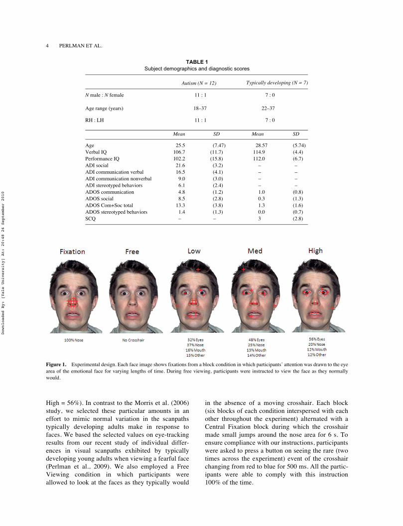
We modified aspects of the basic design of our previ-ous study of typically developing individuals (Morris,Pelphrey, & McCarthy, 2006) to address our currenthypotheses. Throughout the experiment, participantsviewed a single fearful, male face (shown in Figure 1)in full color from the NimStim set of facial expres-sions (Tottenham et al., 2009) in the center of thescreen. Although the Morris et al. (2006) study used asingle neutral face, we chose to maximize the poten-tial for amygdala activation in our participants by ask-ing them to view a fearful face, as fearful faces havebeen repeatedly shown to be strong activators of thehuman amygdalae (e.g., Morris et al., 1996). Partici-pants visually followed a small red crosshair as itmade small jumps across the face every 500 ms suchthat they made a saccade and fixated on the crosshairat each new location.
As illustrated in Figure 1, three types of 12-s blockswere designed to simulate scanpaths with varyingamounts of eye fixation (Low = 32%, Medium = 48%,
Downloaded By: [Yale University] At: 20:48 26 September 2010
4 PERLMAN ET AL.
High = 56%). In contrast to the Morris et al. (2006)study, we selected these particular amounts in aneffort to mimic normal variation in the scanpathstypically developing adults make in response tofaces. We based the selected values on eye-trackingresults from our recent study of individual differ-ences in visual scanpaths exhibited by typicallydeveloping young adults when viewing a fearful face(Perlman et al., 2009). We also employed a FreeViewing condition in which participants wereallowed to look at the faces as they typically would
in the absence of a moving crosshair. Each block(six blocks of each condition interspersed with eachother throughout the experiment) alternated with aCentral Fixation block during which the crosshairmade small jumps around the nose area for 6 s. Toensure compliance with our instructions, participantswere asked to press a button on seeing the rare (twotimes across the experiment) event of the crosshairchanging from red to blue for 500 ms. All the partic-ipants were able to comply with this instruction100% of the time.
TABLE 1 Subject demographics and diagnostic scores
Autism (N = 12) Typically developing (N = 7)
N male : N female 11 : 1 7 : 0
Age range (years) 18–37 22–37
RH : LH 11 : 1 7 : 0
Mean SD Mean SD
Age 25.5 (7.47) 28.57 (5.74)Verbal IQ 106.7 (11.7) 114.9 (4.4)Performance IQ 102.2 (15.8) 112.0 (6.7)ADI social 21.6 (3.2) – –ADI communication verbal 16.5 (4.1) – –ADI communication nonverbal 9.0 (3.0) – –ADI stereotyped behaviors 6.1 (2.4) – –ADOS communication 4.8 (1.2) 1.0 (0.8)ADOS social 8.5 (2.8) 0.3 (1.3)ADOS Com+Soc total 13.3 (3.8) 1.3 (1.6)ADOS stereotyped behaviors 1.4 (1.3) 0.0 (0.7)SCQ – – 3 (2.8)
Figure 1. Experimental design. Each face image shows fixations from a block condition in which participants’ attention was drawn to the eyearea of the emotional face for varying lengths of time. During free viewing, participants were instructed to view the face as they normallywould.
Downloaded By: [Yale University] At: 20:48 26 September 2010
FACE-EVOKED ACTIVITY IN AUTISM 5
FMRI data acquisition
Scanning was performed on a Siemens 3 T Allegrahead-only scanner (Siemens, Erlangen, Germany).High-resolution, T1-weighted anatomical imageswere acquired using an MPRAGE sequence (TR =1630 ms; TE = 2.48 ms; FOV = 20.4 cm; a = 8°;image matrix = 2562; voxel size = 0.8 ! 0.8 ! 0.8 mm;224 slices). Whole-brain functional images wereacquired using a single-shot, gradient-recalled echo-planar pulse sequence (TR = 2000 ms; TE = 30 ms; a= 73°; FOV = 20.4 cm; image matrix = 642; voxel size= 3.2 ! 3.2 ! 3.2 mm; 35 slices) sensitive to blood-oxygenation-level-dependent (BOLD) contrast. Runsconsisted of the acquisition of 225 successive brainvolumes beginning with two discarded RF excitationsto allow for steady-state equilibrium.
RESULTS
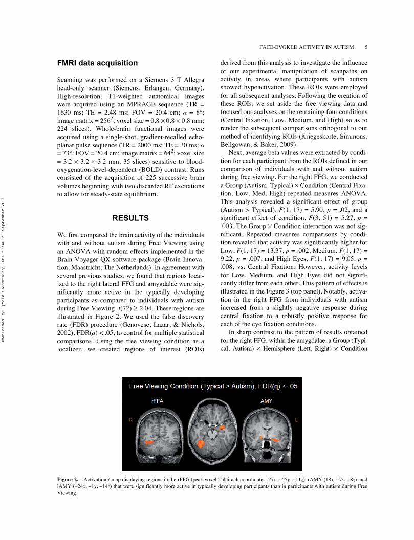
We first compared the brain activity of the individualswith and without autism during Free Viewing usingan ANOVA with random effects implemented in theBrain Voyager QX software package (Brain Innova-tion, Maastricht, The Netherlands). In agreement withseveral previous studies, we found that regions local-ized to the right lateral FFG and amygdalae were sig-nificantly more active in the typically developingparticipants as compared to individuals with autismduring Free Viewing, t(72) " 2.04. These regions areillustrated in Figure 2. We used the false discoveryrate (FDR) procedure (Genovese, Lazar, & Nichols,2002), FDR(q) < .05, to control for multiple statisticalcomparisons. Using the free viewing condition as alocalizer, we created regions of interest (ROIs)
derived from this analysis to investigate the influenceof our experimental manipulation of scanpaths onactivity in areas where participants with autismshowed hypoactivation. These ROIs were employedfor all subsequent analyses. Following the creation ofthese ROIs, we set aside the free viewing data andfocused our analyses on the remaining four conditions(Central Fixation, Low, Medium, and High) so as torender the subsequent comparisons orthogonal to ourmethod of identifying ROIs (Kriegeskorte, Simmons,Bellgowan, & Baker, 2009).
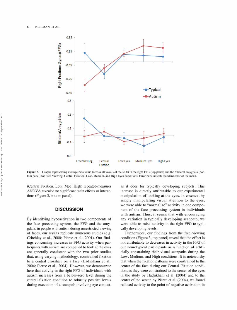
Next, average beta values were extracted by condi-tion for each participant from the ROIs defined in ourcomparison of individuals with and without autismduring free viewing. For the right FFG, we conducteda Group (Autism, Typical) ! Condition (Central Fixa-tion, Low, Med, High) repeated-measures ANOVA.This analysis revealed a significant effect of group(Autism > Typical), F(1, 17) = 5.90, p = .02, and asignificant effect of condition, F(3, 51) = 5.27, p =.003. The Group ! Condition interaction was not sig-nificant. Repeated measures comparisons by condi-tion revealed that activity was significantly higher forLow, F(1, 17) = 13.37, p = .002, Medium, F(1, 17) =9.22, p = .007, and High Eyes, F(1, 17) = 9.05, p =.008, vs. Central Fixation. However, activity levelsfor Low, Medium, and High Eyes did not signifi-cantly differ from each other. This pattern of effects isillustrated in the Figure 3 (top panel). Notably, activa-tion in the right FFG from individuals with autismincreased from a slightly negative response duringcentral fixation to a robustly positive response foreach of the eye fixation conditions.
In sharp contrast to the pattern of results obtainedfor the right FFG, within the amygdalae, a Group (Typi-cal, Autism) ! Hemisphere (Left, Right) ! Condition
Figure 2. Activation t-map displaying regions in the rFFG (peak voxel Talairach coordinates: 27x, #55y, #11z), rAMY (18x, #7y, #8z), andlAMY (–24x, #1y, #14z) that were significantly more active in typically developing participants than in participants with autism during FreeViewing.
Downloaded By: [Yale University] At: 20:48 26 September 2010
6 PERLMAN ET AL.
(Central Fixation, Low, Med, High) repeated-measuresANOVA revealed no significant main effects or interac-tions (Figure 3, bottom panel).
DISCUSSION
By identifying hypoactivation in two components ofthe face processing system, the FFG and the amy-gdala, in people with autism during unrestricted viewingof faces, our results replicate numerous studies (e.g.Critchley et al., 2000; Pierce et al., 2001). Our find-ings concerning increases in FFG activity when par-ticipants with autism are compelled to look at the eyesare generally consistent with the two prior studiesthat, using varying methodology, constrained fixationto a central crosshair on a face (Hadjikhani et al.,2004; Pierce et al., 2004). However, we demonstratehere that activity in the right FFG of individuals withautism increases from a below-zero level during thecentral fixation condition to robustly positive levelsduring execution of a scanpath involving eye contact,
as it does for typically developing subjects. Thisincrease is directly attributable to our experimentalmanipulation of looking at the eyes. In essence, bysimply manipulating visual attention to the eyes,we were able to “normalize” activity in one compo-nent of the face processing system in individualswith autism. Thus, it seems that with encouragingany variation in typically developing scanpath, wewere able to raise activity in the right FFG to typi-cally developing levels.
Furthermore, our findings from the free viewingcondition (Figure 3, top panel) reveal that the effect isnot attributable to decreases in activity in the FFG ofour neurotypical participants as a function of artifi-cially constraining their visual scanpaths during theLow, Medium, and High conditions. It is noteworthythat when the fixation patterns were constrained to thecenter of the face during our Central Fixation condi-tion, as they were constrained to the center of the eyesin the study by Hadjikhani et al. (2004) and to thecenter of the screen by Pierce et al. (2004), we foundreduced activity to the point of negative activation in
Figure 3. Graphs representing average beta value (across all voxels of the ROI) in the right FFG (top panel) and the bilateral amygdala (bot-tom panel) for Free Viewing, Central Fixation, Low, Medium, and High Eyes conditions. Error bars indicate standard error of the mean.
Downloaded By: [Yale University] At: 20:48 26 September 2010
FACE-EVOKED ACTIVITY IN AUTISM 7
both groups of participants, but this effect was morepronounced for typically developing individuals rela-tive to their free viewing data.
Our findings have important repercussions for the-oretical debates concerning the nature of the FFA.Some researchers have argued that the FFA is notspecialized for faces per se; rather, the seeminglyselective response to faces reflects the development ofa high level of visual “expertise” for faces, relative toother categories of objects, in typically developingindividuals (Gauthier, Behrmann, & Tarr, 1999; Tarr& Gauthier, 2000). This theoretical camp has seizedon the findings of FFA hypoactivation in autism asevidence in favor of their theory of visual processing(e.g., Grelotti, Gauthier, & Schultz, 2002). Specifi-cally, they argue that a lack of attention to the eyesleads to a failure to develop an FFA in individualswith autism. Our results are not easily reconciled withthe expertise view of face-processing deficits inautism. We find that the FFA is hypoactive in individ-uals with autism, but we show here that this hypoacti-vation can be reversed with a simple manipulation offixation to the eyes. It appears that an FFA is presentin individuals who presumably lack the necessary“expertise” with faces to have developed such aregion.
In contrast to the FFG, our manipulation of eyecontact had no effect on amygdalae activation in ourgroup of participants with autism. In typically devel-oping individuals, constraining eye movements tothe center of the screen or directing eye movementsto the eyes of the stimulus face served to greatlydecrease amygdalae activity (bottom panel of Figure3). This observation is consistent with previousresearch conceptualizing the role of the amygdalaeas directing attention towards salient environmentalstimuli such as the eyes of faces (e.g., Adolphs et al.,2005). It is possible that we do not observe amy-gdalae activity when we constrain the eye move-ments of typically developing participants becausewe are removing the need for the amygdalae to dir-ect eye movements in response to the presentation ofan emotionally expressive face. In the case ofautism, the lack of amygdalae activity during freeviewing, combined with the observation that thishypoactivation cannot be reversed via the manipula-tion of eye movements, suggests that this brainmechanism for directing eye movements to the key,most socially relevant features of a face is severelyimpaired in individuals with autism.
We must acknowledge an important limitation to ourexperimental design. Eye-tracking data were not col-lected during our fMRI scans. Thus, we cannot be cer-tain that our participants made all the eye movements
we wished them to make and only those eye move-ments, nor can we be sure that participants fixated onthe cross in order to detect color change. Nonetheless,the fact that all participants were able to identify therare and very subtle instances of the crosshair turningfrom red to blue with 100% accuracy provides somereassurance that our manipulations, via attention tothe crosshair, were successful. In addition, we have nodata concerning the scanpaths participants were mak-ing during the Free Viewing condition, although, onthe basis of prior reports (Jones et al., 2008; Klinet al., 2002; Pelphrey et al., 2002), we suspect thatparticipants with autism were looking less at the eyesthen were our typically developing participants.
A second limitation of our study concerns our rela-tively small sample size of matched typically devel-oping individuals. It is possible that our study wasunderpowered for detecting group differences. We donot, however, believe this to be the case given that ourseven typically developing subjects showed signifi-cantly more, rather than less, right FFG and bilateralamygdalae activity during the free viewing condition.Thus, the lack of significant differences between sub-jects with autism and their typically developing coun-terparts on all other conditions in which scanpathswere manipulated should reflect the effects of ourmanipulation.
In summary, the present results add a higherdegree of specificity to our understanding of socialbrain dysfunction in autism. Pivotal questions areraised for future research, however, including thefollowing: If the FFG can be engaged in individualswith autism, why is it not engaged organically, andwhat limits the implementation of this region in eve-ryday situations? Our results offer important impli-cations for behavioral interventions that may helpthose with autism to develop higher levels of socialfunctioning through increased attention to the eyesof social partners. The present findings offer somepreliminary insights into the mechanisms by whichthe common clinical practice of encouraging eyecontact might serve to shape the development of thesocial brain. Requiring individuals with autism, evenhigh-functioning adults, to fixate the eyes appearstemporarily to “normalize” activity within a previ-ously silent component of the face processing system.Future research will be needed to determine if thiskind of manipulation can have lasting effects on brainactivity beyond the time frame of a single experimen-tal session.
Manuscript received 6 March 2007Manuscript accepted 20 September 2007
First published online
Downloaded By: [Yale University] At: 20:48 26 September 2010
8 PERLMAN ET AL.
REFERENCES
Adolphs, R., Gosselin, F., Buchanan, T. W., Tranel, D.,Schyns, P., & Damasio, A. R. (2005). A mechanism forimpaired fear recognition after amygdala damage.Nature, 433, 68–72.
American Psychiatric Association (1994). Diagnostic andStatistical Manual of Mental Disorders (4th ed., textrevision). Washington, DC: APA.
Ashwin, C., Baron-Cohen, S., Wheelwright, S., O’Riordan,M., & Bullmore, E. T. (2007). Differential activation ofthe amygdala and the ‘social brain’ during fearful face-processing in Asperger syndrome. Neuropsychologia,45, 2–14.
Baron-Cohen, S., Ring, H. A., Wheelwright, S., Bullmore,E. T., Brammer, M. J., Simmons, A., et al. (1999).Social intelligence in the normal and autistic brain: AnfMRI study. European Journal of Neuroscience, 11,1891–1898.
Bird, G., Catmur, C., Silani, G., Frith, C., & Frith, U.(2006). Attention does not modulate neural responses tosocial stimuli in autism spectrum disorders. NeuroIm-age, 31(4), 1614–1624.
Bookheimer, S. Y., Wang, A. T., Scott, A., Sigman, M., &Dapretto, M. (2008). Frontal contributions to face pro-cessing differences in autism: Evidence from fMRI ofinverted face processing. Journal of the InternationalNeuropsychological Society, 14, 922–932.
Critchley, H. D., Daly, E. M., Bullmore, E. T., Williams,S. C. R., Van Amelsvoort, T., Robertson, D. M., et al.(2000). The functional neuroanatomy of social behav-ior: Changes in cerebral bloodflow when people withautistic disorder process facial expressions. Brain, 123,2203–2212.
Dalton, K. M., Nacewicz, B. M., Johnstone, T., Schaefer, H.S., Gernsbacher, M. A., Goldsmith, H. H., et al. (2005)Gaze fixation and the neural circuitry of face processingin autism. Nature Neuroscience, 8, 519–526.
Davis, M. (1992). The role of the amygdala in fear and anx-iety. Annual Review of Neuroscience, 15, 353–375.
Gauthier, I., Behrmann, M., & Tarr, M.J. (1999). Can facerecognition really be dissociated from object recogni-tion? Journal of Cognitive Neuroscience, 11, 349–370.
Genovese, C. R., Lazar, N. A., & Nichols, T. (2002). Thresh-olding of statistical maps in functional neuroimaging usingthe false discovery rate. NeuroImage, 15, 870–878.
Grelotti, D. J., Gauthier, I., & Schultz, R. T. (2002). Socialinterest and the development of cortical face specializa-tion: What autism teaches us about face processing.Developmental Psychobiology, 40, 213–225.
Hadjikhani, N., Joseph, R. M., Snyder, J., Chabris, C. F.,Clark, J., Steele, S., et al. (2004). Activation of the fusi-form gyrus when individuals with autism spectrum dis-order view faces. NeuroImage, 22, 1141–1150.
Hubl, D., Bölte, S., Feineis-Matthews, S., Lanfermann, H.,Federspiel, A., Strik, F., et al. (2003). Functional imbal-ance of visual pathways indicates alternative face pro-cessing strategies in autism. Neurology, 61, 1232–1237.
Humphreys, K., Hasson, U., Avidan, G., Minshew, N., &Behrmann, M. (2008) Cortical patterns of category-selective activation for faces, places and objects in adultswith autism. Autism Research, 1, 52–63.
Jones, W., Carr, K., & Klin, A. (2008). Absence of prefer-ential looking to the eyes of approaching adults
predicts level of social disability in 2-year-old toddlerswith autism spectrum disorder. Archives of GeneralPsychiatry, 65, 946–954.
Joseph, R. M., Ehrman, K., McNally, R., & & Keehn, B.(2008). Affective response to eye contact and face recog-nition ability in children with ASD. Journal of the Inter-national Neuropsychological Society, 14, 947–955.
Kanner, L. (1943). Autistic disturbances of affective con-tact. Nervous Child, 2, 217–250.
Kanwisher, N., McDermott, J., & Chun, M. M. (1997). Thefusiform face area: A module in human extrastriate cor-tex specialized for face perception. Journal of Neuro-science, 17(11) 4302–4311.
Kleinhans, N. M., Johnson, L.C., Richards, T., Mahurin, R.,Greenson, J., Dawson, G., et al. (2009). Reduced neuralhabituation in the amygdala and social impairments inautism spectrum disorders. American Journal of Psychi-atry, 166(4), 467–475.
Klin, A. (2008). Three things to remember if you are a func-tional magnetic resonance imaging researcher of faceprocessing in autism spectrum disorders. Biological Psy-chiatry, 64, 549–551.
Klin, A., Jones, W., Schultz, R. T., Volkmar, F. R., &Cohen, D. J. (2002). Visual fixation patterns duringviewing of naturalistic social situations as predictors ofsocial competence in individuals with autism. Archivesof General Psychiatry, 59, 809–816.
Koshino, H., Kana, R. J., Keller, T. A., Cherkassky, V. L.,Minshew, N. J., & Just, M. A. (2008). fMRI investiga-tion of working memory for faces in autism: Visual cod-ing underconnectivity with frontal areas. CerebralCortex, 18, 289–300.
Kriegeskorte, N., Simmons, W. K., Bellgowan, P. S. F., &Baker, C. I. (2009). Circular analysis in systemsneuroscience: The dangers of double dipping. NatureNeuroscience, 12(5), 535–540.
Lord, C., Risi, S., Lambrecht, L., Cook, E. H., Leventhal, B.L., DiLavore, P. C., et al. (2000). The autism diagnosticobservation schedule – generic: A standard measure ofsocial and communication deficits associated with thespectrum of autism. Journal of Autism and Developmen-tal Disorders, 30, 205–223.
Lord, C., Rutter, M., & Le Couteur, A. (1994). Autism diag-nostic interview – revised: A revised version of adaignostic interview for caregivers of individuals withpossible pervasive developmental disorders. Journal ofAutism and Developmental Disorders, 24, 659–685.
Morris, J. P., Pelphrey, K. A., & McCarthy, G. (2006). Con-trolled scanpath variation alters fusiform face activation.Social Cognitive and Affective Neuroscience, 2, 31–38.
Morris, J. S., Frith, C. D., Perrett, D. I., Rowland, D.,Young, A. W., Calder, A. J., et al. (1996). A differentialneural response in the human amygdala to fearful andhappy facial expressions. Nature, 383, 812–815.
Ogai, M., Matsumoto, H., Suzuki, K., Ozawa, F., Fukuda,R., Uchiyama, I., et al. (2003). fMRI study of recogni-tion of facial expression in high-functioning autisticpatients. NeuroReport, 14, 559–563.
Osterling, J., & Dawson, G. (1994). Early recognition ofchildren with autism: A study of first birthday home vid-eotapes. Journal of Autism and Developmental Disor-ders, 24, 247–257.
Pelphrey, K. A., Morris, J. P., McCarthy, G., & LaBar, K. S.(2007). Perception of dynamic changes in facial affect
Downloaded By: [Yale University] At: 20:48 26 September 2010

FACE-EVOKED ACTIVITY IN AUTISM 9
and identity in autism. Social Cognitive and AffectiveNeuroscience, 2(2), 140–150.
Pelphrey, K. A., Sasson, N. J., Reznick, J. S., Paul, G.,Goldman, B. D., & Piven, J. (2002). Visual scanning offaces in autism. Journal of Autism and DevelopmentalDisorders, 32, 249–261.
Perlman, S. B., Morris, J. P., Vander Wyk, B. C., Green, S.R., Doyle, J. L., & Pelphrey, K. A. (2009). Individualdifferences in personality shape how people look atfaces. PLoS ONE 4(6), e5952.
Pierce, K., Haist, F., Sedaghat, F., & Courchesne, E. (2004).The brain response to personally familiar faces inautism: Findings of fusiform activity and beyond. Brain,127, 2703–2716.
Pierce, K., Müller, R. A., Ambrose, J., Allern, G., &Courchesne, E. (2001). Face processing occurs outsidethe fusiform ‘face area’ in autism: Evidence from func-tional MRI. Brain, 124, 2059–2073.
Pierce, K., & Redcay, E. (2008). Fusiform function in chil-dren with an autism spectrum disorder is a matter of‘who’. Biological Psychiatry, 64, 552–560.
Piggot, J., Kwon, H., Mobbs, D., Blasey, C., Lotspeich, L.,Menon, V., et al. (2004). Emotional attribution in high-functioning individuals with autistic spectrum disorder:A functional imaging study. Journal of the AmericanAcademy of Child and Adolescent Psychiatry, 43(4),473–480.
Poldrack, R. A. (2006). Can cognitive processes be inferredfrom neuroimaging data?. Trends in Cognitive Sciences,10(2), 59–63.
Puce, A., Allison, T., Asgari, M., Gore, J. C., McCarthy, G.(1996). Differential sensitivity of human visual cortex tofaces, letterstrings, and textures: A functional magneticresonance imaging study. Journal of Neuroscience,16(16), 5205–5215.
Rutter, M., Bailey, A., Berument, S. K., Lecouteur, A.,Lord, C., & Pickles, A. (2003). Social CommunicationQuestionnaire (SCQ). Los Angeles: Western Psycholog-ical Services.
Schultz, R. T., Gauthier, I., Klin, A., Fulbright, R. K.,Anderson, A. W., Volkmar, F., et al. (2000). Abnormalventral temporal cortical activity during face discrimina-tion among individuals with autism and Asperger syn-drome. Archives of General Psychiatry, 57, 331–340.
Tarr, M. J., & Gauthier, I. (2000). FFA: A flexible fusiformarea for subordinate-level visual processing automatizedby expertise. Nature Neuroscience, 3, 764–769.
Tottenham, N., Tanaka, J., Leon, A. C., McCarry, T., Nurse,M., et al. (2009). The NimStim set of facial expressions:Judgments from untrained research participants. Psychi-atry Research, 168(3), 242–249.
Wang, A. T., Dapretto, M., Hariri, A. R., Sigman, M., &Bookheimer, S. (2004). Neural correlates of facial affectprocessing in children and adolescents with autism spec-trum disorder. Journal of the American Academy ofChild and Adolescent Psychiatry, 43(4), 481–490.
Wing, L., & Gould, J. (1979). Severe impairments of socialinteraction and associated abnormalities in children: Epi-demiology and classification. Journal of Autism andDevelopmental Disorders, 24, 11–29.
Downloaded By: [Yale University] At: 20:48 26 September 2010