Evolution of the larval peripheral nervous system in Drosophila species has involved a change in...
11
Dev Genes Evol (2004) 214: 442–452 DOI 10.1007/s00427-004-0422-4 ORIGINAL ARTICLE Virginie Orgogozo . François Schweisguth Evolution of the larval peripheral nervous system in Drosophila species has involved a change in sensory cell lineage Received: 23 December 2003 / Accepted: 15 June 2004 / Published online: 4 August 2004 # Springer-Verlag 2004 Abstract A key challenge in evolutionary biology is to identify developmental events responsible for morpholog- ical changes. To determine the cellular basis that underlies changes in the larval peripheral nervous system (PNS) of flies, we first described the PNS pattern of the abdominal segments A1–A7 in late embryos of several fly species using antibody staining. In contrast to the many variations reported previously for the adult PNS pattern, we found that the larval PNS pattern has remained very stable during evolution. Indeed, our observation that most of the analysed Drosophilinae species exhibit exactly the same pattern as Drosophila melanogaster reveals that the pattern observed in D. melanogaster embryos has remained constant for at least 40 million years. Further- more, we observed that the PNS pattern in more distantly related flies (Calliphoridae and Phoridae) is only slightly different from the one in D. melanogaster . A single difference relative to D. melanogaster was identified in the PNS pattern of the Drosophilinae fly D. busckii, the absence of a specific external sensory organ. Our analysis of sensory organ development in D. busckii suggests that this specific loss resulted from a transformation in cell lineage, from a multidendritic-neuron-external-sensory- organ lineage to a multidendritic-neuron-solo lineage. Keywords Peripheral nervous system . Evolution . Drosophila . Cell lineage . Cell migration Introduction Evolution of the abdominal larval peripheral nervous system (PNS) in Drosophila melanogaster and closely related species offers an excellent model system to study the developmental modifications responsible for evolu- tionary changes. The D. melanogaster abdominal larval PNS is composed of a constant number of neurons and associated cells, whose characteristics and positions have been well described and are perfectly reproducible between individuals (Bodmer and Jan 1987; Campos- Ortega and Hartenstein 1997; Dambly-Chaudiére and Ghysen 1986; Ghysen et al. 1986; Hertweck 1931). Three main types of sensory organs can be distinguished: external sensory (es) organs, chordotonal (ch) organs and multidendritic (md) neurons. The es and ch organs are composed of one or several neurons associated with accessory cells. The es organs transduce mechanosensory, chemosensory and possibly stretch information (Green and Hartenstein 1997), while the ch organs are thought to function as stretch or proprio-receptors (Jan and Jan 1993). The md neurons do not have any accessory cells and may function as touch-, proprio-, chemo-, thermo-, or noci- receptors (Bodmer et al. 1987; Liu et al. 2003; Tracey et al. 2003), or even as non-sensory secretory neurons (Hewes et al. 2003). The simplicity of the larval abdom- inal PNS therefore allows the study of evolutionary changes at cellular resolution. Since sensory cells occupy very precise positions and exhibit specific orientations within the body, changes that might have occurred during evolution regarding cell number, position and orientation can be detected. Also, the peripheral location of the larval PNS makes it easily accessible for observation in various species. Finally, the development of sensory organs has been well documented in D. melanogaster and many of the genes that regulate the successive steps of sensory development are known (reviewed in Campos-Ortega and Hartenstein 1997; Ghysen and Dambly-Chaudière 1989, 2000; Jan and Jan 2001). Correspondingly, many antibodies have been developed and can be used to follow the different steps of larval sensory organ formation in D. Edited by P. Simpson V. Orgogozo . F. Schweisguth Ecole Normale Supérieure, UMR 8542, 46 rue d’Ulm, 75005 Paris, France Present address: V. Orgogozo (*) Department of Ecology and Evolutionary Biology, Princeton University, Princeton, NJ, 08544, USA e-mail: [email protected]
-
Upload
independent -
Category
Documents
-
view
1 -
download
0
Transcript of Evolution of the larval peripheral nervous system in Drosophila species has involved a change in...
Dev Genes Evol (2004) 214: 442–452DOI 10.1007/s00427-004-0422-4
ORIGINAL ARTICLE
Virginie Orgogozo . François Schweisguth
Evolution of the larval peripheral nervous system in Drosophilaspecies has involved a change in sensory cell lineage
Received: 23 December 2003 / Accepted: 15 June 2004 / Published online: 4 August 2004# Springer-Verlag 2004
Abstract A key challenge in evolutionary biology is toidentify developmental events responsible for morpholog-ical changes. To determine the cellular basis that underlieschanges in the larval peripheral nervous system (PNS) offlies, we first described the PNS pattern of the abdominalsegments A1–A7 in late embryos of several fly speciesusing antibody staining. In contrast to the many variationsreported previously for the adult PNS pattern, we foundthat the larval PNS pattern has remained very stable duringevolution. Indeed, our observation that most of theanalysed Drosophilinae species exhibit exactly the samepattern as Drosophila melanogaster reveals that thepattern observed in D. melanogaster embryos hasremained constant for at least 40 million years. Further-more, we observed that the PNS pattern in more distantlyrelated flies (Calliphoridae and Phoridae) is only slightlydifferent from the one in D. melanogaster. A singledifference relative to D. melanogaster was identified in thePNS pattern of the Drosophilinae fly D. busckii, theabsence of a specific external sensory organ. Our analysisof sensory organ development in D. busckii suggests thatthis specific loss resulted from a transformation in celllineage, from a multidendritic-neuron-external-sensory-organ lineage to a multidendritic-neuron-solo lineage.
Keywords Peripheral nervous system . Evolution .Drosophila . Cell lineage . Cell migration
Introduction
Evolution of the abdominal larval peripheral nervoussystem (PNS) in Drosophila melanogaster and closelyrelated species offers an excellent model system to studythe developmental modifications responsible for evolu-tionary changes. The D. melanogaster abdominal larvalPNS is composed of a constant number of neurons andassociated cells, whose characteristics and positions havebeen well described and are perfectly reproduciblebetween individuals (Bodmer and Jan 1987; Campos-Ortega and Hartenstein 1997; Dambly-Chaudiére andGhysen 1986; Ghysen et al. 1986; Hertweck 1931). Threemain types of sensory organs can be distinguished:external sensory (es) organs, chordotonal (ch) organs andmultidendritic (md) neurons. The es and ch organs arecomposed of one or several neurons associated withaccessory cells. The es organs transduce mechanosensory,chemosensory and possibly stretch information (Green andHartenstein 1997), while the ch organs are thought tofunction as stretch or proprio-receptors (Jan and Jan 1993).The md neurons do not have any accessory cells and mayfunction as touch-, proprio-, chemo-, thermo-, or noci-receptors (Bodmer et al. 1987; Liu et al. 2003; Tracey etal. 2003), or even as non-sensory secretory neurons(Hewes et al. 2003). The simplicity of the larval abdom-inal PNS therefore allows the study of evolutionarychanges at cellular resolution. Since sensory cells occupyvery precise positions and exhibit specific orientationswithin the body, changes that might have occurred duringevolution regarding cell number, position and orientationcan be detected. Also, the peripheral location of the larvalPNS makes it easily accessible for observation in variousspecies. Finally, the development of sensory organs hasbeen well documented in D. melanogaster and many ofthe genes that regulate the successive steps of sensorydevelopment are known (reviewed in Campos-Ortega andHartenstein 1997; Ghysen and Dambly-Chaudière 1989,2000; Jan and Jan 2001). Correspondingly, manyantibodies have been developed and can be used to followthe different steps of larval sensory organ formation in D.
Edited by P. Simpson
V. Orgogozo . F. SchweisguthEcole Normale Supérieure,UMR 8542, 46 rue d’Ulm,75005 Paris, France
Present address:V. Orgogozo (*)Department of Ecology and Evolutionary Biology, PrincetonUniversity,Princeton, NJ, 08544, USAe-mail: [email protected]
melanogaster (see for example Orgogozo et al. 2001,2002). Since some antibodies cross-react in other insectspecies (Meier et al. 1991; Grueber and Truman 1999, andthis study), they may be useful to identify variations insensory organ development between species.
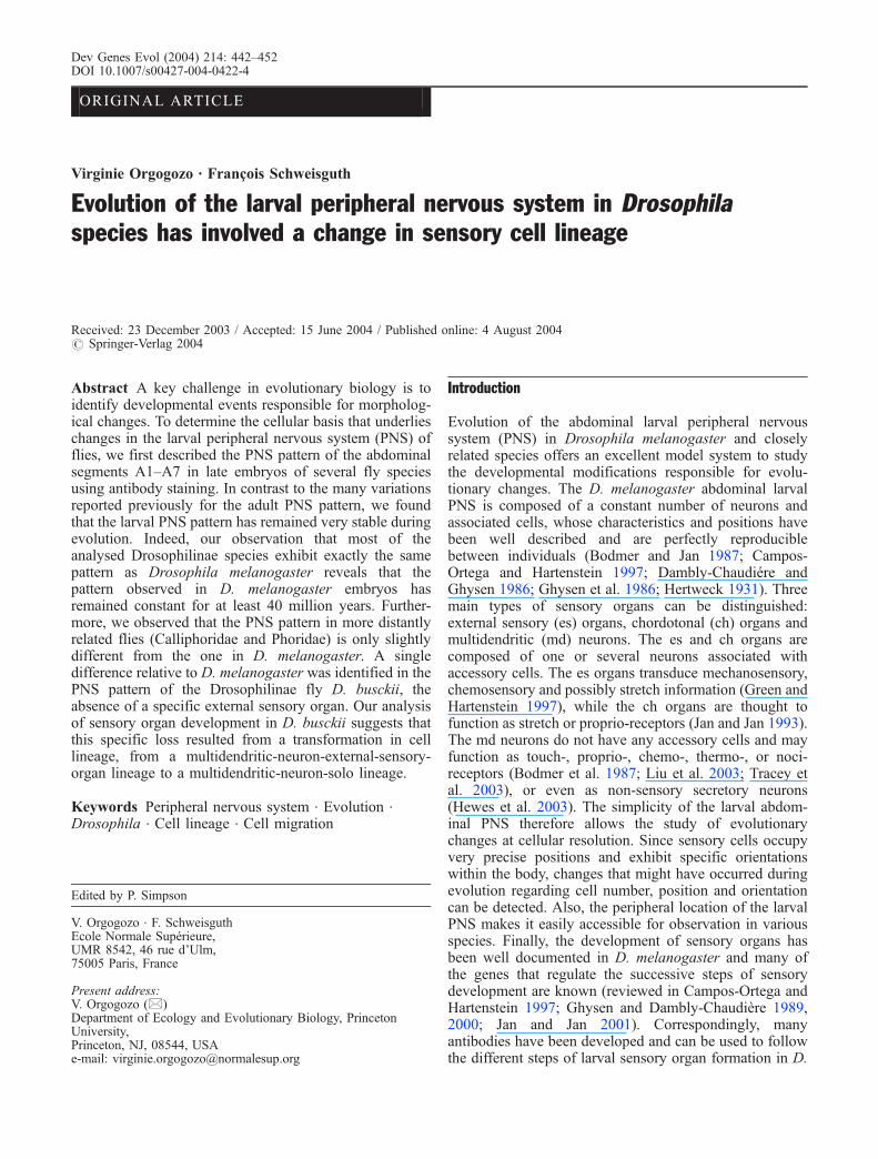
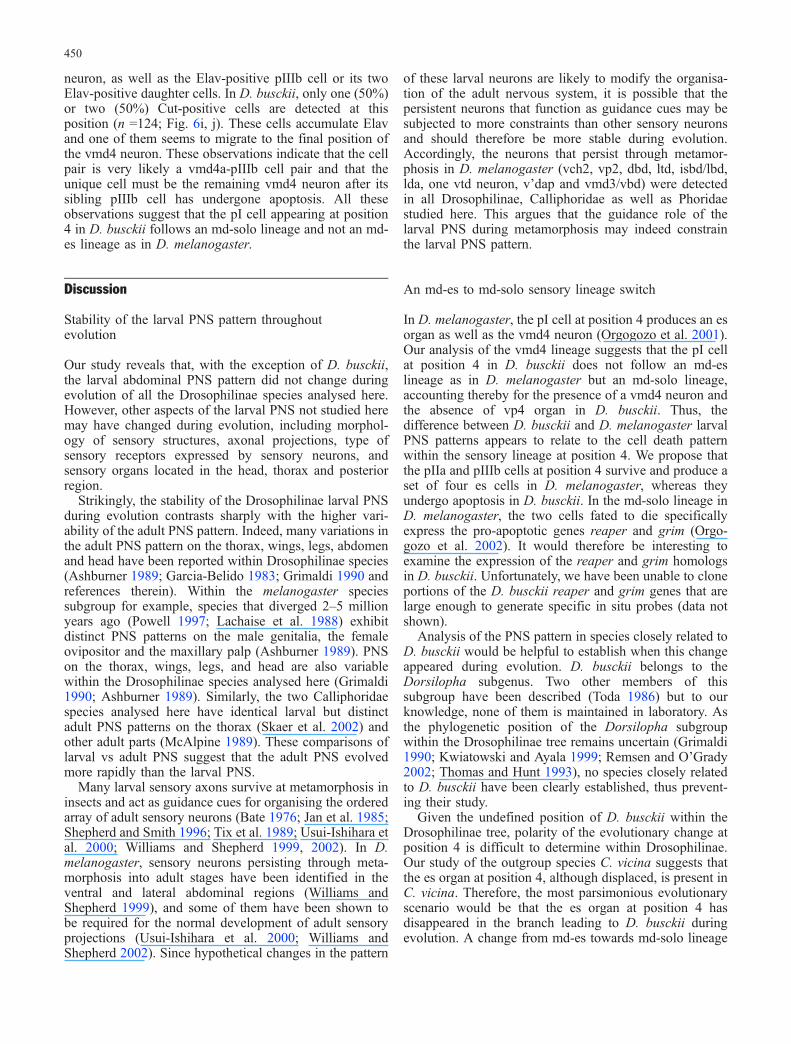
In D. melanogaster, the neurons and associated supportcells that constitute each larval sensory organ are producedduring embryogenesis from a primary precursor (pI) cell.pI cells are singled out from a group of adjacent epithelialcells named the proneural cluster, in a process mediated bythe Notch signalling pathway (reviewed in Simpson et al.1999). Following its specification, each pI cell undergoes astereotyped sequence of asymmetric cell divisions togenerate sensory cells (Bodmer et al. 1989). In the ventralabdominal region, two types of sensory lineages have beenobserved. The md-es lineage, followed by pI cells locatedat positions 1–4 and 4a, consists of a series of four-celldivisions and produces a four-cell es organ and an mdneuron (Orgogozo et al. 2001; Fig. 1a, c, d). The md-sololineage, followed by the pI cell located at position 1a,comprises two cell divisions but generates only one cell,an md neuron, because the other progeny cells undergoapoptosis (Orgogozo et al. 2002; Fig. 1b–d). The md-sololineage has been proposed to be a modified md-es lineagein which two secondary precursor cells undergo apoptosis.In support of this proposal, in embryos in which apoptosisis inhibited, the two cells fated to death divide and togetherproduce an ectopic four-cell es organ indicating that themd-solo lineage has been transformed into an md-eslineage (Orgogozo et al. 2002).
Comparative analysis of first-instar larvae and nymphsfrom several insect orders indicated that larval abdominalPNS patterns fall into three main types: fixed pattern (inDiptera), fully variable pattern, and variable pattern withsome fixed sensory organs (in most other species; Green
and Hartenstein 1997 and references therein). In specieswith a fixed pattern, sensory organs are usually distributedover the entire segment whereas in species with a variablepattern, they typically form one or several rows along thedorsal-ventral axis in the middle of each segment. In bothD. melanogaster and the grasshopper Schistocercagregaria, sensory neurons are grouped into three regions(ventral, lateral, dorsal) and their axons primarily projectonto two nerves (intersegmental and segmental nerves)towards the ventral nerve cord. Although the Schistocercapleural ch organ probably corresponds to the Drosophilalch5 ch organ according to its position, it is difficult tocompare other sensory organs between Drosophila andSchistocerca. In the moth Manduca sexta, severalindividual abdominal MD neurons have been proposedto correspond to particular D. melanogaster md neuronsaccording to their time of development and their positionwithin the larval body wall (Grueber and Truman 1999).
Morphological modifications of the larval PNS patternduring evolution may result from various developmentalchanges, such as: (1) appearance/loss of a particularsensory organ associated with emergence/loss of aproneural cluster or pI cell, or with survival/death ofsensory cells; (2) re-positioning of sensory cells (throughchanges in proneural cluster position or migration ofsensory cells); (3) change in sensory organ types; etc.
To identify changes in larval PNS pattern in speciesrelated to D. melanogaster, we examined the PNS of theabdominal segments A1–A7 in several fly species. Then,in order to investigate the developmental changes under-lying different patterns, we analysed sensory organdevelopment in these species.
Materials and methods
Fly stocks and embryo fixation
The fly stocks used were: D. melanogaster yw ; D.sechellia 14021-0248.7 and D. malerkotliana (gifts of D.Stern); D. yakuba, D. bifasciata, D. busckii 1, andZaprionus vittiger (gifts of P. Santamaria); D. bifurca(collected in Mexico, from Bowling Green Stock Centercollection), D. hydei (collected in Raida, Saudi Arabia),and D. buzzatii (collected in Nahal Oren, Israel; gifts of D.Lachaise); D. ararama (gift of P. Simpson); D. virilis (giftof J.-A. Lepesant); D. busckii 2 and D. duncani (gift of N.Gompel); and Megaselia scalaris (gift of M.-L. Carriou).The D. busckii 1 strain was established from a femalecollected in France while the D. busckii 2 strain was fromthe United States. One-day-laid embryos were collectedand fixed according to standard D. melanogaster proce-dures (Rothwell and Sullivan 2000), with the modificationthat M. scalaris and D. hydei embryos were dechorionatedin bleach for at least 5 min. Fixed embryos fromCalliphora vicina and Phormia terraenovae were kindlyprovided by P. Simpson.
Fig. 1a–d Formation of sensory organs in the ventral region of D.melanogaster. a Md-es lineage (Orgogozo et al. 2001). b Md-sololineage (Orgogozo et al. 2002). The pIIa and pIIIb precursor cellsundergo apoptosis. c Diagram of the Cut-positive pI cells. The pIcell at position 1a (blue cell) follows an md-solo lineage whereas theones at positions 1–4 and 4a (white cells) follow an md-es lineage. dDiagram of the Cut-positive sensory cells in mature PNS. Each esorgan is composed of a socket/shaft cell pair (yellow cell), a sheathcell (yellow cell) and one or several es neurons (pink circular cell).md neurons (diamond-shaped cells) originate either from an md-eslineage (encircled in black) or an md-solo lineage (encircled in blue)
443
Immunostaining and microscopy
Embryos were stained as previously described (Orgogozoet al. 2001). Primary antibodies were used at the followingdilutions: mouse anti-Cut, 1/1,000 (2B10; DSHB), rat anti-Elav, 1/4 (7E8A10, DSHB), rabbit anti-HRP, 1/500(Cappel), rabbit anti-Collier, 1/500 (gift of A. Vincent).The RNA-binding protein Elav specifically accumulates inall neurons in D. melanogaster (Robinow and White1988). Anti-Elav antibodies were previously used to stainneurons in the moth Manduca sexta (Grueber and Truman1999) and are, therefore, likely to cross-react betweendifferent fly species. The anti-HRP antibodies have beenshown to bind to neuronal membranes in all ecdysozoananimals (Haase et al. 2001; Jan and Jan 1982). In D.melanogaster, they are known to recognise an epitope ofcarbohydrate origin, an α1,3-fucosylated, N-linked glycan(Fabini et al. 2001; Snow et al. 1987) associated withseveral cell adhesion molecules and with a Na + K +ATPase (Desai et al. 1994; Sun and Salvaterra 1995; Wanget al. 1994). Staining with anti-HRP antibodies permits usto identify the three types of sensory neurons: es, ch andmd neurons. The ch dendrites are surrounded by ascolopale cell which is also HRP-positive (Bodmer et al.1987), and the es neurons can be reliably distinguishedfrom md neurons because their single dendrite has a smallHRP-positive dot at its tip. In D. melanogaster, thetranscription factor Cut has been observed to accumulatein all the cells that compose an es organ [socket cell, shaftcell, sheath cell, and es neuron(s)] as well as in a subset ofmd neurons (Blochlinger et al. 1988, 1990; Brewster andBodmer 1995; Grueber et al. 2003a). Secondary antibodiesCy3-anti-mouse, Cy5-anti-rat and, Alexa488-anti-rabbitwere purchased from Jackson Laboratories or MolecularProbes. For D. hydei, C. vicina, P. terranovae and M.scalaris, the Cut signal was amplified using biotinylatedanti-mouse (1/2000; Jackson Laboratories), avidin DH-biotinylated HRP complexes (Vector Laboratories), TSA-biotin (NEN Life Science), and streptavidin-Alexa568(Molecular Probes). For each abdominal region (ventral,lateral, dorsal), at least four hemisegments of threeembryos were analysed. Developmental stages of D.busckii and C. vicina embryos were identified according tothe extent of germ band retraction, as for D. melanogasterembryos. Images were collected on a Leica SP2 confocalmicroscope and processed using Adobe Photoshop soft-ware. Figures show the mean projection of severalconfocal z-sections. When necessary, the CNS signaldetected in the bottom z-sections below the sensory cellswas removed to better show the sensory cells in the z-projected images.
Results
To identify changes in the larval PNS pattern among flyspecies related to D. melanogaster, we examined embryosof several species, fixed at the end of peripheral neuro-genesis and stained with anti-Cut, anti-Elav and anti-HRP
antibodies. The triple staining Cut, Elav, HRP worked inall species analysed herein and allowed the identificationof all the es cells as well as all ch and md neurons (seeMaterials and methods). For a given species, we observedthat each abdominal segment A1–A7 displays the samePNS pattern.
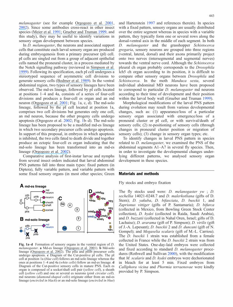
The dorsal sensory cluster contains 13 neurons in D.melanogaster
Clustering of sensory cells in the dorsal cluster hasrendered the number of neurons in this dorsal clusterdifficult to determine. First studies indicated that 10(Ghysen et al. 1986), 11 (Bodmer and Jan 1987), or 12(Bodmer et al. 1989; Ghysen et al. 1986) neurons arepresent in the dorsal cluster. Following these studies, thedorsal cluster was usually estimated to contain 12 sensoryneurons: 5 es neurons (innervating 3 mono-innervated esorgans and 1 doubly innervated es organ), 1 bipolar mdneuron and 6 md neurons exhibiting an extensive dendriticarborisation.
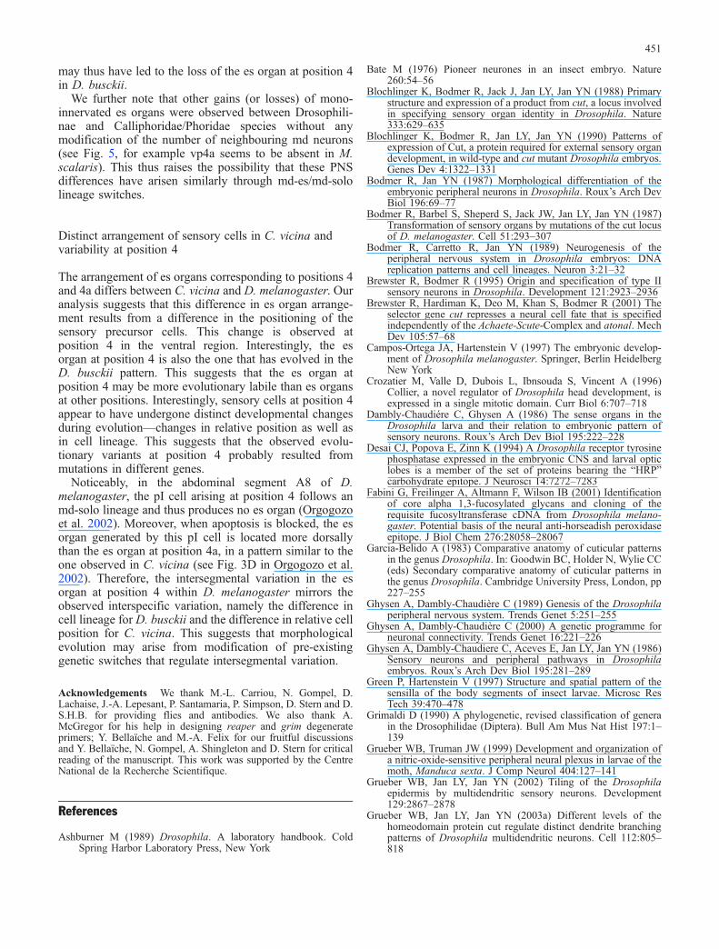
Our examination of the D. melanogaster PNS patternrevealed that there is an additional md neuron in the dorsalcluster, resulting in a total number of 13 dorsal sensoryneurons (Fig. 2a–c). This observation agrees with those ofMoore et al. (2002), who also observed 13 sensoryneurons in the dorsal cluster (see Fig. 1B in Moore et al.
Fig. 2a–c Dorsal sensory cluster in D. melanogaster stage 16embryos. a Dorsal abdominal region stained for Cut (green) andElav (red). b Red channel. c Schematic representation of the Elav-positive neurons seen in a and b. Cut-negative neurons are in redand Cut-positive neurons are in orange. The five es neurons areencircled by a dashed line while the eight md neurons are encircledwith a straight line. Names have been assigned to each md neuronaccording to the nomenclature used in Brewster et al. (2001) andGrueber et al. (2002). In all figures, anterior is left and dorsal is up
444
2002). In former representations of the dorsal sensorycluster, either the Cut-negative dda1 neuron (Grueber et al.2002, 2003a, 2003b) or one of the Cut-positive mdneurons (Brewster et al. 2001; Orgogozo et al. 2002) hasbeen neglected. A complete nomenclature for each dorsalmd neuron is proposed based on the nomenclature used inGrueber et al. (2002) and Brewster et al. (2001; Fig. 2c).
Drosophilinae species exhibit the same abdominalPNS pattern as D. melanogaster
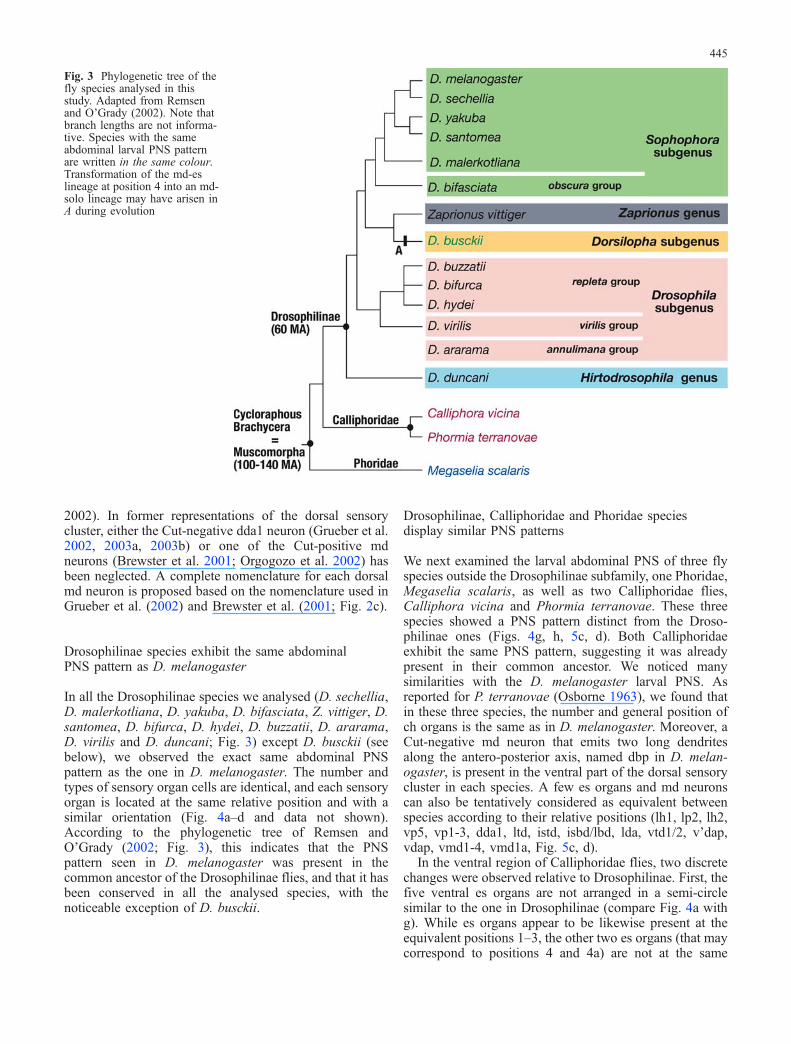
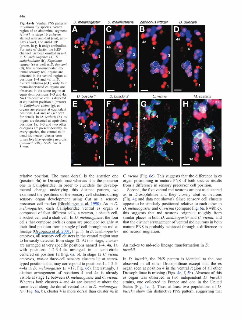
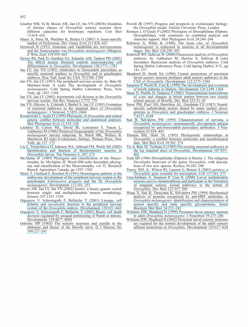
In all the Drosophilinae species we analysed (D. sechellia,D. malerkotliana, D. yakuba, D. bifasciata, Z. vittiger, D.santomea, D. bifurca, D. hydei, D. buzzatii, D. ararama,D. virilis and D. duncani; Fig. 3) except D. busckii (seebelow), we observed the exact same abdominal PNSpattern as the one in D. melanogaster. The number andtypes of sensory organ cells are identical, and each sensoryorgan is located at the same relative position and with asimilar orientation (Fig. 4a–d and data not shown).According to the phylogenetic tree of Remsen andO’Grady (2002; Fig. 3), this indicates that the PNSpattern seen in D. melanogaster was present in thecommon ancestor of the Drosophilinae flies, and that it hasbeen conserved in all the analysed species, with thenoticeable exception of D. busckii.
Drosophilinae, Calliphoridae and Phoridae speciesdisplay similar PNS patterns
We next examined the larval abdominal PNS of three flyspecies outside the Drosophilinae subfamily, one Phoridae,Megaselia scalaris, as well as two Calliphoridae flies,Calliphora vicina and Phormia terranovae. These threespecies showed a PNS pattern distinct from the Droso-philinae ones (Figs. 4g, h, 5c, d). Both Calliphoridaeexhibit the same PNS pattern, suggesting it was alreadypresent in their common ancestor. We noticed manysimilarities with the D. melanogaster larval PNS. Asreported for P. terranovae (Osborne 1963), we found thatin these three species, the number and general position ofch organs is the same as in D. melanogaster. Moreover, aCut-negative md neuron that emits two long dendritesalong the antero-posterior axis, named dbp in D. melan-ogaster, is present in the ventral part of the dorsal sensorycluster in each species. A few es organs and md neuronscan also be tentatively considered as equivalent betweenspecies according to their relative positions (lh1, lp2, lh2,vp5, vp1-3, dda1, ltd, istd, isbd/lbd, lda, vtd1/2, v’dap,vdap, vmd1-4, vmd1a, Fig. 5c, d).
In the ventral region of Calliphoridae flies, two discretechanges were observed relative to Drosophilinae. First, thefive ventral es organs are not arranged in a semi-circlesimilar to the one in Drosophilinae (compare Fig. 4a withg). While es organs appear to be likewise present at theequivalent positions 1–3, the other two es organs (that maycorrespond to positions 4 and 4a) are not at the same
Fig. 3 Phylogenetic tree of thefly species analysed in thisstudy. Adapted from Remsenand O’Grady (2002). Note thatbranch lengths are not informa-tive. Species with the sameabdominal larval PNS patternare written in the same colour.Transformation of the md-eslineage at position 4 into an md-solo lineage may have arisen inA during evolution
445
relative position. The most dorsal is the anterior one(position 4a) in Drosophilinae whereas it is the posteriorone in Calliphoridae. In order to elucidate the develop-mental change underlying this distinct pattern, weexamined the positions of the sensory cell clusters duringsensory organ development using Cut as a sensoryprecursor cell marker (Blochlinger et al. 1990). As in D.melanogaster, each Calliphoridae ventral es organ iscomposed of four different cells, a neuron, a sheath cell,a socket cell and a shaft cell. In D. melanogaster, the fourcells that compose each es organ are produced roughly attheir final position from a single pI cell through an md-eslineage (Orgogozo et al. 2001; Fig. 1). In D. melanogasterembryos, all sensory cell clusters in the ventral region startto be easily detected from stage 12. At this stage, clustersare arranged at very specific positions named 1–4, 4a, 1a,with positions 1-2-3-4-4a arranged in a semi-circlecentered on position 1a (Fig. 6a, b). In stage 12 C. vicinaembryos, two-or three-cell sensory clusters lie at stereo-typed positions that may correspond to positions 1a-1-2-3-4-4a in D. melanogaster (n =17; Fig. 6c). Interestingly, adistinct arrangement of positions 4 and 4a is alreadyvisible at stage 12 between D. melanogaster and C. vicina.Whereas both clusters 4 and 4a are located at about thesame level along the dorsal-ventral axis in D. melanogas-ter (Fig. 6a, b), cluster 4 is more dorsal than cluster 4a in
C. vicina (Fig. 6c). This suggests that the difference in esorgan positioning in mature PNS of both species resultsfrom a difference in sensory precursor cell position.
Second, the five ventral md neurons are not as clusteredas in Drosophilinae and they closely abut es neurons(Fig. 4g and data not shown). Since sensory cell clustersappear to be similarly positioned relative to each other inD. melanogaster and C. vicina (compare Fig. 6a, b with c),this suggests that md neurons originate roughly fromsimilar places in both D. melanogaster and C. vicina, andthat the distinct arrangement of ventral md neurons in bothmature PNS is probably achieved through a difference inmd neuron migration.
An md-es to md-solo lineage transformation in D.busckii
In D. busckii, the PNS pattern is identical to the oneobserved in all other Drosophilinae except that the esorgan seen at position 4 in the ventral region of all otherDrosophilinae is missing (Figs. 4e, f, 5b). Absence of thises organ was observed in two independent D. busckiistrains, one collected in France and one in the UnitedStates (Fig. 4e, f). Thus, at least two populations of D.busckii show this distinctive PNS pattern, suggesting that
Fig. 4a–h Ventral PNS patternsin various fly species. Ventralregion of an abdominal segmentA1–A7 in stage 16 embryosstained with anti-Cut (red), anti-Elav (blue), and anti-HRP(green, in g, h only) antibodies.For sake of clarity, the HRPchannel has been omitted in a–f.In D. melanogaster (a), D.malerkotliana (b), Zaprionusvittiger (c) as well as D. duncani(d), five mono-innervated ex-ternal sensory (es) organs aredetected in the ventral region atpositions 1–4 and 4a. In D.busckii embryos (e,f ), only fourmono-innervated es organs areobserved in the same region atequivalent positions 1–3 and 4a.No Cut-positive cell is detectedat equivalent position 4 (arrow).In Calliphora vicina (g), esorgans are present at equivalentpositions 1–4 and 4a (see textfor detail). In M. scalaris (h), esorgans are detected at equivalentpositions 1a, 1–3 and two otheres organs are present dorsally. Inevery species, the ventral multi-dendritic neuron cluster com-prises five Elav-positive neurons(outlined cells). Scale bar is5 mm.
446
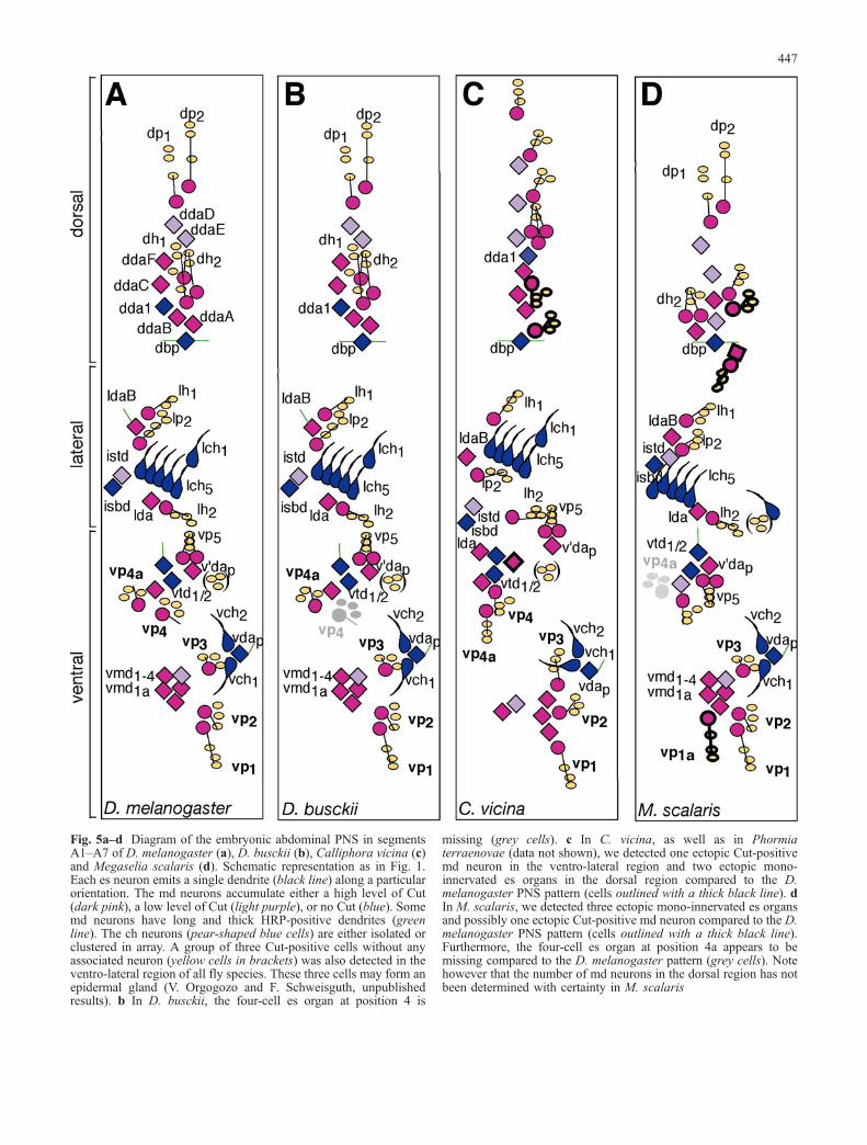
Fig. 5a–d Diagram of the embryonic abdominal PNS in segmentsA1–A7 of D. melanogaster (a), D. busckii (b), Calliphora vicina (c)and Megaselia scalaris (d). Schematic representation as in Fig. 1.Each es neuron emits a single dendrite (black line) along a particularorientation. The md neurons accumulate either a high level of Cut(dark pink), a low level of Cut (light purple), or no Cut (blue). Somemd neurons have long and thick HRP-positive dendrites (greenline). The ch neurons (pear-shaped blue cells) are either isolated orclustered in array. A group of three Cut-positive cells without anyassociated neuron (yellow cells in brackets) was also detected in theventro-lateral region of all fly species. These three cells may form anepidermal gland (V. Orgogozo and F. Schweisguth, unpublishedresults). b In D. busckii, the four-cell es organ at position 4 is
missing (grey cells). c In C. vicina, as well as in Phormiaterraenovae (data not shown), we detected one ectopic Cut-positivemd neuron in the ventro-lateral region and two ectopic mono-innervated es organs in the dorsal region compared to the D.melanogaster PNS pattern (cells outlined with a thick black line). dIn M. scalaris, we detected three ectopic mono-innervated es organsand possibly one ectopic Cut-positive md neuron compared to the D.melanogaster PNS pattern (cells outlined with a thick black line).Furthermore, the four-cell es organ at position 4a appears to bemissing compared to the D. melanogaster pattern (grey cells). Notehowever that the number of md neurons in the dorsal region has notbeen determined with certainty in M. scalaris
447
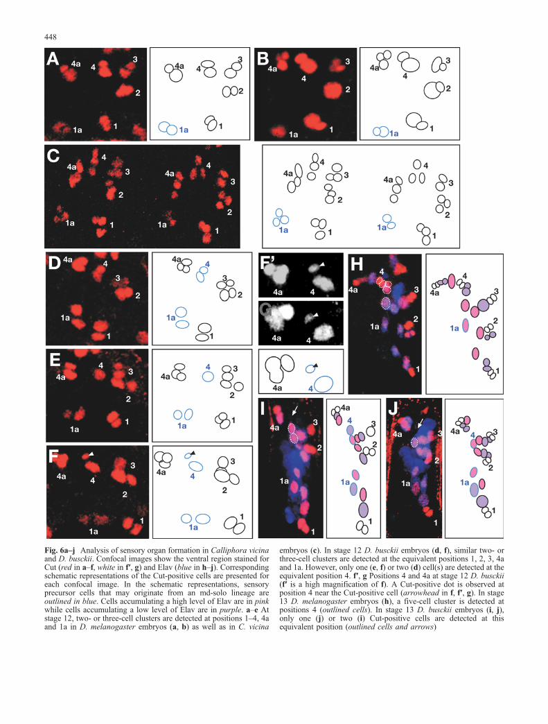
Fig. 6a–j Analysis of sensory organ formation in Calliphora vicinaand D. busckii. Confocal images show the ventral region stained forCut (red in a–f, white in f′, g) and Elav (blue in h–j). Correspondingschematic representations of the Cut-positive cells are presented foreach confocal image. In the schematic representations, sensoryprecursor cells that may originate from an md-solo lineage areoutlined in blue. Cells accumulating a high level of Elav are in pinkwhile cells accumulating a low level of Elav are in purple. a–e Atstage 12, two- or three-cell clusters are detected at positions 1–4, 4aand 1a in D. melanogaster embryos (a, b) as well as in C. vicina
embryos (c). In stage 12 D. busckii embryos (d, f), similar two- orthree-cell clusters are detected at the equivalent positions 1, 2, 3, 4aand 1a. However, only one (e, f) or two (d) cell(s) are detected at theequivalent position 4. f′, g Positions 4 and 4a at stage 12 D. busckii(f′ is a high magnification of f). A Cut-positive dot is observed atposition 4 near the Cut-positive cell (arrowhead in f, f′, g). In stage13 D. melanogaster embryos (h), a five-cell cluster is detected atpositions 4 (outlined cells). In stage 13 D. busckii embryos (i, j),only one (j) or two (i) Cut-positive cells are detected at thisequivalent position (outlined cells and arrows)
448
this pattern may be characteristic of the D. busckii species.According to the phylogenetic tree established by Remsenand O’Grady (2002; Fig. 3), the most parsimoniousevolutionary scenario is that the es organ at position 4 hasbeen lost during evolution in the branch leading to D.busckii (see Discussion).
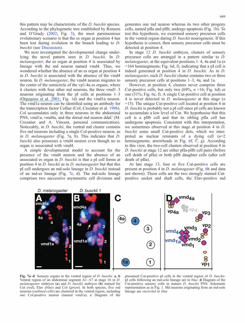
We next investigated the developmental change under-lying the novel pattern seen in D. busckii. In D.melanogaster, the es organ at position 4 is associated bylineage with the md neuron named vmd4. Thus, wewondered whether the absence of an es organ at position 4in D. busckii is associated with the absence of the vmd4neuron. In D. melanogaster, the vmd4 neuron migrates tothe center of the semicircle of the vp1-4a es organs, whereit clusters with four other md neurons, the three vmd1–3neurons originating from the pI cells at positions 1–3(Orgogozo et al. 2001; Fig. 1d) and the vmd1a neuron.The vmd1a neuron can be identified using an antibody forthe transcription factor Collier (Col; Crozatier et al. 1996).Col accumulates only in three neurons in the abdominalPNS, vmd1a, vmd4a, and the dorsal md neuron ddaC (M.Crozatier and A. Vincent, personal communication).Noticeably, in D. busckii, the ventral md cluster containsfive md neurons including a single Col-positive neuron, asin D. melanogaster (Fig. 7a, b). This indicates that D.busckii also possesses a vmd4 neuron even though no esorgan is associated with vmd4.
A simple developmental model to account for thepresence of the vmd4 neuron and the absence of anassociated es organ in D. busckii is that a pI cell forms atposition 4 in D. busckii as in D. melanogaster but that thispI cell undergoes an md-solo lineage in D. busckii insteadof an md-es lineage (Fig. 7c, d). The md-solo lineagecomprises two successive asymmetric cell divisions and
generates one md neuron whereas its two other progenycells, named pIIa and pIIb, undergo apoptosis (Fig. 1b). Totest this hypothesis, we examined sensory precursor cellsin the ventral region during D. busckii neurogenesis. If thishypothesis is correct, then sensory precursor cells must bedetected at position 4.
In stage 12 D. busckii embryos, clusters of sensoryprecursor cells are arranged in a pattern similar to D.melanogaster, at the equivalent positions 1–4, 4a and 1a (n=164 hemisegments; Fig. 6d, f), indicating that a pI cell isindeed generated at position 4 in D. busckii. As in D.melanogaster, each D. busckii cluster contains two or threesensory precursor cells at positions 1–3, 4a, and 1a.
However, at position 4, clusters never comprise threeCut-positive cells, but only two (69%, n =16; Fig. 6d) orone (31%; Fig. 6e, f). A single Cut-positive cell at position4 is never detected in D. melanogaster at this stage (n=15). The unique Cut-positive cell located at position 4 inD. busckii is probably not a pI cell since pI cells are knownto accumulate a low level of Cut. We hypothesise that thiscell is a pIIb cell and that its sibling pIIa cell hasundergone apoptosis. Consistent with this interpretation,we sometimes observed at this stage at position 4 in D.busckii some small Cut-positive dots, which we inter-preted as nuclear remnants of a dying cell (n=2hemisegments; arrowheads in Fig. 6f, f′, g). Accordingto this view, the two-cell clusters observed at position 4 inD. busckii at stage 12 are either pIIa-pIIb cell pairs (beforecell death of pIIa) or both pIIb daughter cells (after celldeath of pIIa).
At late stage 13, four or five Cut-positive cells arepresent at position 4 in D. melanogaster (Fig. 6h and datanot shown). These cells are the two strongly stained Cut-positive socket and shaft cells, the Elav-positive md
Fig. 7a–d Sensory organs in the ventral region of D. busckii. a, bVentral region of an abdominal segment A1–A7 at stage 16 in D.melanogaster embryos (a) and D. busckii embryos (b) stained forCut (red), Elav (blue) and Col (green). In both species, five mdneurons (outlined cells) are clustered in the ventral region, includingone Col-positive neuron (named vmd1a). c Diagram of the
presumed Cut-positive pI cells in the ventral region of D. busckii.pI cells following an md-solo lineage are in blue. d Diagram of theCut-positive sensory cells in mature D. busckii PNS. Schematicrepresentation as in Fig. 1. Md neurons originating from an md-sololineage are encircled in blue
449
neuron, as well as the Elav-positive pIIIb cell or its twoElav-positive daughter cells. In D. busckii, only one (50%)or two (50%) Cut-positive cells are detected at thisposition (n =124; Fig. 6i, j). These cells accumulate Elavand one of them seems to migrate to the final position ofthe vmd4 neuron. These observations indicate that the cellpair is very likely a vmd4a-pIIIb cell pair and that theunique cell must be the remaining vmd4 neuron after itssibling pIIIb cell has undergone apoptosis. All theseobservations suggest that the pI cell appearing at position4 in D. busckii follows an md-solo lineage and not an md-es lineage as in D. melanogaster.
Discussion
Stability of the larval PNS pattern throughoutevolution
Our study reveals that, with the exception of D. busckii,the larval abdominal PNS pattern did not change duringevolution of all the Drosophilinae species analysed here.However, other aspects of the larval PNS not studied heremay have changed during evolution, including morphol-ogy of sensory structures, axonal projections, type ofsensory receptors expressed by sensory neurons, andsensory organs located in the head, thorax and posteriorregion.
Strikingly, the stability of the Drosophilinae larval PNSduring evolution contrasts sharply with the higher vari-ability of the adult PNS pattern. Indeed, many variations inthe adult PNS pattern on the thorax, wings, legs, abdomenand head have been reported within Drosophilinae species(Ashburner 1989; Garcia-Belido 1983; Grimaldi 1990 andreferences therein). Within the melanogaster speciessubgroup for example, species that diverged 2–5 millionyears ago (Powell 1997; Lachaise et al. 1988) exhibitdistinct PNS patterns on the male genitalia, the femaleovipositor and the maxillary palp (Ashburner 1989). PNSon the thorax, wings, legs, and head are also variablewithin the Drosophilinae species analysed here (Grimaldi1990; Ashburner 1989). Similarly, the two Calliphoridaespecies analysed here have identical larval but distinctadult PNS patterns on the thorax (Skaer et al. 2002) andother adult parts (McAlpine 1989). These comparisons oflarval vs adult PNS suggest that the adult PNS evolvedmore rapidly than the larval PNS.
Many larval sensory axons survive at metamorphosis ininsects and act as guidance cues for organising the orderedarray of adult sensory neurons (Bate 1976; Jan et al. 1985;Shepherd and Smith 1996; Tix et al. 1989; Usui-Ishihara etal. 2000; Williams and Shepherd 1999, 2002). In D.melanogaster, sensory neurons persisting through meta-morphosis into adult stages have been identified in theventral and lateral abdominal regions (Williams andShepherd 1999), and some of them have been shown tobe required for the normal development of adult sensoryprojections (Usui-Ishihara et al. 2000; Williams andShepherd 2002). Since hypothetical changes in the pattern
of these larval neurons are likely to modify the organisa-tion of the adult nervous system, it is possible that thepersistent neurons that function as guidance cues may besubjected to more constraints than other sensory neuronsand should therefore be more stable during evolution.Accordingly, the neurons that persist through metamor-phosis in D. melanogaster (vch2, vp2, dbd, ltd, isbd/lbd,lda, one vtd neuron, v’dap and vmd3/vbd) were detectedin all Drosophilinae, Calliphoridae as well as Phoridaestudied here. This argues that the guidance role of thelarval PNS during metamorphosis may indeed constrainthe larval PNS pattern.
An md-es to md-solo sensory lineage switch
In D. melanogaster, the pI cell at position 4 produces an esorgan as well as the vmd4 neuron (Orgogozo et al. 2001).Our analysis of the vmd4 lineage suggests that the pI cellat position 4 in D. busckii does not follow an md-eslineage as in D. melanogaster but an md-solo lineage,accounting thereby for the presence of a vmd4 neuron andthe absence of vp4 organ in D. busckii. Thus, thedifference between D. busckii and D. melanogaster larvalPNS patterns appears to relate to the cell death patternwithin the sensory lineage at position 4. We propose thatthe pIIa and pIIIb cells at position 4 survive and produce aset of four es cells in D. melanogaster, whereas theyundergo apoptosis in D. busckii. In the md-solo lineage inD. melanogaster, the two cells fated to die specificallyexpress the pro-apoptotic genes reaper and grim (Orgo-gozo et al. 2002). It would therefore be interesting toexamine the expression of the reaper and grim homologsin D. busckii. Unfortunately, we have been unable to cloneportions of the D. busckii reaper and grim genes that arelarge enough to generate specific in situ probes (data notshown).
Analysis of the PNS pattern in species closely related toD. busckii would be helpful to establish when this changeappeared during evolution. D. busckii belongs to theDorsilopha subgenus. Two other members of thissubgroup have been described (Toda 1986) but to ourknowledge, none of them is maintained in laboratory. Asthe phylogenetic position of the Dorsilopha subgroupwithin the Drosophilinae tree remains uncertain (Grimaldi1990; Kwiatowski and Ayala 1999; Remsen and O’Grady2002; Thomas and Hunt 1993), no species closely relatedto D. busckii have been clearly established, thus prevent-ing their study.
Given the undefined position of D. busckii within theDrosophilinae tree, polarity of the evolutionary change atposition 4 is difficult to determine within Drosophilinae.Our study of the outgroup species C. vicina suggests thatthe es organ at position 4, although displaced, is present inC. vicina. Therefore, the most parsimonious evolutionaryscenario would be that the es organ at position 4 hasdisappeared in the branch leading to D. busckii duringevolution. A change from md-es towards md-solo lineage
450
may thus have led to the loss of the es organ at position 4in D. busckii.
We further note that other gains (or losses) of mono-innervated es organs were observed between Drosophili-nae and Calliphoridae/Phoridae species without anymodification of the number of neighbouring md neurons(see Fig. 5, for example vp4a seems to be absent in M.scalaris). This thus raises the possibility that these PNSdifferences have arisen similarly through md-es/md-sololineage switches.
Distinct arrangement of sensory cells in C. vicina andvariability at position 4
The arrangement of es organs corresponding to positions 4and 4a differs between C. vicina and D. melanogaster. Ouranalysis suggests that this difference in es organ arrange-ment results from a difference in the positioning of thesensory precursor cells. This change is observed atposition 4 in the ventral region. Interestingly, the esorgan at position 4 is also the one that has evolved in theD. busckii pattern. This suggests that the es organ atposition 4 may be more evolutionary labile than es organsat other positions. Interestingly, sensory cells at position 4appear to have undergone distinct developmental changesduring evolution—changes in relative position as well asin cell lineage. This suggests that the observed evolu-tionary variants at position 4 probably resulted frommutations in different genes.
Noticeably, in the abdominal segment A8 of D.melanogaster, the pI cell arising at position 4 follows anmd-solo lineage and thus produces no es organ (Orgogozoet al. 2002). Moreover, when apoptosis is blocked, the esorgan generated by this pI cell is located more dorsallythan the es organ at position 4a, in a pattern similar to theone observed in C. vicina (see Fig. 3D in Orgogozo et al.2002). Therefore, the intersegmental variation in the esorgan at position 4 within D. melanogaster mirrors theobserved interspecific variation, namely the difference incell lineage for D. busckii and the difference in relative cellposition for C. vicina. This suggests that morphologicalevolution may arise from modification of pre-existinggenetic switches that regulate intersegmental variation.
Acknowledgements We thank M.-L. Carriou, N. Gompel, D.Lachaise, J.-A. Lepesant, P. Santamaria, P. Simpson, D. Stern and D.S.H.B. for providing flies and antibodies. We also thank A.McGregor for his help in designing reaper and grim degenerateprimers; Y. Bellaïche and M.-A. Felix for our fruitful discussionsand Y. Bellaïche, N. Gompel, A. Shingleton and D. Stern for criticalreading of the manuscript. This work was supported by the CentreNational de la Recherche Scientifique.
References
Ashburner M (1989) Drosophila. A laboratory handbook. ColdSpring Harbor Laboratory Press, New York
Bate M (1976) Pioneer neurones in an insect embryo. Nature260:54–56
Blochlinger K, Bodmer R, Jack J, Jan LY, Jan YN (1988) Primarystructure and expression of a product from cut, a locus involvedin specifying sensory organ identity in Drosophila. Nature333:629–635
Blochlinger K, Bodmer R, Jan LY, Jan YN (1990) Patterns ofexpression of Cut, a protein required for external sensory organdevelopment, in wild-type and cut mutant Drosophila embryos.Genes Dev 4:1322–1331
Bodmer R, Jan YN (1987) Morphological differentiation of theembryonic peripheral neurons in Drosophila. Roux’s Arch DevBiol 196:69–77
Bodmer R, Barbel S, Sheperd S, Jack JW, Jan LY, Jan YN (1987)Transformation of sensory organs by mutations of the cut locusof D. melanogaster. Cell 51:293–307
Bodmer R, Carretto R, Jan YN (1989) Neurogenesis of theperipheral nervous system in Drosophila embryos: DNAreplication patterns and cell lineages. Neuron 3:21–32
Brewster R, Bodmer R (1995) Origin and specification of type IIsensory neurons in Drosophila. Development 121:2923–2936
Brewster R, Hardiman K, Deo M, Khan S, Bodmer R (2001) Theselector gene cut represses a neural cell fate that is specifiedindependently of the Achaete-Scute-Complex and atonal. MechDev 105:57–68
Campos-Ortega JA, Hartenstein V (1997) The embryonic develop-ment of Drosophila melanogaster. Springer, Berlin HeidelbergNew York
Crozatier M, Valle D, Dubois L, Ibnsouda S, Vincent A (1996)Collier, a novel regulator of Drosophila head development, isexpressed in a single mitotic domain. Curr Biol 6:707–718
Dambly-Chaudiére C, Ghysen A (1986) The sense organs in theDrosophila larva and their relation to embryonic pattern ofsensory neurons. Roux’s Arch Dev Biol 195:222–228
Desai CJ, Popova E, Zinn K (1994) A Drosophila receptor tyrosinephosphatase expressed in the embryonic CNS and larval opticlobes is a member of the set of proteins bearing the “HRP”carbohydrate epitope. J Neurosci 14:7272–7283
Fabini G, Freilinger A, Altmann F, Wilson IB (2001) Identificationof core alpha 1,3-fucosylated glycans and cloning of therequisite fucosyltransferase cDNA from Drosophila melano-gaster. Potential basis of the neural anti-horseadish peroxidaseepitope. J Biol Chem 276:28058–28067
Garcia-Belido A (1983) Comparative anatomy of cuticular patternsin the genus Drosophila. In: Goodwin BC, Holder N, Wylie CC(eds) Secondary comparative anatomy of cuticular patterns inthe genus Drosophila. Cambridge University Press, London, pp227–255
Ghysen A, Dambly-Chaudière C (1989) Genesis of the Drosophilaperipheral nervous system. Trends Genet 5:251–255
Ghysen A, Dambly-Chaudière C (2000) A genetic programme forneuronal connectivity. Trends Genet 16:221–226
Ghysen A, Dambly-Chaudiere C, Aceves E, Jan LY, Jan YN (1986)Sensory neurons and peripheral pathways in Drosophilaembryos. Roux’s Arch Dev Biol 195:281–289
Green P, Hartenstein V (1997) Structure and spatial pattern of thesensilla of the body segments of insect larvae. Microsc ResTech 39:470–478
Grimaldi D (1990) A phylogenetic, revised classification of generain the Drosophilidae (Diptera). Bull Am Mus Nat Hist 197:1–139
Grueber WB, Truman JW (1999) Development and organization ofa nitric-oxide-sensitive peripheral neural plexus in larvae of themoth, Manduca sexta. J Comp Neurol 404:127–141
Grueber WB, Jan LY, Jan YN (2002) Tiling of the Drosophilaepidermis by multidendritic sensory neurons. Development129:2867–2878
Grueber WB, Jan LY, Jan YN (2003a) Different levels of thehomeodomain protein cut regulate distinct dendrite branchingpatterns of Drosophila multidendritic neurons. Cell 112:805–818
451
Grueber WB, Ye B, Moore AW, Jan LY, Jan YN (2003b) Dendritesof distinct classes of Drosophila sensory neurons showdifferent capacities for homotypic repulsion. Curr Biol13:618–626
Haase A, Stern M, Wachtler K, Bicker G (2001) A tissue-specificmarker of Ecdysozoa. Dev Genes Evol 211:428–433
Hertweck H (1931) Anatomie und Variabilität des nervensystemsund der Sinnesorgane von Drosophila melanogaster (Meigen).Z Wiss Zool 139:559–663
Hewes RS, Park D, Gauthier SA, Schaefer AM, Taghert PH (2003)The bHLH protein Dimmed controls neuroendocrine celldifferentiation in Drosophila. Development 130:1771–1781
Jan LY, Jan YN (1982) Antibodies to horseradish peroxidase asspecific neuronal markers in Drosophila and in grasshopperembryos. Proc Natl Acad Sci USA 79:2700–2704
Jan YN, Jan LY (1993) The peripheral nervous system. In: Bate M,Martinez-Arias A (eds) The development of Drosophilamelanogaster. Cold Spring Harbor Laboratory Press, NewYork, pp 1207–1244
Jan YN, Jan LY (2001) Asymmetric cell division in the Drosophilanervous system. Nat Rev Neurosci 2:772–779
Jan YN, Ghysen A, Cristoph I, Barbel S, Jan LY (1985) Formationof neuronal pathways in the imaginal discs of Drosophilamelanogaster. J Neurosci 5:2453–2464
Kwiatowski J, Ayala FJ (1999) Phylogeny of Drosophila and relatedgenera: conflict between molecular and anatomical analyses.Mol Phylogenet Evol 13:319–328
Lachaise D, Cariou ML, David JR, Lemeunier F, Tsacas L,Ashburner M (1988) Historical biogeography of the Drosophilamelanogaster species subgroup. In: Hetch MK, Wallace B,MacIntyre RJ (eds) Evolutionary biology. Plenum Press, NewYork, pp 127–173
Liu L, Yermolaieva O, Johnson WA, Abboud FM, Welsh MJ (2003)Identification and function of thermosensory neurons inDrosophila larvae. Nat Neurosci 6: 267–273
McAlpine JF (1989) Phylogeny and classification of the Musco-morpha. In: McAlpine JF, Wood DM (eds) Secondary phylog-eny and classification of the Muscomorpha, vol 32. ResearchBranch Agriculture Canada, pp 1397–1502
Meier T, Chabaud F, Reichert H (1991) Homologous patterns in theembryonic development of the peripheral nervous system in thegrasshopper Schistocerca gregaria and the fly Drosophilamelanogaster. Development 112:241–253
Moore AW, Jan LY, Jan YN (2002) hamlet, a binary genetic switchbetween single- and multipledendrite neuron morphology.Science 297:1355–1358
Orgogozo V, Schweisguth F, Bellaiche Y (2001) Lineage, cellpolarity and inscuteable function in the peripheral nervoussystem of the Drosophila embryo. Development 128:631–643
Orgogozo V, Schweisguth F, Bellaiche Y (2002) Binary cell deathdecision regulated by unequal partitioning of Numb at mitosis.Development 129:4677–4684
Osborne MP (1963) The sensory neurones and sensilla in theabdomen and thorax of the blowfly larva. Q J Microsc Sci104:227–241
Powell JR (1997) Progress and prospects in evolutionary biology:the Drosophila model. Oxford University Press, London
Remsen J, O’Grady P (2002) Phylogeny of Drosophilinae (Diptera:Drosophilidae), with comments on combined analysis andcharacter support. Mol Phylogenet Evol 24:249–264
Robinow S, White K (1988) The locus elav of Drosophilamelanogaster is expressed in neurons at all developmentalstages. Dev Biol 126:294–303
Rothwell WF, Sullivan W (2000) Fluorescent analysis of Drosophilaembryos. In: Ashburner M, Hawley S, Sullivan B (eds)Secondary fluorescent analysis of Drosophila embryos. ColdSpring Harbor Laboratory Press, Cold Spring Harbor, N.Y., pp141–157
Shepherd D, Smith SA (1996) Central projections of persistentlarval sensory neurons prefigure adult sensory pathways in theCNS of Drosophila. Development 122:2375–2384
Simpson P, Woehl R, Usui K (1999) The development and evolutionof bristle patterns in Diptera. Development 126:1349–1364
Skaer N, Pistillo D, Simpson P (2002) Transcriptional heterochronyof scute and changes in bristle pattern between two closelyrelated species of blowfly. Dev Biol 252:31–45
Snow PM, Patel NH, Harrelson AL, Goodman CS (1987) Neural-specific carbohydrate moiety shared by many surface glyco-proteins in Drosophila and grasshopper embryos. J Neurosci7:4137–4144
Sun B, Salvaterra PM (1995) Characterization of nervana, aDrosophila melanogaster neuronspecific glycoprotein antigenrecognized by anti-horseradish peroxidase antibodies. J Neu-rochem 65:434–443
Thomas RH, Hunt JA (1993) Phylogenetic relationships inDrosophila: a conflict between molecular and morphologicaldata. Mol Biol Evol 10:362–374
Tix S, Bate M, Technau G (1989) Pre-existing neuronal pathways inthe leg imaginal discs of Drosophila. Development 107:855–862
Toda MJ (1986) Drosophilidae (Diptera) in Burma. I. The subgenusDorsilopha Surtevant of the genus Drosophila, with descrip-tions of two new species. Kontyu 54:282–290
Tracey WD, Jr., Wilson RI, Laurent G, Benzer S (2003) painless, aDrosophila gene essential for nociception. Cell 113:261–273
Usui-Ishihara A, Simpson P, Usui K (2000) Larval multidendriteneurons survive metamorphosis and participate in the formationof imaginal sensory axonal pathways in the notum ofDrosophila. Dev Biol 225:357–369
Wang X, Sun B, Yasuyama K, Salvaterra PM (1994) Biochemicalanalysis of proteins recognized by anti-HRP antibodies inDrosophila melanogaster: identification and characterization ofneuron specific and male specific glycoproteins. InsectBiochem Mol Biol 24:233–242
Williams DW, Shepherd D (1999) Persistent larval sensory neuronsin adult Drosophila melanogaster. J Neurobiol 39:275–286
Williams DW, Shepherd D (2002) Persistent larval sensory neuronesare required for the normal development of the adult sensoryafferent projections in Drosophila. Development 129:617–624
452