Development of Polymer-based Gels for Multimodal Medical ...
This article appeared in a journal published by Elsevier. The attachedcopy is furnished to the author for internal non-commercial researchand education use, including for instruction at the authors institution
and sharing with colleagues.
Other uses, including reproduction and distribution, or selling orlicensing copies, or posting to personal, institutional or third party
websites are prohibited.
In most cases authors are permitted to post their version of thearticle (e.g. in Word or Tex form) to their personal website orinstitutional repository. Authors requiring further information
regarding Elsevier’s archiving and manuscript policies areencouraged to visit:
http://www.elsevier.com/copyright
Author's personal copy
Colloids and Surfaces B: Biointerfaces 75 (2010) 510–519
Contents lists available at ScienceDirect
Colloids and Surfaces B: Biointerfaces
journa l homepage: www.e lsev ier .com/ locate /co lsur fb
Evaluation of boronate-containing polymer brushes and gels as substrates forcarbohydrate-mediated adhesion and cultivation of animal cells
Alexander E. Ivanova,b,∗, Ashok Kumarc, Suthasinee Nilsangd,e, Maria-Rosa Aguilar f,Lyubov I. Mikhalovskaf, Irina N. Savina f, Lars Nilssong, Ivan G. Scheblykinh,Marina V. Kuzimenkovab, Igor Yu. Galaevb
a Protista Biotechnology AB, IDEON, SE-22370, Lund, Swedenb Department of Biotechnology, Lund University, P.O. Box 124, SE-22100, Lund, Swedenc Department of Biological Science and Bioengineering, Indian Institute of Technology Kanpur, 208016 Kanpur, UP, Indiad Department of Food Engineering and Bioprocess Technology, School of Environment, Resources and Development, Asian Institute of Technology, Pathumthani 12120, Thailande Faculty of Science and Technology, Valaya Alongkorn Rajabhat University, Pathumthani 13180, Thailandf School of Pharmacy and Biomolecular Sciences, University of Brighton, Brighton BN2 4GJ, UKg Department of Food Technology, Lund University, P.O. Box 124, SE-22100, Lund, Swedenh Department of Chemical Physics, Lund University, P.O. Box 124, SE-22100, Lund, Sweden
a r t i c l e i n f o
Article history:Received 2 April 2009Received in revised form 28 August 2009Accepted 22 September 2009Available online 26 September 2009
Keywords:Boronic acidCell detachmentGraft polymerizationHydrogelSurface topology
a b s t r a c t
Boronate-containing thin polyacrylamide gels (B-Gel), polymer brushes (B-Brush) and chemisorbedorganosilane layers (B-COSL) were prepared on the surface of glass slides and studied as substratesfor carbohydrate-mediated cell adhesion. B-COSL- and B-Brush-modified glass samples exhibited multi-ple submicron structures densely and irregularly distributed on the glass surface, as found by scanningelectron microscopy and atomic force microscopy. B-Gel was ca. 0.1 mm thick and contained pores witheffective size of 1–2 �m in the middle and of 5–20 �m on the edges of the gel sample as found by confocallaser scanning microscopy. Evidence for the presence of phenylboronic acid in the samples was givenby time-of-flight secondary ion mass-spectrometry (ToF SIMS), contact angle measurements performedin the presence of fructose, and staining with Alizarin Red S dye capable of formation specific, fluo-rescent complexes with boronic acids. A comparative study of adhesion and cultivation of animal cellson the above substrates was carried out using murine hybridoma M2139 cell line as a model. M2139cells adhered to the substrates in the culture medium without glucose or sodium pyruvate at pH 8.0,and then were cultivated in the same medium at pH 7.2 for 4 days. It was found that the substrates ofB-Brush type were superior both regarding cell adhesion and viability of the adhered cells, among thesubstrates studied. MTT assay confirmed proliferation of M2139 cells on B-Brush substrates. Some celladhesion was also registered in the macropores of B-Gel substrate. The effects of surface microstructureof the boronate-containing polymers on cell adhesion are discussed. Transparent glass substrates graftedwith boronate-containing copolymers offer good prospects for cell adhesion studies and development ofcell-based assays.
© 2009 Elsevier B.V. All rights reserved.
1. Introduction
Cell adhesion studies and cell-based assays are expandingresearch fields of cell biology, tissue engineering and biomedi-cal analysis. Adhesion of animal cells to artificial matrices canbe supported by the proteins of extracellular matrix (ECM) suchas fibronectin, laminin or collagen containing specific functionaldomains that interact with the integrin receptors present on the
∗ Corresponding author at: Protista Biotechnology AB, IDEON, SE-22370, Lund,Sweden. Fax: +46 46 286 3889.
E-mail address: [email protected] (A.E. Ivanov).
cell surface [1]. In particular, the amino acid sequence RGD (Arg-Gly-Asp) has been identified to ligate many different integrinsand to facilitate cell adhesion on many artificial matrices [2,3]. Inother adhesion strategies, the selective interactions between theartificial matrices and cells were established using carbohydrate-mediated cell recognition [4], which is important initial step ofmany specific cell–cell interactions, for example, extravasation ofleukocytes from the blood into the inflamed tissue. Relatively weakaffinity interactions can occur between the selectins of endothe-lial cells and their carbohydrate counterparts on leukocytes, andvice versa. This makes possible a single-touch reversible bindingbetween the leukocytes and endothelial cells resulting in leuko-cyte rolling [5]. Reversibility of the lectin interactions with cell
0927-7765/$ – see front matter © 2009 Elsevier B.V. All rights reserved.doi:10.1016/j.colsurfb.2009.09.028
Author's personal copy
A.E. Ivanov et al. / Colloids and Surfaces B: Biointerfaces 75 (2010) 510–519 511
glycocalix has been proven by the dissociation of lectin-containingmagnetic particles from the cell surface induced by high con-centration of N-acetyl-d-galactosamine, a competing sugar withspecificity to the lectin [6]. The competing effect of N-acetyl-d-glucosamine (GlcNAc) confirmed reversibility of adhesion ofchicken hepatocytes on immobilized GlcNAc-terminated oligosac-charides [7]. The reversible character of carbohydrate-mediatedcell adhesion creates possibilities for facile cell attachment as wellas for detachment of the adhered cells. These phenomena can beapplicable to techniques such as isolation of specific cell lines [6,8],quantifying of selective cell adhesion to glycan microarrays [7],or profiling of cell surface carbohydrate expression [9]. It seemslikely that carbohydrate-mediated cell adhesion is more suitablefor the preparation of arrays of individual intact cells [7] ratherthan for formation of confluent cell monolayers. The latter typ-ically involves integrin-ECM interactions and cell spreading. Itwas previously reported that initial adhesion of hepatocytes onsynthetic glycopolymer matrices was enhanced by asialoglycopro-tein receptors of the glycopolymer. In contrast, the post-adhesionprocess of hepatocyte attachment and spreading was negativelyinfluenced by the high surface concentration of the receptor [5].Arrays of single animal cells can be interesting in combination withmicropatterned surfaces, as small patterns do not allow cell spread-ing and initiation of cytoskeleton tension and organization [10]. Thecarbohydrate-mediated cell adhesion can be a relevant techniquefor cell immobilization within repeating units of the patterns.
Recently, we have reported a new type of the carbohydrate-mediated affinity adhesion of microparticles and animal cellsachieved on the surface of glass grafted with boronate-containing polymers (BCP), capable of interaction with cell surfacecarbohydrates [11,12]. The surface-initiated radical copolymer-ization of N,N-dimethylacrylamide (DMAAm) and N-acryloyl-m-aminophenylboronic acid (NAAPBA) resulted in the formationof polymer brushes consisting of end-grafted copolymer chains.Adhesion of animal cells on the affinity polymer brushes yieldedirregular arrays formed by individual cells that could be, however,detached from the surface by means of elution with 0.1 M fruc-tose. More recently, similar cell adhesion experiments have beensuccessfully performed on gold electrodes chemically modified bydiazonium salts of p-aminophenylboronic acid [13], a low molecu-lar weight affinity ligand. The cell adhesion affinity molecules likeRGD-type peptides are commonly immobilized on solid surfacesvia polymeric spacers such as polyethylene glycol (PEO), polylysineor thermoresponsive poly-N-isopropylacrylamide [2]. These poly-mers reduce adsorption of proteins on solid supports and, therefore,enable more specific adhesion of cells induced by the adhesionmolecules. In this study we aim to compare animal cells adhesion onvarious immobilized BCP using identical experimental conditionsand the same cell line. Three different types of substrates were syn-thesized and compared: partially cross-linked chemisorbed layerof organosilane with immobilized phenylboronic acid (B-COSL),polymer brush composed of end group-grafted BCP (B-Brush) andpolyacrylamide gel containing copolymerized NAAPBA (B-Gel) [14](see Fig. 1). Water-soluble BCP were earlier reported to be non-toxic substances, able to bind to cell surface carbohydrates and toinitiate proliferation of lymphocytes, i.e. to display lectin-like bind-ing activity [15]. Since specific complex formation between BCPand polysaccharides is known [16,17], one can presume a mul-tivalent type of interaction between BCP and oligosaccharides ofcell glycocalix. Up to now, effects of immobilized BCP on prolif-eration of animal cells were very rarely studied [18,19], althoughability of boronic acid-containing reagents (“boronolectins”) totrigger biological events in living cells had been supposed [20].There have been reports on encapsulation of animal cells in hydro-gels formed by BCP with poly(vinyl alcohol) with no effect of thepolymers on cell proliferation [19]. One may expect that different
BCP-containing substrates would produce different effects on adhe-sion and proliferation of animal cells due to the different degreesand types of polymer cross-linking, porosity or availability of theboronate affinity ligands. The aims of this study were to investigatethe effects of surface structure and three-dimensional organizationof the affinity polymeric reagents on cell adhesion and to comparethe cell viability observed on the different substrates.
2. Materials and methods
2.1. Materials
Cover glass slides (CGSs) for light microscopy (15 mm × 15 mm)were a product of Knittel Gläser (Germany). Glass capillary tubeswith outer diameter 1.3–1.5 mm were purchased from ModulohmA/S (Herlev, Denmark). 3-Aminophenylboronic acid hydrochloride,N,N-dimethylacrylamide (DMAA), 3-mercaptopropyltrimethoxysilane (3-MPTMS), 3-glycidoxypropyltrimethoxy silane (3-GPTMS)and sodium monosulfonate Alizarin Red S (ARS) were prod-ucts of Sigma–Aldrich (Steinheim, Germany). 2,2′-Azobis(2-methylpropionitrile), AMPN, was purchased from Acros (NewJersey, USA). N-Acryloyl-m-aminophenylboronic acid (NAAPBA)was prepared as described elsewhere [21]. Copolymer of DMAAmand NAAPBA was prepared according to the previously publishedmethod [22]. The murine clone BB5.1 secreting anti-mouse C5 (IgG1subclass) was kindly provided by Dr. B. Stockinger (National Insti-tute for Medical Research, London) and was used for generatingmurine hybridoma cell line M2139. Dulbecco’s modified Eagle’smedium (DMEM), RPMI 1640 medium, McCoy’s medium and fetalcalf serum (FCS) were from Gibco Invitrogen Corporation (GrandIsland, NY, USA).
2.2. Chemical modification of the glass slides by3-glycidoxypropyltrimethoxysilane (3-GPTMS) andimmobilization of 3-aminophenylboronic acid (3-APBA):preparation of B-COSL substrate
The chemical modification was performed as described earlier[12]. Briefly, the glass slides and capillaries were washed by 4 Msodium hydroxide, distilled water, 4 M hydrochloric acid, distilledwater and dried. The clean CGSs and capillaries were kept in theboiling solution of 3-GPTMS in dry 1,4-dioxane for ca. 16 h. Theglasses were rinsed with several portions of fresh solvent and driedunder vacuum in a desiccator over dry calcium chloride. For immo-bilization of 3-APBA, the 3-GPTMS-modified glasses were placedinto 3-APBA solution (4%, w/v) in water adjusted to pH 8.5 by0.5 M sodium bicarbonate and agitated on a rocking table for 24 hat room temperature [23]. The chemically modified glass sampleswere rinsed with several portions of distilled water. To hydrolyzethe residual epoxy groups into diols, the modified slides and capil-laries were treated with diluted hydrochloric acid (pH 2.4) for 24 hat room temperature. The slides intended to be analyzed by elec-tron microscopy were rinsed with ethanol and dried under vacuum.Otherwise, the slides and capillaries were rinsed with ethanol andstored in this solvent. The capillaries chemically modified by 3-APBA were dried on air before the contact angle measurements,which were performed as described elsewhere [11].
2.3. Chemical modification of the glass slides followed by graftpolymerization: preparation of B-Brush substrate
The graft polymerization was performed according to the earlierdescribed method [12]. Briefly, the clean glass slides were treatedwith 2% (v/v) 3-mercaptopropyltrimethoxy silane (3-MPTMS) indry 1,4-dioxane at 100 ◦C for 24 h, rinsed with several portions of
Author's personal copy
512 A.E. Ivanov et al. / Colloids and Surfaces B: Biointerfaces 75 (2010) 510–519
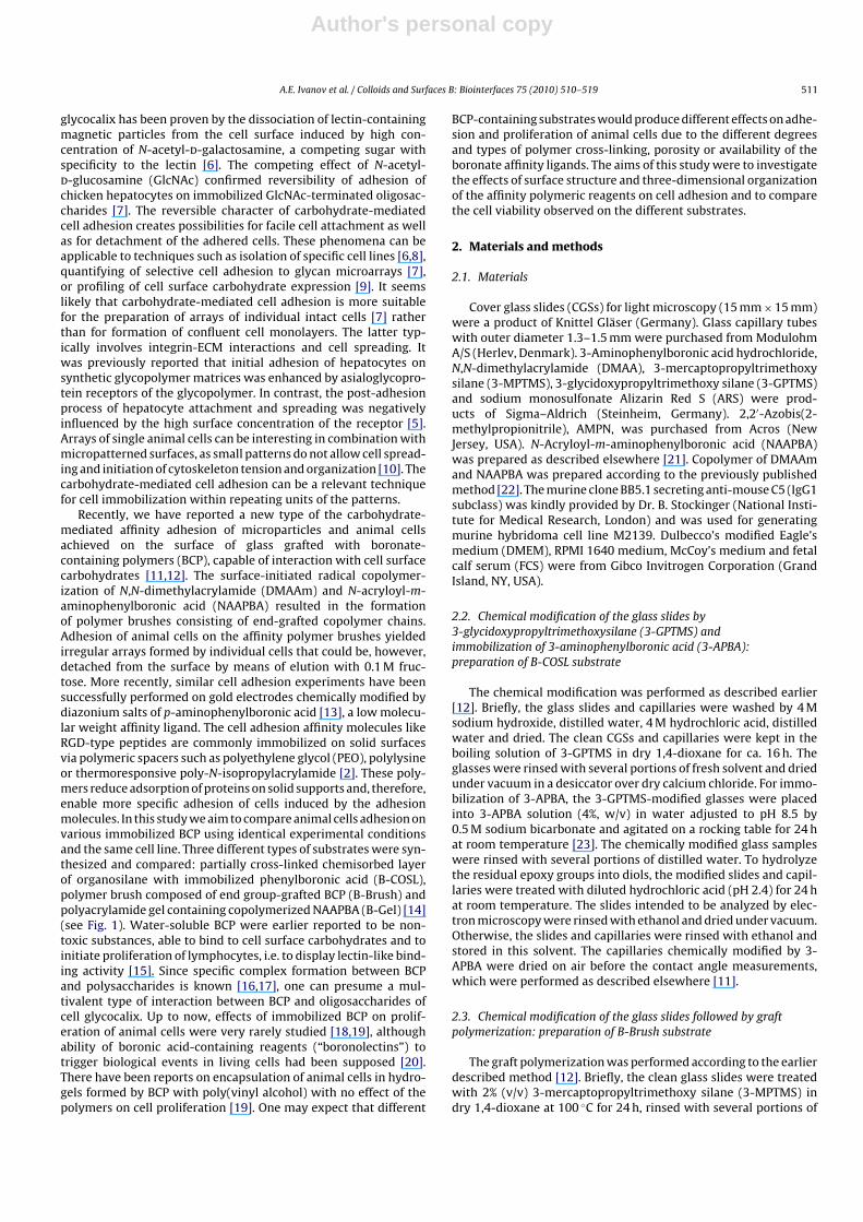
Fig. 1. Different principles of boronic acid immobilization on B-COSL (a), B-Brush (b) and B-Gel (c). The scheme (a) illustrates the structure of siloxane gel formed as resultof polymerization of 3-GPTES accompanied with its reaction with surface silanol groups. The scheme (b) illustrates the structure of polymer brush formed by end-graftedchains. The scheme (c) illustrates the structure of physically cross-linked polyacrylamide gel. The schemes are not intended for comparison of the PBA group contents in thedifferent substrates.
fresh solvent dried under vacuum and placed into the polymer-ization mixture containing NAAPBA (432 mg, 2.27 mmol), DMAA(2.07 ml, 20 mmol), AMPN (54 mg) in 20 ml ethanol. Free radicalpolymerization of the monomers was carried out for 5 h at 75 ◦Cunder bubbling nitrogen. The grafted glass plates or capillaries werewashed with ethanol overnight on a rocking table and then rinsedwith fresh portions of 1,4-dioxane and ethanol. The glass samplesintended to be analyzed by electron microscopy were dried undervacuum. The glass samples intended for cell adhesion were rinsedwith ethanol and stored in this solvent.
2.4. Synthesis of thin boronate-containing gels on the surface ofglass slides: preparation of B-Gel substrate
Free radical copolymerization of acrylamide (AAm) and NAAPBAresulting in the formation of thin gels was performed as describedpreviously [14]. Briefly, NAAPBA (48 mg, 0.25 mmol) and AAm(156 mg, 2.2 mmol) were dissolved in distilled water (2.5 ml) at50 ◦C. TEMED (10 �l) was added to the solution as a componentof the radical initiation system. The reaction mixture (200 �l) wascombined with a freshly prepared ammonium persulphate solu-tion (100 �l, 40 mg/ml) on the surface of a glass slide. The whitecopolymer gel formed immediately after mixing of the solutions.The gel-coated glass slides were kept in a desiccator over distilledwater for 2 h. After that, the gels were either transferred into 70%aqueous ethanol or stained with 4.5 mM Alizarin Red S dye in 0.1 Msodium phosphate buffer solution, pH 7.0. The excess of the dyewas removed by repeated washings with the above buffer solu-tion on a rocking table. The procedure was continued until theconstant optical density (�max = 500 nm) of the washings has beenreached. The stained gels were stored in distilled water. To elute thespecifically adsorbed ARS from the gel, the phosphate buffer solu-tion containing 0.5 M fructose was used. The elution of ARS andabsorbance measurements of the colored eluate (�max = 500 nm)were carried out until the colorless state of the gel had beenattained. The absorbance was recalculated into the amount (�mol)of the eluted dye using the calibration curve, obtained for ARS(ε500 = 4300 M−1 cm−1).
2.5. Characterization of the boronate-containing substrates byscanning electron microscopy (SEM)
SEM analysis of modified glasses was performed in a PhilipsXL20 scanning electron microscope. Samples were covered witha thin layer of palladium in order to obtain a conductive sur-face before the experiment. For B-COSL and B-Brush substratesthe photographs were registered at high magnification (×10,000and ×5000) in all cases, as lower magnifications did not revealedsignificant differences between the analyzed samples.
2.6. Characterization of the copolymer-grafted glass slides byatomic force microscopy
The samples of copolymer-grafted glasses were analyzed ina Nanoscope IIIa instrument (Digital Instruments, Santa Barbara,CA) in a tapping mode. The height, phase and amplitude imagemeasurements were performed and agreed well one another. Theexperiments were run in air at room temperature. A JV 2625 scan-ner with 125 × 125 (x,y) × 25 (z) �m scan and an etched silicon tipNanoprobe attached to a 125 �m long cantilever with a resonancefrequency in the range of 281–319 kHz and a spring constant inthe range of 20–80 N/m were used for imaging. All images wereprocessed using the Nanoscope IIIa software version 4.22 (DigitalInstruments, Santa Barbara, CA) without any filtering.
2.7. Characterization of the gel-coated glass slides (B-Gel) byconfocal microscopy
The samples of B-Gel were stained with 4.5 mM Alizarin RedS as described in Section 2.4. The stained and washed gel sampleswere examined with a Leica TCS SP5 confocal laser scanning micro-scope with excitation and emission wavelengths 488 and 530 nm,respectively.
2.8. Estimation of the boronic groups content in thecopolymer-grafted glass slides (B-Brush) by fluorescentmicroscopy
A wide-field fluorescence microscope based on a commercialOlympus IX-71 inverted microscope was used for registration offluorescence spectra and measurements of fluorescence intensitiesof the ARS–boronate complexes immobilized on a glass surface.Excitation was made using the 458 nm line of a CW Ar-ion laser.The laser beam was directed to the samples and the fluorescencelight was collected by an objective lens (Olympus LUCPlanF1 40×,NA 0.6) and projected onto a CCD camera (Photometrics, Cascade512B). Simultaneous imaging and recording of the spectra wasachieved by placing a holographic grating (150 grooves/mm, Thor-labs) in front of the CCD camera. The resolution of this spectrometerwas ca. 6 nm. The experimental design and principles of fluores-cence microscopy of thin polymer films are discussed in detailselsewhere [24].
The samples of B-Brush substrates were incubated in 0.1 mMARS in 0.1 M sodium phosphate buffer solution, pH 7.0, at roomtemperature for 1.5 h and rinsed by the buffer solution to removethe unbound dye. A 4 �l droplet of the buffer solution was appliedto the glass substrate, which was further covered by a non-modifiedcover glass to prevent evaporation. The distance between the sub-strate and the cover glass was kept at ca. 5 �m using few finely
Author's personal copy
A.E. Ivanov et al. / Colloids and Surfaces B: Biointerfaces 75 (2010) 510–519 513
dispersed particles of 5 �m-size spherical silica gel. The spectrumof the B-Brush substrate without ARS was independently registeredin a similar way and subtracted from that of the B-Brush incu-bated with ARS to produce the spectrum of ARS–PBA complexes(see Fig. 4a).
For calibration purpose, the fluorescence spectra of complexesformed between ARS and soluble DMAAm–NAAPBA copolymerstaken at known concentrations were obtained in a series of inde-pendent experiments. A 5 �m gap between two non-modifiedcover glasses was filled by the copolymer solution (10 mg/ml, ca.8 mM PBA groups) in 0.1 M sodium phosphate buffer solution, pH7.0, containing ARS at concentrations from 3 × 10⁻⁶ to 3 × 10⁻⁴ M.The fluorescence spectra obtained in this concentration range hadthe maxima at 575 nm. Owing to the large molar excess of PBA overARS and the low dissociation constant of the complex (4.4 × 10⁻⁷ M[25]), a virtually complete binding of ARS to the pendant PBA groupsof the copolymer could be expected at all the studied concentra-tions of ARS. Therefore, the concentration of ARS–PBA fluorescentcomplexes increased proportionally to the above concentrations ofARS. Correspondingly, decimal logarithm of fluorescence intensityat 575 nm exhibited a linear dependence from logarithm of the ARSconcentration (see Fig. 4b).
The calculation of surface concentration of ARS–PBA complexes(n, mol/cm2) was performed using the following equations:
Isurface = Const × Nsurface = Const × S × nmol/cm2
nmol/cm2 = Isurface
Const × S
IV0 = Const × V0 × n0mol/l = Const × d × S × n0mol/l
Const = IV0
d × S × n0mol/l
nmol/cm2 = Isurface(IV0
d × S × n0mol/l
)S
= Isurface × d × n0mol/l
IV0
= Isurface × d × 1tg˛
1tg˛
= n0mol/l
IV0
Isurface = 5.59 × 10⁵
tg˛ = 1 × 10¹¹ l/mol
d = 5 �m
n = 5.59 × 105 × 5 × 10−4 cm × 1
1011× 10−3 mol/cm3
= 2.8 × 10⁻¹² mol/cm2
where, Isurface—fluorescence intensity of ARS–PBA complex on theB-Brush substrate; Nsurface—total amount of fluorescent ARS–PBAcomplexes; d—distance between the glass slides; S—surfacearea of the substrate; Const—fluorescent constant of ARS–PBAcomplex; IV0 —fluorescence intensity of DMAAm–NAAPBA/ARScomplex in solution; V0—volume of calibration solution (4 �l);n0—concentration of fluorescent complex in calibration solution(mol/l); tg˛—tangent of calibration slope.
2.9. Maintenance of cell cultures and sedimentation of cells onthe copolymer-grafted glass plates
Murine hybridoma cell line M2139 were maintained in Dul-becco’s modified Eagle’s medium (DMEM) containing l-glutamineand pyridoxine chloride, but no glucose and sodium pyruvate(Gibco, Invitrogen, catalog number 11966025). The medium wassupplemented by 10% (v/v) fetal bovine serum (FCS) and 0.1%
kanamycin monosulfate. The culture was maintained in T-flasksincubated with 7.5% CO2 at 37 ◦C. The glass slides containing immo-bilized boronic acid were washed with 10 mM phosphate buffersolution, pH 8.0, for 30 min in a sterile Petri dish. The cell line M2139suspended in the above culture medium with pH adjusted to 8.0(106 cells/ml, 15 ml) was seeded on the slides. Cells were allowedto sediment on the horizontally situated glass slides for 30 min at4 ◦C. After that, the slides were transferred to separate Petri disheseach containing 3 ml fresh DMEM with pH 7.2 (the normal value).The slides with adhered cells were incubated at 37 ◦C in 7.5% carbondioxide environment. Experiment was run for 5 days, the mediumbeing daily replaced with fresh one. The micrographs of the cul-tured cells were taken by means of a stereomicroscope (Nikon,Eclipse, NY). Detachment of cells from the boronate-containingsubstrates was performed by dropping 0.1 M fructose solution onthe slides with adhered cells, the slides were leaned against thewall of a Petri dish containing 3 ml fresh culture medium.
2.10. MTT assay
In order to determine the viability and proliferation of cellscultured on the modified glass surfaces, 3-(4,5-dimethylthiazol-2-yl)-2,5-diphenyltetrazolium bromide (MTT) assay was carriedout [26]. It is an indirect method for assessing of cell growth andproliferation, since mitochondria oxidize the MTT solution, givinga typical blue-violet end product quantified by spectrophotome-try. M2139 cells were incubated on the modified glass slides asdescribed above, for the purpose of checking the cell proliferationand cytotoxicity effects of the material on the cells. The culturemedium and specimens were removed after allowing the cells togrow for appropriate time period, and cell viability was tested byMTT. The serum free culture medium containing MTT (0.5 mg/ml)were added in each sample. Cells were cultured for 4 h and thenthe medium containing MTT was removed. After that, isopropanol(1.5 ml) was added into each sample to dissolve the formazan crys-tals formed by the living cells and incubated for 15 min. The solutionfrom the samples was removed and the absorbance at 490 nm wasmeasured. All the above experiments were performed in triplicate.
3. Results and discussion
3.1. Characterization of the boronate-containing substrates
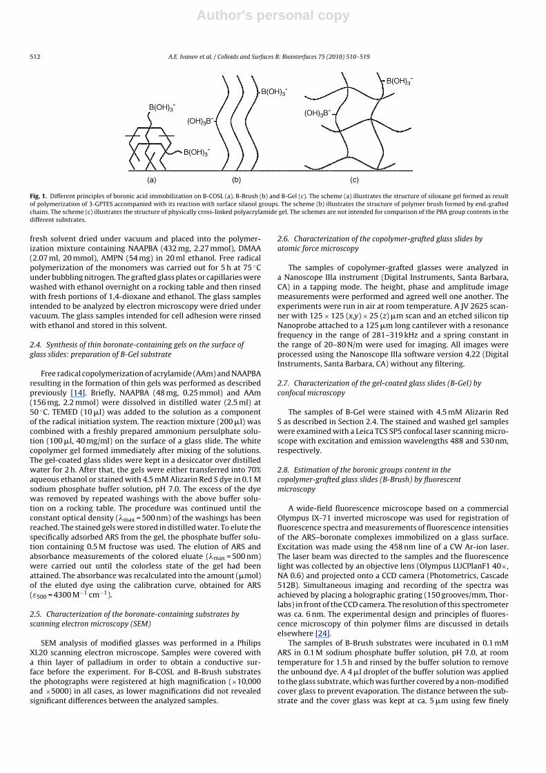
The substrates prepared by immobilization of 3-APBA onchemisorbed organosilane layers containing epoxy groups (B-COSL), by graft copolymerization of NAAPBA (B-Brush) and by theformation of NAAPBA-containing thin gels (B-Gel) were charac-terized by scanning electron microscopy (SEM) (see Fig. 2). TheSEM images of the cleaned glass slides (Fig. 2a) contained a smallnumber of pores of submicron size, possibly formed as result oftreatment with strong alkali. The image of B-COSL contained manynon-homogenously distributed submicron structures of ca. 200 nmand smaller in size, besides the rare pores typical of the parent glass(Fig. 2b). The true monolayers of low molecular weight organosi-lanes can be prepared by the chemical modification carried outin gaseous phase. These chemisorbed layers typically appear onSEM images as highly uniform structures [27]. In liquid phase, par-tial hydrolysis and polymerization of reactive organosilanes occursin parallel to the chemical modification of silanol groups. Thisresults in the formation of submicron crystalline or amorphousdomains of polysiloxane gels exhibiting irregular patterns [27].The chemisorbed layers of 3-glycidoxypropyltrimethoxysilane pre-pared in the present study were of a similar irregular structure(Fig. 2b). The epoxy groups attached to the submicron gels wereused for covalent immobilization of 3-APBA via its amino group, as
Author's personal copy
514 A.E. Ivanov et al. / Colloids and Surfaces B: Biointerfaces 75 (2010) 510–519
Fig. 2. Scanning electron microscopy images of the parent glass slides (a), B-COSL (b), B-Brush (c), B-Gel (the middle of the gel field) (d).
described in Section 2.2. The presence of phenylboronate functionson the glass surface was qualitatively confirmed by the capil-lary rise measurements: due to the specific complex formation ofphenylboronates with fructose, the contact angle between the B-COSL-capillaries and 0.1 M fructose in sodium bicarbonate buffersolution, pH 9.0 was clearly lower (30 ± 4◦) than that between themodified glass and the buffer solution (43 ± 4◦). Gradual dropletspreading accompanied with a decrease in contact angle of fruc-tose solutions is inherent property of boronate-containing solidsurfaces, which was reported by us earlier [11,12].
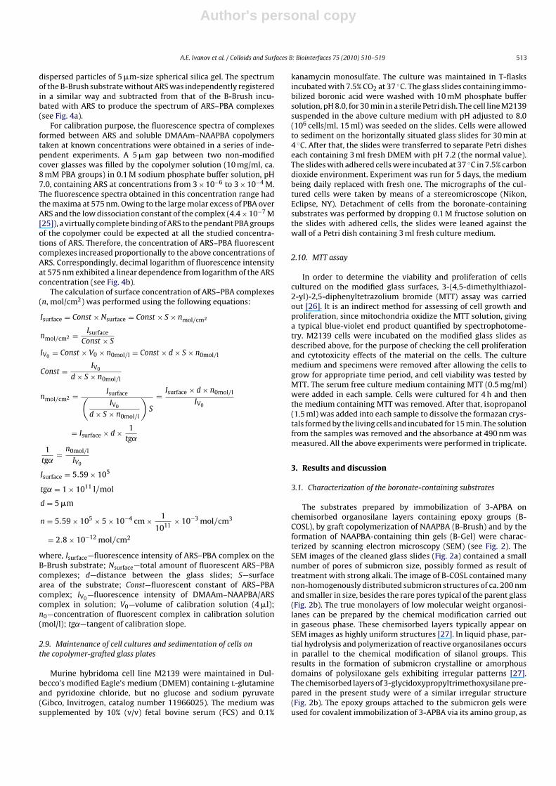
The substrates prepared by graft copolymerization of NAAPBAand DMAAm (B-Brush) exhibited another kind of nanoscopic pat-tern consisting of many dots, typically of ca. 100 nm in size or lower(Fig. 2c). This pattern was in good agreement with the image inde-pendently obtained by atomic force microscopy (AFM) for a samplefrom the same polymerization batch (see Fig. 3) As follows from theimage, the dots observed by AFM were outward extremities withtypical heights up to 80 nm and average diameters within 200 nm.The NAAPBA-containing polymer brushes have been earlier char-acterized by time-of-flight secondary ion mass-spectrometry (ToFSIMS) that proved both the presence of boron and monomericfragments of DMAAm in the polymer brush [13]. The B-Brush sub-strates were also characterized by the contact angles obtained inthe absence and in the presence of 0.1 M fructose in the depositeddroplets of aqueous buffer solutions. The contact angles exhibiteda quick decrease from ca. 50◦ to 30–35◦ in the presence of fruc-tose, in contrast to the more stable contact angles obtained withthe sugar-free solutions [12]. This gave additional evidence for thepresence of reactive phenylboronic acid (PBA) groups on the sub-strate. Quantitative estimation of the PBA groups is difficult becauseof their very low content on the surface. Characterization of verythin (20 nm and lower) polymeric films is possible, however, by flu- Fig. 3. Height AFM image of B-Brush (a) and its section analysis (b).
Author's personal copy
A.E. Ivanov et al. / Colloids and Surfaces B: Biointerfaces 75 (2010) 510–519 515
orescence spectroscopy [24,28]. We attempted to quantify the PBAgroups by measuring intensity of fluorescence exhibited by affin-ity complexes formed by PBA groups of the B-Brush substrate withAlizarin Red S dye. Unlike the dye itself, its complexes with boronicacid exhibit a strong fluorescence [29] and may allow for their reg-istration at low surface concentrations. The fluorescence spectrumof the B-Brush substrate was obtained and subtracted from thatof the B-Brush substrate kept in contact with 0.1 mM solution ofARS to produce the spectrum of ARS–PBA complexes. The resul-tant intensity of fluorescence at �max = 575 nm was recorded (seeFig. 4a). For calibration purpose, the measurements of fluorescenceintensity as a function of ARS concentration varied from 3 × 10⁻⁶ to3 × 10−4 M in a thin layer (5 �m) of solution of DMAAm–NAAPBAcopolymer (10 mg/ml) were performed (for details see Section 2.8).The calibration graph is illustrated in Fig. 4b. The surface concen-tration of fluorescent complexes was found to be 2.8 × 10⁻⁶ �molARS–PBA complexes/cm2.
Fig. 4. (a) Fluorescence spectra of the B-Brush substrate (©), B-Brush substrateincubated in 0.1 mM ARS solution (�), and the difference spectrum attributed tothe ARS–PBA complexes formed by the graft DMAAm–NAAPBA copolymer (- -). (b)Calibration graph obtained with DMAAm–NAAPBA copolymer and ARS (♦) and thefluorescence intensity registered at 575 nm with B-Brush substrate incubated in0.1 mM ARS solution (�).
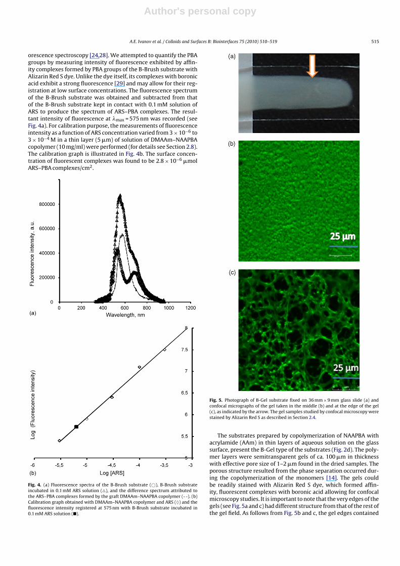
Fig. 5. Photograph of B-Gel substrate fixed on 36 mm × 9 mm glass slide (a) andconfocal micrographs of the gel taken in the middle (b) and at the edge of the gel(c), as indicated by the arrow. The gel samples studied by confocal microscopy werestained by Alizarin Red S as described in Section 2.4.
The substrates prepared by copolymerization of NAAPBA withacrylamide (AAm) in thin layers of aqueous solution on the glasssurface, present the B-Gel type of the substrates (Fig. 2d). The poly-mer layers were semitransparent gels of ca. 100 �m in thicknesswith effective pore size of 1–2 �m found in the dried samples. Theporous structure resulted from the phase separation occurred dur-ing the copolymerization of the monomers [14]. The gels couldbe readily stained with Alizarin Red S dye, which formed affin-ity, fluorescent complexes with boronic acid allowing for confocalmicroscopy studies. It is important to note that the very edges of thegels (see Fig. 5a and c) had different structure from that of the rest ofthe gel field. As follows from Fig. 5b and c, the gel edges contained
Author's personal copy
516 A.E. Ivanov et al. / Colloids and Surfaces B: Biointerfaces 75 (2010) 510–519
much larger pores (5–20 �m in diameter) compared to the otherparts of the gel. The dye could be desorbed from the gel by 0.1 Mfructose, a strong competitor of the affinity binding to boronates.Specific adsorption capacity of the gels estimated from the quantityof the dye adsorbed at its saturating concentration of 4.5 mM wasfound to be ca. 160 �mol/ml gel (or 1.6 �mol/cm2 for the 0.1 mmthick gel), which coincided well with the amount of copolymerizedNAAPBA [14].
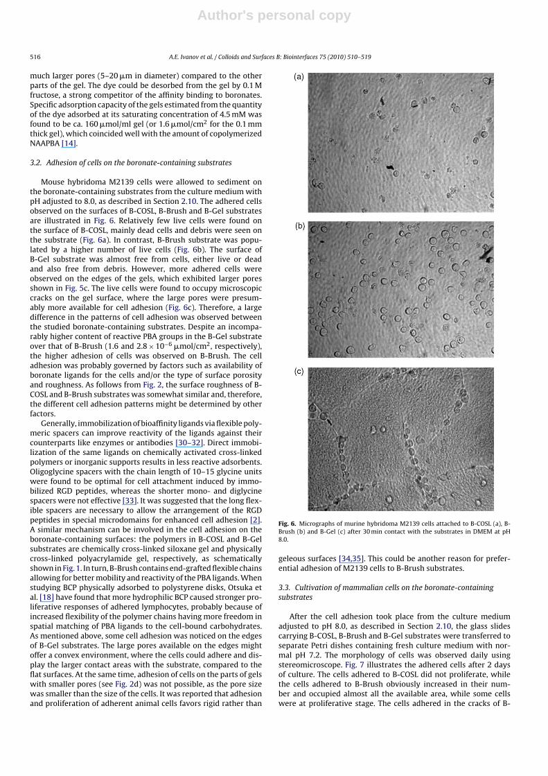
3.2. Adhesion of cells on the boronate-containing substrates
Mouse hybridoma M2139 cells were allowed to sediment onthe boronate-containing substrates from the culture medium withpH adjusted to 8.0, as described in Section 2.10. The adhered cellsobserved on the surfaces of B-COSL, B-Brush and B-Gel substratesare illustrated in Fig. 6. Relatively few live cells were found onthe surface of B-COSL, mainly dead cells and debris were seen onthe substrate (Fig. 6a). In contrast, B-Brush substrate was popu-lated by a higher number of live cells (Fig. 6b). The surface ofB-Gel substrate was almost free from cells, either live or deadand also free from debris. However, more adhered cells wereobserved on the edges of the gels, which exhibited larger poresshown in Fig. 5c. The live cells were found to occupy microscopiccracks on the gel surface, where the large pores were presum-ably more available for cell adhesion (Fig. 6c). Therefore, a largedifference in the patterns of cell adhesion was observed betweenthe studied boronate-containing substrates. Despite an incompa-rably higher content of reactive PBA groups in the B-Gel substrateover that of B-Brush (1.6 and 2.8 × 10⁻⁶ �mol/cm2, respectively),the higher adhesion of cells was observed on B-Brush. The celladhesion was probably governed by factors such as availability ofboronate ligands for the cells and/or the type of surface porosityand roughness. As follows from Fig. 2, the surface roughness of B-COSL and B-Brush substrates was somewhat similar and, therefore,the different cell adhesion patterns might be determined by otherfactors.
Generally, immobilization of bioaffinity ligands via flexible poly-meric spacers can improve reactivity of the ligands against theircounterparts like enzymes or antibodies [30–32]. Direct immobi-lization of the same ligands on chemically activated cross-linkedpolymers or inorganic supports results in less reactive adsorbents.Oligoglycine spacers with the chain length of 10–15 glycine unitswere found to be optimal for cell attachment induced by immo-bilized RGD peptides, whereas the shorter mono- and diglycinespacers were not effective [33]. It was suggested that the long flex-ible spacers are necessary to allow the arrangement of the RGDpeptides in special microdomains for enhanced cell adhesion [2].A similar mechanism can be involved in the cell adhesion on theboronate-containing surfaces: the polymers in B-COSL and B-Gelsubstrates are chemically cross-linked siloxane gel and physicallycross-linked polyacrylamide gel, respectively, as schematicallyshown in Fig. 1. In turn, B-Brush contains end-grafted flexible chainsallowing for better mobility and reactivity of the PBA ligands. Whenstudying BCP physically adsorbed to polystyrene disks, Otsuka etal. [18] have found that more hydrophilic BCP caused stronger pro-liferative responses of adhered lymphocytes, probably because ofincreased flexibility of the polymer chains having more freedom inspatial matching of PBA ligands to the cell-bound carbohydrates.As mentioned above, some cell adhesion was noticed on the edgesof B-Gel substrates. The large pores available on the edges mightoffer a convex environment, where the cells could adhere and dis-play the larger contact areas with the substrate, compared to theflat surfaces. At the same time, adhesion of cells on the parts of gelswith smaller pores (see Fig. 2d) was not possible, as the pore sizewas smaller than the size of the cells. It was reported that adhesionand proliferation of adherent animal cells favors rigid rather than
Fig. 6. Micrographs of murine hybridoma M2139 cells attached to B-COSL (a), B-Brush (b) and B-Gel (c) after 30 min contact with the substrates in DMEM at pH8.0.
geleous surfaces [34,35]. This could be another reason for prefer-ential adhesion of M2139 cells to B-Brush substrates.
3.3. Cultivation of mammalian cells on the boronate-containingsubstrates
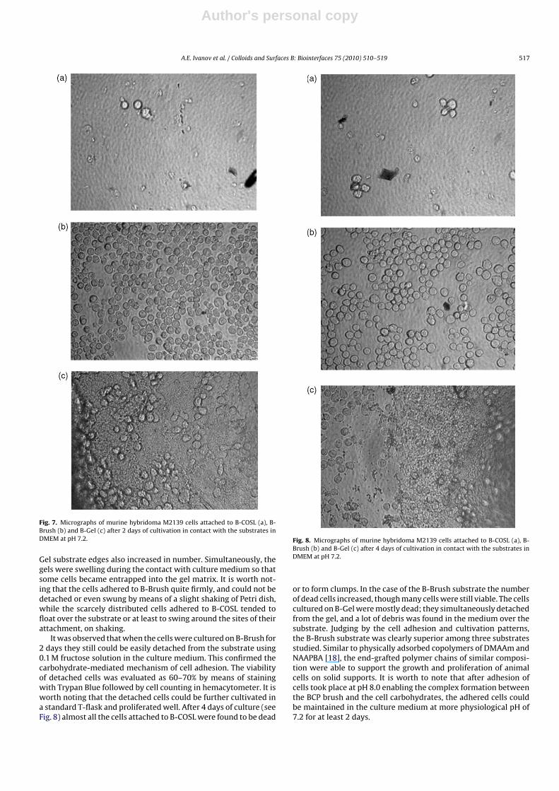
After the cell adhesion took place from the culture mediumadjusted to pH 8.0, as described in Section 2.10, the glass slidescarrying B-COSL, B-Brush and B-Gel substrates were transferred toseparate Petri dishes containing fresh culture medium with nor-mal pH 7.2. The morphology of cells was observed daily usingstereomicroscope. Fig. 7 illustrates the adhered cells after 2 daysof culture. The cells adhered to B-COSL did not proliferate, whilethe cells adhered to B-Brush obviously increased in their num-ber and occupied almost all the available area, while some cellswere at proliferative stage. The cells adhered in the cracks of B-
Author's personal copy
A.E. Ivanov et al. / Colloids and Surfaces B: Biointerfaces 75 (2010) 510–519 517
Fig. 7. Micrographs of murine hybridoma M2139 cells attached to B-COSL (a), B-Brush (b) and B-Gel (c) after 2 days of cultivation in contact with the substrates inDMEM at pH 7.2.
Gel substrate edges also increased in number. Simultaneously, thegels were swelling during the contact with culture medium so thatsome cells became entrapped into the gel matrix. It is worth not-ing that the cells adhered to B-Brush quite firmly, and could not bedetached or even swung by means of a slight shaking of Petri dish,while the scarcely distributed cells adhered to B-COSL tended tofloat over the substrate or at least to swing around the sites of theirattachment, on shaking.
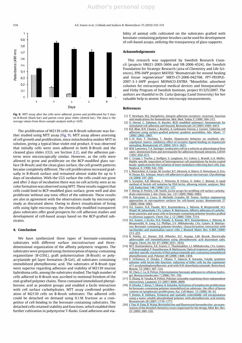
It was observed that when the cells were cultured on B-Brush for2 days they still could be easily detached from the substrate using0.1 M fructose solution in the culture medium. This confirmed thecarbohydrate-mediated mechanism of cell adhesion. The viabilityof detached cells was evaluated as 60–70% by means of stainingwith Trypan Blue followed by cell counting in hemacytometer. It isworth noting that the detached cells could be further cultivated ina standard T-flask and proliferated well. After 4 days of culture (seeFig. 8) almost all the cells attached to B-COSL were found to be dead
Fig. 8. Micrographs of murine hybridoma M2139 cells attached to B-COSL (a), B-Brush (b) and B-Gel (c) after 4 days of cultivation in contact with the substrates inDMEM at pH 7.2.
or to form clumps. In the case of the B-Brush substrate the numberof dead cells increased, though many cells were still viable. The cellscultured on B-Gel were mostly dead; they simultaneously detachedfrom the gel, and a lot of debris was found in the medium over thesubstrate. Judging by the cell adhesion and cultivation patterns,the B-Brush substrate was clearly superior among three substratesstudied. Similar to physically adsorbed copolymers of DMAAm andNAAPBA [18], the end-grafted polymer chains of similar composi-tion were able to support the growth and proliferation of animalcells on solid supports. It is worth to note that after adhesion ofcells took place at pH 8.0 enabling the complex formation betweenthe BCP brush and the cell carbohydrates, the adhered cells couldbe maintained in the culture medium at more physiological pH of7.2 for at least 2 days.
Author's personal copy
518 A.E. Ivanov et al. / Colloids and Surfaces B: Biointerfaces 75 (2010) 510–519
Fig. 9. MTT assay after the cells were adhered, grown and proliferated for 5 dayson B-Brush (black bar) and parent cover glass slides (dotted bar). The data is theaverage values from three-sample analysis with p < 0.05.
The proliferation of M2139 cells on B-Brush substrate was fur-ther studied using MTT assay (Fig. 9). MTT assay allows assessingof cell growth and proliferation, since mitochondria oxidize MTT insolution, giving a typical blue-violet end product. It was observedthat initially cells were seen adhered to both B-Brush and thecleaned glass slides (CGS, see Section 2.2), and the adhesion pat-terns were microscopically similar. However, as the cells wereallowed to grow and proliferate on the BCP-modified glass sur-face (B-Brush) and the clean glass surface, the cell growth patternsbecame completely different. The cell proliferation increased grad-ually in B-Brush surface and remained almost stable for up to 5days of incubation. With the CGS surface the cells could not growand after 2 days of incubation there was no cell activity seen as nocolor formation was observed using MTT. These results suggest thatcells could bind to BCP-modified glass surface, grow well and alsoproliferate without any toxic effect of the substrate. These resultsare also in agreement with the observations made by microscopicstudy as discussed above. Owing to direct visualization of livingcells using light microscopy, the chemically modified transparentglass substrates offer good prospects for cell adhesion studies anddevelopment of cell-based assays based on the BCP-grafted sub-strates.
4. Conclusion
We have synthesized three types of boronate-containingsubstrates with different surface microstructure and three-dimensional organization of the affinity polymeric reagents. Thesubstrates were prepared using chemisorption of epoxy-containingorganosilane (B-COSL), graft polymerization (B-Brush) or poly-acrylamide gel layer formation (B-Gel), all substrates containingimmobilized phenylboronic acid. The substrates of B-Brush typewere superior regarding adhesion and viability of M2139 murinehybridoma cells, among the substrates studied. The high number ofcells adhered to B-Brush was ascribed to motional freedom of theend-grafted polymer chains. These contained immobilized phenyl-boronic acid as pendent groups and enabled a facile interactionwith cell surface carbohydrates. MTT assay confirmed prolifer-ation of M2139 cells on B-Brush substrates. The adhered cellscould be detached on demand using 0.1 M fructose as a com-petitor of cell binding to the boronate-containing substrates. Thedetached cells retained viability and functions, which enabled theirfurther cultivation in polystyrene T-flasks. Good adhesion and via-
bility of animal cells cultivated on the substrates grafted withboronate-containing polymer brushes can be used for developmentof cell-based assays, utilizing the transparency of glass supports.
Acknowledgements
This research was supported by Swedish Research Coun-cil (projects VR621-2005-3604 and VR-2006-4524), the SwedishFoundation for Strategic Research (area of Chemistry and Life Sci-ences), FP6-IAPP project MATISS “Biomaterials for wound healingand tissue regeneration” MKTI-CT-2006-042768, FP7-PEOPLE-2007-3-1-IAPP project MONACO-EXTRA “Monolithic adsorbentcolumns for extracorporeal medical devices and bioseparations”and Visby Program of Swedish Institute, project 01325/2007. Theauthors are thankful to Dr. Carla Quiroga (Lund University) for hervaluable help in atomic force microscopy measurements.
References
[1] P. Newham, M.J. Humphries, Integrin adhesion receptors: structure, functionand implications for biomedicine, Mol. Med. Today 2 (1996) 304–313.
[2] U. Hersel, C. Dahmen, H. Kessler, RGD modified polymers: biomaterials forstimulated cell adhesion and beyond, Biomaterials 24 (2003) 4385–4415.
[3] R.R. Bhat, B.N. Chaney, J. Rowley, A. Liebmann-Vinson, J. Genzer, Tailoring celladhesion using surface-grafted polymer gradient assemblies, Adv. Mater. 17(2005) 2802–2807.
[4] S.-H. Kim, T. Hoshiba, T. Akaike, Hepatocyte behavior on synthetic gly-copolymer matrix: inhibitory effect of receptor-ligand binding on hepatocytespreading, Biomaterials 25 (2004) 1813–1823.
[5] M.B. Lawrence, T.A. Springer, Leukocytes roll on a selectin at physiological flowrates: distinction from and prerequisite for adhesion through integrins, Cell 65(1991) 859–873.
[6] C. Grupp, I. Troche, J. Steffgen, S. Langhans, D.I. Cohen, L. Brandl, G.A. Müller,Highly specific separation of heterogeneous cell populations by lectin-coatedbeads: application for the isolation of inner medullary collecting duct cells, Exp.Nephrol. 6 (1998) 542–550.
[7] L. Nimrichter, A. Gargir, M. Gortler, R.T. Altstock, A. Shtevi, O. Weisshaus, E. Fire,N. Dotan, R.L. Schnaar, Intact cell adhesion to glycan microarrays, Glycobiology14 (2004) 197–203.
[8] S. Scarpino, A.R. Morena, C. Petersen, B. Fröysa, O. Söder, C. Boitani, A rapidmethod of Sertoli cell isolation by DSA lectin, allowing mitotic analyses, Mol.Cell. Endocrinol. 146 (1998) 121–127.
[9] T. Zheng, D. Peelen, L.M. Smith, Lectin arrays for profiling cell surface carbohy-drate expression, J. Am. Chem. Soc. 127 (2005) 9982–9983.
[10] D. Falconnet, G. Csucs, H. Michelle Grandin, M. Textor, Surface engineeringapproaches to micropattern surfaces for cell-based assays, Biomaterials 27(2006) 3044–3063.
[11] A.E. Ivanov, H.A. Panahi, M.V. Kuzimenkova, L. Nilsson, B. Bergenståhl, H.S.Waqif, M. Jahanshahi, I.Yu. Galaev, B. Mattiasson, Affinity adhesion of carbohy-drate particles and yeast cells to boronate-containing polymer brushes graftedto siliceous supports, Chem. Eur. J. 12 (2006) 7204–7214.
[12] A.E. Ivanov, J. Eccles, H.A. Panahi, A. Kumar, M.V. Kuzimenkova, L. Nilsson, B.Bergenståhl, N. Long, G.J. Phillips, S.V. Mikhalovsky, I.Yu. Galaev, B. Mattias-son, Boronate-containing polymer brushes: characterization, interaction withsaccharides and mammalian cancer cells, J. Biomed. Mater. Res. A 88A (2009)213–225.
[13] R. Polsky, J.C. Harper, D.R. Wheeler, D.C. Arango, S.M. Brozik, Electricallyaddressable cell immobilization using phenylboronic acid diazonium salts,Angew. Chem. Int. Ed. 47 (2008) 2631–2634.
[14] M.V. Kuzimenkova, A.E. Ivanov, C. Thammakhet, L.I. Mikhalovska, I.Yu. Galaev,P. Thavarungkul, P. Kanatharana, B. Mattiasson, Optical responses, permeabilityand diol-specific reactivity of thin polyacrylamide gels containing immobilizedphenylboronic acid, Polymer 49 (2008) 1444–1454.
[15] E. Uchimura, H. Otsuka, T. Okano, Y. Sakurai, K. Kataoka, Totally syntheticpolymer with lectin-like function: induction of killer cells by the copolymerof 3-acrylamidophenylboronic acid with N,N-dimethylacrylamide, Biotechnol.Bioeng. 72 (2001) 307–314.
[16] W. Chen, C. Lu, R. Pelton, Polyvinylamine boronate adhesion to cellulose hydro-gel, Biomacromolecules 7 (2006) 701–702.
[17] D. Zhang, H. Tanaka, R. Pelton, Polymer assembly exploiting three independentinteractions, Langmuir 23 (2007) 8806–8809.
[18] H. Otsuka, T. Ikeya, T. Okano, K. Kataoka, Activation of lymphocyte proliferationby boronate-containing polymer immobilized on substrate: the effect of boroncontent on lymphocyte proliferation, Eur. Cell Mater. 12 (2006) 36–43.
[19] T. Konno, K. Ishihara, Temporal and spatially controllable cell encapsulationusing a water-soluble phospholipid polymer with phenylboronic acid moiety,Biomaterials 28 (2007) 1770–1777.
[20] J. Yan, H. Fang, B. Wang, Boronolectins and fluorescent boronolectins: an exam-ination of the detailed chemistry issues important for the design, Med. Res. Rev.25 (2005) 490–520.
Author's personal copy
A.E. Ivanov et al. / Colloids and Surfaces B: Biointerfaces 75 (2010) 510–519 519
[21] A.E. Ivanov, H. Larsson, I.Yu. Galaev, B. Mattiasson, Synthesis of boronate-containing copolymers of N,N-dimethylacrylamide their interaction withpoly(vinyl alcohol) and rheological behaviour of the gels, Polymer 45 (2004)2495–2505.
[22] M.V. Kuzimenkova, A.E. Ivanov, I.Yu. Galaev, Boronate-containing copolymers:polyelectrolyte properties and sugar-specific interaction with agarose gel,Macromol. Biosci. 6 (2006) 170–178.
[23] M. Glad, S. Ohlson, L. Hansson, M. Mansson, K. Mosbach, High-performanceliquid affinity chromatography of nucleosides, nucleotides and carbohydrateswith boronic acid-substituted microparticulate silica, J. Chromatogr. 200 (1980)245–260.
[24] O. Mirzov, I.G. Scheblykin, Photoluminescence spectra of a conjugated polymer:from films and solutions to single molecules, Phys. Chem. Chem. Phys. 8 (2006)5569–5576.
[25] S. Hajizadeh, A.E. Ivanov, M. Jahanshahi, M.H. Sanati, N.V. Zhuravleva, L.I.Mikhalovska, I.Yu. Galaev, Glucose sensors with increased sensitivity based oncomposite gels containing immobilized boronic acid, React. Funct. Polym. 68(2008) 1625–1634.
[26] T. Mosmann, Rapid colorimetric assay for cellular growth and survival: appli-cation to proliferation and cytotoxicity assays, J. Immunol. Methods 65 (1983)55–63.
[27] T. Koga, M. Morit, H. Ishida, H. Yakabe, S. Sasaki, O. Sakami, H. Otsuka, A.Takahara, Dependence of the molecular aggregation state of octadecylsiloxanemonolayers on preparation methods, Langmuir 21 (2005) 905–910.
[28] H. Lin, S.R. Tabaei, D. Thompsson, O. Mirzov, P.-O. Larsson, I.G. Scheblykin,Fluorescence blinking, exciton dynamics, and energy transfer domains insingle conjugated polymer chains, J. Am. Chem. Soc. 190 (2008) 7042–7051.
[29] G. Springsteen, B. Wang, A detailed examination of boronic acid-diol complex-ation, Tetrahedron 58 (2002) 5291–5300.
[30] Y. Byun, H.A. Jacobs, S.W. Kim, Heparin surface immobilization throughhydrophilic spacers–thrombin and antithrombin binding kinetics, J. Biomater.Sci. Polym. Ed. 6 (1994) 1–13.
[31] A.E. Ivanov, N.V. Bovin, E.Y. Korchagina, V.P. Zubov, Favourable biospecificreactivity of blood group B antigenic trisaccharides chemically attached topoly-N(2-hydroxyethyl)acrylamide-coated porous glass, Biomed. Chromatogr.6 (1992) 39–42.
[32] A.E. Ivanov, L.V. Kozlov, B.B. Shojbonov, V.P. Zubov, V.K. Antonov, Inorganicsupports coated with N-substituted polyacrylamides: application to biospecificchromatography of proteins, Biomed. Chromatogr. 5 (1991) 90–93.
[33] J.H. Beer, K.T. Springer, B.S. Coller, Immobilized Arg-Gly-Asp (RGD) peptides ofvarying length as structural probes of the platelet glycoprotein-IIB/IIIA recep-tor, Blood 79 (1992) 117–128.
[34] D.L. Elbert, J.A. Hubbell, Conjugate addition reactions combined with free-radical cross-linking for the design of materials for tissue engineering,Biomacromolecules 2 (2001) 430–441.
[35] C.M. Lo, H.B. Wang, M. Dembo, Y.I. Wang, Cell movement is guided by therigidity of the substrate, Biophys. J. 79 (2000) 144–152.
Copyright © 2022 FDOKUMEN