Comprehensive Leaf Cell Wall Analysis Using Carbohydrate ...
16
polymers Article Comprehensive Leaf Cell Wall Analysis Using Carbohydrate Microarrays Reveals Polysaccharide-Level Variation between Vitis Species with Differing Resistance to Downy Mildew Yu Gao 1 , Xiangjing Yin 2,3 , Haoyu Jiang 1 , Jeanett Hansen 4 , Bodil Jørgensen 4 , John P. Moore 5 , Peining Fu 1 , Wei Wu 1 , Bohan Yang 1 , Wenxiu Ye 1 , Shiren Song 1 and Jiang Lu 1, * Citation: Gao, Y.; Yin, X.; Jiang, H.; Hansen, J.; Jørgensen, B.; Moore, J.P.; Fu, P.; Wu, W.; Yang, B.; Ye, W.; et al. Comprehensive Leaf Cell Wall Analysis Using Carbohydrate Microarrays Reveals Polysaccharide- Level Variation between Vitis Species with Differing Resistance to Downy Mildew. Polymers 2021, 13, 1379. https://doi.org/10.3390/ polym13091379 Academic Editors: Emiliano Bedini and Serena Traboni Received: 27 February 2021 Accepted: 21 April 2021 Published: 23 April 2021 Publisher’s Note: MDPI stays neutral with regard to jurisdictional claims in published maps and institutional affil- iations. Copyright: © 2021 by the authors. Licensee MDPI, Basel, Switzerland. This article is an open access article distributed under the terms and conditions of the Creative Commons Attribution (CC BY) license (https:// creativecommons.org/licenses/by/ 4.0/). 1 Center for Viticulture and Enology, Department of Plant Science, School of Agriculture and Biology, Shanghai Jiao Tong University, Shanghai 200240, China; [email protected] (Y.G.); [email protected] (H.J.); [email protected] (P.F.); [email protected] (W.W.); [email protected] (B.Y.); [email protected] (W.Y.); [email protected] (S.S.) 2 Forestry and Pomology Research Institute, Shanghai Academy of Agricultural Sciences, Shanghai 201403, China; [email protected] 3 Shanghai Key Lab of Protected Horticultural Technology, Shanghai Academy of Agricultural Sciences, Shanghai 201403, China 4 Department of Plant and Environmental Sciences, Faculty of Sciences, University of Copenhagen, 1871 Frederiksberg C, Denmark; [email protected] (J.H.); [email protected] (B.J.) 5 Department of Viticulture and Oenology, South African Grape and Wine Research Institute, Stellenbosch University, Stellenbosch 7602, South Africa; [email protected] * Correspondence: [email protected] Abstract: The cell wall acts as one of the first barriers of the plant against various biotic stressors. Previous studies have shown that alterations in wall polysaccharides may influence crop disease resistance. In the grapevine family, several native species (e.g., Chinese wild grapevine) show a naturally higher resistance to microbial pathogens than cultivated species (e.g., Vitis vinifera), and this trait could be inherited through breeding. Despite the importance of the cell wall in plant immunity, there are currently no comprehensive cell wall profiles of grapevine leaves displaying differing resistance phenotypes, due to the complex nature of the cell wall and the limitations of analytical techniques available. In this study, the cutting-edge comprehensive carbohydrate microar- ray technology was applied to profile uninfected leaves of the susceptible cultivar (Vitis vinifera cv. “Cabernet Sauvignon”), a resistant cultivar (Vitis amurensis cv. “Shuanghong”) and a hybrid offspring cross displaying moderate resistance. The microarray approach uses monoclonal antibodies, which recognize polysaccharides epitopes, and found that epitope abundances of highly esterified homogalacturonan (HG), xyloglucan (with XXXG motif), (galacto)(gluco)mannan and arabinogalac- tan protein (AGP) appeared to be positively correlated with the high resistance of Vitis amurensis cv. “Shuanghong” to mildew. The quantification work by gas chromatography did not reveal any significant differences for the monosaccharide constituents, suggesting that polysaccharide structural alterations may contribute more crucially to the resistance observed; this is again supported by the contact infrared spectroscopy of cell wall residues, revealing chemical functional group changes (e.g., esterification of pectin). The identification of certain wall polysaccharides that showed alterations could be further correlated with resistance to mildew. Data from the use of the hybrid material in this study have preliminarily suggested that these traits could be inherited and may be applied as potential structural biomarkers in future breeding work. Keywords: cell wall; grapevine; oomycete; polysaccharide microarray; monoclonal antibodies 1. Introduction The domesticated grapevine (Vitis vinifera) is susceptible to many pathogens, which may lead to huge losses in production. Unlike the vine trunk, which contains a higher per- Polymers 2021, 13, 1379. https://doi.org/10.3390/polym13091379 https://www.mdpi.com/journal/polymers
-
Upload
khangminh22 -
Category
Documents
-
view
0 -
download
0
Transcript of Comprehensive Leaf Cell Wall Analysis Using Carbohydrate ...

polymers
Article
Comprehensive Leaf Cell Wall Analysis Using CarbohydrateMicroarrays Reveals Polysaccharide-Level Variation betweenVitis Species with Differing Resistance to Downy Mildew
Yu Gao 1 , Xiangjing Yin 2,3, Haoyu Jiang 1, Jeanett Hansen 4, Bodil Jørgensen 4, John P. Moore 5 , Peining Fu 1,Wei Wu 1, Bohan Yang 1, Wenxiu Ye 1, Shiren Song 1 and Jiang Lu 1,*
�����������������
Citation: Gao, Y.; Yin, X.; Jiang, H.;
Hansen, J.; Jørgensen, B.; Moore, J.P.;
Fu, P.; Wu, W.; Yang, B.; Ye, W.; et al.
Comprehensive Leaf Cell Wall
Analysis Using Carbohydrate
Microarrays Reveals Polysaccharide-
Level Variation between Vitis Species
with Differing Resistance to Downy
Mildew. Polymers 2021, 13, 1379.
https://doi.org/10.3390/
polym13091379
Academic Editors: Emiliano Bedini
and Serena Traboni
Received: 27 February 2021
Accepted: 21 April 2021
Published: 23 April 2021
Publisher’s Note: MDPI stays neutral
with regard to jurisdictional claims in
published maps and institutional affil-
iations.
Copyright: © 2021 by the authors.
Licensee MDPI, Basel, Switzerland.
This article is an open access article
distributed under the terms and
conditions of the Creative Commons
Attribution (CC BY) license (https://
creativecommons.org/licenses/by/
4.0/).
1 Center for Viticulture and Enology, Department of Plant Science, School of Agriculture and Biology,Shanghai Jiao Tong University, Shanghai 200240, China; [email protected] (Y.G.);[email protected] (H.J.); [email protected] (P.F.); [email protected] (W.W.);[email protected] (B.Y.); [email protected] (W.Y.); [email protected] (S.S.)
2 Forestry and Pomology Research Institute, Shanghai Academy of Agricultural Sciences,Shanghai 201403, China; [email protected]
3 Shanghai Key Lab of Protected Horticultural Technology, Shanghai Academy of Agricultural Sciences,Shanghai 201403, China
4 Department of Plant and Environmental Sciences, Faculty of Sciences, University of Copenhagen,1871 Frederiksberg C, Denmark; [email protected] (J.H.); [email protected] (B.J.)
5 Department of Viticulture and Oenology, South African Grape and Wine Research Institute,Stellenbosch University, Stellenbosch 7602, South Africa; [email protected]
* Correspondence: [email protected]
Abstract: The cell wall acts as one of the first barriers of the plant against various biotic stressors.Previous studies have shown that alterations in wall polysaccharides may influence crop diseaseresistance. In the grapevine family, several native species (e.g., Chinese wild grapevine) show anaturally higher resistance to microbial pathogens than cultivated species (e.g., Vitis vinifera), andthis trait could be inherited through breeding. Despite the importance of the cell wall in plantimmunity, there are currently no comprehensive cell wall profiles of grapevine leaves displayingdiffering resistance phenotypes, due to the complex nature of the cell wall and the limitations ofanalytical techniques available. In this study, the cutting-edge comprehensive carbohydrate microar-ray technology was applied to profile uninfected leaves of the susceptible cultivar (Vitis viniferacv. “Cabernet Sauvignon”), a resistant cultivar (Vitis amurensis cv. “Shuanghong”) and a hybridoffspring cross displaying moderate resistance. The microarray approach uses monoclonal antibodies,which recognize polysaccharides epitopes, and found that epitope abundances of highly esterifiedhomogalacturonan (HG), xyloglucan (with XXXG motif), (galacto)(gluco)mannan and arabinogalac-tan protein (AGP) appeared to be positively correlated with the high resistance of Vitis amurensiscv. “Shuanghong” to mildew. The quantification work by gas chromatography did not reveal anysignificant differences for the monosaccharide constituents, suggesting that polysaccharide structuralalterations may contribute more crucially to the resistance observed; this is again supported by thecontact infrared spectroscopy of cell wall residues, revealing chemical functional group changes (e.g.,esterification of pectin). The identification of certain wall polysaccharides that showed alterationscould be further correlated with resistance to mildew. Data from the use of the hybrid material inthis study have preliminarily suggested that these traits could be inherited and may be applied aspotential structural biomarkers in future breeding work.
Keywords: cell wall; grapevine; oomycete; polysaccharide microarray; monoclonal antibodies
1. Introduction
The domesticated grapevine (Vitis vinifera) is susceptible to many pathogens, whichmay lead to huge losses in production. Unlike the vine trunk, which contains a higher per-
Polymers 2021, 13, 1379. https://doi.org/10.3390/polym13091379 https://www.mdpi.com/journal/polymers

Polymers 2021, 13, 1379 2 of 16
centage of cellulosic and lignin components that are more recalcitrant to biological degrada-tion, the grapevine leaves with a higher primary cell wall content are markedly more vulner-able to opportunistic pathogenic microorganisms, e.g., fungi [1]. Necrotrophic pathogenscan secrete a wide range of cell wall-degrading enzymes (CWDEs) to effect almost thecomplete deconstruction of plant tissues [2]. In contrast to necrotrophs, which acquirenutrients from dead tissue, biotrophs need the host to be alive during infection/feedingand are found to implement “milder” penetration procedures [3]. Studies of biotrophssuggest that some of the enzymes employed can also be secreted from the haustoria ofbiotrophic pathogens interfacing with plants for more effective invasion; these may in-clude a set of pectinases and hemicellulases depending on their targeted tissue types [4,5].In contrast, plants have also evolved various defense mechanisms at the structural andmolecular levels under different types of stresses [6]. These not only elevate their chancesof survival and continuity, but they also act as selection pressures acting on the Vitis speciesduring plant evolution. The biodiversity of the Vitis genus provides researchers with manyvaluable plant genetic resources for the investigation of molecular defense mechanismsagainst fungi, as well as contributing towards breeding programmes. Several studieshave analyzed the differences between crops with close genetic relatedness from a cellwall structural perspective, and revealed compositional changes in polysaccharides andproteins, which are probably adaptive [4–9]. In addition to the cell wall compositional andstructural differences observed, recent studies have also shown that the host plant is foundto secrete inhibiting proteins that de-activate CWEDs as a means of protection [10–14].These studies have broadened our views on the roles of the cell wall in plant-pathogeninteractions. However, there are very few comparative studies from a cell wall structuralpoint of view in grapevine–pathogen interactions, even though a variation in defenseresponses have been observed in the Vitaceae and genus Vitis [15].
Grapevine downy mildew is recognized as one of the more serious diseases to infectVitis vinifera and is caused by the oomycete Plasmopara viticola. It is found to penetrate thegrapevine leaves through stomata, and then develop haustoria on the hyphae to unravelthe structure of the middle lamella and primary cell wall for nutrient uptake and laterreproduction [16]. Microscopy studies have shown that a number of species in the Vitisgenus could slow down the growth of hyphae in the leaf tissue; these include some Chinesenative species (e.g., Vitis amurensis) and a number of American species [15,17,18]. To date,studies on the identification of resistance genes using QTL localisation and other molecularbiological procedures have resulted in valuable progress [19–21]. Furthermore, studies froma phenotypical and physiological perspective have also provided useful information, inturn, aided by technology development. For instance, callose (beta-1,3-glucan) depositionwas clearly observed near the tip end of hyphae of resistant grapevine species by usingelectron microscopy [15]. This clearly indicated that grapevine leaf cell walls could undergolocalised structural alterations to adapt to external stresses, such as infection. However, itis regrettable that we still lack information on the comprehensive cell wall compositionprofiles of the grapevine leaves, as well as baseline compositional levels before pathogeninfection between those Vitis species with differing resistance levels.
Far beyond providing the mechanical support of living cells, the plant cell wall actsas one of the first barriers in the frontline of plant–pathogen interactions [22]. It is widelyrecognized that the plant cell wall is among the most complex structures in nature, gener-ally thought to consist of cellulose microfibrils embedded in a pectin and hemicellulosematrix [23]. However, the composition and conformation of these polysaccharide polymersare found to vary among the species, varieties and tissue types, as well as the developmen-tal stages sampled [24]. These differences are necessary to ensure the functional processeslinked to biological activities are performed effectively (e.g., fruit ripening, stem elongationand pollen tube formation) [24]. Furthermore, a number of previous research studies haveindicated that some polysaccharide polymers can increase the rigidity and strength of thecell wall for protection [25–27]. However, the work on profiling technologies for the cellwall at the polysaccharide level was a challenge for many decades due to the limitations of

Polymers 2021, 13, 1379 3 of 16
the analytical techniques available. The recent development of carbohydrate microarraytechnology using monoclonal antibodies has overcome these issues and is able to gener-ate high-resolution profiles of polysaccharide epitopes. This provides information-richdatasets for comparative studies on cell wall changes and new insights to be gained [28]. Todate, this method has successfully been applied to a number of grapevine studies, mainlyanalyzing grape berries [29–32] and one on the study of leaves (cultivar “Shiraz”) [33].These studies provide a valuable foundation, which can now be extended to pathogeninfection research.
In this study, we applied carbohydrate microarray technology combined with otheranalytical cell wall methods to profile grapevine leaves for possible associations betweencell wall polymer composition and grapevine downy mildew resistance. The Chinesenative species (V. amurensis cv. “Shuanghong”) with higher downy mildew resistance andEuropean species (V. vinifera cv. “Cabernet Sauvignon”) with lower resistance were chosenfor leaf sampling. Furthermore, one hybrid (crossed from these two species) was alsoprofiled for comparison as it showed partial resistance.
2. Material and Methods2.1. Plant Materials and Disease Resistance Assessment
Vitis vinifera cv. “Cabernet Sauvignon” (susceptible, hereafter shortened as “Cab”),Vitis amurensis cv. Shuanghong (resistant, hereafter shortened as “SH”) and their hybrid(partially resistant, hereafter shortened as “Cross”) were maintained in the germplasmvineyard in the Center for Viticulture and Enology, Shanghai Jiao Tong University, MinhangDistrict, Shanghai, China. To verify the resistance of selected cultivars, the fully expandedleaves (4th leaf from tip) were picked, and the leaf disks (9 discs each, approx. 11 mmin diameter) were generated using a cork borer. The leaf discs were then incubatedin a Petri dish with moist filter paper underneath. The downy mildew broth (35-µL,105 sporangia/mL) was inoculated into the center of the leaf discs, and the Petri disheswere incubated in a culture chamber at 22 ◦C with a photoperiod of 16/8 h (light/dark,respectively) for 7 days. The infection was investigated using microscopy, and the severityindex was calculated according to Yu et al. [15].
2.2. Cell Wall Preparation
The grapevine leaves (3 biological replicates) were processed to alcohol insolubleresidue (AIR) [31] for later cell wall analysis. Briefly, after storage at −80 ◦C overnight, leafsamples were ground with liquid nitrogen using a high-throughput Tissue-Lyser (JXFSTRPseries, Shanghai Jingxin Industrial Development Co. Ltd., Shanghai, China) for 30 s at50 Hz. In order to deactivate any endogenous enzymes, four volumes of 70% ethanolwere added to the homogenized samples and then incubated at 95 ◦C for 15 min using awater bath. The insoluble residue was recovered by centrifugation and then sequentiallywashed with Methanol, Chloroform, Acetone (2 h each and the pelleted residue retainedafter centrifugation). Finally, the washed residue was dried under a fume-hood and thenre-suspended with the same volume of dH2O, stored at −80 ◦C, before being freeze-driedto form AIR powder.
2.3. Comprehensive Microarray Polymer Profiling (CoMPP)
CoMPP was performed on the AIR sourced from the grapevine leaves [28]. Briefly,10 mg of AIR was firstly extracted with CDTA (diamino-cyclohexane-tetraacetic acid,50 mM, pH 7.5), and later, an alkali extraction using NaOH (4 M + 0.1% NaBH4) wasperformed. These two solvents were routinely used for obtaining the pectin-rich fraction(CDTA) and the hemicellulose fraction (NaOH) [28–31]. Each extraction was carried out ina 300 µL solution, and the tubes (each containing a glass bead) were agitated at a frequencyof 30 Hz for 2 min followed by 6 Hz for 2 h. This was performed sequentially for eachextract, so a total of 300 µL CDTA extract and 300 µL NaOH extract was obtained from10 mg AIR. Each sample was prepared as 4 dilutions (first: 1:1, followed by serial five-fold

Polymers 2021, 13, 1379 4 of 16
dilutions of the preceding sample) before they were pipetted into 384-microwell plates.All of the samples (including dilutions) were then printed onto nitrocellulose membranes(pore size 0.45 mm, Whatman) using a microarray instrument (Marathon, Arrayjet, Roslin,UK). The printed arrays were firstly blocked with phosphate buffered saline (140 mMNaCl, 10 mM Na2HPO4, 2,7 mM KCl, 1.7 mM KH2PO4, PH 7.5) with 5% (w/v) low-fat milkpowder for 1 h followed by an overnight incubation at 5 ◦C with selected polysaccharidemonoclonal antibodies (mAbs) or carbohydrate binding modules (CBMs) (see Table 1). Thesecondary binding was later performed in PBS +5% Milk powder for 2 h with anti-rat, anti-mouse, or anti-His tag antibodies conjugated with alkaline phosphatase (Sigma) diluted1:5000 (anti-mouse and anti-rat) or 1:1500 (anti-His tag). Arrays were washed in PBS andthen developed in a chromogenic solution (5-bromo-4-chloro-3-indolylphaphate and nitroblue tetrazolium in alkaline phosphatase buffer (100 mM NaCl, 100 mM diethanolamine,5 mM MgCl2, pH 9.5). The arrays were dried and scanned by a Cano Scan 8800 F scanner(SØborg, Denmark) with a signal intensity at 2400 dots per inch (dpi) and saved as aTIFF image. The probed arrays were quantified by using Array-ProAnalyzer 6.3 (MediaCybernetics, Rockville, MD, USA) software. The data were presented in a heatmap format,as the relative percentage distribution between the samples and antibodies, where themaximal mean spt signal intensity was set to 100%, and the rest of values were normalizedaccordingly. A cut-off of 5% was applied to limit the signal noise.
2.4. Gas Chromatography–Mass Spectrometry (GC–MS) for Monosaccharides
The monosaccharide composition of grapevine leaf AIR was determined by using GC–MS [31]. Briefly, 5 mg of AIR was hydrolyzed with 2 M Trifluoroacetic acid (TFA) at 110 ◦Cfor 2 h in the screwed tubes. The tubes were then cooled and centrifuged to collect thesupernatant. The soluble monosaccharides in the supernatant were converted to methoxysugars using 1 M methanolic HCl at 80 ◦C for 16 h. The methoxy sugars were then used toperform the silylation by adding a pre-mixed solution (HMDS + TMCS + Pyridine, 3:1:9,Sylon HTP kit; Sigma-Aldrich, MO, USA) and then incubated at 80 ◦C for 20 min. The TMSglycosides were dissolved in cyclohexane and injected onto a 7890 B (Agilent, Palo Alto,CA, USA) gas chromatograph coupled to an Agilent 5877 MS Mass spectrometer. TheGC/MS was performed at the Instrumental Analysis Center, Shanghai Jiao Tong University.A sugar mixture that contained 7 main monosaccharides (arabinose, rhamnose, xylose,galactose, galacturonic acid, mannose, glucose) was diluted to five concentrations, andunderwent the same process (silylation and GC–MS) for creating the standard curves. Themain monosaccharides from leaf AIR were quantified using established standard curvesand myo-inositol as the internal standard.
2.5. Fourier Transform Infrared (FTIR) Spectroscopy
AIR sourced from grapevine leaves was scanned using Nicolet 6700 Fourier TransformInfrared Spectroscopy (Thermo Scientific, MA, USA) containing a Golden Gate DiamondAttenuated Total Reflectance (ATR) accessory with a type II diamond crystal; the spectrabetween 4000 and 650 cm−1 were recorded with a Geon-KBr beam splitter and DTGS/Csldetector. Each measurement consisted of 128 co-added scans, and the spectral data wereprocessed using OMNIC software.
2.6. Univariate Statistical and Multivariate Data Analysis
All samples were analyzed for statistical significance by using Microsoft Excel 2020,and Univariate statistical analyses were performed (ANOVA, with p = 0.05). Multivari-ate analysis for CoMPP raw data was performed with the SIMCA 14 software package(Sartorium Stedim Biotech—Umetrics AB, Umea, Sweden).

Polymers 2021, 13, 1379 5 of 16
Table 1. mAbs and CBMs used in the CoMPP analysis. HG: homogalacturonan; RG I: Rhamnogalac-turonan I.
Code Specificity Group
JIM5 Binds to partially methyl esterified HG
HG
JIM7 Binds to heavily methyl esterified HG
LM18 Binds to de-esterified HG, higher affinity to shorterchain (DP < 4)
LM19 Binds to de-esterified HG, higher affinity to longerchain (DP > 4)
LM20 Binds to methyl-esterified HG
LM8 Binds to xylogalacturonan
INRA-RU1Binds to unbranched region of RGI, need >6
disaccharide backbone repeats, maximum binding toDP = 14
RGI and side chainsINRA-RU2
Binds to unbranched region of RGI, significantbinding to 2 disaccharide backbone repeats, need at
least DP = 4
LM5 Binds to (1–4)-ß-D-galactan
LM6 Binds to (1,5)-α-L-arabinan, may bind to AGP
LM13 Binds to linear arabinan, highly sensitiveto arabinanase
LM16 Binds to galactan stub
LM21binds to mannans, glucomannans, and
galactomannans. Binds most strongly tomannotetraose and mannopentaose.
Hemicellulose
BS-400-2 Binds to (1–3)-ß-D-glucan
LM15 Binds to xyloglucan, XXXG motif
LM24 Binds to XLLG or XXLG
LM25 Binds to xyloglucan/unsubstituted glucan,
LM10 (1–4)-ß-D-xylan
LM11 bind to unsubstituted xylans and arabinoxylanscarrying a low degree of arabinose substitution
CBM3a Binds to crystalline cellulose and can detect cellulosein both in vitro assays and directly in plant materials Cellulose
JIM8 Arabinogalactan protein
Glycoprotein
JIM13 Arabinogalactan protein
JIM15 Arabinogalactan protein
JIM16 Arabinogalactan protein
JIM17 Arabinogalactan protein
LM2 Arabinogalactan protein
MAC207 Arabinogalactan protein
3. Results and Discussion3.1. Downy Mildew Resistance Measurement of Leaf Disks Sourced from the Two GrapevineCultivars and the Hybrid Cross
Leaves from cultivar “SH”, “Cab” and “Cross” were subjected to downy mildewresistance measurement before implementing cell wall analysis. The inoculation of Plas-mopara viticola onto the three cultivars is shown in Figure 1. The visual observation clearly

Polymers 2021, 13, 1379 6 of 16
indicated that cultivar “SH’ showed high resistance to mildew and cultivar “Cab” showedthe most susceptibility, whereas the cultivar “Cross” (bred from “SH” and “Cab”) showedpartial resistance. The disease severity index was calculated according to Yu et al. [15]and showed similar trends by visual observation (data not shown); these results generallyconfirmed the previous finding [15] and provide validation of the chosen samples for latercell wall analysis.
Figure 1. Grapevine downy mildew symptoms on leaf disks with different sporulation density observed at 7 days post-inoculation. “Cab”: Vitis vinifera cv. “Cabernet sauvignon”; “SH”: Vitis amurensis cv. “Shuanghong”; “Cross”: the hybridfrom “Cab” and “SH”.
3.2. Comprehensive Microarray Polymer Profiling (CoMPP)
Comprehensive Microarray Polymer Profiling uses a set of mAbs and CBMs [28] torecognize the structural motifs of polysaccharides and glycoproteins present in extractedfractions from cell wall AIR samples. CoMPP generates datasets on the polysaccharide epi-tope occurrences rather than their monosaccharide composition, and, therefore, providesin-depth information on the cell wall epitope structure based on antibody characterisation.In this study, two sequentially extracted fractions (CDTA and NaOH) from the grapevineleaf cell wall AIR were used for analysis, and the datasets obtained were used for construct-ing the heatmaps and multivariate data analysis. The heatmap generated from the CDTAfraction of cultivar “Cab” was found to be rich in pectin, specifically in homogalacturonans(HGs) and rhamnogalacturonan I-associated polymers (Figure 2). The HGs were recog-nized by mAbs JIM5, JIM7, LM18, LM19 and LM20. The selection of various mAbs forHGs depends on their affinities to the pectin and the corresponding specific structures andconformations (Table 1). Another two mAbs (LM7 for partially methyl-esterified epitope ofHG that results from non-blockwise de-esterification processes, and LM8 for xylogalacturo-nan) were also used in the analysis; however, a very weak signal was detected, and theirrelative abundance were recorded as zero in the heatmap due to the cut-off (<5) imposed.The mAbs INRA-RU1 and RU2 were used for probing the grapevine leaf cell wall for thefirst time in this study, as the first CoMPP analysis on grapevine leaf was performed onthe cultivar “Shiraz” without using any INRA RG I backbone mAbs [33]. The leaf cellwall profile shows relatively lower epitope abundance on RG I than HG compared togrape berry cell walls [31,32,34]; this indicates the tissue-specific feature of plant cell wallvariation. Interestingly, INRA-RU2 shows much lower abundance than INRA-RU1. Aprevious specificity study showed that INRA-RU1 recognizes RG I with more than sixdisaccharide backbone repeats, but the affinity decreases steeply as the number of disaccha-rides increases beyond seven, whereas INRA-RU2 requires at least two disaccharides withthe highest affinity to seven disaccharides [35]. The differences between the above twomAbs may be due to the higher avidity of INRA-RU1 to RGI, especially the acetylesterifiedRGI decorated with short galactan side chains [35]. In addition to the RG I main chain, thestructural motif abundance of mAbs LM5 and LM6 also revealed the presence of galactan
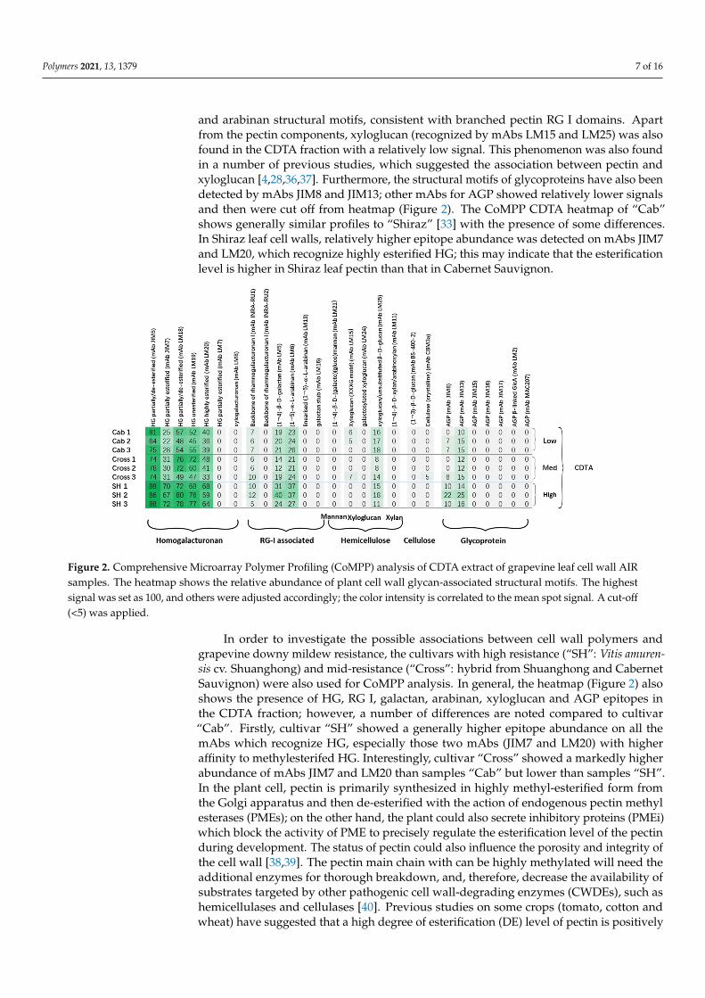
Polymers 2021, 13, 1379 7 of 16
and arabinan structural motifs, consistent with branched pectin RG I domains. Apartfrom the pectin components, xyloglucan (recognized by mAbs LM15 and LM25) was alsofound in the CDTA fraction with a relatively low signal. This phenomenon was also foundin a number of previous studies, which suggested the association between pectin andxyloglucan [4,28,36,37]. Furthermore, the structural motifs of glycoproteins have also beendetected by mAbs JIM8 and JIM13; other mAbs for AGP showed relatively lower signalsand then were cut off from heatmap (Figure 2). The CoMPP CDTA heatmap of “Cab”shows generally similar profiles to “Shiraz” [33] with the presence of some differences.In Shiraz leaf cell walls, relatively higher epitope abundance was detected on mAbs JIM7and LM20, which recognize highly esterified HG; this may indicate that the esterificationlevel is higher in Shiraz leaf pectin than that in Cabernet Sauvignon.
Figure 2. Comprehensive Microarray Polymer Profiling (CoMPP) analysis of CDTA extract of grapevine leaf cell wall AIRsamples. The heatmap shows the relative abundance of plant cell wall glycan-associated structural motifs. The highestsignal was set as 100, and others were adjusted accordingly; the color intensity is correlated to the mean spot signal. A cut-off(<5) was applied.
In order to investigate the possible associations between cell wall polymers andgrapevine downy mildew resistance, the cultivars with high resistance (“SH”: Vitis amuren-sis cv. Shuanghong) and mid-resistance (“Cross”: hybrid from Shuanghong and CabernetSauvignon) were also used for CoMPP analysis. In general, the heatmap (Figure 2) alsoshows the presence of HG, RG I, galactan, arabinan, xyloglucan and AGP epitopes inthe CDTA fraction; however, a number of differences are noted compared to cultivar“Cab”. Firstly, cultivar “SH” showed a generally higher epitope abundance on all themAbs which recognize HG, especially those two mAbs (JIM7 and LM20) with higheraffinity to methylesterifed HG. Interestingly, cultivar “Cross” showed a markedly higherabundance of mAbs JIM7 and LM20 than samples “Cab” but lower than samples “SH”.In the plant cell, pectin is primarily synthesized in highly methyl-esterified form fromthe Golgi apparatus and then de-esterified with the action of endogenous pectin methylesterases (PMEs); on the other hand, the plant could also secrete inhibitory proteins (PMEi)which block the activity of PME to precisely regulate the esterification level of the pectinduring development. The status of pectin could also influence the porosity and integrity ofthe cell wall [38,39]. The pectin main chain with can be highly methylated will need theadditional enzymes for thorough breakdown, and, therefore, decrease the availability ofsubstrates targeted by other pathogenic cell wall-degrading enzymes (CWDEs), such ashemicellulases and cellulases [40]. Previous studies on some crops (tomato, cotton andwheat) have suggested that a high degree of esterification (DE) level of pectin is positively

Polymers 2021, 13, 1379 8 of 16
correlated with plant defense. [4,41,42]. In addition to differences in HG, cultivar “SH”showed the highest abundance for RG I and its associated side chains (arabinan and galac-tan) than the other two cultivars. RG I with side chains is usually called the hairy regionof pectin; its complete deconstruction requires a set of lytic enzymes, such as RG lyases,arabinanases and galactanases. Previous studies have suggested the roles of arabinan andgalactan side chains as cross-linked bridges between pectin and xyloglucan [23,43–45].In addition to structural support, these side chains have been suggested to be involvedin other biological activities, e.g., galactans could be used as structural markers, as theirdecreasing signal is found during fruit ripening [30]. Arabinan has been focused on forits potential roles in the stress tolerance of plants, such as in regard to drought [46] andsalt stress [47]. However, currently no direct connection between arabinans and pathogenresistance has been confirmed. Apart from the pectin changes, the heatmap does not shownotable differences in the epitope abundances of mAbs recognizing hemicelluloses (e.g.,LM15 and LM25), even though some minor variations are present between the replicatesof each of the cultivars. The comparison of glycoprotein profiles revealed the markeddifferences between three cultivars, and “SH” showed the highest abundance on the mAbJIM8 (which recognizes an AGP epitope). Recent reviews have suggested additional rolesfor AGPs in plant stress tolerance compared with more well-established roles in plantgrowth and development; AGP accumulation has been observed during a range of bioticand abiotic stresses [47–49].
The CoMPP heatmap gives a general overview of the polysaccharide profile for initialinterpretation; however, it compresses the data for obtaining the relative values, andnumerous variables (mAb abundance) can lead to a greater difficulty in visually inspectingdatasets for differences. Thus, multivariate data analysis was routinely applied to theraw data to provide additional information (Figure 3). SIMCA software was used in thisstudy to implement an unsupervised method (principal component analysis, PCA), inorder to obtain an unbiased overview of the data structure. The raw CoMPP dataset waspre-processed in Microsoft Excel, and the mAbs were set as variables. The pre-processeddataset was then transferred to SIMCA to construct the PCA plot. The algorithms, if PCAfound the maximum variance in the data assigning it to principal component 1 (PC1),then proceeded to PC2, PC3, etc. In this study, the PCA score plot, which contained twocomponents (PC1 and PC2) with high quality (Figure 3A), was constructed; the scoreplot shows that three cultivars were clustered into three groups, and cultivar “SH” wasseparated from others (PC1 = 41.6%). The loading plot (Figure 3B) shows that the variables(mAbs) that drive this separation mainly include HG, RG I with associated side chainsand AGPs. Interestingly, two replicates of cultivar “Cross” sit at the same side as cultivar“SH 1” and “SH 2” and the major variables driving this separation in the vertical dimension(PC2 = 26.3%) are mAbs LM18 and LM19, which recognize the partially esterified HG.Further group-to-group (SH to Cross) comparisons suggest that corresponding variablesinclude JIM7 and LM 20, which recognize methylesterifed HG (data not shown); this againsupports the heatmap datasets. In addition to pectin, mAb BS-400-2 (recognizes callose),which did not show zero on heatmap, however, shows its association with other pectinrecognizing mAbs in the PCA plot. Callose has been well documented in many studiesfor its roles in plant/microbial interactions; it has been found to be deposited around theinfection sites to block the pathogens [15,40].
The CoMPP datasets of the NaOH fraction of the three cultivars yielded a heatmap(Figure 4) rich in hemicellulosic polymers, including mannans (mAb LM21), xyloglucan(mAbs LM15, LM24 and LM25) and xylan (mAb LM11). Glucan, cellulose and AGP werealso detected by mAbs (mAbs BS-400-2, CBM3a, JIM8, JIM13), as well as the signals ofgalactan (mAb LM5) and arabinan (mAbs LM6 and LM13); these may again support thehypothesis that these side chains act as the linkages between RG I and hemicellulosicpolymers [45]. The comparison between cultivars revealed the increasing trend of xyloglu-can (with XXXG motif, recognized by mAb LM15). Hemicellulose polymers have beensuggested to play critical roles in the strengthening of the plant cell wall structure, therefore

Polymers 2021, 13, 1379 9 of 16
elevating the plant resistance to stress. For example, the marked increase in arabinoxy-loglucan has been stated to positively correlated with the resistance of tobacco to Botrytiscinerea [50]; the silencing of xylanases activity increased the xylan content and siliquedehiscence resistance [51]; and a generally enriched hemicellulose was found in resistantwheat cultivar compared to cultivar susceptible to Fusarium Head Blight [52]. In additionto mAbs LM15, another hemicellulosic polymer, (galacto)(gluco)mannan (mAb LM21),showed a similar increasing pattern. Interestingly, similar to the CDTA fraction (Figure 2),an increasing trend was also observed for AGP epitope abundance, recognized by bothmAbs JIM8 and JIM13, which again suggested their possible roles in grapevine/downymildew interactions. To obtain further information on possible associations between poly-mers, the PCA score plot (Figure 5A) and loading plot (Figure 5B) were constructed fromthe CoMPP raw datasets of the NaOH fraction. In Figure 5A, it is very clear that thosesamples were well clustered into three groups on the horizontal dimension; cultivar “SH”has distinct distance from cultivar “Cab”, whereas cultivar “Cross” sits between them (PC1explained by 28.6% variance). In the loading plot (Figure 4), the main variables drivingthis separation include AGP, mannan, xyloglucan and cellulose, which again support theheatmap datasets (Figure 4).
Figure 3. PCA score plot (A) and loading plot (B) of the CDTA (pectin-rich) extract from AIR sourcedfrom grapevine leaves.

Polymers 2021, 13, 1379 10 of 16
Figure 4. Comprehensive Microarray Polymer Profiling (CoMPP) analysis of NaOH extract ofgrapevine leaf cell wall AIR samples. The heatmap showed the relative abundance of plant cell wallglycan-associated structural motifs. The highest signal was set as 100, and others were adjustedaccordingly; the color intensity is correlated to the mean spot signal. A cut-off (<5) was applied.
3.3. Monosaccharide Composition of Leaf Cell Wall Materials Using GC–MS
Gas chromatography (GC) has been routinely applied in cell wall analysis; it gives ad-ditional monosaccharide composition data for the validation of CoMPP analysis [32,33,37].In this study, GC was applied to the most distinct samples (”SH” and “Cab”), and sevenmonosaccharides were analyzed (Figure 6). In general, leaf AIR from two cultivars containsa substantial amount of GalA (ca. 35–40 mol.%), with the presence of Ara (ca. 14–15%),Rha (ca. 5–7%) and Gal (ca. 13–14%), which suggests that leaf cell walls contain homogalac-turonan (HG), rhamnogalacturonan I (RG I) and possibly rhamnogalacturonan II (RG II).On the other hand, the presence of Xyl (ca. 11–12%), Man (ca. 4–6%) and Glc (ca. 12–14%)suggests that xylan, xyloglucan and mannan are major components of the hemicellulosicfraction of grapevine leaf cell walls. These datasets show high similarity to the leaf sampleof cultivar “Shiraz” [33], but different to the composition of tobacco leaves [36] and grapeberry cell walls [37], this indicates the cell wall differences among the plant organs andtissues. Furthermore, the comparison between “Cab” and “SH” did not reveal significantdifferences among those monosaccharides; In addition to the markedly higher mol.% ofGalA in cultivar “SH”, this may explain the generally higher abundance of mAbs thatrecognize HG (Figure 2). This result highlights the limitations of GC composition datasets,which tend not provide information on subtle structural differences, whereas CoMPPprovides additional richer and complementary data on the polysaccharide structure byvirtue of epitope abundance. In a previous study on grape berry cell wall deconstruction,the GC–MS did not show any significant changes between samples, whereas CoMPP wasable to detect changes such as pectin de-esterification [37].
3.4. IR Spectroscopy
ATR-FT-MIR spectroscopy provides a fast and convenient method to detect the chem-ical functional groups present in cell wall samples [53]; it is usually applied in cell wallstudies to provide a general overview of vibrational chemical bonds linked to polysaccha-rides and protein [31,33]. In this study, AIR samples sourced from cultivar “SH” and “Cab”were used for IR spectroscopy, generating wavescan spectral profiles (Figure 7) which canbe used for comparisons. Two spectra were generated, both consisting of a number ofmaxima, including pectin HG at 1017 cm−1, xyloglucan at 1041 cm−1, amides at 1650 cm−1
and esterified pectin at 1740 cm−1 [53]. The comparison showed a similar spectral profilein general, which confirmed the finding from GC–MS and CoMPP, and reflects that bulk

Polymers 2021, 13, 1379 11 of 16
chemistry does not vary dramatically. However, few differences are worth noting: First,visual inspection indicated subtle difference in the shape of maxima at 1041 cm−1, whichmay suggest the structural differences in hemicellulosic polymers; this may be explained bythe higher abundance of xyloglucan (with XXXG motif) found in Figure 4. The differencein maxima at 1017 cm−1 was also noted, which may indicate the higher pectin HG contentin the “SH” AIR sample; however, the supporting data will be needed in future work asthe GC–MS and CoMPP mainly focused on the non-cellulose fractions. Interestingly, themost significant difference between the two spectra is the maxima (1740 cm−1), which referto esterified HG. This again supports the finding from Figures 2 and 3 that indicated thecultivar “SH” has a higher abundance of esterified HG.
Figure 5. PCA score plot (A) and loading plot (B) of the NaOH (hemicellulose-rich) extract from AIRsourced from grapevine leaves.

Polymers 2021, 13, 1379 12 of 16
Figure 6. The monosaccharide composition of AIR sourced from grapevine leaves. The compositionis expressed in relative mol% of seven main monosaccharides present in grapevine leaf cell wall. Ara:arabinose; Rha: rhamnose; Xyl: xylose; GalA: galacturonic acid; Man: mannose; Gal: galactose; Glc:glucose. Error bars represent the standard derivation of the mean value of three biological repeat.
Figure 7. Fourier transform infrared (FT-IR) spectra generated from the AIR samples sourced from grapevine leaves.
4. Conclusions
In order to achieve the successful penetration of plant tissue, pathogenic microorgan-isms (e.g., fungi) are found to employ various methods in their arsenal. Among these,the secretion of cell wall-degrading enzymes (CWDEs) is recognized as an effective andprecise way for unravelling and degrading one of the most complex structures in the nature.Compared to rot fungi in general, which secretes a wide range of CWDEs, as is the casefor necrotrophs, for thorough degradation, the bioinformatic genome and transcriptomecomparisons suggest that the relatively lower diversity of lytic enzymes is expressed in theoomycete family [54]. Thus, the interactions between plants and oomycetes in the cell wallbattlefield may vary during fungal infection. However, there is very limited informationon the host resistance from the viewpoint of the plant cell wall structure. Previous studies

Polymers 2021, 13, 1379 13 of 16
found that grape hybrids which were bred from native species may acquire parental resis-tance to differing degrees. In this study, cutting-edge cell wall profiling tools were appliedto selected grapevine leaf samples to investigate the possible correlations between grapeleaf cell wall polymers and downy mildew resistance.
The carbohydrate microarray provided much richer information than more classicalmethods (GC–MS and FTIR), and revealed a number of differences between the uninfectedleaves of Vitis amurensis cv. “Shuanghong” (resistance) and Vitis vinifera cv. “CabernetSauvignon” (susceptible), with the aid of multivariate data analysis tools. Significantlyhigher amounts of methyl esterified HG were found in cultivar “SH”, and the increasingtrend of this component from cultivar “Cab” and “Cross” (hybrid with moderate resistance)to “SH” may suggest its potential role as a structural biomarker in downy mildew resistance;this was also supported by FTIR spectral data of the functional chemical groups. Similarpatterns were also noted for xyloglucan with the XXXG motif. Arabinogalactan proteins(AGPs) were found to be the highest in abundance in resistant “SH” but did not showsignificant differences between “Cab” and “Cross”.
In summary, the higher esterification level of HG, high content of mannan and xy-loglucan (XXXG) could be the correlated with the native Chinese grapevine (Vitis amurensiscv. “SH”) natural resistance to downy mildew, and may be inherited from the nativegrapevine species through breeding. However, are these differences common to otherwild grapevine species with similar resistances? How are these polymers regulated inwild grapevines during development? In order to answer these questions, the relevantgenes behind these cell wall polymer changes will need to be focused on, e.g., the balancein expression of endogenous pectin methyl esterase (PME) and PME-inhibiting proteins(PMEI). Furthermore, in addition to the structural enhancement of the cell wall ahead of in-fection, we do not know how these wall polymers are altered by mildew infection/invasion.Future work will be needed to observe the induced changes of these cell wall componentsduring mildew infection, as well as relevant factors (e.g., lytic enzymes), for obtainingan in-depth understanding of the complex network involved in cell wall regulation andmolecular architecture. This is the first comprehensive comparison study of leaf cell wallpolysaccharide polymer/protein variation from Vitis species and hybrids differing in theirresistance to grapevine downy mildew. This provides important baseline reference datasetsfor future investigations on other biotic and abiotic stresses important in agriculturalgrape production.
Author Contributions: Y.G. and X.Y. designed and conducted the experiments, P.F., W.W. and B.Y.did the breeding work, germplasm maintenance and infection experiments. H.J. performed theGC–MS and IR analysis. J.H. and B.J. performed the CoMPP analysis. Y.G. analysed the data anddrafted the manuscript. W.Y., S.S., J.P.M. and J.L. contributed to the improvement of the manuscript.All authors have read and agreed to the published version of the manuscript.
Funding: This work was supported by the Shanghai Agriculture Applied Technology DevelopmentProgram, China (Grant No. 2018-02-08-00-08-F01552); National Natural Science Foundation ofChina (No. 31801830 and 31701775); the Shanghai Jiao Tong University New Scholar Start-Up Fund(AF1500068); Shanghai Municipal Commission for Science and Technology (18391900400); ChinaAgriculture Research System (CARS-29-yc-2).
Institutional Review Board Statement: Not applicable.
Informed Consent Statement: Not applicable.
Data Availability Statement: Not applicable.
Conflicts of Interest: The authors declare no conflict of interests.

Polymers 2021, 13, 1379 14 of 16
References1. Moore, J.P.; Divol, B. Tracking the careers of grape and wine polymers using biotechnology and systems biology. In Biotechnology
in Functional Foods and Nutraceuticals; Bagchi, D., Lau, F.C., Ghosh, D.K., Eds.; CRC Press Inc.: Boca Raton, FL, USA, 2010;pp. 389–406.
2. Kubicek, C.P.; Starr, T.L.; Glass, N.L. Plant cell wall-degrading enzymes and their secretion in plant-pathogen fungi. Annu. Rev.Phytopathol. 2014, 52, 427–451. [CrossRef] [PubMed]
3. Boevink, P.C.; Mclellan, H.; Gilroy, E.M.; Naqvi, S.; He, Q.; Yang, L.; Wang, X.; Turnbull, D.; Armstrong, M.R.; Tian, Z.; et al.Oomycetes seek help from the plant: Phytophthora infestans effectors target host susceptibility factors. Mol. Plant 2016, 9, 636–638.[CrossRef] [PubMed]
4. Johnsen, H.R.; Striberny, B.; Olsen, S.; Vidal-Melgosa, S.; Fangel, J.U.; Willats, W.G.T.; Rose, J.K.C.; Krause, K. Cell wall compositionprofiling of parasitic giant dodder (Cuscuta reflexa) and its hosts: A priori differences and induced changes. New Phytol. 2015, 207,805–816. [CrossRef]
5. Olsen, S.; Striberny, B.; Hollmann, J.; Schwacke, R.; Popper, Z.A.; Krause, K. Getting ready for host invasion elevated expressionand action of xyloglucan endotransglucosylases/hydrolases in developing haustoria of the holoparasitic angiosperm Cuscuta.J. Exp. Bot. 2016, 67, 695–708. [CrossRef]
6. Bacete, L.; Melida, H.; Miedes, E.; Molina, A. Plant cell wall-mediated immunity: Cell wall changes trigger disease resistanceresponses. Plant J. 2018, 93, 614–636. [CrossRef]
7. Beck, M.; Zhou, J.; Faulkner, C.; Maclean, D.; Robatzek, S. Spatio-temporal cellular dynamics of the Arabidopsis flagellin receptorreveal activation status-dependent endosomal sorting. Plant Cell. 2012, 24, 4205. [CrossRef] [PubMed]
8. De Jonge, R.; van Esse, H.P.; Kombrink, A.; Shinya, T.; Desaki, Y.; Bours, R.; Van Der Krol, S.; Shibuya, N.; Joosten, M.H.A.J.;Thomma, B.P.H.J. Conserved fungal lysm effector ecp6 prevents chitin-triggered immunity in plants. Science 2010, 329, 953.[CrossRef]
9. Hernández Blanco, C.; Feng, D.X.; Hu, J.; Sánchez vallet, A.; Deslandes, L.; Llorente, F.; Berrocal-Lobo, M.; Keller, H.; Barlet, X.;Sánchez-Rodríguez, C.; et al. Impairment of cellulose synthases required for Arabidopsis secondary cell wall formation enhancesdisease resistance. Plant Cell. 2007, 19, 890–903. [CrossRef]
10. Sicilia, F.; Fernandez-Recio, J.; Caprari, C.; De Lorenzo, G.; Tsernoglou, D.; Cervone, F.; Federici, L. The Polygalacturonase-inhibiting protein pgip2 of Phaseolus vulgaris has evolved a mixed mode of inhibition of endopolygalacturonase pg1 of Botrytiscinerea 1. Plant Physiol. 2005, 139, 1380–1388. [CrossRef]
11. Joubert, D.A.; Kars, I.; Wagemakers, L.; Bergmann, C.; Kemp, G.; Vivier, M.A.; Van Kan, J.A.L. A polygalacturonase-inhibitingprotein from grapevine reduces the symptoms of the endopolygalacturonase BcPG2 from Botrytis cinerea in Nicotiana benthamianaleaves without any evidence for in vitro interaction. Mol. Plant-Microbe Interact. 2007, 20, 392–402. [CrossRef]
12. Federici, L.; Caprari, C.; Mattei, B.; Savino, C.; Di Matteo, A.; De Lorenzo, G.; Cervone, F.; Tsernoglou, D.; Staskawicz, B.J.Structural requirements of endopolygalacturonase for the interaction with PGIP (polygalacturonase- inhibiting protein). Proc. Natl.Acad. Sci. USA 2001, 98, 13425–13430. [CrossRef]
13. Alexandersson, E.; Becker, J.V.W.; Jacobson, D.; Nguema-Ona, E.; Steyn, C.; Denby, K.J.; Vivier, M.A. Constitutive expression of agrapevine polygalacturonase-inhibiting protein affects gene expression and cell wall properties in uninfected tobacco. BMC Res.Notes. 2011, 4, 493. [CrossRef]
14. Liu, N.; Zhang, X.; Sun, Y.; Wang, P.; Li, X.; Pei, Y.; Li, F.; Hou, Y. Molecular evidence for the involvement of a polygalacturonase-inhibiting protein, GhPGIP1, in enhanced resistance to Verticillium and Fusarium wilts in cotton. Sci. Rep. 2017, 7, 39840.[CrossRef]
15. Yu, Y.; Zhang, Y.L.; Yin, L.; Lu, J. The mode of host resistance to Plasmopara viticola infection of grapevines. Phytopathol. 2012, 102,1094–1101. [CrossRef]
16. Burruano, S. The life-cycle of Plasmopara viticola, cause of downy mildew of vine. Mycologist 2000, 14, 179–182. [CrossRef]17. Boso, S.; Santiago, J.L.; Martinez, M.C. Resistance of eight different clones of the grape cultivar Albariño to Plasmopara viticola.
Plant Dis. 2004, 88, 741–744. [CrossRef] [PubMed]18. Davidson, L.C. Variation within and between Vitis spp. for foliar resistance to the downy mildew pathogen Plasmopara viticola.
Plant Dis. 2008, 92, 1577–1584. [CrossRef]19. Unger, S.; Büche, C.; Boso, S.; Kassemeyer, H.H. The course of colonization of two different Vitis genotypes by Plasmopara viticola
indicates compatible and incompatible host–pathogen interactions. Phytopathology 2007, 97, 780–786. [CrossRef]20. Trouvelot, S.; Varnier, A.L.; Allègre, M.; Mercier, L.; Baillieul, F.; Arnould, C.; Gianinazzi-Pearson, V.; Klarzynski, O.; Joubert, J.M.;
Pugin, A. A β-1,3 glucan sulfate induces resistance in grapevine against Plasmopara viticola through priming of defense responses,including HR-like cell death. Mol. Plant-Microbe Interact. 2008, 21, 232–243. [CrossRef] [PubMed]
21. Wu, J.; Zhang, Y.L.; Zhang, H.Q.; Huang, H.; Folta, K.M.; Lu, J. Whole genome wide expression profiles of Vitis amurensis graperesponding to downy mildew by using Solexa sequencing technology. BMC Plant Biol. 2010, 10, 234–249. [CrossRef] [PubMed]
22. Vorwerk, S.; Somerville, S.; Somerville, C. The role of plant cell wall polysaccharide composition in disease resistance. TrendsPlant Sci. 2004, 9, 203–209. [CrossRef]
23. Somerville, C.; Bauer, S.; Brininstool, G.; Facette, M.; Hamann, T.; Milne, J.; Osborne, E.; Paredez, A.; Persson, S.; Raab, T.; et al.Toward a systems approach to understanding plant cell walls. Science 2004, 306, 2206–2211. [CrossRef] [PubMed]

Polymers 2021, 13, 1379 15 of 16
24. Albersheim, P.; Darvill, A.; Roberts, K.; Sederoff, R.; Staehelin, A. Plant Cell Walls: From Chemistry to Biology; Garland Science:New York, NY, USA, 2011; pp. 365–407.
25. Marcus, S.E.; Blake, A.W.; Benians, T.A.S.; Lee, K.J.D.; Poyser, C.; Donaldson, L.; Leroux, O.; Rogowski, A.; Petersen, H.L.;Boraston, A.; et al. Restricted access of proteins to mannan polysaccharides in intact plant cell walls. Plant J. 2010, 64, 191–203.[CrossRef]
26. Ma, Q.-H.; Zhu, H.-H.; Qiao, M.-Y. Contribution of both lignin content and sinapyl monomer to disease resistance in tobacco.Plant Pathol. 2018, 67, 642–650. [CrossRef]
27. Yang, Y.; Yang, X.; Dong, Y.; Qiu, D. The Botrytis cinerea Xylanase BcXyl1 Modulates Plant Immunity. Front. Microbiol. 2018, 9,2535. [CrossRef] [PubMed]
28. Moller, I.; Sørensen, I.; Bernal, A.; Blaukopf, C.; Lee, K.; Øbro, J.; Pettolino, F.; Roberts, A.; Mikkelsen, J.D.; Knox, J.P.; et al.Highthroughput mapping of cell-wall polymers within and between plants using novel microarrays. Plant J. 2007, 50, 1118–1128.[CrossRef]
29. Gao, Y.; Fangel, J.U.; Willats, W.G.T.; Vivier, M.A.; Moore, J.P. Effect of commercial enzymes on berry cell wall deconstruction inthe context of intravineyard ripeness variation under winemaking conditions. J. Agric. Food Chem. 2016, 64, 3862–3872. [CrossRef]
30. Moore, J.P.; Fangel, J.U.; Willats, W.G.T.; Vivier, M.A. Pectic-(1,4)-galactan, extensin and arabinogalactan-protein epitopesdifferentiate ripening stages in wine and table grape cell walls. Ann. Bot. 2014, 114, 1279–1294. [CrossRef] [PubMed]
31. Gao, Y.; Fangel, J.U.; Willats, W.G.T.; Vivier, M.A.; Moore, J.P. Dissecting the polysaccharide-rich grape cell wall changes duringwinemaking using combined high-throughput and fractionation methods. Carbohydr. Polym. 2015, 133, 567–577. [CrossRef][PubMed]
32. Zietsman, A.J.J.; Moore, J.P.; Fangel, J.U.; Willats, W.G.T.; Vivier, M.A. Profiling the hydrolysis of isolated grape berry skin cellwalls by purified enzymes. J. Agric. Food Chem. 2015, 63, 8267–8274. [CrossRef] [PubMed]
33. Moore, J.P.; Nguema-Ona, E.; Fangel, J.U.; Willats, W.G.; Hugo, A.; Vivier, M.A. Profiling the main cell wall polysaccharides ofgrapevine leaves using high-throughput and fractionation techniques. Carbohydr. Polym. 2014, 99, 190–198. [CrossRef]
34. Garrido-Bañuelos, G.; Buica, A.; Schückel, J.; Zietsman, A.J.J.; Willats, W.G.T.; Moore, J.P.; Du Toit, W.J. Investigating therelationship between grape cell wall polysaccharide composition and the extractability of phenolic compounds into Shiraz wines.Part I: Vintage and ripeness effects. Food Chem. 2019, 278, 36–46. [CrossRef]
35. Ralet, M.C.; Tranquet, O.; Poulain, D.; Moise, A.; Guillon, F. Monoclonal antibodies to rhamnogalacturonan I backbone. Planta2010, 231, 1373–1383. [CrossRef]
36. Nguema-Ona, E.; Moore, J.P.; Fagerstrom, A.; Fangel, J.U.; Willats, W.G.; Hugo, A.; Vivier, M.A. Profiling the main cell wallpolysaccharides of tobacco leaves using high-throughput and fractionation techniques. Carbohydr. Polym. 2012, 88, 939–949.[CrossRef]
37. Gao, Y.; Fangel, J.U.; Willats, W.G.T.; Vivier, M.A.; Moore, J.P. Dissecting the polysaccharide-rich grape cell wall matrix usingrecombinant pectinases during winemaking. Carbohydr. Polym. 2016, 152, 510–519. [CrossRef]
38. Marcus, S.E.; Verhertbruggen, Y.; Herve, C.; Ordaz-Ortiz, J.J.; Farkas, V.; Pedersen, H.L.; Willats, W.G.T.; Knox, J.P. Pectichomogalacturonan masks abundant sets of xyloglucan epitopes in plant cell walls. BMC Plant Biol. 2008, 8, 60. [CrossRef][PubMed]
39. Herve, C.; Rogowski, A.; Blake, A.W.; Marcus, S.E.; Gilbert, H.J.; Knox, J.P. Carbohydrate-binding modules promote the enzymaticdeconstruction of intact plant cell walls by targeting and proximity effects. Proc. Natl. Acad. Sci. USA 2010, 107, 15293–15298.[CrossRef] [PubMed]
40. Tingley, J.P.; Low, K.E.; Xing, X.; Abbott, D.W. Combined whole cell wall analysis and streamlined in silico carbohydrate-activeenzyme discovery to improve biocatalytic conversion of agricultural crop residues. Biotechnol. Biofuels 2021, 14, 16. [CrossRef]
41. Volpi, C.; Janni, M.; Lionetti, V.; Bellincampi, D.; Favaron, F.; D’Ovidio, R. The ectopic expression of a pectin methyl esteraseinhibitor increases pectin methyl esterification and limits fungal diseases in wheat. Mol. Plant-Microbe Interact. 2011, 24, 1012–1019.[CrossRef]
42. Liu, N.; Sun, Y.; Pei, Y.; Zhang, X.; Wang, P.; Li, X.; Li, F.; Hou, Y. A pectin methylesterase inhibitor enhances resistance toverticillium wilt. Plant Physiol. 2018, 176, 2202–2220. [CrossRef] [PubMed]
43. Coenen, G.J.; Bakx, E.J.; Verhoef, R.P.; Schols, H.A.; Voragen, A.G.J. Identification of the connecting linkage between homo- orxylogalacturonan and rhamnogalacturonan type I. Carbohydr. Polym. 2007, 70, 224–235. [CrossRef]
44. Zykwinska, A.; Thibault, J.F.; Ralet, M.C. Organization of pectic arabinan and galactan side chains in association with cellulosemicrofibrils in primary cell walls and related models envisaged. J. Exp. Bot. 2007, 58, 1795–1802. [CrossRef] [PubMed]
45. Popper, Z.A.; Fry, S.C. Widespread occurrence of a covalent linkage between xyloglucan and acidic polysaccharides in suspension-cultured angiosperm cells. Ann. Bot. 2005, 96, 9199. [CrossRef]
46. Moore, J.; Farrant, J.; Driouich, A. A role for pectin-associated arabinans in maintaining the flexibility of the plant cell wall duringwater deficit stress. Plant Signal. Behav. 2008, 3, 102–104. [CrossRef] [PubMed]
47. Zhao, C.; Zayed, O.; Zeng, F.; Liu, C.; Zhang, L.; Zhu, P.; Hsu, C.C.; Tuncil, Y.E.; Tao, W.A.; Carpita, N.C.; et al. Arabinosebiosynthesis is critical for salt stress tolerance in Arabidopsis. New Phytol. 2019, 224, 274–290. [CrossRef] [PubMed]
48. Nguema-Ona, E.; Vicré-Gibouin, M.; Cannesan, M.A.; Driouich, A. Arabinogalactan proteins in root-microbe interactions. TrendsPlant Sci. 2013, 18, 1360–1385. [CrossRef]

Polymers 2021, 13, 1379 16 of 16
49. Mareri, L.; Romi, M.; Cai, G. Arabinogalactan proteins: Actors or spectators during abiotic and biotic stress in plants? PlantBiosyst. Int. J. Deal. Asp. Plant Biol. 2019, 153, 173–185. [CrossRef]
50. Nguema-Ona, E.; Moore, J.P.; Fagerström, A.D.; Fangel, J.U.; Willats, W.G.T.; Hugo, A.; Vivier, M.A. Overexpression of thegrapevine PGIP1 in tobacco results in compositional changes in the leaf arabinoxyloglucan network in the absence of fungalinfection. BMC Plant Biol. 2013, 13, 46. [CrossRef]
51. Li, Y.L.; Yu, Y.K.; Zhu, K.M.; Ding, L.-N.; Wang, Z.; Yang, Y.-H.; Cao, J.; Xu, L.-Z.; Li, Y.-M.; Tan, X.-L. Down-regulation ofMANNANASE7 gene in Brassica napus L. enhances silique dehiscence-resistance. Plant Cell Rep. 2021, 40, 361–374. [CrossRef][PubMed]
52. Lahlali, R.; Kumar, S.; Wang, L.; Forseille, L.; Sylvain, N.; Korbas, M.; Muir, D.; Swerhone, G.; Lawrence, J.R.; Fobert, P.R.; et al.Cell wall biomolecular composition plays a potential role in the host type ii resistance to Fusarium head blight in wheat. Front.Microbiol. 2016, 7, 910. [CrossRef]
53. Kacurakova, M.; Capek, P.; Sasinkova, V.; Wellner, N.; Ebringerova, A. FT-IR study of plant cell wall model compound: Pecticpolysaccharides and hemicelluloses. Carbohydr. Polym. 2000, 43, 195–203. [CrossRef]
54. Cosgrove, D. Microbial expansins. Annu. Rev. Microbiol. 2017, 71, 479–497. [CrossRef] [PubMed]