Carbohydrate−π Interactions: What Are They Worth?
28
Carbohydrate-π Interactions: What Are They Worth? Zachary R. Laughrey, Sarah E. Kiehna ‡ , Alex J. Riemen, and Marcey L. Waters Department of Chemistry, CB 3290, University of North Carolina, Chapel Hill, NC 27599 Abstract Protein-carbohydrate interactions play an important role in many biologically important processes. The recognition is mediated by a number of noncovalent interactions including an interaction between the α-face of the carbohydrate and the aromatic side chain. To this end, this interaction has been studied in the context of a β-hairpin in aqueous solution, in which the interaction can be investigated in the absence of other cooperative noncovalent interactions. In this β-hairpin system both the aromatic side chain as well as the carbohydrate was varied in an effort to gain greater insight into the driving force and magnitude of the carbohydrate-π interaction. The magnitude of the interaction was found to vary from -0.5 to -0.8 kcal/mol, depending on the nature of the aromatic ring and the carbohydrate. Replacement of the aromatic ring with an aliphatic group resulted in a decrease in interaction energy to -0.1 kcal/mol, providing evidence for the contribution of CH-π interactions to the driving force. These findings demonstrate the significance of carbohydrate-π interactions within biological systems and also demonstrate its utility as a molecular recognition element in designed systems Introduction Many biological processes, including bacterial cell wall recognition, viral and bacterial infections, and fertilization, rely on carbohydrate-protein interactions. 1, 2 Additionally, glycosolation as a posttranslational modification affects the hydration and conformation of a protein. 3, 4 Due to its significance in biology, understanding the driving force for binding carbohydrates in water is an active area of research. 5-7 In addition to hydrogen bonding, a common feature of carbohydrate binding proteins is the interaction of the α-face of the carbohydrate with the face of an aromatic side chain (Figure 1). 8, 9 Carbohydrate-π interactions have been investigated through a variety of analytical techniques including NMR, IR, molecular modeling, and X-ray. 10-16 The Simons group utilized IR and modeling to examine the interaction. 17 Jimenez-Barbero et al have used NMR and modeling to examine the driving force for binding of oligosaccharides to the hevein domain and variations thereof. 18-20 These studies indicate that the carbohydrate-π interactions are important to the recognition of carbohydrates and that the interaction is dependent on the electronic nature of the aromatic group. However, there is limited experimental data investigating the favorable contribution of carbohydrate-π interaction in isolation. 15, 16 Given the importance of carbohydrate recognition in biology, a better understanding of the role of carbohydrate-π interactions is warranted. Previously, we reported an attractive interaction between a tryptophan diagonally cross-strand from a tetraacetylglucoserine which stabilized the folding of a β-hairpin. 21 Examining the proton NMR shifts of the carbohydrate protons demonstrated that the interaction was primarily through the α-face of the carbohydrate and the face of the Trp sidechain, suggesting a Correspondence to: Marcey L. Waters, [email protected]. ‡ Current address: Nanotope, Inc., 8025 Lamon Ave. Suite 450, Skokie, IL 60077 NIH Public Access Author Manuscript J Am Chem Soc. Author manuscript; available in PMC 2009 November 5. Published in final edited form as: J Am Chem Soc. 2008 November 5; 130(44): 14625–14633. doi:10.1021/ja803960x. NIH-PA Author Manuscript NIH-PA Author Manuscript NIH-PA Author Manuscript
-
Upload
independent -
Category
Documents
-
view
0 -
download
0
Transcript of Carbohydrate−π Interactions: What Are They Worth?

Carbohydrate-π Interactions: What Are They Worth?
Zachary R. Laughrey, Sarah E. Kiehna‡, Alex J. Riemen, and Marcey L. WatersDepartment of Chemistry, CB 3290, University of North Carolina, Chapel Hill, NC 27599
AbstractProtein-carbohydrate interactions play an important role in many biologically important processes.The recognition is mediated by a number of noncovalent interactions including an interactionbetween the α-face of the carbohydrate and the aromatic side chain. To this end, this interaction hasbeen studied in the context of a β-hairpin in aqueous solution, in which the interaction can beinvestigated in the absence of other cooperative noncovalent interactions. In this β-hairpin systemboth the aromatic side chain as well as the carbohydrate was varied in an effort to gain greater insightinto the driving force and magnitude of the carbohydrate-π interaction. The magnitude of theinteraction was found to vary from -0.5 to -0.8 kcal/mol, depending on the nature of the aromaticring and the carbohydrate. Replacement of the aromatic ring with an aliphatic group resulted in adecrease in interaction energy to -0.1 kcal/mol, providing evidence for the contribution of CH-πinteractions to the driving force. These findings demonstrate the significance of carbohydrate-πinteractions within biological systems and also demonstrate its utility as a molecular recognitionelement in designed systems
IntroductionMany biological processes, including bacterial cell wall recognition, viral and bacterialinfections, and fertilization, rely on carbohydrate-protein interactions.1, 2 Additionally,glycosolation as a posttranslational modification affects the hydration and conformation of aprotein.3, 4 Due to its significance in biology, understanding the driving force for bindingcarbohydrates in water is an active area of research.5-7 In addition to hydrogen bonding, acommon feature of carbohydrate binding proteins is the interaction of the α-face of thecarbohydrate with the face of an aromatic side chain (Figure 1).8, 9 Carbohydrate-π interactionshave been investigated through a variety of analytical techniques including NMR, IR,molecular modeling, and X-ray.10-16 The Simons group utilized IR and modeling to examinethe interaction.17 Jimenez-Barbero et al have used NMR and modeling to examine the drivingforce for binding of oligosaccharides to the hevein domain and variations thereof.18-20 Thesestudies indicate that the carbohydrate-π interactions are important to the recognition ofcarbohydrates and that the interaction is dependent on the electronic nature of the aromaticgroup. However, there is limited experimental data investigating the favorable contribution ofcarbohydrate-π interaction in isolation.15, 16 Given the importance of carbohydraterecognition in biology, a better understanding of the role of carbohydrate-π interactions iswarranted.
Previously, we reported an attractive interaction between a tryptophan diagonally cross-strandfrom a tetraacetylglucoserine which stabilized the folding of a β-hairpin.21 Examining theproton NMR shifts of the carbohydrate protons demonstrated that the interaction was primarilythrough the α-face of the carbohydrate and the face of the Trp sidechain, suggesting a
Correspondence to: Marcey L. Waters, [email protected].‡Current address: Nanotope, Inc., 8025 Lamon Ave. Suite 450, Skokie, IL 60077
NIH Public AccessAuthor ManuscriptJ Am Chem Soc. Author manuscript; available in PMC 2009 November 5.
Published in final edited form as:J Am Chem Soc. 2008 November 5; 130(44): 14625–14633. doi:10.1021/ja803960x.
NIH
-PA Author Manuscript
NIH
-PA Author Manuscript
NIH
-PA Author Manuscript

carbohydrate-π interaction. Upfield shifting and NOE data is consistent with the geometryshown in Figure 1b. The magnitude of the interaction was found to be -0.8 kcal/mol, which isgreater than the magnitude of a Lys-Trp cation-π interaction measured in the same modelsystem.22 However, when the acetyl groups on glucose were removed, the interaction betweenthe carbohydrate and tryptophan was lost. The reduction was attributed to the increaseddesolvation cost of the unprotected glucose.
To further investigate the efficacy of an isolated carbohydrate-π interaction in aqueous solution,two series of peptides were synthesized and studied. In the first series, aromatic or aliphaticresidues (X) were incorporated in close proximity to a tetraacetylglucoserine, Ser(Ac4Glc) onthe face of a β-hairpin (Figure 2). In the second series, Trp was kept constant and the natureof the carbohydrate (Z) was varied.
Results and DiscussionDesign
The 12-residue sequence used in this study is based on peptides previously described, in whicha stabilizing carbohydrate-π interaction between a Trp at position 2 and tetracylated glucoseat position 9 was explored.21 Several key features have been maintained including: a +3 chargeto provide solubility and discourage aggregation; an Asn-Gly turn nucleator sequence; and ahydrophobic cluster on both the HB face of the hairpin (Val-3, Val-5, and Ile-10) and the NHBface (X-2 and Leu-11). Aromatic amino acids and carbohydrates were placed in positions 2(X) and 9 (Z) respectively. These positions have been shown to allow diagonal cross-strandinteraction and provide ample room to allow for the bulky side chains.22-25 The glycosylatedseries were synthesized per literature methods and introduced into the peptide chain as Fmoc-protected amino acids (see Supporting Information).21, 26-31
Characterization of Structureβ-Hairpin structure characterization was accomplished by a number of NMR measurementsdescribed below, including: carbohydrate chemical shifts, α-hydrogen (Hα) chemical shifts;glycine splitting; and cross strand NOEs. NMR spectroscopy provides insight into the geometryof the interaction, as the carbohydrate protons are shifted upfield when in close proximity tothe face of the aromatic sidechains due to ring current effects.32 The extent of downfieldshifting of Hα relative to random coil is an indicator to the extent of β-sheet conformation ateach position along the strand. Downfield shifting of Hα by > 0.1 ppm is indicative of β-hairpinformation.33 The fraction folded at each residue can be determined by comparing the observedHα chemical shifts to those in the unfolded state and in the fully folded state (obtained froman unfolded control peptide and a cyclic control peptide, respectively; see ExperimentalProcedures).[Syud, 1999 #30] Alternatively, glycine splitting, when compared to a cycliccontrol, acts as a global indicator of β-hairpin conformation.34 Fraction folded valuesdetermined from Hα shifting and Gly splitting were generally in good agreement. Finally, longdistance cross strand NOEs between side chains are consistent with β-hairpin structure andwere observed for all peptides.
Variation of the Aromatic side chain (X)To examine the role of the aromatic side chain, a series of peptides were synthesized in whichTrp was replaced with other aromatic or hydrophobic side chains including 1-Nal, 2-Nal, Phe,and Cha (Figure 2) while maintaining the carbohydrate as S(Ac4Glc).
1-Nal was substituted for Trp to investigate the significance of the NH group in Trp. In addition,1-Nal has a greater surface area than Trp (1-Nal: 161Å2 compared to Trp: 147Å2) and theelectron density on the face of the ring is not as great (Figure 3). This substitution produced a
Laughrey et al. Page 2
J Am Chem Soc. Author manuscript; available in PMC 2009 November 5.
NIH
-PA Author Manuscript
NIH
-PA Author Manuscript
NIH
-PA Author Manuscript

well-folded peptide which is as folded as WS(Ac4Glc) within experimental error (Table 1 andFigure 4). The protons on the α-face (H1, H3, and H5) were all shifted upfield, indicating thatthe α-face packs against the aromatic face of 1-Nal (Figure 5), similar to the shifting observedfor WS(Ac4Glc). The protons H6 and H6′ were also found to be shifted to a lesser extent,indicating that the exocyclic CH2 interacts with the aromatic face. This interaction andgeometry has precedent in galactose binding lectins. While the acetyl groups could not beassigned, the maximum upfield shift was ≤ 0.07 ppm (this is assuming that the methyl grouppeak which was farthest downfield in the control peptide is the farthest upfield in the β-hairpin).This is significantly less then the shifts of the protons of the α-face of the carbohydrate, whichrange from -0.6 to -1.2 ppm, and indicates that the acetyl groups play little to no direct role inthe stabilizing interaction. NOESY NMR displayed long distance cross-strand interactions inthe peptide indicating the peptide is properly folded in a β-hairpin structure. Strong NOEs werealso observed between the carbohydrate and 1-Nal (Figure 6), although not as extensively asseen with Trp.
Because the β-sheet propensity of each amino acid influences the stability of the folded state,one cannot directly compare extent of folding of two peptides in which X has been varied andattribute differences exclusively to sidechain-sidechain interactions. To determine theenergetic contribution of the diagonal sidechain-sidechain interaction alone, a double mutantcycle was completed.33-39 A double mutant cycle replaces two interacting side chains withtwo non-interacting side chains. A single mutation disrupts the interaction of interest but couldadditionally cause other changes to stability (i.e. β-sheet propensity, hydrogen bonding, etc).The double mutant corrects for these unintentional changes leaving only the noncovalentinteraction of interest. In this study, X and Z were exchanged for Leu and Ser, respectively.Leu was chosen as it has a high β-sheet propensity that minimizes net loss of β-hairpin stability.Ser was chosen as it has a small polar side chain and that does not interact diagonally. Thedouble mutant cycle reveals that the interaction of 1-Nal with S(Ac4Glc) has a similarmagnitude to the Trp-S(Ac4Glc) interaction (Table 2).
2-Nal was substituted at the X position to determine the influence of orientation differencesbetween 1-Nal and 2-Nal on the carbohydrate-π interaction. The results were similar to both1-Nal and Trp, indicating that the carbohydrate interacts in a favorable manner with 2-Nal viastacking with the aromatic sidechain, despite the differences in orientation of the two sidechains(Table 1 and Figure 4). The upfield shifting of the peptide Hα protons demonstrated that theβ-hairpin conformation was well formed throughout the peptide with a similar stability as WS(Ac4Glc) and 1-NalS(Ac4Glc). Additionally, the carbohydrate shifts were similar in magnitudeto those in the peptides with the other two large aromatic groups (Figure 5), namely, that theprotons of the carbohydrates α-face are shifted significantly upfield indicating an interactionwith the aromatic face. Long-distance NOEs between the carbohydrate's α-face and 2-Nal alsosupport a stacking geometry (Figure 6). The magnitude of the interaction of 2-Nal withAc4Glc was also found to be similar to that of Trp and 1-Nal, as determined from the doublemutant cycle (Table 2).
The situation changes when the smaller Phe was placed in the sequence at position 2. Thepercent folded was reduced from 85% to 57% (Figure 4 and Table 1). Phe is known to have alower β-sheet propensity than Trp, 40[Smith, 1997 #121] but the double mutant cycle indicatesthat the loss in β-hairpin stability is due in part to a weakening of the carbohydrate-π interaction(Table 2). Additionally, the carbohydrate's α-face protons are not upfield shifted nearly as muchas when X is a larger aromatic group. There are significantly fewer unambiguous NOEsbetween the Phe side chain and the face if the carbohydrate than between the larger aromaticside chains and the carbohydrates. The smaller changes in chemical shift of both the Hα andthe carbohydrate protons indicate a less folded hairpin and a less favorable carbohydrate-πinteraction.
Laughrey et al. Page 3
J Am Chem Soc. Author manuscript; available in PMC 2009 November 5.
NIH
-PA Author Manuscript
NIH
-PA Author Manuscript
NIH
-PA Author Manuscript

When the aromatic nature of the X sidechain is removed by replacing Phe with Cha, the stabilityof the hairpin is further reduced (Table 1), despite the fact that Cha has been shown to have ahigher β-sheet propensity than Phe.41 A double mutant cycle indicates that the interaction ofCha with Ac4Glc is weaker than that of Phe with Ac4Glc (Table 2), despite the similar facialsurface area. Unlike the aromatic peptides in this series, there are no unambiguous NOEsbetween the cyclohexane side chain and the carbohydrate (Figure 6). Since Cha is not aromatic,no shifting of the carbohydrate protons is observed.
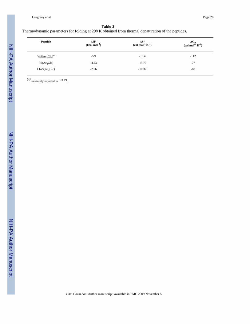
To provide additional insight into the effect of the X group on the interaction with Ac4Glc, weperformed thermal denaturations on WS(Ac4Glc), FS(Ac4Glc), and ChaS(Ac4Glc) by NMR.33 Fitting of the data provides values for ΔH°, ΔS°, and ΔCp for folding (Table 3). Since theonly change in the peptide sequence is the X group at position 2, changes in the driving forcefor folding can be attributed to the role of that residue in stabilizing the folded state. One cansee that for Trp and Phe folding is more enthalpically favorable than for Cha, which does notsignificantly interact with Ac4Glc. This is consistent with an enthalpic driving force for theinteraction of the carbohydrate with the aromatic ring, as has been observed in other systems,5, 42 and is suggestive of CH(δ+)-π and dispersion forces as major contributors to theinteraction.
Variation of the CarbohydrateThe carbohydrate was also varied while holding Trp constant and the impact on the sidechain-sidechain interaction was explored. Previous studies comparing Ac4Glc and Glc suggested thatAc4Glc formed a favorable carbohydrate-π interaction, but the desolvation cost appeared to betoo high for Glc to interact favorably with Trp.[Kiehna, 2007 #85] Two additional acylatedcarbohydrates, Ac4Gal and Ac3GlcNAc (Figure 2) have been substituted for Ac4Glc. Ac4Galinvestigates the effect of the stereochemistry at C4 on the carbohydrate-π interaction.Ac3GlcNAc investigates the replacement of oxygen at C-2 with nitrogen. We also investigatedthe deprotected counterpart, GlcNAc, in which only the nitrogen at C-2 is acetylated. Lastly,we investigated Me4Glc to further investigate the role of desolvation and determine the role(if any) of the acetyl groups.
The only difference between the Ac4Glc and Ac4Gal is the orientation of the alcohol at C-4(axial vs. equatorial) (Figure 2). The binding sites of many galactose binding proteins(galectins) contain an aromatic residue which interacts with the “hydrophobic cluster” madeup of C-4, C-5, and C-6 (Figure 7).43 The upfield shifting of the 6/6′ protons of Ac4Glc in WS(Ac4Glc) suggested that such an interaction at C-4, C-5, and C-6 may be feasible in the β-hairpin. Thus, we replaced Ac4Glc with Ac4Gal and investigated its interaction with Trp. Therewas only a small change in the fraction folded for WS(Ac4Gal) relative to WS(Ac4Glc), asmeasured by the glycine splitting and Hα shifts (Table 4 and Figure 8). NOEs between thesugar and Trp indicate that the interaction occurs on the α-face of the sugar, as was seen forAc4Glc. Inspection of the carbohydrate chemical shifts reveals that the protons of the α-face(C-1, C-3, and C-5) are the most shifted relative to random coil but C-4 is not significantlyshifted (Figure 9), indicating the same geometry as for WS(Ac4Glc) rather than that seen ingalectins. This may be due to conformational restrictions of the system rather than a specificpreference for one geometry over the other. The extent of shifting at positions 1, 3, and 5 ofAc4Gal is similar to that observed in Ac4Glc, suggesting that the interaction with the α–faceis equally as favorable. This is consistent with the interaction energy determined from doublemutant cycles (Table 5), which is within error of that measured for Ac4Glc.
Another common carbohydrate found in nature is GlcNAc. This carbohydrate has twodistinctive features, namely that nitrogen replaces oxygen at C-2, and that the nitrogen isacetylated. Both of these differences change how the carbohydrate interacts with the face ofthe Trp. The tetraacylated sugar, Ac3GlcNAc, was used to compare directly with Ac4Glc. The
Laughrey et al. Page 4
J Am Chem Soc. Author manuscript; available in PMC 2009 November 5.
NIH
-PA Author Manuscript
NIH
-PA Author Manuscript
NIH
-PA Author Manuscript

presence of the amide reduces the interaction relative to WS(Ac4Glc): the fraction folded isreduced from 0.85 to 0.73 (Figure 8 and Table 4) and the double mutant cycle demonstratesthat the interaction energy is reduced by 0.2 kcal mol-1 (Table 5). However, the protons ofAc3GlcNAc α-face are not nearly as upfield shifted as compared to WS(Ac4Glc) (Figure 9).The proton at C-1 is the only one that is significantly shifted, suggesting a change in geometrydue to the amide at C-2. This suggests that the energetic term from the double mutant cyclearises from a favorable interaction other than the carbohydrate-p interaction alone. Indeed,downfield shifting of the NH group of Ac3GlcNAc (9.34 ppm) suggests that it may participatein a hydrogen bond. The Trp NH is not significantly shifted, however (10.16 ppm vs 10.11ppm in an unfolded control peptide).
When the acyl groups are removed from the Ac3GlcNAc the fraction folded only decreasesslightly (0.73 vs. 0.70 for Ac3GlcNAc and GlcNAc, respectively). However, the carbohydrateprotons are not nearly as upfield shifted when compared to the other carbohydrates, with thegreatest shifting occurring at C-6 (Figure 9). In fact, the chemical shifts of GlcNAc are similarto those of Glc, which did not display and significant interaction with Trp.21 There are severalweak unambiguous NOEs between the α-face of the carbohydrate (Figure 10). The doublemutant cycle indicates that the interaction energy for GlcNAc is comparable to Ac3GlcNAc.Thus, it appears that some sort of a favorable interaction occurs, but via a different geometrythan seen with other carbohydrates. However, NMR provides no evidence of hydrogen bondinginvolving either the GlcNAc NH (7.82 vs 7.88 ppm in the unfolded control peptide) or the TrpNH (10.17 vs 10.11 ppm in the unfolded control peptide).
We also investigated the peptide in which the acetyl protecting groups of Ac4Glc were replacedwith methyl groups to probe the role of desolvation and to determine if there is a specificinfluence of the acetyl groups. The peptide WS(Me4Glc) is equally well folded as WS(Ac4Glc) (Table 4, Figure 8), and exhibits numerous NOEs between the Trp residue and α-face of the sugar (Figure 10), indicating that Me4Glc also forms a favorable interaction withTrp. Double mutant cycles indicate that the magnitude of the interaction is within error forMe4Glc and Ac4Glc. This appears to suggest that protection of the hydroxyl groups, and hencereduction of the desolvation cost, is indeed the primary difference between peptides WS(Ac4Glc) and WS(Me4Glc) relative to the unprotected glucose in WS(Glc). However, Ac4Glcand Me4Glc do not behave identically; peptide WS(Me4Glc) does not demonstrate the sameextent of upfield shifting of the carbohydrate protons at positions C-1, C-3, and C-5 as doespeptide WS(Ac4Glc) (0.4-0.6 ppm for peptide WS(Me4Glc) versus 0.6-1.35 ppm for peptideWS(Ac4Glc)), despite the similar stability of the β-hairpins. This may be due to competitionof the methyl groups for interaction with the Trp, as the methyl groups are also upfield shiftedby up to 0.25 ppm. Indeed, the magnitude of upfield shifting of the methyl groups is verysimilar to the shifting that Cuevas and coworkers observed in their study of carbohydrate-πinteractions with Me5Glc.44 Thus, it appears that while Me4Glc forms a favorable interactionwith Trp, the interaction of the α-face of the sugar is not the only contributor to the interaction.
DiscussionThe system described here has allowed for the systematic investigation of carbohydrate-aromatic interactions in the absence of other cooperative noncovalent interactions, such ashydrogen bonds, thus allowing for the quantification of the binding energy and an explorationof the factors that contribute to the interaction. Variation of the X sidechain provides significantinsight into the nature and driving force of the carbohyrate-aromatic interaction. The similarinteractions of Ac4Glc with Trp, 1-Nal, and 2-Nal confirm that Ac4Glc interacts primarily withthe face of the aromatic ring and that any hydrogen bonding to the NH of Trp is a minorcontributor to the interaction at best (≤ 0.1 kcal/mol). The fact that 1-Nal and 2-Nal interactsimilarly indicates that this model system has enough flexibility to optimize the interaction
Laughrey et al. Page 5
J Am Chem Soc. Author manuscript; available in PMC 2009 November 5.
NIH
-PA Author Manuscript
NIH
-PA Author Manuscript
NIH
-PA Author Manuscript

when the orientation of the aromatic ring is varied. Comparison of Trp to Phe indicates thatthe surface area of the aromatic ring impacts the magnitude of the interaction (-0.8 kcal/molfor Trp versus -0.5 kcal/mol for Phe).
The interaction between carbohydrates and aromatic groups has been variously described interms of the hydrophobic effect, dispersion forces, and CH–π interactions.20, 45, 46Comparison of Phe versus Cha at position X indicates that the carbohydrate-π interaction ismore favorable than an equivalent hydrophobic interaction between Ac4Glc and an aliphaticsidechain (-0.5 kcal/mol for Phe versus -0.1 kcal/mol for Cha). This is similar to what has beenseen in protein mutation studies, in which mutation of an aromatic residue in the binding pocketabolishes binding of the carbohydrate.20, 47 Because naturally occurring aliphatic amino acidshave a different size and shape than an aromatic residue, the results from protein mutationstudies have been difficult to attribute solely to the loss of aromaticity. Since Phe and Cha havethe same facial surface area, the comparison within the β-hairpin model system is more direct,and clearly indicates that aromaticity influences the interaction energy. The preference forinteraction of Ac4Glc with Phe over Cha and the greater enthalpic driving force for folding ofFS(Ac4Glc) relative to ChaS(Ac4Glc) points to CH(δ+)–π interactions as a significantcontributor to the driving force of the interaction, as cyclohexane is more polarizable thanbenzene, and so dispersion forces would be expected to be stronger with Cha than Phe.48Moreover, Cha is also more hydrophobic than Phe, arguing against the hydrophobic effect asthe primary driving force for interaction. This is consistent with Jimenez-Barbero's finding thatvariation of the electronics of the aromatic ring influences carbohydrate binding in the heveindomain.20
Variation of the carbohydrate provides insight into the balance of features that influence thisinteraction. Within the β-hairpin model system, it appears that interaction on the α-face of thecarbohydrate is most favorable, even when another “hydrophobic” surface is present, as inAc4Gal. In the case of Ac3GlcNAc and GlcNAc, the interaction energy decreases and thegeometry of the interaction changes, likely because of the presence of the amide at C-2 whichis expected to have stronger interactions with solvent than the corresponding ester.
A comparison of Ac4Glc to Me4Glc was made to address the roles of desolvation andelectrostatics to the interaction. We have previously shown that Ac4Glc interacts favorablywith Trp, with an interaction energy of approximately -0.8 kcal/mol, but that removal of theacetyl groups results in loss of the favorable interaction. We attributed this to differences indesolvation cost, although the electron-withdrawing nature of the acetyl groups also results indifferences in the partial charge on the α-face of the sugar between Ac4Glc and Glc, as indicatedby the electrostatic potential maps (Figure 11). Thus, we investigated the interaction ofMe4Glc with Trp because it has a similar electrostatic potential map to Glc, but its desolvationcost is significantly reduced. The observed stabilizing interaction (ΔΔG = -0.8 kcal/mol)indicates that paying the desolvation cost is indeed enough to allow for a favorablecarbohydrate-π interaction, and that there is nothing unique about the acetylated glucose.However, NMR shifts of Me4Glc indicate that interaction of Trp with the α-face is reducedand that direct interaction with the polarized methyl groups is also occurring. Thus, a directcomparison of the role of electrostatics in the interaction of Trp with Ac4Glc and Me4Glc isnot possible, as there are different contributors to the interaction energy. Nonetheless, the NMRdata suggest that the weaker polarization of the α-face of the sugar may reduce thecarbohydrate-π interaction and that the interaction between the methyl groups and Trp providea compensating interaction.
Since the carbohydrate–π interaction is only observed when the hydroxyl groups are protectedin this system, the question arises as to whether this interaction is significant in carbohydratebinding proteins, where the carbohydrate is unprotected. We have shown that the role of the
Laughrey et al. Page 6
J Am Chem Soc. Author manuscript; available in PMC 2009 November 5.
NIH
-PA Author Manuscript
NIH
-PA Author Manuscript
NIH
-PA Author Manuscript

protecting groups is to desolvate the sugar to allow for interaction with the aromatic ring.Within a carbohydrate binding protein, hydrogen bonding groups are pre-organized for thesame task. Thus, it appears that Nature uses cooperative interactions between the aromatic ringand hydrogen bonding groups to desolvate and bind the carbohydrate.42 Indeed, obtaining thissort of cooperative binding may be the primary challenge in designing synthetic receptors forcarbohydrates in water.7
We have measured a wide range of noncovalent interactions within the same peptide modelsystem, and so a direct comparison can be made between them. Surprisingly, the interactionenergy between Ac4Glc and Trp is larger than the cation–π interaction between Lys and Trp(-0.4 kcal/mol),22, 25 but is similar in magnitude to the interaction between KMe3 and Trp(-1.0 kcal/mol).23 Thus, the carbohydrate–π interaction is a considerable interaction. Incontrast, the interaction of Cha with Ac4Glc is the same magnitude as its interaction with Lys(-0.1 kcal/mol).22
Lastly, these studies also provide evidence for a novel method of influencing protein structure.In structural studies of glycosylated proteins and peptides, it has generally been found thatstabilization of the folded state occurs because glycosylation rigidifies the peptide backbone,thereby destabilizing the unfolded state.49-55 In contrast, in the system reported here,incorporation of a carbohydrate-π interaction results in enthalpic stabilization of the foldedstructure through a specific interaction.
ConclusionThis study provides insight into the role of carbohydrate-π interactions in carbohydraterecognition by proteins. The energetic contribution of the carbohydrate-π interaction betweenthe α-face of the pyranose ring and the face of an aromatic ring was found to range from -0.5and -0.8 kcal mol-1, and is dependent on the nature of both the aromatic ring and thecarbohydrate. Of significance is the fact that a favorable interaction is only observed when thehydroxyl groups of the carbohydrate are protected, whether with acetyl groups or methylgroups. This implies a significant cost for desolvation of the sugar. However, NMR datasuggests that the interaction of Ac4Glc is more favorable than for Me4Glc, which may implyan electronic tuning of the interaction. Moreover, the preferential interaction of the pyranosering with the face of an phenyl group relative to a cyclohexyl ring suggests that CH(δ+)-πinteractions play a measurable role in the interaction. These studies provide a better physicalunderstanding of the driving force behind the carbohydrate-π interaction as well as insight intotheir magnitude and significance relative to other noncovalent interactions which have beenmeasured in the same model system. In addition to providing insight into the recognition ofcarbohydrates by proteins, we expect that the findings of this study will be useful in thedevelopment of new and improved receptors for carbohydrate recognition.
Experimental SectionPeptide Synthesis and Purification
All peptides were synthesized on Fmoc-PAL-PEG-PS amide resin using standard solid-phaseprotocols on a continuous flow Pioneer Peptide Synthesizer (Applied Biosystems). Fmoc-amino acids (4-6 equiv) were activated and coupled with 0.45M HBTU/HOBt in DMF. Thefollowing protecting groups were used: Arg(Pbf), Asn(Trt), Cys(Trt), Gln(Trt), Lys(Boc), Ser(tBu), Thr(tBu), Trp(Boc). Deprotection of the Fmoc groups was achieved with 2% piperidine,2% 1,8-diazabicyclo[5.4.0]undec-7-ene (DBU) in DMF. All peptides were acylated at the N-terminus using a 5% acetic anhydride/6% lutidine/DMF solution and amidated at the C-terminus. Peptide resin cleavage and deprotection was performed simultaneously by treatmentwith 95% trifluoroacetic acid (TFA)/2.5% triisopropylsilane (TIPS)/2.5% H2O, for 2-3 hours
Laughrey et al. Page 7
J Am Chem Soc. Author manuscript; available in PMC 2009 November 5.
NIH
-PA Author Manuscript
NIH
-PA Author Manuscript
NIH
-PA Author Manuscript

under nitrogen. The TFA was removed by distillation under vacuum. The crude peptides wereprecipitated with cold ether, extracted into water and lyophilized.
Crude peptides were then dissolved and purified by reverse phase HPLC using a Vydac C18semipreparative column. Peptides were eluted with a linear gradient of 95% H2O /5%acetonitrile with 0.1% TFA (Solvent A) and 95% acetonitrile/5% water with 0.1% TFA(Solvent B) from 0-30% B. Peptides were detected by monitoring at 220 and 280 nm. Molecularweights were determined using ESI mass spectrometry. Disulfide bonds were formed byDMSO oxidation of purified peptides in PBS (pH 7.4). Peptides were then repurified by RP-HPLC.
NMR SpectroscopyNMR samples were made to concentrations of 1-3 mM and analyzed on a Varian Inova 600MHz instrument. Samples were dissolved in D2O buffered with 10 mM acetate-d3 pH 4.2 andreferenced to DSS. NMR spectra were collected with between 8-64 scans using a 1.5 spresaturation. All 2D NMR experiments used pulse sequences from the chempack softwareincluding TOCSY, gCOSY, and ROESY. TOCSY and gCOSY experiments were performedwith 4-8 scans in the 1st dimension and 256 in the 2nd dimension. ROESY experiments wereperformed with 32 scans in the 1st dimension and 256-512 in the 2nd dimension. All spectrawere analyzed using standard window functions (sinebell and gaussian with shifting).Assignments were made using standard methods. Thermal denaturations were performed induplicate in 5-10 degree increments. The temperature was calibrated with methanol andethylene glycol standards using Varian macros.
Determination of Fraction FoldedTo determine the chemical shifts of the fully folded state, 14-residue disulfide-linked analoguesof peptides were synthesized with the sequence Ac-CRXVTVNGKZILQC-NH2 where X =Trp, 1-Nal, 2-Nal, Cha, or Phe and Z = Ser(Ac4Glc), Ser(Ac4Gal), Ser(Ac3GlcNAc), Ser(GlcNAc), and Ser(Me4Glc) and characterized by NMR. To determine the chemical shifts ofthe unfolded state, a series of 7-residue peptides were synthesized and characterized. Thesequences of these peptides were Ac-RXVTVNG-NH2 (X = 1-Nal, 2-Nal, Cha, and Phe) andAc-NGKZILQ-NH2 (Z = Ser(Ac4Gal), Ser(Ac3GlcNAc), Ser(GlcNAc), and Ser(Me4Glc)).The 7-mers with either Trp or Ser(Ac4Glc) had previously been described.21 The fractionfolded was determined from eq 1.
(eq 1)
where δobs is the observed chemical shift, δ100 is the chemical shift of the cyclic peptide andδ0 is the chemical shift of the unfolded control peptides. The fraction folded as determined byglycine splitting was determined with the equation:
(eqn 2)
Double Mutant CycleDouble mutant cycles were performed to quantify the interaction between the series ofcarbohydrates and the sidechain X. Single mutant peptide in which Ser(Ac4Glc), Ser(Ac4Gal),Ser(Ac3GlcNAc), Ser(GlcNAc), or Ser(Me4Glc) were replaced by Ser and 1-Nal, 2-Nal, Phe,
Laughrey et al. Page 8
J Am Chem Soc. Author manuscript; available in PMC 2009 November 5.
NIH
-PA Author Manuscript
NIH
-PA Author Manuscript
NIH
-PA Author Manuscript

and Cha were replaced by Leu. The double mutant contained both substitutions. The singlymutated peptides RWVTVNGKSILQ and RLVTVNGKS(Ac4Glc)ILQ as well as the doublemutant RLVTVNGKSILQ were previously reported.21 Difficulties arose in the synthesis ofthe cyclic RLVTVNGKS(Me4Glc)ILQ control. The glycine splitting value of cyclicRLVTVNGKS(Ac4Glc)ILQ control was used in its place. The energy of folding for eachpeptide was determined from the difference in chemical shift of the glycine hydrogens. Theside chain interaction energy was then determined using equation 3.
(eqn 3)
Thermal DenaturationVariable temperature NMR was used to perform the thermal denaturation experiments. Atemperature range of 275 to 330K was explored in five-degree increments. The temperaturewas calibrated using methanol and ethylene glycol standards. The change in glycine chemicalshift difference was used to determine the fraction folded at each temperature. The fractionfolded of the peptide was plotted against temperature, and the curve was fitted with equation4.
(eqn 4)
Supplementary MaterialRefer to Web version on PubMed Central for supplementary material.
AcknowledgementsThis work was supported by funding from the National Institutes of Health, Institute of General Medical Sciences(GM072691 and GM071589).
References1. Gabius HJ, Siebert HC, André S, Jiménez-Barbero J, Rüdiger H. Chemical Biology of the Sugar Code.
CHEMBIOCHEM 2004;5:740–764. [PubMed: 15174156]2. Bertozzi CR, Kiessling LL. Chemical Glycobiology. Science 2001;291:2357–2364. [PubMed:
11269316]3. Liang FU, Chen RPY, Lin CC, Huang KT, Chan SI. Tuning the Conformational Properties of a Peptide
by Glycosylation and Phosphorlation. Biochem Biophys Res Commun 2006;342:482–488. [PubMed:16487934]
4. Bosques CJ, Tschampel SM, Woods RJ, Imperiali B. Effects of Glycosylation on PeptideConformation: A Synergistic Experimental and Computational Study. J Am Chem Soc 2004;126(27):8421–8425. [PubMed: 15237998]
5. Davis AP, Wareham RS. Carbohydrate Recognition through Noncovalent Interactions: A Challengefor Biomimetic Supramolecular Chemistry. Angew Chem Int Ed 1999;38:2978–2996.
6. Klein E, Ferrand Y, Barwell NP, Davis AP. Solvent Effects in Carbohydrate Binding by SyntheticReceptors: Implications for the Role of Water in Natural Carbohydrate Recognition. Angew Chem IntEd 2008;47:2693–2696.
7. Ferrand Y, Crump MP, Davis AP. A Synthetic Lectin Analog for Biomimetic DisaccharideRecognition. Science 2007;318:619–622. [PubMed: 17962557]
Laughrey et al. Page 9
J Am Chem Soc. Author manuscript; available in PMC 2009 November 5.
NIH
-PA Author Manuscript
NIH
-PA Author Manuscript
NIH
-PA Author Manuscript

8. Elgavish S, Shaanan B. Lectin-Carbohydrate Interactions: Diffrent Folds, Common RecognitionPrinciples. Trends Biochem Sci 1997;22:462–467. [PubMed: 9433125]
9. Vyas NK, Vyas MN, Quiocho FA. Sugar and Signal-Transducer Binding Sites of the Escherichia coliGalactose Chemoreceptor Protein. Science 1988;242:1290–1295. [PubMed: 3057628]
10. Jiménez-Barbero J, Asensio JL, Cañada FJ, Poveda A. Free and Protein-Bound CarbohydrateStructures. Curr Opin Struct Biol 1999;9:549–555. [PubMed: 10508763]
11. Bernardi A, Arosio D, Potenza D, Sánchez-Medina I, Mari S, Cañada FJ, Jiménez-Barbero J.Intramolecular Carbohydrate-Aromatic Interactions and Intermolecular van der Waals InteractionsEnhance the Molecular Recognition Ability of GM1 Glycomimetics for Cholera Toxin. Chem EurJ 2004;10:4395–4406.
12. Terraneo G, Potenza D, Canales A, Jiménez-Barbero J, Baldridge KK, Bernardi A. A Simple Modelfor the Study of Carbohydrate-Aromatic Interactions. J Am Chem Soc 2007;129(10):2890–2900.[PubMed: 17309255]
13. Sujatha MS, Sasidhar YU, Balaji PV. Energetics of Galactose- and Glucose-aromatic Amino AcidInteractions: Implications for Binding in Galactose-Specific Proteins. Pro Sci 2004;13:2502–2514.
14. Sujatha MS, Sasidhar YU, Balaji PV. Insights into the Role of the Aromatic Residue in Galactose-Binding Sites: MP2/6-311G++** Study on Galactose- and Glucose-Aromatic Residue AnalogueComplexes. Biochemistry 2005;44(23):8554–8562. [PubMed: 15938646]
15. Vandenbussche S, Díaz D, Fernández-Alonzo MC, Pan W, Vincent SP, Cuevas G, Cañada FJ,Jiménez-Barbero J, Bartik K. Aromatic-Carbohydrate Interactions: An NMR and ComputationalStudy of Model Systems. Chem Eur J. 2008Early View
16. Fernández-Alonso MC, Cañada FJ, Jiménez-Barbero J, Cuevas G. Molecular Recognition ofSaccharides by Proteins. Insights on the Origin of the Carbohydrate-Aromatic Interactions. J AmChem Soc 2005;127(20):7379–7386. [PubMed: 15898786]
17. Screen J, Stanca-Kaposta EC, Gamblin DP, Liu B, Macleod NA, Snoek LC, Davis BG, Simons JP.IR-Spectral Signitures of Aromatic-Sugar Complexes: Probing Carbohydrate-Protein Interactions.Angew Chem Int Ed 2007;46:3644–3648.
18. Asensio JL, Siebert HC, von der Lieth CW, Laynez J, Bruix M, Soedjanaamadja UM, Beintema JJ,Cañada FJ, Gabius HJ, Jiménez-Barbero J. NMR Investigations of Protein-CarbohydrateInteractions: Studies on the Relevance of Trp/Tyr Variations in Lectin Binding Sites as Deducedfrom Titration Microcalorimetry and NMR Studies on Hevein Domains. Determination of the NMRStructure of the Complex Between Pseudohevein and N,N′,N″-Triacetylchitotriose. Proteins2000;40:218–236. [PubMed: 10842338]
19. Aboitiz N, Vila-Perelló M, Groves P, Asensio JL, Andreu D, Cañada FJ, Jiménez-Barbero J. NMRand Modeling Studies of Protein-Carbohydrate Interactions: Synthesis, Three-DimensionalStructure, and Recognition Properties of a Minimum Hevein Domain with Binding Affinity forChitooligoscaaharides. CHEMBIOCHEM 2004;5:1245–1255. [PubMed: 15368576]
20. Chåvez MI, Andreu C, Vidal P, Aboitiz N, Freire F, Groves P, Asensio G, Muraki M, Cañada FJ,Jiménez-Barbero J. On the Importance of Carbohydrat-Aromatic Interactions for the MolecularRecognition of Oligosaccharides by Proteins: NMR Studies of the Structure and Binding Affinity ofAcAMP2-like Peptides with Non-Natural Naphthyl and Fluoroaromatic Residues. Chem Eur J2005;11:7060–7074.
21. Kiehna SE, Laughrey ZR, Waters ML. Evaluation of a Carbohydrate-π Interactions in a Peptide ModelSystem. Chem Commun 2007:4026–4028.
22. Tatko CD, Waters ML. Comparison of C-H…π and Hydrophobic Interactions in a β-Hairpin Peptide:Impact on Stability and Specificity. J Am Chem Soc 2004;126(7):2028–2034. [PubMed: 14971936]
23. Hughes RM, Waters ML. Influence of N-Methylation on a Cation-π Interaction Produces aRemarkably Stabile β-Hairpin Peptide. J Am Chem Soc 2005;127(18):6518–6519. [PubMed:15869257]
24. Hughes RM, Waters ML. Effects of Lysine Acetylation in a β-Hairpin Peptide: Comparison of anAmide-π and Cation-π Interaction. J Am Chem Soc 2006;128(41):13586–13591. [PubMed:17031973]
25. Tatko CD, Waters ML. The Geometry and Efficacy of Cation-π Interactions in a Diagonal Positionof a Designed β-Hairpin. Protein Sci 2003;12:2443–2452. [PubMed: 14573858]
Laughrey et al. Page 10
J Am Chem Soc. Author manuscript; available in PMC 2009 November 5.
NIH
-PA Author Manuscript
NIH
-PA Author Manuscript
NIH
-PA Author Manuscript

26. Salvador LA, Elofsson M, Kihlberg J. Preparation of Building Blocks for Glycopeptide Synthesis byGlycosylation of Fmoc Amino Acids Having Unprotected Carboxyl Groups. Tetrahedron 1995;51(19):5643–5656.
27. Macmillan D, Daines AM, Bayrhuber M, Slitsch SL. Solid-Phase Synthesis of Thioether-LinkedGlycopeptide Mimics for Application to Glycoprotein Semisynthesis. Org Lett 2002;4(9):1467–1470. [PubMed: 11975605]
28. Takasu A, Houjyou T, Inai Y, Hirabayashi T. Three-Dimensional Arrangement of Sugar Residuesalong a Helical Polypeptide Backbone: Synthesis of a New Type of Periodic Glycopeptide byPolymerization of a β-O-Glycosylation Tripeptide Containing α-Aminoisobutyric Acid.Biomacromolecules 2002;3:775–782. [PubMed: 12099822]
29. Wang H, Sun L, Glazeback S, Zhao K. Peralkylation of Saccharides under Aqueous Conditions.Tetraherdron Letters 1995;36(17):2953–2956.
30. Mitchell SA, Pratt MR, Hruby VJ, Polt R. J Org Chem 2001;66:2327–2342. [PubMed: 11281773]31. Sjolin P, Kihlberg J. J Org Chem 2001;66:2957–2965. [PubMed: 11325260]32. Pople JA. J Chem Phys 1956;24:1111.33. Maynard AJ, Sharman GJ, Searle MS. Origin of β-Hairpin Stability in Solution: Structural and
Thermodynamic Analysis of the Folding of a Model Peptide Supports Hydrophobic Stabilization inWater. J Am Chem Soc 1998;120(9):1996–2007.
34. Searle MS, Griffiths-Jones SR, Skinner-Smith H. Energetics of Weak Interactions in a β–HairpinPeptides: Electrstatic and Hydrophobic Contributions to Stability from Lysine Salt Bridges. J AmChem Soc 1999;121(50):11615–11620.
35. Serrano L, Bycroft M, Fersht AR. J Mol Biol 1991;218:465–475. [PubMed: 2010920]36. Schreiber G, Fersht AR. J Mol Biol 1995;248:478–486. [PubMed: 7739054]37. Sharman GJ, Searle MS. J Am Chem Soc 1998;120:5291–5300.38. Blanco FJ, Serrano L. Eur J Biochem 1995;230:634–649. [PubMed: 7607238]39. Carver FJ, Hunter CA, Jones PS, Livingstone DJ, McCabe JF, Seward EM, Tiger P, Spey SE. Chem
Eur J 2001;7:4854–4862.40. Minor DLJ, Kim PS. Context is a Major Determinant of β-Sheet Propensity. Nature 1994;371:264–
267. [PubMed: 8078589]41. Tatko CD, Waters ML. Selective Aromatic Interactions in β-Hairpin Peptides. J Am Chem Soc
2002;124(32):9372–9373. [PubMed: 12167022]42. Lemieux RU. Acc Chem Res 1996;29:373–380.43. Leonidas DD, Vatzaki EH, Vorum H, Celis JE, Madsen P, Acharya KR. Biochemistry
1998;37:13930–13940. [PubMed: 9760227]44. Bautista-Ibanez L, Ramirez-Gualito K, Quiroz-Garcia B, Rojas-Anguilar A, Cuevas G. Calorimetric
Measurement of the CH/π Interaction Involved in the Molecular Recognition of Saccharides byAromatic Compounds. J Org Chem 2008;73(3):849–857. [PubMed: 18173283]
45. Muraki M, Morii H, Harata K. Protein Pept Lett 1998;5:193–198.46. Muraki M, Morii H, Harata K. Protein Eng 2000;13:385–389. [PubMed: 10877847]47. Flint J, Bolam DN, Nurizzo D, Taylor EJ, Williamson MP, Walters C, Davies GJ, Gilbert HJ. Probing
the Mechanism of Ligand Recognition in Family 29 Carbohydrate-binding Modules. J Biol Chem2005;280(25):23718–23726. [PubMed: 15784618]
48. Ma JC, Dougherty DA. The Cation-π interaction. Chem Rev 1997;97(5):1303–1324. [PubMed:11851453]
49. Andreotti AH, Kahne D. J Am Chem Soc 1993;115:3352–3.50. Wyss DF, Choi JS, Li J, Knoppers MH, Willis KJ, Arulanandam ARN, Smolyar A, Reinherz EL,
Wagner G. Science 1995;269:1273–1278. [PubMed: 7544493]51. Live DH, Kumar RA, Beebe X, Danishefsky SJ. Proc Natl Acad Sci USA 1996;93:12759–12761.
[PubMed: 8917491]52. O'Connor SE, Imperiali B. J Am Chem Soc 1997;119:2295–2296.53. Imperiali B, O'Connor SE. Chem Biol 1998;5:427–437. [PubMed: 9710565]54. Imperiali B, O'Connor SE. Curr Opin Chem Bio 1999;3:643–649. [PubMed: 10600722]
Laughrey et al. Page 11
J Am Chem Soc. Author manuscript; available in PMC 2009 November 5.
NIH
-PA Author Manuscript
NIH
-PA Author Manuscript
NIH
-PA Author Manuscript

55. Bann JG, Peyton DH, Bächinger HP. FEBS Lett 2000;473:237–240. [PubMed: 10812082]
Laughrey et al. Page 12
J Am Chem Soc. Author manuscript; available in PMC 2009 November 5.
NIH
-PA Author Manuscript
NIH
-PA Author Manuscript
NIH
-PA Author Manuscript

Figure 1.(a) Interaction between glucose and Trp 183 in the E. coli chemoreceptor protein (pdb: 2GBP).9 (b) Interaction geometry for Trp and Ac4Glc in the context of a beta-hairpin peptide.21
Laughrey et al. Page 13
J Am Chem Soc. Author manuscript; available in PMC 2009 November 5.
NIH
-PA Author Manuscript
NIH
-PA Author Manuscript
NIH
-PA Author Manuscript

Figure 2.β–hairpin structure and X/Z side chains.
Laughrey et al. Page 14
J Am Chem Soc. Author manuscript; available in PMC 2009 November 5.
NIH
-PA Author Manuscript
NIH
-PA Author Manuscript
NIH
-PA Author Manuscript

Figure 3.Electrostatic potential maps of the sidechains at position X in the β-hairpin. (a) indole; (b)naphthalene; (c) benzene; (d) cyclohexane. Electrostatic potential maps were generated withMacSpartan: HF/6-31g*; Isodensity value = 0.02; range = -25 (red, electron rich) to 25 kcal/mol (blue, electron poor).
Laughrey et al. Page 15
J Am Chem Soc. Author manuscript; available in PMC 2009 November 5.
NIH
-PA Author Manuscript
NIH
-PA Author Manuscript
NIH
-PA Author Manuscript

Figure 4.Fraction folded as determined at from Hα chemical shifts. WS(AcGlc) values originallyreported in Ref 19.
Laughrey et al. Page 16
J Am Chem Soc. Author manuscript; available in PMC 2009 November 5.
NIH
-PA Author Manuscript
NIH
-PA Author Manuscript
NIH
-PA Author Manuscript
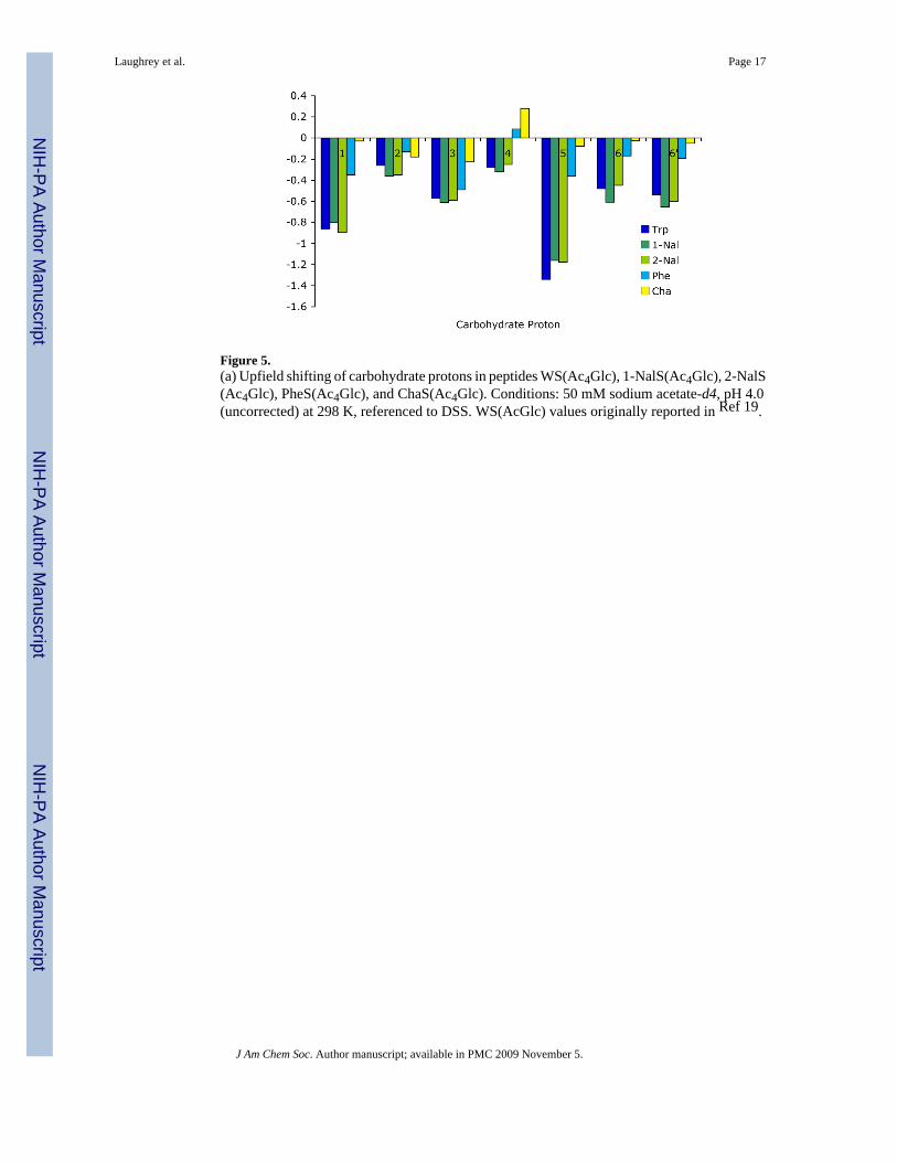
Figure 5.(a) Upfield shifting of carbohydrate protons in peptides WS(Ac4Glc), 1-NalS(Ac4Glc), 2-NalS(Ac4Glc), PheS(Ac4Glc), and ChaS(Ac4Glc). Conditions: 50 mM sodium acetate-d4, pH 4.0(uncorrected) at 298 K, referenced to DSS. WS(AcGlc) values originally reported in Ref 19.
Laughrey et al. Page 17
J Am Chem Soc. Author manuscript; available in PMC 2009 November 5.
NIH
-PA Author Manuscript
NIH
-PA Author Manuscript
NIH
-PA Author Manuscript
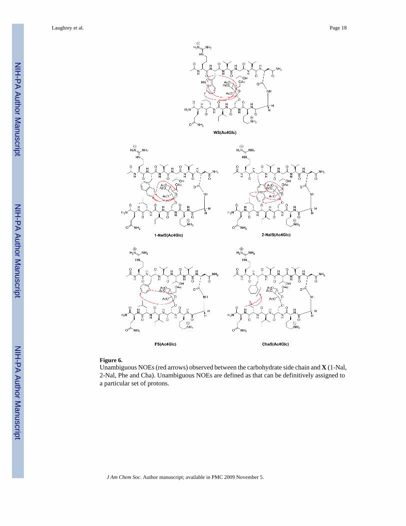
Figure 6.Unambiguous NOEs (red arrows) observed between the carbohydrate side chain and X (1-Nal,2-Nal, Phe and Cha). Unambiguous NOEs are defined as that can be definitively assigned toa particular set of protons.
Laughrey et al. Page 18
J Am Chem Soc. Author manuscript; available in PMC 2009 November 5.
NIH
-PA Author Manuscript
NIH
-PA Author Manuscript
NIH
-PA Author Manuscript

Figure 7.A Trp-Gal interaction in the binding pocket of galectin.43
Laughrey et al. Page 19
J Am Chem Soc. Author manuscript; available in PMC 2009 November 5.
NIH
-PA Author Manuscript
NIH
-PA Author Manuscript
NIH
-PA Author Manuscript

Figure 8.Fraction folded as determined from Hα chemical shifts. WS(Ac4Glc) values were originallyreported in Ref 19.
Laughrey et al. Page 20
J Am Chem Soc. Author manuscript; available in PMC 2009 November 5.
NIH
-PA Author Manuscript
NIH
-PA Author Manuscript
NIH
-PA Author Manuscript

Figure 9.Upfield shifting of carbohydrate protons in peptides. WS(Ac4Glc) values were originallyreported in Ref 19.
Laughrey et al. Page 21
J Am Chem Soc. Author manuscript; available in PMC 2009 November 5.
NIH
-PA Author Manuscript
NIH
-PA Author Manuscript
NIH
-PA Author Manuscript

Figure 10.Unambiguous NOEs (red arrows) observed between the carbohydrate side-chain (Ac4Gal,Ac3GlcNAc, GlcNAc, and Me4Glc) and Trp. Unambiguous NOEs are defined as that can bedefinitively assigned to a particular set of protons.
Laughrey et al. Page 22
J Am Chem Soc. Author manuscript; available in PMC 2009 November 5.
NIH
-PA Author Manuscript
NIH
-PA Author Manuscript
NIH
-PA Author Manuscript

Figure 11.Electrostatic potential maps of the sidechains at position Z in the β-hairpin. (a) Ac4Glc; (b)Glc; (c) Me4Glc. Electrostatic potential maps were generated with MacSpartan: HF/6-31g*;Isodensity value = 0.02; range = -25 (red, electron rich) to 25 kcal/mol (blue, electron poor).
Laughrey et al. Page 23
J Am Chem Soc. Author manuscript; available in PMC 2009 November 5.
NIH
-PA Author Manuscript
NIH
-PA Author Manuscript
NIH
-PA Author Manuscript

NIH
-PA Author Manuscript
NIH
-PA Author Manuscript
NIH
-PA Author Manuscript
Laughrey et al. Page 24Ta
ble
1Fr
actio
n Fo
lded
and
ΔG
°(fo
ldin
g) a
t 298
K fo
r β-h
airp
ins.a
XS(
Z)
ΔδG
ly (p
pm)
(± 0
.005
ppm
)Fr
actio
n Fo
lded
(Gly
split
ting)
bFr
actio
n Fo
lded
(Hα)
cΔG
(fold
ing)
(kca
l mol
-1)d
Trpe
Ac 4
Glc
0.48
40.
850.
83 (0
.02)
-1.0
3
1-N
alA
c 4G
lc0.
484
0.86
0.83
(0.0
3)-1
.08
2-N
alA
c 4G
lc0.
471
0.83
0.81
(0.0
4)-0
.94
Phe
Ac 4
Glc
0.32
50.
570.
51 (0
.13)
-0.1
7
Cha
Ac 4
Glc
0.24
20.
450.
42 (0
.12)
0.12
(a) C
ondi
tions
: 50
mM
sodi
um a
ceta
te-d
4, p
H 4
.0 (u
ncor
rect
ed) a
t 298
K, r
efer
ence
d to
DSS
.
(b) Er
ror i
s ± 0
.01.
(c) Hα
frac
tion
fold
ed w
as d
eter
min
ed fr
om th
e av
erag
e of
the
valu
es fo
r Val
3, V
al 5
, Lys
8, a
nd Il
e 10
. The
stan
dard
dev
iatio
n is
show
n in
par
enth
eses
.
(d) Δ
G d
eter
min
ed u
sing
gly
cine
-spl
ittin
g va
lues
; err
or is
± 0
.05
kcal
/mol
.
(e) Pr
evio
usly
repo
rted
in R
ef 1
9 .
J Am Chem Soc. Author manuscript; available in PMC 2009 November 5.

NIH
-PA Author Manuscript
NIH
-PA Author Manuscript
NIH
-PA Author Manuscript
Laughrey et al. Page 25
Table 2The Diagonal Interaction Energies Between Residues 2 and 9 as Determined by Double Mutant Cycles
X S(Z) ΔΔG (kcal mol-1)a
Trpb Ac4Glc -0.8
1-Nal Ac4Glc -0.7
2-Nal Ac4Glc -0.7
Phe Ac4Glc -0.5
Cha Ac4Glc -0.1
aThe error in the diagonal interaction energy is ±0.1 kcal mol-1
bPreviously reported in Ref 19
J Am Chem Soc. Author manuscript; available in PMC 2009 November 5.
NIH
-PA Author Manuscript
NIH
-PA Author Manuscript
NIH
-PA Author Manuscript
Laughrey et al. Page 26
Table 3Thermodynamic parameters for folding at 298 K obtained from thermal denaturation of the peptides.
Peptide ΔH°(kcal mol-1)
ΔS°(cal mol-1 K-1)
ΔCP(cal mol-1 K-1)
WS(Ac4Glc)a -5.9 -16.4 -112
FS(Ac4Glc) -4.23 -13.77 -77
ChaS(Ac4Glc) -2.96 -10.32 -88
(a)Previously reported in Ref 19.
J Am Chem Soc. Author manuscript; available in PMC 2009 November 5.

NIH
-PA Author Manuscript
NIH
-PA Author Manuscript
NIH
-PA Author Manuscript
Laughrey et al. Page 27Ta
ble
4β-
Hai
rpin
Fra
ctio
n fo
lded
and
ΔG
(fol
ding
) at 2
98 K
.
XS(
Z)
ΔδG
ly(±
0.0
05 p
pm)
Frac
tion
Fold
ed(G
ly sp
littin
g)b
Frac
tion
Fold
ed(Hα)
cΔG
(fold
ing)
(kca
l/mol
)d
Trp
eA
c 4G
lc0.
484
0.85
0.83
(0.0
2)-1
.03
Trp
Ac 4
Gal
0.46
60.
820.
77 (0
.04)
-0.9
0
Trp
Ac 3
Glc
NA
c0.
421
0.73
0.66
(0.0
9)-0
.59
Trp
Glc
NA
c0.
392
0.70
0.64
(0.0
2)-0
.50
Trp
Me 4
Glc
0.50
0.88
0.81
(0.0
1)-1
.18
Trp
cG
lc0.
383
0.65
0.63
(0.0
2)-0
.37
Trp
OH
0.37
50.
640.
60 (0
.01)
-0.3
4
(a) C
ondi
tions
: 50
mM
sodi
um a
ceta
te-d
4, p
H 4
.0 (u
ncor
rect
ed) a
t 298
K, r
efer
ence
d to
DSS
.
(b) Er
ror i
s ± 0
.01.
(c) Hα
frac
tion
fold
ed w
as d
eter
min
ed fr
om th
e av
erag
e of
the
valu
es fo
r Val
3, V
al 5
, Lys
8, a
nd Il
e 10
; the
stan
dard
dev
iatio
n is
show
n in
par
enth
eses
.
(d) Δ
G d
eter
min
ed u
sing
gly
cine
-spl
ittin
g va
lues
; err
or is
± 0
.05
kcal
/mol
.
(e) Pr
evio
usly
repo
rted
in R
ef 1
9 .
J Am Chem Soc. Author manuscript; available in PMC 2009 November 5.

NIH
-PA Author Manuscript
NIH
-PA Author Manuscript
NIH
-PA Author Manuscript
Laughrey et al. Page 28
Table 5The diagonal interaction energies as determined by Double Mutant Cycles
X Y ΔΔG (kcal mol-1)a
Trpb Ac4Glc -0.8
Trp Ac4Gal -0.7
Trp Ac3GlcNAc -0.6c
Trp GlcNAc -0.5c
Trp Me4Glc -0.8c
aThe error in the diagonal interaction energy is ±0.1 kcal mol-1
bPreviously reported in Ref 19.
cAlthough the interaction enery for this mutant is similar to that of the Trp-Ac4Glc interaction, the NMR data suggests that an interaction other than the
carbohydrate-π interaction is contributing. See text for details.
J Am Chem Soc. Author manuscript; available in PMC 2009 November 5.