Evaluation of a plasmid-based 16S–23S rDNA intergenic spacer region array for analysis of...
15
Evaluation of a plasmid-based 16S–23S rDNA intergenic spacer region array for analysis of microbial diversity in industrial wastewater $ Kimberly L. Cook a,b,c , Alice C. Layton a,b , Hebe M. Dionisi a,1 , James T. Fleming a,b , Gary S. Sayler a,b, * a Center for Environmental Biotechnology, The University of Tennessee, 676 Dabney Hall, Knoxville, TN 37996, USA b Department of Microbiology, The University of Tennessee, Knoxville, TN 37996, USA c National Center for Agricultural Utilization Research, USDA, Agricultural Research Service, 1815 North University Street, Peoria, IL 61604, USA Received 18 July 2003; received in revised form 2 December 2003; accepted 3 December 2003 Abstract A plasmid-based 16S – 23S rDNA intergenic spacer region (ISR) array was developed and optimized for analysis of microbial diversity within complex environmental samples. Plasmid probes with 16S – 23S rDNA ISR inserts (800 – 1500 bp) from industrial wastewater treatment plant (WWTP) microorganisms were arrayed onto glass slides. Hybridization of fluorescently labeled target sequences from two clones from the ISR WWTP library to arrayed probes showed that there was a good linear relationship between hybridization intensity and ISR similarity (r 2 = 0.82). Hybridization was highly specific (average background from arrayed probes with less than 80% similarity in ISR sequence was less than 7%). Strong fluorescence intensity corresponded to near-perfect match clones (99% or greater similarity in ISR sequence). A majority of probes (79%) showed no background hybridization. However, weak background (less than 50% for arrayed probes with 90% and 95% similarity in the 16S rRNA genes) was observed from closely related microorganisms. Background fluorescence from the negative control (plasmid vector with no insert) was similar to water and dimethyl sulfoxide (DMSO)-negative controls. Hybridization using fluorescently labeled ISR sequences from a mixed community sample produced strong fluorescent signals with no background from negative controls. A Cy5-labeled reference standard, part of the vector and present in every spotted probe, was used to normalize hybridization values. These results indicate that arrayed plasmid containing ISR probe insert sequences provides specificity and sensitivity for microbial community analysis in a high-throughput array format. D 2004 Elsevier B.V. All rights reserved. Keywords: Intergenic spacer; Array; Microarray; ISR; Activated sludge; Wastewater 0167-7012/$ - see front matter D 2004 Elsevier B.V. All rights reserved. doi:10.1016/j.mimet.2003.12.008 $ Names are necessary to report factually on available data; however, the USDA neither guarantees nor warrants the standard of the product, and the use of the name by the USDA implies no approval of the product to the exclusion of others that may also be suitable. * Corresponding author. Tel.: +1-865-974-8080; fax: +1-865-974-8086. E-mail address: [email protected] (G.S. Sayler). 1 Present address: CENPAT-CONICET, Puerto Madryn, Chubut, Argentina. www.elsevier.com/locate/jmicmeth Journal of Microbiological Methods 57 (2004) 79 – 93
-
Upload
independent -
Category
Documents
-
view
3 -
download
0
Transcript of Evaluation of a plasmid-based 16S–23S rDNA intergenic spacer region array for analysis of...

www.elsevier.com/locate/jmicmeth
Journal of Microbiological Methods 57 (2004) 79–93
Evaluation of a plasmid-based 16S–23S rDNA intergenic
spacer region array for analysis of microbial diversity
in industrial wastewater$
Kimberly L. Cooka,b,c, Alice C. Laytona,b, Hebe M. Dionisia,1,James T. Fleminga,b, Gary S. Saylera,b,*
aCenter for Environmental Biotechnology, The University of Tennessee, 676 Dabney Hall, Knoxville, TN 37996, USAbDepartment of Microbiology, The University of Tennessee, Knoxville, TN 37996, USA
cNational Center for Agricultural Utilization Research, USDA, Agricultural Research Service, 1815 North University Street,
Peoria, IL 61604, USA
Received 18 July 2003; received in revised form 2 December 2003; accepted 3 December 2003
Abstract
A plasmid-based 16S–23S rDNA intergenic spacer region (ISR) array was developed and optimized for analysis of
microbial diversity within complex environmental samples. Plasmid probes with 16S–23S rDNA ISR inserts (800–1500 bp)
from industrial wastewater treatment plant (WWTP) microorganisms were arrayed onto glass slides. Hybridization of
fluorescently labeled target sequences from two clones from the ISR WWTP library to arrayed probes showed that there was a
good linear relationship between hybridization intensity and ISR similarity (r2 = 0.82). Hybridization was highly specific
(average background from arrayed probes with less than 80% similarity in ISR sequence was less than 7%). Strong fluorescence
intensity corresponded to near-perfect match clones (99% or greater similarity in ISR sequence). A majority of probes (79%)
showed no background hybridization. However, weak background (less than 50% for arrayed probes with 90% and 95%
similarity in the 16S rRNA genes) was observed from closely related microorganisms. Background fluorescence from the
negative control (plasmid vector with no insert) was similar to water and dimethyl sulfoxide (DMSO)-negative controls.
Hybridization using fluorescently labeled ISR sequences from a mixed community sample produced strong fluorescent signals
with no background from negative controls. A Cy5-labeled reference standard, part of the vector and present in every spotted
probe, was used to normalize hybridization values. These results indicate that arrayed plasmid containing ISR probe insert
sequences provides specificity and sensitivity for microbial community analysis in a high-throughput array format.
D 2004 Elsevier B.V. All rights reserved.
Keywords: Intergenic spacer; Array; Microarray; ISR; Activated sludge; Wastewater
0167-7012/$ - see front matter D 2004 Elsevier B.V. All rights reserved.
doi:10.1016/j.mimet.2003.12.008
$ Names are necessary to report factually on available data; however, the USDA neither guarantees nor warrants the standard of the
product, and the use of the name by the USDA implies no approval of the product to the exclusion of others that may also be suitable.
* Corresponding author. Tel.: +1-865-974-8080; fax: +1-865-974-8086.
E-mail address: [email protected] (G.S. Sayler).1 Present address: CENPAT-CONICET, Puerto Madryn, Chubut, Argentina.

K.L. Cook et al. / Journal of Microbiological Methods 57 (2004) 79–9380
1. Introduction
In recent years, evaluation of microbial diversity in
environmental systems has been aided by molecular
techniques that are less selective than traditional cul-
tural techniques and provide a more complete view of
the complexity within environmental systems (Ogram
and Sayler, 1988; Amann et al., 1995; Massol-Deya et
al., 1995; Gurtler and Stanisich, 1996). In particular,
molecular methods based on the 16S rRNA gene
analysis have provided unique insights into the breadth
of microbial diversity (Hugenholtz et al., 1998). This
sequence is universally present in all bacteria and
contains highly conserved regions interspersed with
variable regions, thereby facilitating the design of
primers and probes for analysis of bacterial phylogeny
at different levels of specificity (i.e., family, genus, or
species level)(Amann et al., 1995; Cilia et al., 1996).
Many 16S rRNA-based molecular techniques [dena-
turing gradient gel electrophoresis (DGGE), restriction
fragment length polymorphism (tRFLP), and ARDRA
(amplified rDNA restriction analysis)] have been de-
veloped for community-level analysis of differences in
microbial composition associated with environmental,
spatial, or temporal changes. However, these methods
yield complex community profiles that provide only
superficial information about community ecology
(Kent and Triplett, 2002; Valinsky et al., 2002). Addi-
tionally, the size of the 16S rRNA gene is extremely
constant, limiting its use in gel-based community-
fingerprinting techniques, and 16S rRNA gene sequen-
ces may be too highly conserved to permit analysis of
phylogenetic relationships between closely related spe-
cies (Cilia et al., 1996; Mendoza et al., 1998; Aakra et
al., 1999).
In contrast to the rRNA genes, the intergenic
spacer region (ISR) located between the 16S rRNA
and 23S rRNA genes is under less selective pressure
(Buchan et al., 2001). Therefore, the 16S–23S rDNA
ISR exhibits greater sequence diversity and length
variation than the 16S rRNA gene (Jensen et al., 1993;
Gurtler and Stanisich, 1996; Nagpal et al., 1998). This
variability is due primarily to insertion and deletion
events and the presence or absence of tRNA sequen-
ces. Divergence within the ISR has been used to
confirm 16S rRNA gene-based phylogeny and has
been shown to provide better discrimination of closely
related species than 16S rRNA characterization (Cilia
et al., 1996; Mendoza et al., 1998; Aakra et al., 1999;
Adb-El-Haleem et al., 2002). However, ISR sequen-
ces among closely related microorganisms (species
level) are conserved due to the location of the ISR
between the highly conserved 16S rRNA and 23
rRNA genes and transcription of the rrn operon as a
single pre-RNA transcript (Garcia-Martinez et al.,
1999). Concerted evolution of multigene families
and homologous recombination between similar ISRs
also contributes to sequence conservation within the
ISR despite limited evolutionary constraints (Anton et
al., 1998; Gurtler, 1999). Therefore, ISR sequences
are often conserved within a species, but rarely
beyond the genus or family level.
The 16S–23S rDNA ISR has been used to evaluate
mixed microbial communities within environmental
samples via clonal library construction and commu-
nity-level fingerprinting (Acinas et al., 1999; Fisher
and Triplett, 1999; Ranjard et al., 2000). Conserved
sequences within the 16S rRNA and 23S rRNA genes
were used as primer targets for amplifying the spacer
region of a broad range of microorganisms in a single
amplification reaction (Jensen and Straus, 1993).
Polymerase chain reaction (PCR) products, including
a portion of the 16S rRNA gene, can be used for
phylogenetic analysis (Acinas et al., 1999). ISR com-
munity level profiling was used to characterize com-
munity samples from Amazonian soils (Borneman
and Triplett, 1997), freshwater bacterial communities
(Fisher and Triplett, 1999), plant rhizosphere (Robleto
et al., 1998), and Mediterranean offshore waters
(Acinas et al., 1999). Microbial community profiling
based on ISR size differences is limited by the fact
that multiple microorganisms (either closely related
strains or completely unrelated species) may possess
ISR regions of identical length (Garcia-Martinez et al.,
1999). Alternatively, one microorganism may possess
multiple ISRs with significant length differences, so
that a single microorganism may contribute multiple
bands to a community profile (Fisher and Triplett,
1999).
Array technology is a high-throughput methodolo-
gy that permits parallel analysis of hundreds or
thousands of genetic sequences. For reviews on the
application of array technologies to environmental
sample analysis, see Cook and Sayler (2003) and
Zhou and Thompson (2002). Alternative array meth-
odologies have been applied for analysis of environ-

K.L. Cook et al. / Journal of Microbiological Methods 57 (2004) 79–93 81
mental samples through gene expression analysis
(Murray et al., 2001; Schut et al., 2001), bacterial
identification (Cho and Tiedje, 2001; Loy et al., 2002;
Wilson et al., 2002), and evaluation of functional
genes important in environmental ecosystems (Wu et
al., 2001). However, many of these arrays rely on
highly conserved 16S rRNA gene sequences and/or
require costly and time-consuming selection of oligo-
nucleotide probe sequences (Guschin et al., 1997; Loy
et al., 2002; Valinsky et al., 2002; Wilson et al., 2002).
This report describes the development and optimi-
zation of a plasmid-based 16S–23S rDNA ISR array
for characterization of microbial diversity within an
industrial wastewater treatment plant (WWTP). Con-
served sequences in the 16S rRNA and 23S rRNA
genes were used as primer targets to amplify ISR
sequences from a mixed microbial community. Phylo-
genetic information was obtained through analysis of
500 bp of 16S rRNA gene sequence. The PCR products
were cloned into plasmid vector and extracted plasmid
was arrayed onto glass slides. Hybridization of fluo-
rescently labeled samples against arrayed plasmid with
16S–23S rDNA ISR insert sequences provided a
strong, specific fluorescent signal. This plasmid-based
ISR array represents a new application of array tech-
nology in which community analysis can be achieved
with a high level of specificity without the need for
culture-based approaches or sequence analysis.
2. Materials and methods
2.1. Evaluation of the ISR as a probe target for
characterization of mixed microbial communities
2.1.1. DNA extraction and preparation of clonal
library
DNAwas extracted from industrial WWTP-activat-
ed sludge samples collected from an industrial waste-
water treatment system in September 1999. A 50-ml
aliquot of each sample was pelleted by centrifugation
and genomic DNA was extracted from 100 mg wet
weight, as described by Layton et al. (2000). 16S–23S
rDNA ISR sequences from industrialWWTPmicrobial
communities were amplified using Ready-To-Go-PCR
Beads (Amersham Pharmacia, Piscataway, NJ), with
800-nM primers and 5–10 ng of template DNA. The
PCR primers were 1055f (5V ATG GCT GTC GTC
AGC T 3V) (Amann et al., 1995) and 23Sr (5V GGG
TTB CCC CAT TCR G 3V) (Borneman and Triplett,
1997). The touchdown PCR program used for 16S–
23S rDNA ISR analysis was as follows: 5 min at 94 jC,then 10 cycles consisting of 15 s at 94 jC, 45 s at 65 jC,followed by a 1 jC decrease for each of the 10 cycles,
and 60 s at 72 jC, followed by an additional 20 cycles
of 15 s at 94 jC, 45 s at 55 jC, and 60 s at 72 jC, and afinal cycle of 60 s at 72 jC.Aliquots of 20 Al of the PCRproduct were electrophoresed in 2% agarose gels in
0.5� TBE solution (0.045M Tris–borate, 0.001 M
EDTA, pH 8.0).
PCR products were cloned into the pCRR 2.1-
TOPO TA vector (TA vector; Invitrogen, Carlsbad,
CA). Large-scale plasmid DNA extractions were per-
formed by using an alkaline lysis procedure from
Promega Tech Bulletin .009 (Promega, Madison,
WI). Contaminating RNAwas removed by incubating
the plasmid preparations with 10 Ag of DNAase-free
RNAase per milliliter of preparation followed by
phenol–chloroform extraction and ethanol precipita-
tion. Clones were characterized using phylogenetic
information obtained from sequencing approximately
450 bp of the 16S rRNA using the 1492 r primer (5VTAC GGY TAC CTT GTT ACG ACT T 3V) (Lane,1991). All sequencings were performed at the Univer-
sity of Tennessee Molecular Biology Resource Facility
using an Applied Biosystems 373 DNA sequencer
(Perkin-Elmer, Foster City, CA).
2.1.2. Construction of a prototype nylon membrane
ISR array
The 16S–23S rDNA ISR inserts were amplified
from purified plasmid from 11 WWTP clones as
described above, except that 0.8–1.1 ng of plasmid
template DNA was used along with the primers
1492f (5V AAG TCG TAA CAA GGT ARC CGT
A 3V) and 23Sr. The amplified fragments were
cleaned using the QIAquick PCR purification kit
(QIAGEN, Valencia, CA). Purified PCR products
(probes) were quantified using Hoechst Dye (Biorad,
Hercules, CA) measured at an excitation of 346 nm
and emission of 460 nm with a DyNA Quant 200
fluorometer (Amersham Biosciences, Piscataway,
NJ). The PCR product was diluted to produce probes
at final concentrations of 0.4, 0.2, and 0.04 pmol.
The probes were blotted onto BIOTRANS nylon
membranes (ICN Pharmaceuticals, Costa Mesa,

K.L. Cook et al. / Journal of Microbiological Methods 57 (2004) 79–9382
CA) using the dot blot procedure described by
Dionisi et al. (2002).
2.1.3. Cross-hybridization analysis
Arrays were prehybridized in solution containing
0.5 M NaH2PO4, 1 mM EDTA, and 7% (wt/vol)
sodium dodecyl sulfate (SDS) for 1 h at 65 jC in a
shaking water bath. Hybridizations were carried out
overnight following addition of 32P-labeled target
ISR DNA (1.0� 106 to 3.0� 106 cpm). Eleven
separate arrays were hybridized using labeled target
ISR sequences from each of the different clones.
Hybridization was performed at 65 jC and mem-
branes were washed in high stringency wash buffer
(10 mM NaCl, 10 mM Tris base, 1 mM EDTA, and
0.5% SDS) at 65, 70, and 75 jC. Following each
wash, the arrays were exposed to a phosphor screen
(Molecular Dynamics, Sunnyvale, CA) and the ex-
posed screen was scanned on a STORM 840 Phos-
phorImager (Molecular Dynamics). The resulting
images were analyzed by determining pixel density
for each spot using ImageQuaNTk 5.0 analysis
software (Molecular Dynamics).
2.2. Evaluation of glass slide plasmid-based ISR
array
2.2.1. Construction of plasmid-based 16S–23S RDNA
ISR array
Samples from the same industrial WWTP were
collected in July 2001 and genomic DNAwas extracted
from 2 ml of mixed liquor using the FastDNA kit (Bio
101, Vista, CA) as previously described (Dionisi et al.,
2002). 16S–23S rDNA ISRs of microorganisms from
the industrial WWTP were amplified, PCR products
were cloned into the pCRR 2.1-TOPO TA vector, and
plasmid DNAwas extracted. The ISR array was com-
posed of 160 plasmids from the industrial WWTP
16S–23S rDNA ISR library. Each glass slide contained
triplicate ISR arrays. TAvector with no insert, dimethyl
sulfoxide (DMSO; Sigma, St. Louis, MO), and water
blanks served as negative controls. Probes consisting of
plasmid containing a 16S–23S rDNA ISR insert were
spotted in 50%DMSO at a final concentration between
400 and 550 ng/Al. Ten microliters of plasmid probe in
an equal volume of DMSO was placed into 384-well
microtiter plates. Probes were arrayed at a spacing of
350 Am onto Cel Associates (Houston, TX) poly-L-
lysine-coated slides in a grid of eight rows by six
columns. Arrays were printed at 57% relative humidity
and 22jC using a Virtek robotic printer (Waterloo,
Canada) with eight pins (TeleChem International, Sun-
nyvale, CA). Following printing, ISR arrays were
rehydrated in a glass Petri dish containing moist filter
paper (Qualitative; Whatman, Ann Arbor, MI) for 5
min and snap-dried at 80 jC for 5 s. ISR arrays were
then UV cross-linked at 150 mJ in a UV Spectrolinker
XL1500 (Spectronics, Westbury, NY). Arrays were
blocked in a solution of 1.5% succinic anhydride
(Sigma), 90% 1-methyl-2-pyrrolidinone (Sigma), and
43 mM boric acid (pH 8.0) (Sigma) for 15 min at room
temperature. Following blocking, DNA on the arrays
was denatured by boiling at 95 jC for 2 min. Slides
were dried by centrifugation using a microarray cen-
trifuge (TeleChem International) at 2000� g for 20 s.
Two slides from each printed batch were evaluated for
quality of printing by staining with Sybr Green II
(Molecular Probes, Eugene, OR) diluted 1:10,000 in
0.5� TBE. Arrays were stained for 1 min and then
washed in 0.5� TBE and sterile deionizedwater for 30
s each prior to scanning using the Scan Array 5000
MicroArray Analysis System (GSI Lumonics, Water-
town, MA) at an emission wavelength of 522 nm.
A subarray containing probes from six 16S–23S
rDNA ISR clones from theWWTP library was used for
analysis of cross-hybridization and optimization of
hybridization conditions for the ISR array (Table 1).
The probes, as well as negative controls (TA vector
with no insert and DMSO), were spotted under the
conditions described above. Five replicates of the
subarray were printed on each slide.
2.2.2. Preparation of fluorescently labeled target
DNA
16S–23S rDNA ISR sequences from industrial
WWTP samples genomic DNA extracts (2–5 ng) were
amplified using the PCR conditions described above to
increase overall target signal intensity. 16S–23S rDNA
ISRs from plasmid insert sequences were selectively
amplified using PCR to minimize background from
vector. The resultant PCR products from the first
reactions were purified using the Zymo DNA Clean
and Concentrator-5 Kit (Zymo Research, Orange, CA).
Cleaned PCR product was diluted 1:1000 and 1 Al wasused as template for an incorporation reaction using the
PCR bead, primers (1492f and 23Sr), and 4 AM Cy3-

Table 1
Industrial WWTP 16S–23S rDNA ISR clones used for optimization of ISR array hybridization
Clone name Closest match (% similarity) Phylogenetic placement % GC ISR size
(bp)
Tma
(A) Prototype ISR array clonesb
TED99917 Hyphomicrobium hollandicum Y14303 (95%) Alpha subdivision, Hyphomicrobium 56 728 101
TED39909 Magnetospirillum gryphiswaldense Y10109 (94%) Alpha subdivision, Rhodospirillaceae 57 829 101
TED99907 Asticcacaulis excentricus AJ247194 (93%) Alpha subdivision, Caulobacter group 61 643 103
TED99915 Leptothrix discophora L33974 (98%) Beta subdivision, Comamonadaceae 56 558 100
TED99903 Thaurera aromatica AJ315680 (99%) Beta subdivision, Rhodocyclus branch 56 537 100
TED99928 Pseudomonas lanceolata AB021390 (99%) Beta subdivision, Comamonadaceae 49 616 98
TED99910 Methylococcus sp. X72769 (93%) Gamma subdivision, Methylococcaceae 54 548 100
TED99902 Lewinella persicus AF039295 (90%) Sphingobacteria, Sphingobacteriales 48 876 98
TED99906 Flexibacter tractuosus AB078076 (91%) Sphingobacteria, Sphingobacteriales 44 978 96
TED99901 Nitrosphira sp. Y14638 (98%) Nitrospirales, Nitrospiraceae 53 511 99
TED99926 Verrucomicrobium sp. AF027005 (97%) Verrucomicrobia 54 440 99
(B) ISR subarray clonesc
TED70103 Leptothrix mobilis X97071 (98%) Beta subdivision, Comamonadaceae 55 577 100
TED70101 L. mobilis X97071 (99%) Beta subdivision, Comamonadaceae 53 692 99
TED70129 P. lanceolata AB021390 (99%) Beta subdivision, Comamonadaceae 54 521 100
TED70105 Thauera aromatica AJ315680 (99%) Beta subdivision, Rhodocyclus branch 56 511 100
TED70113 Rhodobacter sphaeroides X53855 (94%) Alpha subdivision, Rhodobacter group 54 621 100
TED70109 Haliscomenobacter hydrossis M58790 (90%) Sphingobacteria, Sphingobacteriales 42 305 94
a Melting temperature of ISR sequence.b Clones used on phototype ISR array for cross-hybridization analysis.c Clones used on ISR subarray for formamide optimization experiments.
K.L. Cook et al. / Journal of Microbiological Methods 57 (2004) 79–93 83
labeled dCTP (Amersham Pharmacia, Peapack, NJ).
The labeled target DNA was purified by precipitation
with 2.5 vol of ethanol and 0.1 vol of sodium acetate
(Sigma) at � 20 jC overnight.
2.2.3. Optimization of hybridization specificity and
intensity using a formamide gradient
To optimize hybridization specificity and signal
intensity, 16S–23S rDNA ISR subarrays were hybrid-
ized in hybridization solution containing 30%, 40%,
50%, 60%, or 70% formamide. The array was replicat-
ed five times on each slide and hybridization at each
formamide concentration was replicated on three to
five separate slides. Labeled target DNA was resus-
pended in hybridization solution containing different
concentrations of formamide (Ambion, Austin, TX),
5� SSC, 0.1% SDS, and 0.5 Ag of unlabeled herring
sperm DNA (Sigma) in a total volume of 60 Al.Fluorescently labeled target was denatured at 95 jCfor 2 min and cooled to ambient temperature by
centrifugation for 2 min. Denatured probe was depos-
ited into an adhesive-backed Frame Seal (MJ Research,
Waltham, MA) chamber placed around the arrayed
DNA on the glass slide array, and the chamber was
then sealed with an adhesive plastic coverslip. All
hybridizations were performed in anMJ Research Twin
Tower Sample Block (MJ Research) at 45 jC. Follow-ing hybridization, the arrays were washed in separate
chambers with 2� SSC and 0.1% SDS for 5 min at the
hybridization temperature, and then with 0.1� SSC
and 0.1% SDS for 5 min at room temperature, followed
by 0.1� SSC for 1min four times at room temperature.
Residual salts were removed by centrifugation. Slides
were scanned at a resolution of 5 AM with the Sca-
nArray 3000 confocal laser scanner. Signal intensities
for each spot were determined using QuantArray 3.0
image analysis software (Perkin Elmer, Boston, MA).
ANOVA was carried out using SPSS version 11.0
(SPSS, Chicago, IL).
2.2.4. Analysis of industrial WWTP sludge library
using the 16S–23S rDNA ISR array
Labeled target sequences from clones TED70103 or
TED70109 (Table 1) from the industrial WWTP library
were hybridized to arrayed plasmid probes on the ISR
array. A formamide concentration of 50% was used in

K.L. Cook et al. / Journal of Microbiological Methods 57 (2004) 79–9384
the hybridization solution and all hybridizations were
performed at 45 jC. Following hybridization, arrays
were scanned and analyzed as described above. Plas-
mids from selected spots were sequenced for phyloge-
netic analysis, using the 16S rRNA gene portion of the
insert (1055f primer), and for ISR similarity, using the
1492f primer. Spearman correlation coefficients (r)
were calculated to find relationships between hybrid-
ization ratio and ISR or 16S rRNA gene sequences.
2.3. Hybridization of plasmid-based ISR array with
DNA from mixed community
Genomic DNA was extracted from WWTP micro-
organisms that were inoculated into plant rhizosphere
and grown for 13 days. DNAwas extracted from 10 to
15 ml of plant rhizosphere suspension using theMoBio
Soil Extraction Kit (Solana Beach, CA). Genomic
extract (2–5 ng) was labeled with Cy3-dCTP as
described above. The kanamycin gene from the pCRR2.1-TOPO TA vector was used as reference DNA by
incorporation of Cy5-dCTP (Amersham Pharmacia)
using primers KanF (5VTCA GGG CGC AAG GGC
3V; corresponding to sequence positions 963–977 of
the TAvector) and KanR (5VACT CTTCCT TTT TCA
ATT CAG AAG AAC 3V; corresponding to sequence
positions 2107–2133 of the TA vector). The Cy3-
labeled target and the Cy5-labeled reference standard
were mixed in equal portions and hybridized to the
array simultaneously. The arrays were hybridized,
washed, and analyzed as described above.
2.4. Nucleotide sequence accession numbers
Clone sequences for 16S–23S rDNA ISR inserts
from plasmids used for ISR array analysis were
deposited into GenBank and received GenBank ac-
cession numbers AY484706 to AY484734,
AY484743, and AF420301.
3. Results
3.1. Evaluation of the ISR as a probe target for
characterization of mixed microbial communities
16S–23S rDNA ISR probes from a phylogeneti-
cally diverse group of WWTP clones were used in a
prototype array to evaluate the feasibility of using the
16S–23S rDNA ISR as probes for characterization of
mixed microbial communities in an array format. The
1492f primer was used to produce labeled target
sequences to avoid amplification of conserved
sequences present in the 16S rRNA portion of the
ISR insert. The 23Sr primer used in these studies
anneals 115 bp into the 23S rRNA. This primer was
chosen because it is more conserved than previously
used 23S rRNA-targeted primers that are specific for
sequences closer to the ISR region (Gurtler and
Stanisich, 1996). Clones were sequenced from the
16S rRNA end of the PCR product using the 1055f
primer. The approximately 500 bp of partial 16S
rRNA gene sequence was used to obtain phylogenetic
information. The names and accession numbers of
microorganisms from GenBank that most closely
matched each of the 11 clones used for cross-hybrid-
ization analysis, as well as the tentative phylogenetic
placement, clone number, percent GC of the ISR, ISR
size, and Tm are given in Table 1A. The 16S–23S
rDNA ISR library produced from WWTP mixed
communities was diverse with representatives from
the alpha, beta, and gamma proteobacteria as well as
Nitrospira, Verrucomicrobia, and Sphingobacteria.
ISR size varied between 440 and 978 bp. Tm for the
11 clones varied between 96 and 103 jC, with an
average of 99 jC (F 2 jC).Cross-hybridization analysis using selected clones
revealed that at low stringency (65 jC wash), some
cross-hybridization occurred, that is, a labeled target
using clone TED39909 (Magnetospirillum sp.), an
alpha proteobacterium, produced 35.8% background
signal from spotted probe sequence TED99907
(Asticcacaulis sp.), another alpha proteobacterium
(Table 2). This background was reduced at higher
wash stringency (75 jC). At a wash temperature of 75
jC, the same labeled target (TED39909) showed only
2.1% background from the alpha proteobacterium
TED99909. At the lowest stringency, wash tempera-
ture (65 jC) background averaged 10% (F 20%) over
the entire group. At the highest stringency, wash
temperature (75 jC) background averaged 1.5%
(F 2%). Based on these results, it was determined
that arrayed 16S–23S rDNA ISR provided a useful
target for hybridization analysis with minimum po-
tential for cross-hybridization given sufficiently strin-
gent hybridization conditions.

Table 2
Characterization of percent cross-hybridization between ISRs of different phylogenetic groups
Wash Clone Phylogeny Alpha proteobacteria Beta proteobacteria Gamma CFBa Nitrospira Verrucob
temperature
(jC)name
TED99917 TED39909 TED99907 TED99915 TED99903 TED99928 TED99910 TED99902 TED99906 TED99901 TED99926
65 TED99917 Hyphomicrobium 100.00 47.70 41.85 1.73 6.39 4.30 2.16 1.03 1.21 6.37 18.80
65 TED39909 Magnetosprillum 17.46 100.00 56.00 11.08 3.47 6.26 3.42 0.29 1.75 23.83 62.20
65 TED99907 Asticcacaulis 3.16 35.76 100.00 0.74 15.25 4.80 1.05 3.38 3.67 6.19 22.91
65 TED99915 Leptothrix 1.44 5.40 18.18 100.00 73.20 86.77 4.13 0.13 6.69 5.82 5.19
65 TED99903 Thauera 1.05 0.85 4.42 18.63 100.00 21.63 8.38 0.14 1.96 0.34 2.14
65 TED99928 Pseudomonas 0.13 0.66 1.52 0.51 14.14 100.00 0.20 0.11 1.26 � 0.02 3.68
65 TED99910 Methylococcus 1.10 12.24 24.29 3.89 76.01 17.37 100.00 3.19 1.42 1.81 11.20
65 TED99902 Lewinella 0.69 0.25 0.92 0.51 0.70 1.53 2.04 100.00 6.32 1.21 1.62
65 TED99906 Flexibacter 0.86 0.67 1.54 2.60 0.85 2.27 2.29 21.33 100.00 5.79 1.52
65 TED99901 Nitrospira 2.76 54.72 61.67 0.97 0.30 4.34 3.42 0.38 1.35 100.00 57.77
65 TED99926 Verrucomicrobium 1.42 38.68 40.26 0.18 8.52 1.46 1.34 0.30 0.21 15.14 100.00
70 TED99917 Hyphomicrobium 100.00 21.22 18.56 0.95 0.50 3.92 1.60 0.73 0.81 4.69 0.86
70 TED39909 Magnetosprillum 12.55 100.00 28.88 7.60 0.36 3.22 2.42 0.44 0.71 5.07 2.82
70 TED99907 Asticcacaulis 2.02 15.17 100.00 3.53 0.47 2.62 1.14 3.06 3.08 4.76 1.24
70 TED99915 Leptothrix 1.43 1.17 9.35 100.00 2.76 66.37 0.78 0.26 4.29 3.99 0.34
70 TED99903 Thauera 0.82 0.50 3.05 8.27 100.00 13.01 2.65 0.16 1.18 0.43 0.15
70 TED99928 Pseudomonas 0.02 0.38 1.24 1.31 1.20 100.00 0.22 0.25 0.34 0.21 0.05
70 TED99910 Methylococcus 0.79 6.58 10.93 1.89 0.32 9.73 100.00 4.41 0.58 0.82 0.64
70 TED99902 Lewinella 0.73 0.13 0.50 0.34 0.15 0.85 0.46 100.00 1.61 0.71 0.17
70 TED99906 Flexibacter 0.69 0.23 0.96 0.77 0.12 0.75 0.29 5.33 100.00 2.53 0.18
70 TED99901 Nitrospira 1.17 26.16 31.41 0.58 0.10 2.08 0.42 0.24 0.80 100.00 1.51
70 TED99926 Verrucomicrobium 0.66 15.74 20.24 0.10 0.78 0.95 0.12 0.19 0.05 3.71 100.00
75 TED99917 Hyphomicrobium 100.00 2.68 2.65 1.73 0.28 2.24 1.60 0.34 0.47 5.96 2.06
75 TED39909 Magnetosprillum 3.97 100.00 4.47 2.83 0.16 1.50 2.42 0.88 0.19 1.03 3.49
75 TED99907 Asticcacaulis 0.59 2.13 100.00 0.94 0.19 1.46 1.14 10.01 3.35 6.51 1.73
75 TED99915 Leptothrix 0.93 0.41 5.08 100.00 1.09 8.88 0.78 2.81 2.23 4.21 1.29
75 TED99903 Thauera 0.61 0.23 1.73 3.18 100.00 2.53 2.65 1.81 0.73 0.85 0.88
75 TED99928 Pseudomonas 0.03 0.12 0.80 0.14 0.47 100.00 0.22 1.77 0.22 0.50 2.03
75 TED99910 Methylococcus 0.40 0.76 2.51 0.87 0.14 3.26 100.00 4.98 0.44 1.07 1.22
75 TED99902 Lewinella 0.58 0.06 0.29 0.34 0.07 0.62 0.46 100.00 1.70 0.31 0.70
75 TED99906 Flexibacter 0.60 0.17 0.65 1.03 0.06 0.58 0.29 4.51 100.00 1.76 0.95
75 TED99901 Nitrospira 0.25 2.50 2.64 0.54 0.06 1.29 0.42 1.90 0.77 100.00 2.89
75 TED99926 Verrucomicrobium 0.15 1.69 1.32 0.11 0.32 0.58 0.12 0.67 0.10 1.67 100.00
a CFB=Cytophaga, Flavobacterium, Bacteroides group.b Abbreviation for Verrucomicrobium.
K.L.Cooket
al./JournalofMicro
biologica
lMeth
ods57(2004)79–93
85

robiol
3.2. Construction of plasmid-based 16S–23S rDNA
ISR array
Plasmid probes extracted from a clonal library of
16S–23S rDNA ISR sequences from industrial
WWTP microorganisms were spotted onto glass
slides at a concentration of 400–550 ng/Al. This
concentration of starting material was necessary to
achieve saturating concentrations of spotted probe
since approximately one-fourth of the plasmid is
probe ISR sequence, the rest being vector sequence.
No effect of this concentration of starting material
was observed (i.e., there was no evidence of the
‘‘comet effect’’ typically seen when excessively high
concentrations of probe are spotted) (Eisen and
Brown, 1999). TA vector with no insert was spotted
onto the array and its intensity following hybridiza-
tion was subtracted from all other background-sub-
tracted intensity values to eliminate the effect of
nonspecific hybridization to the plasmid. Initial back-
ground levels from TA vector were minimal (5–7%)
and were only slightly higher than values for the
DMSO control (2–3%).
K.L. Cook et al. / Journal of Mic86
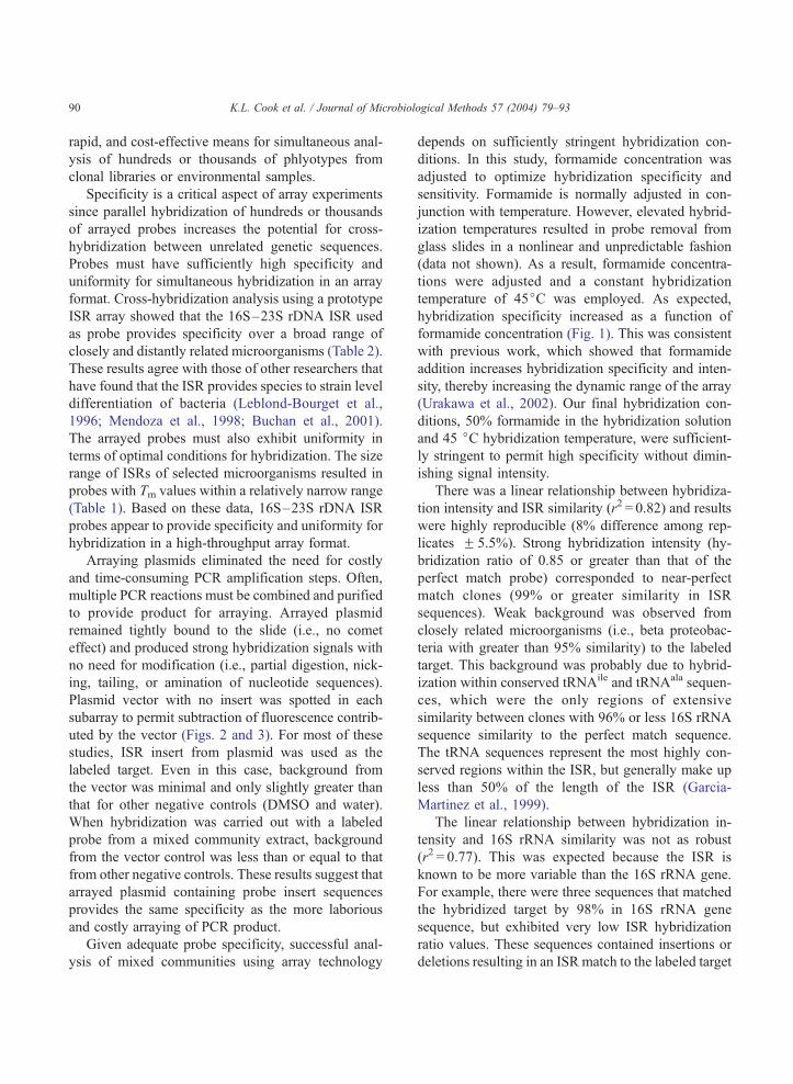
Fig. 1. Effect of formamide concentration on hybridization specificity. Each
subarrays were tested at each formamide concentration. The hybridization
each spot in the subarray to the final fluorescence intensity of the 100% 16
rRNA, and 80% ISR match to target sequence (.); TED70129, 94% 16S r
16S rRNA, and 70% ISR match to target sequence (E); TED70113, 8
TED70109, 80% 16S rRNA, and 58% ISR match to target sequence (x).
3.3. Optimization of hybridization specificity and
intensity using a formamide gradient
Hybridization specificity was optimized using a
subarray of six probes from the WWTP ISR array.
Spotted probe 16S–23S rDNA ISR insert sequences
matched the labeled target 16S–23S rDNA ISR
sequence (TED70103) between 58% and 100% (Fig.
1). The names and accession numbers of microorgan-
isms from GenBank that most closely matched each of
the six plasmid probes spotted onto the subarray, as
well as their tentative phylogenetic placements, clone
number, percent GC of the ISR, ISR size, and Tm are
given in Table 1B. The subarray also contained TA
vector with no insert. The array was replicated five
times on each slide and hybridization at each form-
amide concentration was replicated in three to five
separate slides. Replicate spots from each array were
averaged to obtain one value per probe per slide. Final
fluorescence intensity (IV) for each spot resulted from
subtraction of local mean background and negative
control (TA vector with no insert) values from the
initial fluorescence intensity (I). The hybridization
ogical Methods 57 (2004) 79–93
subarray consisted of five replicate arrays and three to five separate
ratio is derived from the ratio of final fluorescence intensity (IV) forS rRNA and ISR matched probe (TED70103). TED70101, 98% 16S
RNA, and 74% ISR match to target sequence (n); TED70105, 90%
0% 16S rRNA, and 64% ISR match to target sequence (z); and

K.L. Cook et al. / Journal of Microbiological Methods 57 (2004) 79–93 87
ratio for each array spot was measured as the ratio of
IV for the test spot to IV for the control spot (the spot
matching the labeled target 100%; hybridization ra-
tio= IV test/IV control).ISR insert from the 100% matched clone
(TED70103) was labeled with Cy3-dCTP and hybrid-
ized to the subarray in hybridization solution with
formamide concentrations ranging between 30% and
70%. Specificity was found to increase with formam-
ide concentration (Fig. 1). ANOVA suggested that
hybridization specificity significantly increased at a
formamide concentration of 50% or greater (P < 0.05;
Fig. 1). At low hybridization stringency, the closest
sequence matches exhibited some background hybrid-
ization. At a formamide concentration of 70%, there
was no detectable background from other clones;
however, overall intensity was also lower (30% lower
than the intensity at the lowest concentration of
formamide). At a formamide concentration of 50%,
background hybridization was less than 25%, and
overall intensity was 95% of the intensity at the lowest
concentration of formamide. Based on the level of
background hybridization and overall intensity, all
further experiments were conducted at 45 jC in
hybridization solution containing 50% formamide.
3.4. Analysis of industrial WWTP sludge library using
the 16S–23S rDNA ISR array
The 16S–23S rDNA ISR inserts of two clones
from the industrial WWTP library were used as
labeled targets to investigate the occurrence of these
sequences in the arrayed portion of the industrial
WWTP library and to evaluate the specificity of
hybridization. A common clone (TED70109; 16% of
sequenced clones in the library matched 99% in ISR
sequence) was chosen to evaluate hybridization spec-
ificity. A beta proteobacterium (TED70103) was less
common in the sequenced portion of the WWTP
library, but was chosen to evaluate the potential for
cross-hybridization between closely related microor-
ganisms. Fig. 2A shows the relationship between
hybridization ratio, 16S rRNA sequence similarity,
and ISR sequence similarity for 69 sequenced clones
hybridized against labeled target sequences from
clones TED70103 and TED70109. Spearman correla-
tion analysis of these data showed a positive correla-
tion between hybridization ratio and 16S–23S rDNA
ISR sequence (r = 0.63, P < 0.0005). Slide-to-slide va-
riation in signal intensity for replicate spots averaged
8.0% with a standard deviation of 5.5%. Given that
labeled targets are complementary to the ISR portion
of arrayed probes, it was not surprising to find that the
linear relationship between hybridization ratio and
ISR sequence similarity (r2 = 0.82) was better than that
for hybridization ratio and 16S rRNA gene sequence
similarity (r2 = 0.77). Two of four ISR subarrays from
the ISR array, hybridized with labeled target from
TED70103 and TED70109, are shown in Fig. 2B
and C, respectively.
Hybridization of labeled target from clone
TED70109 to the array resulted in 10 clones with a
hybridization ratio greater than 0.85 (Fig. 2A). These
clones were all greater than 99% matches to the
labeled target sequence in terms of ISR and 16S rRNA
gene sequence. A cluster of four clones had 99% 16S
rRNA gene sequence similarity to clone TED70109,
but had low hybridization ratios (between 0.60 and
0.38) (Fig. 2A). This discrepancy was due to the low
level of ISR sequence similarity (84%) between the
100% match clone and this cluster of clones. Differ-
ences in the ISR sequence were due to insertions and
single base-pair mismatches in comparison to the ISR
sequence of the perfect match clone.
Hybridizat ion of the less common clone
(TED70103) to the array produced only two matches
with a hybridization ratio greater than 0.55; both were
98% similar in 16S rRNA gene sequence, but only 78–
80% similar in ISR sequence to the perfect match (Fig.
2A). Only one other 98% 16S rRNA gene match to
TED70103 was identified in the sequenced portion of
the library. The clone had a hybridization ratio of 0.36.
This finding was consistent with a significantly longer
ISR region (692 bp versus 577 bp for clone
(TED70103) containing several long insertions. De-
spite fewer matched clones, hybridization with
TED70103, a beta proteobacterium, resulted in great-
er background hybridization by related beta proteo-
bacteria than that observed for the Sphingobacterium
clone TED70109, a more common clonal library
sequence (Fig. 2B and C). Background hybridization
ratio was less than 0.50 for arrayed probes matching
between 90% and 95% in 16S rRNA gene sequence
(less than 80% ISR match) and less than 0.10 for
arrayed probes with less than 90% match in 16S
rRNA sequence. More than 79% of the probes pro-

Fig. 2. (A) Relationship between hybridization ratio, ISR sequence similarity, and 16S rRNA sequence similarity. ISR sequences from two clones,
TED70103 (.) and TED70109 (o), were labeled with Cy3-dCTP and hybridized to separate arrays in triplicate. Cluster of four clones with 99%
16S rRNA sequence similarity to clone TED70109, but low ISR similarity and low hybridization ratios (o). (B) Two subarrays of the ISR array
hybridized with Cy3-dCTP-labeled ISR from clone TED70103. Red boxes indicate TA vector with no insert (negative control); white boxes
indicate the 100% match to the labeled target sequence. (C) Two subarrays of the ISR array hybridized with Cy3-dCTP-labeled ISR from clone
TED70109. Red boxes indicate TA vector with no insert (negative control); white boxes indicate the 100% match to the labeled target sequence.
K.L. Cook et al. / Journal of Microbiological Methods 57 (2004) 79–9388

K.L. Cook et al. / Journal of Microbiological Methods 57 (2004) 79–93 89
duced little or no signal (hybridization ratio less than
0.20) in comparison to the hybridization intensity of
the perfect match probe for both of the labeled targets
used for hybridization.
Fig. 3. Reproducibility of hybridization to triplicate arrays on one
slide. (A) Hybridization of Cy3-dCTP-labeled ISR from mixed
community to one array. The figure shows three subarrays
(horizontal row) of the array and triplicate arrays (vertical rows)
that are present on each slide. Red boxes indicate TA vector with no
insert (negative control). (B) Hybridization of Cy5-dCTP-labeled
kanamycin gene to the same array. The kanamycin gene is present in
the TA vector portion of every spot on the array. White boxes
indicate dropped spot.
3.5. Hybridization of plasmid-based 16S–23S rDNA
ISR array with DNA from mixed community
Hybridization using fluorescently labeled target
DNA from sludge organisms inoculated into plant
rhizosphere resulted in a strong fluorescent signal
from matching probe sequences. Hybridization to
ISR arrays was reproducible among triplicate arrays
on each slide and hybridization produced strong,
distinctive hybridization patterns that corresponded
to the overall diversity of organisms present in the
sample (Fig. 3). Normalization of spot intensity from
the sample (Cy3-dCTP-labeled ISR) to the intensity of
the reference sequence (Cy5-dCTP-labeled kanamy-
cin) present on every spotted plasmid on the array
minimized slide-to-slide variability due to differences
in the amount of arrayed probe material and some
differences in hybridization. Additionally, this nor-
malization procedure reduced within slide variability
(for replicate spots) due to differential hybridization
occurring over the surface of the slide. The kanamycin
hybridization also provided a visible measure of spot
quality (i.e., a spot dropped during printing was de-
tected in each of the replicate arrays seen in Fig. 3,
white boxes). As expected, hybridization with labeled
target from the environmental samples resulted in no
background from plasmid vector sequences (vector
control intensity less than or equal to DMSO and water
blanks; Fig. 3A). Coefficient of variation (CV) be-
tween replicate spots on the slide was fairly high (40%)
with a high standard deviation (54%). However, the
CV for spots with strong hybridization signals (hybrid-
ization ratio of 0.75 or greater) was very low (5F 4%).
These results indicate that variability was highest
among spots with low overall fluorescence, but was
very consistent among strongly positive responses.
4. Discussion
In this study, we report the use of a plasmid-based
16S–23S rDNA ISR array for analysis of microbial
diversity within complex environmental samples. The
ISR array utilizes the sequence diversity of the 16S–
23S rDNA ISR to obtain greater specificity than
traditional 16S rRNA gene-based methods. The array
format eliminates the need for length-based evaluation
of ISR differences and provides a high-throughput,
K.L. Cook et al. / Journal of Microbiological Methods 57 (2004) 79–9390
rapid, and cost-effective means for simultaneous anal-
ysis of hundreds or thousands of phlyotypes from
clonal libraries or environmental samples.
Specificity is a critical aspect of array experiments
since parallel hybridization of hundreds or thousands
of arrayed probes increases the potential for cross-
hybridization between unrelated genetic sequences.
Probes must have sufficiently high specificity and
uniformity for simultaneous hybridization in an array
format. Cross-hybridization analysis using a prototype
ISR array showed that the 16S–23S rDNA ISR used
as probe provides specificity over a broad range of
closely and distantly related microorganisms (Table 2).
These results agree with those of other researchers that
have found that the ISR provides species to strain level
differentiation of bacteria (Leblond-Bourget et al.,
1996; Mendoza et al., 1998; Buchan et al., 2001).
The arrayed probes must also exhibit uniformity in
terms of optimal conditions for hybridization. The size
range of ISRs of selected microorganisms resulted in
probes with Tm values within a relatively narrow range
(Table 1). Based on these data, 16S–23S rDNA ISR
probes appear to provide specificity and uniformity for
hybridization in a high-throughput array format.
Arraying plasmids eliminated the need for costly
and time-consuming PCR amplification steps. Often,
multiple PCR reactions must be combined and purified
to provide product for arraying. Arrayed plasmid
remained tightly bound to the slide (i.e., no comet
effect) and produced strong hybridization signals with
no need for modification (i.e., partial digestion, nick-
ing, tailing, or amination of nucleotide sequences).
Plasmid vector with no insert was spotted in each
subarray to permit subtraction of fluorescence contrib-
uted by the vector (Figs. 2 and 3). For most of these
studies, ISR insert from plasmid was used as the
labeled target. Even in this case, background from
the vector was minimal and only slightly greater than
that for other negative controls (DMSO and water).
When hybridization was carried out with a labeled
probe from a mixed community extract, background
from the vector control was less than or equal to that
from other negative controls. These results suggest that
arrayed plasmid containing probe insert sequences
provides the same specificity as the more laborious
and costly arraying of PCR product.
Given adequate probe specificity, successful anal-
ysis of mixed communities using array technology
depends on sufficiently stringent hybridization con-
ditions. In this study, formamide concentration was
adjusted to optimize hybridization specificity and
sensitivity. Formamide is normally adjusted in con-
junction with temperature. However, elevated hybrid-
ization temperatures resulted in probe removal from
glass slides in a nonlinear and unpredictable fashion
(data not shown). As a result, formamide concentra-
tions were adjusted and a constant hybridization
temperature of 45jC was employed. As expected,
hybridization specificity increased as a function of
formamide concentration (Fig. 1). This was consistent
with previous work, which showed that formamide
addition increases hybridization specificity and inten-
sity, thereby increasing the dynamic range of the array
(Urakawa et al., 2002). Our final hybridization con-
ditions, 50% formamide in the hybridization solution
and 45 jC hybridization temperature, were sufficient-
ly stringent to permit high specificity without dimin-
ishing signal intensity.
There was a linear relationship between hybridiza-
tion intensity and ISR similarity (r2 = 0.82) and results
were highly reproducible (8% difference among rep-
licates F 5.5%). Strong hybridization intensity (hy-
bridization ratio of 0.85 or greater than that of the
perfect match probe) corresponded to near-perfect
match clones (99% or greater similarity in ISR
sequences). Weak background was observed from
closely related microorganisms (i.e., beta proteobac-
teria with greater than 95% similarity) to the labeled
target. This background was probably due to hybrid-
ization within conserved tRNAile and tRNAala sequen-
ces, which were the only regions of extensive
similarity between clones with 96% or less 16S rRNA
sequence similarity to the perfect match sequence.
The tRNA sequences represent the most highly con-
served regions within the ISR, but generally make up
less than 50% of the length of the ISR (Garcia-
Martinez et al., 1999).
The linear relationship between hybridization in-
tensity and 16S rRNA similarity was not as robust
(r2 = 0.77). This was expected because the ISR is
known to be more variable than the 16S rRNA gene.
For example, there were three sequences that matched
the hybridized target by 98% in 16S rRNA gene
sequence, but exhibited very low ISR hybridization
ratio values. These sequences contained insertions or
deletions resulting in an ISR match to the labeled target

K.L. Cook et al. / Journal of Microbiological Methods 57 (2004) 79–93 91
of less than 80%. Evidence suggests that there can be
significant variation in the number of rrn operons
within a single microorganism and variation in ISR
sequence between multiple rrn operons within a single
microorganism (Brosius et al., 1981; Anton et al.,
1998; Nagpal et al., 1998; Boyer et al., 2001). This
may be an issue in array analysis, since only the
arrayed 16S–23S rDNA ISRs would be represented.
Additionally, there may be PCR-based biases affecting
the number or kind of ISRs amplified. For example,
Boyer et al. (2001) found that ISRs lacking tRNA
sequences were preferentially amplified. Shorter tem-
plates and those without secondary structure also may
be preferentially amplified (Fisher and Triplett, 1999).
Gurtler and Stanisich (1996) found that differences in
primers specific for 23S rRNA gene sequences affect-
ed the detection of spacer variation. For these reasons,
it may be prudent to improve the resolution of the array
by including clonal libraries from diverse environ-
ments using primers specific for different regions in
the bacterial 16S rRNA or 23S rRNA genes (Gurtler
and Stanisich, 1996; Garcia-Martinez et al., 1999), or
primer sets targeting specific taxa (Berridge et al.,
1998; Jiang and Fu, 2001).
Hybridization using fluorescently labeled 16S–23S
rDNA ISRs from a mixed community sample pro-
duced strong fluorescent signal with no background
from negative controls (Fig. 3). Simultaneous hybrid-
ization with a Cy5-labeled standard targeting the
kanamycin gene served as a reference for normalizing
hybridization signal. This is important as differences in
signal intensity may result from overall differences in
the amount of the microorganism in the sample pop-
ulation, as well as variations in the amount of DNA
probe deposited during arraying (Wu et al., 2001; Cho
and Tiedje, 2002). Using the kanamycin gene present
in the TA vector and therefore part of every arrayed
probe, it was possible to normalize for differences in
the amount of arrayed material and to account for some
differences in hybridization conditions.
The ISR array format described here permits the use
of 16S–23S rDNA ISR sequences from mixed micro-
bial communities as probe targets for characterization
of community diversity. The method is based on PCR
amplification for labeling of target sequences, making
it semiquantitative and prone to PCR biases. However,
for detection of target microorganisms or for analysis
of microbial community dynamics or community
characterization, PCR amplification of target sequen-
ces diminishes issues relating to array sensitivity.
Detection of target sequences in environmental sam-
ples is limited by low concentrations of biomass and
high concentrations of inhibitors and by the limited
loading capacity of glass slide arrays (Hoheisel and
Vingron, 2000; Cho and Tiedje, 2002; Zhou and
Thompson, 2002). Target labeling through PCR am-
plification thereby enhances the ability to effectively
characterize mixed populations of microorganisms by
increasing sensitivity.
Using the 16S–23S rDNA ISR, it was possible to
obtain clone probes from a mixed population of
microorganisms in one PCR reaction, to hybridize
with a high level of specificity, and to obtain uniform
hybridization due to the narrow Tm range of ISR
sequences. The ISR array provides a sensitive, rapid,
and cost-effective method for analyzing mixed micro-
bial communities. These arrays are now being used
for analysis of microbial community dynamics within
microbial populations exposed to temporal and envi-
ronmental perturbations.
Acknowledgements
We gratefully acknowledge the assistance of
Victoria Garrett, Jacque Caprio, Fu Men, and Jim
Easter for valuable technical assistance. We also thank
Arthur Meyers of Eastman Chemical (Kingsport, TN)
for technical advice and collection of samples. H.D. is
a recipient of a postdoctoral fellowship from CONI-
CET. This research was supported by grants from
NASA’s Graduate Student Researcher Program, the
Waste Management Research, and Education Institute
at the University of Tennessee.
References
Aakra, A., Utaker, J.B., Nes, I.F., 1999. RFLP of rRNA genes and
sequencing of the 16S–23S rDNA intergenic spacer region of
ammonia-oxidizing bacteria: a phylogenetic approach. Int. J.
Syst. Bacteriol. 49, 123–130.
Acinas, S.G., Anton, J., Rodriguez-Valera, F., 1999. Diversity of
free-living and attached bacteria in offshore Western Mediterra-
nean waters as depicted by analysis of genes encoding 16S
rRNA. Appl. Environ. Microbiol. 65, 514–522.
Adb-El-Haleem, D., Layton, A.C., Sayler, G.S., 2002. Long PCR-
amplified rDNA for PCR-RFLP- and Rep-PCR-based ap-

K.L. Cook et al. / Journal of Microbiological Methods 57 (2004) 79–9392
proaches to recognize closely related microbial species. J. Micro-
biol. Methods 49, 315–319.
Amann, R.I., Ludwig, W., Schleifer, K.-H., 1995. Phylogenetic
identification and in situ detection of individual microbial cells
without cultivation. Microbiol. Rev. 59, 143–169.
Anton, A.I., Martinez-Murcia, A.J., Rodriquez-Valera, F., 1998.
Sequence diversity in the 16S–23S intergenic spacer region
(ISR) of the rRNA operons in representatives of the Escherichia
coli ECOR collection. J. Mol. Evol. 47, 62–72.
Berridge, B.R., Fuller, J.D., Azavedo, J.D., Low, D.E., Bercovier,
H., Frelier, P.F., 1998. Development of specific nested oligo-
nucleotide PCR primers for the Streptococcus iniae 16S–23S
ribosomal DNA intergenic spacer. J. Clin. Microbiol. 36,
2778–2781.
Borneman, J., Triplett, E.W., 1997. Molecular microbial diversity in
soils from Eastern Amazonia: evidence for unusual microorgan-
isms and microbial population shifts associated with deforesta-
tion. Appl. Environ. Microbiol. 63, 2647–2653.
Boyer, S.L., Flechtner, V.R., Johansen, J.R., 2001. Is the 16S–23S
rRNA internal transcribed spacer region a good tool for use in
molecular systematics and population genetics? A case study in
Cyanobacteria. Mol. Biol. Evol. 18, 1057–1069.
Brosius, J., Dull, T.J., Sleeter, D.D., Noller, H.F., 1981. Gene or-
ganization and primary structure of ribosomal RNA operon from
Escherichia coli. J. Mol. Biol. 148, 107–127.
Buchan, A., Alber, M., Hodson, R.E., 2001. Strain-specific differ-
entiation of environmental Escherichia coli isolates via denatur-
ing gradient gel electrophoresis (DGGE) analysis of 16S–23S
intergenic spacer region. FEMS Microbiol. Ecol. 35, 313–321.
Cho, J.-C., Tiedje, J.M., 2001. Bacterial species determination from
DNA–DNA hybridization by using genome fragments and
DNA microarrays. Appl. Environ. Microbiol. 67, 3677–3682.
Cho, J.-C., Tiedje, J.M., 2002. Quantitative detection of microbial
genes by using DNA microarrays. Appl. Environ. Microbiol. 68,
1425–1430.
Cilia, V., Lafay, B., Christen, R., 1996. Sequence heterogeneities
among 16S ribosomal RNA sequences, and their effect on phy-
logenetic analyses at the species level. Mol. Biol. Evol. 13,
451–461.
Cook, K.L., Sayler, G.S., 2003. Environmental applications of array
technology: promise, problems and practicalities. Curr. Opin.
Biotechnol. 14, 311–318.
Dionisi, H.M., Layton, A.C., Harms, G., Gregory, I.R., Robin-
son, K.G., Sayler, G.S., 2002. Quantification of Nitrosomonas
oligotropha-like ammonia oxidizing bacteria and Nitrospira
spp. from full-scale wastewater treatment plants by competi-
tive PCR. Appl. Environ. Microbiol. 68, 245–253.
Eisen, M.B., Brown, P.O., 1999. DNA arrays for analysis of gene
expression. Methods Enzymol. 303, 179–205.
Fisher, M.M., Triplett, E.W., 1999. Automated approach for ribo-
somal intergenic spacer analysis of microbial diversity and its
application to freshwater bacterial communities. Appl. Environ.
Microbiol. 65, 4630–4636.
Garcia-Martinez, J., Acinas, S.G., Anton, A.I., Rodriguez-Valera,
F., 1999. Use of the 16S–23S ribosomal genes spacer region
in studies of prokaryotic diversity. J. Microbiol. Methods 36,
55–64.
Gurtler, V., 1999. The role of recombination and mutation in
16S–23S rDNA spacer rearrangements. Gene 238, 241–252.
Gurtler, V., Stanisich, V.A., 1996. New approaches to typing and
identification of bacteria using the 16S–23S rDNA spacer re-
gion. Microbiology 142, 3–16.
Guschin, D.Y., Mobarry, B.K., Proudnikov, D., Stahl, D.A.,
Rittmann, B.E., Mirzabekov, A.D., 1997. Oligonucleotide
microchips as genosensors for determinative and environ-
mental studies in microbiology. Appl. Environ. Microbiol. 63,
2397–2402.
Hoheisel, J.D., Vingron, M., 2000. Transcriptional profiling: is it
worth the money? Res. Microbiol. 151, 113–119.
Hugenholtz, P., Goebel, B.M., Pace, N.R., 1998. Impact of culture-
independent studies on the emerging phylogenetic view of bac-
terial diversity. J. Bacteriol. 180, 4465–4474.
Jensen, M.A., Straus, N., 1993. Effect of PCR conditions on the
formation of heteroduplex and single-stranded DNA products in
the amplification of bacterial ribosomal DNA spacer regions.
PCR Methods Appl. 3, 186–194.
Jensen, M.A., Webster, J.A., Straus, N., 1993. Rapid identification
of bacteria on the basis of polymerase chain reaction-amplified
ribosomal DNA spacer polymorphisms. Appl. Environ. Micro-
biol. 59, 945–952.
Jiang, S.C., Fu, W., 2001. Seasonal abundance and distribution of
Vibrio cholerae in coastal waters quantified by a 16S–23S inter-
genic spacer probe. Microbiol. Ecol. 42, 540–548.
Kent, A.D., Triplett, E.W., 2002. Microbial communities and their
interactions in soil and rhizosphere ecosystems. Annu. Rev.
Microbiol. 56, 211–236.
Lane, D.J., 1991. 16S/23S rRNA sequencing. In: Stackebrandt, E.,
Goodfellow, M. (Eds.), Nucleic Acid Techniques in Bacterial
Systematics. Wiley, New York, NY, pp. 115–148.
Layton, A.C., Karanth, P.N., Lajoie, C.A., Meyers, A.J., Greg-
ory, I.R., Stapleton, R.D., Taylor, D.E., Sayler, G.S., 2000.
Quantification of Hyphomicrobium populations in activated
sludge from an industrial wastewater treatment system as
determined by 16S rRNA analysis. Appl. Environ. Microbiol.
66, 1167–1174.
Leblond-Bourget, N., Philippe, H., Mangin, I., Decaris, B., 1996.
16S rRNA and 16S to 23S internal transcribed spacer sequence
analyses reveal inter- and intraspecific Bifidobacterium phy-
logeny. Int. J. Sys. Bacteriol. 46, 102–111.
Loy, A., Lehner, A., Lee, N., Adamczyk, J., Meier, H., Ernst, J.,
Schleifer, K.-H., Wagner, M., 2002. Oligonucleotide microarray
for 16S rRNA gene-based detection of all recognized lineages of
sulfate-reducing prokaryotes in the environment. Appl. Environ.
Microbiol. 68, 5064–5081.
Massol-Deya, A.A., Odelson, D.A., Hickey, R.F., Tiedje, J.M.,
1995. Bacterial community fingerprinting of amplified 16S
and 16S–23S ribosomal DNA gene sequences and restriction
endonuclease analysis (ARDRA). Molecular Microbial Ecolo-
gy Manual. Kluwer Academic Publishing, The Netherlands,
pp. 1–8.
Mendoza, M., Meugnier, H., Bes, M., Etienne, J., Freney, J., 1998.
Identification of Staphylococcus species by 16S–23S rDNA
intergenic spacer PCR analysis. Int. J. Sys. Bacteriol. 48,
1049–1055.

K.L. Cook et al. / Journal of Microbiological Methods 57 (2004) 79–93 93
Murray, A.E., Lies, D., Li, G., Nealson, K., Zhou, J.Z., Tiedje, J.M.,
2001. DNA/DNA hybridization to microarrays reveals gene-
specific differences between closely related microbial genomes.
Proc. Natl. Acad. Sci. U. S. A. 98, 9853–9858.
Nagpal, M.L., Fox, K.F., Fox, A., 1998. Utility of 16S–23S rRNA
spacer region methodology: how similar are interspace regions
within a genome and between strains for closely related organ-
isms? J. Microbiol. Methods 33, 211–219.
Ogram, A.V., Sayler, G.S., 1988. The use of gene probes in the
rapid analysis of natural microbial communities. J. Ind. Micro-
biol. 3, 281–292.
Ranjard, L., Brothier, E., Nazaret, S., 2000. Sequencing bands of
ribosomal intergenic spacer analysis fingerprints for character-
ization and microscale distribution of soil bacterium populations
responding to mercury spiking. Appl. Environ. Microbiol. 66,
5334–5339.
Robleto, E.A., Borneman, J., Triplett, E.W., 1998. Effects of bac-
terial antibiotic production on rhizosphere microbial communi-
ties from a culture-independent perspective. Appl. Environ.
Microbiol. 64, 5020–5022.
Schut, G.J., Zhou, J.Z., Adams, M.W.W., 2001. DNA microarray
analysis of the hyperthermophilic archaeon Pyrococcus furiosus:
evidence for a new type of sulfur-reducing enzyme complex.
J. Bacteriol. 183, 7027–7036.
Urakawa, H., Noble, P.A., Fantroussi, S.E., Kelly, J.J., Stahl, D.A.,
2002. Single-base-pair discrimination of terminal mismatches
by using oligonucleotide microarrays and neural network ana-
lyses. Appl. Environ. Microbiol. 68, 235–244.
Valinsky, L., Vedova, G.D., Scupham, A.J., Alvey, S., Figueroa, A.,
Yin, B., Hartin, R.J., Chrobak, M., Crowley, D.E., Jiang, T.,
Borneman, J., 2002. Analysis of bacterial community composi-
tion by oligonucleotide fingerprinting of rRNA genes. Appl.
Environ. Microbiol. 68, 3243–3250.
Wilson, W.J., Strout, C.L., DeSantis, T.Z., Stilwell, J.L., Car-
rano, A.V., Andersen, G.L., 2002. Sequence-specific identifi-
cation of 18 pathogenic microorganisms using microarray
technology. Mol. Cell. Probes 16, 119–127.
Wu, L., Thompson, D.K., Li, G., Hurt, R.A., Tiedje, J.M., Zhou, J.,
2001. Development and evaluation of functional gene arrays for
detection of selected genes in the environment. Appl. Environ.
Microbiol. 67, 5780–5790.
Zhou, J.Z., Thompson, D.K., 2002. Challenges in applying micro-
arrays to environmental studies. Curr. Opin. Biotechnol. 13,
204–207.