Epidermal growth factor receptor-induced activator protein 1 activity controls density-dependent...
8
EGFR-Inducecd AP-1 101 MOLECULAR BIOTECHNOLOGY Volume 34, 2006 RESEARCH 101 Molecular Biotechnology © 2006 Humana Press Inc. All rights of any nature whatsoever reserved. ISSN: 1073–6085/Online ISSN: 1559–0305/2006/34:2/101–108/$30.00 *Author to whom all correspondence and reprint requests should be addressed. 1 Department of Molecular Cell Physiology, Faculty of Earth and Life Sciences, Vrije Universiteit, Amsterdam, the Netherlands. E-mail: [email protected]. 2 Department of Medical Oncol- ogy, VU Medical Center, Amsterdam, the Netherlands. 3 Department of Cell Biology, Faculty of Science, University of Nijmegen, Nijmegen, the Netherlands. 4 Manchester Centre for Integrative Systems Biology, Manchester Interdisciplinary Biocentre, University of Manchester, Manchester, UK. 5 Presently at N.V. Organon, Department of Molecular Pharmacology, Oss, The Netherlands. Abstract Epidermal Growth Factor Receptor-Induced Activator Protein 1 Activity Controls Density-Dependent Growth Inhibition in Normal Rat Kidney Fibroblasts Jorrit J. Hornberg, 1,5,* Henk Dekker, 2 Peter H.J. Peters, 3 Petra Langerak, 1 Hans V. Westerhoff, 1,4 Jan Lankelma, 1,2 and Everardus J. J. van Zoelen 3 Density-dependent growth inhibition secures tissue homeostasis. Dysfunction of the mechanisms, which regulate this type of growth control is a major cause of neoplasia. In confluent normal rat kidney (NRK) fibroblasts, epidermal growth factor (EGF) receptor levels decline, ultimately rendering these cells irresponsive to EGF. Using an activator protein (AP)-1 sensitive reporter construct, we show that AP-1 activity is strongly decreased in density-arrested NRK cells, but is restored after relaxation of density- dependent growth inhibition by removing neighboring cells. EGF could not induce AP-1 activity or S-phase entry in density-arrested cells, but could do so after pretreatment with retinoic acid, which enhances EGF receptor expression. Our results support a model in which the EGF receptor regulates density-dependent growth control in NRK fibroblasts, which is reflected by EGF-induced mitogenic signaling and consequent AP-1 activity. Index Entries: EGF receptor; AP-1; density-dependent growth inhibition; contact inhibition; NRK fibroblasts; GFP. 1. Introduction Insensitivity to antiproliferative signals is a major characteristic by which tumor cells differ from normal cells (1). The disturbed tissue homeo- stasis in tumors is partly caused by the failure of tumor cells to stop proliferating when a critical cell density has been reached. Several mecha- nisms that lead to density-dependent growth in- hibition have been identified and discussed (2), one of which is the downregulation of receptors for growth stimulating polypeptides. Normal rat kidney (NRK) fibroblasts have been widely used as a model system to study the molecular alterations that accompany cellular transformation, and experimental evidence sup- ports the concept that epidermal growth factor (EGF) receptor (EGFR) density regulates den- sity-dependent growth control of this cell line (2). Increased cell density is accompanied by a decrease in EGFR levels and a concomitant decrease in the ability to bind EGF (3,4). When confluent serum- deprived NRK cultures are treated with EGF, they undergo one additional cell cycle before they become density-arrested. These density-arrested cells are unresponsive to EGF, unless additional stimuli such as transforming growth factor (TGF)- β or retinoic acid (RA) are added, which induce phenotypic transformation in an EGF-dependent
Transcript of Epidermal growth factor receptor-induced activator protein 1 activity controls density-dependent...

EGFR-Inducecd AP-1 101
MOLECULAR BIOTECHNOLOGY Volume 34, 2006
RESEARCH
101
Molecular Biotechnology © 2006 Humana Press Inc. All rights of any nature whatsoever reserved. ISSN: 1073–6085/Online ISSN: 1559–0305/2006/34:2/101–108/$30.00
*Author to whom all correspondence and reprint requests should be addressed. 1Department of Molecular Cell Physiology, Faculty of Earthand Life Sciences, Vrije Universiteit, Amsterdam, the Netherlands. E-mail: [email protected]. 2Department of Medical Oncol-ogy, VU Medical Center, Amsterdam, the Netherlands. 3Department of Cell Biology, Faculty of Science, University of Nijmegen, Nijmegen,the Netherlands. 4Manchester Centre for Integrative Systems Biology, Manchester Interdisciplinary Biocentre, University of Manchester,Manchester, UK. 5Presently at N.V. Organon, Department of Molecular Pharmacology, Oss, The Netherlands.
Abstract
Epidermal Growth Factor Receptor-Induced ActivatorProtein 1 Activity Controls Density-Dependent Growth
Inhibition in Normal Rat Kidney Fibroblasts
Jorrit J. Hornberg,1,5,* Henk Dekker,2 Peter H.J. Peters,3 Petra Langerak,1
Hans V. Westerhoff,1,4 Jan Lankelma,1,2 and Everardus J. J. van Zoelen3
Density-dependent growth inhibition secures tissue homeostasis. Dysfunction of the mechanisms, whichregulate this type of growth control is a major cause of neoplasia. In confluent normal rat kidney (NRK)fibroblasts, epidermal growth factor (EGF) receptor levels decline, ultimately rendering these cellsirresponsive to EGF. Using an activator protein (AP)-1 sensitive reporter construct, we show that AP-1activity is strongly decreased in density-arrested NRK cells, but is restored after relaxation of density-dependent growth inhibition by removing neighboring cells. EGF could not induce AP-1 activity or S-phaseentry in density-arrested cells, but could do so after pretreatment with retinoic acid, which enhances EGFreceptor expression. Our results support a model in which the EGF receptor regulates density-dependentgrowth control in NRK fibroblasts, which is reflected by EGF-induced mitogenic signaling and consequentAP-1 activity.
Index Entries: EGF receptor; AP-1; density-dependent growth inhibition; contact inhibition; NRKfibroblasts; GFP.
1. IntroductionInsensitivity to antiproliferative signals is a
major characteristic by which tumor cells differfrom normal cells (1). The disturbed tissue homeo-stasis in tumors is partly caused by the failure oftumor cells to stop proliferating when a criticalcell density has been reached. Several mecha-nisms that lead to density-dependent growth in-hibition have been identified and discussed (2),one of which is the downregulation of receptorsfor growth stimulating polypeptides.
Normal rat kidney (NRK) fibroblasts havebeen widely used as a model system to study themolecular alterations that accompany cellular
transformation, and experimental evidence sup-ports the concept that epidermal growth factor(EGF) receptor (EGFR) density regulates den-sity-dependent growth control of this cell line (2).Increased cell density is accompanied by a decreasein EGFR levels and a concomitant decrease in theability to bind EGF (3,4). When confluent serum-deprived NRK cultures are treated with EGF, theyundergo one additional cell cycle before theybecome density-arrested. These density-arrestedcells are unresponsive to EGF, unless additionalstimuli such as transforming growth factor (TGF)-β or retinoic acid (RA) are added, which inducephenotypic transformation in an EGF-dependent
101_108_Horn_F 10/9/06, 10:03 AM101

102 Hornberg et al.
MOLECULAR BIOTECHNOLOGY Volume 34, 2006
manner (5). These transforming agents are them-selves not mitogenic for NRK cells, but are ableto increase EGFR gene transcription (6,7) andprotein levels (8).
EGF-induced signaling leads to the activationof the mitogen-activated protein kinase (MAPK)cascade that comprises the extracellular signal-regulated kinase (ERK). Upon binding of EGF,its receptor recruits adaptor proteins leading toactivation of the Ras protein. Ras causes phospho-rylation of Raf, which is thereby activated. Rafphosphorylates MAP/ERK kinase (MEK), whichin turn phosphorylates ERK (9,10). Phosphory-lated ERK (ERK-PP) translocates to the nucleuswhere it influences the activity of a variety of tran-scription factors (9,11), such as the activator pro-tein (AP)-1 complex. AP-1 acts as a heterodimerictranscriptional activator consisting of Jun and Fosfamily proteins (12). ERK activation (e.g., inresponse to serum stimulation) leads to rapidinduction of various Fos and Jun genes (13–15)and the expressed repertoire of Jun and Fos pro-teins is determined by the duration of ERK acti-vation (16). Expression of both Jun and Fos
proteins is necessary for fibroblast proliferation(17), which follows from the induction of cyclinD1 by AP-1 (18).
Taken together, the central role of the EGFR indensity-dependent growth arrest should thus bereflected by its ability to cause MAPK-inducedactivation of AP-1 driven gene expression. Weinvestigate this in the present study.
2. Materials and Methods
2.1. DNA Manipulations
A reporter vector was constructed containingan enhanced green fluorescent protein (EGFP)gene downstream of an AP-1 responsive elementand suitable for stable transfection (Fig. 1A).The AP-1 box was amplified from pAP1-Luc(Stratagene) containing seven repetitive AP-1sites (TGACTAA)7 and a TATA-box using theHigh Fidelity PCR system (Roche) with primers5'-gtagagctcccccctgaacctgaaacata-3' (introducinga SacI-site) and 5'-aacagtaccggaatgccaag-3'. Thefragment was digested (SacI/EcoRI) and clonedinto pd2EGFP-1 (Clontech), which contains a
Fig. 1. Monitoring activator protein (AP)-1 activity in living cells. (A) The AP-1 reporter construct comprisesthe gene encoding enhanced green fluorescent protein (EGFP) downstream of seven AP-1 binding sequences. Aneomycin resistance marker (Neo) was used to select stable transfectants. (B) The transfected normal ratkidney:AP1-EGFP cells were serum-starved for 3 d and left untreated (1) or stimulated with 10% FCS in theabsence (2) or presence (3) of the MEK inhibitor PD098059. The fluorescent images shown are representative forthree independent experiments. Surface plots of the fluorescent images are depicted to visualize the difference influorescence intensity.
101_108_Horn_F 10/9/06, 10:03 AM102

EGFR-Inducecd AP-1 103
MOLECULAR BIOTECHNOLOGY Volume 34, 2006
neomycin resistance marker. The d2EGFP-1gene, which encodes a relatively unstable GFP,was excised from the construct (SalI/NotI) andreplaced by the EGFP gene, which was excised(NotI/SalI) from pEGFP (Clontech). The finalreporter construct was designated pAP1-EGFP. AllDNA manipulations were carried out as describedin ref. 19.
2.2. Cell Culture and TransfectionNRK fibroblasts were cultured in Dulbecco’s
modified Eagle’s medium (DMEM; BiowhittakerEurope) with 10% fetal bovine serum (FBS;Gibco), 0.10 g/L penicillin, and 0.10 g/L strepto-mycin, in a humidified 5% CO2 incubator at 37°C.For serum starvation, cells were washed with 1XHank’s buffered salt solution (Gibco) and cul-tured in DMEM, supplemented with 0.5% bovineserum albumin (Applichem) instead of FBS.Approximately 60% confluent cells weretransfected with pAP1-EGFP using SuperFect(Qiagen), according to the manufacturer’s instruc-tions (5 µg DNA, DNA:reagent = 1 µg:5 µL).Stable transfectants were selected by exposure toG418 (Calbiochem) and scored for fluorescenceafter treatment with serum. One of the resultingcell lines, named NRK:AP1-EGFP, was used forfurther experiments and maintained in the con-tinuous presence of 400 µg/mL G418. Whereindicated, a so-called “wound healing assay”was performed, in which cells were locally removedfrom the confluent monolayer by a gentle scratchwith a yellow pipet tip.
2.3. Stimulation ExperimentsCells were grown to 80–90% confluency and
made quiescent by serum-starvation for 3 d.Where indicated, cells were stimulated with FBS,5 ng/mL EGF (Becton Dickinson) and/or 50 ng/mL RA (Sigma). All EGF stimulations were car-ried out in the additional presence of 5 µg/mL insu-lin. MEK was inhibited by preincubation for 1 hwith 100 µM PD098059 (Calbiochem) prior to thestimulation with growth factors.
2.4. Fluorescence MicroscopyFluorescence was detected using a scanning laser
microscope (Leica TCS 4D). GFP was excited with
a krypton-argon laser (488 nm), and emitted lightwas selected with a long pass filter (515 nm; witha beam splitter at 510 nm). Images are displayedin glow color mode. ImageJ v.1.33j software, apublic domain image analysis program availablevia http://rsb.info.nih.gov/ij/, was used to gener-ate surface plots of the images.
2.5. [3H]-Thymidine Incorporation AssayDNA synthesis was determined by a [3H]-thy-
midine incorporation assay. Cells were stimulatedas indicated, and cumulative incorporation of[3H]-thymidine (Amersham, 0.5 µCi/mL added)was measured between 4 and 20 h after stimula-tion (20).
3. Results3.1. A Reporter for MAPK and AP-1Activity in Living Cells
The EGFR density has been shown to controlthe mitogenic activation of NRK cells by EGF(21). To investigate the role of EGF-inducedMAPK signaling to AP-1 in density-dependentcell cycle arrest, we constructed a GFP reportervector for AP-1 activity (see Subheading 2.), andstable transfected NRK fibroblasts with this reportervector, resulting in NRK:AP1-EGFP cells.
Fluorescence was highly increased by serumstimulation of serum-deprived quiescent NRK:AP1-EGFP cells (Figs. 1B and 2). To examine ifGFP expression indeed depended on MAPK activ-ity, we stimulated the cells with serum after prein-cubation with the MEK inhibitor PD098059 (22).This caused a strong decrease in the serum-inducedfluorescence (Figs. 1B and 3). These results con-firm that the developed construct can be used as atool for monitoring AP-1-driven gene expressionin living cells.
3.2. Reduced AP-1 Activity in Density-Dependent Growth-Arrested Cells
With fluorescence as a reporter for MAPK sig-naling to AP-1, we tested whether AP-1 drivengene expression is reduced during density-depen-dent growth arrest. NRK:AP1-EGFP cells weregrown in the presence of serum and fluorescencewas measured daily (Fig. 2). Cells growing at low
101_108_Horn_F 10/9/06, 10:03 AM103

104 Hornberg et al.
MOLECULAR BIOTECHNOLOGY Volume 34, 2006
Fig. 2. Decreased activator protein (AP)-1 activityin confluent cells. Normal rat kidney:AP1-enhancedgreen fluorescent protein cells were grown for 8 d inthe presence of serum and fluorescence was monitoreddaily. Depicted here are fluorescent images of day 2(A), 4 (B), 6 (C), and 8 (D). Representative images offive independent experiments are shown.
Fig. 3. Re-entry into the cell cycle is accompaniedby restoration of activator protein (AP)-1 activity.Normal rat kidney:AP1-enhanced green fluorescentprotein (EGFP) cells were grown until they wereconfluent after which a so-called “wound assay” wasperformed. Cells were locally removed from theconfluent monolayer (A), after which cells started todivide again, thereby filling the empty space and GFPexpression was induced (B,C). When the experimentwas carried out in the absence of serum, cells did notstart to divide and no GFP induction was observed(D). The fluorescent images depicted here are repre-sentative of four independent experiments.
density expressed high levels of GFP, but wefound a strong decrease when cells reachedconfluence, indicating that AP-1 activity waslower in cells subject to cell–cell contact-medi-ated growth inhibition.
3.3. AP-1 Activity is Restored When CellsAre Relieved From Density Arrest
To further establish the correlation betweenAP-1 activity and density-dependent growth con-trol, we locally removed cells from the confluentmonolayer in a so-called wound healing assay,and allowed cells to fill the empty space. Within24 h, cells at the edge of the wound started to pro-liferate, which continued until the empty spacewas completely covered by a confluent mono-layer. Figure 3 shows that this reentry of cellsflanking the empty space into the cell cycle wasaccompanied by an increase in GFP expression inthose cells. We measured high fluorescence in allcells that were dividing in the open space, whereas
the cells in the remaining confluent monolayerretained at the same low fluorescence level asbefore. In a control experiment, which was per-formed in the absence of serum, cells flanking theopen space did not start to divide at all nor wasGFP expression induced (Fig. 3D). This indicatedthat the restoration of fluorescence in Fig. 3B,Cwas not induced by potential mechanical stressowing to the removal of cells.
3.4. RA Induces Growth-Arrested Cells toReengage in EGF-Induced Signaling to AP-1
In the previous sections, we have shown thatAP-1-driven gene expression takes place in grow-ing cells, but not (detectably) in density-arrestedcells. The number of EGFRs per cell is known to
101_108_Horn_F 10/9/06, 10:03 AM104

EGFR-Inducecd AP-1 105
MOLECULAR BIOTECHNOLOGY Volume 34, 2006
be downregulated at high cell density (4), and asa result density-arrested NRK cells are unresponsiveto EGF. However, EGFR levels can be increasedby addition of RA or TGF-β (6–8), and this isaccompanied by restoration of responsiveness toEGF (21,23). Because EGF triggers MAPK sig-naling and subsequently AP-1 activity, we hypoth-esized that the control exercised by the EGFRdensity on growth of NRK cells is mediated byAP-1-driven gene expression.
To test this hypothesis, density-arrested NRK:AP1-EGFP cells were stimulated with EGF. Thisdid not cause a significant increase in fluorescence(Fig. 4), which corroborates the fact that density-arrested NRK cells are no longer responsive toEGF. When we treated the density-arrested cellswith RA in combination with EGF, we found asignificant increase in fluorescence, whereas RAalone did not result in GFP induction. Increased fluo-rescence was also induced by EGF when growth-arrested cells had been pretreated for 8 h withRA, even if RA was removed prior to stimulationwith EGF (Fig. 4). These data indicate that RA-induced responsiveness to EGF in density-arrestedcells correlates with increased activity of AP-1.
3.5. RA Causes Cell Cycle Progressionin EGF-Stimulated Density-Arrested Cells
Next, we tested whether the EGF-induced AP-1 activity in density-arrested NRK cells, enabledby RA, correlates with increased progressionthrough the cell cycle. A [3H]-thymidine incorpo-ration assay was performed to measure new DNAsynthesis (cell cycle progression). Stimulation ofdensity-arrested NRK:AP1-EGFP cells with serumdramatically increased thymidine incorporation ascompared to nonstimulated cells (Fig. 5). EGFstimulation resulted only in a marginal increasein DNA synthesis rate, whereas a combinedstimulation of density-arrested cells with EGFand RA resulted in a much higher cell cycle pro-gression rate. Thus, RA restores EGF responsive-ness in density-arrested NRK cells in terms ofboth AP-1 driven gene expression and cell cycleprogression.
3.6. DiscussionIn this study, we have investigated the role of
EGFR-induced AP-1 activity in density-depen-dent growth inhibition. The acquired capability toescape this type of growth control is a major dif-
Fig. 4. RA induces MAPK signaling to activator protein (AP)-1 by epidermal growth factor (EGF). Normal ratkidney:AP1-enhanced green fluorescent protein (EGFP) cells were grown to confluency, serum-starved for 3 dand subsequently stimulated (as indicated) with fetal bovine serum (FBS), EGF, RA, or EGF plus RA, or leftuntreated (control). The asterisk indicates that cells were preincubated with RA for 8 h, washed, and then stimu-lated with EGF. Fluorescence was monitored 2 d after stimulation. Surface plots are depicted for clearer visual-ization of the differences in fluorescence. Representative images of three independent experiments are shown.
101_108_Horn_F 10/9/06, 10:03 AM105

106 Hornberg et al.
MOLECULAR BIOTECHNOLOGY Volume 34, 2006
ference between tumor cells and normal cells. Ithas been postulated that, in NRK fibroblasts, cel-lular density-dependent growth control is regu-lated by the number of EGFRs present on the cellsurface (2). These cells can acquire a transformedphenotype upon treatment with EGF in combina-tion with factors that upregulate EGFR expres-sion, such as RA. This results not only in loss ofdensity-dependent growth arrest but also in acqui-sition of anchorage-independent growth, suggest-ing that most likely similar mechanisms underliethese various aspects of phenotypic transforma-tion.
Binding of EGF to its receptor initiates signal-ing through the MAPK cascade (reviewed in refs.9,10, and 24), which subsequently causes activa-tion of AP-1 (reviewed in refs. 12 and 13). Becausethe transcriptional activator AP-1 is known to beinvolved in cell cycle regulation, we hypothesizedthat the role of the EGFR in density-dependentgrowth control is reflected by its ability to causeAP-1-driven gene expression. By using a new
GFP reporter construct for AP-1 activity, we estab-lished that gene transcription driven by AP-1 wasreduced in cells that had been growth-arrested byserum deprivation, as compared to growing cells.Serum stimulation of these arrested cells inducedAP-1 activity dramatically, which is in agreementwith the serum-induced upregulation of the genesencoding Jun and Fos, that constitute the AP-1dimer (13–15). AP-1-driven GFP expression washardly detectable in density-arrested cells, butwas highly induced if cells were relieved fromdensity-dependent growth inhibition by removingtheir neighboring cells. These data are in accor-dance with the established role of AP-1 in cellproliferation: activation of AP-1 leads to induc-tion of cyclin D1 and, hence, to cell cycle pro-gression (18).
Our results confirm the model that cell density-dependent growth control in NRK cells is regu-lated by the number of EGFRs present on the cellsurface (2). This model is supported by a con-siderable body of experimental evidence. Forinstance, at high cell density, EGFR levels decrease,which is accompanied by irresponsiveness to EGF(3). Furthermore, density-arrested NRK cells canbe forced to reenter the cell cycle by treatmentwith a combination of EGF and a transformingagent, such as RA (5). Cells that are relieved fromdensity-arrest by RA have induced EGFR genetranscription, express increased levels of the recep-tor protein levels, and are more responsive to EGF(6–8,23). Even at submaximal cell density, RAwas shown to increase EGFR mRNA levels, bind-ing of EGF to the cells and EGF-induced mitoge-nesis (21). This is in accordance with our findingsthat RA restores the EGF-induced AP-1 activityand that RA dramatically increases the EGF-inducedthymidine incorporation rate in density-arrestedcells. Clearly, treatment with only RA did notresult in a change in AP-1 activity. RA does nothave to be present during EGF stimulation, becauseEGF-induced AP-1 activity is also induced if RAis washed away prior to the EGF stimulation. Thisfits a model in which RA causes the EGFR to beupregulated, after which EGF can induce mitoge-nic MAPK signaling, leading to AP-1 activation.We confirmed that our construct indeed reports
Fig. 5. Retinoic acid (RA) facilitates epidermalgrowth factor (EGF)-induced cell cycle progressionin confluent normal rat kidney (NRK) cells. NRK:AP1-enhanced green fluorescent protein cells weregrown to confluency and serum-starved for 3 d. Thencells were stimulated (as indicated) with fetal bovineserum (FBS), EGF, or EGF and RA, or left untreated(control). Cumulative [3H]-thymidine incorporationbetween 4 and 20 h after stimulation was measured.Error bars indicate the standard error of the mean,based on a triplicate experiment.
101_108_Horn_F 10/9/06, 10:03 AM106

EGFR-Inducecd AP-1 107
MOLECULAR BIOTECHNOLOGY Volume 34, 2006
AP-1 activity induced by MAPK signaling, byshowing that a MEK inhibitor, which in NRKcells decreases the amplitude of EGF-inducedMAPK signaling (25), caused a strong reductionof the serum-induced AP-1 activity.
Growth factor-induced MAPK activation aswell as the expression of Jun and Fos proteins areabsolute requirements for proliferation of fibro-blasts (17,26). The time profile of MAPK activityupon growth factor stimulation is known to deter-mine which Jun and Fos target genes are expressed(16) and, ultimately, it is critical for the decision asto which cellular response is evoked by the MAPKpathway (27,28). In NRK cells, EGF induces ahighly dynamic ERK-PP profile, consisting of arapid activation peak and a relaxation to a lowquasi-steady-state phase (29). Previously, it hasbeen shown that RA increases the rate of MAPKphosphorylation upon EGF stimulation in thesecells (21). The resulting view is that RA relievesa density-arrested cell from growth inhibition bycausing an increase in the number of EGFRs onthe cell surface, thereby influencing the EGF-induced MAPK activity profile and, hence, theexpression of Jun and Fos proteins. These thenconstitute the AP-1 complex, which in turn acti-vates transcription of genes that promote cellcycle progression.
AcknowledgmentsThe authors thank Dr. Rob van Spanning for
the kind gift of pEGFP, Dr. Marc Heijn forpd2EGFP-1, and Dr. Frank Kruyt for advice onthe transfection procedure.
References1. Hanahan, D. and Weinberg, R. A. (2000) The hall-
marks of cancer. Cell 100, 57–70.2. van Zoelen, E. J. (1991) Phenotypic transformation
of normal rat kidney cells: a model for studying cel-lular alterations in oncogenesis. Crit. Rev. Oncog. 2,311–333.
3. Rizzino, A., Kazakoff, P., Ruff, E., et al. (1988)Regulatory effects of cell density on the binding oftransforming growth factor beta, epidermal growthfactor, platelet-derived growth factor, and fibroblastgrowth factor. Cancer Res. 48, 4266–4271.
4. Rizzino, A., Kazakoff, P., and Nebelsick, J. (1990)Density-induced down regulation of epidermal growthfactor receptors. In Vitro Cell Dev. Biol. 26, 537–542.
5. van Zoelen, E. J., van Oostwaard, T. M., and de Laat,S. W. (1988) The role of polypeptide growth factorsin phenotypic transformation of normal rat kidneycells. J. Biol. Chem. 263, 64–68.
6. Thompson, K. L., Assoian, R., and Rosner, M. R.(1988) Transforming growth factor-beta increasestranscription of the genes encoding the epidermalgrowth factor receptor and fibronectin in normal ratkidney fibroblasts. J. Biol. Chem. 263, 19,519–19,524.
7. Thompson, K. L. and Rosner, M. R. (1989) Regula-tion of epidermal growth factor receptor gene expres-sion by retinoic acid and epidermal growth factor. J.Biol. Chem. 264, 3230–3234.
8. Assoian, R. K., Frolik, C. A., Roberts, A. B., et al. (1984)Transforming growth factor-beta controls receptor lev-els for epidermal growth factor in NRK fibroblasts. Cell36, 35–41.
9. Lewis, T. S., Shapiro, P. S., and Ahn, N. G. (1998)Signal transduction through MAP kinase cascades.Adv. Cancer Res. 74, 49–139.
10. Cobb, M. H. (1999) MAP kinase pathways. Prog.Biophys. Mol. Biol. 71, 479–500.
11. Treisman, R. (1996) Regulation of transcription byMAP kinase cascades. Curr. Opin. Cell Biol. 8, 205–215.
12. Wisdom, R. (1999) AP-1: one switch for many sig-nals. Exp. Cell. Res. 253, 180–185.
13. Whitmarsh, A. J. and Davis, R. J. (1996) Transcrip-tion factor AP-1 regulation by mitogen-activated pro-tein kinase signal transduction pathways. J. Mol.Med. 74, 589–607.
14. Lallemand, D., Spyrou, G., Yaniv, M., et al. (1997)Variations in Jun and Fos protein expression and AP-1 activity in cycling, resting and stimulated fibro-blasts. Oncogene 14, 819–830.
15. Iyer, V. R., Eisen, M. B., Ross, D. T., et al. (1999)The transcriptional program in the response of humanfibroblasts to serum. Science 283, 83–87.
16. Cook, S. J., Aziz, N., and McMahon, M. (1999) Therepertoire of fos and jun proteins expressed duringthe G1 phase of the cell cycle is determined by theduration of mitogen-activated protein kinase activa-tion. Mol. Cell. Biol. 19, 330–341.
17. Kovary, K. and Bravo, R. (1991) The jun and fos pro-tein families are both required for cell cycle progres-sion in fibroblasts. Mol. Cell. Biol. 11, 4466–4472.
18. Brown, J. R., Nigh, E., Lee, R. J., et al. (1998) Fosfamily members induce cell cycle entry by activatingcyclin D1. Mol. Cell. Biol. 18, 5609–5619.
19. Sambrook, J., Fritsch, E. F., and Maniatis, T. (1989)Molecular Cloning: A Laboratory Manual, 2 ed. ColdSpring Harbor Laboratory, Cold Spring Harbor, NY.
20. van Zoelen, E. J., Peters, P. H., Afink, G. B., et al.(1994) Bradykinin-induced growth inhibition of nor-mal rat kidney (NRK) cells is paralleled by a decrease
101_108_Horn_F 10/9/06, 10:03 AM107
108 Hornberg et al.
MOLECULAR BIOTECHNOLOGY Volume 34, 2006
in epidermal-growth-factor receptor expression.Biochem. J. 298, 335–340.
21. Lahaye, D. H., Camps, M. G., Erp, P. E., et al. (1998)Epidermal growth factor (EGF) receptor density con-trols mitogenic activation of normal rat kidney (NRK)cells by EGF. J. Cell. Physiol. 174, 9–17.
22. Alessi, D. R., Cuenda, A., Cohen, P., et al. (1995)PD098059 is a specific inhibitor of the activation ofmitogen-activated protein kinase kinase in vitro andin vivo. J. Biol. Chem. 270, 27,489–27,494.
23. Roberts, A. B., Anzano, M. A., Lamb, L. C., et al.(1984) Antagonistic actions of retinoic acid and dex-amethasone on anchorage-independent growth andepidermal growth factor binding of normal rat kidneycells. Cancer Res. 44, 1635–1641.
24. Carpenter, G. (2000) The EGF receptor: a nexus fortrafficking and signaling. Bioessays 22, 697–707.
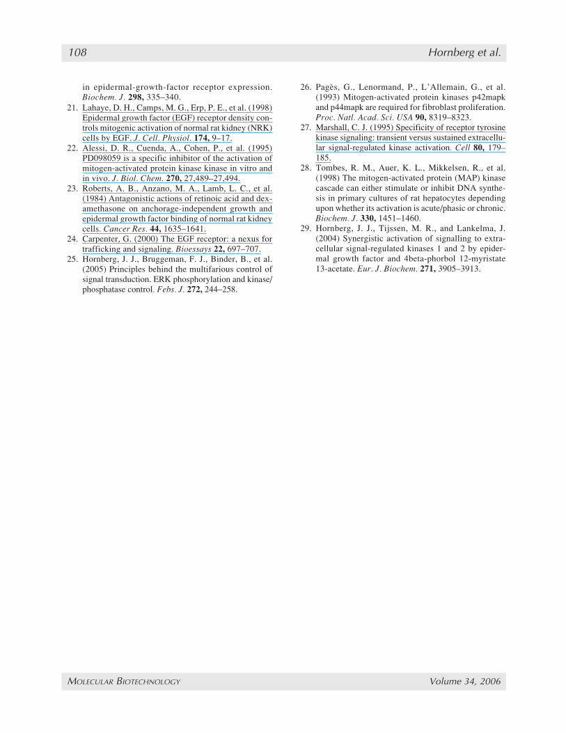
25. Hornberg, J. J., Bruggeman, F. J., Binder, B., et al.(2005) Principles behind the multifarious control ofsignal transduction. ERK phosphorylation and kinase/phosphatase control. Febs. J. 272, 244–258.
26. Pagès, G., Lenormand, P., L’Allemain, G., et al.(1993) Mitogen-activated protein kinases p42mapkand p44mapk are required for fibroblast proliferation.Proc. Natl. Acad. Sci. USA 90, 8319–8323.
27. Marshall, C. J. (1995) Specificity of receptor tyrosinekinase signaling: transient versus sustained extracellu-lar signal-regulated kinase activation. Cell 80, 179–185.
28. Tombes, R. M., Auer, K. L., Mikkelsen, R., et al.(1998) The mitogen-activated protein (MAP) kinasecascade can either stimulate or inhibit DNA synthe-sis in primary cultures of rat hepatocytes dependingupon whether its activation is acute/phasic or chronic.Biochem. J. 330, 1451–1460.
29. Hornberg, J. J., Tijssen, M. R., and Lankelma, J.(2004) Synergistic activation of signalling to extra-cellular signal-regulated kinases 1 and 2 by epider-mal growth factor and 4beta-phorbol 12-myristate13-acetate. Eur. J. Biochem. 271, 3905–3913.
101_108_Horn_F 10/9/06, 10:03 AM108