Mechanisms of Transforming Growth Factor-b Receptor Endocytosis and Intracellular Sorting Differ...
10
Molecular Biology of the Cell Vol. 12, 675– 684, March 2001 Mechanisms of Transforming Growth Factor-b Receptor Endocytosis and Intracellular Sorting Differ between Fibroblasts and Epithelial Cells Jules J.E. Dore ´, Jr., Diying Yao, Maryanne Edens, Nandor Garamszegi, Elizabeth L. Sholl, and Edward B. Leof* Thoracic Diseases Research Unit and Department of Biochemistry and Molecular Biology, Mayo Clinic, Rochester, Minnesota 55905 Submitted November 13, 2000; Revised November 13, 2000; Accepted January 16, 2000 Monitoring Editor: Suzanne R. Pfeffer Transforming growth factor-bs (TGF-b) are multifunctional proteins capable of either stimulating or inhibiting mitosis, depending on the cell type. These diverse cellular responses are caused by stimulating a single receptor complex composed of type I and type II receptors. Using a chimeric receptor model where the granulocyte/monocyte colony-stimulating factor receptor ligand bind- ing domains are fused to the transmembrane and cytoplasmic signaling domains of the TGF-b type I and II receptors, we wished to describe the role(s) of specific amino acid residues in regulating ligand-mediated endocytosis and signaling in fibroblasts and epithelial cells. Specific point mutations were introduced at Y182, T200, and Y249 of the type I receptor and K277 and P525 of the type II receptor. Mutation of either Y182 or Y249, residues within two putative consensus tyrosine-based internalization motifs, had no effect on endocytosis or signaling. This is in contrast to mutation of T200 to valine, which resulted in ablation of signaling in both cell types, while only abolishing receptor down-regulation in fibroblasts. Moreover, in the absence of ligand, both fibroblasts and epithelial cells constitutively internalize and recycle the TGF-b receptor complex back to the plasma membrane. The data indicate fundamental differences between mesenchymal and epithelial cells in endocytic sorting and suggest that ligand binding diverts heteromeric receptors from the default recycling pool to a pathway mediating receptor down-regulation and signaling. INTRODUCTION Transforming growth factor-bs (TGF-b) control a variety of cellular processes as diverse as mitotic inhibition or stimu- lation (Massague ´, 1996; Moses and Serra, 1996). It is unclear how the same receptor complex can mediate such different cellular phenotypes. The most commonly accepted receptor model for TGF-b action consists of a heteromeric complex composed of type I and type II receptors (Wrana et al., 1992, 1994). Once associated, the type I receptor becomes phos- phorylated primarily within the juxtamembrane GS domain (amino acids 185–192) by the constitutive serine/threonine kinase activity of the type II receptor (Franze ´n et al., 1995; Wieser et al., 1995). Phosphorylation of the GS domain is proposed to activate the type I receptor, resulting in signal propagation to downstream effector molecules (Massague ´, 1998). In addition, specific residues in nearby regions have also been suggested to have both positive and negative regulatory functions (Wieser et al., 1995; Charng et al., 1996; Souchelnytskyi et al., 1996; Dore ´ et al., 1998). For instance, threonine 200 has been shown to have a fundamental role in mediating all aspects of TGF-b signaling (Wieser et al., 1995), whereas replacement of threonine 204 with an acidic resi- due, such as aspartate, can generate a type I receptor capable of signaling (albeit to a lesser extent) independent of ligand or an associated type II receptor (Wieser et al., 1995; Charng et al., 1996; Luo and Lodish, 1996). What makes these data most intriguing is that none of the aforementioned residues have been shown to be phosphorylated (Heldin et al., 1997). As such, the mechanism(s) through which they might act is still enigmatic. Additionally, there have been no reports as to whether these amino acids might modulate TGF-b recep- tor endocytic activity. Once ligand binding occurs, typically a growth factor receptor is removed from the plasma membrane by an en- docytic process, resulting in receptor down-regulation. En- docytosis consists of several interconnected pathways, in- cluding the initial internalization of receptors, sorting endosomes, recycling of receptors back to the plasma mem- brane, and/or shunting to proteosome/lysosomes for deg- * Corresponding author. E-mail address: [email protected]. © 2001 by The American Society for Cell Biology 675
Transcript of Mechanisms of Transforming Growth Factor-b Receptor Endocytosis and Intracellular Sorting Differ...
Molecular Biology of the CellVol. 12, 675–684, March 2001
Mechanisms of Transforming Growth Factor-bReceptor Endocytosis and Intracellular Sorting Differbetween Fibroblasts and Epithelial CellsJules J.E. Dore, Jr., Diying Yao, Maryanne Edens, Nandor Garamszegi,Elizabeth L. Sholl, and Edward B. Leof*
Thoracic Diseases Research Unit and Department of Biochemistry and Molecular Biology, MayoClinic, Rochester, Minnesota 55905
Submitted November 13, 2000; Revised November 13, 2000; Accepted January 16, 2000Monitoring Editor: Suzanne R. Pfeffer
Transforming growth factor-bs (TGF-b) are multifunctional proteins capable of either stimulatingor inhibiting mitosis, depending on the cell type. These diverse cellular responses are caused bystimulating a single receptor complex composed of type I and type II receptors. Using a chimericreceptor model where the granulocyte/monocyte colony-stimulating factor receptor ligand bind-ing domains are fused to the transmembrane and cytoplasmic signaling domains of the TGF-btype I and II receptors, we wished to describe the role(s) of specific amino acid residues inregulating ligand-mediated endocytosis and signaling in fibroblasts and epithelial cells. Specificpoint mutations were introduced at Y182, T200, and Y249 of the type I receptor and K277 and P525of the type II receptor. Mutation of either Y182 or Y249, residues within two putative consensustyrosine-based internalization motifs, had no effect on endocytosis or signaling. This is in contrastto mutation of T200 to valine, which resulted in ablation of signaling in both cell types, while onlyabolishing receptor down-regulation in fibroblasts. Moreover, in the absence of ligand, bothfibroblasts and epithelial cells constitutively internalize and recycle the TGF-b receptor complexback to the plasma membrane. The data indicate fundamental differences between mesenchymaland epithelial cells in endocytic sorting and suggest that ligand binding diverts heteromericreceptors from the default recycling pool to a pathway mediating receptor down-regulation andsignaling.
INTRODUCTION
Transforming growth factor-bs (TGF-b) control a variety ofcellular processes as diverse as mitotic inhibition or stimu-lation (Massague, 1996; Moses and Serra, 1996). It is unclearhow the same receptor complex can mediate such differentcellular phenotypes. The most commonly accepted receptormodel for TGF-b action consists of a heteromeric complexcomposed of type I and type II receptors (Wrana et al., 1992,1994). Once associated, the type I receptor becomes phos-phorylated primarily within the juxtamembrane GS domain(amino acids 185–192) by the constitutive serine/threoninekinase activity of the type II receptor (Franzen et al., 1995;Wieser et al., 1995). Phosphorylation of the GS domain isproposed to activate the type I receptor, resulting in signalpropagation to downstream effector molecules (Massague,1998). In addition, specific residues in nearby regions havealso been suggested to have both positive and negativeregulatory functions (Wieser et al., 1995; Charng et al., 1996;
Souchelnytskyi et al., 1996; Dore et al., 1998). For instance,threonine 200 has been shown to have a fundamental role inmediating all aspects of TGF-b signaling (Wieser et al., 1995),whereas replacement of threonine 204 with an acidic resi-due, such as aspartate, can generate a type I receptor capableof signaling (albeit to a lesser extent) independent of ligandor an associated type II receptor (Wieser et al., 1995; Charnget al., 1996; Luo and Lodish, 1996). What makes these datamost intriguing is that none of the aforementioned residueshave been shown to be phosphorylated (Heldin et al., 1997).As such, the mechanism(s) through which they might act isstill enigmatic. Additionally, there have been no reports asto whether these amino acids might modulate TGF-b recep-tor endocytic activity.
Once ligand binding occurs, typically a growth factorreceptor is removed from the plasma membrane by an en-docytic process, resulting in receptor down-regulation. En-docytosis consists of several interconnected pathways, in-cluding the initial internalization of receptors, sortingendosomes, recycling of receptors back to the plasma mem-brane, and/or shunting to proteosome/lysosomes for deg-* Corresponding author. E-mail address: [email protected].
© 2001 by The American Society for Cell Biology 675
radation. Previous reports regarding the TGF-b receptorhave been contradictory and ranged from no significantdown-regulation (Massague, 1985; Wakefield et al., 1987) toa 50% decrease in surface binding (Frolik et al., 1984). Thediscrepancy is likely due to the existence of several differentTGF-b receptor complexes on the plasma membrane withdistinct endocytic activities (Chen and Derynck, 1994; Heniset al., 1994; Anders et al., 1997; Dore et al., 1998; Gilboa et al.,1998). Using a chimeric receptor model consisting of theligand binding domain of the granulocyte/monocyte colo-ny-stimulating factor (GM-CSF) a or b receptor fused to thetransmembrane and cytoplasmic tail of the type I or type IITGF-b receptor, we have shown (in fibroblasts) that onlyheteromeric receptors (type I/II interactions) are down-reg-ulated, whereas homomeric receptors (types I/I and II/IIinteractions) were proposed to recycle back to the plasmamembrane following initial internalization (Anders andLeof, 1996; Anders et al., 1997).
Although TGF-b receptors appear to be endocytosedthrough a clathrin-mediated process (Anders et al., 1997), themechanism(s) regulating the endocytic event(s) remains un-known. In the present report we wished to test whetherspecific amino acid residues in TGF-b type I and type IIreceptors known to modulate signaling, as well as key res-idues within motifs that regulate endocytosis in other recep-tor systems, might also control endocytosis of the TGF-breceptor complex. The data from the present study indicate1) that TGF-b receptor endocytic activity in both fibroblastsand epithelial cells appears to be controlled independentlyof type I receptor tyrosine-based sorting elements; 2) al-though type I receptor phosphorylation plays an essentialrole in regulating TGF-b receptor signaling in fibroblastsand epithelial cells, it is only obligate for receptor endocy-tosis in fibroblasts, delineating fundamental differences inthe intracellular recognition of the receptor complex be-tween the two cell types; 3) TGF-b receptors constitutivelyrecycle in the absence of ligand; and 4) ligand bindingdiverts heteromeric receptor complexes from the defaultrecycling pathway to one associated with down-regulationand signaling.
MATERIALS AND METHODS
MaterialsRecombinant human GM-CSF was generously provided by DNAXResearch Institute (Palo Alto, CA), whereas recombinant humanTGF-b2 was purchased from R&D Systems (Minneapolis, MN). Cellculture media, horse serum, and geneticin (G418 sulfate) were pur-chased from Life Technologies (Gaithersburg, MD). Fetal bovineserum (FBS) was obtained from Summit (Fort Collins, CO) andhygromycin B was purchased from Boehringer-Mannheim (India-napolis, IN). Monensin was purchased from Sigma Chemical Co.(St. Louis, MO), whereas antibodies were acquired from Santa CruzBiotechnology (Santa Cruz, CA).
Cell CultureParental AKR-2B and NIH-3T3 fibroblasts were maintained inDMEM supplemented with 5% (vol/vol) FBS, whereas parentalMv1Lu and R1B (Mv1Lu clone lacking type I TGF-b receptors;Laiho et al., 1990) mink lung epithelium, Madin-Darby canine kid-ney (MDCK), and NRK-49F cultures were maintained in DMEMsupplemented with 10% (vol/vol) FBS. Chimeric receptor express-ing clones were constructed in a two-step process with the cDNA
constructs described previously (Anders and Leof, 1996). Initially, awild-type chimeric bI or bII receptor was transfected into each cellline. The designation bI or bII refers to the ligand binding domainof the GM-CSF b receptor coupled to the transmembrane and cyto-plasmic domains of the TGF-b type I or type II receptor, respec-tively. Cells were plated at 105 cells/well in six-well dishes 24 hbefore transfection. Cells were washed with serum-free DMEM andincubated in 2 ml of DMEM for 10 min before transfection with amixture of 2 mg of plasmid and 4 ml of TransIt-LT2 (Pan Vera,Madison, WI) in a final volume of 100 ml of Opti-MEM for 8 h at37°C. Medium was replaced with 10% FBS/DMEM for 16 h andthen changed to selection medium (5% FBS/DMEM supplementedwith 300 mg/ml hygromycin B for AKR-2B and NIH-3T3 or 10%FBS/DMEM 300 mg/ml hygromycin for Mv1Lu, MDCK and NRK-49F) for 24 h before trypsinization and replating at 1:40 dilution.Fourteen days posttransfection isolated colonies were expanded.Clones were screened by fluorescent activated cell sorting (FACS)for plasma membrane expression of the chimeric receptor as previ-ously described (Anders and Leof, 1996). One representative clonewas chosen for the second transfection based upon high membraneexpression of the chimeric receptor and normal induction of plas-minogen activator inhibitor protein-1 (PAI-1, fibroblasts) or normalinhibition of thymidine incorporation in response to TGF-b2 (epi-thelial cells).
The second transfection used mutant aI or aII chimeric receptors(ligand binding domain from GM-CSF a receptor fused to thetransmembrane and cytoplasmic region of the TGF-bI or II receptor)to produce the chimeric high-affinity ligand-binding a/b complex.Chimeric b receptor-expressing clones were plated at 7.5 3 104 cellsin six-well plates and transfected with 2 mg of a mutant a receptorplasmid in TransIt-LT2 and Opti-MEM. After recovery, cells wereplaced in selection medium (5% FBS/DMEM or 10%FBS/DMEMsupplemented with 600 mg/ml geneticin and 350 mg/ml hygromy-cin B) for 24 h before 1:40 dilution.
Site-directed Mutagenesis of Chimeric cDNAChimeric aI receptor cDNA was mutated at amino acid threonine200 to valine by using the QuickChange mutagenesis kit (Strat-agene, La Jolla, CA) and mutagenic primers CAGGTTTACCATT-GCTTGTTCAGAGAgtATTGCGAGAACTATTGTG and CACAAT-AGTTCTCGCAATTacTCTCTGAACAAGCAATGGTAAACCTG,where the lowercase letters indicate the base changes necessary tointroduce the appropriate amino acid change. The chimeric aI re-ceptor containing the tyrosine to serine mutation at position 182(Y182S) was generated with mutagenic primers CGTTGAAAGACT-TAATTTcTGATATGACAACGTCAGG and CCTGACGTTGTCA-TATCAgAAATTAAGTCTTTCAACG, whereas the Y249S constructused the primers GGCAGAGATTTcTCAAACTGTAATG and CAT-TACAGTTTGAgAAATCTCTGCC. Introduction of K277R andP525L mutations, which inhibit type II receptor kinase activity, intothe chimeric aII constructs was previously described (Anders et al.,1998). These constructs were transfected into MB-102 parentalMv1Lu cells and clones selected. Mutant constructs were verified byautomated DNA sequencing and ligated into the eukaryotic expres-sion vectors pNa at the SalI and HindIII sites for aI and aII con-structs and into pHa at the SalI or XbaI and BamHI sites for the bI orbII constructs, respectively.
Receptor Function and ExpressionReceptor binding assays were used to determine expression ofchimeric receptors on the plasma membrane, as described previ-ously (Anders and Leof, 1996; Dore et al., 1998). Functional analysisof the chimeric receptors in AKR-2B clones involved both the in-duction of endogenous PAI-1 protein and transiently transfectedPAI-1 luciferase (3TP-Lux) reporter gene activity by GM-CSF,through the chimeric receptors, and TGF-b2, through the endoge-nous TGF-b receptor (Anders and Leof, 1996). Representative cell
J.J.E. Dore et al.
Molecular Biology of the Cell676
lines from each transfection group chosen for further analysis wereY182S clone 1, T200V clones 10 and 12, and Y249S clone 7. Func-tional analysis of Mv1Lu clones involved inhibition of [3H]thymi-dine incorporation in the absence or presence of 10 ng/ml GM-CSFor TGF-b2. Representative clones chosen from each transfectiongroup were wild-type receptor clones 9 and 18; Y182S clones 1, 4,and 9; T200V clones 2, 7, and 13; Y249S clones 8, 16, and 22; K277Rclones 102-1, -3, and -18, 117-10 and -15, 126-1 and -12; and P525Lclones 102-5, -10, and -13, 117-7 and -9, 126-2 and -5.
EndocytosisThe endocytic response to ligand binding was performed as previ-ously described (Anders et al., 1997; Dore et al., 1998). Briefly, todetermine receptor down-regulation, cells were incubated at 37°Cwith 10 ng/ml (500 pM) cold GM-CSF for the times indicated. Wellswere then washed twice at 4°C with phosphate-buffered saline, pH3.0, and the remaining surface binding determined by incubating for2 h at 4°C with 100 pM 125I-GM-CSF alone or in the presence of25-fold molar excess of cold GM-CSF before cell lysis. A modifica-tion of the down-regulation assay was used to assess the effect ofmonensin on receptor trafficking. Cells were pretreated with 100 mMmonensin in 5% FBS/DMEM for 30 min at 37°C. Medium was thenreplaced with fresh 5% FBS/DMEM/monensin containing 10ng/ml cold GM-CSF for the indicated times and processed as de-scribed above. To describe receptor recycling, 37°C 5% FBS/DMEM/monensin-containing medium was added directly to cellsfor the indicated times.
RESULTS
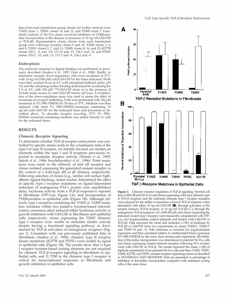
Chimeric Receptor SignalingTo determine whether TGF-b receptor endocytosis was con-trolled by specific amino acids in the cytoplasmic tails of thetype I or type II receptor, we initially focused our studies onelements within the type I and II receptors previously re-ported to modulate receptor activity (Wieser et al., 1995;Saitoh et al., 1996; Souchelnytskyi et al., 1996). Point muta-tions were made in the chimeric aI and aII receptor andclones isolated expressing the generated mutant receptor inthe context of a wild-type bII or bI chimera, respectively.Following selection of clones (e.g., similar cell surface high-affinity ligand binding), initial studies determined the effectof specific type I receptor mutations on ligand-dependentinduction of endogenous PAI-1 protein (our unpublisheddata), luciferase activity from a TGF-b–responsive reporterin fibroblasts (3TP-Lux; Figure 1A), and incorporation of[3H]thymidine in epithelial cells (Figure 1B). Although chi-meric type I receptors containing the Y182S or Y249S muta-tion (residues within two putative tyrosine-based internal-ization consensus sites) induced either luciferase activity orgrowth inhibition with GM-CSF in fibroblasts and epithelialcells, respectively, clones expressing the T200V chimerictype I receptor were unable to stimulate similar activitydespite having a functional signaling pathway as docu-mented by TGF-b activation of endogenous receptors (Fig-ure 1). Consistent with our previously published data infibroblasts (Anders et al., 1998), chimeric type II receptorkinase mutations (K277R and P525L) were unable to signalin epithelial cells (Figure 1B). The results show that 1) typeI receptor tyrosine-based sorting elements are not requiredfor chimeric TGF-b receptor signaling in fibroblasts or epi-thelial cells; and 2) T200 in the chimeric type I receptor iscritical for transcriptional responses in fibroblasts andgrowth inhibition in epithelial cells.
Figure 1. Chimeric receptor regulation of TGF-b signaling. Parental celllines (AKR-2B and Mv1Lu) and clones expressing wild-type chimeric typeII TGF-b receptors and the indicated chimeric type I receptor mutationwere assayed for the ability to transduce a known TGF-b response whenstimulated with either 10 ng/ml GM-CSF (f), through activation of themutant chimeric TGF-b receptor, or 10 ng/ml TGF-b2 (M), through theendogenous TGF-b receptors. (A) AKR-2B cells and clones expressing theindicated mutant type I receptor were transiently cotransfected with 3TP-Lux and b-galactosidase control plasmids and treated with GM-CSF orTGF-b2. Data represent the mean fold induction (6SE) of luciferase byTGF-b2 or GM-CSF from two experiments on clones Y182S-1, Y249S-7,and T200V-10 and -12. Fold induction is corrected for b-galactosidaseexpression and then calculated relative to unstimulated basal expression(5% FBS/DMEM) by the same clone during each experiment. (B) Inhibi-tion of thymidine incorporation was determined in parental Mv1Lu cellsand clones expressing mutant chimeric receptors following 20 h of treat-ment with GM-CSF or TGF-b. The results represent the mean (6SE) oftriplicate experiments from parental Mv1Lu cells and three Y182S, T200V,Y249S, K277R, and P525L mutant receptor expressing clones as describedin MATERIALS AND METHODS. Data are presented as percentage ofinhibition of thymidine incorporation compared with untreated cyclingcells of the same clone.
Cell Type-specific TGF-b Receptor Endocytosis
Vol. 12, March 2001 677

Tyrosine-based Endocytosis of Type I ReceptorBecause TGF-b receptor down-regulation in fibroblasts re-quires both formation of a type I/II receptor complex andtype II receptor kinase activity (Anders et al., 1997, 1998), thissuggests that a role of the type I receptor is to providespecific residues/motifs that facilitate receptor endocytosis.One such motif shown to enhance the endocytosis of severalreceptors is tyrosine-based and can be represented by thesequence Y-polar-polar-hydrophobic (Ohno et al., 1995;Marks et al., 1996; Rapoport et al., 1997). Although identicaltyrosine-based motifs seen in other proteins were not foundin the type I receptor cytoplasmic region, tyrosines at 182and 249 were selected based on location to known regula-tory regions within the receptor and similarity to previouslyidentified adaptor protein-2 recognition sites (Rapoport etal., 1997). Residue Y182 (sequence YDMT) was chosen due toits proximity to the regulatory GS domain (amino acids185–192), whereas Y249 (sequence YQTV) for its proximityto the putative ATP binding site (K232) and similarity to thelamp-1 internalization sequence YQTI (Honing et al., 1996).It would not be unexpected that either of these residueswould be exposed and interact with the endocytic machin-ery following GS domain phosphorylation and activation ofthe type I receptor. To address this possibility, the endocyticactivity of chimeric TGF-b receptors containing the Y182S orY249S mutations in the type I receptor was determined inboth fibroblasts and epithelium. As shown in Table 1, al-though differences were observed in the endocytic rate offibroblasts and epithelial cells (Dore et al., 1998), mutation ofY182 or Y249 to serine had no adverse effect on ligand-induced down-regulation in either cell type compared withthe wild-type heteromeric chimera control (A105 or MB-18).For instance, by 4 h there was an ;80% decrease in receptorbinding in all tested lines (Table 1). Thus, in contrast to thatobserved for other plasma membrane receptors, endocytosisof chimeric TGF-b receptors is controlled independent oftyrosine-based sorting elements in the type I receptor.
Role of Type I Receptor T200 in Down-regulationand SignalingThe preceding data indicate that type I receptor tyrosine-based endocytic motifs do not significantly modulate the
initial endocytic activity of TGF-b receptors in either fibro-blasts or epithelial cells. Because we had previously showna delineation of endocytosis and signaling in fibroblasts(Anders et al., 1998), we next determined whether othermutations reported to effect receptor signaling (Franzen etal., 1995; Wieser et al., 1995) would modulate receptor endo-cytosis. Similar to that shown previously in Mv1Lu epithe-lial cells (Dore et al., 1998), mutation of type I receptorresidues S172 or T176 in fibroblasts had no significant effecton either the rate or extent of receptor down-regulation (ourunpublished data). In contrast, when T200V clones wereassayed for ligand-induced down-regulation of the chimericreceptors, a differential response was observed dependentupon the cell type (Figure 2). Although an initial decrease inreceptor binding was observed following 30-min ligandtreatment in fibroblasts (Figure 2A), by 60 min binding hadreturned to 94% of control levels and remained constantover the next 3 h. However, whereas mutation of T200resulted in a type I TGF-b receptor with impaired endocyticactivity in fibroblasts, epithelial clones expressing the samereceptor mutation down-regulate similar to wild-type recep-tors (Figure 2B).
To determine whether this cell type-specific response tothe T200V mutation was a common theme in epithelial andmesenchymal cells, we transfected the a1 T200V constructsin conjunction with wild-type b2 constructs into MDCKepithelial cells, as well as NIH-3T3 and NRK-49F fibroblasts(Figure 2C). As noted previously in Mv1Lu and AKR-2Bcells, the T200V mutation expressed in other cell lines dem-onstrated a similar response. The combined down-regula-tion response of the T200V mutant-expressing clones, inepithelial cells, was similar to that of clones expressing wild-type receptors (54% decrease in surface binding for T200Vcompared with 62% for wild-type, at 4 h). This is in contrastto fibroblasts in which the T200V mutation significantlyinhibited chimeric receptor down-regulation over 4 h. (21%for T200V compared with 74% for wild-type). Thus, theT200V mutation represents the first type I receptor site ca-pable of modulating both signaling and endocytic activity ofthe TGF-b receptor complex and documents differential en-docytic sorting between fibroblasts and epithelial cells.
Table 1. Down-regulation of chimeric receptors is not affected by type I TGF-b receptor tyrosine mutations
Cell line Slope Percent receptor binding at 4 h n
A105 20.661 6 0.082 32.2 6 2.7 3A-Y182S 20.664 6 0.057 26.6 6 5.7 3A-Y249S 20.962 6 0.090 25.7 6 4.0 3M-Heteromer 21.301 6 0.067 17.1 6 2.3 2M-Y182S 20.981 6 0.082 18.4 6 0.8 2M-Y249S 20.906 6 0.059 26.2 6 2.1 2
Decreased surface binding of 125I-GM-CSF was determined for AKR-2B and Mv1Lu clones expressing a wild-type chimeric type II TGF-breceptor and a type I TGF-b receptor with tyrosine to serine mutations at either Y182 or Y249, when treated with 10 ng/ml GM-CSF at 37°C.Down-regulation of the normal mesenchymal (A105) and epithelial (M-Heteromer) heteromer containing wild-type type I and type IIchimeric receptors is shown for comparison. The rate of down-regulation was determined from the slope of the line calculated from four datapoints over the first 60 min. Data for the mesenchymal cells represent 1 AKR-2B clone for each receptor type (A105, Y182S-1, and Y249S-7),whereas the epithelial cell data represent the mean of 2 M-heteromers (MB-9 and -18) and three separate M-Y182S (Y182S-1, -4, and -9) andY249S (Y249S-8, -16, and -22) mutant clones. Assays from the indicated (n) number of experiments were each performed in duplicate.
J.J.E. Dore et al.
Molecular Biology of the Cell678

Role of Type II Receptor Kinase in EndocytosisWe have previously shown that type II receptor kinaseactivity is critical for both receptor down-regulation andsignaling in fibroblasts (Anders et al., 1998). For instance,cells expressing a wild-type chimeric type I receptor and akinase-impaired chimeric type II receptor have diminishedendocytic activity, whereas cultures expressing a wild-typechimeric type II receptor and a kinase-deficient type I recep-tor internalize ligand and down-regulate like wild-type re-ceptors (Anders et al., 1998). Because the T200V mutationblocks type I receptor phosphorylation (Wieser et al., 1995),and epithelial cells expressing this receptor mutation down-
regulate similar to wild-type receptors (Figure 2), this sug-gests that the kinase activity of the type II receptor might notbe obligate for chimeric TGF-b receptor down-regulation inepithelial cells. As such, we wished to directly assess the roleof the type II receptor kinase in regulating endocytosis inMv1Lu epithelial cells. Consistent with previously pub-lished data (Anders et al., 1998), a mutation (P525L) in thechimeric type II receptor that alters the ability of the receptorto transphosphorylate dramatically reduces receptor down-regulation in fibroblasts (Figure 3A). However, when thesame mutation or the K277R mutation in the ATP bindingsite was expressed in epithelial cells, a distinct endocytic
Figure 2. Down-regulation of chimeric mutant T200V type Ireceptors. Cell surface binding in response to 10 ng/ml GM-CSFwas performed as described in MATERIALS AND METHODS.Each point represents the mean (6 SE) for all clones of the seriesassayed in duplicate from two independent experiments. (A)AKR-2B fibroblast clones T200V-10 and -12 (E) are compared withA105 wild-type control (F). (B) Mv1Lu epithelial clones T200V-2and -7 (M) are compared with the mean of MB-9 and -18 aswild-type control (f). (C) Mean percentage of down-regulation oftwo different epithelial (f, M) and three fibroblast (F, E) cell lines.Stable clones expressing the T200V mutation (open symbols) inMv1Lu (clones 2 and 7) and MDCK (clones 13, 17, 32, and 38)epithelial lines and 3T3-NIH (clones 6, 7, and 9), AKR-2B (clones10 and 12), and NRK-49F (clones 1 and 3) fibroblast lines wereused. Control wild-type expressing clones (closed symbols) usedfor comparison were Mv1Lu-9 and -18, MDCK-1 and -5, NIH-3T3-2 and -10, AKR-2B clone A105, and NRK-49F-7.
Cell Type-specific TGF-b Receptor Endocytosis
Vol. 12, March 2001 679
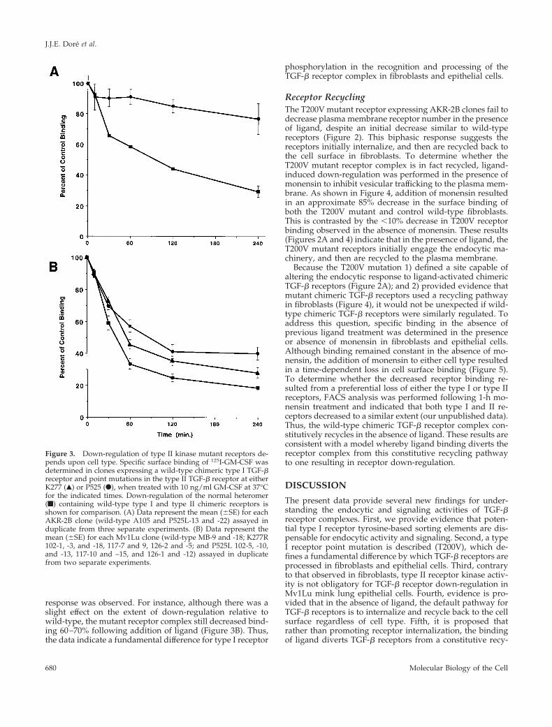
response was observed. For instance, although there was aslight effect on the extent of down-regulation relative towild-type, the mutant receptor complex still decreased bind-ing 60–70% following addition of ligand (Figure 3B). Thus,the data indicate a fundamental difference for type I receptor
phosphorylation in the recognition and processing of theTGF-b receptor complex in fibroblasts and epithelial cells.
Receptor RecyclingThe T200V mutant receptor expressing AKR-2B clones fail todecrease plasma membrane receptor number in the presenceof ligand, despite an initial decrease similar to wild-typereceptors (Figure 2). This biphasic response suggests thereceptors initially internalize, and then are recycled back tothe cell surface in fibroblasts. To determine whether theT200V mutant receptor complex is in fact recycled, ligand-induced down-regulation was performed in the presence ofmonensin to inhibit vesicular trafficking to the plasma mem-brane. As shown in Figure 4, addition of monensin resultedin an approximate 85% decrease in the surface binding ofboth the T200V mutant and control wild-type fibroblasts.This is contrasted by the ,10% decrease in T200V receptorbinding observed in the absence of monensin. These results(Figures 2A and 4) indicate that in the presence of ligand, theT200V mutant receptors initially engage the endocytic ma-chinery, and then are recycled to the plasma membrane.
Because the T200V mutation 1) defined a site capable ofaltering the endocytic response to ligand-activated chimericTGF-b receptors (Figure 2A); and 2) provided evidence thatmutant chimeric TGF-b receptors used a recycling pathwayin fibroblasts (Figure 4), it would not be unexpected if wild-type chimeric TGF-b receptors were similarly regulated. Toaddress this question, specific binding in the absence ofprevious ligand treatment was determined in the presenceor absence of monensin in fibroblasts and epithelial cells.Although binding remained constant in the absence of mo-nensin, the addition of monensin to either cell type resultedin a time-dependent loss in cell surface binding (Figure 5).To determine whether the decreased receptor binding re-sulted from a preferential loss of either the type I or type IIreceptors, FACS analysis was performed following 1-h mo-nensin treatment and indicated that both type I and II re-ceptors decreased to a similar extent (our unpublished data).Thus, the wild-type chimeric TGF-b receptor complex con-stitutively recycles in the absence of ligand. These results areconsistent with a model whereby ligand binding diverts thereceptor complex from this constitutive recycling pathwayto one resulting in receptor down-regulation.
DISCUSSION
The present data provide several new findings for under-standing the endocytic and signaling activities of TGF-breceptor complexes. First, we provide evidence that poten-tial type I receptor tyrosine-based sorting elements are dis-pensable for endocytic activity and signaling. Second, a typeI receptor point mutation is described (T200V), which de-fines a fundamental difference by which TGF-b receptors areprocessed in fibroblasts and epithelial cells. Third, contraryto that observed in fibroblasts, type II receptor kinase activ-ity is not obligatory for TGF-b receptor down-regulation inMv1Lu mink lung epithelial cells. Fourth, evidence is pro-vided that in the absence of ligand, the default pathway forTGF-b receptors is to internalize and recycle back to the cellsurface regardless of cell type. Fifth, it is proposed thatrather than promoting receptor internalization, the bindingof ligand diverts TGF-b receptors from a constitutive recy-
Figure 3. Down-regulation of type II kinase mutant receptors de-pends upon cell type. Specific surface binding of 125I-GM-CSF wasdetermined in clones expressing a wild-type chimeric type I TGF-breceptor and point mutations in the type II TGF-b receptor at eitherK277 (Œ) or P525 (F), when treated with 10 ng/ml GM-CSF at 37°Cfor the indicated times. Down-regulation of the normal heteromer(f) containing wild-type type I and type II chimeric receptors isshown for comparison. (A) Data represent the mean (6SE) for eachAKR-2B clone (wild-type A105 and P525L-13 and -22) assayed induplicate from three separate experiments. (B) Data represent themean (6SE) for each Mv1Lu clone (wild-type MB-9 and -18; K277R102-1, -3, and -18, 117-7 and 9, 126-2 and -5; and P525L 102-5, -10,and -13, 117-10 and –15, and 126-1 and -12) assayed in duplicatefrom two separate experiments.
J.J.E. Dore et al.
Molecular Biology of the Cell680

cling pathway to one resulting in down-regulation and sig-naling. Together these observations indicate that mesenchy-mal and epithelial cell types have similar, as well as distinctendocytic sorting mechanisms for the same receptor com-plex, possibly accounting for some of the diverse cellularresponses to TGF-b.
Tyrosine-based Motifs Are Not Involved in ChimericTGF-b Receptor Endocytosis or SignalingThe endocytic process is regulated through the coordinateinterplay of a number of plasma membrane and cytosolicproteins (Mellman, 1996; Mukherjee et al., 1997; Schmid,1997; McNiven, 1998). Receptor endocytosis is initiated bythe recognition of distinct sequence elements within thecytoplasmic (primarily) domain of various membrane recep-tors that control the internalization process, as well as asso-ciation with coated pit proteins (Pearse and Robinson, 1990;Trowbridge et al., 1993; Robinson et al., 1996). Once internal-ized, receptors are shuttled to various intracellular compart-ments where they are sorted for recycling back to the cellsurface or down-regulated through the degradative path-ways. Whereas no canonical sequence or receptor domainhas been identified, a common tyrosine-based motif foundto regulate growth factor receptor internalization consists ofTyr-polar-polar-hydrophobic residues (Chang et al., 1993;
Carpentier and McClain, 1995; Lamaze and Schmid, 1995).Two similar sites near regions known to regulate type Ireceptor activity are Y182 (YDMT) and Y249 (YQTV). Whenthese tyrosines were individually mutated to serine, ratherthan being inhibitory, chimeric TGF-b receptors were down-regulated as effectively as wild-type receptors in either fi-broblasts or epithelial cells (Table 1). Although we cannotconclusively eliminate a potential site at Y284 (YASW)within the type II receptor, the results indicate that type Ireceptor tyrosine-based motifs do not have as fundamental arole in regulating the endocytic activity of chimeric TGF-breceptors as they do for other growth factor receptors(Chang et al., 1993; Carpentier and McClain, 1995; Lamazeand Schmid, 1995).
T200 Defines a Type I Receptor Site DifferentiallyRegulating Endocytosis in Fibroblasts andEpithelial CellsBecause tyrosine-based internalization signals are not likelyinvolved in TGF-b receptor endocytosis, we next deter-mined whether other type I receptor residues with knownregulatory activity would affect the endocytic response. Thetwo questions we wished to address were 1) whether a siteknown to regulate receptor signaling would also modulatereceptor endocytosis; and 2) whether there would be distinctresponses dependent upon cell type. To that end, both mes-enchymal and epithelial cell lines were isolated expressing awild-type chimeric type II TGF-b receptor and a chimerictype I receptor with S172A, T176V, or T200V mutations. Aswas previously shown in Mv1Lu epithelial cells (Dore et al.,1998), S172A and T176V mutations had no effect on either
Figure 4. Monensin induced down-regulation of T200V chimericreceptor. Down-regulation assays were performed on AKR-2Bclones containing wild-type type I and II chimeric receptors (M) ora wild-type type II and T200V type I mutant (‚) in the presence of100 mM monensin and 10 ng/ml GM-CSF. The data represent themean (6 SE) of three separate experiments done in duplicate on twoindependent mutant clones (T200V-10 and -12) and wild-type(A105) control cells. The ligand-induced down-regulation of theT200V clones in the absence of monensin reported in Figure 2 isshown for comparison (Œ). Preliminary experiments demonstratedthat monensin had no effect on ligand binding and decreased wild-type cell surface receptor binding in a dose-dependant manner, with100 mM being maximally effective (our unpublished data).
Figure 5. Constitutive internalization of chimeric TGF-b receptors.Ligand-independent decrease of GM-CSF surface binding in thepresence (closed symbols) or absence (open symbols) of 100 mM inwild-type chimeric A105 fibroblasts (M, f) and MB-18 epithelialcells (F, E). Each point represents the mean (6 SE) of three separateexperiments done in duplicate.
Cell Type-specific TGF-b Receptor Endocytosis
Vol. 12, March 2001 681

endocytosis or signaling in fibroblasts (our unpublisheddata). This is contrasted by the T200V mutation that defineda fundamental difference in endocytic control between thecell types. For instance, although T200 had no affect onepithelial cell receptor down-regulation, fibroblasts express-ing this mutation were unable to down-regulate followingaddition of ligand (Figure 2). However, because the down-regulation assay detects cell surface binding and not recep-tors per se, an alternative possibility was that the differentialresponse observed in fibroblasts and epithelial cells did notreflect a fundamental cell type difference in how the sortingmachinery recognizes T200, but a sequestering or inactivat-ing of receptors in epithelial cells such that they were unableto bind ligand. To directly address this question, hemagglu-tinin-tagged chimeric type I receptors were generated andhistological analysis performed. Preliminary results indi-cated differential plasma membrane localization of theT200V mutant receptor in fibroblasts and epithelial cellsfollowing ligand addition in direct support of the down-regulation data (our unpublished data).
The divergent response of fibroblasts and epithelial cellsto ligand-induced down-regulation of the T200V hetero-meric receptor complex indicates significant differences inearly endocytic events between the two cell types. Althoughepithelial cells process the T200V receptor complex similarto a wild-type heteromer, the down-regulation response ofthe T200V mutation in fibroblasts suggests that initially in-ternalized receptors are either recycled back to the cell sur-face or replaced from intracellular stores. To address thisquestion, the sodium ionophore monensin was used to blockthe recycling of internalized receptors back to the plasmamembrane (Gladhaug and Christoffersen, 1988; Smith andHunt, 1990; Lenferink et al., 1998; Shitara et al., 1998). In thepresence of monensin, both wild-type and T200V mutantreceptor complexes showed a similar decrease in surfacebinding over time (Figure 4). Although monensin has beenshown to interfere with specific receptor transport to the
plasma membrane from the trans-Golgi network (Sandersonet al., 1993; Sato et al., 1993), it is unlikely that the continuousdecrease in binding noted with monensin treatment resultsfrom a defect in trafficking of newly synthesized receptorsbecause we previously showed that 6–8 h was required toobtain control binding levels following receptor down-reg-ulation (Anders et al., 1997). Thus, this replacement ratewould not be sufficient to maintain the stable level of surfacebinding shown in Figure 2A.
Chimeric TGF-b Receptors Constitutively RecycleThe finding that fibroblasts expressing TGF-b receptors withthe T200V mutation underwent recycling following initialinternalization indicated 1) a mechanism existed by whichreceptors could be returned to the cell surface; and 2) TGF-breceptor down-regulation depends upon an internalizationmotif(s) as well as an intracellular targeting motif(s) thatdiverts receptors from the recycling pathway. Although pre-vious reports had suggested that TGF-b receptors undergorecycling (Massague and Kelly, 1986; Sathre et al., 1991),these studies were unable to differentiate the multiple ligandbinding species on the cell surface (type II, type I/II, andtype III receptors). Because unoccupied EGF receptors areknown to constitutively recycle (Gladhaug and Christof-fersen, 1988; French et al., 1994), we hypothesized that TGF-breceptors might use a similar endocytic strategy. To addressthat question, cells expressing chimeric receptors weretreated with monensin in the absence of ligand and the effecton subsequent cell surface binding determined. As shown inFigure 5, monensin induced a time-dependant decrease inchimeric TGF-b receptor binding. Because GM-CSF bindingto the chimeric receptors requires the formation of an a/bcomplex (Anders and Leof, 1996), the decreased bindingcould reflect a loss in only one of the two receptor types. Toaddress that question, FACS analysis was performed andshowed that both type I and II receptors were internalized to
Figure 6. Schematic diagram illustrating poten-tial endocytic routes for heteromeric TGF-b re-ceptor complexes in fibroblasts and epithelialcells. We propose this working model for thetrafficking of TGF-b receptor complexes for thetwo cell types studied. Although ligand is in-cluded, it is presently unknown whether it re-mains receptor bound or dissociates from thereceptor complex. Initially, receptors are inter-nalized into a vesicular compartment (sortingendosome), which separates heteromeric and ho-momeric receptor complexes regardless of celltype (for simplicity only heteromeric complexesare shown). In the absence of ligand, heteromericreceptors are recycled back to the cell surface. Akey feature for mesenchymal cells is the phos-phorylation status of the type I receptor. Type Ireceptor phosphorylation is decreased by theP525L or K277R mutations in the type II receptoror the type I receptor T200V mutation; any ofthese receptor mutations will keep the receptorcomplex within the recycling pathway. How-ever, if the type I receptor is phosphorylated in
fibroblasts, the complex is diverted from the default recycling pathway to one associated with receptor down-regulation. This is in contrastto epithelial cells, which shuttle ligand-bound heteromeric receptor complexes to the down-regulation pathway(s) regardless of phosphor-ylation status.
J.J.E. Dore et al.
Molecular Biology of the Cell682
a similar extent (our unpublished data). This result indicatesthat neither receptor is preferentially recognized by the in-ternalization machinery. Moreover, no significant differencein the rate of down-regulation was observed in monensin-treated cells, with or without ligand (compare the monensin-treated cultures in Figures 4 and 5 to wild-type control inFigures 2 and 3). Thus, in the absence of ligand wild-typechimeric TGF-b receptors are constitutively internalized andrecycled in AKR-2B fibroblasts and Mv1Lu epithelial cells.
Proposed Model for Chimeric TGF-b ReceptorEndocytosis in Fibroblasts and Epithelial CellsTo help define the differences in endocytic control betweenthe epithelial and mesenchymal cell types studied, we pro-pose the following model (Figure 6). TGF-b receptors areconstitutively internalized regardless of ligand occupancy.The functional significance of ligand appears to be related tothe formation of specific heteromeric complexes recogniz-able by the endocytic machinery. In fibroblasts, type I recep-tor phosphorylation is necessary to divert heteromeric re-ceptors from the default recycling pathway. This wouldaccount for the lack of down-regulation observed in the typeI T200V and type II kinase mutants (Figures 2 and 3). This isin contrast to epithelial cells where the requirement of typeI receptor phosphorylation is not as stringent; it is the li-gand-occupied heteromeric complex that is recognized.Whether there are other receptor elements or activities reg-ulating receptor trafficking in epithelial cells is presentlyunknown. In this model we have described common anddistinct endocytic mechanisms that are consistent withinmultiple cell lines. It is unlikely to be a universal theme forall mesenchymal and epithelial cells, because few conceptsin biology are absolute. However, a key component of thismodel is that it lends itself to readily testable questions,including 1) determining whether the results reflect funda-mental differences in other mesenchymal and epithelial cells;2) defining and characterizing endocytic motifs in the type Iand/or type II receptor; 3) determining the role(s) of cyto-solic proteins such as adaptor protein family members andrab proteins in trafficking of the receptor complex; and 4)determining the relationship between TGF-b receptor sig-naling and endocytosis and whether this is differentiallyregulated in fibroblasts and epithelial cells.
ACKNOWLEDGMENTS
We thank Dr. Richard Pagano for helpful discussions and com-ments. This work was supported by Grants GM-54200 and GM-55816 from the National Institutes of Health.
REFERENCES
Anders, R.A., Arline, S.L., Dore, J.J.E., and Leof, E.B. (1997). Distinctendocytic responses of heteromeric and homomeric transforminggrowth factor b receptors. Mol. Biol. Cell 8, 2133–2143.
Anders, R.A., Dore, J.E.J., Arline, S.A., Garamszegi, N., and Leof,E.B. (1998). Differential requirements for type I and type II TGFbreceptor kinase activity in ligand-mediated receptor endocytosis.J. Biol. Chem. 273, 23118–23125.
Anders, R.A., and Leof, E.B. (1996). Chimeric granulocyte/macro-phage colony-stimulating factor/transforming growth factor-b(TGF-b) receptors define a model system for investigating the role of
homomeric and heteromeric receptors in TGF-b signaling. J. Biol.Chem. 271, 21758–21766.
Carpentier, J.L., and McClain, D. (1995). Insulin receptor kinaseactivation releases a constraint maintaining the receptor on mi-crovilli. J. Biol. Chem. 270, 5001–5006.
Chang, C.P., Lazar, C.S., Walsh, B.J., Komuro, M., Collawn, J.F.,Kuhn, L.A., Tainer, J.A., Trowbridge, I.S., Farquhar, M.G., Rosen-feld, M.G., Wiley, H.S., and Gill, G.N. (1993). Ligand-induced inter-nalization of the epidermal growth factor receptor is mediated bymultiple endocytic codes analogous to the tyrosine motif found inconstitutively internalized receptors. J. Biol. Chem. 268,19312–19320.
Charng, M.-J., Kinnunen, P., Hawker, J., Brand, T., and Schneider,M.D. (1996). FKBP-12 recognition is dispensable for signal genera-tion by type I transforming growth factor-b receptors. J. Biol. Chem.271, 22941–22944.
Chen, R.H., and Derynck, R. (1994). Homomeric interactions be-tween type II transforming growth factor-beta receptors. J. Biol.Chem. 269, 22868–22874.
Dore, J.J.E., Jr., Edens, M., Garamszegi, N., and Leof, E.B. (1998).Heteromeric and homomeric transforming growth factor-b recep-tors show distinct signaling and endocytic responses in epithelialcells. J. Biol. Chem. 273, 31770–31777.
Franzen, P., Heldin, C.H., and Miyazono, K. (1995). The GS domainof the transforming growth factor-beta type I receptor is importantin signal transduction. Biochem. Biophys. Res. Commun. 207, 682–689.
French, A.R., Sudlow, G.P., Wiley, H.S., and Lauffenburger, D.A.(1994). Postendocytic trafficking of epidermal growth factor-recep-tor complexes is mediated through saturable and specific endoso-mal interactions. J. Biol. Chem. 269, 15749–15755.
Frolik, C.A., Wakefield, L.M., Smith, D.M., and Sporn, M.B. (1984).Characterization of a membrane receptor for transforming growthfactor-b in normal rat kidney fibroblasts. J. Biol. Chem. 259, 10995–11000.
Gilboa, L., Wells, R.G., Lodish, H.F., and Henis, Y.I. (1998). Oligo-meric structure of type I and type II transforming growth factor betareceptors - homodimers form in the ER and persist at the plasmamembrane. J. Cell Biol. 140, 767–777.
Gladhaug, I.P., and Christoffersen, T. (1988). Rapid constitutiveinternalization and externalization of epidermal growth factor re-ceptors in isolated rat hepatocytes. Monensin inhibits receptor ex-ternalization and reduces the capacity for continued endocytosis ofepidermal growth factor. J. Biol. Chem. 263, 12199–12203.
Heldin, C.-H., Miyazono, K., and ten Dijke, P. (1997). TGF-b signal-ing from cell membrane to nucleus through SMAD proteins. Nature390, 465–471.
Henis, Y.I., Moustakas, A., Lin, H.Y., and Lodish, H.F. (1994). Thetypes II and III transforming growth factor-beta receptors formhomo-oligomers. J. Cell Biol. 126, 139–154.
Honing, S., Griffith, J., Geuze, H.J., and Hunziker, W. (1996). Thetyrosine-based lysosomal targeting signal in lamp-1 mediates sort-ing into Golgi-derived clathrin-coated vesicles. EMBO J. 15, 5230–5239.
Laiho, M., Weis, F.M.B., and Massague, J. (1990). Concomitant lossof transforming growth factor (TGF)-b receptor types I and II inTGF-b-resistant cell mutants implicates both receptor types in signaltransduction. J. Biol. Chem. 265, 18518–18524.
Lamaze, C., and Schmid, S.L. (1995). Recruitment of epidermalgrowth factor receptors into coated pits requires their activatedtyrosine kinase. J. Cell Biol. 129, 47–54.
Cell Type-specific TGF-b Receptor Endocytosis
Vol. 12, March 2001 683
Lenferink, A.E.G., Pinkas-Kramarski, R., van de Poll, M.L.M., vanVugt, M.J.H., Klapper, L.N., Tzahar, E., van Zoelen, E.J.J., andYarden, Y. (1998). Differential endocytic routing of homo-and het-ero-dimeric Erb-B tyrosine kinases confers signaling superiority toreceptor heterodimers. EMBO J. 17, 3385–3397.
Luo, K., and Lodish, H.F. (1996). Signaling by chimeric erythropoi-etin-TGFb receptors: homodimerization of the cytoplasmic domainof the type I TGFb receptor and heterodimerization with the type IIreceptor are both required for intracellular signal transduction.EMBO J. 15, 4485–4496.
Marks, M.S., Woodruff, L., Ohno, H., and Bonifacino, J.S. (1996).Protein targeting by tyrosine- and di-leucine-based signals: evi-dence for distinct saturable components. J. Cell Biol. 135, 341–354.
Massague, J. (1985). Type-b transforming growth factor receptors incells chronically exposed to the ligand. Cancer Cells 3, 73–78.
Massague, J. (1996). TGFb signaling: receptors, transducers, andMad proteins. Cell 85, 947–950.
Massague, J. (1998). TGF-b signal transduction. Annu. Rev. Bio-chem. 67, 753–791.
Massague, J., and Kelly, B. (1986). Internalization of transforminggrowth factor-b and its receptor in Balb/c 3T3 fibroblasts. J. CellPhysiol. 128, 216–222.
McNiven, M.A. (1998). Dynamin: a molecular motor with pinchaseaction. Cell 94, 151–154.
Mellman, I. (1996). Endocytosis and molecular sorting. Annu. Rev.Cell Dev. Biol. 12, 575–626.
Moses, H.L., and Serra, R. (1996). Regulation of differentiation byTGF-b. Curr. Opin. Genet. Dev. 6, 581–586.
Mukherjee, S., Ghosh, R.N., and Maxfield, F.R. (1997). Endocytosis.Physiol. Rev. 77, 759–803.
Ohno, H., Stewart, J., Fournier, M.-C., Bosshart, H., Rhee, I.,Miyatake, S., Saito, T., Gallusser, A., Kirchhausen, T., and Boni-facino, J.S. (1995). Interaction of tyrosine-based sorting signals withclathrin-associated proteins. Science 269, 1872–1875.
Pearse, B.M.F., and Robinson, M.S. (1990). Clathrin, adaptors, andsorting. Annu. Rev. Cell Biol. 6, 151–171.
Rapoport, I., Miyazaki, M., Boll, W., Duckworth, B., Cantley, L.C.,Shoelson, S., and Kirchhausen, T. (1997). Regulatory interactions inthe recognition of endocytic sorting signals by AP-2 complexes.EMBO J. 16, 2240–2250.
Robinson, M.S., Watts, C., and Zerial, M. (1996). Membrane dynam-ics in endocytosis. Cell 84, 13–21.
Saitoh, M., Nishitoh, H., Amagasa, T., Miyazono, K., Takagi, M., andIchijo, H. (1996). Identification of important regions in the cytoplas-mic juxtamembrane domain of type I receptor that separate signal-ing pathways of transforming growth factor-b. J. Biol. Chem. 271,2769–2775.
Sanderson, C.M., McQueen, N.L., and Nayak, D.P. (1993). Sendaivirus assembly: M protein binds to viral glycoproteins in transitthrough the secretory pathway. J. Virol. 67, 651–663.
Sathre, K.A., Tsang, M.L.-S., Weatherbee, J.A., and Steer, C.J. (1991).Binding and internalization of transforming growth factor-b1 byhuman hepatoma cells: evidence for receptor recycling. Hepatology14, 287–295.
Sato, S., Burdett, I., and Hughes, R.C. (1993). Secretion of the babyhamster kidney 30-kDa galactose-binding lectin from polarized andnonpolarized cells: a pathway independent of the endoplasmic re-ticulum-golgi complex. Exp. Cell Res. 207, 8–18.
Schmid, S.L. (1997). Clathrin-coated vesicle formation and proteinsorting: an integrated process. Annu. Rev. Biochem. 66, 511–548.
Shitara, Y., Kato, Y., and Sugiyama, Y. (1998). Effect of brefeldin Aand lysosomotropic reagents on intracellular trafficking of epider-mal growth factor and transferrin in Madin-Darby canine kidneyepithelial cells. J. Control Release 55, 35–43.
Smith, A., and Hunt, R.C. (1990). Hemopexin joins transferrin asrepresentative members of a distinct class of receptor-mediatedendocytic transport systems. Eur. J. Cell Biol. 53, 234–245.
Souchelnytskyi, S., ten Dijke, P., Miyazono, K., and Heldin, C.-H.(1996). Phosphorylation of Ser165 in TGF-Symbol“ § 12type I recep-tor modulates TG.F-Symbol” § 121-induced cellular responses.EMBO J. 15, 6231–6240.
Trowbridge, I.S., Collawn, J.F., and Hopkins, C.R. (1993). Signal-dependent membrane protein trafficking in the endocytic pathway.[Review] Annu. Rev. Cell Biol. 9, 129–161.
Wakefield, L.M., Smith, D.M., Masui, T., Harris, C.C., and Sporn,M.B. (1987). Distribution and modulation of the cellular receptor fortransforming growth factor-b. J. Cell Biol. 105, 965–975.
Wieser, R., Wrana, J.L., and Massague, J. (1995). GS domain muta-tions that constitutively activate TGF-beta R-I, the downstreamsignaling component in the TGF-beta receptor complex. EMBO J. 14,2199–208.
Wrana, J.L., Attisano, L., Carcamo, J., Zentella, A., Doody, J., Laiho,M., Wang, X.F., and Massague, J. (1992). TGF beta signals through aheteromeric protein kinase receptor complex. Cell 71, 1003–1014.
Wrana, J.L., Attisano, L., Wieser, R., Ventura, F., and Massague, J.(1994). Mechanism of activation of the TGF-beta receptor. Nature370, 341–347.
J.J.E. Dore et al.
Molecular Biology of the Cell684