Glucokinase activator PSN-GK1 displays enhanced antihyperglycaemic and insulinotropic actions
11
ARTICLE Glucokinase activator PSN-GK1 displays enhanced antihyperglycaemic and insulinotropic actions M. C. T. Fyfe & J. R. White & A. Taylor & R. Chatfield & E. Wargent & R. L. Printz & T. Sulpice & J. G. McCormack & M. J. Procter & C. Reynet & P. S. Widdowson & P. Wong-Kai-In Received: 16 January 2007 / Accepted: 2 February 2007 / Published online: 6 April 2007 # Springer-Verlag 2007 Abstract Aims/hypothesis We evaluated the insulinotropic and anti- hyperglycaemic actions of glucokinase activators (GKAs), especially through acute and subchronic studies in rodent diabetes models with (2R)-2-(4-cyclopropanesulphonyl- phenyl)-N-(5-fluorothiazol-2-yl)-3-(tetrahydropyran-4-yl) propionamide (PSN-GK1), a novel and potent GKA. Materials and methods The action of PSN-GK1 on or in the following were investigated: (1) on human liver glucokinase, insulin secretion from MIN6 cells and 2- deoxy-D-[ 3 H]glucose (2-DG) uptake into rat hepatocytes; and (2) in Zucker diabetic fatty rats and in non-diabetic C57Bl/6, diabetic db/db and ob/ob mice. Results At 5 mmol/l glucose, PSN-GK1 activated glucoki- nase (4.3-fold, median effective concentration [EC 50 ] 130 nmol/l), increased MIN6 insulin secretion (26-fold, EC 50 267 nmol/l) and 2-DG hepatocytic uptake (threefold, EC 50 1 μmol/l); at higher glucose concentrations, EC 50 s and fold-effectiveness were both lower. In C57Bl/6 mice, PSN-GK1 reduced blood glucose at 1 and 10 mg/kg (by mouth), but insulin was increased significantly at only the higher dose. In hyperinsulinaemic 10-mmol/l glucose clamps, PSN-GK1 increased 2-DG incorporation into liver glycogen sixfold, directly demonstrating liver effects. PSN- GK1 improved glycaemic profiles in db/db mice and Zucker diabetic fatty rats, diabetic animal models in which GKA efficacy has not previously been described, without causing hypoglycaemia. In ob/ob mice, it dose-dependently reduced excursions in OGTTs. Moreover, after subchronic administration, no tachyphylaxis was evident and glycae- mia was improved without alterations to lipid levels, liver weight, glycogen content or body weight. Conclusions/interpretation PSN-GK1 was potently antihy- perglycaemic through its effects on insulin release and hepatic glucose metabolism. It is one of the most potent GKAs described in the literature and is active in diabetic animal models where GKAs have not been reported to show efficacy to date. Ongoing human trials are investigat- ing the potential of this novel therapeutic approach. Keywords Diabetic animal models . Glucokinase . Glucokinase activator . Hepatic glucose uptake . Insulin secretion . Liver glycogen synthesis . Oral antihyperglycaemics . PSN-GK1 Abbreviations 2-DG 2-deoxy-D-[ 3 H]glucose EC 50 median effective concentration G6PDH glucose-6-phosphate dehydrogenase GKA glucokinase activator GSIR glucose-stimulated insulin release GST glutathione S-transferase Diabetologia (2007) 50:1277–1287 DOI 10.1007/s00125-007-0646-8 M. C. T. Fyfe (*) : J. R. White : A. Taylor : R. Chatfield : J. G. McCormack : M. J. Procter : C. Reynet : P. S. Widdowson : P. Wong-Kai-In (OSI)Prosidion, Windrush Court, Watlington Road, Oxford OX4 6LT, UK e-mail: [email protected] E. Wargent The Clore Laboratory, University of Buckingham, Buckingham, UK R. L. Printz Vanderbilt University School of Medicine, Vanderbilt University, Nashville, TN, USA T. Sulpice Physiogenex, Toulouse, France
-
Upload
independent -
Category
Documents
-
view
0 -
download
0
Transcript of Glucokinase activator PSN-GK1 displays enhanced antihyperglycaemic and insulinotropic actions
ARTICLE
Glucokinase activator PSN-GK1 displays enhancedantihyperglycaemic and insulinotropic actions
M. C. T. Fyfe & J. R. White & A. Taylor & R. Chatfield &
E. Wargent & R. L. Printz & T. Sulpice &
J. G. McCormack & M. J. Procter & C. Reynet &P. S. Widdowson & P. Wong-Kai-In
Received: 16 January 2007 /Accepted: 2 February 2007 / Published online: 6 April 2007# Springer-Verlag 2007
AbstractAims/hypothesis We evaluated the insulinotropic and anti-hyperglycaemic actions of glucokinase activators (GKAs),especially through acute and subchronic studies in rodentdiabetes models with (2R)-2-(4-cyclopropanesulphonyl-phenyl)-N-(5-fluorothiazol-2-yl)-3-(tetrahydropyran-4-yl)propionamide (PSN-GK1), a novel and potent GKA.Materials and methods The action of PSN-GK1 on or inthe following were investigated: (1) on human liverglucokinase, insulin secretion from MIN6 cells and 2-deoxy-D-[3H]glucose (2-DG) uptake into rat hepatocytes;and (2) in Zucker diabetic fatty rats and in non-diabeticC57Bl/6, diabetic db/db and ob/ob mice.Results At 5 mmol/l glucose, PSN-GK1 activated glucoki-nase (4.3-fold, median effective concentration [EC50]130 nmol/l), increased MIN6 insulin secretion (26-fold,EC50 267 nmol/l) and 2-DG hepatocytic uptake (threefold,
EC50 1 μmol/l); at higher glucose concentrations, EC50sand fold-effectiveness were both lower. In C57Bl/6 mice,PSN-GK1 reduced blood glucose at 1 and 10 mg/kg (bymouth), but insulin was increased significantly at only thehigher dose. In hyperinsulinaemic 10-mmol/l glucoseclamps, PSN-GK1 increased 2-DG incorporation into liverglycogen sixfold, directly demonstrating liver effects. PSN-GK1 improved glycaemic profiles in db/db mice andZucker diabetic fatty rats, diabetic animal models in whichGKA efficacy has not previously been described, withoutcausing hypoglycaemia. In ob/ob mice, it dose-dependentlyreduced excursions in OGTTs. Moreover, after subchronicadministration, no tachyphylaxis was evident and glycae-mia was improved without alterations to lipid levels, liverweight, glycogen content or body weight.Conclusions/interpretation PSN-GK1 was potently antihy-perglycaemic through its effects on insulin release andhepatic glucose metabolism. It is one of the most potentGKAs described in the literature and is active in diabeticanimal models where GKAs have not been reported toshow efficacy to date. Ongoing human trials are investigat-ing the potential of this novel therapeutic approach.
Keywords Diabetic animal models . Glucokinase .
Glucokinase activator . Hepatic glucose uptake . Insulinsecretion . Liver glycogen synthesis . Oralantihyperglycaemics . PSN-GK1
Abbreviations2-DG 2-deoxy-D-[3H]glucoseEC50 median effective concentrationG6PDH glucose-6-phosphate dehydrogenaseGKA glucokinase activatorGSIR glucose-stimulated insulin releaseGST glutathione S-transferase
Diabetologia (2007) 50:1277–1287DOI 10.1007/s00125-007-0646-8
M. C. T. Fyfe (*) : J. R. White :A. Taylor :R. Chatfield :J. G. McCormack :M. J. Procter : C. Reynet : P. S. Widdowson :P. Wong-Kai-In(OSI)Prosidion,Windrush Court, Watlington Road,Oxford OX4 6LT, UKe-mail: [email protected]
E. WargentThe Clore Laboratory, University of Buckingham,Buckingham, UK
R. L. PrintzVanderbilt University School of Medicine, Vanderbilt University,Nashville, TN, USA
T. SulpicePhysiogenex,Toulouse, France
PSN-GK1
(2R)-2-(4-cyclopropanesulphonylphenyl)-N-(5-fluorothiazol-2-yl)-3-(tetrahydropyran-4-yl)propionamide
S0.5 substrate concentration at half maximal velocityVmax maximal velocityZDF Zucker diabetic fatty
Introduction
The incidence of type 2 diabetes is increasing dramaticallyworldwide, largely due to the ongoing obesity epidemic. Atpresent, no single agent is capable of achieving acceptable,long-lasting blood glucose control in the majority ofpatients [1]. Although combinations of available drugs cangive superior glycaemic control [2], they tend to loseefficacy over time and produce unwanted side effects, suchas weight gain. Thus, there is a pressing need for safe,novel drugs with improved efficacy [3].
Because of its key role in glucose homeostasis, gluco-kinase is a potential target for small-molecule type 2diabetes therapeutics [4, 5]. Its pivotal position in bloodglucose control is determined by its actions in pancreas andliver [6], although it is expressed in numerous other celltypes that complete the glucose-sensing network [7, 8]. Inbeta cells, glucokinase is the glucose sensor that dictates thethreshold for insulin secretion, while in hepatocytes it israte-determining for glucose metabolism. Additionally, thisenzyme plays a crucial role in neuronal glucosensing in thebrain [9] and is also produced in incretin-releasing K and Lcells in the gut [10], as well as in the pituitary [11].Deactivating glucokinase mutations lead to maturity-onsetdiabetes of the young type 2, a condition characterised byelevated plasma glucose resulting from glucokinase hap-loinsufficiency [12, 13]. On the other hand, individualswith activating glucokinase mutations [14] have hyper-insulinaemic hypoglycaemia [15], a disorder attributed to adecreased threshold for glucose-stimulated insulin release(GSIR) and possibly also to elevated hepatic glucose use,although effects on liver glucokinase flux remain unin-vestigated. The existence of patients with hyperinsulinae-mic hypoglycaemia suggests that small-moleculeglucokinase activators (GKAs) could exhibit powerfulantihyperglycaemic properties both by augmenting GSIRand by altering hepatic glucose balance [16, 17]. Thus,GKAs could combine [18] the glucose-lowering effects ofinsulin secretagogues with biguanide-like, hepatic glucose-lowering actions to provide enhanced glycaemic control.
Interest in glucokinase is intensifying [19] following thediscovery of Ro-28-1675 [20]. This compound activatesglucokinase by increasing affinity for glucose (substrateconcentration at half maximal velocity, S0.5) and maximalvelocity (Vmax). It enhances GSIR from isolated rat
pancreatic islets, increases hepatocyte glucose uptake anddemonstrates antihyperglycaemic effects in various rodentmodels. Moreover, chronic administration of Ro-28-1675prevents the development of hyperglycaemia in diet-induced obese mice [21]. Like Ro-28-1675, other GKAsreported to date [22–25] bind an allosteric site found wherethe activating mutations cluster [26, 27].
Here, we evaluate the GKA (2R)-2-(4-cyclopropanesul-phonylphenyl)-N-(5-fluorothiazol-2-yl)-3-(tetrahydropyran-4-yl)propionamide (PSN-GK1) [28] (Fig. 1) and thetranslation of this compound’s improved activity and excel-lent pharmacokinetic profile into potent acute andsubchronic antihyperglycaemic effects in rodents. Impor-
PSN-GK1 (μmol/l)
0.01 0.1 1 10 100
Fol
d ac
tivat
ion
1
3 O
SO
O O
NH
N
SF
PSN-GK1
a
Glucose (mmol/l)
0 5 10 15 20
Rat
e (μ
mol
min
−1 m
g−1)
0
1
2
3
4
5
6
b
2
4
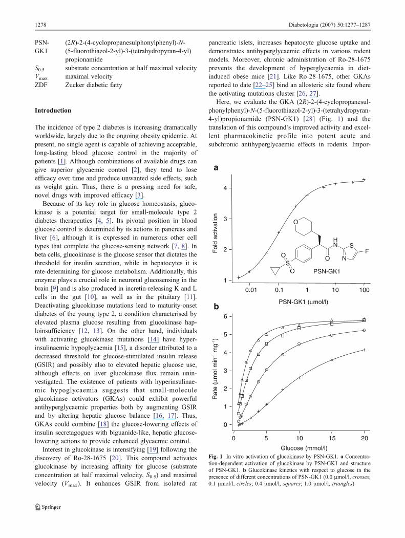
Fig. 1 In vitro activation of glucokinase by PSN-GK1. a Concentra-tion-dependent activation of glucokinase by PSN-GK1 and structureof PSN-GK1. b Glucokinase kinetics with respect to glucose in thepresence of different concentrations of PSN-GK1 (0.0 μmol/l, crosses;0.1 μmol/l, circles; 0.4 μmol/l, squares; 1.0 μmol/l, triangles)
1278 Diabetologia (2007) 50:1277–1287
tantly, these antihyperglycaemic effects do not appear to beaccompanied by adverse events, such as hypoglycaemia orhyperlipidaemia.
Materials and methods
Animals
Male C57Bl/6 (5–6 weeks), C57Bl/6 ob/ob (6–7 weeks)and female C57Bl/KsJ db/db (9 weeks) mice from HarlanOlac, Bicester, UK were housed under controlled condi-tions (21±2°C; 45–65% humidity; 12-h light–dark cycle,lights on 08.00 h) with free access to rat and mouse no. 1diet (Bantin and Kingman, Hull, UK). Female Zuckerdiabetic fatty (ZDF) rats (6 weeks old; Charles River,Manston, UK) were housed similarly, except that they wereput on a 10/14-h light–dark cycle (lights off at 22.00 h) andhad free access to high-fat diet D1245 (Research Diets,New Brunswick, NJ, USA) (45% of energy from fat) for4 weeks prior to a meal-feeding regimen. For clamp studies,male C57Bl/6J mice (12 weeks) from Charles River werehoused as above but fed pelleted chow (UAR, Nillemoison,France). Studies were approved by local ethical boards andanimal husbandry and procedures carried out according toinstitutional guidelines.
Materials
PSN-GK1 was prepared [29], dissolved in DMSO and usedin vitro at concentrations indicated with a final DMSOconcentration of ≤5%. For mice studies, PSN-GK1 wasdissolved in warm 90% water–10% Gelucire 44/14 (Gatte-fossé, Gennevilliers, France), with stirring and/or sonicationat 0.1–2.0 mg/ml, for dosing at 1–20 mg/kg via oral gavageat 10 ml/kg. For ZDF rats, PSN-GK1 was administered at10 mg 2 ml–1 kg–1 in warm 90% water–10% Gelucire 44/14.Recombinant human hexokinases I, II and III and glucoki-nase (liver form) were expressed as glutathione S-transferase(GST) fusion proteins in E. coli, purified by glutathioneaffinity chromatography to >98% purity and stored at –80 °Cin 50 mmol/l Tris/HCl pH 7.4, 1 mmol/l DL-dithiothreitol,50 mmol/l NaCl and 10% glycerol.
Enzyme assays
Glucokinase activity was measured in a coupled reaction withglucose-6-phosphate dehydrogenase (G6PDH) by monitoringNADPH production at A340 in a plate reader (SpectraMax190; Molecular Devices, Wokingham, UK) after 15 minincubation at 24°C, in a final volume of 100 μl containing25 mmol/l HEPES pH 7.1, 25 mmol/l KCl, 5 mmol/l glucose,1 mmol/l ATP, 2 mmol/l MgCl2, 1 mmol/l DL-dithiothreitol,
1 mmol/l NADP, 2.5 U/ml G6PDH, 0.4 μg GST-glucoki-nase. These conditions were also used for hexokinases I toIII, except that the glucose concentration was 0.25 mmol/l.We tested ten dilutions of PSN-GK1 from 0.004 to100 μmol/l, calculating and fitting fold changes in activityvs controls to sigmoidal curves using a four-parameterlogistic model. For effects on kinetic parameters (S0.5, Vmax,Hill coefficient), a series of velocity plots at different PSN-GK1 and glucose concentrations (0.67–20 mmol/l) werefitted to the Hill equation using XLfit version 4.1 (IDBS,Guildford, UK).
Cell incubations and assays
MIN6 cells, a mouse pancreatic beta cell line, were cultured inDMEM containing 25 mmol/l glucose, 1 mmol/l sodiumpyruvate, 50 μmol/l 2-mercaptoethanol, 15% heat-inactivatedFCS, 50 U/ml penicillin, 50 μg/ml streptomycin. Culture andincubations were at 37°C. Cells were seeded at 4×10⁵ cells perwell in 12-well plates, cultured for 3 days, then washed twicewith KRB containing 119 mmol/l NaCl, 4.74 mmol/l KCl,2.54 mmol/l CaCl2, 1.19 mmol/l MgSO4, 1.19 mmol/lKH2PO4, 25 mmol/l NaHCO3, 10 mmol/l HEPES pH 7.4,0.1% BSA and 5 mmol/l glucose. Cells were pre-incubatedfor 1 h in KRB, followed by 1 h static incubations with PSN-GK1 and glucose at indicated concentrations. Media sampleswere removed, cleared of debris by centrifugation at 1,000 gand insulin was measured in triplicate against standards using arat insulin ELISA kit (Mercodia, Uppsala, Sweden). Cells werelysed in 150 mmol/l NaCl, 1% glycerol, 1% Triton X-100,1mmol/l EGTA, 50mmol/l HEPES pH 7.5 supplemented withComplete Protease Inhibitor Cocktail (Roche Diagnostics,Penzberg, Germany) for protein measurement by bicincho-ninic acid assay (Pierce, Rockford, IL, USA) with BSAstandard.
Cryopreserved rat hepatocytes (XenoTech LLC, Lenexa,KS, USA) were centrifuged at 75 g over 25% Percoll andseeded at 2×10⁵ to 4×10⁵ cells per well in 12-well platescoated with collagen-I. Cells were attached, cultured andassayed for 2-deoxy-D-[3H]glucose (2-DG) uptake as de-scribed [30]. Media contained 5.55 mmol/l glucose and after2 days of culture cells were incubated for 4 h in fresh mediacontaining 74 kBq/ml 2-DG (Perkin Elmer, Beaconsfield,UK). Cells were washed three times, lysed and portions oflysates used for scintillation counting and protein measure-ment as above. Net uptake was calculated as pmol 2-DG permg protein and expressed as a percentage of the basal value.
Animal studies
C57Bl/6J mice Food was withdrawn 5 h before dosing,while water was available throughout. A blood sample wastaken from the tail tip under local anaesthetic for glucose
Diabetologia (2007) 50:1277–1287 1279
and insulin measurement. Thereafter, mice were weighedand dosed orally with PSN-GK1 (1 or 10 mg/kg) or vehicle.Blood samples were taken 15, 30, 60, 120 and 240 minafter dosing, samples (20 μl) for glucose being taken intodisposable micro-pipettes and added to 480 μl haemolysisreagent. Duplicate 20-μl aliquots haemolysed blood wereadded to 180 μl Trinder’s glucose reagent (Sigma enzy-matic colorimetric method) in a 96-well plate. After mixing,samples were left (room temperature) for 30 min beforereading against standards; correction was made for haemo-globin. Samples for insulin (30 μl) were collected intoheparin/Li-containing tubes, kept on ice for <30 min,centrifuged (2,800 g, 10 min, 4°C) and stored at −20°Cfor subsequent measurements using an insulin ELISA kit(Crystal Chem, Downers Grove, IL, USA) and SpectroMax250 plate reader (Molecular Devices).
Hyperinsulinaemic–hyperglycaemic clamps After 8 daysacclimatisation, during which mice were periodicallyhandled, an indwelling catheter was placed into the femoralvein under anaesthesia, sealed under the back skin andglued on top of the skull. After 4 to 5 days, mice werefasted for 6 h before dosing and throughout the remainderof the study. PSN-GK1 (10 mg/kg) or vehicle wasadministered orally 30 min before 180 min i.v. infusion ofa steady rate of [3H]glucose (to ensure detectable glucoseisotopic dilution into blood and measurable incorporationinto liver glycogen) and of pharmacological insulin at18 mU kg−1 min−1. Non-radiolabelled glucose infusionmaintained plasma glucose at 10 mmol/l. Plasma glucoseconcentrations and [3H]glucose-specific activity weredetermined in 5 μl blood from the tail tip every 10 minduring the last hour. For glucose turnover, [3H]glucoseenrichments were determined in deproteinised blood by Zn(OH)2 precipitation; aliquots of the supernatant fractionwere evaporated to dryness to determine radioactivity. In asecond aliquot, glucose concentration was assessed by theglucose oxidase method. Plasma insulin was determined byELISA (Mercodia) at study end to check that mice werestimulated comparably. Mice were killed by cervicaldislocation. The liver glycogen synthesis rate was deter-mined as described previously [31, 32] following extractionwith 3% perchloric acid and precipitation with ethanol. Theradioactive glycogen was counted and divided by the [3H]glucose-specific activity to determine the rate of synthesis.
C57Bl/KsJ db/db mice Blood (20 μl) was obtained forglucose levels 45 min before dosing. Just prior to dosing, foodwas removed (free access to water), a further blood sampletaken and mice dosed orally with PSN-GK1 (20 mg/kg) orvehicle, blood samples being removed 30, 60, 120, 180 and300 min thereafter. Glucose was determined in whole blood asabove.
ZDF rats At age 10 to 11 weeks, rats were housed singlyand fed high-fat diet (45% energy from fat, D1245;Research Diets) as follows during the 14-h dark phase: (1)free access (0–3 h); (2) food removed (3–7 h); (3) 8 g (7–14 h). After 30 days, the diet was changed to one in which60% of energy was from fat (D12492; Research Diets).Treatment commenced at age 14 to 15 weeks and wasconducted as follows: 1 h before lights out, a blood samplewas obtained from the tail tip under local anaesthesia forbasal glucose. Thirty minutes later, rats were dosed orallywith PSN-GK1 (10 mg/kg) or vehicle. At the start of thedark phase, blood samples were taken for glucose, then theanimals were fed as described above with hourly glucosesampling.
OGTTs in ob/ob mice Food was withdrawn 5 h beforeOGTTs and throughout with free access to water provided.Blood (20 μl) was removed for basal glucose 45 min beforeOGTTs. Then mice were weighed and dosed orally withPSN-GK1 (3, 5 or 10mg/kg) or vehicle 30min before glucose(2 g/kg). Blood samples were taken 0, 30, 60, 90, 120, 180and 240 min thereafter and glucose determined as above.
Subchronic study in ob/ob mice On day 0, 23.5 h beforefirst dose, blood glucose was determined and mice allocatedto groups. The day after, an OGTTwas performed as above,with oral PSN-GK1 (10 mg/kg) or vehicle administered at11.30 h, 30 min before the glucose load (2 g/kg). After theOGTT, food was returned. PSN-GK1 or vehicle was doseddaily at 11.30 h. On days 3 and 6, glucose was measured inblood samples 15 min before and 60 min after dosing.Another OGTT was performed on day 8. On day 9, 3 hafter dosing, terminal anaesthesia was induced and blood(500–600 μl) collected via the abdominal aorta intoheparin/Li-containing tubes and kept on ice (<30 min),before centrifugation (2,800 g, 10 min, 4°C) to give plasma(≥300 μl), which was stored at −20°C for subsequentanalysis (using kits) of fructosamine (Randox, Crumlin,Northern Ireland, UK), NEFA (Wako, Neuss, Germany),triacylglycerol (Thermotrace, VIC, Australia) and alanineaminotransferase (Randox). Livers were removed, freeze-clamped, weighed and stored (wrapped in foil) at −80°C forsubsequent glycogen analysis by ethanol precipitation afteralkali digestion and for glucosyl measurement after amylo-glucosidase treatment.
Plasma PSN-GK1 concentrations
A satellite exposure study, linked to basal blood glucose-lowering/insulin secretion in C57Bl/6 mice, was performed,in which 12 mice were dosed with 10 mg/kg PSN-GK1.Following terminal anaesthesia, blood (500–600 μl) was
1280 Diabetologia (2007) 50:1277–1287
collected from three animals via the abdominal aorta at 30,60, 120 and 240 min after dosing. Blood was collected intoheparin/Li-containing tubes and kept on ice (<30 min),before centrifugation (2,800 g, 10 min, 4°C) and storage at−20°C for subsequent compound measurement. A similarexperiment was performed following a 5 mg/kg dose to ob/ob mice, with terminal blood collected after 30, 60, 150 and270 min; these timepoints were used in a satellite exposurestudy paired with the OGTTs on days 1 and 8 (dosed daily)of the ob/ob study above.
Aliquots (50 μl) of plasma calibration standards, qualitycontrols, unknown samples and blanks were placed into a96-well protein precipitation plate (Argonaut Technologies,Hengoed, Wales, UK). The plate was placed on a vacuummanifold over a 96-well, 1-ml collection plate andacetonitrile (200 μl) added to each well. The plate was leftfor precipitation to occur and supernatant fractions to dripthrough under gravity for 10 min. Vacuum was applied andthe block removed and centrifuged (3,000 g, 10 min) priorto injection on to a liquid chromatography/mass spectrom-etry/mass spectrometry system, comprising an HPLCcolumn (Hichrom RPB; Highchrom, Theale, Berkshire,UK), 50×2.1 mm column, mobile phase of acetonitrile–10 mmol/l ammonium formate, flow rate 0.25 ml/min. TheMicromass Quattro Micro spectrometer (Waters, Milford,MA, USA) was fitted with an electrospray ionisationinterface. PSN-GK1 was detected by selected reactionmonitoring of daughter ions in negative ion mode.
Statistics
Analyses consisted of one-way ANOVA coupled with ttests. In cases where normality tests failed, the Mann-Whitney U test was used.
Results
Glucokinase activation in vitro
PSN-GK1 raised the glucose phosphorylating activity ofglucokinase at 5 mmol/l glucose by 4.3±0.2-fold with anEC50 of 130±10 nmol/l (Fig. 1a); 54±6 nmol/l PSN-GK1doubled activity. At 30 μmol/l, PSN-GK1 did not affect any
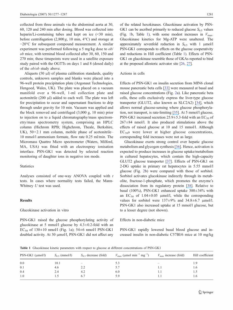
of the related hexokinases. Glucokinase activation by PSN-GK1 can be ascribed primarily to reduced glucose S0.5 values(Fig. 1b, Table 1), with some modest increases in Vmax.Glucokinase kinetics for Mg-ATP were unaltered. Theapproximately sevenfold reduction in S0.5 with 1 μmol/lPSN-GK1 corresponds to effects on the glucose cooperativityand reductions in Hill coefficient (Table 1). Effects of PSN-GK1 on glucokinase resemble those of GKAs reported to bindat the proposed allosteric activator site [26, 27].
Actions in cells
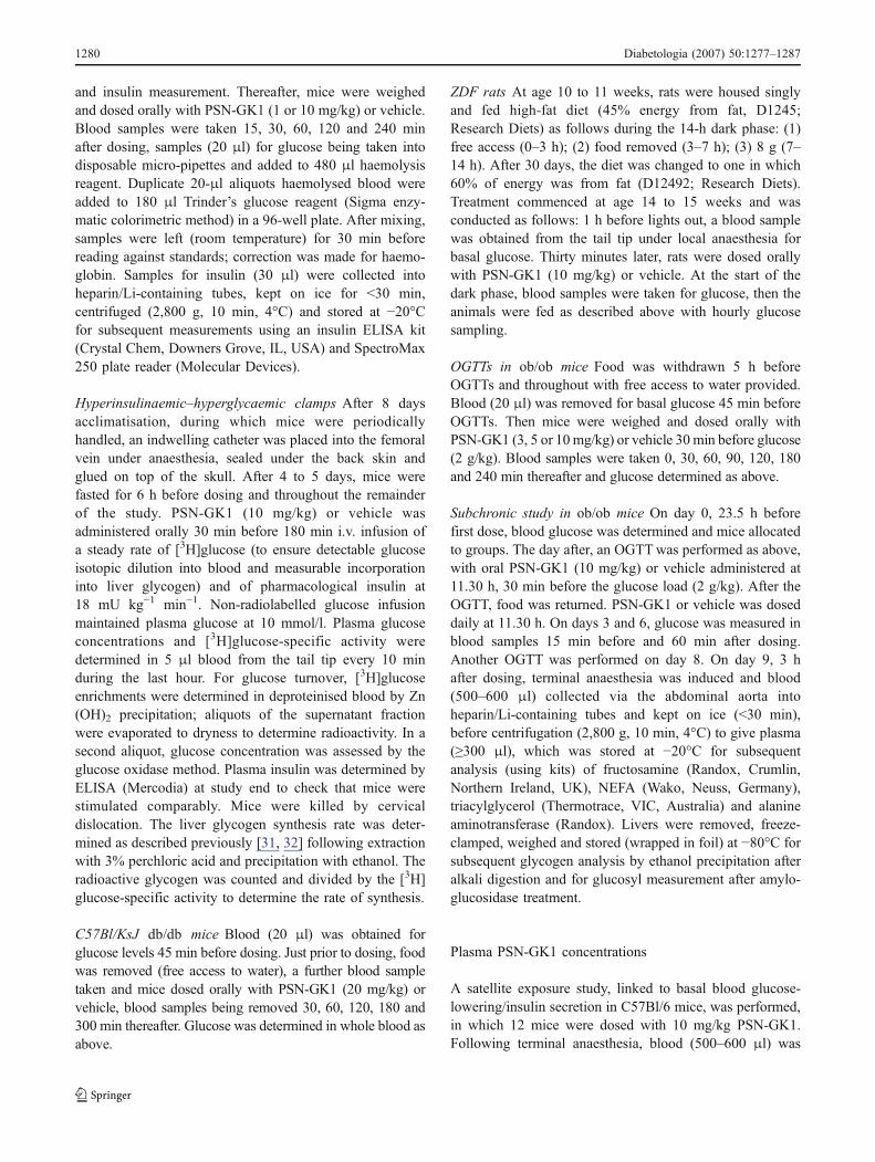
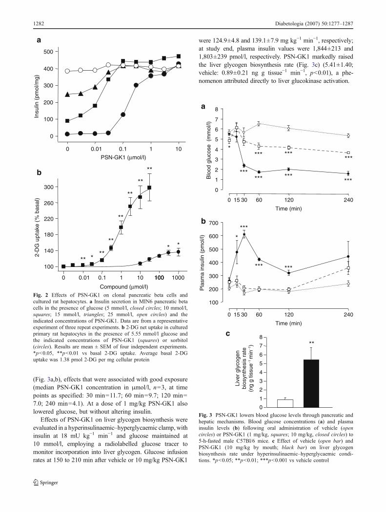
Effects of PSN-GK1 on insulin secretion from MIN6 clonalmouse pancreatic beta cells [33] were measured at basal andraised glucose concentrations (Fig. 2a). Like pancreatic betacells, these cells exclusively express the liver-type glucosetransporter (GLUT2, also known as SLC2A2) [34], whichallows normal glucose-sensing where glucose phosphoryla-tion, not transport, is rate-limiting [35]. At 5 mmol/l glucose,PSN-GK1 increased secretion 25.9±5.3-fold with an EC50 of267±84 nmol/l. It also produced stimulations above theeffects of raised glucose at 10 and 15 mmol/l. AlthoughEC50s were lower at higher glucose concentrations,corresponding fold increases were not as large.
Glucokinase exerts strong control over hepatic glucosemetabolism and glycogen synthesis [36]. Hence, activation isexpected to produce increases in glucose uptake/metabolismin cultured hepatocytes, which contain the high-capacityGLUT2 glucose transporter [37]. Effects of PSN-GK1 on2-DG uptake in primary rat hepatocytes in 5.55 mmol/lglucose (Fig. 2b) were compared with those of sorbitol.Sorbitol activates glucokinase indirectly through its metab-olite, fructose-1-phosphate, which promotes the enzyme’sdissociation from its regulatory protein [38]. Relative tobasal (100%), PSN-GK1 enhanced uptake 300±34% withan EC50 of 1.04±0.05 μmol/l, while the correspondingvalues for sorbitol were 137±9% and 34.8±6.7 μmol/l.PSN-GK1 also increased uptake at 15 mmol/l glucose, butto a lesser degree (not shown).
Effects in non-diabetic mice
PSN-GK1 rapidly lowered basal blood glucose and in-creased insulin in non-diabetic C57Bl/6 mice at 10 mg/kg
Table 1 Glucokinase kinetic parameters with respect to glucose at different concentrations of PSN-GK1
PSN-GK1 (μmol/l) S0.5 (mmol/l) S0.5 decrease (fold) Vmax (μmol min−1 mg−1) Vmax increase (fold) Hill coefficient
0.0 10.1 – 5.3 – 1.90.1 4.6 2.2 5.7 1.1 1.60.4 2.4 4.2 6.0 1.1 1.51.0 1.5 6.7 5.9 1.1 1.6
Diabetologia (2007) 50:1277–1287 1281
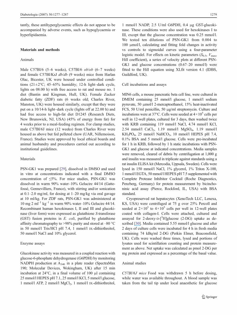
(Fig. 3a,b), effects that were associated with good exposure(median PSN-GK1 concentration in μmol/l, n=3, at timepoints as specified: 30 min=11.7; 60 min=9.7; 120 min=7.0; 240 min=4.1). At a dose of 1 mg/kg PSN-GK1 alsolowered glucose, but without altering insulin.
Effects of PSN-GK1 on liver glycogen biosynthesis wereevaluated in a hyperinsulinaemic–hyperglycaemic clamp,withinsulin at 18 mU kg−1 min−1 and glucose maintained at10 mmol/l, employing a radiolabelled glucose tracer tomonitor incorporation into liver glycogen. Glucose infusionrates at 150 to 210 min after vehicle or 10 mg/kg PSN-GK1
were 124.9±4.8 and 139.1±7.9 mg kg−1 min−1, respectively;at study end, plasma insulin values were 1,844±213 and1,803±239 pmol/l, respectively. PSN-GK1 markedly raisedthe liver glycogen biosynthesis rate (Fig. 3c) (5.41±1.40;vehicle: 0.89±0.21 ng g tissue−1 min−1, p<0.01), a phe-nomenon attributed directly to liver glucokinase activation.
0
1
2
3
4
5
6
7
8
Blo
od g
luco
se (
mm
ol/l)
2400 15 30 60 120
100
200
300
400
500
600
700
***
Time (min)
2400 15 30 60 120
Time (min)P
lasm
a in
sulin
(pm
ol/l)
a
b
***
****
****** ***
***
*********
*
0
1
2
3
4
5
6
7
8c
Live
r gl
ycog
enbi
osyn
thes
is r
ate
(ng
g tis
sue−1
min
−1) **
Fig. 3 PSN-GK1 lowers blood glucose levels through pancreatic andhepatic mechanisms. Blood glucose concentrations (a) and plasmainsulin levels (b) following oral administration of vehicle (opencircles) or PSN-GK1 (1 mg/kg, squares; 10 mg/kg, closed circles) to5-h-fasted male C57Bl/6 mice. c Effect of vehicle (open bar) andPSN-GK1 (10 mg/kg by mouth; black bar) on liver glycogenbiosynthesis rate under hyperinsulinaemic–hyperglycaemic condi-tions. *p<0.05; **p<0.01; ***p<0.001 vs vehicle control
10001001
*****
**
**
**
**
**
* *
100100100.10.010
Compound (μmol/l)
2-D
G u
pta
ke (
% b
asal
)
100
140
180
220
260
300
b
0
100
200
300
400
500
Insu
lin (
pmol
/mg)
1010.10.010
PSN-GK1 (μmol/l)
a
Fig. 2 Effects of PSN-GK1 on clonal pancreatic beta cells andcultured rat hepatocytes. a Insulin secretion in MIN6 pancreatic betacells in the presence of glucose (5 mmol/l, closed circles; 10 mmol/l,squares; 15 mmol/l, triangles; 25 mmol/l, open circles) and theindicated concentrations of PSN-GK1. Data are from a representativeexperiment of three repeat experiments. b 2-DG net uptake in culturedprimary rat hepatocytes in the presence of 5.55 mmol/l glucose andthe indicated concentrations of PSN-GK1 (squares) or sorbitol(circles). Results are mean ± SEM of four independent experiments.*p<0.05, **p<0.01 vs basal 2-DG uptake. Average basal 2-DGuptake was 1.38 pmol 2-DG per mg cellular protein
1282 Diabetologia (2007) 50:1277–1287
Effects in db/db mice and ZDF rats
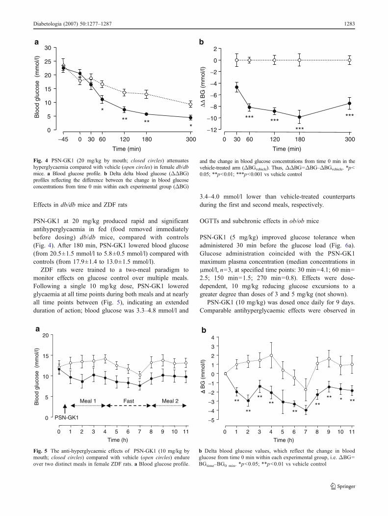
PSN-GK1 at 20 mg/kg produced rapid and significantantihyperglycaemia in fed (food removed immediatelybefore dosing) db/db mice, compared with controls(Fig. 4). After 180 min, PSN-GK1 lowered blood glucose(from 20.5±1.5 mmol/l to 5.8±0.5 mmol/l) compared withcontrols (from 17.9±1.4 to 13.0±1.5 mmol/l).
ZDF rats were trained to a two-meal paradigm tomonitor effects on glucose control over multiple meals.Following a single 10 mg/kg dose, PSN-GK1 loweredglycaemia at all time points during both meals and at nearlyall time points between (Fig. 5), indicating an extendedduration of action; blood glucose was 3.3–4.8 mmol/l and
3.4–4.0 mmol/l lower than vehicle-treated counterpartsduring the first and second meals, respectively.
OGTTs and subchronic effects in ob/ob mice
PSN-GK1 (5 mg/kg) improved glucose tolerance whenadministered 30 min before the glucose load (Fig. 6a).Glucose administration coincided with the PSN-GK1maximum plasma concentration (median concentrations inμmol/l, n=3, at specified time points: 30 min=4.1; 60 min=2.5; 150 min=1.5; 270 min=0.8). Effects were dose-dependent, 10 mg/kg reducing glucose excursions to agreater degree than doses of 3 and 5 mg/kg (not shown).
PSN-GK1 (10 mg/kg) was dosed once daily for 9 days.Comparable antihyperglycaemic effects were observed in
0
5
10
15
20
Blo
od g
luco
se (
mm
ol/l)
0 1 2 3 4 5 6 7 8 9 10 11
−5
−4
−3
−2
−1
0
1
2
3
4
****
**
****
****
** *
Time (h)
0 1 2 3 4 5 6 7 8 9 10 11
Time (h)
Δ Δ B
G (
mm
ol/l)
Meal 1 Meal 2Fast
PSN-GK1
a b
Fig. 5 The anti-hyperglycaemic effects of PSN-GK1 (10 mg/kg bymouth; closed circles) compared with vehicle (open circles) endureover two distinct meals in female ZDF rats. a Blood glucose profile.
b Delta blood glucose values, which reflect the change in bloodglucose from time 0 min within each experimental group, i.e. ΔBG=BGtime–BG0 min. *p<0.05; **p<0.01 vs vehicle control
−45 0 30 60 120 180 300
0
5
10
15
20
25
30
Time (min)
Blo
od g
luco
se (
mm
ol/l)
a
*****
*
0 30 60 120 180 300
−12
−10
−8
−6
−4
−2
0
2
Time (min)
ΔΔ B
G (
mm
ol/l)
b
*** ******
***
Fig. 4 PSN-GK1 (20 mg/kg by mouth; closed circles) attenuateshyperglycaemia compared with vehicle (open circles) in female db/dbmice. a Blood glucose profile. b Delta delta blood glucose (ΔΔBG)profiles reflecting the difference between the change in blood glucoseconcentrations from time 0 min within each experimental group (ΔBG)
and the change in blood glucose concentrations from time 0 min in thevehicle-treated arm (ΔBGvehicle). Thus, ΔΔBG=ΔBG–ΔBGvehicle. *p<0.05; **p<0.01; ***p<0.001 vs vehicle control
Diabetologia (2007) 50:1277–1287 1283
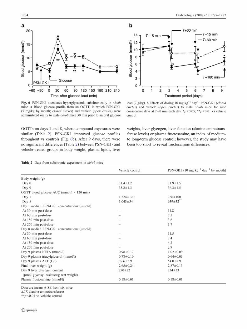
OGTTs on days 1 and 8, where compound exposures weresimilar (Table 2). PSN-GK1 improved glucose profilesthroughout vs controls (Fig. 6b). After 9 days, there wereno significant differences (Table 2) between PSN-GK1- andvehicle-treated groups in body weight, plasma lipids, liver
weights, liver glycogen, liver function (alanine aminotrans-ferase levels) or plasma fructosamine, an index of medium-to long-term glucose control; however, the study may havebeen too short to reveal fructosamine differences.
Table 2 Data from subchronic experiment in ob/ob mice
Vehicle control PSN-GK1 (10 mg kg−1 day−1 by mouth)
Body weight (g)Day 0 31.4±1.2 31.9±1.5Day 9 35.2±1.3 36.3±1.5OGTT blood glucose AUC (mmol/l × 120 min)Day 1 1,224±120 786±100Day 8 1,043±54 639±32**
Day 1 median PSN-GK1 concentrations (μmol/l)At 30 min post-dose – 11.8At 60 min post-dose – 7.1At 150 min post-dose – 3.6At 270 min post-dose – 1.7Day 8 median PSN-GK1 concentrations (μmol/l)At 30 min post-dose – 11.5At 60 min post-dose – 7.4At 150 min post-dose – 4.2At 270 min post-dose – 2.9Day 9 plasma NEFA (mmol/l) 0.98±0.17 1.02±0.09Day 9 plasma triacylglycerol (mmol/l) 0.78±0.10 0.64±0.03Day 9 plasma ALT (U/l) 39.6±5.9 54.0±8.9Final liver weight (g) 2.65±0.24 2.87±0.13Day 9 liver glycogen content(μmol glycosyl residues/g wet weight)
270±22 234±33
Plasma fructosamine (mmol/l) 0.18±0.01 0.18±0.01
Data are means ± SE from six miceALT, alanine aminotransferase**p<0.01 vs vehicle control
Fig. 6 PSN-GK1 attenuates hyperglycaemia subchronically in ob/obmice. a Blood glucose profile from an OGTT, in which PSN-GK1(5 mg/kg by mouth; closed circles) and vehicle (open circles) wereadministered orally to male ob/ob mice 30 min prior to an oral glucose
load (2 g/kg). b Effects of dosing 10 mg kg−1 day−1 PSN-GK1 (closedcircles) and vehicle (open circles) to male ob/ob mice for nineconsecutive days at T=0 min each day. *p<0.05; **p<0.01 vs vehiclecontrol
1284 Diabetologia (2007) 50:1277–1287
Discussion
PSN-GK1 is a potent activator of glucokinase in vitro,possessing enhanced activity relative to the archetypalGKA Ro-28-1675. At 5 mmol/l glucose, PSN-GK1 at0.05 μmol/l doubles glucokinase activity, whereas0.18 μmol/l Ro-28-1675 increases activity only by 1.5-fold[21]. Although the recently disclosed GKA50 has an EC50
of 0.03 μmol/l [24, 39], no fold-activation data areavailable. Thus, PSN-GK1 is one of the most potent GKAsdescribed to date. Like Ro-28-1675 and GKA50, PSN-GK1principally increases catalytic effectiveness, expressed asthe Vmax:S0.5 ratio, by lowering S0.5, as indicated by the factthat Vmax increases were modest. As expected, PSN-GK1does not activate other hexokinases that do not possessglucokinase’s allosteric activator site [26].
PSN-GK1 markedly increases GSIR from MIN6 cells,most notably at lower glucose concentrations, where basalsecretion is lower. However, effects are also observed at 10and 15 mmol/l glucose, values more typical of the diabeticcondition. Here, concentration-responses for PSN-GK1 areleft-shifted, suggesting that, at higher glucose concentra-tions, smaller doses would still provide maximum secretion.The fact that higher PSN-GK1 concentrations are requiredto stimulate secretion at lower glucose concentrations mayprovide GKAs with an inbuilt safety mechanism thatglucose-insensitive sulfonylureas [40] do not have. At25 mmol/l glucose, PSN-GK1 does not augment secretion,consistent with a mechanism chiefly involving reduction inS0.5 rather than increased Vmax.
The antihyperglycaemic properties of GKAs may also bemediated through enhanced hepatic glucose utilisation, sothat activation will cause increases in uptake of glucose orother glucokinase substrates, such as non-metabolisable 2-DG. Glucokinase activation in hepatocytes, either pharma-cologically or by adenoviral overexpression, results inincreased 2-DG uptake [22, 30]. Stimulation of hepaticglucose phosphorylation by sorbitol, a precursor to theindirect GKA fructose-1-phosphate [38], has been reportedwith an EC50 of 20–30 μmol/l and stimulation of ∼1.5-fold[41], values similar to those seen in our hands in ratprimary hepatocytes. PSN-GK1 has a substantially greatereffect on 2-DG uptake, indicating that glucokinase activa-tion occurs by a different mechanism.
In vitro effects in pancreatic and hepatic cells aretranslated in vivo in non-diabetic mice, where PSN-GK1stimulated insulin secretion, reduced blood glucose andraised liver glycogen biosynthesis. The increased potencyof PSN-GK1 compared with Ro-28-1675 is evident in thismodel: 15 mg/kg Ro-28-1675 maximally raised insulin∼1.5-fold [21], while 10 mg/kg PSN-GK1 induced anapprox. threefold increase. As mentioned, the hypoglycae-mia provoked by GKAs in a non-diabetic setting may be
more manageable than that resulting from sulfonylureas[40] because the glucose-lowering effected by GKAs isglucose-dependent [42, 43]. At 1 mg/kg, PSN-GK1 reducedblood glucose without hypoglycaemia, indicating that thereis a reasonable window between glucose-lowering andsevere hypoglycaemia. At this dose, no effects on insulinsecretion were apparent, indicating that severe hypoglycae-mic actions may be associated with insulinotropic effects.However, the fact that this dose was able to decrease bloodglucose possibly indicates a hepatic action, although insulinlevels were raised in view of the prevailing low glucose.Hyperinsulinaemic–hyperglycaemic clamps directly con-firmed the hepatic contribution (Fig. 3c).
To date, there have been no reports of GKAs displayingantihyperglycaemic actions in db/db mice. Indeed, someGKAs, such as Ro-28-1675, lose their effectiveness in olderdb/db mice with blood glucose ∼16.7 mmol/l and hypo-insulinaemia [20]. In our study, by contrast, PSN-GK1normalised blood glucose in db/db mice with initial bloodglucose >20 mmol/l without hypoglycaemia. The potentialof GKAs to control blood glucose is underscored by thefact that a single dose of PSN-GK1 can elicit antihyper-glycaemic effects in high-fat-diet-fed female ZDF rats overmultiple ‘meals’ and an extended time period withouthypoglycaemia. To our knowledge, PSN-GK1 is the firstGKA to show efficacy in this model.
PSN-GK1 improved glucose tolerance dose-dependently inob/ob mice, again without hypoglycaemia. Like Ro-28-1675,PSN-GK1 appears to normalise blood glucose in rodent type2 diabetes models, even though it produces hypoglycaemiain non-diabetic animals. It has been postulated [21] thatGKAs do not provoke hypoglycaemia in diabetic rodentsbecause of underlying defects in glucose homeostasis. Theresults seen acutely in OGTTs translate into subchronicefficacy, comparable antihyperglycaemic actions being notedat the beginning and end of the 9-day experiment, i.e. PSN-GK1’s effects did not appear to suffer from tachyphylaxis.Moreover, although PSN-GK1 improved blood glucosethroughout this study, it did not alter liver glycogen,suggesting that GKAs lower blood glucose principally by amechanism unrelated to increased glycogen storage.
One concern with GKAs is the possibility that theycould increase lipid levels. In normal rodents, chronicglucokinase overexpression leads to raised hepatic lipogen-esis and circulating lipids [44, 45]. However, humans withactivating glucokinase mutations have normal lipids [15],hinting that long-term glucokinase activation may notproduce adverse lipid effects. Effects of other GKAs onlipids following repeated administration have not beendescribed. In our study, subchronic administration of PSN-GK1 did not affect plasma lipids, indicating that concernssurrounding GKAs and increased lipogenesis may beunfounded. However, longer term studies will be required
Diabetologia (2007) 50:1277–1287 1285
ultimately to ensure that GKAs are safe in this regard. Itwas also discovered that PSN-GK1 did not induce changesin liver weight or plasma alanine aminotransferase, anindicator of liver toxicity and steatosis.
In summary, we have demonstrated that PSN-GK1’sability to activate glucokinase results in robust hypogly-caemic effects in normal mice and antihyperglycaemicactions in diabetic rodents. The overall efficacy, plus thefact that no adverse events were observed subchronically,further supports the premise that GKAs may be among thenext generation of oral glucose-lowering therapies. Incontrast to current therapies that target a single organ, e.g.sulfonylureas (pancreas) or metformin (liver), GKAs couldachieve greater efficacy by targeting multiple sites in thebody. Several GKAs are now in human clinical trials.Results from these trials will determine whether they candistinguish themselves from the widely prescribed sulfonyl-ureas, which are associated with increased mortality [46] asa result of raised cardiovascular risk [47] and mayaccelerate beta cell apoptosis and exhaustion [48, 49].
Acknowledgements We are grateful to F. Naud of Solvias (Basel,Switzerland) for the synthesis of PSN-GK1 and to Gattefossé(Gennevilliers, France) for generously supplying us with Gelucire44/14. The MIN6 cells were kindly supplied by J. Miyazaki of OsakaUniversity Graduate School of Medicine, Osaka, Japan. We alsoacknowledge M. Cawthorne, M. Sennitt and D. Hislop fromBuckingham University for in vivo pharmacology experiments, aswell as L. Bertram, C. Rasamison, V. Shah and G. Williams from(OSI)Prosidion (Oxford, UK) for technical support. The following arethanked for helpful discussion: K. Lindhardt and M. Thomsen from(OSI)Prosidion, R. Burcelin from University Paul Sabatier (Toulouse,France) and A. Cherrington and D. Granner from VanderbiltUniversity School of Medicine, USA.
Duality of interest E. Wargent, R. L. Printz and T. Sulpiceconducted studies on a fee-for-service basis. M. C. T. Fyfe, J. R.White, A. Taylor, R. Chatfield, M. J. Procter, C. Reynet, J. G.McCormack, P. S. Widdowson and P. Wong-Kai-In are all employedby (OSI)Prosidion and own stock or have stock options in (OSI)Pharmaceuticals (Melville, NY, USA).
References
1. Gershell L (2005) Type 2 diabetes market. Nature Rev DrugDiscov 4:367–368
2. Bell DSH (2004) A comparison of agents used to manage type 2diabetes mellitus: need for reappraisal of traditional approaches.Treat Endocrinol 3:67–76
3. Wagman AS, Nuss JM (2001) Current therapies and emergingtargets for the treatment of diabetes. Curr Pharmaceut Design7:417–450
4. Kietzmann T, Ganjam GK (2005) Glucokinase: old enzyme, newtarget. Expert Opin Therap Patents 15:705–713
5. Printz RL, Granner DK (2005) Tweaking the glucose sensor:adjusting glucokinase activity with activator compounds.Endocrinology 146:3693–3695
6. Magnuson MA, Matschinsky FM (2004) Glucokinase as aglucose sensor: past, present, and future. In: Matschinsky FM,
Magnuson MA (eds) Glucokinase and glycemic disease: frombasics to novel therapeutics. Karger, Basel, pp 1–17
7. Matschinsky FM, Magnuson MA, Zelent D et al (2006) Thenetwork of glucokinase-expressing cells in glucose homeostasisand the potential of glucokinase activators for diabetes therapy.Diabetes 55:1–12
8. Baltrusch S, Tiedge M (2006) Glucokinase regulatory network inpancreatic β-cells and liver. Diabetes 55(Suppl 2):S55–S64
9. Kang L, Dunn-Meynell AA, Routh VH et al (2006) Glucokinaseis a critical regulator of ventromedial hypothalamic neuronalglucosensing. Diabetes 55:412–420
10. Theodorakis MJ, Carlson O, Michopoulos S et al (2006) Humanduodenal enteroendocrine cells: source of both incretin peptides,GLP-1 and GIP. Am J Physiol Endocrinol Metab 290:E550–E559
11. Zelent D, Golson ML, Koeberlein B et al (2006) A glucose sensorrole for glucokinase in anterior pituitary cells. Diabetes 55:1923–1929
12. Velho G, Froguel P, Gloyn A, Hattersley A (2004) Maturity onsetdiabetes of the young type 2. In: Matschinsky FM, Magnuson MA(eds) Glucokinase and glycemic disease: from basics to noveltherapeutics. Karger, Basel, pp 42–64
13. Sagen JV, Odili S, Bjørkhaug L et al (2006) From clinicogeneticstudies of maturity-onset diabetes of the young to unraveling com-plex mechanisms of glucokinase regulation. Diabetes 55:1713–1722
14. Heredia VV, Carlson TJ, Garcia E, Sun S (2006) Biochemicalbasis of glucokinase activation and the regulation by glucokinaseregulatory protein in naturally occurring mutations. J Biol Chem281:40201–40207
15. Christesen HBT, Herold K, Noordam K, Gloyn AL (2004)Glucokinase-linked hypoglycemia: Clinical aspects of activatingglucokinase mutations. In: Matschinsky FM, Magnuson MA (eds)Glucokinase and glycemic disease: from basics to novel thera-peutics. Karger, Basel, pp 75–91
16. Al-Hasani H, Tschöp MH, Cushman SW (2003) Two birds withone stone: Novel glucokinase activator stimulates glucose-inducedpancreatic insulin secretion and augments hepatic glucose metab-olism. Mol Interventions 3:367–370
17. Leighton B, Atkinson A, Coghlan MP (2005) Small moleculeglucokinase activators as novel anti-diabetic agents. Biochem SocTrans 33:371–374
18. Van Gaal LF, De Leeuw IH (2003) Rationale and options forcombination therapy in the treatment of Type 2 diabetes.Diabetologia 46(Suppl 1):M44–M50
19. Guertin KR, Grimsby J (2006) Small molecule glucokinaseactivators as glucose lowering agents: a new paradigm fordiabetes therapy. Curr Med Chem 13:1839–1843
20. Grimsby J, Sarabu R, Corbett WL et al (2003) Allostericactivators of glucokinase: potential role in diabetes therapy.Science 301:370–373
21. Grimsby J, Matschinsky FM, Grippo JF (2004) Discovery andactions of glucokinase activators. In: Matschinsky FM, MagnusonMA (eds) Glucokinase and glycemic disease: from basics to noveltherapeutics. Karger, Basel, pp 360–378
22. Efanov AM, Barrett DG, Brenner MB et al (2005) A novelglucokinase activator modulates pancreatic islet and hepatocytefunction. Endocrinology 146:3696–3701
23. Sarabu R, Grimsby J (2005) Targeting glucokinase activation forthe treatment of type 2 diabetes—a status review. Curr Opin DrugDiscov Dev 8:631–637
24. McKerrecher D, Allen JV, Caulkett PWR et al (2006) Design of apotent, soluble glucokinase activator with excellent in vivoefficacy. Bioorg Med Chem Lett 16:2705–2709
25. Futamura M, Hosaka H, Kadotani A et al (2006) An allostericactivator of glucokinase impairs the interaction of glucokinase andglucokinase regulatory protein and regulates glucose metabolism.J Biol Chem 281:37668–37674
1286 Diabetologia (2007) 50:1277–1287
26. Dunten P, Swain A, Kammlott U et al (2004) Crystal structure ofhuman liver glucokinase bound to a small molecule allostericactivator. In: Matschinsky FM, Magnuson MA (eds) Glucokinaseand glycemic disease: from basics to novel therapeutics. Karger,Basel, pp 145–154
27. Kamata K, Mitsuya M, Nishimura T, Eiki J-I, Nagata Y (2004)Structural basis for allosteric regulation of the monomericallosteric enzyme human glucokinase. Structure 12:429–438
28. Fyfe MCT, Gardner LS, Nawano M et al (2004) Tri(cyclo)substituted amide compounds. International Patent PublicationWO 2004/072031
29. Fyfe MCT, Naud F (2006) Fluorination process of protectedaminothiazole. International Patent Publication WO 2006/016174
30. Yang R, Cao L, Gasa R, Brady MJ, Sherry AD, Newgard CB(2002) Glycogen-targeting subunits and glucokinase differentiallyaffect pathways of glycogen metabolism and their regulation inhepatocytes. J Biol Chem 277:1514–1523
31. Massillon D, Chen W, Hawkins M, Liu R, Barzilai N, Rossetti L(1995) Quantitation of hepatic glucose fluxes and pathways ofhepatic glycogen synthesis in conscious mice. Am J PhysiolEndocrinol Metab 269:E1037–E1043
32. Perrin C, Knauf C, Burcelin R (2004) Intracerebroventricularinfusion of glucose, insulin, and the adenosine monophosphate-activated kinase activator, 5-aminoimidazole-4-carboxamide-1-β-D-ribofuranoside, controls muscle glycogen synthesis.Endocrinology 145:4025–4033
33. Miyazaki J, Araki K, Yamato E et al (1990) Establishment of apancreatic beta cell line that retains glucose inducible insulinsecretion: special reference to expression of glucose transporterisoforms. Endocrinology 127:126–132
34. Leturque A, Brot-Laroche E, Le Gall M, Stolarczyk E, Tobin V(2006) The role of GLUT2 in dietary sugar handling. J PhysiolBiochem 61:529–538
35. Tal M, Wu Y, Leiser M et al (1992) [Val12]HRAS downregulatesGLUT2 in β cells of transgenic mice without affecting glucosehomeostasis. Proc Natl Acad Sci U S A 89:5744–5748
36. Agius L (1998) The physiological role of glucokinase bindingand translocation in hepatocytes. Adv Enzyme Regul 38:303–331
37. Zheng Q, Levitsky LL, Mink K, Rhoads DB (1995) Glucoseregulation of glucose transporters in cultured adult and fetalhepatocytes. Metabolism 44:1553–1558
38. Veiga-Da-Cunha M, Van Schaftingen E (2002) Identification offructose 6-phosphate- and fructose 1-phosphate-binding residues inthe regulatory protein of glucokinase. J Biol Chem 277:8466–8473
39. Coope GJ, Atkinson AM, Allott C et al (2006) Predictive bloodglucose lowering efficacy by glucokinase activators in high fat fedfemale Zucker rats. Br J Pharmacol 149:328–335
40. Light PE (2002) The ABCs of sulfonylurea receptors, islet KATP
channels and the control of insulin secretion. Can J Diabetes26:223–231
41. Brocklehurst KJ, Payne VA, Davies RA et al (2004) Stimulationof hepatocyte glucose metabolism by novel small moleculeglucokinase activators. Diabetes 53:535–541
42. Matschinsky FM, Glaser B, Magnuson MA (1998) Pancreatic β-cell glucokinase: closing the gap between theoretical concepts andexperimental realities. Diabetes 47:307–315
43. Zelent D, Najafi H, Odili S et al (2005) Glucokinase and glucosehomeostasis: proven concepts and new ideas. Biochem Soc Trans33:306–310
44. O’Doherty RM, Lehman DL, Telemaque-Potts S, Newgard CB(1999) Metabolic impact of glucokinase overexpression in liver.Lowering of blood glucose in fed rats is accompanied byhyperlipidemia. Diabetes 48:2022–2027
45. Ferre T, Riu E, Franckhauser S, Agudo J, Bosch F (2003) Long-term overexpression of glucokinase in the liver of transgenic miceleads to insulin resistance. Diabetologia 46:1662–1668
46. Simpson SH, Majumdar SR, Tsuyuki RT, Eurich DT, Johnson JA(2006) Dose–response relation between sulfonylurea drugs andmortality in type 2 diabetes mellitus: a population-based cohortstudy. Can Med Assoc J 174:169–174
47. Bell DSH (2006) Do sulfonylurea drugs increase the risk ofcardiac events? Can Med Assoc J 174:185–186
48. Donath MY, Ehses JA, Maedler K et al (2005) Mechanisms of β-cell death in type 2 diabetes. Diabetes 54(Suppl 2):S108–S113
49. Del Prato S, Pulizzi N (2006) The place of sulfonylureas in thetherapy for type 2 diabetes mellitus. Metab Clin Exp 55(Suppl 1):S20–S27
Diabetologia (2007) 50:1277–1287 1287