Multidetector Computed Tomography Myocardial Perfusion Imaging During Adenosine Stress

© 2015 Liguori et al. This work is published by Dove Medical Press Limited, and licensed under Creative Commons Attribution – Non Commercial (unported, v3.0) License. The full terms of the License are available at http://creativecommons.org/licenses/by-nc/3.0/. Non-commercial uses of the work are permitted without any further
permission from Dove Medical Press Limited, provided the work is properly attributed. Permissions beyond the scope of the License are administered by Dove Medical Press Limited. Information on how to request permission may be found at: http://www.dovepress.com/permissions.php
Medical Devices: Evidence and Research 2015:8 265–278
Medical Devices: Evidence and Research Dovepress
submit your manuscript | www.dovepress.com
Dovepress 265
R E v i E w
open access to scientific and medical research
Open Access Full Text Article
http://dx.doi.org/10.2147/MDER.S70630
Emerging clinical applications of computed tomography
Carlo Liguori1
Giulia Frauenfelder2
Carlo Massaroni3
Paola Saccomandi3
Francesco Giurazza4
Francesca Pitocco4
Riccardo Marano5
Emiliano Schena3
1Radiology Unit, AORN A Cardarelli, 2Radiology Unit, AOU Federico ii, Naples, 3Measurement and Biomedical instrumentation Unit, 4Radiology Unit, Università Campus Bio-Medico di Roma, 5Department of Radiological Sciences, institute of Radiology, Catholic University of Rome, A Gemelli University Hospital, Rome, italy
Correspondence: Emiliano Schena Measurement and Biomedical instrumentation Unit, Università Campus Bio-Medico di Roma, via Álvaro del Portillo 21, 00128 Rome, italy Tel +39 06 22541 9650 Email [email protected]
Abstract: X-ray computed tomography (CT) has recently been experiencing remarkable growth
as a result of technological advances and new clinical applications. This paper reviews the essential
physics of X-ray CT and its major components. Also reviewed are recent promising applications
of CT, ie, CT-guided procedures, CT-based thermometry, photon-counting technology, hybrid
PET-CT, use of ultrafast-high pitch scanners, and potential use of dual-energy CT for material
differentiations. These promising solutions and a better knowledge of their potentialities should
allow CT to be used in a safe and effective manner in several clinical applications.
Keywords: computed tomography, X-ray, thermometry, dual-energy, ultrafast scanner,
guidance, photon-counting technology
IntroductionMany diagnostic imaging techniques, including ultrasound imaging, magnetic reso-
nance (MR) and computed tomography (CT), have gained widespread acceptance
in several fields of medicine. In spite of the concern related to the patient dose, CT
imaging has become a “cannot-do-without tool” in many branches of medicine.
The first application of CT dates back to shortly after its invention, when between
1957 and 1963 Cormack applied this technology to improve radiotherapy planning.1
The first successful implementation of CT was performed a few years later by
Hounsfield, who surprised the entire medical community with his experiments.2
From the introduction of CT in the early 1970s, its importance in clinical imaging
has exceeded even the most optimistic hopes of researchers. The steadily increasing
numbers of examinations based on this technique and the introduction of new proce-
dures performed under CT guidance in clinical practice have promoted research on
new applications in this scenario. Nowadays, CT is widely used for both diagnostic
and therapeutic procedures.
This article reviews some medical applications of X-ray CT. After a brief descrip-
tion of its major components and some essential physics, particular attention is focused
on the characteristics of each type of CT scanner, on the applications of multidetector
CT (MDCT) and dual-energy CT (DECT), as well as emerging applications proposed
to improve the outcomes of some diagnostic and therapeutic procedures.
History and essential physicsHistoryThe discovery of X-radiation in 1895 by Röntgen, who investigated radiance during
electric discharges inside an evacuated glass tube, revolutionized the diagnosis of

Medical Devices: Evidence and Research 2015:8submit your manuscript | www.dovepress.com
Dovepress
Dovepress
266
Liguori et al
several diseases.3,4 After 1895, X-ray research developed very
rapidly, and the first picture of a whole skeleton obtained
by X-rays dates back to 1897.3 Over the years, the design
of X-ray equipment was improved to obtain high quality
two-dimensional images of the inside of the human body.3
Among others, Edison made a significant contribution to
the development of medical imaging techniques, and von
Helmholtz investigated the mathematical equations describ-
ing properties of X-rays and their penetration through dif-
ferent materials.3 Thompson investigated the possibility of
obtaining a three-dimensional X-ray image.5 These studies
led to development of some investigational techniques, such
as the ones patented by Baese in 1915 and Bocage in 1922.
A further step in the direction of contemporary scanners
was the use of gamma radiation to obtain a layered image of
tissues, proposed by Kuhn in 1963. However, the discovery
of X-rays was not enough to lead to the advent of CT; in
fact, its development was more related to the development
of computational techniques. The first commercial fan-beam
CT scanner come onto the market in 1973, with an acquisition
time of 20 seconds and 30 detectors. In 1979, Cormack and
Hounsfield were awarded the Nobel Prize and credited with
inventing the modern CT.1,2 Since the end of 1970s, a lot of
progress has been made in CT design and manufacture. In
comparison with the first scans, contemporary ones can scan
in a few hundred milliseconds, and reconstruct an image of
2,048×2,048 pixels from hundreds of spiral slices.
Essential physicsCT is based on the principle that the density of the tissue passed
through by the X-ray beam can be measured by calculation
of the attenuation coefficient. The X-ray emitter discharges
monochromatic photons that produce a high kV X-ray beam
with an average energy of 75 keV.6 X-rays are generated by
physical processes that take place within matter at the atomic
level. During generation of X-radiation in X-ray tubes, transi-
tion of electrons between the inner shells of of an atom, and the
deceleration of charged particles caused by electromagnetic
fields within matter occur.7 The X-ray spectrum may be recog-
nized as the sum of the energies of both the above-mentioned
processes, resulting in discrete characteristic X-rays and
continuous X-ray emission, respectively. After X-rays pass
through a layer of biological material, the detector measures
an attenuated X-ray intensity.6 The unattenuated intensity of
the X-ray beam, I0, is also measured by the CT scanner.
X-ray monochromatic intensity, I, is defined as the
amount of photon energy (N ⋅ hv) passing through a unit
area (S) in unit time (t):
I
N hv
S t=
⋅⋅
(1)
where h is Planck’s constant and v is the frequency of the
photon of radiation emitted.
The relationship between the It and I
0 can be expressed
by Lambert-Beer’s law:6
ln I I xt 0( ) = ⋅µ (2)
where x is the thickness of the biological tissue and µ is the
linear attenuation coefficient. CT reconstruction algorithms
use Equation (2) in a pre-processing step before image
reconstruction to reduce dependence of the CT image on
machine-dependent parameters (ie, It and I
0).
The attenuation of radiation due to interaction with mat-
ter is related to some phenomena (ie, photoelectric effect
and coherent and incoherent scattering).3 The total value of
the linear attenuation depends on the substance and tracks
linearly with density.8
After CT reconstruction, before storing and displaying,
CT images are normalized to integer values comprising the
CT number (also known as the Hounsfield unit),8 which
defines the degree of attenuation of radiation by various
substances. The number CT(x,y) in each image pixel (x,y)
is expressed as:6
CT x yx y
( , )( , )
=−
1000 2
2
µ µµ
H O
H O
(3)
where µH2O
is the attenuation coefficient of water. This nor-
malization results in CT numbers ranging from −1,000 to
+3,000 (eg, −1,000 for air and +3,000 for dense bone or areas
filled with contrast agents). Contrast in CT images mainly
derives from the physical properties of tissue that influence
incoherent scattering (known as Compton scattering), which
depends on tissue electron density, ρe = NZ/A (where Z and
A are the atomic number and atomic mass, respectively).9 As
a consequence, a tissue containing a relative abundance of
hydrogen (eg, fat) is well visualized by CT. The CT number
allows accurate diagnosis in some clinical settings, and accu-
rate estimation of, eg, tumor volume and lesion diameter.
The DECT scanner is gaining acceptance in the clinical
setting.10 The principle underpinning the clinical use of DECT
is the dependency of µ on the X-ray energy. Indeed, each
type of material demonstrates a relatively specific change in
attenuation between images obtained with different energy
spectra, and this difference in attenuation allows a more

Medical Devices: Evidence and Research 2015:8 submit your manuscript | www.dovepress.com
Dovepress
Dovepress
267
Emerging clinical applications of CT
nuanced characterization of the features depicted. With
this technique, two image datasets are acquired in the same
anatomic location with two different X-ray spectra. Different
types of DECT scanners are available, as explained in the
section on physics and applications of DECT scanners. Two
major advantages of DECT are material decomposition by
the almost simultaneous acquisition of two image series
with different kVp values (80 and 140) and elimination of
misregistration artifacts.
Key performance parameters and major components of CT scannersThe main components of CT scanners are the following: the
X-ray genera the system for X-ray detection, the system of
collimators and filters, and the system for the reconstruction
of images. The patient is positioned within the gantry on a
table that moves automatically during the scans. The X-ray
tube is responsible for generation of X-rays. These are emit-
ted when matter is hit by charged particles with high kinetic
energy (103–106 eV) that are able to knock electrons out of
their atomic orbits. Basically, the X-ray tube is composed
of a cathode and an anode within a vacuum glass envelope.
Electrons from the cathode tungsten filament are acceler-
ated toward the anode, represented by a tungsten target
electrode maintained at a positive voltage potential with
respect to the cathode. Peak voltage is applied to the cath-
ode, and the tube current, ie, the rate of electron flow from
the cathode to the anode, in the order of hundreds of mA, is
used to produce X-rays.6 The X-rays pass through the body
in the gantry and carry the information about the structure
of the body to the detectors. The information, represented by
radiation intensities, is in the form of a series of projections,
measured by the detection system. Usually, the whole detec-
tor system rotates synchronously with the X-ray tube around
the body. Different detectors are used (ie, xenon detector,
solid-state detector, and multiple detector arrays) to detect
the projections and convert them into electrical quantities.
Such quantities are used by the reconstruction system to
form the diagnostic image. Collimators and filters are used
to limit unnecessary radiation exposure and to improve the
image quality.
A series of X-rays passing through the patient at the same
orientation is known as a projection. The main projection
geometries used are: parallel beam geometry, where all the
rays are parallel to each other; fan beam geometry, in which
the rays diverge, looking like a fan; and cone beam geometry,
where the beam has a conical shape. Each geometry has a
dedicated algorithm for reconstruction of images from the
projections. The modern CT scanner incorporates fan beam
geometry in the acquisition and reconstruction processes.
CT scanner technology has evolved over several genera-
tions, and CT scanner technology has evolved over several
generations.
The first-generation scanner used a parallel beam system
and lateral movement to make a single projection, along with
a circular movement about the central opening in the gantry to
gather all the projections necessary to reconstruct the image.
Only two detectors measured the X-rays transmitted by the
patients. The second-generation scanner used a fan beam
system and a linear array of 30 detectors, which improved
utilization of the X-ray beam by 30-fold. The slow scanning
due to the translational motion of the first-generation and
second-generation scanners was overcome by the third-gen-
eration scanners, where the X-ray tube and detector array are
mechanically joined and rotated together around the patient.
The problem of artifact with the third-generation scanner was
partially overcome by the fourth-generation scanner. The
fifth-generation scanners differ significantly from the others,
in that they do not contain moving parts and the conventional
X-ray tube is substituted by an arc of tungsten encircling the
patient, and an electron beam is steered around the patient,
hitting the arc. The sixth-generation scanner is known as
the helical CT scanner, because data are acquired while
the patient’s table is moving. Lastly, the seventh-generation
scanner uses a multiple detector array (solid-state detectors),
allowing an increased number of X-rays to be detected, and
enabling better and more efficient patient imaging.
Many parameters can influence the quality and performance
of CT imaging, and can be related to generation of X-rays,
phase of acquisition, or steps in image reconstruction.
The performance parameters of CT scanners are indica-
tors of the quality of the system in terms of its physical and
technical capabilities, and allow standardized, comparative,
and quantitative criteria to be established. The key parameters
are: spatial resolution, ie, the minimum area of the image in
which changes are detectable; low-contrast resolution, ie, the
ability to detect changes in tissue attenuation (typical values
are close to 0.4%); slice thickness, ie, the nominal thickness
of the image cross-section (typical values 0.4 mm ÷ 10 mm);
pitch, ie, the ratio between displacement of the table and the
thickness of the scanned layer for one revolution of the scan-
ner (relevant only for helical CT scanners); and dose, ie, an
indicator of the damage that X-rays absorbed by the patient
can cause. The CT dose index is the most commonly used
dose indicator. The value of the dose absorbed by the patient
during a scan is strongly related to a number of parameters

Medical Devices: Evidence and Research 2015:8submit your manuscript | www.dovepress.com
Dovepress
Dovepress
268
Liguori et al
(eg, tube voltage, current-time product, and thickness and
number of slices). The total dose absorbed by the patient
should not exceed permissible levels; consequently, efforts
of manufacturers of tomographic equipment are directed
towards minimizing this dose.3
Clinical applications and technical parametersWith remarkable growth in its applications and use, evolution
of MDCT technology has resulted in significant changes in the
scanning parameters, hardware, and radiation exposure associ-
ated with CT scanning. In comparison with single-detector CT,
conventional MDCT allows acquisition of different image slabs
from the same dataset by using helical reconstruction, weighting
algorithms, and interpolation of adjacent helical datasets.
Salient MDCT characteristics are: a cone-shaped X-ray
beam, up to 0.5 seconds of rotation time (2 Hz of fre-
quency), faster scan coverage, larger z-axis coverage/rotation
(18/24/32 mm), isotropic voxel possible, greater temporal
resolution and less image noise, beam pitch, and reduced
radiation dose efficiency.11
MDCT has multiple detector rows (each consisting of
500–900 detector elements) along the scanning direction,
which create a two-dimensional curved detector array.
MDCT acquires more than one slice in a single X-ray tube
rotation with thin isotropic voxels. At a beam pitch of 1:1,
table speed and z-axis coverage from dual-slice CT scanners
(20 mm per second) have steadily increased to 40 mm per
second for four-slice, 80 mm per second for eight-slice, and
160 mm per second for 16-slice MDCT scanners. Currently
available MDCT scanners have as many as 40 rows of detec-
tors in the z-axis, which comprise more than 30,000 indi-
vidual detector elements with high efficiency and minimal
afterglow. In addition to changes in detector design, evolution
of CT technology has resulted in substantial alteration in
the image reconstruction techniques used to accommodate
changes in image geometry; the filtered back-projection
technique of image reconstruction is used in single-slice axial
scanners, and interpolated filtered back-projection is used in
single-detector CT scanners and four-slice MDCT.
Recent implementation of computer calculation perfor-
mances has made it possible to introduce into clinical CT
scanners a different image reconstruction model known as
the iterative reconstruction protocol. This protocol is mainly
based on comparison of the acquired CT image with a CT
model in order to reduce CT image noise using lower Kv
levels or to improve image quality using the same kV levels.12
Isotropic scanning may be defined as acquisition of images
with equal voxel size in three axes: MDCT scanners permit
acquisition of thin slices with isotropic voxel size.
Images obtained using conventional MDCT provide use-
ful but only limited material-specific information, because
the representation of structures on images depends solely on
the linear attenuation coefficient of each of the constituent
materials and is independent of the material density and mass
attenuation coefficient.
The introduction of DECT technology made possible new
diagnostic scenarios: two datasets (80 kVp and 140 kVp) are
loaded on the workstation, and virtual non-contrast, iodine
map, and mixed (with adjustable blending of 80 kVp and 140
kVp data) images can be obtained.10
Single-energy CT applications of multidetector scannersMDCT offers several improvements when performing CT
angiography. Because scans are faster, less intravenous
contrast is necessary, resulting in cost-savings and a reduced
contrast load to the patient’s kidneys. Moreover, the scan
distance is increased so that the entire volume (ie, chest, abdo-
men, and pelvis) can be scanned in one acquisition; this is a
definite advantage when scanning the aorta, because only one
acquisition is necessary. Thinner collimation (0.5–1.0 mm)
improves visualization of small vessels (,1 mm), which is
helpful when staging malignancies, evaluating organ donors
and patients with suspected pulmonary embolism, and visual-
izing the mesenteric vessels.
The introduction of MDCT resulted in advances in
musculoskeletal imaging. Multiplanar reconstruction can be
created in any plane, maintaining high resolution (the joint
needs to be scanned only in one plane, preferably oblique to
the joint surface). CT colonography involves volumetric CT
imaging after colonic cleansing combined with sophisticated
processing software to create three-dimensional endoluminal
views.12
MDCT also allows scanning of the heart during a single
breath hold, eliminating artifact caused by respiratory motion;
moreover, it allows noninvasive characterization of cardiac
arrhythmias using an integrative approach that combines
electrical and structural data (electrocardiography and
MDCT).13
Physics and applications of DECT scannersPhysicsThe basic principle of DECT or spectral imaging is to obtain
two datasets with different kVp values (usually 80 and 140)

Medical Devices: Evidence and Research 2015:8 submit your manuscript | www.dovepress.com
Dovepress
Dovepress
269
Emerging clinical applications of CT
from the same anatomic region. This allows performing mate-
rial decomposition thanks to the attenuation differences at
different energy levels. X-ray attenuation of materials in the
diagnostic energy range varies according to the specific organ
composition and X-ray beam energy. Consequently, different
substances show a different CT number at different energies.
The increase in photon energy results in a small decrease in
CT values for materials with a low atomic number while it
causes a rapid decrease in Hounsfield unit values for materials
with a high atomic number.14
At low kVp, the photoelectric effect predominates for
elements with a high atomic number, such as calcium and
iodine. With a reduction in kVp, the incidence of photoelec-
tric interaction increases. In contrast with single-spectrum
imaging, DECT is sensitive to the chemical composition.
This means that DECT is capable of differentiating materials
with different atomic numbers, despite similar attenuation
coefficients, because the data are acquired using two dif-
ferent spectra. Consequently, materials with almost similar
coefficient attenuation values at different energy levels can
be distinguished by DECT.15
Post-processingThere are two approaches to extracting dual-energy infor-
mation from projection data. A straightforward method is
to subtract equivalent projections and apply filtered back-
projection to reconstruct the difference as spectral informa-
tion. Another way is to consider the CT number of voxels
in standard CT images, and then to use post-processing
algorithms to extract specific spectral information from the
difference between the corresponding voxels. Currently, the
most commonly used approach is the latter, with the image
reconstruction system providing low-kVp and high-kVp
images and a series of weighted average images. Three main
types of algorithms are in use: the first one optimizes images;
the second one identifies or differentiates certain materials;
and the third one quantifies a substance in the dataset. The
output of the first algorithm consists of altered gray-level
CT images, whereas the output of the latter two algorithms
usually color-code substances, either several substances in
different colors or the quantity of one substance on a color
palette.
Optimization algorithms are monoenergetic images in
which the CT number for each voxel is extrapolated to a cer-
tain energy from the two density values at the acquired photon
energies, and nonlinear blending algorithms. Differentiation
algorithms define a slope between the density values obtained
by scanning the material at the two spectra, and differentiate
materials on the basis of the photoelectric effect. Quantifica-
tion algorithms use a three-material decomposition, quantify-
ing one of three materials.16
Research in the technical development of DECT led to
four different scanner models, sharing the same technological
concept but being different in terms of acquisition structure
and produced by different manufacturers as follows:
• Dual-source and dual-energy scanner (Definition Flash
and Force; Siemens Healthcare, Forchheim, Germany)
which obtains high-energy scans at 120/140 kVp and low-
energy scans simultaneously at 80/100 kVp (limited in
temporal registration and field of view in large patients).
This technology is consistently helped by high-end
(Stellar) detectors with miniaturized electronic compo-
nents able to generate ultra-thin slices with a very high
spatial resolution. Moreover, a selective photon shield
panel blocks low-energy photons out of the high-energy
X-ray spectrum and so prevents unnecessary exposure.
• Single-source and dual-energy scanner with fast kV
switching (Revolution CT; GE Healthcare, Milwaukee,
WI, USA). As a result of Gemstone detector technol-
ogy, a fast (0.25 msec) temporal sampling of 80 kVp
and 140 kVp switching during a single gantry rotation
is achievable in order to generate high-energy and low-
energy X-ray spectra (they are limited in individual modi-
fication of the high-energy and low-energy X-ray beams
because of the single X-ray source, not yet possible on
commercially available scanners; moreover, the spectral
overlap increases).
• Single-source and dual-energy scanner with dual detector
layers (Brilliance CT; Philips Healthcare, Andover, MA,
USA) with a modified detector array (two scintillation layers
arranged one atop the other) to receive separate high-energy
and low-energy image data streams from a single X-ray
source (not yet available for routine clinical use).
• Single-source and dual-energy scanner with faster gantry
rotation and able to generate high-energy and low-energy
X-ray spectra on same anatomic position in course of
two separate rotations (Aquilion ONE; Toshiba, Nasu,
Japan).
Applications of DECT scannersTwo types of clinically useful quantitative information can
be generated by DECT, ie, the physical densities of known
basis material pairs (eg, iodine and water) and the effective
atomic number of a substance located in a selected region of
interest. The physical density of a material can be measured
directly from material density images.

Medical Devices: Evidence and Research 2015:8submit your manuscript | www.dovepress.com
Dovepress
Dovepress
270
Liguori et al
Using DECT, noncontrast (unenhanced) images can be
avoided by using the dual-energy mode for different clini-
cal applications: iodine can be removed from the image and
a virtual noncontrast (water) image can be acquired. The
major advantage of 80 kVp images compared with 140 kVp
images is a higher image contrast. Typically, a combination
of 80/140 kVp is used for DECT, but 100/140 kVp is pre-
ferred for some applications.10 With material characterization
algorithms, iodine can be differentiated from other tissues on
a contrast-enhanced DECT scan. The dual-energy software
then subtracts iodine from all regions of the image, generat-
ing a virtual unenhanced image. On this image, enhancing
lesions can be distinguished from calcification and other high-
attenuation lesions, without having the patient undergo scan-
ning before administration of contrast.17 Moreover, DECT
can generate iodine distribution images or maps on which
the calculated iodine distribution on an image is color-coded
and superimposed on the virtual unenhanced images.
Potential applications of DECT according to anatomic
regions usage can be grouped as follows.
Head-neckExcellent anatomic detail is preserved and lesions can be
easily delineated from their surroundings because of super-
imposition of a color map on the original CT images.12 For
instance, invasion of laryngeal cartilage by squamous cell
carcinoma can be challenging to assess on single-energy
CT images because uncalcified or unossified cartilage has
attenuation similar to that of the enhancing tumor. A DECT
protocol using 100 kVp and 140 kVp is really useful for
evaluation of potential cartilage involvement.18
LungDECT may improve detection of pulmonary embolism in
comparison with conventional CT and may assist in evalu-
ation of lung perfusion (Figure 1A and B). Regional distri-
bution of ventilation can also be assessed by administerinh
xenon to the patient as a contrast material instead of iodine.
DECT can also evaluate pulmonary nodule characteristics
by using virtual nonenhanced images.19
AbdomenKidneyDECT may improve the characterization of smaller inde-
terminate renal lesions: a hyperattenuating renal lesion on a
conventional single-phase (venous-phase) CT scan would be
an indeterminate finding that necessitates further work-up.
Water and iodine material density images generated from
a single-phase DECT dataset can be used to differentiate
a small simple cyst from a hemorrhagic cyst or small renal
mass: a simple cyst appears dark on both, while a hemor-
rhagic cyst appears bright on the water display and dark on
the iodine display; a solid mass appears isodense to adjacent
solid renal parenchyma on water material density images,
but its iodine content makes it look brighter than either a
simple or a complicated cyst on iodine material density
images.20,21 Water material density images may also be useful
to identify calculi at excretory phase CT because they are
Figure 1 (A) Dual-energy CT axial image in a patient with chest pain and shortness of breath. Color-coded perfusion map demonstrates a wedge-shaped perfusion defect in the right lower lobe, coupled with an opacification defect of the proximal interlobar pulmonary artery in the weighted average CT mediastinal image. Small emboli can be seen in the left arterial pulmonary branches without parenchyma perfusion defects. (B) Dual-energy CT coronal multiplanar reconstruction in a patient with dyspnea. Color-coded perfusion map shows patchy area of reduced perfusion in the upper and lower right lobe; a weighted average CT image in the mediastinum demonstrates an incomplete obstruction of the upper and lower pulmonary artery. Minimal thrombotic obstructions in the left main pulmonary artery without perfusion alterations in the left lung can be appreciated.Abbreviation: CT, computed tomography.

Medical Devices: Evidence and Research 2015:8 submit your manuscript | www.dovepress.com
Dovepress
Dovepress
271
Emerging clinical applications of CT
reconstructed by extracting all iodine-containing pixels from
the renal collecting system and ureters. Another emerging
application of DECT is renal stone characterization, distin-
guishing between those with and without uric acid.22,23
LiverA single-phase scan performed with DECT can allow dif-
ferentiation of small masses from cysts by use of water and
iodine material density displays. Simple cysts in the liver
appear dark in comparison with adjacent hepatic parenchyma
on water material density images and show no iodine-induced
enhancement on iodine material density images. Conversely,
solid metastases are relatively less well defined and appear
isodense or nearly isodense to solid hepatic parenchyma
on water material density images and show enhancement on
iodine material density images.24 DECT can detect liver iron
load: in recent years, several noninvasive methods have been
pursued for quantification of iron concentration in the liver.
Of these, MR imaging using gradient multiecho sequences
has proven to be the gold standard method. Fisher et al per-
formed an ex vivo study to evaluate the accuracy of DECT in
quantifying liver iron concentration, showing the added value
of this technique.25 The three-material decomposition algo-
rithm used in that study was specifically designed to quantify
iron; similar to iodine, iron shows an energy-dependent
change in CT attenuation. Using this algorithm, the authors
showed a high accuracy of DECT in quantification of liver
iron concentration even in the presence of fat.25
PancreasDECT may also be helpful for differentiating enhancing
pancreatic parenchyma from acute hemorrhage without
needing an additional unenhanced acquisition. In addition,
DECT may have a role in delineating organ perfusion in the
setting of severe acute pancreatitis, helping clarify which
regions show reduced perfusion and which regions show
necrosis, aiding patient care and the prognosis.24
vascular systemImaging evaluation after endovascular repair of abdomi-
nal aortic aneurysm generally involves a multiphasic CT
protocol. DECT, with a single-phase examination, has the
potential to be as accurate as conventional dual-phase CT,
with a global reduction in the radiation dose.26,27
HeartClinical applications of cardiac DECT have been described
for dual-energy perfusion with or without the adenosine
stress test, viability imaging, and cardiac iron detection.28–30
Ruzsics et al described the combination of dual-energy car-
diac perfusion and coronary CT to diagnose coronary artery
stenosis and myocardial ischemia.28 Zhang et al investigated
the detection of acute myocardial infarction in a canine
model using DECT, showing a sensitivity and specificity of
92% and 80%, respectively.31 Bauer et al investigated late
enhancement DECT in order to identify areas of chronic
myocardial infarction and viability, comparing their results
(eg, a sensitivity of 77% and specificity of 97%) with 3 T
MR imaging data.32 Several studies have also shown the role
of DECT in characterization of plaques, in calcific plaque
removal from coronary arteries, and in evaluation of coronary
stents in in vitro and ex vivo settings.33,34 Cardiac iron load
can be detected by DECT, and this technique could be useful
for patients who cannot undergo cardiac MR imaging due
to claustrophobia or other contraindications.35 El-Sayed et al
showed the power of DECT for evaluating iron overload in
the clinical setting with accuracy similar to that of the MR
T2* imaging technique.36 DECT, in fact, allows evaluation
of iron overload without being affected by energy-dependent
CT attenuations, having the potential to be an alternative
modality for assessing tissue iron overload. The low radiation
dose of the imaging protocols on new scanners, very short
scan times, ability to identify large iron concentrations, and
high resolution make DECT a promising tool for evaluating
myocardial iron overload.
Dose exposure issue and indications for usage of ultra-high pitch scannersHistorically, initial dual-energy imaging approaches came at
the expense of a doubled radiation dose and sometimes the
need for a second injection of contrast medium. Only since
2006, with the introduction of dual-source CT, have multiple
energy image acquisition methods achieved clinical signifi-
cance and widespread application for diagnostic purposes.
Since then, various strategies for acquiring DECT data have
been proposed for use with recent generations of advanced
MDCT systems: simultaneously applying two X-ray tubes
and two corresponding detectors at different kVp and tube
current settings with dual-source CT, rapid kVp switching
based on single-source CT, compartmentalization of detected
X-ray photons into energy bins by double-layer detectors of a
single-source CT scanner operating at constant kVp and tube
current settings, synchronized double rotations at different
kVp levels with volume CT, and counting of photons and
integrating the X-ray energy flux.37,38 There is strong evidence
that DECT imaging with dual-source CT technology is not

Medical Devices: Evidence and Research 2015:8submit your manuscript | www.dovepress.com
Dovepress
Dovepress
272
Liguori et al
associated with increased radiation dose levels. In recent
years, several studies have evaluated the use of DECT and
shown substantial clinical benefits. However, this exciting
advance coincided with a general awareness regarding the
use of ionizing radiation for medical imaging and concern
about the cumulative radiation dose. Nowadays, the litera-
ture suggests that there is no increase in radiation exposure
when DECT protocols, based on dual-source CT technology,
are used instead of single-energy techniques; Moreover, a
potential decrease in radiation dose via virtual unenhanced
DECT has been shown in several studies.21,39
Several studies have underlined the prognostic value of
MDCT in patients suspected to have coronary artery disease,
still having concern about radiation exposure and related lifetime
cancer risk, especially in younger patients.40,41 The helical cardiac
CT scan with the retrospective gating technique is still the most
commonly used acquisition mode in cardiac CT angiography,
using low pitch values (0.2–0.4) and retrospectively adapting the
reconstruction time windows, but may still result in a relatively
high effective radiation dose (10–18 mSv).42
Prospective triggering has recently entered routine clini-
cal practice: suing this acquisition mode, the tube is switched
on only during a certain phase of the cardiac cycle, which is
prospectively determined from the electrocardiogram, using
the R-wave as the scan trigger. This scan technique works best
for a regular heart rate below 70 beats per minute, lowering
the radiation dose to values around 2–4 mSv.43
Highly reduced temporal resolutions only became possible
after introduction of DECT, obtaining the required data for
image generation in just over a quarter gantry rotation, scan-
ning the entire heart in a fraction of a second with so-called
high-pitch protocols; these scanners achieve pitch values up
to 3.4 without image reconstruction gaps.44–46 A high-pitch
fast protocol allows imaging of the entire coronary circulation
in a single heart beat (0.27 seconds for 12 cm coverage): in
that way, image quality is more independent of heart rate and
phase of the cardiac cycle.46 The second-generation 128-slice
DECT uses a pitch value of 3.2 in the high-pitch spiral scan
mode, in which prospective electrocardiography-triggered
spiral data acquisition is completed within a single cardiac
cycle for sufficiently short scan ranges and sufficiently long
inter-beat (R-R) intervals; in this way, the chest is covered in
700 msec, obtaining information about the heart and entire
chest, opening up new and interesting scenarios in terms of
comprehensive cardiothoracic imaging. The second detector
array is used to fill in the sampling gaps, and cross-sectional
images are obtained from the acquired data with a temporal
resolution of 75 msec and an offset of approximately 0.65
msec.47 Radiation exposure is also minimized in high-pitch
DECT, obtaining a radiation dose ,1 mSv and reducing
the dose by .50% compared with systems from previous
generations.36,48,49 Sommer et al showed that patients with
regular and low heart rates (,65 beat/min), and scanned with
high-pitch protocols, obtained high image quality and a low
number of nonevaluable segments. Their study showed that, for
the high-pitch protocol, there is a potential reduction of patient
dose of 89.8%, compared with the retrospective gating, and
of 61.2%, compared with prospective triggering: hence, the
high-pitch protocol showed a great benefit in terms of lifetime
attributable to cancer risk of cardiac CT.43
Future researchTwo of the most promising emerging solutions in terms of
the potential clinical impact of CT are: CT thermometry,
which aims at controlling the amount of tissue damaged
during hyperthermal procedures; and CT-guided procedures,
which allow accurate positioning of either a needle or an
applicator for diagnostic and therapeutic purposes. In this
section, the basis for and the most significant challenges of
these promising solutions are described. Moreover, there is a
section devoted to description of other emerging applications,
including integration of positron emission tomography (PET)
with CT scanners, X-ray/MR systems, and phase-contrast
X-ray imaging (PCI).
CT thermometryMuch recent research effort has been devoted to introduction
of noninvasive techniques for monitoring of temperature
during hyperthermal treatment.49 Knowledge of tissue tem-
perature may be particularly beneficial, allowing the operator
to visualize the running procedure and to be notified in real
time about its outcome.50 Noninvasive techniques are recom-
mended in this field.51,52
Although thermometry based on MR imaging is the
most widely employed technique for monitoring the effects
of thermal procedures, several research groups have now
focused their attention on assessment of the feasibility of CT
thermometry in hyperthermal treatments.53,54
This technique is based on the dependency of the
attenuation coefficient, and consequently the CT number, on
temperature. This dependency can be explained by the phe-
nomenon of thermal expansion of the tissue. This relationship
can be linearized over a large temperature range, although
some authors have proposed quadratic or cubic models.55,56
Bydder and Kreel initially investigated the variation in CT
number of water with temperature during the 1970s, and in the
Medical Devices: Evidence and Research 2015:8 submit your manuscript | www.dovepress.com
Dovepress
Dovepress
273
Emerging clinical applications of CT
early 1980s Fallone et al assessed the feasibility of this tech-
nique to monitor the temperature of biological tissue.57,58 After
these studies, CT thermometry fell into obscurity due to the
poor stability and precision of CT scanners.59 However, in the
last decade, the high stability of the modern CT scanners have
fostered application of CT thermometry. Its feasibility has been
assessed during different types of hyperthermal procedures in ex
vivo models, on phantoms, and in a preliminary study involving
an in vivo pig model.60–62 Although CT-based thermometry is in
its infancy, recent improvement in the performance of CT scan-
ners and the increasing interest in monitoring of temperature
during thermal procedures are helping to increase the number
of studies focusing on this technique.
CT-guided proceduresCT also plays an essential role in the field of interventional
radiology. CT-guided interventions consist of a wide set of
procedures, divided in diagnostic and therapeutic, which are
part of extravascular interventional radiology.
Diagnostic proceduresThe first reported case of use of CT to guide a biopsy dates
back to 1975.63 The operator handles a needle that needs
to be advanced into the tissues up to the target. Different
needles can be used, and the choice depends on the site and
dimensions of the lesion. CT is considered by far the most
accurate method to guide tissue sampling. Almost all sus-
picious lesions in the human body, except for those in the
central nervous system, can be histologically characterized
by CT-guided biopsies. Further, biopsy is also required for
reassessment of patients with cancer, in order to plan per-
sonalized therapies.64
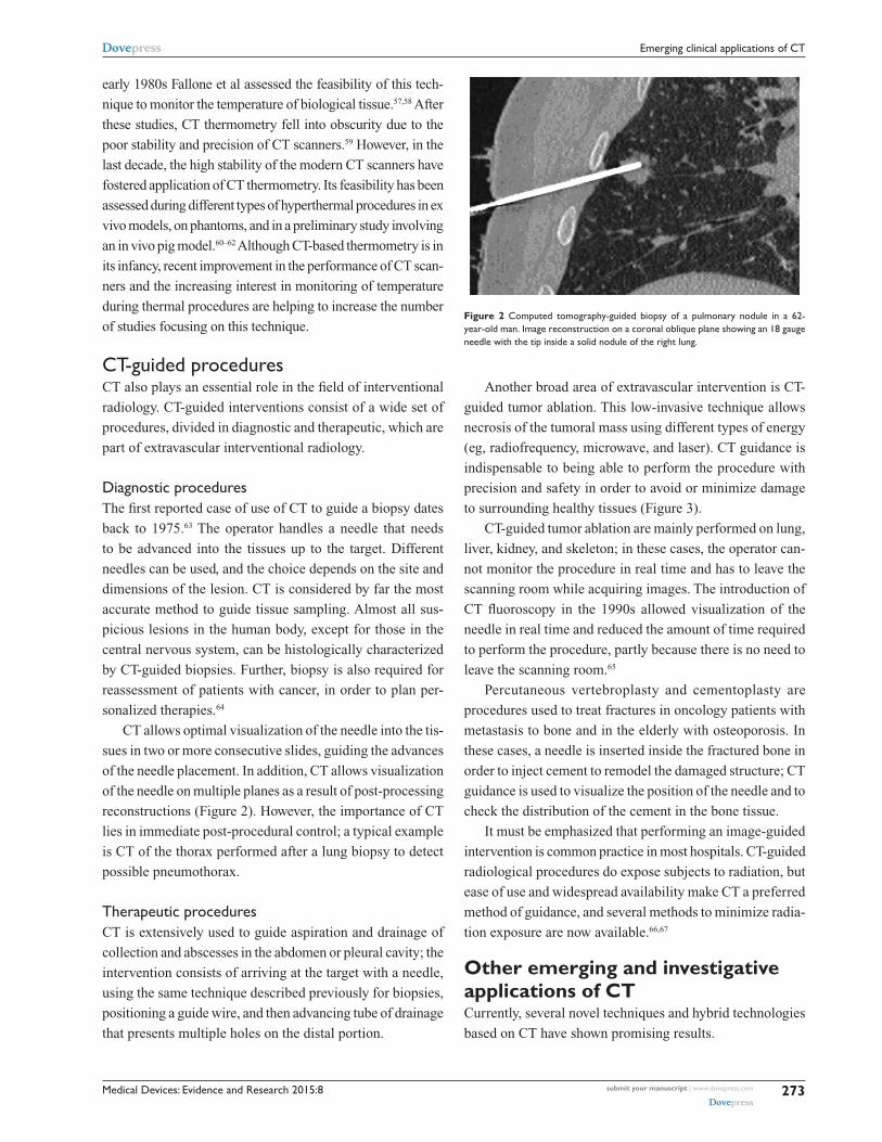
CT allows optimal visualization of the needle into the tis-
sues in two or more consecutive slides, guiding the advances
of the needle placement. In addition, CT allows visualization
of the needle on multiple planes as a result of post-processing
reconstructions (Figure 2). However, the importance of CT
lies in immediate post-procedural control; a typical example
is CT of the thorax performed after a lung biopsy to detect
possible pneumothorax.
Therapeutic proceduresCT is extensively used to guide aspiration and drainage of
collection and abscesses in the abdomen or pleural cavity; the
intervention consists of arriving at the target with a needle,
using the same technique described previously for biopsies,
positioning a guide wire, and then advancing tube of drainage
that presents multiple holes on the distal portion.
Figure 2 Computed tomography-guided biopsy of a pulmonary nodule in a 62-year-old man. image reconstruction on a coronal oblique plane showing an 18 gauge needle with the tip inside a solid nodule of the right lung.
Another broad area of extravascular intervention is CT-
guided tumor ablation. This low-invasive technique allows
necrosis of the tumoral mass using different types of energy
(eg, radiofrequency, microwave, and laser). CT guidance is
indispensable to being able to perform the procedure with
precision and safety in order to avoid or minimize damage
to surrounding healthy tissues (Figure 3).
CT-guided tumor ablation are mainly performed on lung,
liver, kidney, and skeleton; in these cases, the operator can-
not monitor the procedure in real time and has to leave the
scanning room while acquiring images. The introduction of
CT fluoroscopy in the 1990s allowed visualization of the
needle in real time and reduced the amount of time required
to perform the procedure, partly because there is no need to
leave the scanning room.65
Percutaneous vertebroplasty and cementoplasty are
procedures used to treat fractures in oncology patients with
metastasis to bone and in the elderly with osteoporosis. In
these cases, a needle is inserted inside the fractured bone in
order to inject cement to remodel the damaged structure; CT
guidance is used to visualize the position of the needle and to
check the distribution of the cement in the bone tissue.
It must be emphasized that performing an image-guided
intervention is common practice in most hospitals. CT-guided
radiological procedures do expose subjects to radiation, but
ease of use and widespread availability make CT a preferred
method of guidance, and several methods to minimize radia-
tion exposure are now available.66,67
Other emerging and investigative applications of CTCurrently, several novel techniques and hybrid technologies
based on CT have shown promising results.

Medical Devices: Evidence and Research 2015:8submit your manuscript | www.dovepress.com
Dovepress
Dovepress
274
Liguori et al
Figure 3 (A and B) Computed tomography-guided microwave ablation of metastasis from breast carcinoma of the left iliac bone in a 54-year-old woman. The patient stands prone and the tip of a 14 gauge needle is inserted into the lesion.
Hybrid PET-CTIntegration of PET with CT scanners has allowed acquisi-
tion of noninvasive three-dimensional images of functional
processes occurring in the human body by fusion of images
combining anatomy (CT) with function (PET). Since the
first prototype was developed in 1990, use of PET/CT in
the clinical setting for diagnostic and therapeutic purposes
in oncology, neurology, and cardiovascular disease has been
growing. Beyer and Pichler reported that hybrid PET/CT
has led to a 10%–15% increase in diagnostic accuracy when
compared with standalone PET or CT.68 This technique uses
CT images for anatomic reference of PET tracer uptake pat-
terns as well as for correction of the PET attenuation data for
quantification purposes.69 Hybrid PET/CT scanners improve
both the detection and treatment of cancer, such as detecting
spread of cancer to lymph nodes and assessing the possibility
of treatment with radiotherapy. One of the main diagnostic
advantages of adding functional data with CT is the ability to
isolate regions of cancer cell metabolism with fluorodeoxyg-
lucose or hypoxic regions using other PET tracers.
X-ray/MR systemsX-ray/MR systems are hybrid technologies where the com-
bined modality is more than the simple sum of each method.
At the same time, they combine the high spatial and temporal
resolution of X-ray fluoroscopy, provided by the three-dimen-
sional imaging capabilities, with the soft tissue contrast of
MRI. This type of system has become increasingly popular
in several diagnostic and minimally invasive applications, eg,
for biopsies and arthrograms.70 Other potential applications of
X-ray/MR systems are: guiding radiation therapy, investigat-
ing atherosclerotic plaques, and assessing acute strokes and
brain injuries. Two major issues in designing such a system
are the bulkiness of the imaging scanners and the conflict
in imaging physics, due to rotating metallic parts of X-ray
system and magnetic fields of MR one.71
Phase-contrast imagingPromising results have recently been obtained in material
science and biological applications by use of PCI, which can
be implemented at third-generation synchrotron radiation
sources.72 PCI is an innovative method that is also sensi-
tive to the refraction of X-rays in matter. This technique is
based upon the recording attenuation and phase changes
of the transmitted X-ray beam and allows evaluation of
planar or three-dimensional distribution of the scattering
properties of the investigated object. Five techniques have
been developed to explore the phase-contrast in the X-ray
regime. Of these, the propagation-based phase-contrast
imaging method is the simplest way to visualize the phase-
contrast.73 In the case of biological samples showing weakly
absorbing details, use of phase information for imaging is
an attractive alternative to conventional X-ray CT. PCI is
very appealing because it may help to decrease the total
dose absorbed, enhancing the conditions of the entire imag-
ing procedure. Currently, this technique is widely used in
preclinical research, but has yet to be tested in pilot clinical
trials.74 The breast, lung, joints, bone, vasculature, and brain
have been mostly imaged in ex vivo and in vitro samples,
as well as in a few animal models. Phase-contrast imaging
of the breast has been one of the first medical applications
of X-ray PCI, reported by Pisano et al. In the musculosk-
eletal field, PCI allows early and accurate visualization of
osteoarthritis and rheumatoid arthritis.75 Several groups
are exploring the possibility of using PCI in the research
and diagnosis of diseases that decrease the alveolar area

Medical Devices: Evidence and Research 2015:8 submit your manuscript | www.dovepress.com
Dovepress
Dovepress
275
Emerging clinical applications of CT
of the lung (eg, emphysema), to investigate lung structure
and function, and to detect atelectasis in the injured lung.
Some experiments have been performed using PCI on brain
tissue for neuroimaging applications, obtaining images of
both tumoral and healthy rat brain.
Photon-counting technology for spectral CTA new frontier in CT is photon-counting technology. When
an X-ray passes though the body, its spectral distribution
changes. However, unlike traditional detectors, photon-
counting detectors used in photon-counting spectral CT are
able to measure the energy of a single photon. In particular,
photon-counting technology is based on selection of a nar-
row subrange of the spectrum, and measurement of energy
attenuation in each window can be used to classify different
materials. The challenge of this technology is to record each
single photon, with the aim to reduce dose delivered to the
patient. Photon-counting detectors are mostly comprised
of cadmium telluride, cadmium zinc telluride, and silicon
semiconductors. These detectors allow X-ray photons to be
counted separately, and cadmium telluride and cadmium
zinc telluride detectors also allow measurement of photon
energies with a level of accuracy appropriate for clinical
application.76 Moreover, photon-counting spectral CT
overcomes artifact or loss of data due to patient motion,
and can achieve good spectral separation between images
without requiring heavy prefiltration. Despite some limita-
tions still presented by such detectors (eg, cost of materials,
intensity-dependent image artifacts associated with defects
in cadmium zinc telluride and cadmium telluride crystals,
and limited energy resolution associated with hole trapping
and leakage current in cadmium zinc telluride and cadmium
telluride materials), many clinical applications are under
investigation.77
Several studies have investigated the use of photon-
counting technology for K-edge imaging. Selective and
quantitative imaging of contrast medium can be achieved
by exploiting K-edge discontinuity in the photoelectric
component of X-ray absorption. An ideal application for
K-edge imaging is CT imaging of target-specific and con-
ventional contrast agents that have been designed to be
spectral CT-sensitive.78
Novel reconstruction algorithmsThe aim of reducing the radiation dose delivered to patient
has encouraged the investigation of novel reconstruction
algorithms. Unlike traditional back-projection algorithms,
iterative reconstruction algorithms use a forward reconstruc-
tion model and a precise modeling of scanner geometry and
the underlying physics, aiming to produce higher-resolution
and low-artifact images than back-projection reconstruction.
Among them, the adaptive statistical iterative reconstruction
algorithm utilizes information contained in the image recon-
structed by back-projections as an initial “building block” in
the reconstruction process, to decrease reconstruction time.
Model-based iterative reconstruction is demanding in terms
of the big number of computers to process data to produce a
complete scan, but has the crucial advantage of reducing the
radiation dose to 80% of that for standard reconstruction.79
ConclusionThe introduction of CT is undoubtedly one of the most
important milestones achieved in the last 40 years of clinical
and biomedical research. The socio-economic impact of CT
can be summarized by the installation of more than 50,000
scanners for the year 2010 and by the fact that use of CT in
the USA has increased more than three times since 1993 to
approximately 70 million scans annually.80
In the past, the widespread use of CT was mainly related
to its ability to create three-dimensional detailed pictures of
areas inside the body and to discriminate soft tissues with
good contrast and spatial resolution. These features made
the use of CT crucial in many branches of medicine.
However, over the years, different solutions have been pro-
posed to minimize the patient dose and to improve image
characteristics. For example, since the 1970s, the minimum
scan time decreased from about 5 minutes to a fraction of
a second, the slice thickness decreased from more than
1 cm to less than 1 mm, and the resolution improved from
2 pl/cm to more than 20 pl/cm. The first-generation CTs
were time-consuming and were replaced by other solutions,
leading rapidly to the fourth-generation CTs. An important
milestone in the improvement of CT performance was slip
ring technology, which enabled continuous acquisition of
data with a reduced scan time. A further revolution was the
advancement of MDCT in the late 1990s, which improved
temporal and spatial resolution.
The current landscape of CT applications in medicine is
growing rapidly, along with new and exciting developments,
such as CT-based thermometry and CT-guided invasive diag-
nostic and therapeutic procedures. Moreover, the introduction
of DECT opened up a new perspective regarding the use of
CT images. Given that clinical use of spectral CT imaging
will be defined by using dual-energy acquisition, progressive
utilization of CT data for accurate tissue characterization will

Medical Devices: Evidence and Research 2015:8submit your manuscript | www.dovepress.com
Dovepress
Dovepress
276
Liguori et al
be possible, gaining significant improvement in oncological
and emergency clinical practice. Implementation of DECT
technology will decrease the radiation dose exposure for
the patient in the daily clinical scenario, which is the main
issue of concern with CT scanning at present. Combination
of dual-energy acquisition, reduction of X-ray dose, and
implementation of techniques for reducing the acquisition
time velocity will make CT imaging more robust and reliable
as an evaluation technique for patients in all clinical settings
in the future.
DisclosureThe authors report no conflicts of interest in this work.
References 1. Cormack AM. Reconstruction of densities from their projections,
with applications in radiological physics. Phys Med Biol. 1973;18: 195–207.
2. Hounsfield GN. Computerized transverse axial scanning (tomography) part 1. Description of the system. Br J Radiol. 1973;46:1016–1022.
3. Cierniak R. X-Ray Computed Tomography in Biomedical Engineering. London, UK: Springer-Verlag; 2011.
4. Röntgen WC. Über eine neue Art von Strahlen. Sitzungsberichte der Physikalisch-medizinischen Gesellschaft zu Würzburg [on a new kind of rays]. Science. 1896;3:227–231. German.
5. Thomson E. Stereoscopic roentgen pictures. Electr Eng. 1896;21:256. 6. Bushberg JT. The Essential Physics of Medical Imaging. 2nd ed.
Philadelphia, PA, USA: Lippincott Williams and Wilkins; 2002. 7. Cho ZH, Jones JP, Singh M. Foundations of Medical Imaging. 1st ed.
New York, NY, USA: John Wiley and Sons; 1993. 8. Kak AC, Slanley M. Principles of Computerized Tomographic Imaging.
1st ed. New York, NY, USA: IEEE Press; 1988. 9. Glover GH. Compton scatter effects in CT reconstructions. Med Phys.
1982;9:860–867. 10. Karçaaltıncaba M, Aktaş A. Dual-energy CT revisited with multide-
tector CT: review of principles and clinical applications. Diagn Interv Radiol. 2011;17:181–194.
11. Karen MH, Sheila S, Frank C, et al. Multidetector row CT: principles and clinical applications. Crit Rev Comput Tomogr. 2002;43:143–181.
12. Kaza RK, Platt JF, Goodsitt MM, et al. Emerging techniques for dose optimization in abdominal CT. Radiographics. 2014;34:4–17.
13. Cochet H, Dubois R, Sacher F, et al. Cardiac arrythmias: multimodal assessment integrating body surface ECG mapping into cardiac imaging. Radiology. 2014;271:239–247.
14. Godoy MC, Naidich DP, Marchiori E, et al. Basic principles and postprocessing techniques of dual-energy CT: illustrated by selected congenital abnormalities of the thorax. J Thorac Imaging. 2009;24: 152–159.
15. Aran S, Shaqdan KV, Abujudeh HH. Dual-energy computed tomogra-phy (DECT) in emergency radiology: basic principles, techniques, and limitations. Emerg Radiol. 2014;21:391–405.
16. Johnson TRC. Dual-energy CT: general principles. Am J Roentgenol. 2012;199(5 Suppl):3–8.
17. Vogl TJ, Schulz B, Bauer RW, et al. Dual-energy CT applications in head and neck imaging. AJR Am J Roentgenol. 2012;199:S34–S39.
18. Tawfik AM, Kerl JM, Bauer RW, et al. Dual-energy CT of head and neck cancer: average weighting of low- and high-voltage acquisitions to improve lesion delineation and image quality-initial clinical experience. Invest Radiol. 2012;47:306–311.
19. Kang M, Park CM, Lee CH, et al. Dual-energy CT: clinical applications in various pulmonary diseases. Radiographics. 2010;30:685–698.
20. Brown CL, Hartman RP, Dzyubak OP, et al. Dual-energy CT iodine overlay technique for characterization of renal masses as cyst or solid: a phantom feasibility study. Eur Radiol. 2009;19:1289–1295.
21. Graser A, Johnson TR, Hecht EM, et al. Dual-energy CT in patients suspected of having renal masses: can virtual nonenhanced images replace true nonenhanced images? Radiology. 2009;252:433–440.
22. Stolzmann P, Scheffel H, Rentsch K, et al. Dual-energy computed tomography for the differentiation of uric acid stones: ex vivo perfor-mance evaluation. Urol Res. 2008;36:133–138.
23. Stolzmann P, Leschka S, Scheffel H, et al. Characterization of urinary stones with dual-energy CT: improved differentiation using a tin filter. Invest Radiol. 2010;45:1–6.
24. Silva AC, Morse BG, Hara AK, Paden RG, Hongo N, Pavlicek W. Dual-energy (spectral) CT: applications in abdominal imaging. Radiographics. 2011;31:1031–1046.
25. Fischer MA, Reiner CS, Raptis D, et al. Quantification of liver iron content with CT-added value of dual-energy. Eur Radiol. 2011;21: 1727–1732.
26. Stolzmann P, Frauenfelder T, Pfammatter T, et al. Endoleaks after endo-vascular abdominal aortic aneurysm repair: detection with dual-energy dual-source CT. Radiology. 2008;249:682–691.
27. Chandarana H, Godoy MC, Vlahos I, et al. Abdominal aorta: evaluation with dual-source dual-energy multidetector CT after endovascular repair of aneurysms – initial observations. Radiology. 2008;249:692–700.
28. Ruzsics B, Schwarz F, Schoepf UJ, et al. Comparison of dual-energy computed tomography of the heart with single photon emission com-puted tomography for assessment of coronary artery stenosis and of the myocardial blood supply. Am J Cardiol. 2009;104:318–326.
29. Nagao M, Kido T, Watanabe K, et al. Functional assessment of coronary artery flow using adenosine stress dual-energy CT: a preliminary study. Int J Cardiovasc Imaging. 2011;27:471–481.
30. Schwarz F, Ruzsics B, Schoepf UJ, et al. Dual-energy CT of the heart – principles and protocols. Eur J Radiol. 2008;68:423–433.
31. Zhang LJ, Peng J, Wu SY, Yeh BM, Zhou CS, Lu GM. Dual source dual-energy computed tomography of acute myocardial infarction: correlation with histopathologic findings in a canine model. Invest Radiol. 2010;45:290–297.
32. Bauer RW, Kerl JM, Fischer N, et al. Dual-energy CT for the assessment of chronic myocardial infarction in patients with chronic coronary artery disease: comparison with 3-T MRI. AJR Am J Roentgenol. 2010;195: 639–646.
33. Barreto M, Schoenhagen P, Nair A, et al. Potential of dual-energy computed tomography to characterize atherosclerotic plaque: ex vivo assessment of human coronary arteries in comparison to histology. J Cardiovasc Comput Tomogr. 2008;2:234–242.
34. Boll DT, Merkle EM, Paulson EK, Mirza RA, Fleiter TR. Calcified vascular plaque specimens: assessment with cardiac dual-energy multi-detector CT in anthropomorphically moving heart phantom. Radiology. 2008;249:119–126.
35. Hazirolan T, Akpinar B, Unal S, Gümrük F, Haliloglu M, Alibek S. Value of dual energy computed tomography for detection of myocardial iron deposition in thalassaemia patients: initial experience. Eur J Radiol. 2008;68:442–445.
36. Ibrahim E-SH, Bowman AW. Characterization of myocardial iron overload by dual-energy computed tomography compared to T2* MRI. A phantom study. Conf Proc IEEE Eng Med Biol Soc. 2014;2014:5133–5136.
37. Henzler T, Fink C, Schoenberg SO, Schoepf UJ. Dual-energy CT: radiation dose aspects. Am J Roentgenol. 2012;199:S16–S25.
38. Lin XZ, Wu ZY, Tao R, et al. Dual energy spectral CT imaging of insulinoma – value in preoperative diagnosis compared with conven-tional multi-detector CT. Eur J Radiol. 2012;8:2487–2494.
39. Leschka S, Stolzmann P, Baumüller S, et al. Performance of dual-energy CT with tin filter technology for the discrimination of renal cysts and enhancing masses. Acad Radiol. 2010;17:526–534.
40. Van Werkhoven JM, Gaemperli O, Schuijf JD, et al. Multislice computed tomography coronary angiography for risk stratification in patients with an intermediate pretest likelihood. Heart. 2009;95: 1607–1611.
Medical Devices: Evidence and Research 2015:8 submit your manuscript | www.dovepress.com
Dovepress
Dovepress
277
Emerging clinical applications of CT
41. Hadamitzky M, Freissmuth B, Meyer T, et al. Prognostic value of coronary computed tomographic angiography for prediction of car-diac events in patients with suspected coronary artery disease. JACC Cardiovasc Imaging. 2009;2:404–411.
42. Shuman WP, Branch KR, May JM, et al. Prospective versus retrospec-tive ECG gating for 64-detector CT of the coronary arteries: comparison of image quality and patient radiation dose. Radiology. 2008;248: 431–437.
43. Sommer WH, Albrecht E, Bamberg F, et al. Feasibility and radiation dose of high-pitch acquisition protocols in patients undergoing dual-source cardiac CT. AJR Am J Roentgenol. 2010;195:1306–1312.
44. Einstein AJ, Henzlova MJ, Rajagopalan S. Estimating risk of cancer associated with radiation exposure from 64-slice computed tomography coronary angiography. JAMA. 2007;298:317–323.
45. Einstein AJ, Sanz J, Dellegrottaglie S, et al. Radiation dose and cancer risk estimates in 16-slice computed tomography coronary angiography. J Nucl Cardiol. 2008;15:232–240.
46. Achenbach S, Marwan M, Schepis T, et al. High pitch spiral acquisition: a new mode for coronary angiography. J Cardiovasc Comput Tomogr. 2009;3:117e2.
47. Chinnaiyan KM, Bilolikar AN, Walsh E, et al. CT dose reduction using prospectively triggered or fast-pitch spiral technique employed in cardiothoracic imaging (the CT dose study). J Cardiovasc Comput Tomogr. 2014;8:205–214.
48. Bruder H, Petersilker M, Mehldau H, et al. Flash imaging in dual source CT (DSCT). Proc SPIE 7258, Medical Imaging; 2009. Physics of Medical Imaging, 72580D. Available from: http://proceedings.spiedigitallibrary.org/proceeding.aspx?articleid=1335243. Accessed May 9, 2015.
49. Chinnaiyan KM, Boura JA, DePetris A, et al. Progressive radiation dose reduction from coronary computed tomography angiography in a statewide collaborative quality improvement program: results from the Advanced Cardiovascular Imaging Consortium. Circ Cardiovasc Imaging. 2013;6:646e654.
50. Stafford RJ, Fuentes D, Elliott AA, Weinberg JS, Ahrar K. Laser-induced thermal therapy for tumor ablation. Crit Rev Biomed Eng. 2010;38:79–100.
51. Lepetit-Coiffé M, Laumonier H, Seror O, et al. Real-time monitoring of radiofrequency ablation of liver tumors using thermal-dose calculation by MR temperature imaging: initial results in nine patients, including follow-up. Eur Radiol. 2010;20:193–201.
52. Saccomandi P, Schena E, Silvestri S. Techniques for temperature monitoring during laser-induced thermotherapy: an overview. Int J Hyperthermia. 2013;29:609–619.
53. Vogl TJ, Straub R, Eichler K, Woitaschek D, Mack MG. Malignant liver tumors treated with MR imaging-guided laser-induced thermo-therapy: experience with complications in 899 patients (2,520 lesions). Radiology. 2002;225:367–377.
54. Fani F, Schena E, Saccomandi P, Silcestri S. CT-based thermometry: an overview. Int J Hyperthermia. 2014;30:219–227.
55. Pandeya G, Greuter M, de Jong K, Schmidt B, Flohr T, Outkerk M. Feasibility of noninvasive temperature assessment during radiofrequency liver ablation on computed tomography. J Comput Assist Tomogr. 2011;35:356–360.
56. Li M, Abi-Jaoudeh N, Kapoor A, et al. Towards cone-beam CT thermometry. Proc SPIE. 2013;8671:1–7.
57. Bydder GM, Kreel L. The temperature dependence of computed tomography attenuation values. J Comput Assist Tomogr. 1979;3: 506–510.
58. Fallone BG, Moran PR, Podgorsak EB. Noninvasive thermometry with a clinical X-ray CT scanner. Med Phys. 1982;9:715–721.
59. Mahnken AH, Bruners P. CT thermometry: will it ever become ready for use? Int J Clin Pract. 2011;65 Suppl 171:1–2.
60. Schena E, Saccomandi P, Giurazza F, et al. Monitoring of temperature increase and tissue vaporization during laser interstitial thermotherapy of ex vivo swine liver by computed tomography. Conf Proc IEEE Eng Med Biol Soc. 2013;2013:378–381.
61. Pandeya GD, Klaessens JH, Greuter MJ, et al. Feasibility of computed tomography based thermometry during interstitial laser heating in bovine liver. Eur Radiol. 2011;21:1733–1738.
62. Schena E, Saccomandi P, Giurazza F, et al. Experimental assessment of CT-based thermometry during laser ablation of porcine pancreas. Phys Med Biol. 2013;58:1–12.
63. Alfidi RJ, Haaga J, Meaney TF, et al. Computed tomography of the thorax and abdomen: a preliminary report. Radiology. 1975;117:257–264.
64. Crommelin DJ, Storm G, Luijten P. ‘Personalized medicine’ through ‘personalized medicines’: time to integrate advanced, non-invasive imaging approaches and smart drug delivery systems. Int J Pharm. 2011;415:5–8.
65. Katada K, Anno H, Takeshita G, et al. [Development of real-time CT fluoroscopy]. Nippon Igaku Hoshasen Gakkai Zasshi. 1994;54:1172–1174. Japanese.
66. Sarti M, Brehmer WP, Gay SB. Low-dose techniques in CT-guided interventions. Radiographics. 2012;32:1109–1119.
67. Grasso RF, Cazzato RL, Luppi G, et al. Percutaneous lung biopsies: performance of an optical CT-based navigation system with a low-dose protocol. Eur Radiol. 2013;23:3071–3076.
68. Beyer T, Pichler B. A decade of combined imaging: from a PET attached to a CT to a PET inside an MR. Eur J Nucl Med Mol Imaging. 2009;36:S1–S2.
69. Kinahan P, Townsend D, Beyer T, Sashin D. Attenuation correction for a combined 3D PET/CT scanner. Med Phys. 1998;25:2046–2053.
70. Pelc NJ. Hybrid x-ray/MR system and other hybrid imaging modalities. In: Medical Imaging 2003: Physiology and Function: Methods, Systems, and Applications. Clough AV; Amini AA, editors. Proceedings of SPIE; San Diego, CA. Volume 5031 February 15; 2003.
71. Ganguly A, Wen Z, Daniel B, et al. Truly hybrid X-ray/MR imaging: towards a streamlined clinical system. Acad Radiol. 2005;12: 1167–1177.
72. Baik S, Kim HS, Jeong MH, et al. International consortium on phase contrast imaging and radiology beamline at the Pohang light source. Rev Sci Instrum. 2004;75:4355–4358.
73. Snigirev A, Snigireva I, Kohn V, et al. On the possibilities of x-ray phase contrast microimaging by coherent high-energy synchrotron radiation. Rev Sci Instrum. 1995;66:5486–5492.
74. Castelli E, Tonutti M, Arfelli F, et al. Mammography with synchrotron radiation: first clinical experience with phase-detection technique. Radiology. 2011;259:684–694.
75. Pisano ED, Johnston RE, Chapman D, et al. Human breast cancer specimens: diffraction-enhanced imaging with histologic correlation- improved conspicuity of lesion detail compared with digital radiography. Radiology. 2000;214:895–901.
76. Shikhaliev PM, Shannon GF. Photon counting spectral CT versus con-ventional CT: comparative evaluation for breast imaging application. Phys Med Biol. 2011;56:1905–1930.
77. Feuerlein S, Roessl E, Proksa R, et al. Multienergy photon-counting K-edge imaging: potential for improved luminal depiction in vascular imaging. Radiology. 2008;249:1010–1016.
78. Schlomka J, Roessl E, Dorscheid R, et al. Experimental feasibility of multi-energy photon-counting K-edge imaging in pre-clinical computed tomography. Phys Med Biol. 2008;53:4031–4037.
79. Singh S, Kalra MK, Hsieh J, et al. Abdominal CT: comparison of adaptive statistical iterative and filtered back projection reconstruction techniques. Radiology. 2010;257:373–383.
80. Berrington de González A, Mahesh M, Kim KP, et al. Projected cancer risks from computed tomographic scans performed in the United States in 2007. Arch Intern Med. 2009;169:2071–2077.

Medical Devices: Evidence and Research
Publish your work in this journal
Submit your manuscript here: http://www.dovepress.com/medical-devices-evidence-and-research-journal
Medical Devices: Evidence and Research is an international, peer-reviewed, open access journal that focuses on the evidence, technology, research, and expert opinion supporting the use and application of medical devices in the diagnosis, treatment and management of clini-cal conditions and physiological processes. The identification of novel
devices and optimal use of existing devices which will lead to improved clinical outcomes and more effective patient management and safety is a key feature. The manuscript management system is completely online and includes a quick and fair peer-review system. Visit http://www.dovepress.com/testimonials.php to read real quotes from authors.
Medical Devices: Evidence and Research 2015:8submit your manuscript | www.dovepress.com
Dovepress
Dovepress
Dovepress
278
Liguori et al
Copyright © 2022 FDOKUMEN