Early Lung Cancer Detection using Spiral Computed Tomography and Positron Emission Tomography
32
Early Lung Cancer Detection using Spiral Computed Tomography and Positron Emission Tomography Gorka Bastarrika, M.D. 1 , María José García-Velloso, M.D. 2 , Maria Dolores Lozano, M.D. 3 , Usua Montes, R.N. 4 , Wenceslao Torre, M.D. 5 , Natalia Spiteri, M.D. 5 , Arantza Campo, M.D. 4 , Luis Seijo, M.D. 4 , Ana Belén Alcaide, M.D. 4 , Jesús Pueyo, M.D. 1 , David Cano, M.D. 1 , Isabel Vivas, M.D. 1 , Octavio Cosín, M.D. 1 Pablo Domínguez, M.D. 1 , Patricia Serra, M.D. 2 , José A. Richter, M.D. 2 , Luis Montuenga, Ph.D. 6,7 , and Javier J. Zulueta, M.D 4 . Departments of Radiology 1 , Nuclear Medicine 2 , Pathology 3 , Pulmonary Medicine 4 , and Thoracic Surgery 5 , Clínica Universitaria, Department of Histology and Pathology 6 , School of Medicine, and Division of Oncology 7 , Center for Applied Medical Research (CIMA), Universidad de Navarra, Pamplona, Spain. Running Head: Lung Cancer Screening with CT & PET Descriptor: 85 Manuscript word count: 3352 Corresponding author: Javier J. Zulueta, M.D. Pulmonary Medicine Clínica Universitaria Avda. Pio XII, 36 31008 Pamplona Email: [email protected] Tel.: (34) 948-296694 Fax: (34) 948-296500 AJRCCM Articles in Press. Published on March 24, 2005 as doi:10.1164/rccm.200411-1479OC Copyright (C) 2005 by the American Thoracic Society.
Transcript of Early Lung Cancer Detection using Spiral Computed Tomography and Positron Emission Tomography

Early Lung Cancer Detection using Spiral Computed
Tomography and Positron Emission Tomography
Gorka Bastarrika, M.D.1, María José García-Velloso, M.D.2, Maria Dolores
Lozano, M.D.3, Usua Montes, R.N.4, Wenceslao Torre, M.D.5, Natalia Spiteri,
M.D.5, Arantza Campo, M.D.4, Luis Seijo, M.D.4, Ana Belén Alcaide, M.D.4,
Jesús Pueyo, M.D.1, David Cano, M.D.1, Isabel Vivas, M.D.1, Octavio Cosín,
M.D.1 Pablo Domínguez, M.D.1, Patricia Serra, M.D.2, José A. Richter, M.D.2,
Luis Montuenga, Ph.D.6,7, and Javier J. Zulueta, M.D4.
Departments of Radiology1, Nuclear Medicine2, Pathology3, Pulmonary
Medicine4, and Thoracic Surgery5, Clínica Universitaria, Department of
Histology and Pathology6, School of Medicine, and Division of Oncology7,
Center for Applied Medical Research (CIMA), Universidad de Navarra,
Pamplona, Spain.
Running Head: Lung Cancer Screening with CT & PET
Descriptor: 85
Manuscript word count: 3352
Corresponding author:Javier J. Zulueta, M.D.Pulmonary MedicineClínica UniversitariaAvda. Pio XII, 3631008 PamplonaEmail: [email protected].: (34) 948-296694Fax: (34) 948-296500
AJRCCM Articles in Press. Published on March 24, 2005 as doi:10.1164/rccm.200411-1479OC
Copyright (C) 2005 by the American Thoracic Society.

1
This article has an online data supplement, which is accessible from this issue's table of content online at www.atsjournals.org
2
ABSTRACT
Rationale: Lung cancer screening using CT is effective in detecting lung cancer
in early stages. Concerns regarding false positive rates and unnecessary
invasive procedures have been raised. Objective: To study the efficiency of a
lung cancer protocol spiral CT and FDG-PET. Methods: High risk individuals
underwent screening with annual spiral CTs. Follow-up CTs were done for non-
calcified nodules of ≥ 5 mm, and FDG-PET was done for nodules ≥ 10 mm or
smaller (> 7mm) growing nodules. Results: 911 individuals completed a
baseline CT study and 424 at least one annual follow-up study. Of the former,
14% had non-calcified nodules ≥ 5 mm, and 3.6% had nodules ≥ 10mm.
Eleven non-small cell- (NSCLC) and 1 small cell (SCLC) lung cancers were
diagnosed in the baseline study (prevalence rate of 1.32%), and 2 NSCLC in
the annual study (incidence rate of 0.47%). All NSCLC (92% of prevalence
cancers) were diagnosed in stage I (12 stage IA, 1 stage IB). FDG-PET was
helpful for the correct diagnosis in 19 out of 25 indeterminate nodules. The
sensitivity, specificity, positive predictive value, and negative predictive value of
FDG-PET for the diagnosis of malignancy were 69%, 91%, 90% and 71%,
respectively. However, the sensitivity and negative predictive value of the
screening algorithm, which included a 3-month follow-up CT for nodules with a
negative FDG-PET, was 100%. Conclusion: A protocol for early lung cancer
detection using spiral CT and FDG-PET is useful and may minimize
unnecessary invasive procedures for benign lesions.
Word count: 251
Keywords: lung neoplasm, smoking, COPD, pulmonary nodule

3
Introduction
Lung cancer is the most frequent and most lethal malignancy in the world (1-3).
Advances in the treatment of locally advanced lung cancer have had no impact
on overall 5-year survival rates from this disease (1, 2). In countries where
gains against tobacco smoking have been made, mortality rates from lung
cancer have started to decrease, but future projections are not optimistic due to
the recent surge in tobacco consumption among young people (4). And even if
smoking could be reduced significantly, the long lag time between peak tobacco
consumption and the development of clinical lung cancer will assure a long life
for this epidemic.
The main reason why more than 80% of patients with lung cancer die soon after
diagnosis is that most patients are diagnosed in late non-surgical stages (4).
Large screening trials have investigated the use of conventional radiology and
sputum cytology for early detection of lung cancer but failed to show a reduction
in disease-specific mortality, resulting in public policies against screening for
this disease (5-8). However, recently very promising reports using spiral-
computed tomography (CT) have brought lung cancer screening back to the
forefront. The Early Lung Cancer Action Project (ELCAP) demonstrated that low
dose spiral CT detects lung cancer in early stages much more effectively than
conventional chest-x-ray (9). Following a carefully designed protocol based on
follow-up CTs to detect growth of small nodules, 86% of lung cancers
diagnosed in asymptomatic individuals were in stage I (9). Other studies using
spiral CT have found similar results, although with variations in the percentage
of individuals with positive CTs (i.e., with non-calcified nodules that need further

4
work-up) (10-14). Swensen et al. report non-calcified nodules in close to 70% of
the individuals participating in a lung cancer screening trial with spiral CT (10).
Diederich et al. found non-calcified nodules in 43% of their population (11).
Although the ELCAP investigators only performed one invasive procedure on
one benign lesion (9), the high prevalence of non-calcified nodules may result
in unnecessary invasive procedures for benign diseases. This will be
particularly concerning when these protocols become widely used in the
community following less rigorous standards.. Positron emission tomography
with the glucose analogue F-18-fluorodeoxyglucose (FDG-PET) is an accurate
non-invasive imaging test for diagnosis of pulmonary nodules, although few
data exist for nodules smaller than 1 cm in diameter (15). Pastorino et al.
showed that selective use of FDG-PET in a spiral CT-based lung cancer
screening trial may be useful in avoiding biopsies for benign lesions (12). The
hypothesis formulated in the study presented herein is that adding FDG-PET to
a spiral CT-based early lung cancer detection protocol will avoid unnecessary
procedures for benign lesions. Furthermore, false negative lesions (i.e., cancers
with no pathological uptake on the FDG-PET) will be diagnosed with follow-up
CTs to detect growth, and therefore such a protocol will not result in missed
cancers due to negative PET scans.
Following publication of their original results, the ELCAP group lead two
important initiatives: the NY-ELCAP screening trial, a New York area
multicenter trial projecting to screen 10,000 high risk individuals with spiral CT,
and the I-ELCAP, an international consortium which brings together
investigators from over 30 centers from around the world (16). One of the main
objectives of I-ELCAP is to pool data from screening trials underway in these

5
centers. Herein we present results from an ongoing trial of lung cancer
screening using spiral CT and FDG-PET conducted at our institution as a
member of I-ELCAP since year 2000. Some of the results of these studies have
been previously reported in the form of an abstract (17).
MATERIALS AND METHODS
(Word count 520)
Population
Current and former smokers of at least 40 years of age, a minimum 10 pack-
year smoking history, and with no symptoms of lung cancer were invited to
participate. A background questionnaire and written informed consent were
obtained. The study protocol was approved by our center’s Ethics Committee.
CT
CT examinations were done in a single breath hold at end-inspiration. The initial
297 subjects were studied with a single slice helical scanner (Somatom Plus 4;
Siemens, Erlangen, Germany) at low dose settings (140 kVp, 43 mAs) and 1.5
pitch with a collimation of 8 mm. Pulmonary nodules were characterized with a
limited high resolution CT (1 mm slice-thickness). For the subsequent 614
patients, a four-row multi-slice helical CT scanner was used (Somatom Volume
Zoom; Siemens, Forchheim, Germany) at a low-dose setting (120 kVp, 20 mAs,
1.25 mm slice thickness). Studies were analyzed independently by two
radiologists.

6
Pulmonary nodules
Calcifications, size, location, edges and spiculations were documented for each
nodule. Size was the average of length and width and classified nodules into
three categories: < 5 mm, 5 - <10 mm, and ≥ 10 mm.
FDG-PET
Except for nodules with benign characteristics, non-calcified nodules ≥ 10 mm,
or smaller nodules (> 7 mm) showing growth, were evaluated with FDG-PET.
Imaging acquisition was performed with a full ring BGO tomograph (Ecat Exact
HR+, CTI/Siemens, Erlangen, Germany). Visual analysis was performed by two
nuclear medicine physicians, and was considered positive for malignancy when
focal uptake was observed in the lung nodule. Metabolic activity was assessed
by the maximum Standardized Uptake Value (SUVmax).
Diagnostic Algorithm
The result of the baseline screening test was considered positive if there were
one to six non-calcified nodules, or more than six nodules with the largest one
being ≥ 5 mm. When the initial low-dose CT study was negative or the initial
baseline study did not lead to the diagnosis of a malignancy, repeat screening
was performed after 12 months. A positive study prompted further work-up
according to an algorithm developed by I-ELCAP(16). A three-month follow-up
CT was done for nodules between 5- <10 mm, and if growth was detected,
appropriate diagnostic studies were done. If no growth was seen, an annual CT
was scheduled. Growth was assessed visually by the radiologist comparing two
CTs side by side on the workstation. FDG-PET was done for nodules ≥ 10 mm

7
or smaller nodules (> 7 mm) showing growth, and if positive, a percutaneous
fine needle aspiration or an intraoperative biopsy were performed. For nodules
≥ 10 mm with benign characteristics a 3-month follow-up CT was done instead
of a FDG-PET.
Spirometry
FVC and FEV1 were measured with a computerized spirometer (Vmax 22,
SensorMedics, CA). Results were expressed as percent of the predicted value
according to the European Community Lung Health Survey (18). The presence
and severity of airflow obstruction was determined following the criteria
established by the Global Initiative for Chronic Obstructive Lung Disease
(GOLD) (19).
Data analysis
Normally distributed data were summarized as means (SD) and skewed data
were summarized as medians (interquartile range, IQR). Data analysis was
performed with SPSS for Windows version 11 (SPSS Inc., Chicago, IL).
RESULTS
Study Population and Risk Factors
911 (74 % male) subjects participated in the study. Mean (SD) age was 54.7
(8.6) years. Median (IQR) tobacco consumption was 30 (19-49.5) pack-years.
Asbestos exposure was reported in only one subject. Other risk factors for lung
cancer, such as radon, nickel or chromium were not reported.

8
Low-Dose CT Findings
In 440 (48.3%) individuals at least one pulmonary nodule was observed at
baseline examination. Non-calcified nodules of any size were observed in 291
(31.9%) subjects. At least one non-calcified nodule of 5 mm or more was found
in 131 (14.4%) subjects. Of the 291 individuals with non-calcified nodules, 164
(56.4%) had one nodule, 70 (24.1% had two nodules, 31 (10.7%) had three
nodules, 11 (3.8%) had four nodules, and 15 (5.2%) had five or more nodules.
Nodule Size and Attenuation
A total of 973 nodules were detected on baseline low-dose CT studies (table 1).
Of these, 434 (44.6%) were homogeneously calcified nodules and 539 (55.4%)
showed no calcification. Of the 539 non-calcified pulmonary nodules, 369
(68.5%) had a maximum diameter less than 5 mm, 136 (25.2%) between 5-10
mm, and 34 (6.3%) 10 mm or larger.
Of the 291 individuals with non-calcified pulmonary nodules, 160 (17.6% of
participants) only had nodules less than 5 mm, 98 (10.7% of participants) had at
least one nodule between 5-10 mm (no nodules greater than 10 mm), and 33
(3.6 % of participants) had at least one nodule of 10 mm or more (table 2).
FDG-PET for Nodules ≥ 10 mm or Smaller (> 7 mm) Growing Nodules
A total of 24 FDG-PET scans were done on 23 individuals (25 pulmonary
nodules, table 3). One individual had 2 FDG-PET scans (# 10 and # 16, table 3)
for a nodule that grew from 10.5 mm to 14.5 mm in 7 months, and turned out to
be a malignancy. The first FDG-PET was negative (false negative), but the
second showed an SUVmax of 1.6 (true positive). Three of the nodules studied

9
were slightly smaller than 10 mm. An 8 mm nodule (# 25, malignant) seen on
the first annual repeat CT (new incidence nodule), and an 8.7 mm nodule (# 23,
benign) seen on the baseline study in a patient in which the sputum cytology
showed moderate to severe squamous metaplasia. FDG-PET scans for these
two nodules were negative (1 false negative and 1 true negative). A third 8 mm
nodule (# 24, malignant) was seen in a patient with an additional 10 mm nodule
(# 19, benign) that prompted the FDG-PET study. Both nodules had negative
FDG-PET scans (1 false negative, one true negative), but the 8 mm nodule
showed growth on follow-up CT (squamous cell carcinoma).
Altogether, 11 nodules were positive on FDG-PET, of which 9 were malignant
(median SUVmax of 1.8, IQR 1.6 – 3.1), 1 was benign (SUVmax of 2.3 and
necrosis on FNA with no growth on annual CT), and 1 was indeterminate
(SUVmax 1.2) since the patient refused further work up, and was lost to follow-
up. This indeterminate nodule was excluded from the analysis.
Fourteen nodules were negative on FDG-PET, of which 4 were malignant
These 4 malignancies were adenocarcinomas (2 of them, #15 and # 25 on table
3, had bronchoalveolar features in part of the tumor) and were all diagnosed in
stage IA after growth was seen on short-term follow-up CTs. The sensitivity and
specificity of FDG-PET to detect malignancy were 69% (95% CI, 41 – 89) and
91% (95% CI, 63 – 99), respectively. Positive and negative predictive values
were 90% (95% CI, 57 – 98) and 71% (95% CI, 52 - 85), respectively.
Lung Cancer Stages
Lung cancer was detected in 14 (13 non-small cell- and 1 small cell lung
cancer) out of 911 individuals entered in the study. Eleven of the 13 non-small

10
cell lung cancers (NSCLC), and 1 small cell lung cancer (SCLC) were detected
on the baseline study (prevalence of 1.32%). Out of 424 individuals who have
completed at least one annual repeat CT, 2 were diagnosed with non-small cell
lung cancer (incidence of 0.47%). Of the prevalence cancers, 10 (86% ) were
stage IA (T1N0M0) and 1 was stage IB (T2N0M0). The only small cell lung
cancer was diagnosed in stage T3N3M0. The incidence cancers were both
detected in stage IA (T1N0M0). Considering all cancers, 13 out of 14 (92.8%)
were diagnosed in stage I.
Invasive procedures and surgery
There were no preoperative surgical biopsies performed. Six preoperative
percutaneous fine needle aspirations (FNA) were performed on 4 PET-positive
nodules and 2 growing PET-negative nodules. Four of these were positive for
malignancy and two were negative. One negative FNA (PET positive) was
followed up with a yearly CT showing no growth (False Positive PET). The
possibility that this individual actually has a slow growing cancer cannot be
ruled out, although unlikely in view of the lack of growth. The other negative
FNA (PET positive) was inadequate due to a pneumothorax. This individual had
an intraoperative biopsy showing a malignancy. The patient with SCLC was
diagnosed with a bronchoscopy. The rest of the patients were diagnosed with
intraoperative biopsies.
All NSCLC (prevalence and incidence) underwent surgery. Eleven out of 13
(84.6%) had lobectomies and 2 (15.4%) had wedge resections because of
COPD.

11
Spirometry
Of 834 subjects who had spirometry at baseline, 220 (26.4%) had an
FEV1/FVC < 70%. Of these, 113 (51.4%) had and FEV1 ≥ 80% (mild airflow
obstruction), 90 (40.9%) had an FEV1 < 80% but ≥ 50 (moderate airflow
obstruction), 14 (6.4%) had an FEV1 < 50% but ≥ 30 (severe airflow
obstruction), and 3 (1.4%) had an FEV1 < 30% (very severe airflow
obstruction). Of the 14 patients with non-small cell lung cancer, 11 (79%) had
airflow obstruction (FEV1/FVC < 70%). Six had mild, 4 had moderate and 1 had
severe airflow obstruction.
DISCUSSION
Following recent reports (9, 20-22), this study confirms that a low dose spiral
CT-based protocol for early detection of lung cancer is useful in leading to early
stage diagnosis in a high proportion of cases and may increase the chances for
cure. Furthermore, this study shows that a carefully followed multidisciplinary
diagnostic algorithm that includes FDG-PET minimizes unnecessary invasive
procedures for benign lesions, without resulting in missed cancers due to false
negative PET scans.
Lung cancer is one of the leading causes of death in the world. It is the most
common cancer in many countries and, since most patients are diagnosed in
late stages, it is also the most lethal (1-3). Less than 20% of individuals
suffering this disease are diagnosed in stages in which curative surgery is an
option (2). Survival rates for early stage disease are quite high, approaching

12
80% in some series but overall 5-year survival rates are approximately 15% (2).
Five-year survival rates for patients who have already survived 2 years
(conditional survival) remains significantly higher for those diagnosed in early
stages, stressing the importance of early diagnosis (23).
Lung cancer screening with low dose CT has shown to be effective in
diagnosing lung cancer in early stages. In the Early Lung Cancer Action Project
(ELCAP) 89% of lung cancers detected in the prevalence phase of the trial were
diagnosed in stage I and II (9). In other screening trials the proportion of
cancers diagnosed in stages I and II varies between 75% and 90% (10-14).
Whether this will result in a reduction in mortality from lung cancer has not been
answered yet, and is being addressed in several trials underway. However, it is
clear from a clinical point of view that lung cancer diagnosed and treated in
early phases has a much better prognosis. In spite of the fact that earlier
screening trials with chest x-rays and sputum cytology failed to show a
reduction in mortality, the results obtained from CT screening trials published
thus far, and those presented herein, are strong enough to warrant more
research in the field.
One of the major concerns regarding lung cancer screening with spiral CT is the
rate of false positive tests resulting in potentially harmful work-ups for benign
diseases. The prevalence of non-calcified nodules on baseline low dose spiral
CT in different trials ranges between 19% and 69% (9, 12, 20-22). Limiting
further work-up to nodules of 5 mm or more has been shown to be appropriate
by Henschke et al. in a recent study reviewing diagnoses of lung cancer in a CT
screening trial according to size (24). In a population of 2,897 participants, lung
cancer was diagnosed only in individuals with nodules of at least 5 mm in

13
diameter. Out of 378 participants with the largest non-calcified nodule less than
5 mm in diameter, none was diagnosed with a malignancy. In our series, 32% of
participants had non-calcified nodules of any size, but only 14% required further
work-up due to the presence of at least one non-calcified nodule of 5 mm or
more (positive CT scans). Among this group of individuals, the majority had
small nodules between 5-10 mm. Following these nodules with short-term
follow-up CTs for detection of growth results in a low rate of invasive
procedures for benign lesions. Larger nodules or nodules that show growth on
follow-up CTs, all of which are more likely to be malignant, pose a more difficult
problem and are more likely to result in unnecessary invasive procedures. Our
findings suggest that the addition of FDG-PET to the protocol does not eliminate
the risk of unnecessary invasive procedure completely, but may reduce it to a
minimum. Excluding the one nodule that was positive on FDG-PET but was lost
to follow-up, the specificity and positive predictive value of FDG-PET in our
series was 91% and 90%, respectively. What price in terms of invasive
procedures or even surgical interventions for benign lesions is acceptable in
screening protocols remains to be seen. In this series, no surgical intervention
was done for a benign lesion, but one benign lesion did undergo a FNA. The
risk for complications and the anxiety generated in the work-up of benign
lesions has to be considered as endpoints in future studies. On the other hand,
using FDG-PET the diagnosis of cancer in 9 of 13 patients was achieved 3
months earlier than if obtained by short-term follow-up CT. The implications this
might have in terms of overall prognosis are unknown.
Although FDG-PET had a positive influence on the diagnostic management of
19 out of 25 cases, the sensitivity and negative predictive value of FDG-PET for

14
the diagnosis of malignant nodules in this population was 69% and 71%, The
overall sensitivity of FDG-PET for the diagnosis of pulmonary malignancies
greater than 1 cm is over 95% (25). However, recent studies show that for
pulmonary malignant nodules of less than 1 cm in size, the sensitivity of FDG-
PET is very low (26). Four malignant nodules in our series did not show any
FDG uptake. Two were smaller than 1 cm (8 mm), and 2 only slightly larger
(10.5 mm and 11.5 mm). Furthermore, all 4 false negative nodules were
adenocarcinomas and 2 had a non-solid component on the CT and
bronchoalveolar features on the histologic analysis (table 3, nodules # 13 & 25).
These characteristics (adenocarcinoma, part-solid component and
bronchoalveolar carcinoma), have all been reported by others as causes of
false negative nodules on FDG-PET scans (26). Therefore, it is the
combination of FDG-PET with a short-term follow up CT for negative studies
that makes the screening algorithm useful. Additionally, although the protocol
calls for percutaneous fine needle aspirations of suspicious nodules (those
greater than 10 mm or smaller nodules that grow), the results of this study
support FDG-PET as a non-invasive substitute if caution in following up
negative studies is taken. The optimal interval between a negative FDG-PET
and a follow-up CT to detect growth remains to be determined. It may be longer
that the 3-month interval selected in this protocol, thus decreasing the potential
hazards caused by radiation, as well as the costs of the algorithm.
Standardized uptake values (SUVs) may provide numerical thresholds to
differentiate malignant and benign lesions, but in our series visual assessment
of FDG-PET scans classified the indeterminate non-calcified pulmonary nodules
better. Visual analysis of FDG-PET scans was performed for diagnosis because

15
it has been shown that SUV methodology is less straightforward than has often
been assumed (27). Furthermore, the SUV of a lesion on FDG-PET decreases
when the diameter is smaller than twice the spatial resolution of the system,
which is 7-8 mm for our BGO scanner. In our study, if only lung nodules with an
SUVmax greater than 2 would have been considered positive, FDG-PET would
have contributed to establish the proper diagnosis in 14 cases instead of the 19
cases correctly diagnosed with visual analysis. The sensitivity and the negative
predictive value of FDG-PET for the diagnosis of malignant nodules would have
decreased to 23% and 52%, respectively. The efficiency of a screening program
will depend largely on the pre-test probability (prevalence) of having the
disease. The higher the prevalence the lower the risk of false positive results.
The prevalence will be higher as the inclusion of individuals in the screening
program becomes more selective focusing on higher risk. One future possibility
is the use of biomarkers, not only for early detection, but for risk stratification as
well (28). From a clinical point of view, the presence of airways disease may be
important for risk stratification. In this study, 79% of the patients with lung
cancer had airflow obstruction by GOLD’s criteria (19). Therefore, limiting
enrolment to individuals with evidence of airflow obstruction might enhance
efficiency. Related to this finding is the fact that 30% of the participants showed
evidence of airflow obstruction. A recent preliminary randomized controlled lung
cancer screening trial, in which patients were classified according to the
presence of airways disease into high risk (airflow obstruction) and moderate
risk (no airflow obstruction) individuals, showed that the probability of finding
non-calcified nodules is significantly higher (40% vs 22%) in patients with
airways obstruction (29). This also raises the issue whether a screening

16
program for lung cancer might be useful for screening other important diseases
such as COPD, which is the fifth leading cause of death world-wide (30).
This report and others point out one very important aspect. Early lung cancer
detection is complex and requires a multidisciplinary approach. With the data
available to date, lung cancer screening, although promising, still requires
intensive research before wide implementation. But it is quite clear that the
diagnosis and management of lung cancer are due for a change. The need for a
new staging system that addresses smaller lung cancers is emerging. Data
from Europe and the United States strongly suggests that the current size
threshold for staging lung cancers (i.e., 3 cm) is not adequate. and support the
notion that detecting small volume lung cancers provides a better chance for
cure. The Spanish Bronchogenic Carcinoma Co-operative Group has found that
classifying patients into 4 groups according to tumor size (0-2 cm, 2.1-4 cm,
4.1-7 cm, and > 7cm) better predicted prognosis (31). In the US, using the 2003
Surveillance, Epidemiology, and End Results (SEER) registry, Wisnivesky et al.
show that 12-year survival among patients with stage I lung cancer who had
undergone surgical resection, was inversely proportional to the initial tumor size
(32).
In conclusion, early detection of lung cancer following a carefully designed low
dose spiral CT- based protocol with the addition of FDG-PET for nodules of 10
mm or more, or for smaller (> 7mm) growing nodules, is possible. The addition
of FDG-PET to the protocol may reduce unnecessary invasive procedures to a
minimum without resulting in missed cancers. This strategy may improve
chances for cure since epidemiological data strongly suggest that the prognosis
of lung cancer is significantly better when diagnosed in early stages.

17
Aknowledgements
We would like to aknowledge Ms. Marta Moreno for her invaluable technical assistance.

18
REFERENCES
1. Jemal, A., T. Murray, A. Samuels, A. Ghafoor, E. Ward, and M. J. Thun.
2003. Cancer Statistics, 2003. CA Cancer J Clin 53(1):5-26.
2. Jemal, A., L. X. Clegg, E. Ward, L. A. Ries, X. Wu, P. M. Jamison, P. A.
Wingo, H. L. Howe, R. N. Anderson, and B. K. Edwards. 2004. Annual report to
the nation on the status of cancer, 1975-2001, with a special feature regarding
survival. Cancer 101(1):3-27.
3. Franco, J., S. Perez-Hoyos, and P. Plaza. 2002. Changes in lung-cancer
mortality trends in Spain. Int J Cancer 97(1):102-5.
4. Mulshine, J. L. 2003. Screening for lung cancer: in pursuit of pre-
metastatic disease. Nat Rev Cancer 3(1):65-73.
5. Melamed, M., B. Flehinger, M. Zaman, R. Heelan, W. Perchick, and N.
Martini. 1984. Screening for early lung cancer. Results of the Memorial Sloan-
Kettering study in New York. CHEST 86:44-53.
6. Fontana, R. S., D. R. Sanderson, L. B. Woolner, W. F. Taylor, W. E.
Miller, and J. R. Muhm. 1986. Lung cancer screening: the Mayo program.
Journal of Occupational Medicine 28:746-750.
7. Tockman, M. 1986. Survival and mortality from lung cancer in a screened
population. The John Hopkins study. Chest 89:325S.
8. Kubik, A., and J. Polak. 1986. Lung cancer detection: results of a
randomized prospective study in Czechoslovakia. Cancer 57:2428-2437.
9. Henshcke, C. I., D. I. McCauley, D. F. Yankelevitz, D. P. Naidich, G.
McGuinness, O. S. Miettinen, D. M. Libby, M. W. Pasmantier, J. Koizumi, N. K.
Altorki, and J. P. Smith. 1999. Early Lung Cancer Action Project: overall design
and findings from baseline screening. The Lancet 354:99-105.

19
10. Swensen, S. J., J. R. Jett, J. A. Sloan, D. E. Midthun, T. E. Hartman, A.
M. Sykes, G. L. Aughenbaugh, F. E. Zink, S. L. Hillman, G. R. Noetzel, R. S.
Marks, A. C. Clayton, and P. C. Pairolero. 2002. Screening for lung cancer with
low-dose spiral computed tomography. Am J Respir Crit Care Med 165(4):508-
13.
11. Diederich, S., D. Wormanns, M. Semik, M. Thomas, H. Lenzen, N. Roos,
and W. Heindel. 2002. Screening for early lung cancer with low-dose spiral CT:
prevalence in 817 asymptomatic smokers. Radiology 222(3):773-81.
12. Pastorino, U., M. Bellomi, C. Landoni, E. De Fiori, P. Arnaldi, M. Picchio,
G. Pelosi, P. Boyle, and F. Fazio. 2003. Early lung-cancer detection with spiral
CT and positron emission tomography in heavy smokers: 2-year results. The
Lancet 362(9384):593-597.
13. Sone, S., F. Li, Z. G. Yang, T. Honda, Y. Maruyama, S. Takashima, M.
Hasegawa, S. Kawakami, K. Kubo, M. Haniuda, and T. Yamanda. 2001.
Results of three-year mass screening programme for lung cancer using mobile
low-dose spiral computed tomography scanner. Br J Cancer 84(1):25-32.
14. Sobue, T., N. Moriyama, M. Kaneko, M. Kusumoto, T. Kobayashi, R.
Tsuchiya, R. Kakinuma, H. Ohmatsu, K. Nagai, H. Nishiyama, E. Matsui, and K.
Eguchi. 2002. Screening for lung cancer with low-dose helical computed
tomography: anti-lung cancer association project. J Clin Oncol 20(4):911-20.
15. Gould, M. K., C. C. Maclean, W. G. Kuschner, C. E. Rydzak, and D. K.
Owens. 2001. Accuracy of positron emission tomography for diagnosis of
pulmonary nodules and mass lesions: a meta-analysis. Jama 285(7):914-24.

20
16. Henschke, C. I., D. F. Yankelevitz, J. P. Smith, and O. S. Miettinen.
2002. Screening for lung cancer: the early lung cancer action approach. Lung
Cancer 35(2):143-8.
17. Zulueta, J. J., G. Bastarrika, M. J. García Velloso, M. D. Lozano, W.
Torre, J. Pueyo, U. Montes, A. Gúrpide, A. Campo, J. J. García-López, J. A.
Richter, and L. Montuenga. 2004. Early lung cancer detection with Low Dose
Spiral Computed Tomography and Positron Emission Tomography. American
Thoracic Society, Orlando, FL.
18. Roca, J., F. Burgos, J. Sunyer, M. Saez, S. Chinn, J. Anto, R. Rodriguez-
Roisin, P. Quanjer, D. Nowak, and P. Burney. 1998. References values for
forced spirometry. Group of the European Community Respiratory Health
Survey. Eur Respir J 11(6):1354-1362.
19. Pauwels, R. A., A. S. Buist, P. M. A. Calverley, C. R. Jenkins, and S. S.
Hurd. 2001. Global Strategy for the Diagnosis, Management, and Prevention of
Chronic Obstructive Pulmonary Disease . NHLBI/WHO Global Initiative for
Chronic Obstructive Lung Disease (GOLD) Workshop Summary. Am. J. Respir.
Crit. Care Med. 163(5):1256-1276.
20. Henschke, C., D. Naidich, D. Yankelevitz, G. McGuinness, D. McCauley,
J. Smith, D. Libby, M. Pasmantier, M. Vazquez, J. Koizumi, D. Flieder, N.
Altorki, and O. Miettinen. 2001. Early lung cancer action project: initial findings
on repeat screenings. Cancer 92(1):153-159.
21. Swensen, S. J., J. R. Jett, T. E. Hartman, D. E. Midthun, J. A. Sloan, A.
M. Sykes, G. L. Aughenbaugh, and M. A. Clemens. 2003. Lung cancer
screening with CT: Mayo Clinic experience. Radiology 226(3):756-761.

21
22. Diederich, S. 2003. Screening for early lung cancer with low-dose spiral
computed tomography. Lancet 362(9384):588-589.
23. Merrill, R. M., D. E. Henson, and M. Barnes. 1999. Conditional survival
among patients with carcinoma of the lung. Chest 116(3):697-703.
24. Henschke, C. I., D. F. Yankelevitz, D. P. Naidich, D. I. McCauley, G.
McGuinness, D. M. Libby, J. P. Smith, M. W. Pasmantier, and O. S. Miettinen.
2004. CT screening for lung cancer: suspiciousness of nodules according to
size on baseline scans. Radiology 231(1):164-168.
25. Schrevens, L., N. Lorent, C. Dooms, and J. Vansteenkiste. 2004. The
role of PET scan in diagnosis, staging, and management of non-small cell lung
cancer. Oncologist 9(6):633-43.
26. Nomori, H., K. Watanabe, T. Ohtsuka, T. Naruke, K. Suemasu, and K.
Uno. 2004. Evaluation of F-18 fluorodeoxyglucose (FDG) PET scanning for
pulmonary nodules less than 3 cm in diameter, with special reference to the CT
images. Lung Cancer 45(1):19-27.
27. Herder, G. J., R. P. Golding, O. S. Hoekstra, E. F. Comans, G. J. Teule,
P. E. Postmus, and E. F. Smit. 2004. The performance of( 18)F-
fluorodeoxyglucose positron emission tomography in small solitary pulmonary
nodules. Eur J Nucl Med Mol Imaging 31(9):1231-6.
28. Chanin, T. D., D. T. Merrick, W. A. Franklin, and F. R. Hirsch. 2004.
Recent developments in biomarkers for the early detection of lung cancer:
perspectives based on publications 2003 to present. Curr Opin Pulm Med
10(4):242-7.
29. Garg, K., R. L. Keith, T. Byers, K. Kelly, A. L. Kerzner, D. A. Lynch, and
Y. E. Miller. 2002. Randomized controlled trial with low-dose spiral CT for lung

22
cancer screening: feasibility study and preliminary results. Radiology
225(2):506-10.
30. Pauwels, R. A., and K. F. Rabe. 2004. Burden and clinical features of
chronic obstructive pulmonary disease (COPD). Lancet 364(9434):613-20.
31. Wexler, L., B. Brundage, J. Crouse, R. Detrano, V. Fuster, J. Maddahi, J.
Rumberger, W. Stanford, R. White, and K. Taubert. 1996. Coronary Artery
Calcification: Pathophysiology, Epidemiology, Imaging Methods, and Clinical
Implications: A Statement for Health Professionals From the American Heart
Association. Circulation 94(5):1175-1192.
32. Lopez-Encuentra, A., J. L. Duque-Medina, R. Rami-Porta, A. G. de la
Camara, and P. Ferrando. 2002. Staging in lung cancer: is 3 cm a prognostic
threshold in pathologic stage I non-small cell lung cancer? A multicenter study
of 1,020 patients. Chest 121(5):1515-20.
33. Wisnivesky, J. P., D. Yankelevitz, and C. I. Henschke. 2004. The effect of
tumor size on curability of stage I non-small cell lung cancers. Chest
126(3):761-5.

23
Table 1. Distribution of nodules according to presence of calcification and size
NCN (% of all NCN) # Calcified nodules
< 5mm 369 (68.5)
5-10 mm 136 (25.2)
≥ 10 mm 34 (6.3)
Total 539 (100)
(55.4% of all nodules)
434
(44.6% of all nodules)
NCN, non-calcified nodules
Table 2. Distribution of individuals according to size of non-calcified nodules
Individuals with NCN
n (% of total cohort)
< 5mm * 160 (17.6)
5-10 mm † 98 (10.7)
≥ 10 mm ‡ 33 (3.6)
Total 291 (31.9)
NCN, non-calcified nodules; * individuals with only nodules < 5 mm; † with at
least one nodule between 5-10 mm, no nodule > 10 mm; ‡ with at least one
nodule ≥ 10 mm.

24
Table 3. Nodules studied with FDG-PET
Size on CT
(mm)
Location CT morphology
FDG uptake
SUVmax Follow Up Pathology Stage Result
1 20 LLL Solid Positive 2.3 Lost ? ?2 19 RUL Solid Positive 1.2 1 yr: no growth
– lostNecrosis ?
3 17.5 RUL Solid Absent No growth TN4 17.2 LUL Solid Absent No growth TN5 17 RUL Solid Absent No growth TN6 16 LUL Solid Positive 1.6 - NSCLC (Squa) T1N0M0 TP7 16 LUL Solid Absent No growth TN8 15 RUL‡ Solid Positive 5.7 New nodule NSCLC (Squa) T1N0M0 TP9 15 RLL Solid Positive 1.5 - NSCLC (Aden) T1N0M0 TP10 14.5 LUL† Non-solid Positive 1.6 8 months: 4
mm growthNSCLC (Aden) T1N0M0 TP
11 14 RUL Part-solid Positive 4 - NSCLC (Aden) T1N0M0 TP12 13 LLL Solid Absent No growth TN13 11.5 LLL Part-solid Absent 5 months: 1.5
mm growth NSCLC (Aden,
20% BAC)T1N0M0 FN
14 11 LUL Solid Positive 2.2 - NSCLC (Undif) T1N0M0 TP15 10.5 LUL Solid Positive 2 - NSCLC (Aden) T2N0M0 TP16 10.5 LUL† Solid Absent 12 months: 4
mm growth NSCLC (Aden) T1N0M0 FN
17 10 LUL Solid Positive 1.8 - NSCLC (Squa) T1N0M0 TP18 10 RUL Part-solid Positive 1.6 - Lymphoepitelio
maT1N0M0 TP
19 10 RUL* Solid Absent No growth TN20 10 RUL Solid Absent No growth TN21 10 RUL Solid Absent No growth TN22 10 LLL Solid Absent No growth TN23 8.7 RLL Solid Absent No growth TN24 8 RUL* Solid Absent 15 months: 3
mm growth & change in
shape
NSCLC (Aden) T1N0M0 FN
25 8 LUL‡ Part-solid Absent New nodule NSCLC (Aden, 80-90% BAC)
T1N0M0 FN
FU, Follow-up; LUL, left upper lung; LLL, left lower lung; RLL, right lower lung; RUL, right upper lung; Aden, adenocarcinoma; Squa, squamous cell carcinoma; Undif, undifferentiated carcinoma; BAC, bronchoalveolarcarcinoma* two different nodules on same FDG-PET scan, † same nodule on two different FDG-PET scans (baseline & follow-up). ‡ Incidence cancers

25
Early Lung Cancer Detection using Low Dose Spiral Computed
Tomography (CT) and Positron Emission Tomography (PET)
Gorka Bastarrika, M.D., María José García-Velloso, M.D., Maria Dolores
Lozano, M.D., Usua Montes, R.N., Wenceslao Torre, M.D., Natalia Spiteri,
M.D., Arantza Campo, M.D., Luis Seijo, M.D., Ana Belén Alcaide, M.D.,
Jesús Pueyo, M.D., David Cano, M.D., Isabel Vivas, M.D., Octavio Cosín,
M.D. Pablo Domínguez, M.D., Patricia Serra, M.D., José A. Richter, M.D.,
Luis Montuenga, Ph.D., and Javier J. Zulueta, M.D.
Online Supplement

26
MATERIALS AND METHODS
Inclusion criteria
Individuals wishing to enroll in the study had to be at least 40 years of age, have
a minimum history of smoking of 10 pack-years, and not have any history of
prior cancer, other than non-melanoma skin cancer and adequately treated “in
situ” carcinoma of the cervix. Individuals referring potential manifestations of
lung cancer, such as new onset or worsening cough, new onset dyspnea,
hemoptysis, and unexplained weight loss were excluded.
A questionnaire which included personal data, smoking history, other risk
factors for lung cancer, history of prior cancers and a symptom profile was
completed by each participant in the presence of the study’s nurse coordinator
(UM). Written informed consent was obtained from each subject to perform two
annual low-dose CTs of the chest and any further procedure required to
determine the nature of lung nodules according to the study protocol. Consent
was also given to obtain biological samples (sputum and blood) which have
been stored for further studies. Our center’s Ethics Committee approved the
study protocol.
Low-dose CT examination
Participants underwent low dose CT examination in a single breath hold (about
15-20 s) at end-inspiration after a brief period of hyperventilation. Slices were
obtained contiguously from the thoracic inlet to the adrenal glands. Contrast
material was not administered in the baseline or annual studies. The initial two
hundred ninety-seven subjects were studied with a single slice helical scanner

27
Somatom Plus 4; Siemens, Erlangen, Germany) at low dose settings (140 kVp,
43 mAs) and 1.5 pitch with a collimation (slice thickness) of 8 mm. The images
were reconstructed with a high-resolution algorithm at a 4-mm reconstruction
interval. Mediastinal (width 350 HU, level 40 HU) and lung windows (width 1200
HU, level –500 HU) were provided for reading. To precisely characterize any
pulmonary nodule, additional limited high resolution CT (HRCT) (1 mm slice-
thickness) was performed the same day. For the subsequent 614 patients, a
four-row multi-slice helical CT scanner was used (Somatom Volume Zoom;
Siemens, Forchheim, Germany) at a low-dose setting (120 kVp, 20 mAs) and
1.25 slice thickness. Mediastinal (width 350 HU, level 40 HU) and lung windows
(width 1500 HU, level –600) were provided for reading. Studies were analyzed
image-by-image at the workstation independently by two radiologists. All studies
were archived (PACS, CD-ROM) and further analysis was available.
Consensus was reached when both radiologists disagreed.
Pulmonary nodules
For each identified nodule, manifested as a focal non-linear opacity surrounded
by normal lung parenchyma, the reader documented the presence of
calcifications, size, location, edges and spiculations. A nodule was classified as
non-calcified if it failed to meet the usual criteria for benign, calcified nodules.
Nodule attenuation was demonstrated after a specific reconstruction of the
solitary nodule with a soft tissue reconstruction algorithm. Nodule diameter
(size) was the average of length and width. Length was measured on a single
CT image that showed the maximum length. Width, defined as the longest
perpendicular to the length, was measured on the same CT image. A final
classification was made according to the maximum diameter of non-calcified

28
pulmonary nodules into three categories: nodules less than 5 mm, nodules 5-
<10 mm, and nodules of 10 mm or more.
FDG-PET scan
With the exception of nodules with benign characteristics in spite of the absence
of calcifications (broad pleural base in the apices, fat densities within, etc.), non-
calcified lesions of 10 mm or more, or nodules > 7mm showing growth on
control CTs, were evaluated with FDG-PET. Patients were asked to fast for at
least 6h, received an i.v. injection of 370 to 400 MBq of FDG and then rested for
50 minutes before undergoing imaging. Imaging acquisition was performed with
a full ring BGO tomograph (Ecat Exact HR+, CTI/Siemens, Erlangen,
Germany). PET image data sets were corrected for decay, scatter and
randoms, and reconstructed with segmented correction for attenuation with use
of 68Ge transmission data and ordered subset expectation maximization
(OSEM) with 2 iterations an 8 subsets. Visual analysis of the FDG-PET scan
was performed by two experienced nuclear medicine physicians who were
blinded to all clinical information other than the localization of the nodule. FDG-
PET was considered positive for malignancy when positive focal uptake was
observed in the lung nodule, and metabolic activity was assessed by the
maximum Standard Uptake Value (SUVmax). The region of interest was
selected by manually drawing around the nodule on transaxial images.
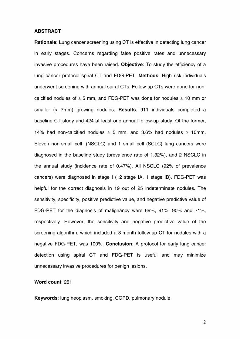
Diagnostic Algorithm
At baseline the result of the initial screening test was considered positive if there
were one to six non-calcified nodules, or more than six nodules were observed
with the largest one being 5 mm in diameter or more. The result was considered
29
negative if there were no non-calcified nodules, or more than six of them were
observed with the largest one being less than 5 mm in diameter. When the
result of the initial low-dose CT study was negative or the initial baseline study
did not lead to the diagnosis of a malignancy, repeat screening was performed
12 months after the initial baseline screening. When the result was positive,
further diagnostic work-up was followed according to a slightly modified
diagnostic algorithm developed by ELCAP(1). Based on the size, radiological
features and growth pattern of the pulmonary nodules. If the nodule was
between 5 and <10 mm a follow-up CT was done three months after the initial
study, and if growth was detected, further diagnostic studies such as a
percutaneous fine needle aspiration, a FDG-PET and/or a diagnostic surgical
biopsy were done. If no growth was seen or if the nodule had decreased in size,
a repeat CT was scheduled 12 months after the initial baseline study. Finally, if
the largest non-calcified nodule was 10 mm or more, an FDG-PET was
scheduled. If positive uptake was demonstrated, a diagnostic procedure such
as a fine needle biopsy was performed when possible. If not, the nodule was
resected after an intraoperative diagnostic fine needle aspiration was performed
to establish the appropriate surgical procedure. Growth was defined as an
increase of the diameter of the nodule in at least one dimension and was
determined by visual assesment by the radiologist comparing two CTs side-by-
side on the workstation. If a nodule of 10 mm or more showed benign
characteristics such as an apical broad pleural base or fat densities within, a 3-
month follow-up CT was done instead of an FDG-PET.
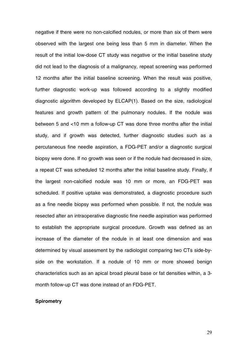
Spirometry
30
FVC and FEV1 were measured with a computerized spirometer (Vmax 22,
SensorMedics, CA). Results were expressed as percent of the predicted value
calculated on the basis of age, weight and height according to the European
Community Lung Health Survey (2). The presence (FEV1/FVC < 70%) and
severity (mild, FEV1 ≥ 80% of predicted; moderate, 50% ≤ FEV1 < 80%;
severe, 30% ≤ FEV1 < 50%; and very severe, FEV1 < 30%) of airways
obstruction was determined following the criteria established by the Global
Initiative for Chronic Obstructive Lung Disease (GOLD) (3).
Data analysis
Normally distributed data were summarized as means (SD) and skewed data
were summarized as medians (interquartile range, IQR). Data analysis was
performed with SPSS for Windows version 11 (SPSS Inc., Chicago, IL).
REFERENCES
E1. Henschke, C. I., D. F. Yankelevitz, J. P. Smith, and O. S. Miettinen.
2002. Screening for lung cancer: the early lung cancer action approach. Lung
Cancer 35(2):143-8.
E2. Roca, J., F. Burgos, J. Sunyer, M. Saez, S. Chinn, J. Anto, R. Rodriguez-
Roisin, P. Quanjer, D. Nowak, and P. Burney. 1998. References values for
forced spirometry. Group of the European Community Respiratory Health
Survey. Eur Respir J 11(6):1354-1362.
E3. Pauwels, R. A., A. S. Buist, P. M. A. Calverley, C. R. Jenkins, and S. S.
Hurd. 2001. Global Strategy for the Diagnosis, Management, and Prevention of
Chronic Obstructive Pulmonary Disease . NHLBI/WHO Global Initiative for

31
Chronic Obstructive Lung Disease (GOLD) Workshop Summary. Am. J. Respir.
Crit. Care Med. 163(5):1256-1276.


![Altered Brain Serotonin 5HT1A Receptor Binding After Recovery From Anorexia Nervosa Measured by Positron Emission Tomography and [Carbonyl11C]WAY100635](https://static.fdokumen.com/doc/165x107/6316dca13ed465f0570c3ef2/altered-brain-serotonin-5ht1a-receptor-binding-after-recovery-from-anorexia-nervosa.jpg)


![Molecular Imaging of Murine Intestinal Inflammation With 2-Deoxy-2-[ 18F]Fluoro- d-Glucose and Positron Emission Tomography](https://static.fdokumen.com/doc/165x107/6344fff26cfb3d4064097a1a/molecular-imaging-of-murine-intestinal-inflammation-with-2-deoxy-2-18ffluoro-.jpg)

