Review of Positron Emission Tomography at Royal Prince Alfred Hospital
35
Correspondence to: The Centre for Health Economics Research and Evaluation University of Sydney Level 6, Building F 88 Mallett St CAMPERDOWN NSW 2050 Telephone 61 2 9351 0900 Facsimile 61 2 9351 0930 email [email protected] www.chere.usyd.edu.au PROJECT REPORT 18 Review of Positron Emission Tomography at Royal Prince Alfred Hospital by Rosalie Viney, Anastasia Lowin, Christine Pollicino, Philip Haywood & Michael Fulham
Transcript of Review of Positron Emission Tomography at Royal Prince Alfred Hospital
Correspondence to: The Centre for Health Economics Research and Evaluation
University of Sydney Level 6, Building F
88 Mallett St CAMPERDOWN NSW 2050
Telephone 61 2 9351 0900 Facsimile 61 2 9351 0930
email [email protected] www.chere.usyd.edu.au
PROJECT REPORT 18
Review of Positron Emission Tomography at Royal Prince Alfred Hospital
by
Rosalie Viney, Anastasia Lowin, Christine Pollicino, Philip Haywood
& Michael Fulham

THE CENTRE FOR HEALTH ECONOMICS RESEARCH AND EVALUATION (CHERE) was established in 1991. CHERE is a centre of excellence in health economics and health services research. CHERE is funded by NSW Health under a Research and Development Infrastructure Grant, with additional support from Central Sydney Area Health Service and funding from external research. It is an affiliated research unit of the Faculty of Medicine, The University of Sydney. The centre aims to contribute to the development and application of health economics through research, teaching and policy support. CHERE’s research program encompasses both the theory and application of health economics. The main theoretical research theme pursues valuing benefits, including understanding what individuals value from health and health care, how such values should be measured, and exploring the social values attached to these benefits. The applied research focuses on economic and the appraisal of new programs or new ways of delivering and/or funding services. CHERE’s teaching includes introducing clinicians, health services managers, public health professionals and others to health economic principles. Training programs aim to develop practical skills in health economics and health services research. Policy support is provided at all levels of the health care system by undertaking commissioned projects, through the provision of formal and informal advice as well as participation in working parties and committees.
TABLE OF CONTENTS ACKNOWLEDGEMENTS…………………………………………………………….. i 1. INTRODUCTION ................................................................................................... 1 2 LITERATURE REVIEW ......................................................................................... 3 3 OVERVIEW OF THE OPERATION OF THE PET UNIT ................................... 11
3.1 Background..................................................................................................... 11 3.2 Operation of the PET Unit ............................................................................. 11 3.3 Throughput...................................................................................................... 13 3.4 Applications of PET at RPAH ........................................................................ 14
4 ESTIMATION OF THE COSTS OF PET ............................................................. 21 4.1 Estimation of the Total Costs of the PET Unit for 1997-98 ........................... 21 4.2 Estimation of the Average Costs per Patient for the PET Unit ...................... 23
5 CONCLUSION....................................................................................................... 26 6 REFERENCES ....................................................................................................... 27


i
ACKNOWLEDGEMENTS We are grateful to the NSW Health Department for providing funding to make this
review possible. We would also like to thank staff at the Department of PET and
Nuclear Medicine, Royal Prince Alfred Hospital, for their cooperation in assisting us in
the collection of data.
NOTE This report was written and submitted to NSW Health Department in January 2000.
Since this time circumstances relating to PET may have changed considerably. In
particular, the costs cited in this report may have altered. The cost estimates prepared
for the report have been re-estimated using 1998/99 and1999/00 costs, but this does not
capture change in resource use.

Viney, Lowin, Pollicino, Haywood & Fulham
CHERE Project Report 18 – January 2002 1
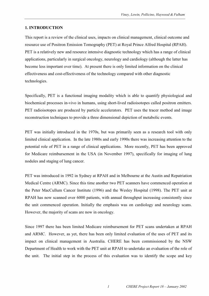
1. INTRODUCTION This report is a review of the clinical uses, impacts on clinical management, clinical outcome and
resource use of Positron Emission Tomography (PET) at Royal Prince Alfred Hospital (RPAH).
PET is a relatively new and resource intensive diagnostic technology which has a range of clinical
applications, particularly in surgical oncology, neurology and cardiology (although the latter has
become less important over time). At present there is only limited information on the clinical
effectiveness and cost-effectiveness of the technology compared with other diagnostic
technologies.
Specifically, PET is a functional imaging modality which is able to quantify physiological and
biochemical processes in-vivo in humans, using short-lived radioisotopes called positron emitters.
PET radioisotopes are produced by particle accelerators. PET uses the tracer method and image
reconstruction techniques to provide a three dimensional depiction of metabolic events.
PET was initially introduced in the 1970s, but was primarily seen as a research tool with only
limited clinical application. In the late 1980s and early 1990s there was increasing attention to the
potential role of PET in a range of clinical applications. More recently, PET has been approved
for Medicare reimbursement in the USA (in November 1997), specifically for imaging of lung
nodules and staging of lung cancer.
PET was introduced in 1992 in Sydney at RPAH and in Melbourne at the Austin and Repatriation
Medical Centre (ARMC). Since this time another two PET scanners have commenced operation at
the Peter MacCullum Cancer Institute (1996) and the Wesley Hospital (1998). The PET unit at
RPAH has now scanned over 6000 patients, with annual throughput increasing consistently since
the unit commenced operation. Initially the emphasis was on cardiology and neurology scans.
However, the majority of scans are now in oncology.
Since 1997 there has been limited Medicare reimbursement for PET scans undertaken at RPAH
and ARMC. However, as yet, there has been only limited evaluation of the uses of PET and its
impact on clinical management in Australia. CHERE has been commissioned by the NSW
Department of Health to work with the PET unit at RPAH to undertake an evaluation of the role of
the unit. The initial step in the process of this evaluation was to identify the scope and key

Review of Positron Emission Tomography
CHERE Project Report 18 – January 2002 2
components of a comprehensive evaluation. In particular, two approaches to evaluation were
identified as being necessary:
• Retrospective analysis, addressing the range of clinical applications of PET, the throughput
of the Unit, the costs of undertaking a PET scan for different patient groups, and the impact
of PET on patient outcomes; and
• Prospective analysis, addressing the impact of PET in specific identified applications, to
provide an unbiased assessment of the impact of PET on clinical management, outcomes for
patients and resource use.
Both components are seen as necessary to a comprehensive evaluation. The retrospective analysis
is important in assessing how the role of PET has changed and in providing an overview of the
role of PET. However, retrospective analyses are limited in that they are subject to a range of
confounding factors, most importantly, there may be bias in the way patients were selected for
PET scanning. Thus, the overall evaluation strategy has incorporated both components. The
project commissioned by the NSW Department of Health was to undertake the retrospective
analysis of throughput, costs and the outcomes of PET scans. In addition, CHERE, together with
clinicians from Royal Prince Alfred Hospital developed an NHMRC project application to
undertake a prospective evaluation of the role of PET in management of non-small cell lung
cancer (NSCLC), which is one of the major current clinical applications of PET at RPAH. The
prospective evaluation took the form of a randomised controlled trial incorporating economic
evaluation. The project application was successful and recruitment for the trial commenced in
April 1999, and was completed in December 2000. Follow-up of patients is continuing until
December 2001.
This report presents the findings of the retrospective analysis of the role of the PET unit,
particularly:
• Review of Australian and international literature on effectiveness and cost-effectiveness of
PET compared with other diagnostic technologies, particularly for oncology;
• Summary of patient throughput, characteristics and reasons for scans for the period 1992-
1998;
• Detailed analysis of reasons for scans for the period 1997-98;
• Estimated total and average costs for the PET unit, based on costing information for the
period 1997-98.

Viney, Lowin, Pollicino, Haywood & Fulham
CHERE Project Report 18 – January 2002 3
2 Literature Review In this section we briefly review the Australian and international literature evaluating the role of
PET in clinical and research applications. In particular, we have focussed on the evaluation of
PET as a diagnostic test, either as an adjunct or an alternative to existing diagnostic tests. As
noted, clinical and research applications for PET have emerged in three broad disease groups:
oncology, cardiology and neurology, with the most recent emphasis in both clinical applications
and in the evaluation literature being on oncology. There is now a growing body of literature on
evaluation of the role of PET in oncology, and, Robert and Milne (1999) report that more than
70% of referrals at the majority of international clinical PET centres now come from oncology
departments.
Three recent reviews of the role of PET have been undertaken. In 1996, a report commissioned by
the US Veterans Health Administration was finalised which included detailed systematic reviews
of PET in a range of oncology applications and Alzheimer’s disease (Flynn, Adams et al. 1996).
This report found that the majority of the literature focussed on the feasibility of the use of PET
and on diagnostic accuracy, with relatively few studies assessing efficacy or impact of PET on
patient management. The report concluded that, at that stage, the evaluation literature was
relatively under-developed, and it was difficult to draw conclusions about the utility of PET. For
example, for diagnosis of Alzheimer’s disease, while there was good evidence to support the
accuracy of PET, there was not yet evidence available to support more widespread use of PET in
management of Alzheimer’s disease. For the use of PET in diagnosis and management of cancer,
the report concluded that the literature was even less developed. Most studies were retrospectively
analysed case series, with small patient numbers, lack of control groups, poor use of blinding and
no randomisation.
In 1999, the National Health Service Research and Development Technology Assessment
Program published a review of the state of knowledge regarding clinical applications of PET,
partly as a basis for determining research priorities in relation to PET in the UK (Robert and Milne
1999). The conclusion of this report was similar to the previous US report. In particular, the
authors note that “there is no good evidence to suggest how PET will affect the cost-effectiveness
of the diagnosis, prognosis and management of patients”, and they comment on the lack of large
prospective studies. Thus, although there were increasing numbers of studies supporting the
diagnostic accuracy of PET in a range of conditions, there was still very little information to
assess the utility of PET in routine clinical practice, in terms of effectiveness or cost-effectiveness.

Review of Positron Emission Tomography
CHERE Project Report 18 – January 2002 4
In 2000, the Department of Health and Aged Care produced a Commonwealth review of PET in
Australia (Commonwealth Department of Health and Aged Care 2000). A key component of the
review process was an evaluation of PET conducted by a Supporting Committee of the Medicare
Services Advisory Committee (MSAC). MSAC explored the use of PET in six clinical indications
including lung and colorectal cancer, coronary revascularisation, epilepsy, melanoma and glioma.
The findings were largely consistent with the conclusions of existing reviews. It was concluded
that there was insufficient evidence on PET’s clinical or cost effectiveness with respect to the
clinical indications reviewed and that conclusive evidence such as a randomised controlled trial is
needed to explore the impact of PET on clinical management.
In our review of the literature we have focussed principally on the use of PET in oncology. Given
the relatively recent publication of the MSAC review we have not replicated their work, but have
focussed on identifying more recent papers. The main uses of PET in oncology identified were:
• Diagnosis Differentiating between benign and malignant conditions and
establishing the source of metastatic disease
• Staging Defining the extent of disease
• Monitoring Surveillance of treatment response
• Recurrence Identifying recurrence
The most well-established uses of PET are in staging of lung cancer and in diagnosis of solitary
pulmonary nodules. However, because of differences in clinical practice between the USA and
Australia, the latter is less relevant in the Australian context. Potential uses of PET identified in
the literature include the prediction of prognosis, tissue diagnosis and determining the best site for
biopsy (Sarinas, Chitkara et al. 1999).
The findings of our review of the literature confirmed the findings of the three previous studies.
The key issue that needs to be emphasised in relation to the evaluation of PET is that there are
specific challenges in evaluating a diagnostic technology. The decision to perform a diagnostic
test should be based on the usefulness of the information provided by the test. In other words, it
should provide an accurate diagnosis, support the application of a specific efficacious treatment,
and ultimately lead to a better or more cost-effective clinical outcome for the patient. In their
review Robert and Milne (1999) note that Fineberg has classified three stages in the diagnostic
process: production of a diagnostic output; the inclusion of that output into a diagnostic strategy

Viney, Lowin, Pollicino, Haywood & Fulham
CHERE Project Report 18 – January 2002 5
and choice of treatment; and the health outcome conditional upon treatment. In terms of evidence
for the value of a diagnostic test, studies which focus on technical performance and diagnostic
accuracy are only addressing the first of these stages. Ultimately, either prospective evaluations of
management impact, or, at minimum, formal decision analyses based on rigorous assessment of
sensitivity and specificity are necessary to address the second and third stages.
To date, published studies have focused on defining the accuracy of PET as a diagnostic test. A
number of prospective studies have been published, but few of these incorporate assessment of
how PET affects clinical decision making. There has been limited assessment of the impact of
PET on clinical management, and virtually no assessment of the impact on patient outcomes and
resource use. Some studies have extrapolated from the diagnostic properties of PET to the impact
on patient management (Wahl, Quint et al. 1994; Lowe, Fletcher et al. 1998; Weder, Schmid et al.
1998). However, even among these studies, some authors note that they believe it would be
unlikely that clinical management would be changed by the availability of the PET scan (Lowe,
Fletcher et al. 1998). One recent paper, not included in either of the previous systematic reviews,
does incorporate prospective assessment of the impact of PET on patient management, as well as
follow-up of clinical outcomes (confirmation of diagnosis at surgery or through histology, as well
as morbidity and mortality outcomes) (Saunders, Dussek et al. 1999). In this study 97 patients
with confirmed or suspected resectable lung cancer were staged based on CT and conventional
staging; CT alone; PET alone; and conventional, CT and PET. Management decisions were based
on all diagnostic information and patients were followed up for up to 41 months. The study found
that PET changed management in 37% of patients, although this figure included 15 patients for
whom the operation was “enabled” by PET, suggesting that the inclusion criteria were not clearly
defined. Of the 97 patients, 15 had a planned operation cancelled as a result of the PET scan.
There has been limited clinical evaluation of PET in the Australian context. A recent paper reports
on the experience with PET at one Australian centre (Hicks, Binns et al. 1999). However, while
the paper reports experience across a range of cancers suggesting that PET may have a role in
changing management, the conclusions are based on case series, without clear description of the
patient selection.
Economic evaluation
A Medline search (1990-2001; ‘positron emission tomography’ and ‘cost-benefit analysis’
exploded to all subheadings) was undertaken to locate papers addressing the cost-effectiveness of
Review of Positron Emission Tomography
CHERE Project Report 18 – January 2002 6
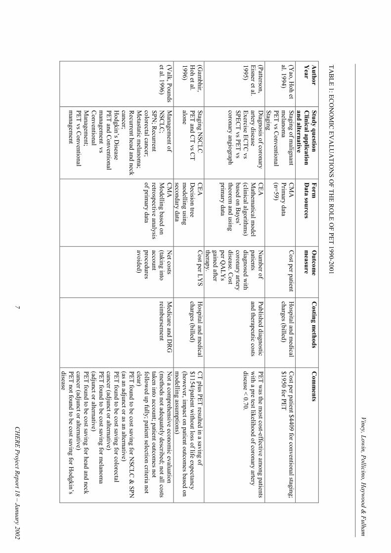
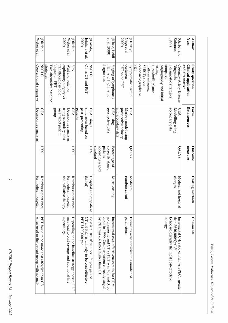
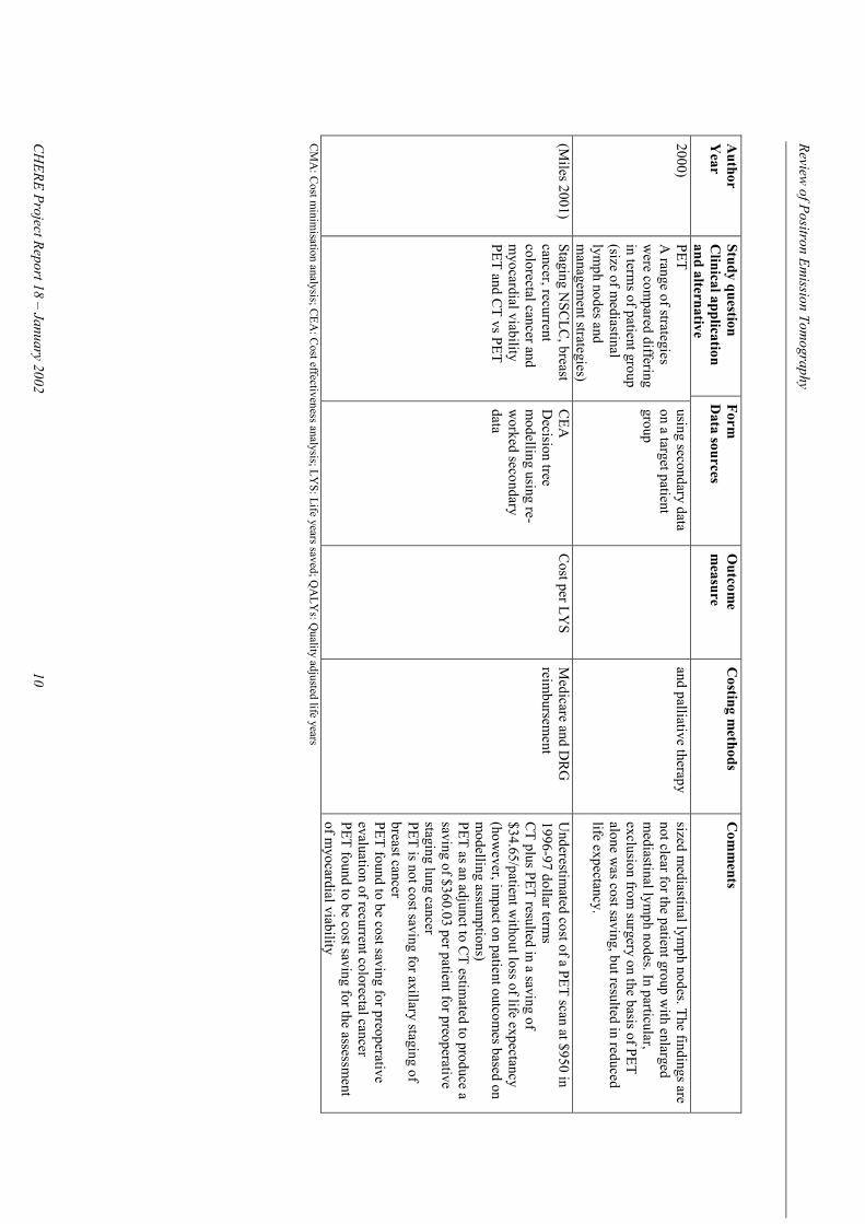
PET. This was supplemented by other search strategies to locate the maximum number of papers.
In all, 17 published economic evaluations were located, and are summarised in Table 1.
While the majority of papers conclude that PET is likely to be cost saving or to fall within an
acceptable cost-effectiveness range relative to conventional management of oncology patients, this
conclusion must be treated with considerable caution. All of the economic evaluations are subject
to methodological flaws. In particular, none incorporate adequate follow-up of patient outcomes.
Thus, even though decision tree modelling studies find that the use of PET in management of non-
small cell lung cancer results in no change or an increase in life expectancy, this is the result of an
assumption that avoidance of surgery in this group of patients reduces mortality. This assumption
is not supported by any studies following up patient outcomes following incorporation of PET in
the management strategy. In other studies there is no assessment of patient outcomes, and the
analysis is restricted to comparison of costs based on the assumption that management will change
and/or that patient outcomes will not be affected. Further, most studies incorporate only limited
assessment of the costs of alternative strategies. More fundamentally, relative cost-effectiveness
results are sensitive to the relative costs of procedures and it cannot be assumed that these costs
are comparable across different settings.
Viney, Lowin, Pollicino, Haywood &
Fulham
CHERE Project Report 18 – January 2002
7
TAB
LE 1: ECO
NO
MIC
EVA
LUA
TION
S OF TH
E RO
LE OF PET 1990-2001
Author
Year
Study question C
linical application and alternative
Form
Data sources
Outcom
e m
easure C
osting methods
Com
ments
(Yao, H
oh et al. 1994)
Staging of malignant
melanom
a PET vs C
onventional Staging
CM
A
Primary data
(n=59)
Cost per patient
Hospital and m
edical charges (billed)
Cost per patient $4409 for conventional staging;
$1950 for PET
(Patterson, Eisner et al. 1995)
Diagnosis of coronary
artery disease Exercise EC
TC vs
SPECT vs PET vs
coronary angiograph
CEA
M
athematical m
odel (clinical algorithm
s) based on B
ayes’ theorem
and using prim
ary data
Num
ber of patients diagnosed w
ith coronary artery disease. C
ost per Q
ALY
s gained after therapy.
Published diagnostic and therapeutic costs
PET was the m
ost cost-effective among patients
with a pre test likelihood of coronary artery
disease < 0.70.
(Gam
bhir, H
oh et al. 1996)
Staging NSC
LC
PET and CT vs C
T alone
CEA
D
ecision tree m
odelling using secondary data
Cost per LY
S H
ospital and medical
charges (billed) C
T plus PET resulted in a saving of $1154/patient w
ithout loss of life expectancy (how
ever, impact on patient outcom
es based on m
odelling assumptions)
(Valk, Pounds
et al. 1996)
Managem
ent of N
SCLC
; SPN
; Recurrent
colorectal cancer; M
etastatic melanom
a; R
ecurrent head and neck cancer; H
odgkin’s Disease
PET and Conventional
managem
ent vs C
onventional M
anagement;
PET vs Conventional
managem
ent
CM
A
Modelling based on
retrospective analysis of prim
ary data
Net costs
(taking into account procedures avoided)
Medicare and D
RG
reim
bursement
Not a com
prehensive economic evaluation
(methods not adequately described; not all costs
taken into account; patient outcomes not
followed up fully; patient selection criteria not
clear) PET found to be cost saving for N
SCLC
& SPN
(as an adjunct or as an alternative) PET found to be cost saving for colorectal cancer (adjunct or alternative) PET found to be cost saving for m
elanoma
(adjunct or alternative) PET found to be cost saving for head and neck cancer (adjunct or alternative) PET not found to be cost saving for H
odgkin’s disease

Review of Positron Emission Tom
ography
CHERE Project Report 18 – January 2002
8
Author
Year
Study question C
linical application and alternative
Form
Data sources
Outcom
e m
easure C
osting methods
Com
ments
(Hoh, G
laspy et al. 1997)
Diagnosis of H
odgkin’s D
isease and Lymphom
a PET vs C
onventional (to diagnosis)
CM
A
Primary data
Cost
comparison
Average prices for
procedures from 5 local
hospitals
PET found to be cost saving to the point of diagnosis: how
ever, this was not a
comprehensive econom
ic evaluation and final m
anagement decisions w
ere based on PET and conventional im
aging results (A
dler, Faulhaber et al. 1997)
Breast cancer; Staging
and managem
ent C
MA
M
odelling based on prospective prim
ary data
Cost per patient
Hospital and m
edical charges
PET would potentially save $2300 per patient
and avoid surgery
(Holm
berg, M
ohiuddin et al. 1997)
Coronary artery disease
Com
parison of two PET
modalities (adenosine
PET and dipyridamole
PET)
CM
A
Primary data
Retrospective, case-
control
Cost per patient
Hospital costs
Adenosine found to be low
er cost
(Scott, Shepherd et al. 1998)
NSC
LC
PET + CT versus CT
alone (4 different PET plus C
T strategies com
pared)
CEA
D
ecision tree m
odelling using secondary data
Cost per LY
S M
edicare reimbursed
costs Extends G
ambhir, H
oh et al (1996) Strategy of PET follow
ing a negative CT scan
resulted in a cost of $25000 per LYS com
pared w
ith CT alone; O
ther strategies (PET + CT resulted in a cost of $70000-$137000 per LY
S com
pared with C
T alone) N
ote that the estimated reduction in m
ortality is based on the assum
ptions incorporated in the m
odel and not based on any follow-up of patient
outcomes
(Gam
bhir, Shepherd et al. 1998)
SPN
4 strategies compared
watchful w
aiting; surgery; C
T; CT + PET
Decision tree
modelling using
secondary data
LYS
Medicare reim
bursed costs
CT plus PET found to be m
ore cost-effective and potentially cost saving com
pared with C
T alone for a pre-test probability of m
alignancy of 0.12-0.69
(von Schulthess, Steinert et al. 1998)
NSC
LC
PET vs CT
Melanom
a; PET vs C
onventional Staging
CM
A
Modelling using
primary data
Cost
comparison
Medical and hospital
charges N
ot a comprehensive econom
ic evaluation (im
pact on patient managem
ent only assessed hypothetically). PET found to be cost saving, but this is based on assum
ptions about the impact of
PET on patient managem
ent
Viney, Lowin, Pollicino, Haywood &
Fulham
CHERE Project Report 18 – January 2002
9
Author
Year
Study question C
linical application and alternative
Form
Data sources
Outcom
e m
easure C
osting methods
Com
ments
(Garber and
Solomon
1999)
Coronary A
rtery Disease
Diagnosis
5 diagnostic strategies com
pared: A
ngiography and initial testing + treadm
ill; planar thallium
imaging;
SPECT; stress
echocardiography or PET
CEA
M
odelling using secondary data
QA
LYs
Medical and hospital
charges Increm
ental C-E ratio of PET vs SPEC
T greater than $640,000/Q
ALY
Echocardiography the m
ost cost-effective strategy
(Derdeyn,
Gage et al.
2000)
Symptom
atic carotid occlusion PET vs no PET
CEA
M
arkov model using
prospective primary
and secondary data
QA
LYs
Medicare
reimbursem
ent Estim
ates were sensitive to a num
ber of param
eters.
(Klose, Leidl
et al. 2000) Staging of lym
phomas
PET vs CT, C
T vs no diagnostics
CEA
using prospective data
Percentage of correctly staged patients according a gold standard
Micro costing
Incremental cost-effectiveness ratio for C
T vs no diagnostic and C
T vs PET was 478 and 3133
euros in 1999. Cost per patient correctly staged
by PET was 6.6 tim
es higher than CT.
(Kosuda,
Ichihara et al. 2000)
NSC
LC
CT vs C
T and PET C
EA using a
simulation based on
past presenting patients
LYS
Hospital and outpatient
(billed) C
ost is 2.18x10⁵ yen per life year gained
CT and PET is unlikely to be cost-effective;
PET $100,000 yen
(Dietlein,
Weber et al.
2000)
SPN
Wait and w
atch or exploratory surgery vs transthoracic needle biopsy or PET Tw
o alternative baseline strategies
CEA
D
ecision tree analysis using secondary data on a target patient group
LYS
Reim
bursement rates
for medical, hospital
and palliative therapy
Depending on the baseline strategy chosen, PET
may lead to cost savings and additional life
expectancy.
(Dietlein,
Weber et al.
NSC
LC
Conventional staging vs
CEA
D
ecision tree analysis LY
S R
eimbursem
ent rates for m
edical, hospital PET found to be m
ore cost effective than CS
when used in the patient group w
ith normal-
Review of Positron Emission Tom
ography
CHERE Project Report 18 – January 2002
10
Author
Year
Study question C
linical application and alternative
Form
Data sources
Outcom
e m
easure C
osting methods
Com
ments
2000) PET A
range of strategies w
ere compared differing
in terms of patient group
(size of mediastinal
lymph nodes and
managem
ent strategies)
using secondary data on a target patient group
and palliative therapy sized m
ediastinal lymph nodes. The findings are
not clear for the patient group with enlarged
mediastinal lym
ph nodes. In particular, exclusion from
surgery on the basis of PET alone w
as cost saving, but resulted in reduced life expectancy.
(Miles 2001)
Staging NSC
LC, breast
cancer, recurrent colorectal cancer and m
yocardial viability PET and C
T vs PET
CEA
D
ecision tree m
odelling using re-w
orked secondary data
Cost per LY
S M
edicare and DR
G
reimbursem
ent U
nderestimated cost of a PET scan at $950 in
1996-97 dollar terms
CT plus PET resulted in a saving of
$34.65/patient without loss of life expectancy
(however, im
pact on patient outcomes based on
modelling assum
ptions) PET as an adjunct to C
T estimated to produce a
saving of $360.03 per patient for preoperative staging lung cancer PET is not cost saving for axillary staging of breast cancer PET found to be cost saving for preoperative evaluation of recurrent colorectal cancer PET found to be cost saving for the assessm
ent of m
yocardial viability C
MA
: Cost m
inimisation analysis; CEA
: Cost effectiveness analysis; LY
S: Life years saved; QA
LYs: Q
uality adjusted life years
Viney, Lowin, Pollicino, Haywood & Fulham
CHERE Project Report 18 – January 2002 11
3 Overview of the Operation of the PET Unit
3.1 Background
The PET Unit at RPAH was established in 1992. The PET scanner was purchased at a
cost of approximately $5 million dollars, funded by a collaborative effort involving
Royal Prince Alfred Hospital (RPAH), the NSW Department of Health and private
donations. The National Medical Cyclotron (NMC) was funded by the Commonwealth
government, and is owned and operated by the Australian Nuclear Science and
Technology Organisation (ANSTO). It was established on the campus of RPAH, at a
cost of approximately $20 million.
The PET Unit began scanning in June 1992, and the annual throughput of patients in
each year has increased consistently since then. Over 7000 patients have now been
scanned. In 1999 (until November 30), 1356 studies were undertaken, comprising 400
neurological scans, 21 cardiac scans and 935 whole body (oncology) scans.
3.2 Operation of the PET Unit
The establishment of the PET Unit and the NMC in 1992 required extensive
refurbishment of the PET suite within the hospital, involving the floor level being raised
and complex air-conditioning being fitted. It also involved the installation of a rapid
transport system under Missenden Road, to transport the PET radiotracers to the PET
Suite. The total area of the PET Suite is 311 square metres. Since 1992 there has been
extensive upgrading of the computer systems of the PET unit, to improve storage and
retrieval of data.
From the outset of the PET program, RPAH has had staff members from the PET unit
located in the NMC who are responsible for production of PET tracers for RPAH. A
typical production run for RPAH involves:
• set-up for production beginning at 0700 hrs,
• cyclotron irradiates PET target for 90 mins from 0730 - 0900 hrs,
• 18F1 is transferred to an automated radiochemistry box and synthesis takes place over
60 mins,
• a sample of the product is taken for quality control (QC) and
• product released 30 minutes later for human use at 1030 hrs.
1 18F is a radioactive isotope with a half life of 110 minutes.
Review of Positron Emission Tomography
CHERE Project Report 18 – January 2002 12
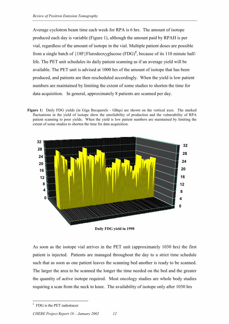
Average cyclotron beam time each week for RPA is 6 hrs. The amount of isotope
produced each day is variable (Figure 1), although the amount paid by RPAH is per
vial, regardless of the amount of isotope in the vial. Multiple patient doses are possible
from a single batch of {18F}Flurodeoxyglucose (FDG)2, because of its 110 minute half-
life. The PET unit schedules its daily patient scanning as if an average yield will be
available. The PET unit is advised at 1000 hrs of the amount of isotope that has been
produced, and patients are then rescheduled accordingly. When the yield is low patient
numbers are maintained by limiting the extent of some studies to shorten the time for
data acquisition. In general, approximately 8 patients are scanned per day.
Figure 1: Daily FDG yields (in Giga Becquerels - GBqs) are shown on the vertical axes. The marked fluctuations in the yield of isotope show the unreliability of production and the vulnerability of RPA patient scanning to poor yields. When the yield is low patient numbers are maintained by limiting the extent of some studies to shorten the time for data acquisition.
0
4812162024
28
32
0
4812
16
20
24
28
32
Daily FDG yield in 1998
As soon as the isotope vial arrives in the PET unit (approximately 1030 hrs) the first
patient is injected. Patients are managed throughout the day to a strict time schedule
such that as soon as one patient leaves the scanning bed another is ready to be scanned.
The larger the area to be scanned the longer the time needed on the bed and the greater
the quantity of active isotope required. Most oncology studies are whole body studies
requiring a scan from the neck to knee. The availability of isotope only after 1030 hrs
2 FDG is the PET radiotracer
Viney, Lowin, Pollicino, Haywood & Fulham
CHERE Project Report 18 – January 2002 13
has cost implications, because most hospital activity is scheduled to occur during the
normal working day. PET unit staff are often required to work overtime, and patients
are often required to be scanned in the evening.
The process for each patient involves arrival at the unit followed by a consultation with
either the PET unit director or the registrar to ensure they understand the process and to
facilitate signing of a consent form. Patients are injected with the isotope approximately
45 minutes prior to scanning. Once injected, the patient is asked to remain still – this
period is referred to as the uptake period. Patients for whom the area scanned includes
the pelvis are fitted with a catheter. The duration of the scanning period is dependent
mainly on the amount of the body to be scanned. After the scanning has finished it
takes approximately 2 hours for the image reconstruction techniques to provide a 3-D
depiction of dynamic metabolic events. The scan is assessed by the Director of the PET
unit, and a report is sent to the referring physician. The findings may be passed to the
referring physician by phone in cases where surgery is booked immediately following
the scan.
The availability of the cyclotron is a limiting factor for the PET unit throughput. The
NMC cyclotron is closed at weekends. It is also closed for maintenance every Monday
and for 3 weeks over the Christmas / New Year period and for 2 weeks midyear,
however in 1998 the midyear shutdown was not carried out.
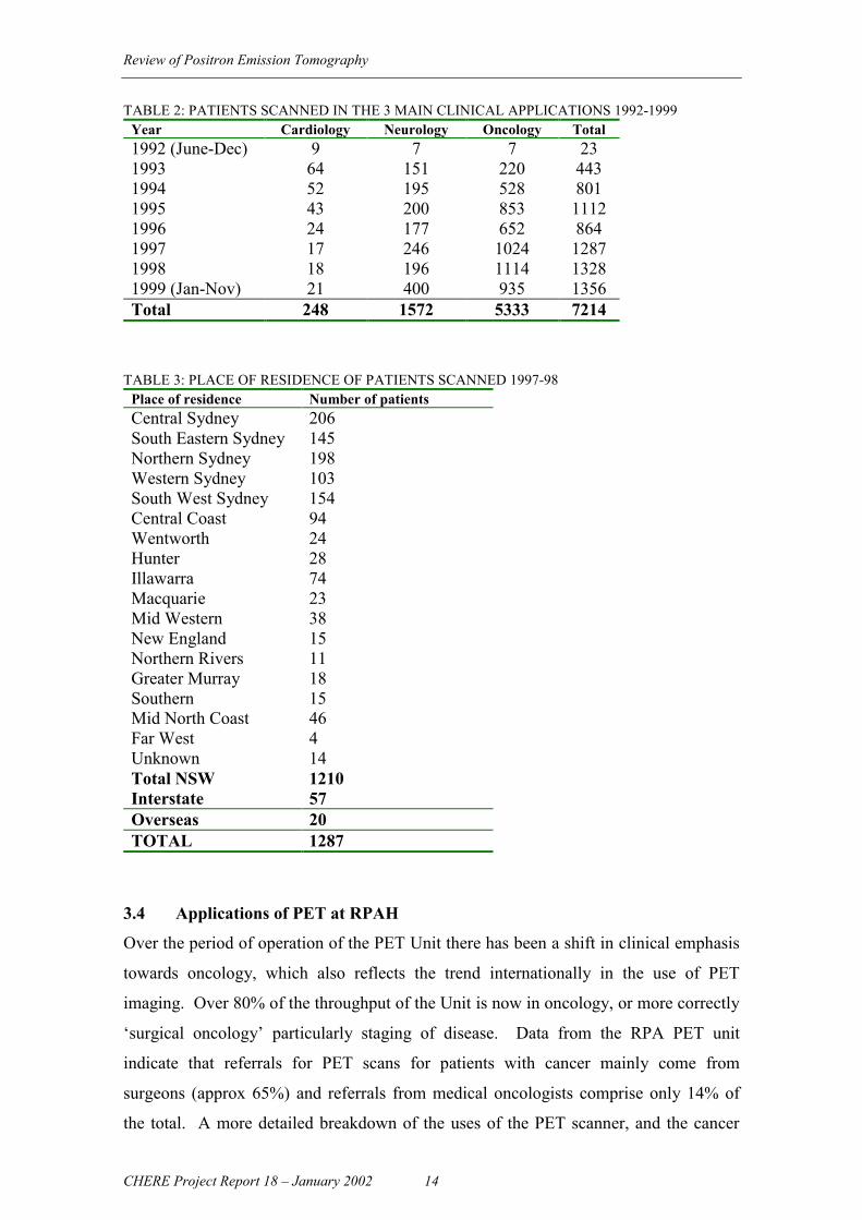
3.3 Throughput
Table 2 summarises the annual throughput. The current annual throughput is
approximately 1300 patients, and this has been relatively stable over the past three
years. Given the current arrangements for provision of the service, which involves
supply by the NMC of a single vial of isotope per day to the Unit on 4 days of the week,
this throughput is likely to remain stable.
The Unit provides a state-wide PET imaging service for patients, and is the only PET
unit in NSW. Less than 20 per cent of patients scanned reside within Central Sydney
Area Health Service. Table 3 provides a summary of the place of residence of patients
scanned during 1997-98.
Review of Positron Emission Tomography
CHERE Project Report 18 – January 2002 14
TABLE 2: PATIENTS SCANNED IN THE 3 MAIN CLINICAL APPLICATIONS 1992-1999 Year Cardiology Neurology Oncology Total 1992 (June-Dec) 9 7 7 23 1993 64 151 220 443 1994 52 195 528 801 1995 43 200 853 1112 1996 24 177 652 864 1997 17 246 1024 1287 1998 18 196 1114 1328 1999 (Jan-Nov) 21 400 935 1356 Total 248 1572 5333 7214
TABLE 3: PLACE OF RESIDENCE OF PATIENTS SCANNED 1997-98 Place of residence Number of patients Central Sydney 206 South Eastern Sydney 145 Northern Sydney 198 Western Sydney 103 South West Sydney 154 Central Coast 94 Wentworth 24 Hunter 28 Illawarra 74 Macquarie 23 Mid Western 38 New England 15 Northern Rivers 11 Greater Murray 18 Southern 15 Mid North Coast 46 Far West 4 Unknown 14 Total NSW 1210 Interstate 57 Overseas 20 TOTAL 1287
3.4 Applications of PET at RPAH
Over the period of operation of the PET Unit there has been a shift in clinical emphasis
towards oncology, which also reflects the trend internationally in the use of PET
imaging. Over 80% of the throughput of the Unit is now in oncology, or more correctly
‘surgical oncology’ particularly staging of disease. Data from the RPA PET unit
indicate that referrals for PET scans for patients with cancer mainly come from
surgeons (approx 65%) and referrals from medical oncologists comprise only 14% of
the total. A more detailed breakdown of the uses of the PET scanner, and the cancer
Viney, Lowin, Pollicino, Haywood & Fulham
CHERE Project Report 18 – January 2002 15
sites is provided later in this report. However, it should be noted that the current use of
the scanner reflects existing knowledge of the effectiveness of PET as a diagnostic tool.
Thus, there is an emphasis in the RPAH PET unit on lung cancer and melanoma, for
which there is more evaluation evidence, both here and internationally. As new
research evidence becomes available, there may be a further shift in clinical emphasis.
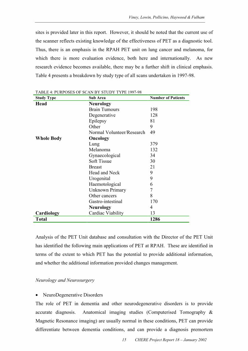
Table 4 presents a breakdown by study type of all scans undertaken in 1997-98.
TABLE 4: PURPOSES OF SCAN BY STUDY TYPE 1997-98 Study Type Sub Area Number of Patients Head Neurology Brain Tumours 198 Degenerative 128 Epilepsy 81 Other 9 Normal Volunteer/Research 49 Whole Body Oncology Lung 379 Melanoma 132 Gynaecological 34 Soft Tissue 30 Breast 21 Head and Neck 9 Urogenital 9 Haemotological 6 Unknown Primary 7 Other cancers 8 Gastro-intestinal 170 Neurology 4 Cardiology Cardiac Viability 13 Total 1286 Analysis of the PET Unit database and consultation with the Director of the PET Unit
has identified the following main applications of PET at RPAH. These are identified in
terms of the extent to which PET has the potential to provide additional information,
and whether the additional information provided changes management.
Neurology and Neurosurgery
• NeuroDegenerative Disorders
The role of PET in dementia and other neurodegenerative disorders is to provide
accurate diagnosis. Anatomical imaging studies (Computerised Tomography &
Magnetic Resonance imaging) are usually normal in these conditions, PET can provide
differentiate between dementia conditions, and can provide a diagnosis premortem

Review of Positron Emission Tomography
CHERE Project Report 18 – January 2002 16
when anatomical imaging is unhelpful. The principal benefit of the PET scan in this
case is the value of the information to the individual, the referring doctor and
carers/family members. As treatments for dementia become available (already there are
a number of agents that have been shown to delay the rate of disease progression in
patients with suspected dementia in clinical trials) it is imperative that patients are
correctly identified. In this context a PET scan will assist in determining more
appropriate treatment, or in limiting unnecessary investigations. In 1997-98, 128
patients were scanned for neurological degenerative conditions.
• Brain Tumours
PET is used to provide information about the grade of a tumour, and to provide more
accurate information to localise the most active tumour area for biopsy or excision by a
neurosurgeon. In both cases, the benefit of PET is in determining the most appropriate
treatment, particularly surgical treatment. Information about the grade of the tumour is
used to determine whether patients are appropriate surgical candidates. In 1997-98, 198
patients were evaluated for brain tumours. Thus, almost half of the neurological
referrals were for evaluation of tumours. PET can also be used for the surveillance of
low-grade brain tumours avoiding alternative methods of monitoring. This use of PET
may allow the earlier diagnosis of malignant transformation and earlier treatment may
provide improved outcomes.
• Epilepsy
PET is used in epileptic patients who have refractory focal epilepsy to localise seizure
foci. Thus, the PET scan may be used to provide additional diagnostic information to
assist in management of epilepsy, in particular to select patients for whom surgery is
likely to provide a benefit (that is, to determine whether surgery is likely to affect
seizure control) but also to exclude patients with generalised epilepsy where surgery is
of no benefit] and therefore to increase the probability of successful surgery. In 1997-
98, 81 patients were scanned for epilepsy.
Cardiology PET has been used to establish cardiac viability, to determine which patients are most
likely to be successful candidates for revascularisation surgery. In 1992, this
represented approximately 30% of the workload of the PET unit, but, in line with
research evidence, this role has changed considerably. In 1997-98, only 13 patients
Viney, Lowin, Pollicino, Haywood & Fulham
CHERE Project Report 18 – January 2002 17
were scanned for this purpose. The benefit of a PET scan in this case is that it
potentially provides better selection of surgical candidates, which may improve surgical
outcomes.
Oncology
The main role of the PET Unit at RPAH over the past six years has been in a range of
oncology applications in the pre-surgical setting. During 1997-98, patients were
scanned for a range of cancers, including breast cancer, colorectal cancer, lung cancer,
melanoma, gynaecological cancers, brain tumours and head and neck cancers. In
general these patients have a whole body scan, and there are a number of possible
purposes of the scan, as outlined below.
• Lung Cancer
PET is used to provide additional diagnostic information for patients with lung cancer.
In 1997-98, 349 patients were referred for a whole body PET scan related to a primary
diagnosis of lung cancer. The majority of patients referred for a PET scan have non-
small cell lung cancer and the other large group is patients who are thought to have
solitary pulmonary metastases from a range of malignancies including colorectal
carcinoma, melanoma, breast cancer and soft tissue sarcomas.
The main use of the PET scan is to determine whether a patient is a suitable candidate
for surgery prior to the final decision about treatment being made. PET is used to
determine whether patients who have been diagnosed with StageI-II non-small cell lung
cancer on other diagnostic tests have mediastinal node involvement (Stage III) or distant
metastatic disease (Stage IV). Patients who have Stage III or IV cancer are generally
not considered to be suitable candidates for surgery, although some patients with Stage
IIIa non-small cell lung cancer may still have surgery following chemotherapy and PET
in this instance is used to exclude Stage IV disease both before and at the completion of
therapy. Thus, the main benefit of PET in this case is that it may avoid unnecessary and
potentially fatal painful surgery.
PET is also used in some cases post-surgically to provide information about recurrence
or to assess response to chemotherapy or radiotherapy. In this case, the benefit of PET
is that it may provide information to determine the most appropriate treatment.
Review of Positron Emission Tomography
CHERE Project Report 18 – January 2002 18
• Colorectal cancer
The PET scanner can be used for the accurate staging of disease, especially in those
who are thought to have a solitary liver metastasis that would be amenable to surgery if
there is not more widespread disease. This allows appropriate selection of candidates
and enhances surgical outcomes.
• Melanoma
The PET scanner can be used for the staging of melanoma after initial diagnosis but it is
mainly used in patients with metastatic melanoma prior to them undergoing surgical
intervention that may include thoracic surgery, node dissection, hepatic resection or
craniotomy. This may allow more appropriate treatment to be given.
• Head and Neck Cancer
Head and Neck surgery for ontological reasons distorts the anatomy of the region.
Because of this the more functional PET scan may be more sensitive to early local
recurrence than more structural diagnostic tools such as CT scanners. Early detection
may offer more definitive and successful treatment.
• Gynaecological Cancer
The PET scanner is being used in ovarian cancer for two purposes; one to help aid the
diagnosis of recurrence and the second is to assess the response to chemotherapeutic
treatment. Earlier diagnosis of recurrence may allow improved therapeutic option
selection. Assessing the response to treatment of ovarian cancer to chemotherapy may
allow the early cessation of chemotherapy in those for whom it is not affecting the
tumour, with quality of life and resource saving implications.
Viney, Lowin, Pollicino, Haywood & Fulham
CHERE Project Report 18 – January 2002 19
Summary Thus, in summary, a number of potential benefits can be identified from the applications
of PET at RPAH:
• Information for patients and clinicians
Even where the information from a PET scan does not affect clinical management, there
may be benefits, particularly to patients from additional diagnostic information. This is
particularly relevant in diagnosis of dementia conditions and in patients with other
chronic conditions where treatment does not eradicate the disease and the natural history
of these disorders is that there is disease progression over time.
• More appropriate clinical management
The PET scan may provide information that changes clinical management of the
patient’s condition. In many cases this may not provide a survival benefit (for example,
in changed management of lung cancer), but there may be short term improvements in
quality of life for patients, from avoidance of unnecessary surgery. However, as has
been noted earlier, caution must be applied in assessing the extent to which PET
changes clinical management. To evaluate this impact it is necessary to have an
unbiased assessment of the management plan before and after the PET scan.
• Improved outcomes from treatment
The additional diagnostic information from the PET scan may lead to more localised
and less invasive surgery or other treatment (for example, in surgical management of
brain tumours, colorectal cancer, melanoma). This may improve the survival and
quality of life outcomes from surgery.
• Resource use
Where PET changes clinical management it may avoid unnecessary resource use, for
example, in the case of avoiding surgery. Whether this results in a net saving depends
on the relative costs of the PET scan and the interventions avoided, and the proportion
of patients for whom there is a change in management. As in the case of changes to
clinical management, it is difficult to assess the amount of resource savings
retrospectively.

Review of Positron Emission Tomography
CHERE Project Report 18 – January 2002 20
• Allocative efficiency
Because PET may provide information which leads to better selection of patients for
particular interventions, it may improve the allocative efficiency of service provision. In
particular, there are allocative efficiency gains where there are resource constraints and
where PET provides information which leads to more accurate decisions about which
patients are likely to benefit from treatment. However, it should be noted that there are a
number of questions that must be addressed in assessing the value of a PET scan in
particular clinical applications:
• Will the PET scan provide additional diagnostic information? Is the information
from the PET scan likely to change the diagnosis? Is the information more sensitive
and/or more specific?
• Will the additional diagnostic information change the choice of treatment for the
patient?
• Will any change in management result in changes in survival or quality of life
outcomes for patients?
• Are there any other benefits/disbenefits to patients from the additional information?
• Is the change in management likely to lead to a net reduction in resource use, given
the resource use associated with undertaking the scan?
• Is it feasible that PET would replace another diagnostic test, or is it likely to be used
as an adjunct to other diagnostic tests?
Viney, Lowin, Pollicino, Haywood & Fulham
CHERE Project Report 18 – January 2002 21
4 Estimation of the Costs of PET
This section of the report outlines the methods and results of the costing undertaken for
the PET Unit. Costs were estimated for the 1997-98 period, because a full year of
financial data was available. The aims of the costing component of the project were:
• To estimate the total costs of the PET unit;
• To estimate the average cost of a PET scan;
• To determine whether average costs differ for different groups of patients;
• To estimate the short run marginal cost of a PET scan;
• To investigate how the costs of the PET unit would vary under different scenarios.
4.1 Estimation of the Total Costs of the PET Unit for 1997-98
The assumptions used to estimate the different components of total cost are outline
below.
Recurrent Costs
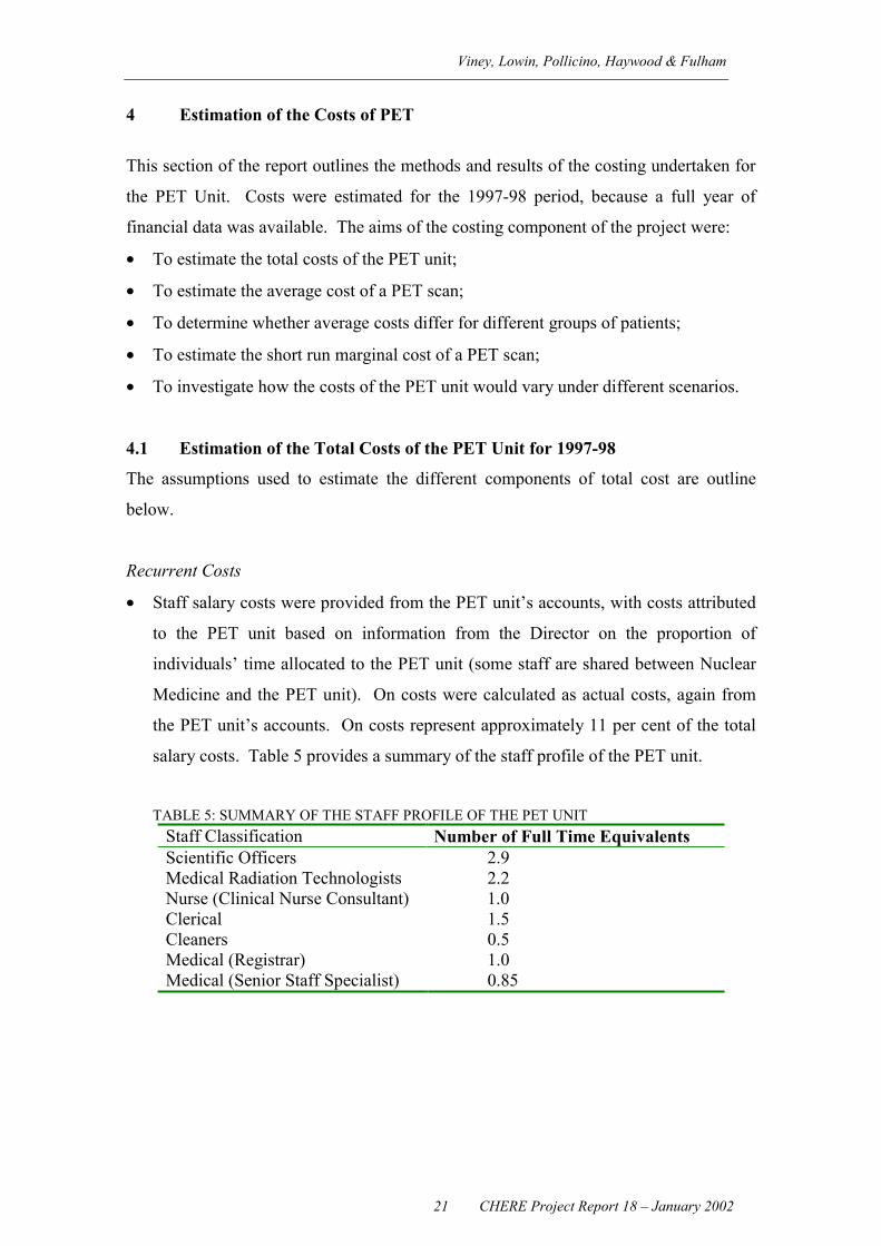
• Staff salary costs were provided from the PET unit’s accounts, with costs attributed
to the PET unit based on information from the Director on the proportion of
individuals’ time allocated to the PET unit (some staff are shared between Nuclear
Medicine and the PET unit). On costs were calculated as actual costs, again from
the PET unit’s accounts. On costs represent approximately 11 per cent of the total
salary costs. Table 5 provides a summary of the staff profile of the PET unit.
TABLE 5: SUMMARY OF THE STAFF PROFILE OF THE PET UNIT
Staff Classification Number of Full Time Equivalents Scientific Officers 2.9 Medical Radiation Technologists 2.2 Nurse (Clinical Nurse Consultant) 1.0 Clerical 1.5 Cleaners 0.5 Medical (Registrar) 1.0 Medical (Senior Staff Specialist) 0.85

Review of Positron Emission Tomography
CHERE Project Report 18 – January 2002 22
• Goods and services costs were provided from the PET unit’s cost centre accounts.
These costs included costs of medical supplies (other than isotope), stationery,
computer software support, radiation monitoring and other operational goods and
services.
• Isotope costs were based on actual charges made by ANSTO to Royal Prince Alfred
Hospital. During 1997-98 177 vials of FDG-18, 10 vials of NH3 and 10 vials of
015 were provided to the PET Unit. Charging is on the basis of a set price per vial.
• Estimates of overhead costs were provided by the Finance Department of Royal
Prince Alfred Hospital. These were based on the overhead cost allocation methods
recommended in the NSW 1997/98 Costing Standards Manual. Overhead costs
were estimated and attributed for Royal Prince Alfred Hospital and for Central
Sydney Area Health Service.
• Repairs and Maintenance costs for the PET Unit were taken from two sources.
Some maintenance costs for the Unit are separately reported in the Royal Prince
Alfred financial accounts. However, this does not include the total cost of
maintenance contracts because of the financial arrangements within the hospital,
whereby global maintenance contracts are held, and not disaggregated to separate
clinical units. Therefore estimates of the maintenance costs were also based on
information provided by the Director of the PET unit and in a separate report on the
activity of the PET Unit.
Capital Costs
• Building costs were estimated on the basis of an equivalent rental cost for the floor
space occupied by the PET Unit. This was based on commercial rents for the area,
of $225 per square metre (this was the mid point of estimates provided by local
commercial real estate agents). The total floorspace of the PET unit was 311 square
metres.
• Information on the costs of capital equipment for the operation of the PET unit was
provided by the Director of the PET Unit and from NSW Department of Health
files. This included the original purchase of the PET scanner and the refurbishment
Viney, Lowin, Pollicino, Haywood & Fulham
CHERE Project Report 18 – January 2002 23
of the PET unit (this cost was covered by ANSTO), but does not include the cost of
construction of the cyclotron. The information provided was the best estimate of the
original purchase price and the date of purchase for each item of capital equipment.
The GDP implicit price deflator was used to convert the purchase price to 1997-98
dollars. It was assumed that the useful life of computer hardware was 5 years, and
the useful life of other capital equipment was 10 years, and that there would be no
residual value. Using these assumptions and a discount rate of 5%, an equivalent
annual cost of capital equipment was calculated.
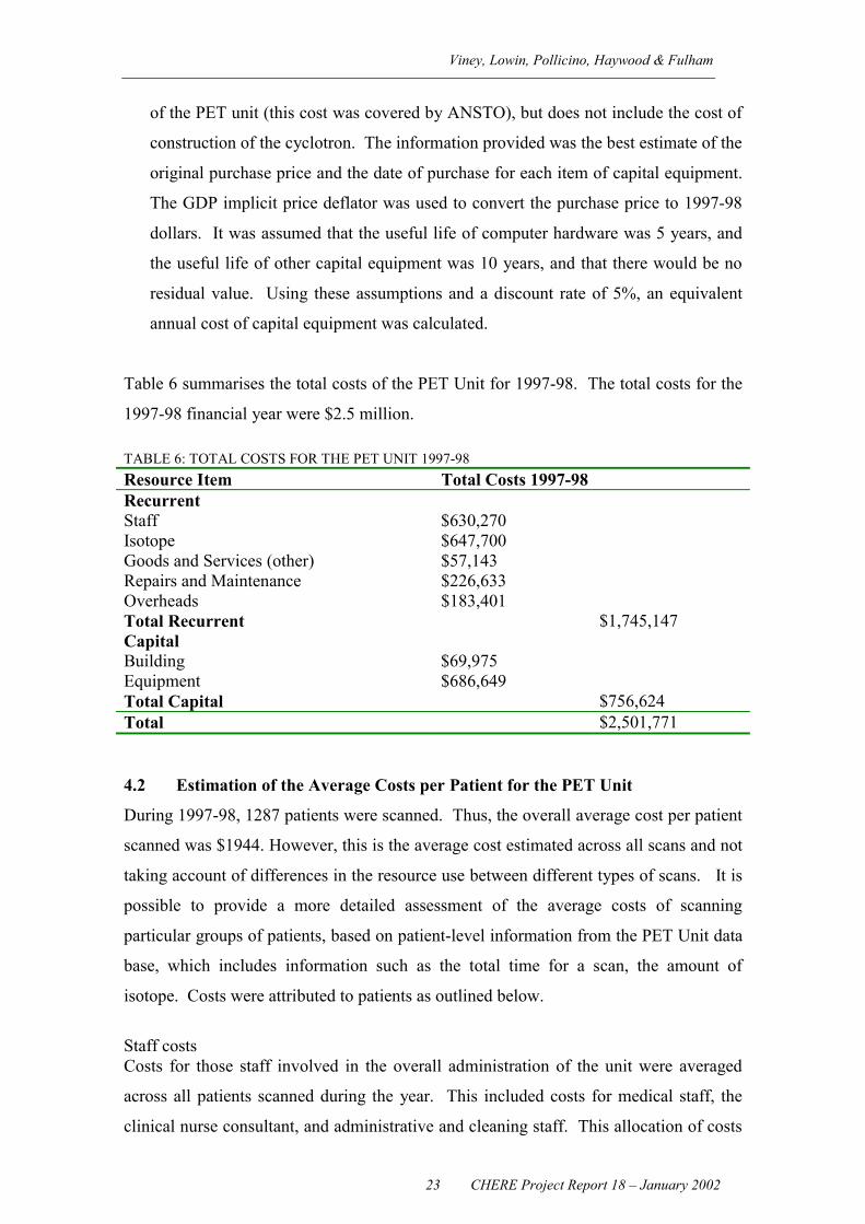
Table 6 summarises the total costs of the PET Unit for 1997-98. The total costs for the
1997-98 financial year were $2.5 million. TABLE 6: TOTAL COSTS FOR THE PET UNIT 1997-98 Resource Item Total Costs 1997-98 Recurrent Staff $630,270 Isotope $647,700 Goods and Services (other) $57,143 Repairs and Maintenance $226,633 Overheads $183,401 Total Recurrent $1,745,147 Capital Building $69,975 Equipment $686,649 Total Capital $756,624 Total $2,501,771 4.2 Estimation of the Average Costs per Patient for the PET Unit
During 1997-98, 1287 patients were scanned. Thus, the overall average cost per patient
scanned was $1944. However, this is the average cost estimated across all scans and not
taking account of differences in the resource use between different types of scans. It is
possible to provide a more detailed assessment of the average costs of scanning
particular groups of patients, based on patient-level information from the PET Unit data
base, which includes information such as the total time for a scan, the amount of
isotope. Costs were attributed to patients as outlined below.
Staff costs Costs for those staff involved in the overall administration of the unit were averaged
across all patients scanned during the year. This included costs for medical staff, the
clinical nurse consultant, and administrative and cleaning staff. This allocation of costs
Review of Positron Emission Tomography
CHERE Project Report 18 – January 2002 24
was made on the basis that where these staff were involved in direct patient care, the
resource use was not likely to substantial vary with the nature of the scan and the length
of time for the scan.
Costs for other staff, directly involved in scanning the patients were attributed to each
patient on the basis of the total time for the scan.
Other recurrent costs Isotope costs were calculated on a per patient basis, although this involves averaging
across patients as follows. While the pet unit is charged a fixed amount for a vial of
isotope, the amount of isotope in a vial is variable. Thus, in practice the per patient cost
of isotope will vary on a daily basis, depending on the amount of isotope produced.
However, as this variability in cost cannot be predicted, in calculating a cost per patient,
an effective price per unit of isotope (measured in megabecs) was estimated over the
whole year, and this was used to attribute costs to patients based on the amount of
isotope the patient received.
Other goods and services were averaged across all patients scanned during the year, on
the basis that the amount of consumables did not vary with the length of time for a scan
or the nature of the scan.
Overhead costs were averaged across all patients, on the basis that these costs cover
items of resource use which do not vary with the nature of the scan or the length of time
for the scan.
Repairs and maintenance costs were attributed to patient on the basis of the total time
for the scan. This reflects the fact that the majority of these costs relate to upkeep of the
pet scanner, and so they should be allocated on the same basis as the capital equipment
(see below).
Capital costs Capital costs were attributed to patients based on the total time for the scan. This
reflects the fact that the opportunity cost of additional time spent scanning a patient is
the time forgone scanning additional patients.

Viney, Lowin, Pollicino, Haywood & Fulham
CHERE Project Report 18 – January 2002 25
Table 7 provides a summary of the average costs for different groups of patients. As
would be expected, the most costly scans are whole body scans, because they require
more time and more isotope to provide a full image of the body. A whole body scan
generally requires 8 ‘beds’ to complete the study (one ‘bed’ represents approximately
10 cm).
TABLE 7 AVERAGE COST OF A PET SCAN BY TYPE OF SCAN
Type of scan 1997/1998 1998/19991 1999/20001 All Scans $1,944 $1,994 $2,040
Whole Body $1,963 $2,013 $2,060
Lung $1,975 $2,026 $2,072 Melanoma $2,106 $2,160 $2,210 Gynaecological $1,929 $1,978 $2,024 Soft Tissue $1,869 $1,917 $1,961 Breast $1,981 $2,032 $2,079 Head and Neck $2,086 $2,139 $2,189 Urogenital $1,949 $1,999 $2,045 Haemotological $2,036 $2,088 $2,136 Unknown Primary $1,889 $1,937 $1,982 Other cancers $2,076 $2,129 $2,178 Gastro-intestinal $1,841 $1,888 $1,932 Neurological $1,755 $1,800 $1,841 Head $1,856 $1,904 $1,947 Brain Tumours $1,681 $1,724 $1,764 Degenerative $1,842 $1,889 $1,933 Epilepsy $1,815 $1,862 $1,904 Other $1,852 $1,899 $1,943 Normal Volunteer/
Research $2,664 $2,732 $2,795
Cardiology Cardiac Viability $3,900 $4,000 $4,092
1. Average costs have been re-estimated for the 1998-1999 and1999-2000 period using the total health price index.
Review of Positron Emission Tomography
CHERE Project Report 18 – January 2002 26
5. Conclusion
This report evaluated the clinical uses, impacts on clinical management, clinical
outcome and resource use of Positron Emission Tomography (PET) at Royal Prince
Alfred Hospital in Sydney.
A current literature review emphasised the increasing role PET scanning in oncology
diagnosis and management. However, the clinical studies and economic evaluations of
this role are limited and not generaliseable to the Australian context. To date, studies
have focused on determining the accuracy of PET as a diagnostic tool. Few studies
have incorporated assessment of how PET affects clinical decision making or impact on
patient outcomes and resource use.
Since the introduction of the PET unit at RPA in 1992, throughput has increased three
fold from 443 patients in 1993 to1328 patients in 1998. Consistent with international
trends, the use of the PET scanner at RPA has shifted towards oncology applications,
namely lung cancer, colorectal cancer and melanoma.
The total cost of the PET unit for the 1997-98 financial year was reported at $2,501,771.
The average cost per patient scan was approximately $1,950. At a more detailed level,
average costs were found to vary by type of scan with cardiology scans ($3,900) found
to be more expensive than whole body ($1,963) and neurology scans ($1,856). The
high relative neurology scan cost was partially attributed to the relatively expensive
normal volunteer/research neurology scans. The increasing trend towards oncological
scans may result in a decrease in the number of patients scanned as the demand for
whole body scans increases.
Further evaluation of the activity of the PET unit at RPAH is necessary. In part this
requirement will be fulfilled by the results of the randomised controlled trial currently
being conducted on the role of PET in management of patients with Non-Small Cell
Lung Cancer. However, it can also be supplemented by further analysis of existing
throughput. Such analysis can only be undertaken when the appropriate data become
available.

Viney, Lowin, Pollicino, Haywood & Fulham
CHERE Project Report 18 – January 2002 27
6 References
Adler, L. P., P. F. Faulhaber, et al. (1997). Axillary lymph node metastases: screening
with [F-18]2-deoxy-2-fluoro-D-glucose (FDG) PET. Radiology 203(2): 323-7.
Commonwealth Department of Health and Aged Care (2000). Report of the
Commonwealth review of positron emission tomography. Canberra, Australian
Commonwealth Government.
Derdeyn, C. P., B. F. Gage, et al. (2000). Cost-effectiveness analysis of therapy for
symptomatic carotid occlusion: PET screening before selective extracranial-to-
intracranial bypass versus medical treatment. Journal of Nuclear Medicine
41(5): 800-7.
Dietlein, M., K. Weber, et al. (2000). Cost-effectiveness of FDG-PET for the
management of potentially operable non-small cell lung cancer: priority for a
PET-based strategy after nodal-negative CT results. European Journal of
Nuclear Medicine 27(11): 1598-1609.
Dietlein, M., K. Weber, et al. (2000). Cost-effectiveness of FDG-PET for the
management of solitary pulmonary nodules: a decision analysis based on cost
reimbursement in Germany. European Journal of Nuclear Medicine 27(10):
1441-56.
Flynn, K., E. Adams, et al. (1996). Positron emission tomography : descriptive analysis
of experience with PET in VA. Canada, Health Services Research &
Development Service.
Gambhir, S. S., C. K. Hoh, et al. (1996). Decision tree sensitivity analysis for cost-
effectiveness of FDG-PET in the staging and management of non-small-cell
lung carcinoma [see comments]. Journal of Nuclear Medicine 37(9): 1428-36.
Gambhir, S. S., J. E. Shepherd, et al. (1998). Analytical decision model for the cost-
effective management of solitary pulmonary nodules. Journal of Clinical
Oncology 16(6): 2113-25.
Garber, A. M. and N. A. Solomon (1999). Cost-effectiveness of alternative test
strategies for the diagnosis of coronary artery disease. Annals of Internal
Medicine 130(9): 719-28.
Hicks, R. J., D. S. Binns, et al. (1999). Positron emission tomography (PET): experience
with a large-field-of-view hree-dimensional PET scanner. Medical Journal of
Australia 171: 529-532.
Review of Positron Emission Tomography
CHERE Project Report 18 – January 2002 28
Hoh, C. K., J. Glaspy, et al. (1997). Whole-body FDG-PET imaging for staging of
Hodgkin's disease and lymphoma. Journal of Nuclear Medicine 38(3): 343-8.
Holmberg, M. J., S. M. Mohiuddin, et al. (1997). Outcomes and costs of positron
emission tomography: comparison of intravenous adenosine and intravenous
dipyridamole. Clinical Therapeutics 19(3): 570-81; discussion 538-9.
Klose, T., R. Leidl, et al. (2000). Primary staging of lymphomas: cost-effectiveness of
FDG-PET versus computed tomography. European Journal of Nuclear
Medicine 27(10): 1457-64.
Kosuda, S., K. Ichihara, et al. (2000). Decision-tree sensitivity analysis for cost-
effectiveness of chest 2-fluoro-2-D-[(18)F]fluorodeoxyglucose positron
emission tomography in patients with pulmonary nodules (non-small cell lung
carcinoma) in Japan. Chest 117(2): 346-53.
Lowe, V. J., J. W. Fletcher, et al. (1998). Prospective investigation of positron emission
tomography in lung nodules. Journal of Clinical Oncology 16(3): 1075-84.
Miles, K. A. (2001). Evaluation of the role of positron emission tomography in
oncology. Medical Journal of Australia 174(2): 105.
Patterson, R. E., R. L. Eisner, et al. (1995). Comparison of cost-effectiveness and utility
of exercise ECG, single photon emission computed tomography, positron
emission tomography, and coronary angiography for diagnosis of coronary
artery disease. [see comments]. Circulation 91(1): 54-65.
Robert, G. and R. Milne (1999). A Delphi study to establish national cost-effectiveness
research priorities for positron emission tomography. European Journal of
Radiology 30: 54-60.
Sarinas, P. S., R. K. Chitkara, et al. (1999). Usefulness of positron emission tomography
imaging in the management of lung cancer. Current Opinion In Pulmonary
Medicine 5(4): 201-7.
Saunders, C. A., J. E. Dussek, et al. (1999). Evaluation of fluorine-18-
fluorodeoxyglucose whole body positron emission tomography imaging in the
staging of lung cancer. Annals of Thoracic Surgery 67(3): 790-7.
Scott, W. J., J. Shepherd, et al. (1998). Cost-effectiveness of FDG-PET for staging non-
small cell lung cancer: a decision analysis. Annals of Thoracic Surgery 66(6):
1876-83; discussion 1883-5.
Valk, P. E., T. R. Pounds, et al. (1996). Cost-effectiveness of PET imaging in clinical
oncology. Nuclear Medicine & Biology 23(6): 737-43.

Viney, Lowin, Pollicino, Haywood & Fulham
CHERE Project Report 18 – January 2002 29
von Schulthess, G. K., H. C. Steinert, et al. (1998). Cost-effectiveness of whole-body
PET imaging in non-small cell lung cancer and malignant melanoma. Academic
Radiology 5(Suppl 2): S300-2.
Wahl, R. L., L. E. Quint, et al. (1994). Staging of mediastinal non-small cell lung cancer
with FDG PET, CT, and fusion images: preliminary prospective evaluation.
Radiology 191(2): 371-7.
Weder, W., R. A. Schmid, et al. (1998). Detection of extrathoracic metastases by
positron emission tomography in lung cancer. Annals of Thoracic Surgery 66(3):
886-92; discussion 892-3.
Yao, W. J., C. K. Hoh, et al. (1994). Whole body FDG PET imaging for the staging of
malignant melanoma: is it cost-effective? Journal of Nuclear Medicine 35
(suppl): 8P.