
Functional localization of the system for visuospatial attention using positron emission tomography
19
braini0212 Brain (1997), 120, 515–533 Functional localization of the system for visuospatial attention using positron emission tomography A. C. Nobre, 1,2,3 G. N. Sebestyen, 1 D. R. Gitelman, 3 M. M. Mesulam, 3 R. S. J. Frackowiak 2 and C. D. Frith 2 1 Department of Experimental Psychology, Oxford Correspondence to: Dr Anna C Nobre, Department of University, Oxford, 2 Wellcome Department of Cognitive Experimental Psychology, Oxford University, Neurology, Institute of Neurology, London, UK and South Parks Road, Oxford OX1 3UD, UK 3 Department of Neurology, Northwestern University, Chicago, USA Summary PET was used to image the neural system underlying agreed closely with the cortical regions recently proposed to form the core of a neural network for spatial attention. The visuospatial attention. Analysis of data at both the group and individual-subject level provided anatomical resolution two attention tasks evoked largely overlapping patterns of neural activation, supporting the existence of a general neural superior to that described to date. Six right-handed male subjects were selected from a pilot behavioural study in which system for visuospatial attention with regional functional specialization. Specifically, neocortical activations were behavioural responses and eye movements were recorded. The attention tasks involved covert shifts of attention, where observed in the right anterior cingulate gyrus (Brodmann area 24), in the intraparietal sulcus of right posterior parietal peripheral cues indicated the location of subsequent target stimuli to be discriminated. One attention condition cortex, and in the mesial and lateral premotor cortices (Brodmann area 6). emphasized reflexive aspects of spatial orientation, while the other required controlled shifts of attention. PET activations Keywords: PET; covert visuospatial attention; anterior cingulate; frontal eye fields; posterior parietal cortex Abbreviations: BA 5 Brodmann area; ERP 5 event-related potential; fMRI 5 functional MRI; FWHM 5 full-width half- maximum; LIP 5 lateral bank of the intraparietal sulcus; LVF 5 left visual field; MANCOVA 5 multivariate analyses of covariance; MANOVA 5 multivariate analyses of variance; rCBF 5 regional cerebral blood flow; RVF 5 right visual field; SMA 5 supplementary motor area; SOAs 5 stimulus-onset asynchronies; SPM 5 statistical parametric mapping; STS 5 superior temporal sulcus Introduction The ability to direct attention to a location in extrapersonal from perceptual or motor problems, and specific behavioural characteristics depend on the brain regions affected space is a requisite step toward conscious perception (James, 1890). Knowledge about the system of brain regions involved (Mesulam, 1990). The precise anatomical regions important to spatial in spatial attention has come primarily from the study of patients with brain lesions. Deficits in spatial attention can attention in the human brain remain unresolved. In monkeys, the localization of relevant brain regions has been possible result from lesions to different cortical and subcortical regions. Visuospatial deficits occur most frequently and through neuroanatomical, neurophysiological and lesion research. Critical areas are located in the inferior parietal are more enduring following lesions to the right cerebral hemisphere, suggesting dominance of the right hemisphere lobule of the posterior parietal cortex (Heilman et al., 1970; Lynch and McLaren, 1989) and in the general area of the (Heilman and Van Abell, 1980; Mesulam, 1981; Weintraub and Mesulam, 1987). Visuospatial neglect can be dissociated frontal eye fields (Kennard, 1939; Welsh and Stutteville, 1958; © Oxford University Press 1997
Transcript of Functional localization of the system for visuospatial attention using positron emission tomography

braini0212
Brain (1997), 120, 515–533
Functional localization of the system forvisuospatial attention using positron emissiontomographyA. C. Nobre,1,2,3 G. N. Sebestyen,1 D. R. Gitelman,3 M. M. Mesulam,3 R. S. J. Frackowiak2 andC. D. Frith2
1Department of Experimental Psychology, Oxford Correspondence to: Dr Anna C Nobre, Department ofUniversity, Oxford, 2Wellcome Department of Cognitive Experimental Psychology, Oxford University,Neurology, Institute of Neurology, London, UK and South Parks Road, Oxford OX1 3UD, UK3Department of Neurology, Northwestern University,Chicago, USA
SummaryPET was used to image the neural system underlying agreed closely with the cortical regions recently proposed to
form the core of a neural network for spatial attention. Thevisuospatial attention. Analysis of data at both the groupand individual-subject level provided anatomical resolution two attention tasks evoked largely overlapping patterns of
neural activation, supporting the existence of a general neuralsuperior to that described to date. Six right-handed malesubjects were selected from a pilot behavioural study in which system for visuospatial attention with regional functional
specialization. Specifically, neocortical activations werebehavioural responses and eye movements were recorded. Theattention tasks involved covert shifts of attention, where observed in the right anterior cingulate gyrus (Brodmann
area 24), in the intraparietal sulcus of right posterior parietalperipheral cues indicated the location of subsequent targetstimuli to be discriminated. One attention condition cortex, and in the mesial and lateral premotor cortices
(Brodmann area 6).emphasized reflexive aspects of spatial orientation, while theother required controlled shifts of attention. PET activations
Keywords: PET; covert visuospatial attention; anterior cingulate; frontal eye fields; posterior parietal cortex
Abbreviations: BA 5 Brodmann area; ERP 5 event-related potential; fMRI 5 functional MRI; FWHM 5 full-width half-maximum; LIP 5 lateral bank of the intraparietal sulcus; LVF 5 left visual field; MANCOVA 5 multivariate analyses ofcovariance; MANOVA 5 multivariate analyses of variance; rCBF 5 regional cerebral blood flow; RVF 5 right visual field;SMA 5 supplementary motor area; SOAs 5 stimulus-onset asynchronies; SPM 5 statistical parametric mapping; STS 5superior temporal sulcus
IntroductionThe ability to direct attention to a location in extrapersonal from perceptual or motor problems, and specific behavioural
characteristics depend on the brain regions affectedspace is a requisite step toward conscious perception (James,1890). Knowledge about the system of brain regions involved (Mesulam, 1990).
The precise anatomical regions important to spatialin spatial attention has come primarily from the study ofpatients with brain lesions. Deficits in spatial attention can attention in the human brain remain unresolved. In monkeys,
the localization of relevant brain regions has been possibleresult from lesions to different cortical and subcorticalregions. Visuospatial deficits occur most frequently and through neuroanatomical, neurophysiological and lesion
research. Critical areas are located in the inferior parietalare more enduring following lesions to the right cerebralhemisphere, suggesting dominance of the right hemisphere lobule of the posterior parietal cortex (Heilman et al., 1970;
Lynch and McLaren, 1989) and in the general area of the(Heilman and Van Abell, 1980; Mesulam, 1981; Weintrauband Mesulam, 1987). Visuospatial neglect can be dissociated frontal eye fields (Kennard, 1939;Welsh and Stutteville, 1958;
© Oxford University Press 1997

516 A. C. Nobre et al.
Latto and Cowey, 1971; Schiller et al., 1980). Neurons in the present when attention was directed to the contralateral visualposterior parietal cortex and in the frontal eye fields are field. Gitelman et al. (1996a) extended the study of spatialsensitive to attentional demands in tasks and show functional attention to examine the regions involved in non-visualspecialization for spatial orientation and exploratory eye exploratory-motor aspects of attention. Subjects explored amovements, respectively (Bushnell et al., 1981; Andersen surface with the right hand to identify targets or performedet al., 1985a; Bruce and Goldberg, 1985; Goldberg and a repetitive circular motion. The cortical regions of activationSegraves, 1987; Andersen, 1989). These two brain regions during exploration were similar to those obtained forare directly interconnected and have convergent patterns of visuospatial attention: posterior parietal, premotor andefference to the cingulate gyrus and subcortical sites in the anterior cingulate cortex. The activations were stronglythalamus and striatum (Mesulam et al, 1977; Seltzer and lateralized to the right hemisphere, despite the usage of thePandya, 1980; Barbas and Mesulam, 1981; Petrides and right hand.Pandya, 1984; Selemon and Goldman-Rakic, 1988). Posterior parietal activation has also been reported duringMesulam (1990) has proposed a neural model for spatial tasks involving attention to feature conjunctions (Corbetta
attention which integrates data across methodologies in et al., 1995), vigilance (Pardo et al., 1991) and cued armmonkey and man. The right hemispheric dominance for movements (Deiber et al., 1991). Activation of anteriorspatial attention was hypothesized to result from the ability cingulate has been reliable in a variety of tasks that engageof the right hemisphere to direct attention to both sides of cognitive effort and decisions, such as Stroop interferenceextrapersonal space and the ability of the left hemisphere to (Pardo et al., 1990), willed action (Frith et al., 1991) anddirect attention only contralaterally (Mesulam, 1981). Three semantic categorization (Petersen et al., 1988). Activation ofcortical regions with distinct functional properties form the premotor cortex has been reported consistently incore of the network: a dorsolateral posterior parietal region, neuroimaging studies of spatial working memory (seethe frontal eye fields and the cingulate cortex. The parietal McCarthy, 1995) and in tasks requiring attention to orregion builds a sensory representation of extrapersonal space. decisions about movements (Deiber et al., 1991; MitzThe frontal regions map orienting and exploratory movements et al., 1993).in space. The cingulate area apportions motivational potential. In monkeys, the frontal eye fields are located in theSubcortical regions also participate. The pulvinar nucleus of posterior part of area 8 (Schiller, 1980). The location of thethe thalamus and the striatum are interconnected with all frontal eye fields in humans has been investigated directlythree cortical regions (Yeterian and VanHoesen, 1978). The by neuroimaging studies of eye movements. PET studiesreticular activating system, which has a well-established role using grouped data have suggested that eye movementsin arousal (Goodman, 1968; Plum and Posner, 1972; Ray engage brain regions in motor and premotor regions, includinget al., 1982), has a distributed but specific pattern of Brodmann areas (BA) 4 and 6 (Melamed and Larsen, 1979;innervation which includes regions of the proposed attentional Fox et al., 1985; Petit et al., 1993; Anderson et al., 1994).network (Scheibel and Scheibel, 1967). The location of the human frontal eyefields was also recentlyNeuroimaging studies have begun to unveil the architecture investigated in individual subjects by Darby et al. (1996)and functional properties of the attentional system in the
using a novel functional magnetic resonance method linkedhuman brain. Most of the studies have been performed usingto blood perfusion (Edelman et al., 1994). Lateral brain areasPET and have relied on data averaged across subjects. Theengaged by voluntary saccades included precentral area 4resulting spatial resolution has been insufficient to resolveand premotor area 6 in most subjects. Combined, thesethe anatomical regions involved. Nevertheless, the overallstudies support a relatively more posterior location for thepattern of results has been consistent with evidence fromfrontal eye fields in man than would have been expected oncognitive neurology. Two PET studies have investigated thethe basis of cytoarchitectonic homologies to BA 8 in monkeybrain regions comprising the system of spatial attention(Brodmann, 1909).directly. Corbetta et al. (1993) used tasks of visuospatialThe main goal of the present paper was to improve theattention and observed activation of superior parietal cortex,
resolution of the anatomical localization of regions of thesuperior frontal cortex in the premotor region and midlinevisuospatial attention network using PET. To this end, theareas which may have included the anterior cingulate gyrus.experiment was adapted for single-subject analysis.The tasks involved many parameters thought to contributeHemispheric lateralization of the brain regions and variabilityto attention, such as spatial priming, expectancy, visualof activation patterns across subjects were analysed. Thefield location, direction of shifts and differential responsesecond objective was to establish a simple behaviouralrequirements. Parietal and frontal regions displayed differentprotocol for systematic study of the functional specializationsensitivities. The superior parietal cortex was sensitive toof brain regions involved in attention. The attention tasksstimulus location, whereas the frontal activation was moreinvolved covert peripheral shifts of attention directed bybound to overt motor responses. Furthermore, the parietalperipheral cues. The proportion of shifts to either visual fieldactivation showed hemispheric asymmetry. Two foci wereand the requirement for non-reflexive spatial shifts werepresent in the right hemisphere, linked to shifts toward each
visual field. Only one focus appeared in the left hemisphere, manipulated across conditions.

Functional anatomy of visuospatial attention 517
cues and targets, and were encouraged to use the cues toMethodsimprove performance.SubjectsSixty trials comprised one experimental block, in whichSix subjects participated in the PET experiment. These
all task contingencies were satisfied (48 valid trials, 12subjects were selected on the basis of performance in ainvalid trials; 30 left targets, 30 right targets; 30 ‘3’ targets;pilot behavioural experiment. All subjects showed significant30 ‘1’ targets; 20 short SOAs, 20 medium SOAs; 20 longeffects of attentional cueing and were able to maintain centralSOAs). Each experimental block lasted 2 min.eye fixation during the pilot tasks. Eye movements wereThe tasks were designed specifically to study covertmonitored using a head-mounted infra-red eye tracker as
peripheral cueing of attention with neurophysiologicalwell as with horizontal and vertical electrooculogram. Theprocedures. In addition to the present PET experiment, theeye tracker had a resolution superior to 1° of visual angletasks were also used in combination with functional MRIand was calibrated before each experimental block. Detectable(fMRI) (Nobre et al., 1996a, b) and electrophysiologicaleye movements occured in 11% of the trials (range acrossevent-related potentials (ERPs) (Sebestyen and Nobre, 1996).subjects, 5–28%) and did not differ across experimentalThe advantage of this task design is that it enabled theconditions.investigation of different directions and types of attentionalHandedness was assessed by the modified Edinburghshifts following the identical physical stimulus. DependingInventory (Oldfield, 1971). All subjects were right-handed,on instruction and stimulus contingencies, a given peripheralwith an average handedness score of 93% (range 73–100%).cue could signal a shift to either hemisphere and with differentSubjects were briefed on the procedures and risks of PET,contributions from reflexive and controlled processes. Aand participated voluntarily after signing informed-consentdiscrimination response was chosen instead of simpleforms. The study protocols were approved by the localdetection in order to fractionate processes linked to responsehospital ethics committee and the Administration ofexecution using ERPs. The presence of validly and invalidlyRadioactive Substances Advisory Committee (UK).cued targets enabled the confirmation of the ability of theseparticular tasks to direct visuospatial attention. The threeSOAs permitted some analysis of the time-course of theunderlying cognitive processes.Behavioural tasks
There were two attention tasks. The tasks were identicalexcept for the spatial contingency between the cue and targetstimuli. In both cases, the background display consisted of a Behavioural procedures
Twelve PET scans were performed. In total there were foursmall central diamond (0.5° wide) and two peripheral squares(1° wide), centred at 7° eccentricity in each visual field. In replications of each attention task condition (same-side and
opposite-side) and four replications of a rest condition, inthe same-side condition, a brief brightening of one peripheralsquare (100 ms duration) indicated the ensuing appearance which the subject was asked to relax and look toward a static
display, which contained only the background display.of a target within that box 80% of the time. In this taskcondition, the location of the target was spatially primed by In all cases, subjects were engaged in the task for 1 min
prior to the onset of signal measurement from the brain.the cue on valid trials. This condition emphasized reflexiveaspects of spatial orientation, since the attentional shifts During the first 30 s of PET scanning, the most sensitive
period, the shifts of attention were biased to either thecould be carried out reflexively. However, contribution fromcontrolled attentional processes such as spatial expectancy left visual field (LVF) or the right visual field (RVF) in
the attention conditions. This was achieved by controllingcould not be ruled out. In the opposite-side condition, thebrief brightening of one of the peripheral boxes (100 ms the trial order so that only valid trials involving shifts to
one visual field were presented during this interval. Allduration) indicated the ensuing appearance of a target withinthe box in the opposite visual field 80% of the time. This other task parameters remained intermixed throughout the
scanning time. The resulting protocol followed a factorialcondition emphasized controlled aspects of attention, sinceit required a non-reflexive shift from the brightened box design which manipulated type of shift (same-side and
opposite-side) and side of shift (LVF and RVF). Theretoward the contralateral location.In both conditions, targets followed cues at stimulus-onset were two replications of each experimental cell: same-side
LVF, same-side RVF, opposite-side LVF, opposite-sideasynchronies (SOAs) of 200, 400 or 800 ms in pseudo-randomized and balanced order. Inter-trial intervals were co- RVF. Rest conditions were always performed as the first,
sixth, seventh and twelfth scans. The four conditions ofvaried with the SOAs so that each trial lasted 2 s. Targetstimuli were either a diagonal cross (3) or an upright cross each attention task were imaged in a blocked fashion
(scans 2–5 or scans 7–11) that was counterbalanced across(1) which appeared briefly (50 ms duration). Subjects wererequired to discriminate between these stimuli covertly, using subjects. The order of the scans emphasizing RVF and
LVF shifts within each task was also counterbalanced.only peripheral vision, and to respond as quickly and asaccurately as possible every time they detected the ‘3’ target. A repeated-measures multivariate analysis of variance
(MANOVA) assessed differences in reaction time across taskSubjects were informed about the contingencies between the
518 A. C. Nobre et al.
condition (same-side, opposite-side), cue validity (valid trials, tested the effects of task conditions and side of visual shifts.SPMs were obtained, in which the value of each voxel wasinvalid trials), target side (LVF, RVF) and SOA (short,
medium, long). a t statistic (SPM{t}) or a Z score (SPM{z}). Voxels wereconsidered significant if their Z scores were significantat P , 0.01 after correction for multiple comparisons. Inaddition, voxels within the brain regions hypothesized toImaging procedures
Subjects were positioned in the PET scanner to sample the be involved in spatial attention were considered significantat P , 0.001 uncorrected. Cortical areas hypothesized to besuperior part of the brain. A venous line was placed in their
left arm. A computer monitor was positioned perpendicular engaged during the attention tasks were the anterior cingulategyrus, the posterior parietal cortex and frontal cortex in theto the subject’s natural forward gaze at the distance required
to maintain the correct visual angles. The display was premotor and prefrontal areas. Subcortical areas hypothesizedto be involved included the pulvinar nucleus of the thalamuscontrolled by a Macintosh Powerbook. Subjects responded
with their right hand on the space-bar of the Powerbook and the striatum. The superior colliculi were not imagedconsistently across subjects.keyboard, which was placed at their side at a comfortable
position. PET data from each individual subject were also analysedseparately with MANCOVAs using the task replications asImages of brain regional cerebral blood flow (rCBF) were
obtained using a CTI Model 953B PET scanner (CTI, factors. The hypotheses for these analyses were guided bythe results from the group analysis. Single-subject analysisKnoxville, Tenn., USA) with the collimating septa retracted.
Twelve scans were obtained at 10-min intervals by measuring added precision to the anatomical localization of brainactivations and assessed individual variability. A thresholdthe distribution of radioactivity following a 20-s intravenous
bolus of H215O at a concentration of 55 Mbq/ml and a flow of P , 0.01 was set for brain regions activated in the groupanalyses.rate of 10 ml/min. Structural images of the subjects’ brains
were obtained with T1-weighted MRI on a separate day.
ResultsImage analysisImages were reconstructed with a Hanning filter (cut-off Behavioural results
Behavioural performance during the PET experiment yieldedfrequency 0.5 cycles per pixel) into 31 transaxial planes with8.538.534.3 mm3 resolution at full-width half-maximum main effects of task condition [F(1,5) 5 8.04, P , 0.05],
cue validity [F(1,5) 5 18.01, P , 0.01] and SOA [F(2,10) 5(FWHM). A transmission scan was used to correct for theattenuating effects of the tissues of the head. 5.49, P , 0.05]. On average subjects responded faster in
the same-side task (398 ms) than in the opposite-side taskPET images were analysed using statistical parametricmapping (SPM), which combines the approaches of general (415 ms), and they responded much faster to validly cued
targets (375 ms) than to invalidly cued targets (439 ms).linear model and the theory of Gaussian fields to makestatistical inferences about regional changes in signal (Friston Subjects responded more slowly to the trials with short SOAs
(415 ms) and about the same to trials with medium and longet al., 1991, 1994). PET scans from each subject wererealigned to the structural MRI using a least squares approach SOAs (403 and 402 ms, respectively). Subjects responded
more quickly to targets on the RVF (401 ms) than the(Friston et al., 1995). The structural MRI and the realignedPET images were spatially normalized into a standardized LVF (412 ms), but this was not statistically significant. A
significant interaction between task condition and cue validityneuroanatomical space (Talairach and Tournoux, 1988) usinga reference template image (Friston et al., 1995). PET images [F(1,5) 5 32.81, P , 0.01] indicated that subjects differed
more on invalid trials than on valid trials across the taskwere smoothed using an isotropic Gaussian kernel in orderto conform the data to a Gaussian-fields model. Two values conditions. In the opposite-side condition subjects were
more slowed to respond to invalidly cued targets (373 msof smoothing were used. A 16-mm kernel was chosen as thesuggested practical value of smoothing to twice the original valid, 458 ms invalid) than in the same-side task (377 ms
valid, 420 ms invalid). They performed more similarly toFWHM of the data (J.-P. Poline, personal communication).The image matrix was interpolated into 65387326 voxels the valid trials in each task condition.
Reaction times were also analysed for the individualwith 23234 mm3 dimension and 16.6318.6317.5 mm3FWHM resolution. An 8-mm smoothing kernel was also subjects in the PET experiment using t tests adjusted for
multiple comparisons. Five of the six subjects hadused in order to evaluate the effects of spatial filtering onthe patterns of activation. The resulting resolution at FWHM significantly faster reaction times to valid trials relative to
invalid trials in the same-side task. All six showed validitywas 9.9311.5311.6 mm3.The PET data from the group of subjects were analysed effects in the opposite-side task. In the same-side task, three
of the six subjects showed significant speeding of the reactionwith multivariate analyses of covariance (MANCOVA), inwhich global flow was treated as a covariate of no interest times to targets presented in the RVF, while one subject
showed the opposite effect. Two subjects showed noand the twelve scans were treated as factors. Linear contrasts

Functional anatomy of visuospatial attention 519
Table 1 All regions of significant activation in the same-side task, relative to baseline
Region Coordinates Structure (BA) Z P P(Zmax . u)
1 0, 4, 52 Medial SMA (6) 5.71 0.000 0.00010, 8, 56 Medial SMA (6) 5.03 0.000 0.0028, 16, 44 Anterior cingulate (24) 4.35 0.000 0.033
2 42, 22, 44 Right lateral premotor (6) 5.24 0.000 0.00144, 212, 44 Right lateral premotor (6) 4.36 0.000 0.033
3 240, 0, 40 Left lateral premotor (6) 4.41 0.000 0.0274 222, 228, 4 Left thalamus (pulvinar) 4.15 0.000 0.0715 34, 268, 36 Right posterior parietal 4.01 0.000 0.1126 58, 250, 16 Right superior temporal sulcus 3.97 0.000 0.1287 12, 276, 228 Right cerebellum 3.63 0.000 0.345
Table 2 Locations of sub-peaks of activation in the rightsignificant differences. In the opposite-side task, five of theand left cortex when shifts of attention were biased towardsix subjects responded more rapidly to right visual-fieldone visual fieldtargets, but only two of the effects were statistically
significant. The sixth subject had the identical average Right hemisphere Left hemispherereaction time across visual fields.
x, y, z Z x, y, z Z
Premotor cortexPET activations during same-side task Right–rest 38, –4, 44 4.27 244, 22, 40 4.37Four main cortical areas were significantly activated by the 42, 2, 40 4.20 238, 210, 40 3.86
46, 10, 36 3.73same-side task relative to the control condition. These areasLeft–rest 42, 22, 44 4.63 240, 0, 0 3.01were located in the right anterior cingulate gyrus, right
42, 14, 40 3.01posterior parietal cortex, bilateral premotor frontal cortex Parietal cortexand medial frontal cortex. In addition, a focus in the Right–rest 36, 270, 32 3.84 226,262, 6 2.81(right) superior temporal sulcus (STS) was observed, which 34, –60, 36 3.54
Left–rest 34, 268, 36 2.86had not been hypothesized, but which tended towardSuperior temporal sulcussignificance after correction for multiple comparisons.Right–rest 58, 250, 16 3.74 240, 256, 4 3.82Subcortical activation was observed in the thalamus. 54, 260, 12 3.38 240, 46, 16 2.84Activation in the right cerebellum did not reach the imposed Left–rest 54, 254, 16 3.12
threshold of significance. No other brain areas were activated 58, 248, 4 2.56at P ¯ 0.001. Table 1 summarizes all the significantactivations obtained. Table 2 shows the sites of activationfor each significant lateral cortical region when the shifts ofattention were biased to either the LVF or RVF. respectively, superimposed upon the average of the subjects’
structural MRIs. Analysis using the narrow spatial filter didnot alter the pattern of activation. Again, three foci wereobserved: two in the medial premotor area and one in theAnterior cingulate and medial premotor cortexright anterior cingulate area. When the narrow filter wasused, the medial prefrontal activation appeared more bilateral.Group analysis. A large focus of activation was obtained
with its primary peak in the medial supplementary motor Two foci were identified in the region, one in each hemisphere[right hemisphere coordinates: 4, 14, 44; Z(54) 5 4.82, P ,area (SMA) in premotor cortex [Talairach and Tournoux
coordinates: 0, 4, 52; Z(54) 5 5.71, P , 0.001, P(Zmax . 0.001, P(Zmax . u) 5 0.02; left hemisphere coordinates: –4,4, 52; Z(54) 5 5.87, P , 0.001, P(Zmax . u) 5 0.000].u) 5 0.000]. According to the Talairach and Tournoux (1988)
atlas, the activation was located in BA 6. The activatedregion had two additional sub-peaks, whose magnitudes were Individual analysis. Five of the six subjects had
significant foci of activity in the medial premotor cortexalso statistically significant after multiple comparisons. Oneof the subpeaks also fell in the medial premotor area and four of the six subjects had significant foci in the anterior
cingulate gyrus. In the individual analysis these foci often[coordinates: 10, 8, 56; Z(54) 5 5.03, P , 0.001, P(Zmax .u) 5 0.002]. The other was located in the anterior portion appeared as separate regions. In four of the five subjects with
medial frontal activation, the focus fell in BA 6. In one caseof the right cingulate gyrus [BA 24, coordinates: 8, 16, 44;Z(54) 5 4.35, P , 0.05, P(Zmax . u) 5 0.03]. Figures 1A the focus was more anterior, and might have been located in
medial area 8. The activations were on the midline. Thereand 2A show the activations in SMA and anterior-cingulate,

520 A. C. Nobre et al.
Fig. 1 Significant PET activations in the medial premotor cortex during the same-side task relative to baseline. The format forpresentation of PET activations is the same across figures. The orientation of the brain follows radiological convention. The right side ofthe brain is shown on the left side of the brain image. In sagittal and axial sections, the anterior part of the brain is on the right of theimage. Part A shows the location of the group activation superimposed upon the average MRI from the six subjects. The threshold forgroup-activation maps is P , 0.001. Part B shows the locations of the activations in two individual subjects. The threshold forindividual-activation maps is P , 0.01. The red lines bisect the peak of the activations, and the numbers on the bottom right of eachcolumn are the corresponding standardized normalized coordinates (Talairach and Tournoux, 1988). Individual data are presented fromone subject (Subject 1) in common across figures and from one additional different subject each time.
was no systematic bias toward either cerebral hemisphere. both cases the activation of the medial premotor cortexoccurred along the midline and the activation of the anteriorFigure 1B shows the location of the medial frontal activation
in two representative subjects. The first individual-subject cingulate was right-sided.activation shown comes from the same subject (Subject1) in all figures. The second case always comes from adifferent subject. Lateral premotor and prefrontal cortexActivation in the anterior cingulate occurred in the right
hemisphere and in BA 24 in all cases. Examples from two Group analysis. The lateral frontal cortex was activatedbilaterally during peripheral shifts of attention. Thesubjects are shown in Fig. 2B. Two subjects had two foci
in the cingulate gyrus. In one case the additional focus magnitudes of the activations in both hemispheres weresignificant at the thresholds for multiple comparisons. Thewas more posterior, towards area 23. In the other case
the additional focus was more anterior, near the head of the peak foci of the activations were located in BA 6 in theanterior precentral or premotor gyri. Two significant focicorpus callosum.were observed in the right hemisphere [one with coordinates42, –2, 44; Z(54) 5 5.24, P , 0.001, P(Zmax . u) 5 0.001,Laterality. The visual field, toward which peripheral shifts
were made did not alter the location of the peak activations the other with coordinates 44, –12, 44; Z(54) 5 4.36, P ,0.001, P(Zmax . u) 5 0.03]. One peak was obtained inin the medial premotor cortex or anterior cingulate gyrus. In

Functional anatomy of visuospatial attention 521
Fig. 2 Significant PET activations in the anterior cingulate cortex during the same-side task relative to baseline. See legend to Fig. 1 fordetails.
the left hemisphere [coordinates: –40, 0, 40; Z(54) 5 4.41, two sub-peaks associated with right shifts and only oneassociated with left shifts. The activations in the rightP , 0.001, P(Zmax . u) 5 0.03]. The strength and reliability
of the premotor effects were augmented by the wider spatial hemisphere also reached more anterior locations, whichmight have been situated in prefrontal rather than premotorfilter, but the location of the peak activations were unchanged.
Figure 3A shows the premotor activation superimposed upon sites. Figure 4 shows the locations of the activations forshifts directed to each visual field.the average structural MRI. No separate foci of activation
were observed in primary motor cortex in either hemisphere.Individual analysis. Five of the six subjects had significantactivations in premotor cortex. The majority of these subjects Right posterior parietal cortexhad more prominent activations in the right hemisphere. Inone case, however, the activation was restricted to the left Group analysis. The right posterior parietal cortex was
engaged in peripheral shifts of attention [coordinates: 34,hemisphere. In all cases the activations were centred overBA 6. In two cases, subjects had another focus of activation –68, 36; Z(54) 5 4.01, P 5 0.000, P (Zmax . u) 5 0.11].
Using the standardized Talairach and Tournoux atlas, wemore anteriorly in the right prefrontal cortex, in BA 8. Figure3B shows the location of premotor activations in two subjects. found that the activation was located in the general area of
the intraparietal sulcus, which is straddled by the superiorIn Subject 6, an activation in the right prefrontal cortex canbe observed. parietal lobule and the supramarginal and angular gyri. The
anatomical features in this region were not well defined inthe average structural MRI because of the high variability inLaterality. Differences in sites of activation across right
and left peripheral shifts fell within the limits of the FWHM sulcal and gyral anatomy in this part of the human brain.The posterior parietal activation for the group of subjects isresolution. In both cases, however, there were more sub-
peaks located in the right hemisphere than in the left shown in Fig. 5A.hemisphere (see Table 2). The right premotor cortex hadthree sub-peaks associated with right shifts of attention and Individual analysis. The sulcal and gyral anatomy was
much clearer in the individual MRI scans, enablingtwo associated with left shifts. The left premotor cortex had
522 A. C. Nobre et al.
Fig. 3 Significant PET activations in the lateral premotor cortex during the same-side task relative to baseline. See legend to Fig. 1 fordetails.
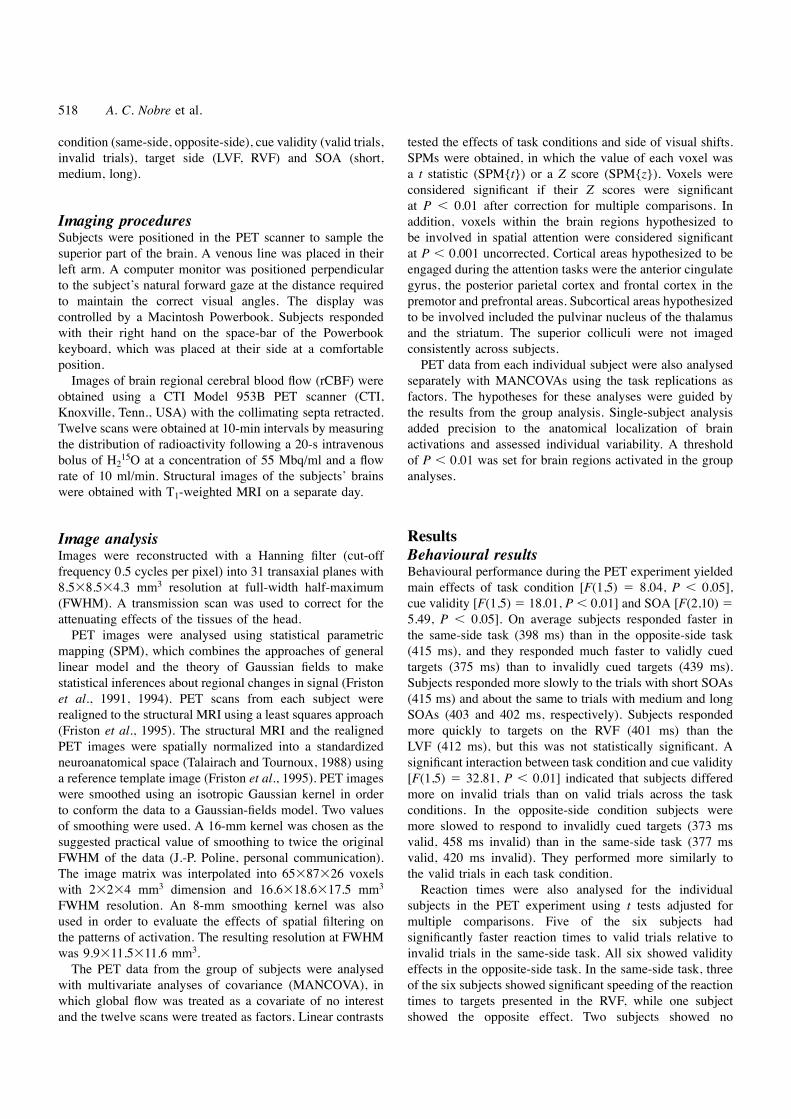
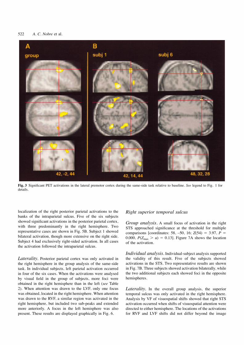
localization of the right posterior parietal activations to the Right superior temporal sulcusbanks of the intraparietal sulcus. Five of the six subjectsshowed significant activations in the posterior parietal cortex, Group analysis. A small focus of activation in the rightwith three predominantly in the right hemisphere. Two STS approached significance at the threshold for multiplerepresentative cases are shown in Fig. 5B. Subject 1 showed comparisons [coordinates: 58, –50, 16; Z(54) 5 3.97, P 5bilateral activation, though more extensive on the right side. 0.000, P(Zmax . u) 5 0.13]. Figure 7A shows the locationSubject 4 had exclusively right-sided activation. In all cases of the activation.the activation followed the intraparietal sulcus.
Individual analysis. Individual-subject analysis supportedthe validity of this result. Five of the subjects showedLaterality. Posterior parietal cortex was only activated inactivations in the STS. Two representative results are shownthe right hemisphere in the group analysis of the same-sidein Fig. 7B. Three subjects showed activation bilaterally, whiletask. In individual subjects, left parietal activation occurredthe two additional subjects each showed foci in the oppositein four of the six cases. When the activations were analysedhemispheres.by visual field in the group of subjects, more foci were
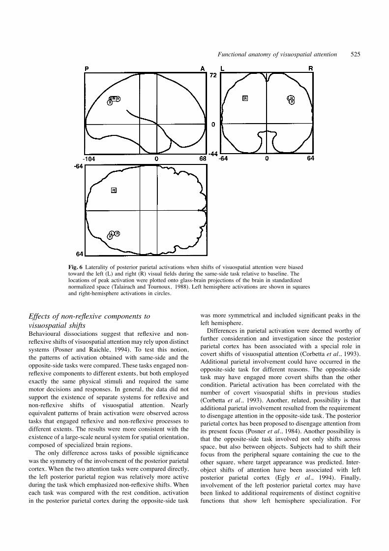
obtained in the right hemisphere than in the left (see Table2). When attention was drawn to the LVF, only one focus Laterality. In the overall group analysis, the superiorwas obtained, located in the right hemisphere. When attention temporal sulcus was only activated in the right hemisphere.was drawn to the RVF, a similar region was activated in the Analysis by VF of visuospatial shifts showed that right STSright hemisphere, but included two sub-peaks and extended activation occurred when shifts of visuospatial attention weremore anteriorly. A focus in the left hemisphere was also directed to either hemisphere. The locations of the activations
for RVF and LVF shifts did not differ beyond the imagepresent. These results are displayed graphically in Fig. 6.

Functional anatomy of visuospatial attention 523
Fig. 4 Laterality of premotor activations when shifts of visuospatial attention were biased toward theleft (L) and right (R) visual fields during the same-side task relative to baseline. The locations of peakactivation were plotted onto glass-brain projections of the brain in standardized normalized space(Talairach and Tournoux, 1988). Left hemisphere activations are shown in squares and right-hemisphereactivations in circles.
resolution. Left STS activation occurred only after shifts no areas of significantly different activation after correctionfor multiple comparisons. The left parietal cortex wasdirected toward the RVF (see Table 2).relatively more active during the opposite-side task[coordinates: –34, –76, 40; Z(54) 5 3.32, P , 0.001, P(Zmax. u) 5 0.64; and coordinates: –26, –76, 44; Z(54) 5 3.26,Subcortical regions
The only subcortical activation which tended to be significant P 5 0.001, P(Zmax . u) 5 0.70]. Since the posterior parietalcortex was hypothesized a priori to participate in visuospatialoccurred in the left thalamus [coordinates: –22, –28, 4;
Z(54) 5 4.15, P , 0.001, P(Zmax . u) 5 0.07]. According shifts, its differential activation was considered significant.to the Talairach and Tournoux atlas (1988) this activationwas located in the pulvinar nucleus. This activation wasalso observed when only shifts to the RVF were analysed Discussion[coordinates: –22, –28, 4; Z(54) 5 4.40, P , 0.001, P(Zmax Behavioural consequences of visuospatial shifts. u) 5 0.03]. No significant activation of the pulvinar was A network of brain regions was engaged by tasks involvingobtained during shifts to the LVF. Foci in the striatum and shifts of visuospatial attention. The tasks differed mainly incerebellum were less significant, and did not survive the their spatial cueing properties. In one case, an attention-statistical thresholds. grabbing stimulus predicted the subsequent appearance of a
target stimulus to be discriminated at that same location (i.e.same-side task) with 80% probability. This type of task has
Activations during opposite-side task been used widely to probe reflexive shifts of attention, sincethe cueing stimulus primes its location in space. However,Cortical structures
The cortical pattern of activation obtained during opposite- nothing prevents subjects from developing controlledstrategies or expectancies based upon the predictiveside task was very similar to that obtained during same-side
task. Significant cortical activations were obtained in medial information carried in the cue stimulus. The second taskrequired non-reflexive attentional processes. In the opposite-frontal cortex, premotor cortex and posterior parietal cortex.
A direct contrast between the two active conditions revealed side task, the appearance of a peripheral cue predicted the

524 A. C. Nobre et al.
Fig. 5 Significant PET activations in the posterior parietal cortex during the same-side task relative to baseline. See legend to Fig. 1 fordetails.
subsequent appearance of the target in the symmetrical supported by the significant interaction obtained betweentask condition and cue validity. Subjects were relativelylocation in the opposite visual field. Controlled shifts were
required and reflexive shifts might have needed to be slower on invalid trials in the opposite-side task, suggestingan additional cost of remaining at, or returning to, theinhibited or overridden.
The ability of the tasks to drive covert shifts of visuo- invalid location.spatial attention was demonstrated by the significantbehavioural benefits of valid cues in both tasks. Subjects
PET activations during peripheral shifts ofwere significantly faster in response to targets that occurredin the predicted location in space. The highly statistically visuospatial attention
The network of regions imaged by PET in the main same-significant validity effects replicated the well-establishedadvantage that spatial cueing confers to behaviour. In both side task relative to the rest control included all the cortical
regions hypothesized to form the core of the network fortasks, subjects were additionally speeded following inter-stimulus intervals .150 ms. The added advantage conferred visuospatial attention proposed by Mesulam (1990). Analysis
of the group results included foci of activation in the rightby longer-latency intervals has been interpreted as evidencefor the contribution of a controlled, non-reflexive process to posterior parietal cortex, the right anterior cingulate, and in
the lateral and medial premotor cortex bilaterally. In additionattentional systems (Posner and Snyder, 1975; Neely, 1977).In this light, both tasks can be interpreted to have combined to the cortical areas that were predicted a priori, a small
focus of activation tended toward significance in the rightreflexive and non-reflexive processes. Overall, reaction timeswere faster for targets in the same-side task during the PET superior temporal sulcus.
During the most sensitive portion of the PET scans, theexperiment, suggesting that additional controlled aspects ofattention may have operated in the opposite-side task. Perhaps tasks were biased to contain only valid trials. Cognitive
processes linked to invalid trials, such as the breachingthe requirement to inhibit or override reflexive aspects duringvisuospatial shifts contralateral to an attention-grabbing or updating of expectations probably did not contribute
substantially to the pattern of brain activation observed.stimulus delayed cognitive processing. This possibility was
Functional anatomy of visuospatial attention 525
Fig. 6 Laterality of posterior parietal activations when shifts of visuospatial attention were biasedtoward the left (L) and right (R) visual fields during the same-side task relative to baseline. Thelocations of peak activation were plotted onto glass-brain projections of the brain in standardizednormalized space (Talairach and Tournoux, 1988). Left hemisphere activations are shown in squaresand right-hemisphere activations in circles.
was more symmetrical and included significant peaks in theEffects of non-reflexive components toleft hemisphere.visuospatial shiftsDifferences in parietal activation were deemed worthy ofBehavioural dissociations suggest that reflexive and non-
further consideration and investigation since the posteriorreflexive shifts of visuospatial attention may rely upon distinctparietal cortex has been associated with a special role insystems (Posner and Raichle, 1994). To test this notion, covert shifts of visuospatial attention (Corbetta et al., 1993).the patterns of activation obtained with same-side and the Additional parietal involvement could have occurred in theopposite-side tasks were compared. These tasks engaged non- opposite-side task for different reasons. The opposite-sidereflexive components to different extents, but both employed task may have engaged more covert shifts than the otherexactly the same physical stimuli and required the same condition. Parietal activation has been correlated with themotor decisions and responses. In general, the data did not number of covert visuospatial shifts in previous studies
support the existence of separate systems for reflexive and (Corbetta et al., 1993). Another, related, possibility is thatnon-reflexive shifts of visuospatial attention. Nearly additional parietal involvement resulted from the requirementequivalent patterns of brain activation were observed across to disengage attention in the opposite-side task. The posteriortasks that engaged reflexive and non-reflexive processes to parietal cortex has been proposed to disengage attention fromdifferent extents. The results were more consistent with the its present focus (Posner et al., 1984). Another possibility isexistence of a large-scale neural system for spatial orientation, that the opposite-side task involved not only shifts acrosscomposed of specialized brain regions. space, but also between objects. Subjects had to shift theirThe only difference across tasks of possible significance focus from the peripheral square containing the cue to the
was the symmetry of the involvement of the posterior parietal other square, where target appearance was predicted. Inter-cortex. When the two attention tasks were compared directly, object shifts of attention have been associated with leftthe left posterior parietal region was relatively more active posterior parietal cortex (Egly et al., 1994). Finally,during the task which emphasized non-reflexive shifts. When involvement of the left posterior parietal cortex may haveeach task was compared with the rest condition, activation been linked to additional requirements of distinct cognitive
functions that show left hemisphere specialization. Forin the posterior parietal cortex during the opposite-side task
526 A. C. Nobre et al.
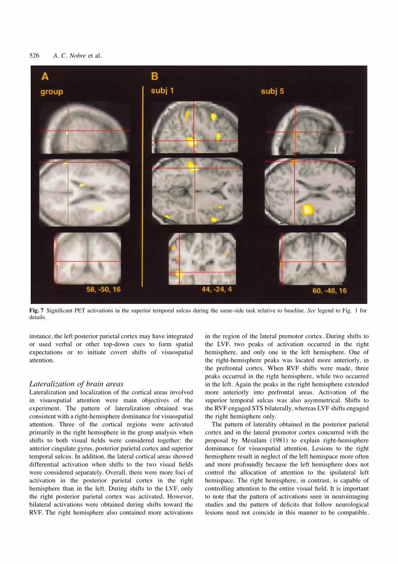
Fig. 7 Significant PET activations in the superior temporal sulcus during the same-side task relative to baseline. See legend to Fig. 1 fordetails.
instance, the left posterior parietal cortex may have integrated in the region of the lateral premotor cortex. During shifts tothe LVF, two peaks of activation occurred in the rightor used verbal or other top-down cues to form spatial
expectations or to initiate covert shifts of visuospatial hemisphere, and only one in the left hemisphere. One ofthe right-hemisphere peaks was located more anteriorly, inattention.the prefrontal cortex. When RVF shifts were made, threepeaks occurred in the right hemisphere, while two occurredin the left. Again the peaks in the right hemisphere extendedLateralization of brain areas
Lateralization and localization of the cortical areas involved more anteriorly into prefrontal areas. Activation of thesuperior temporal sulcus was also asymmetrical. Shifts toin visuospatial attention were main objectives of the
experiment. The pattern of lateralization obtained was the RVF engaged STS bilaterally, whereas LVF shifts engagedthe right hemisphere only.consistent with a right-hemisphere dominance for visuospatial
attention. Three of the cortical regions were activated The pattern of laterality obtained in the posterior parietalcortex and in the lateral premotor cortex concurred with theprimarily in the right hemisphere in the group analysis when
shifts to both visual fields were considered together: the proposal by Mesulam (1981) to explain right-hemispheredominance for visuospatial attention. Lesions to the rightanterior cingulate gyrus, posterior parietal cortex and superior
temporal sulcus. In addition, the lateral cortical areas showed hemisphere result in neglect of the left hemispace more oftenand more profoundly because the left hemisphere does notdifferential activation when shifts to the two visual fields
were considered separately. Overall, there were more foci of control the allocation of attention to the ipsilateral lefthemispace. The right hemisphere, in contrast, is capable ofactivation in the posterior parietal cortex in the right
hemisphere than in the left. During shifts to the LVF, only controlling attention to the entire visual field. It is importantto note that the pattern of activations seen in neuroimagingthe right posterior parietal cortex was activated. However,
bilateral activations were obtained during shifts toward the studies and the pattern of deficits that follow neurologicallesions need not coincide in this manner to be compatible.RVF. The right hemisphere also contained more activations

Functional anatomy of visuospatial attention 527
Neuroimaging is inherently a correlational measure, and areas in eye-movement and visuospatial-attention studies is notnegligible, and remains a point for further discussion andof activation could be observed in brain regions which are
not critical for the behaviour under study. investigation. One possibility is that processes linked todecision or execution of hand movements contributed to theThe pattern of right-hemisphere bias in activations in the
posterior parietal cortex was also consistent with previous premotor activations, since the control condition did notrequire motor responses. This possibility seems unlikelystudies. Corbetta et al. (1993) found a similar pattern of
posterior parietal laterality. Bilateral activations occurred for given the low frequency of the responses made (Jenkinset al., 1995). Final assessment of the contribution of handshifts to the RVF, but only right-hemisphere activations
occurred for shifts to the LVF. However, in their study, the and eye-movement control to the premotor activations mustawait further investigations in which motor decisions, handfrontal activations were not biased to the right hemisphere,
but contralateral to the visual field of the stimuli. In the movements and eye movements are manipulated directly insingle subjects.present study, the lateral premotor activation was bilateral,
but tended to be more extensive in the right hemisphere, If the activations in premotor areas represent engagementof frontal eye fields, it is worthy of note that the tasks weredespite the fact that subjects used the right hand to respond.
Similar predominant right-hemisphere involvement was covert in nature. Eye monitoring during a separate sessionindicated that only few trials were contaminated by saccades.observed by Gitelman et al. (1996a) in a spatial exploratory
motor task, despite the use of the right hand for exploration. The involvement of the frontal eye fields in a task of covertattention is not obvious. It is possible that some activity inthe frontal eye field is independent of the motor executionof eye movements, but still sensitive to attentional shifts orLocalization of brain areasexploration. Alternatively, the frontal eye field could beengaged automatically by activity in other regions of thePrimary sensory and motor areas. Areas linked to
visual processing or to motor output were not conspicuous attentional network, as if primed to elicit eye movements.The activation could represent such priming, or the activein the results, despite the passive nature of the rest control
condition. Motor responses occurred at an average rate of inhibition of activity. Imagery of eye movements could alsohave contributed.one every 4 s. This may have been too infrequent to observe
the engagement of the primary motor areas (Jenkins et al., Frontal eye field activation did not differ across the twoactive task conditions. To the extent that the opposite-side1994). The visual stimuli were presented very briefly as
small black line drawings over a bright white background. task engaged additional non-reflexive attentional components,these did not affect activation in the frontal eye fieldThey may not have been salient enough to activate the visual
areas significantly more than during the passive viewing of significantly. Null findings do not carry much weight, sincethe failure to detect change might have resulted fromthe background display. This finding is orthogonal to and
does not contradict studies which have shown selective insufficient statistical power or methodological limitations.Nevertheless, the findings appear consistent with the recentmodulation of visual areas by visuospatial attention
(VanVoorhis and Hillyard, 1977; Mangun, 1987; McCarthy proposal by Paus (1996) that the frontal eye field is moresensitive to oculomotor variables than to attentional or otherand Nobre, 1993; Heinze et al., 1994).cognitive variables in the tasks employed to date. However,the role of the frontal eye field in the mapping or control ofPremotor and prefrontal areas. Strong activations
were observed in lateral premotor cortex bilaterally, as well exploratory movements cannot be fully assessed with thedata available (Table 3). Most oculomotor or visuospatialas in the medial premotor cortex. The major peaks of these
activations were located in BA 6. The location of these attention tasks, including this one, have not manipulatedexploratory motor variables. One exception is the study byactivations are consistent with the location of the frontal
eye fields in the human brain as indicated by previous Gitelman et al. (1996a), which did report enhanced premotoractivation to exploratory hand movements as compared withneuroimaging studies of eye movements (Paus, 1996).
Activations during eye-movement studies in humans have non-attentional repetitive movements. The correspondencebetween premotor regions which control hand and eyeconsistently been observed in the lateral premotor cortex
(BA 6), occasionally extending posteriorly to the anterior movements, however, remains to be drawn.portion of the precentrally gyrus (BA 4) (Fox et al., 1985;Petit et al., 1993; Anderson et al., 1994; Darby et al., 1996). Anterior cingulate. The region of activation in medial
premotor cortex extended into the right anterior cingulateMedial premotor activations, consistent with those seen inthe present study, have been proposed as the location of the cortex, where a distinct local peak was obtained in BA 24.
The focus was located near the level of the anteriorsupplementary eye fields in the human brain (Petit et al.,1993; Darby et al., 1996). Figure 8 compares the locations commissure along the anterior–posterior dimension. In
individual subjects, cingulate activation was observed asof lateral premotor foci in this and previous oculomotor andvisual attention studies. separate from premotor activation. Additional foci were
occasionally observed more posteriorly and more anteriorly,The variability in the reported loci of premotor activation
528 A. C. Nobre et al.
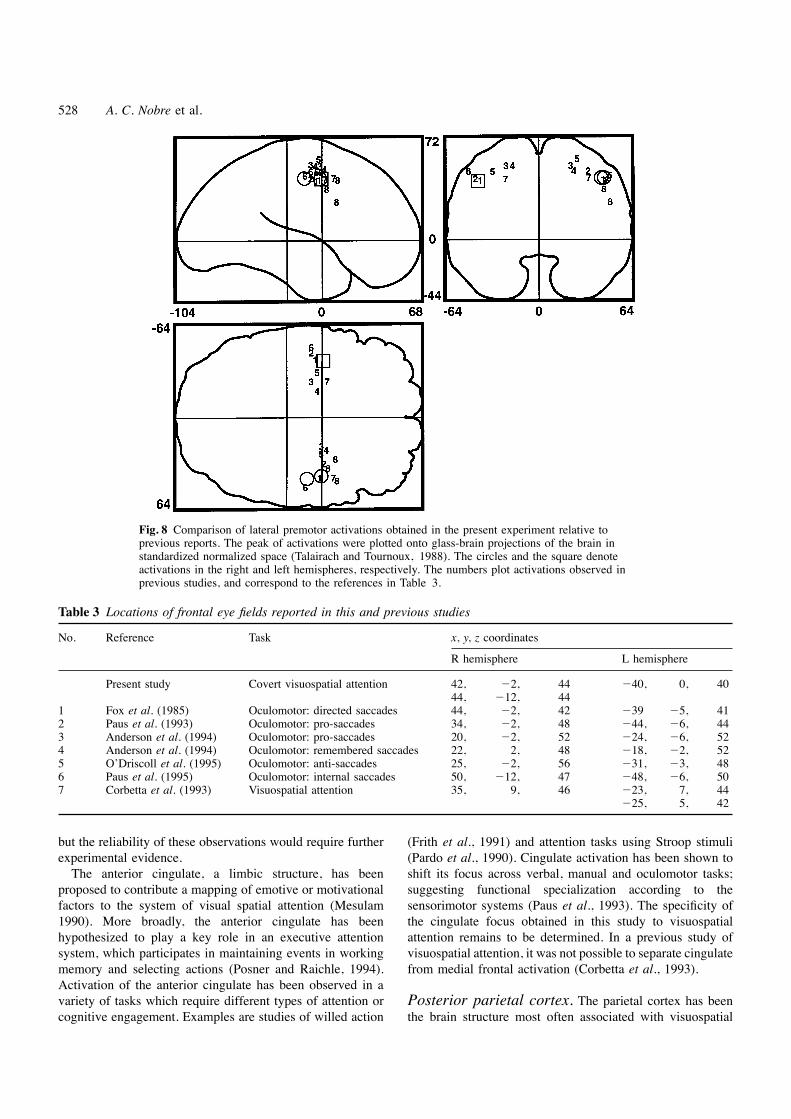
Fig. 8 Comparison of lateral premotor activations obtained in the present experiment relative toprevious reports. The peak of activations were plotted onto glass-brain projections of the brain instandardized normalized space (Talairach and Tournoux, 1988). The circles and the square denoteactivations in the right and left hemispheres, respectively. The numbers plot activations observed inprevious studies, and correspond to the references in Table 3.
Table 3 Locations of frontal eye fields reported in this and previous studies
No. Reference Task x, y, z coordinates
R hemisphere L hemisphere
Present study Covert visuospatial attention 42, 22, 44 240, 0, 4044, 212, 44
1 Fox et al. (1985) Oculomotor: directed saccades 44, 22, 42 239 25, 412 Paus et al. (1993) Oculomotor: pro-saccades 34, 22, 48 244, 26, 443 Anderson et al. (1994) Oculomotor: pro-saccades 20, 22, 52 224, 26, 524 Anderson et al. (1994) Oculomotor: remembered saccades 22, 2, 48 218, 22, 525 O’Driscoll et al. (1995) Oculomotor: anti-saccades 25, 22, 56 231, 23, 486 Paus et al. (1995) Oculomotor: internal saccades 50, 212, 47 248, 26, 507 Corbetta et al. (1993) Visuospatial attention 35, 9, 46 223, 7, 44
225, 5, 42
but the reliability of these observations would require further (Frith et al., 1991) and attention tasks using Stroop stimuli(Pardo et al., 1990). Cingulate activation has been shown toexperimental evidence.
The anterior cingulate, a limbic structure, has been shift its focus across verbal, manual and oculomotor tasks;suggesting functional specialization according to theproposed to contribute a mapping of emotive or motivational
factors to the system of visual spatial attention (Mesulam sensorimotor systems (Paus et al., 1993). The specificity ofthe cingulate focus obtained in this study to visuospatial1990). More broadly, the anterior cingulate has been
hypothesized to play a key role in an executive attention attention remains to be determined. In a previous study ofvisuospatial attention, it was not possible to separate cingulatesystem, which participates in maintaining events in working
memory and selecting actions (Posner and Raichle, 1994). from medial frontal activation (Corbetta et al., 1993).Activation of the anterior cingulate has been observed in avariety of tasks which require different types of attention or Posterior parietal cortex. The parietal cortex has been
the brain structure most often associated with visuospatialcognitive engagement. Examples are studies of willed action
Functional anatomy of visuospatial attention 529
Table 4 Locations of posterior parietal activations reported in this and previous studies
No. Reference Task x, y, z coordinates
R hemisphere L hemisphere
1 Present study Covert visuospatial attentionsame-side task minus baseline 34, 268, 36opposite-side minus same-side task 234, 276, 40
226, 276, 442 Anderson et al. (1994) Oculomotor: pro-saccades 218, 268, 364 Anderson et al. (1994) Oculomotor: remembered saccades 214, 244, 52
218, 256, 48230, 234, 40
3 Corbetta et al. (1993) Visuospatial attention 33, 245, 46 229, 251, 36227, 243, 46
4 Corbetta et al. (1995) Visual shifts 21, 261, 50 217, 259, 5823, 247, 52
5 Corbetta et al. (1995) Attention to feature conjunctions 23, 279, 46 227, 255, 5431 247, 54 231, 253, 4433, 269, 50
6 Gitelman et al. (1996a) Sensorimotor exploration 29, 244, 5230, 244, 4845, 241, 40
attention and hemispatial neglect (e.g. Mesulam, 1981; neglect and visuospatial deficits in monkeys (Heilman et al.,1970; Petrides and Iversen, 1979; Lynch and McLaren,Kinsbourne, 1987; Rafal and Robertson, 1994). The precise
region critical for visuospatial attention in the human brain, 1989). Neuronal firing is modulated by stimulus relevancein visuospatial tasks (Bushnell et al., 1981; Goldberg andhowever, had not yet been settled (Table 4). Neuroimaging
studies have implicated the superior parietal lobule (Corbetta Segraves, 1987). Area 7a appears capable of integrating eye-position and retinotopic information to form a head-centredet al., 1993, 1995; Anderson et al., 1994). However, these
studies have relied upon group analysis and have not spatial map (Zipser and Andersen, 1988; Barash et al., 1991).Area 7a is interconnected with higher-order areas in theincluded structural brain-imaging of the individuals studied.
The human posterior parietal cortex is highly variable and cingulate gyrus, superior temporal sulcus and dorsolateralprefrontal cortex (Mesulam et al., 1977; Andersen et al.,asymmetrical in surface anatomy (Witelson and Kigar, 1992).
Therefore, analyses relying upon average activations in 1985b; Selemon and Goldman Rakic, 1988). Area LIP inmonkeys also appears to be involved in visuospatial functions,groups of subjects and standardized anatomical atlases may be
misleading. Results from neuropsychological and behavioural perhaps more closely related to the planning and control ofeye movements. Area LIP contains neurons that respond inneurological studies have implicated the inferior parietal
lobule, in the area of the temporal parietal junction (Rafal association with eye movements (Andersen et al., 1985a)and stimulation of the area can generate saccades (Shibutaniand Robertson, 1995). Localization based upon lesions in
the human brain, however, can also be misleading because et al., 1984). LIP also has strong interconnections with thefrontal eye fields (Gnadt and Andersen, 1988).of the variable relationship between the lesions and the
functional anatomical boundaries, and because lesions can In the present study, the location of the posterior parietalactivation across the group of subjects was difficult todisconnect functional regions instead of damaging the regions
per se. interpret relative to Brodmann’s areas. Using the Talairachand Tournoux atlas as a guide, the activation appeared toThe correspondence between the areas in the posterior
parietal cortex of monkeys and humans has not been straddle BAs 7, 39 and 40. Figure 9 compares the locationsof posterior parietal activations in this and previous oculo-straightforward (see Andersen, 1989). Understanding these
relationships would greatly help clarify the specializations of motor and visual attention studies.Given the high degree of variability in the anatomicalthe posterior parietal areas in the human brain. Two areas in
the posterior parietal lobe of monkeys have been linked to surface features in this region, localization relied uponanalysis of data from single subjects. The most parsimoniousvisuospatial attention and oculomotor functions: area 7a and
an area in the lateral bank of the intraparietal sulcus (LIP). description of the site of posterior parietal activation is thatit followed the intraparietal sulcus. The inherent resolutionThe homologues of both of these regions are likely to
participate in visual attention functions in humans and to of the PET images did not permit a finer grain description,such as resolving which bank of the sulcus was primarilycause aspects of neglect when damaged by brain lesions.
Area 7a in the inferior parietal lobule in monkeys has been engaged. These activations are likely to have reflected activityin the human homologues of both areas LIP and 7a in thelinked to visuospatial attention by lesion and neuro-
physiological studies. Lesions to area 7a result in hemispatial monkey. More precise localization within the intraparietal

530 A. C. Nobre et al.
Fig. 9 Comparison of posterior parietal activations obtained in the present experiment relative toprevious reports. The peak of activations were plotted onto glass-brain projections of the brain instandardized normalized space (Talairach and Tournoux, 1988). Activation in the same-side task relativeto baseline was plotted as a square, and activation in the opposite-side task as squares. The numbersplot the activations observed in previous studies, and correspond to the references in Table 4.
sulcus and further fractionation of the functional properties Subcortical structures. In general, the subcortical activa-tions were less reliable than cortical activations, and wereof this region may have to be evaluated using fMRI, in
which repeated studies with higher spatial resolution can be difficult to pinpoint anatomically. The only subcortical regionperformed in individuals (Gitelman et al., 1996b, c; Nobre activated significantly during this experiment was the pulvinaret al., 1996a, b). nucleus of the thalamus, suggested to partake in visuospatial
attention (Petersen et al., 1985). Some subcortical regionsSuperior temporal sulcus. A region of the right superior thought to be involved in visuospatial attention were nottemporal sulcus tended to be activated by visuospatial shifts imaged consistently because of the limited size of the arrayof attention. Technically, this region did not meet the criteria of PET detectors. This was the case with the superior colliculiestablished for significance. However, it was also observed and with brainstem structures of the reticular activatingin the majority of the individual analyses. The involvement system. The involvement of these regions in the present task,of the STS in a task of visuospatial shifts of attention was therefore, could not be evaluated.not surprising. The STS in monkeys is polysensory (Bruceet al., 1981; Hikosaka et al., 1988) and some neurons arelinked to saccades and smooth pursuit (Dursteler et al., 1987).
Concluding remarksThe STS is strongly interconnected to other regions in theThe present experiment has helped resolve longstandingspatial attentional network, such as the posterior parietalquestions about the anatomical loci of brain regions involvedcortex, the frontal eye fields and the pulvinar nucleus of thein visuospatial attention. The results support the existence ofthalamus (Barbas and Mesulam, 1981; Cavada and Golmana large-scale neural system for visuospatial orientation. TheRakic, 1989; Stanton et al., 1989; Seltzer and Pandya, 1989,cortical regions involved displayed a right-hemispheric bias1994). Lesions to the STS in the monkey have also beenin their layout. Future studies that vary systematically thereported to result in neglect (Watson et al., 1994). Itsrelevant factors for attention should build upon the presentinvolvement in visuospatial attention in humans and its rolefindings to provide understanding of the regional functionalwithin the spatial attentional network remains to be validated
and investigated further. specializations of the attention system in the human brain.

Functional anatomy of visuospatial attention 531
References Mesulam MM, et al. Qualitative mapping of cerebral blood flowand functional localization with echo-planar MR imaging and signalAndersen RA. Visual and eye movement functions of the posterior
parietal cortex. [Review]. Annu Rev Neurosci 1989; 12: 377–403. targeting with alternating radio frequency. Radiology 1994; 192:513–20.Andersen RA, Essick GK, Siegel RM. Encoding of spatial location
by posterior parietal neurons. Science 1985a, 230:456–8. Egly R, Driver J, Rafal RD. Shifting visual attention betweenobjects and locations: evidence from normal and parietal lesionAndersen RA, Asanuma C, Cowan WM. Callosal and prefrontal subjects. J Exp Psychol Gen 1994; 123: 161–77.associational projecting cell populations in area 7a of the macaque
monkey: a study using retrogradely transported fluorescent dyes. Fox PT, Fox JM, Raichle ME, Burde RM. The role of cerebralcortex in the generation of voluntary saccades: a positron emissionJ Comp Neurol 1985b; 232:443–55.tomographic study. J Neurophysiol 1985; 54: 348–69.Anderson TJ, Jenkins IH, Brooks DJ, Hawken MB Frackowiak RS,
Kennard C, et al. Cortical control of saccades and fixation in man: Friston KJ, Frith CD, Liddle PF, Frackowiak RS. Comparingfunctional (PET) images: the assessment of significant change. Ja PET study. Brain 1994; 117: 1073–84.Cereb Blood Flow Metab 1991; 11: 690–9.Barash S, Bracewell RM, Fogassi L, Gnadt JW, Andersen RA.
Saccade-related activity in the lateral intraparietal area: I. Temporal Friston KJ, Worsley KJ, Frackowiak RSJ, Mazziotta JC, Evans AC.Assessing the significance of focal activations using their spatialproperties: comparison with area 7a. J Neurophysiol 1991; 66:
1095–108. extent. Hum Brain Map 1994; 1: 210–20.
Friston KJ, Holmes AP, Worsley KJ, Poline J-P, Frith CD, Frackow-Barbas H, Mesulam MM. Organization of afferent input to sub-divisions of area 8 in the rhesus monkey. J Comp Neurol 1981; iak RSJ. Statistical parametric maps in functional imaging: a general
linear approach. Hum Brain Map 1995; 2: 189–210.200: 407–31.
Brodmann K. Vergleichende Lokalisationslehre der Grosshirnrinde Frith CD, Friston K, Liddle PF, Frackowiak RS. Willed action andthe prefrontal cortex in man: a study with PET. Proc R Soc Londin ihren Prinzipien dargestellt auf Grund des Zellenbaues. Leipzig:
J.A.Barth, 1909. B Biol Sci 1991; 244: 241–6.
Gitelman DR, Alpert NM, Kosslyn S, Daffner K, Scinto L,Bruce CJ, Goldberg ME. Primate frontal eye fields: I. Single neuronsdischarging before saccades. J Neurophysiol 1985; 53: 603–35. Thompson W, et al. Functional imaging of human right hemispheric
activtion for exploratory movements. Ann Neurol 1996a; 39: 174–9.Bruce C, Desimone R, Gross CG. Visual properties of neurons ina polysensory area in superior temporal sulcus of the macaque. J Gitelman DR, Nobre AC, Meyer JR, Parrish TB, Callahan C,
Russell J, Mesulam MM. Functional magnetic resonance imagingNeurophysiol 1981; 46: 369–84.of covert spataial attention. International Conference on FunctionalBushnell MC, GoldbergME, Robinson DL. Behavioral enhancement Mapping of the Human Brain. Neuroimage 1996b; 3: 5180.of visual responses in monkey cerebral cortex: 1. Modulation in
posterior parietal cortex related to selective visual attention. J Gitelman DR, Nobre AC, Meyer JR, Parrish TB, Callahan C,Russell J, et al. Soc Neurosci Abstr 1996c; 22: 1692.Neurophysiol 1981; 46: 755–72.
Cavada C, Goldman-Rakic PS. Posterior parietal cortex in rhesus Gnadt JW, Andersen RA. Memory related motor planning activityin posterior parietal cortex of macaque. Exp Brain Res 1988; 70:monkey: II. Evidence for segregated cortico-cortical networks link-
ing sensory and limbic areas with the frontal lobe. J Comp Neurol 216–20.1989; 287: 422–45. Goldberg ME, Segraves MA Visuospatial and motor attention in
the monkey. Neuropsychologia 1987; 25: 107–18.Corbetta M, Miezin FM, Shulman GL, Petersen SE. A PET studyof visuospatial attention. J Neurosci 1993; 13: 1202–26. Goodman SJ. Visuo-motor reaction times and brain stem multiple-
unit activity. Exp Neurol 1968; 22: 367–78.Corbetta M, Shulman GL, Miezin FM, Petersen SE. Superiorparietal cortex activation during spatial attention shifts and visual Heilman KM, Van Den Abell T. Right hemisphere dominance forfeature conjunction. 1995; 270: 802–5. attention: the mechanism underlying hemispheric asymmetries of
inattention (neglect). Neurology 1980; 30: 327–30.Darby DD, Nobre AC, Thangaraj V, Edelman R, Mesulam MM,Warach S. Cortical activation in the human brain during lateral Heilman KM, Pandya DN, Geschwind N. Trimodal inattentionsaccades using EPISTAR functional magnetic resonance imaging. following parietal lobe ablations. Trans Am Neurol Assoc 1970;Neuroimage 1996; 3: 53–62. 95: 258–61.Deiber M-P, Passingham RE, Colebatch JG, Friston KJ, Nixon PD, Heinze HJ, Mangun GR, Burchert W, Hinrichs H, Scholz M, MuenteFrackowiak RS. Cortical areas and the selection of movements: a TF, et al. Combined spatial and temporal imaging of brain activitystudy with positron emission tomography. Exp Brain Res 1991; 84: during visual selective attention in humans. Nature 1994; 372: 543–6.393–402.
Hikosaka K, Iwai E, Saito H, Tanaka K. Polysensory properties ofDursteler MR, Wurtz RH, Newsome WT. Directional pursuit deficits neurons in the anterior bank of the caudal superior temporal sulcusfollowing lesions of the foveal representation within the superior of the macaque monkey. J Neurophysiol 1988; 60: 1615–37.temporal sulcus of the macaque monkey. J Neurophysiol 1987; 57:1262–87. James W. The principles of psychology. London: Macmillan, 1890.
Kennard MA. Alterations in response to visual stimuli followingEdelman RR, Siewert B, Darby DG, Thangaraj V, Nobre AC,

532 A. C. Nobre et al.
lesions of frontal lobe in monkeys. Arch Neurol Psychiatry 1939; for sustained attention by positron emission tomography. Nature1991; 349: 61–4.41: 1153–65.
Kinsbourne M. Mechanisms of unilateral neglect. In: Jeannerod M, Paus T. Location and function of the human frontal eye-field: aselective review. Neuropsychologia 1996; 34: 475–83.editor. Neurophysiological and neuropsychological aspects of spatial
neglect. Amsterdam: Elsevier, 1987: 69–86. Paus T, Petrides M, Evans AC, Meyer E. Role of the human anteriorcingulate cortex in the control of oculomotor, manual, and speechLatto R, Cowey A. Visual field defects after frontal eye-field lesions
in monkeys. Brain Res 1971; 30: 1–24. responses: a positron emission tomography study. J Neurophysiol1993; 70: 453–69.Lynch JC, McLaren JW. Deficits of visual attention and saccadic
eye movements after lesions of parietooccipital cortex in monkeys. Petersen S, Robinson D, Keys W. Pulvinar nuclei of the behavingrhesus monkey: visual responses and their modulation. J Neuro-J Neurophysiol 1989; 61: 74–90.physiol 1985; 54: 867–86.Mangun GR, Hillyard SA. The spatial allocation of visual attention
as indexed by event-related brain potentials. Hum Factors 1987; Petersen SE, Fox PT, Posner MI, Mintun M, Raichle ME. Positronemission tomographic studies of the cortical anatomy of single-29: 195–211.word processing. Nature 1988; 331: 585–9.McCarthy G, Nobre AC. Modulation of semantic processing by
spatial selective attention. Electroencephalogr Clin Neurophysiol Petit L, Orssaud C, Tzourio N, Salamon G, Mazoyer B, Berthoz A.PET study of voluntary saccadic eye movements in humans: basal1993; 88: 210–9.ganglia-thalamocortical system and cingulate cortex involvement. JMcCarthy G. Functional neuroimaging of memory. 1995; 1: 155–63. Neurophysiol 1993; 69: 1009–17.
Melamed E, Larsen B. Cortical activation pattern during saccadic Petrides M, Iversen SD. Restricted posterior parietal lesions in theeye movements in humans: localization by focal cerebral blood rhesus monkey and performance on visuospatial tasks. Brain Resflow increases. Ann Neurol 1979; 5: 79–88. 1979; 161: 63–77.Mesulam M-M. A cortical network for directed attention and Petrides M, pandya DN. Projections to the frontal cortex from theunilateral neglect. [Review]. Ann Neurol 1981; 10: 309–25. posterior parietal region in the rhesus monkey. J Comp Neurol
1984; 228: 105–16MesulamM-M. Large-scale neurocognitive networks and distributedprocessing for attention, language, and memory. [Review]. Ann Pierrot-Deseilligny C, Rivaud S, Gaymard B, Muri R, VermerschNeurol 1990; 28: 597–613. AI. Cortical control of saccades. [Review]. Ann Neurol 1995; 37:
557–67.Mesulam M-M, Van Hoesen GW, Pandya DN, Geschwind N.Limbic and sensory connections of the inferior parietal lobule (area Plum F, Posner JB. The diagnosis of stupor and coma. ContempPG) in the rhesus monkey: a study with a newmethod for horseradish Neurol Ser 1972; 10: 1–286.peroxidase histochemistry. Brain Res 1977; 136: 393–414.
Posner M, Raichle M. Images of Mind. New York: ScientificMitz A, Zeffiro S, Wise S. Premotor cortex and the selection of American Library, 1994.actions [abstract]. Soc Neurosci Abstr 1993; 19; 1209.Posner M, Snyder S Facilitation and inhibition in the processing ofNeely JH. Semantic priming and retrieval from lexical memory: signals. In: Rabbitt PMA, Dornic S editors. Attention and perform-roles of inhibitionless spreading activation and limited-capacity ance V. New York: Academic Press, 1975; 669–82.attention. 1977; 106: 226–254.Posner MI, Snyder CRR. Attention and cognitive control. In:Nobre AC, Gitelman DR, Sebestyen GN, Meyer J, Frackowiak RSJ, Solso RL, editor. Information processing and cognition: the LoyolaFrith CD, et al. Functional localisation for the neural system of symposium. Hillside (NJ): Erlbaum, 1975.visual spatial attention using PET and fMRI. 2nd Meeting of
European Neuroscience, Strasbourg. European J Neurosci 1996a; Posner MI, Cohen Y, Rafal RD. Neural systems control of spatialorienting. Philos Trans R Soc Lond B Biol Sci 1982; 298: 187–98.Suppl 9: 122.
Nobre AC, Gitelman DR, Sebestyen GN, Meyer J, Frackowiak RSJ, Posner MI, Walker JA, Friedrich FJ, Rafal RD. Effects of parietalinjury on covert orienting of attention. J Neurosci 1984; 4: 1863–74.Frith CD, et al. Cortical network for visuospatial attention imaged
using PET and fMRI [abstract]. Soc Neurosci Abstr 1996b; 22: 1692. Rafal R, Robertson L. The neurology of visual attention. In:Gazzaniga MS, editor. The cognitive neurosciences. CambridgeO’Driscoll GA, Alpert NM, Matthysse SW, Levy DL, Rauch SL,
Holzman PS. Functional neuroanatomy of antisaccade eye movments (MA): MIT Press, 1995: 625–48.investigated with positron emission tomography. Proc Natl Acad Ray CL, Mirsky AF, Pragay EB. Functional analysis of attention-Sci USA 1995; 92: 925–9. related unit activity in the reticular formation of the monkey. Exp
Neurol 1982; 77: 544–62.Oldfield RC. The assessment and analysis of handedness: theEdinburgh inventory. Neuropsychologia 1971; 9: 97–113. Scheibel ME, Scheibel AB. Anatomical basis of attention mechan-
isms in vertebrate brains. In: Quarton GC, Melnechuk T, SchmittPardo JV, Pardo PJ, Janer KW, Raichle ME. The anterior cingulatecortex mediates processing selection in the Stroop attentional conflict FO editors. The neurosciences. New York: Rockefeller University
Press, 1967: 577–602.paradigm. Proc Natl Acad Sci USA 1990; 87: 256–9.
Pardo JV, Fox PT, Raichle ME. Localization of a human system Schiller PH, True SD, Conway JL. Deficits in eye movements

Functional anatomy of visuospatial attention 533
following frontal eye-field and superior colliculus ablations. J Talairach J, Tournoux P. Co-planar stereotaxic atlas of the humanbrain. Stuttgart: Thieme, 1988.Neurophysiol 1980; 44: 1175–89.
Van Voorhis S, Hillyard SA. Visual evoked potentials and selectiveSebestyen GN, Nobre AC. Temporal correlates of covert shifts ofattention to points in space. Percept Psychophys 1977; 22: 54–62.visual spatial attention using event-related potentials. 2nd Meeting
of European Neuroscience, Strasbourg. European J Neurosci 1996; Watson RT, Valenstein E, Day A, Heilman KM. Posterior neocorticalSuppl 9: 123. systems subserving awareness and neglect. Neglect associated with
superior temporal sulcus but not area 7 lesions. Arch Neurol 1994;Segraves MA, Goldberg ME. Functional properties of corticotectal51:1014–21.neurons in the monkey’s frontal eye field. J Neurophysiol 1987; 58:
1387–419. Weintraub S, Mesulam M-M. Right cerebral dominance in spatialattention. Further evidence based on ipsilateral neglect. Arch NeurolSelemon LD, Goldman-Rakic PS. Common cortical and subcortical1987; 44: 621–625.targets of the dorsolateral prefrontal and posterior parietal cortices
in the rhesus monkey: evidence for a distributed neural network Welch K, Stuteville P. Experimental production of unilateral neglectsubserving spatially guided behavior. J Neurosci 1988; 8: 4049–68. in monkeys. Brain 1958; 81: 341–7.
Witelson SF, Kigar DL. Sylvian fissure morphology and asymmetrySeltzer B, Pandya DN. Converging visual and somatic sensoryin men and women: bilateral differences in relation to handednesscortical input to the intraparietal sulcus of the rhesus monkey. Brainin men. J Comp Neurol 1992; 323: 326–40.Res 1980; 192: 339–51.
Yeterian EH, Van Hoesen GW. Cortico-striate projections in theSeltzer B, Pandya DN. Parietal, temporal, and occipital projectionsrhesus monkey: the organization of certain cortico-caudate connec-to cortex of the superior temporal sulcus in the rhesus monkey: ations. Brain Res 1978; 139: 43–63.retrograde tracer study. J Comp Neurol 1994; 343: 445–63.Zipser D, Andersen RA. A back-propagation programmed networkShibutani H, Sakata H, Hyvarinen J. Saccade and blinking evokedthat simulates response properties of a subset of posterior parietalby microstimulation of the posterior parietal association cortex ofneurons. Nature 1988; 331: 679–84.the monkey. Exp Brain Res 1984; 55: 1–8.
Stanton GB, Deng S-Y, Goldberg ME, McMullen NT. Cytoarchitec-tural characteristics of the frontal eye fields in macaque monkeys. Received July 4, 1996. Revised October 2, 1996.
Accepted November 5, 1996J Comp Neurol 1989; 282: 415–27.


![Molecular Imaging of Murine Intestinal Inflammation With 2-Deoxy-2-[ 18F]Fluoro- d-Glucose and Positron Emission Tomography](https://static.fdokumen.com/doc/165x107/6344fff26cfb3d4064097a1a/molecular-imaging-of-murine-intestinal-inflammation-with-2-deoxy-2-18ffluoro-.jpg)


![Altered Brain Serotonin 5HT1A Receptor Binding After Recovery From Anorexia Nervosa Measured by Positron Emission Tomography and [Carbonyl11C]WAY100635](https://static.fdokumen.com/doc/165x107/6316dca13ed465f0570c3ef2/altered-brain-serotonin-5ht1a-receptor-binding-after-recovery-from-anorexia-nervosa.jpg)


