
Synthesis and Evaluation of Radioligands for Imaging Brain Nociceptin/Orphanin FQ Peptide (NOP)...
14
Published: March 25, 2011 r2011 American Chemical Society 2687 dx.doi.org/10.1021/jm101487v | J. Med. Chem. 2011, 54, 2687–2700 ARTICLE pubs.acs.org/jmc Synthesis and Evaluation of Radioligands for Imaging Brain Nociceptin/Orphanin FQ Peptide (NOP) Receptors with Positron Emission Tomography Victor W. Pike,* ,† Karen S. Rash, ‡ Zhaogen Chen, ‡ Concepci on Pedregal, § Michael A. Statnick, ‡ Yasuyuki Kimura, † Jinsoo Hong, † Sami S. Zoghbi, † Masahiro Fujita, † Miguel A. Toledo, § Nuria Diaz, § Susan L. Gackenheimer, ‡ Johannes T. Tauscher, ‡ Vanessa N. Barth, ‡ and Robert B. Innis † † Molecular Imaging Branch, National Institute of Mental Health, National Institutes of Health, Building 10, Room B3 C346A, 10 Center Drive, Bethesda, Maryland 20892, United States ‡ Eli Lilly & Co., Lilly Research Laboratories, Indianapolis, Indiana 46285, United States § Lilly S.A., Avenida de la Industria 30, 28108-Alcobendas, Madrid, Spain b S Supporting Information ABSTRACT: Positron emission tomography (PET) coupled to an effective radioligand could provide an important tool for understanding possible links between neuropsychiatric disorders and brain NOP (nociceptin/orphanin FQ peptide) receptors. We sought to develop such a PET radioligand. High-affinity NOP ligands were synthesized based on a 3-(2 0 -fluoro-4 0 ,5 0 -dihydrospiro[piperidine- 4,7 0 -thieno[2,3-c]pyran]-1-yl)-2(2-halobenzyl)-N-alkylpropanamide scaffold and from experimental screens in rats, with ex vivo LC-MS/MS measures, three ligands were identified for labeling with carbon-11 and evaluation with PET in monkey. Each ligand was labeled by 11 C-methylation of an N-desmethyl precursor and studied in monkey under baseline and NOP receptor-preblock conditions. The three radioligands, [ 11 C](S)-10ac, gave similar results. Baseline scans showed high entry of radioactivity into the brain to give a distribution reflecting that expected for NOP receptors. Preblock experiments showed high early peak levels of brain radioactivity, which rapidly declined to a much lower level than seen in baseline scans, thereby indicating a high level of receptor- specific binding in baseline experiments. Overall, [ 11 C](S)-10c showed the most favorable receptor-specific signal and kinetics and is now selected for evaluation in human subjects. ’ INTRODUCTION Nociceptin, also known as orphanin FQ, is the most recently discovered peptide member of the endogenous opioid family. 1,2 Nociceptin acts on specific G-protein coupled receptors that are linked to the cAMP cascade and to voltage-gated Ca 2þ and K þ channels. 3 These receptors were formerly known as ORL-1 (opioid receptor-like receptor-1) and are now called NOP (nociceptin opioid peptide) receptors. They are present in various organs, including the brain, and have roles in many normal physiological responses such as cognition, locomotion, and neuroendocrine control. 3 Additionally, many neuropsychia- tric disorders are possibly linked to the NOP receptor, including pain, anxiety, depression, anorexia, obesity, and drug abuse. 4,5 Accordingly, the brain NOP receptor is a target not only for biomedical investigation but also for drug therapies. 4 Positron emission tomography (PET), 6 in tandem with selective radioactive probes (radioligands), is a molecular ima- ging technique with a unique capability for investigating the living human brain and in particular for investigating specific proteins as possible players in pathophysiology 7 or as targets for therapeutic interventions. 8 Effective PET radioligands now exist for a wide range of targets, selected from among brain enzymes, transporters, plaque, ion channels, and neurotransmitter receptors. 9 Received: November 19, 2010
-
Upload
independent -
Category
Documents
-
view
0 -
download
0
Transcript of Synthesis and Evaluation of Radioligands for Imaging Brain Nociceptin/Orphanin FQ Peptide (NOP)...

Published: March 25, 2011
r 2011 American Chemical Society 2687 dx.doi.org/10.1021/jm101487v | J. Med. Chem. 2011, 54, 2687–2700
ARTICLE
pubs.acs.org/jmc
Synthesis and Evaluation of Radioligands for Imaging BrainNociceptin/Orphanin FQ Peptide (NOP) Receptors with PositronEmission TomographyVictor W. Pike,*,† Karen S. Rash,‡ Zhaogen Chen,‡ Concepci�on Pedregal,§ Michael A. Statnick,‡
Yasuyuki Kimura,† Jinsoo Hong,† Sami S. Zoghbi,† Masahiro Fujita,† Miguel A. Toledo,§ Nuria Diaz,§
Susan L. Gackenheimer,‡ Johannes T. Tauscher,‡ Vanessa N. Barth,‡ and Robert B. Innis†
†Molecular Imaging Branch, National Institute of Mental Health, National Institutes of Health, Building 10, Room B3 C346A, 10 CenterDrive, Bethesda, Maryland 20892, United States‡Eli Lilly & Co., Lilly Research Laboratories, Indianapolis, Indiana 46285, United States§Lilly S.A., Avenida de la Industria 30, 28108-Alcobendas, Madrid, Spain
bS Supporting Information
ABSTRACT:
Positron emission tomography (PET) coupled to an effective radioligand could provide an important tool for understandingpossible links between neuropsychiatric disorders and brain NOP (nociceptin/orphanin FQ peptide) receptors. We sought todevelop such a PET radioligand. High-affinity NOP ligands were synthesized based on a 3-(20-fluoro-40,50-dihydrospiro[piperidine-4,70-thieno[2,3-c]pyran]-1-yl)-2(2-halobenzyl)-N-alkylpropanamide scaffold and from experimental screens in rats, with ex vivoLC-MS/MSmeasures, three ligands were identified for labeling with carbon-11 and evaluationwith PET inmonkey. Each ligandwaslabeled by 11C-methylation of an N-desmethyl precursor and studied in monkey under baseline and NOP receptor-preblockconditions. The three radioligands, [11C](S)-10a�c, gave similar results. Baseline scans showed high entry of radioactivity into thebrain to give a distribution reflecting that expected for NOP receptors. Preblock experiments showed high early peak levels of brainradioactivity, which rapidly declined to a much lower level than seen in baseline scans, thereby indicating a high level of receptor-specific binding in baseline experiments. Overall, [11C](S)-10c showed the most favorable receptor-specific signal and kinetics andis now selected for evaluation in human subjects.
’ INTRODUCTION
Nociceptin, also known as orphanin FQ, is the most recentlydiscovered peptide member of the endogenous opioid family.1,2
Nociceptin acts on specific G-protein coupled receptors that arelinked to the cAMP cascade and to voltage-gated Ca2þ and Kþ
channels.3 These receptors were formerly known as ORL-1(opioid receptor-like receptor-1) and are now called NOP(nociceptin opioid peptide) receptors. They are present invarious organs, including the brain, and have roles in manynormal physiological responses such as cognition, locomotion,and neuroendocrine control.3 Additionally, many neuropsychia-tric disorders are possibly linked to the NOP receptor, includingpain, anxiety, depression, anorexia, obesity, and drug abuse.4,5
Accordingly, the brain NOP receptor is a target not only forbiomedical investigation but also for drug therapies.4
Positron emission tomography (PET),6 in tandem withselective radioactive probes (radioligands), is a molecular ima-ging technique with a unique capability for investigating theliving human brain and in particular for investigating specificproteins as possible players in pathophysiology7 or as targets fortherapeutic interventions.8 Effective PET radioligands now existfor a wide range of targets, selected from among brain enzymes,transporters, plaque, ion channels, and neurotransmitter receptors.9
Received: November 19, 2010
2688 dx.doi.org/10.1021/jm101487v |J. Med. Chem. 2011, 54, 2687–2700
Journal of Medicinal Chemistry ARTICLE
Even so, many specific molecular targets of potential interest,including the NOP receptor, still lack a specific radioligand forinvestigation with PET. PET coupled to an effective NOPreceptor radioligand could be an important tool for furtheringthe understanding of neuropsychiatric disorders in which NOPreceptors are implicated and also for the development of newdrugs that may target NOP receptors in the treatment of suchdisorders. We therefore sought to develop a radioligand forimaging brain NOP receptors with PET.
Effective PET radioligands for imaging in brain must displayan array of favorable attributes, including amenability to labelingwith a short-lived positron-emitter such as carbon-11 (t1/2 =20.4 min) or fluorine-18 (t1/2 = 109.7 min), ability to cross theblood�brain barrier, high affinity and selectivity for binding tothe target protein, relatively low nonspecific binding, and absenceof troublesome brain-penetrant radiometabolites.10�13 The dif-ficult challenge ofmeeting such a combination of requirements inany proposed PET radioligand is a major reason for the currentprevalence of nonimageable protein targets. A further importantconsideration is that the target protein should be present inadequate concentration for potential imaging. As a guidelinethe product of receptor density (Bmax, nM) and ligand affinity(1/KD, nM
�1) should exceed a value of 5 in target-rich regionsof brain.
The distribution of NOP receptors in rat brain has beenstudied by autoradiography.14�17 These studies show a wide-spread distribution ofNOP receptors with, for example, relativelyhigh levels in cortex, hypothalamus, and amygdala and low levelin striatum. The density of NOP receptors in rat brain mem-branes was estimated at 237 fmol/mg protein (roughly equatingto 24 nM in whole brain).17 Data on the distribution and densityof NOP receptors in the brains of higher species are sparse exceptfor a single study on the macaque central nervous system.18 Thisstudy found a distribution of NOP quite similar to that in rat butwith some definite differences. Receptor densities of >6 fmol/mg(equating to >6 nM in whole brain) were found in receptor-richregions such as cortex, caudate, and amygdala. Thus, prospectivePET radioligands for brain NOP receptors should have highaffinity, with a Kd value in the low nanomolar range.
The aforementioned autoradiographic studies were performedwith peptide-like radioligands which would not be expected toenter the brain freely. Intense effort has been expended overrecent years to discover nonpeptide small molecule ligands withhigh affinity as potential NOP receptor-targetted therapeutics.19
A published example arising from this effort is SB-612111 (1;[(�)-cis-1-methyl-7-[[4-(2,6-dichlorophenyl)piperidin-1-yl]methyl]-6,7,8,9-tetrahydro-5H-benzocyclohepten-5-ol) (Chart 1).20,21
From our own medicinal chemistry effort, we discovered a newseries of high-affinity small molecule NOP receptor ligands,
based on a 3-(20-fluoro-40,50-dihydrospiro[piperidine-4,70-thieno[2,3-c]pyran]-1-yl)-2(2-halobenzyl)-N-alkylpropanamidescaffold, which we disclose here. In this study, we explored thischemotype for the development of a PET radioligand for theNOP receptor, initially through screening of nonradioactiveligands in rat with ex vivo LC-MS/MS measures22,23 and thenthrough evaluation of selected 11C-labeled ligands in rhesusmonkey with PET. A promising NOP radioligand, [11C](S)-10c, was found, which now merits further evaluation in humansubjects.
’RESULTS AND DISCUSSION
Chemistry. The syntheses of the potential NOP ligands areoutlined in Scheme 1. The spiro[4,5-dihydrothieno[2,3-c]pyran-7,40-piperidine] 2 was synthesized from 2-(3-thienyl)ethanol bytreatment of t-butylcarbonyl-protected (Boc-protected) piper-idin-4-one in the presence of trifluoroacetic acid (TFA) in goodyield. After the piperidine nitrogen was protected with Boc, fluorinewas selectively introduced at the C2 position of the thienopyr-anyl ring via metalation with n-BuLi and subsequent reactionwith the fluorination reagent N-fluoro-N-(phenylsulfonyl)-benzenesulfonamide (NFSI) in 40�50% yield. Michael additionbetween the piperidine intermediate 5 and t-butyl acrylate undermild conditions gave the common intermediate propanoate 6 in65% yield. R-Alkylation of propanoate 6 with either 2-fluoro-benzyl bromide or 2-chloro-benzyl bromide was achieved withLiN(TMS)2 as base. Ester hydrolysis of compounds 7a and 7b,followed by conventional amide formation with the appropriateamine afforded the final amides 9a�c and 10a�c, which wereeach separated into enantiomers with chiral HPLC. The biolo-gically active enantiomer of 9c was determined by single-crystalX-ray crystallography to have S-configuration (see SupportingInformation). The R-enantiomer of 9c has markedly loweraffinity (Table 1). The biologically less active enantiomer of 9awas also determined by single-crystal X-ray crystallography tohave R-configuration (see Supporting Information). We there-fore reasoned that the higher affinity enantiomers of the NOPligands 9a, 9b, and 10a�c also have S-configurationTo assess the stereochemical stability of the chiral center in the
new ligands, as well as explore possible future conditions ofradiolabeling with [11C]methyl iodide, compound (S)-9a wastreated with sodium hydride in dimethyl sulfoxide (DMSO)solution at ambient temperature and then with methyl iodide.Chiral HPLC showed that this gave a single enantiomer of theN-methylated product 10a, plus unchanged starting material, (S)-9a. Therefore, these basic alkylation conditions do not causesignificant racemization and the obtained product also has S-configuration (i.e., the product is (S)-10a).Ligand Lipophilicity. Consideration of ligand lipophilicity is
important in the development of PET radioligands. Moderatelipophilicity is usually sought to promote measurable plasma freefraction (fP) and adequate brain entry, avoid excessive nonspe-cific binding to brain tissue, and avoid the generation of lipophilicbrain-penetrant radiometabolites.10,13,24 The lipophilicity (log D)of (S)-10c was measured to be 3.41 by a radiometric methodand 3.50 by a shake-flask method (Table 1). This lipophilicity iswithin a range that is considered favorable for achieving adequatebrain entry of a PET radioligand from blood without incurringexcessive nonspecific binding to brain tissue.10,13,24 For ligands10a�c, the calculated lipophilicities (cLogD values at pH 7.4)were only slightly lower than those measured, so indicating that
Chart 1. Structure of 1

2689 dx.doi.org/10.1021/jm101487v |J. Med. Chem. 2011, 54, 2687–2700
Journal of Medicinal Chemistry ARTICLE
Scheme 1. Synthesis of NOP Ligands 9a�c and 10a�c and Their Enantiomers
Reagents and conditions: (a) (1) TFA/CH2Cl2, rt, (2) 10%NaOH aq; (b) di-t-butyl dicarbonate, TEA, CH2Cl2; (c) n-BuLi,�78 �C,NFSI, THF; (d) 4M HCl in dioxane, CH2Cl2, rt; (e) t-butyl acrylate, TEA,THF, reflux; (f) (1) LiN(TMS)2, 1 M THF, 1,3-dimethyl-tetrahydropyrimidin-2(1H)-one,THF,�78 �C, (2) 2-F-benzyl bromide or 2-Cl-benzyl bromide; (g) TFA/CH2Cl2, rt; (h) EDCI, HOBt, DIPEA, CH2Cl2, NH4Cl or appropriate amine;(i) chiral HPLC separation.
Table 1. Receptor Binding Affinities, Antagonist Activities, and Lipophilicities of NOP Ligands
ligand
NOP receptor
bindinga Ki (nM)
NOP antagonist
activitya Kb (nM) cLogDb
1 0.253( 0.151 0.258( 0.129 4.98
(S)-9a 0.231( 0.050 0.228( 0.050 4.08
(R)-9a 4.51( 1.05 5.64 ( 1.05 4.08
(S)-9b 0.101( 0.029 0.110( 0.021 3.41
(R)-9b 17.1 ( 2.54 7.36( 0.019 3.41
(S)-9c 0.106( 0.049 0.173( 0.084 2.95
(R)-9c 5.61( 2.74 8.65 ( 5.19 2.95
(S)-10a 0.137( 0.048 0.141( 0.032 4.39
(R)-10a 10.2( 2.85 15.8( 3.48 4.39, (4.47)c
(S)-10b 0.090( 0.016 0.099( 0.048 3.68
(R)-10b 9.08( 1.04 5.79( 1.05 3.68, (3.90)c
(S)-10c 0.150( 0.062 0.069( 0.015* 3.27, (3.50)c, (3.41( 0.07, n = 6)d
(R)-10c 5.32( 1.03 7.92( 1.05 3.27aAssays were performed in triplicate except for that with an asterisk (n = 2). Data are mean ( SE. [3H]Nociceptin was used as reference NOPradioligand in a binding assay using human recombinant NOP receptors expressed in CHO cells. Antagonist activity was determined in an assay ofreceptor-mediated G-protein activation using [35S]GTPγS and membranes expressing cloned human NOP receptors. See Materials and Methods forassay details. bComputed with Pallas software. Values in parentheses are measured values. c log Dmeasured with nonradioactive compound and shake-flask method. d log D value (mean ( SD) measured with [11C](S)-10c.
2690 dx.doi.org/10.1021/jm101487v |J. Med. Chem. 2011, 54, 2687–2700
Journal of Medicinal Chemistry ARTICLE
the computation on this type of structure was quite accurate. Asexpected, from the extra presence of an iso-propyl group, thecLogD andmeasured logD values of ligand 10awere appreciablyhigher than those of 10c, as where those of 10b in which a fluorosubstituent of 10c is replaced with a chloro substituent. Thecorresponding desmethyl compounds were computed to haveless lipophilicity, as would also be expected.Ligand Pharmacology. Ligands (S)-10a�c were found to
have high binding affinity to human recombinant NOP receptorsas represented by sub-nM Ki values in a binding assay with[3H]nociceptin as reference NOP radioligand (Table 1). In afunctional assay of receptor-mediated G-protein activation with[35S]GTPγS, these ligands were each found to be potentantagonists at NOP receptors with sub-nM Kb values (Table 1).Thus, (S)-10a�c meet the high-affinity criterion previouslymentioned for prospective NOP PET radioligands. The R-enantio-mers were found to have substantially lower binding affinities andantagonist activities. Eudismic ratios ranged from 35 to 115.The enantiomers of the corresponding N-desmethyl ligands,9a�c, showed a similar pattern of binding affinity andantagonist activity and displayed eudismic ratios ranging from20 to 169 (Table 1).Studies in Rats. Initially, we applied our previously published
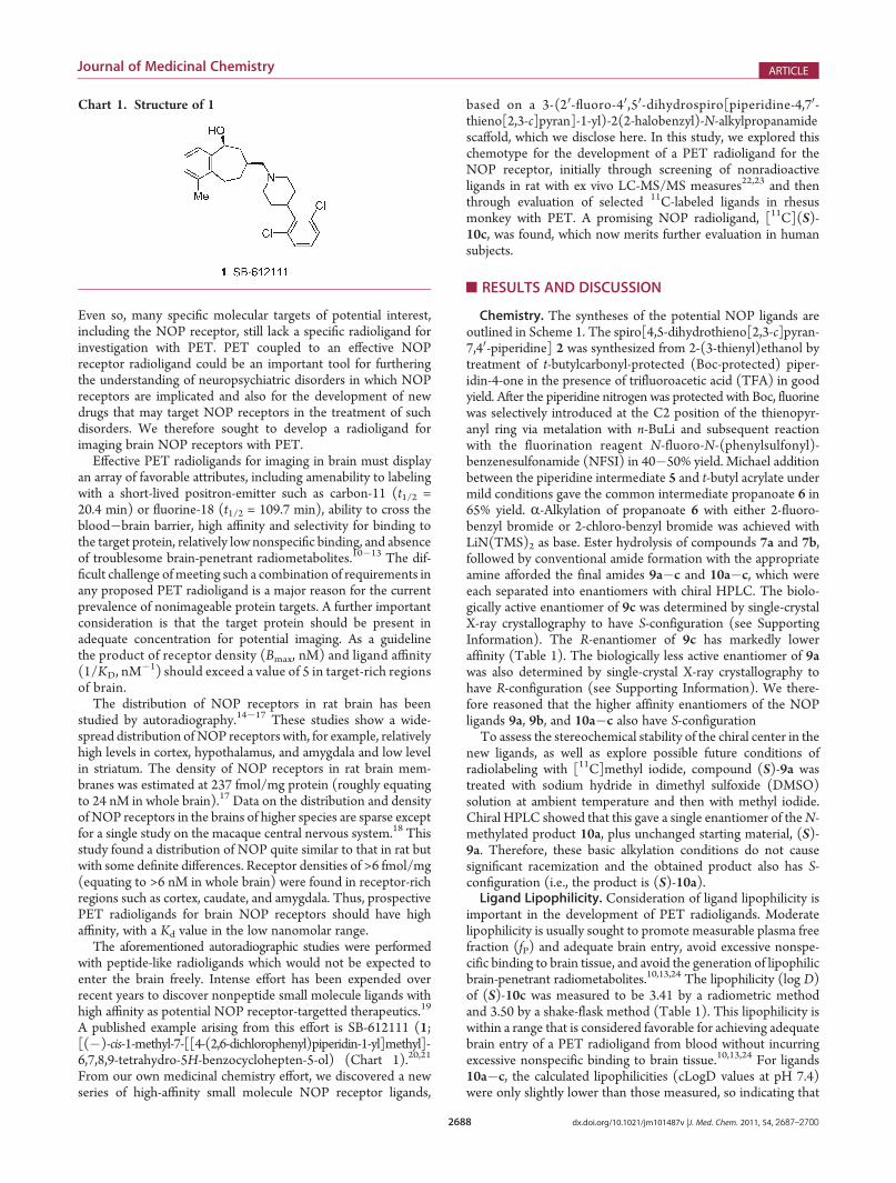
in vivo/ex vivo LC-MS/MSmethod to characterize NOP ligandsas potential tracers in rats because this method avoids the need touse radiolabels.22,23 Essentially, a low amount of nonradioactiveligand was injected intravenously into a group of rats, and after aset time the animals were sacrificed. Brain tissue regions,corresponding to high NOP and low NOP receptor expression,namely hypothalamus and striatum, respectively,15,16 were dis-sected out and their concentrations of ligand measured with LC-MS/MS. As a result, we identified three high-affinity ligands, (S)-10a�c, with potential for labeling to produce effective PETradioligands for brain NOP receptors. After intravenous admin-istration into nonanesthetized rat, all three ligands showed quitefast kinetics with rapid and high rat brain uptake and washoutoccurring within 1 h, a time-span consistent with the prospectiveuse of short-lived carbon-11 as a radiolabel (Figure 1). Eachligand showed high brain uptake with ligand (S)-10a havingslightly higher uptake (453% SUV at 10 min) than (S)-10b or(S)-10c (each ∼280% SUV). All three ligands robustly demon-strated about 3-fold higher uptake in NOP-rich hypothalamusthan in NOP-poor striatum by 40 min after injection, therebyindicating a sizable amount of NOP receptor-specific binding.Ligand (S)-10b had the largest ratio of NOP receptor-specific tononspecific binding (i.e., hypothalamus/striatum ligand concen-tration ratio minus 1 at 40 min), with a value of 3.5, whereas
ligands (S)-10a and (S)-10c had ratios of 2.7 and 2.3, respec-tively. These ratios are in fact underestimates of specific tononspecific binding in hypothalamus because rat striatum isnot entirely devoid of NOP receptors.15,16
Radioligand Syntheses. Ligands (S)-10a�c and (R)-10cwere rapidly labeled with carbon-11 by treating their respectiveN-desmethyl precursors, (S)-9a�c and (R)-9c, with [11C]methyliodide in the presence of KOH as base (Scheme 2). The radi-oligands were isolated with reverse phase HPLC in usefulactivities (mean yields for n = 3 were 47, 75, and 85 mCi for[11C](S)-10a�c, respectively), and in high radiochemicalpurities (>99%) and specific radioactivities (mean values forn = 3 were, 7.8, 5.1, 3.6 Ci/μmol at end of synthesis for [11C](S)-10a�c, respectively). Chiral HPLC analyses of the radioligandsverified almost complete absence of racemization in the labelingprocedure (see Supporting Information for chiral HPLC analysisof [11C](S)-10c). Each radioligand was readily formulated inethanol (10% v/v)/saline for intravenous administration intomonkey. Each was radiochemically stable in formulationmedium. The radiosyntheses times were about 45 min fromthe end of radionuclide production.PET Imaging in Monkeys. After intravenous administration
into monkey, each high-affinity radioligand, [11C](S)-10a�c,showed similar brain radioactivity kinetics, characterized by rapidhigh uptake into brain followed by progressive washout ofradioactivity from all examined regions over the duration of thescanning session (either 90 or 120 min) (Figure 2). Cerebellumshowed highest initial uptake and fastest washout of radioactivityin each case, possibly due to greater regional blood flow in thisregion of brain during anesthesia. The rank order of initial radio-activity uptake was [11C](S)-10c > [11C](S)-10b > [11C](S)-10a.[11C](S)-10c also gave the greatest separation of time�activity
Figure 1. Time-courses of normalized ligand concentrations (%SUV) in rat hypothalamus (4) and striatum (O) after iv injection of nonradioactiveNOP ligand (S)-10a, (S)-10b or (S)-10c (3 μg/kg, iv), measured ex vivo with LC-MS/MS. Error bars are SD (where not visible they are within thesymbol size).
Scheme 2. Radiolabeling of NOP Ligands

2691 dx.doi.org/10.1021/jm101487v |J. Med. Chem. 2011, 54, 2687–2700
Journal of Medicinal Chemistry ARTICLE
curves across brain regions. Compound 1 is a highly potent(Table 1) and selective antagonist for NOP receptors, showingmore than 1000-fold lower affinity for classical opioid receptors.21 Incorresponding experiments in which brain NOP receptors werepreblocked by administration of 1, brain radioactivity kineticswere altered in a consistent manner (Figure 2). Thus, in eachmonkey, maximal initial radioactivity uptake was again high butwas followed by a much faster washout of radioactivity from allexamined brain regions than in the corresponding baseline scansand with very little separation in the time�activity curves fornoncerebellar regions. Such differences in kinetics imply thepresence of a significant proportion of NOP receptor-specificbinding of the radioligand in the baseline experiments. Majordifferences were also clearly apparent in the PET imagesobtained under baseline and preblock conditions. Thus, thebaseline experiments showing a regional distribution of radio-activity reflecting the distribution of NOP receptors expectedfrom the autoradiographic study in macaque brain,18 with, forexample, high uptake in caudate and relatively low uptake inhypothalamus, the reverse of relative levels in rat.15,16 The preblockexperiments in monkey showed low and uniform distributions of
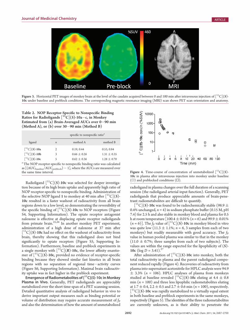
radioactivity consistent with nonspecific binding of radioligandand/or radiometabolites. These image differences are exempli-fied in Figure 3 for [11C](S)-10c.Comparison of the areas-under-the-curves (AUCs) between
baseline and preblock PET experiments provided further evi-dence for an appreciable proportion of NOP receptor-specificbinding under baseline conditions. Thus, for each radioligand,the brain-averaged AUCs (%SUV � min) between 0 and 90minor between 30 and 90 min were appreciably greater in the baselineexperiments than in the preblock experiments. From these data,crude estimates of ratios of NOP receptor-specific binding to non-specific binding were derived as [(AUCbaseline/AUCpreblock)� 1]for the time intervals 0�90 and 30�90 min after radioligandinjection. These estimates (Table 2) confirm that each radio-ligand produced a high proportion of NOP receptor-specificbinding in baseline PET scans, with radioligands [11C](S)-10band [11C](S)-10c showing appreciably higher proportionsthan [11C](S)-10a. (A full biomathematical compartmentalanalysis of monkey PET scans to derive accurate bindingpotentials is beyond the scope of this report and will bepublished subsequently.)
Figure 2. Brain region time�activity curves inmonkey after injection of radioligands [11C](S)-10a�c in baseline and in preblock experiments, in whichNOP preblocker 1 (2 mg/kg, iv) was given at 10 min before radioligand. For each radioligand, the baseline and preblock experiments were conductedabout 3 h apart in the same monkey. Key: hypothalamus (O), cerebellum (]), caudate (r), amygdala (4), lateral temporal cortex (0). Time�activitycurves for other examined regions as listed in the experimental were generally similar to the noncerebellar curves.
2692 dx.doi.org/10.1021/jm101487v |J. Med. Chem. 2011, 54, 2687–2700
Journal of Medicinal Chemistry ARTICLE
Radioligand [11C](S)-10c was selected for deeper investiga-tion because of its high brain uptake and apparently high ratio ofNOP receptor-specific to nonspecific binding. Administration ofthe selective NOP ligand 1 to monkey at 40 min after [11C](S)-10c resulted in a faster washout of radioactivity from all brainregions down to a low level, so demonstrating the reversibility ofthe specific binding of [11C](S)-10c to NOP receptors (FigureS4, Supporting Information). The opiate receptor antagonistnaloxone is effective at displacing opiate receptor radioligandsfrom primate brain.25,26 In another monkey PET experiment,administration of a high dose of naloxone at 37 min after[11C](S)-10c had no effect on the washout of radioactivity frombrain, thereby showing that this radioligand does not bindsignificantly to opiate receptors (Figure S5, Supporting In-formation). Furthermore, baseline and preblock experiments ina single monkey with [11C](R)-10c, the lower affinity enantio-mer of [11C](S)-10c, provided no evidence of receptor-specificbinding because they showed similar fast kinetics in all brainregions with no separation of regional time�activity curves(Figure S6, Supporting Information). Maximal brain radioactiv-ity uptake was in fact higher in the preblock experiment.Emergence of Radiometabolites of [11C](S)-10c inMonkey
Plasma in Vivo. Generally, PET radioligands are appreciablymetabolized over the short time-span of a PET scanning session.Detailed quantitative analysis of radioligand behavior in vivo toderive important output measures such as binding potential orvolume of distribution may require accurate measurement of fPand also the determination of how the amount of unmetabolized
radioligand in plasma changes over the full duration of a scanningsession (the radioligand arterial input function). Generally, PETradioligands that produce appreciable amounts of brain-pene-trant radiometabolites are difficult to quantify.[11C](S)-10c was found to be radiochemically stable (98.9 (
0.4% unchanged, n = 4) in sodium phosphate buffer (0.15 M, pH7.4) for 2.5 h and also stable in monkey blood and plasma for 0.5h at room temperature [100.4( 0.01% (n = 6) and 99.9( 0.01%(n = 6)]. The fP value of [
11C](S)-10c in monkey blood in vitrowas quite low (11.3 ( 1.1%; n = 6, 3 samples from each of twomonkeys) but readily measurable with good accuracy. The fPvalue in human pooled plasma was similar to that in the monkey(11.0 ( 0.7%; three samples from each of two subjects). Thevalues are within the range expected for the lipophilicity of (S)-10c (log D = 3.41).27
After administration of [11C](S)-10c into monkey, both thetotal radioactivity in plasma and the parent radioligand compo-nent reduced rapidly (Figure 4). Recoveries of radioactivity fromplasma into supernatant acetonitrile for HPLC analysis were 94.9( 3.3% (n = 106). HPLC analyses of plasma from monkeysstudied at baseline revealed [11C](S)-10c eluting at 4.4 ( 0.8min (n = 100) and three less lipophilic radiometabolites elutingat 1.7( 0.4, 2.2( 0.5 and 2.7( 0.6 min (n = 100), respectively.[11C](S)-10c was rapidly metabolized to a virtually equal extentin both baseline and preblock experiments in the same monkeys,respectively (Figure 5). The identities of the three radiometabolitesare currently unknown, as is their ability to penetrate the
Figure 3. Horizontal PET images of monkey brain at the level of the caudate acquired between 0 and 100 min after intravenous injection of [11C](S)-10c under baseline and preblock conditions. The corresponding magnetic resonance imaging (MRI) scan shows PET scan orientation and anatomy.
Table 2. NOP Receptor-Specific to Nonspecific BindingRatios for Radioligands [11C](S)-10a�c, in MonkeyEstimated from (a) Brain-Averaged AUCs over 0�90 min(Method A), or (b) over 30�90 min (Method B)
specific to nonspecific ratioa
ligand n method A method B
[11C](S)-10a 2 0.19, 0.44 0.53, 0.84
[11C](S)-10b 3 0.66 ( 0.26 1.31 ( 0.35
[11C](S)-10c 5 0.62 ( 0.36 1.28 ( 0.70aThe NOP receptor-specific to nonspecific binding ratio was calculatedas [(AUCbaseline/AUCpreblock)� 1], where the AUCs are measured overthe same time interval. Figure 4. Time-course of concentration of unmetabolized [11C](S)-
10c in plasma after intravenous injection into monkey under baseline(O) and preblocked conditions (0).
2693 dx.doi.org/10.1021/jm101487v |J. Med. Chem. 2011, 54, 2687–2700
Journal of Medicinal Chemistry ARTICLE
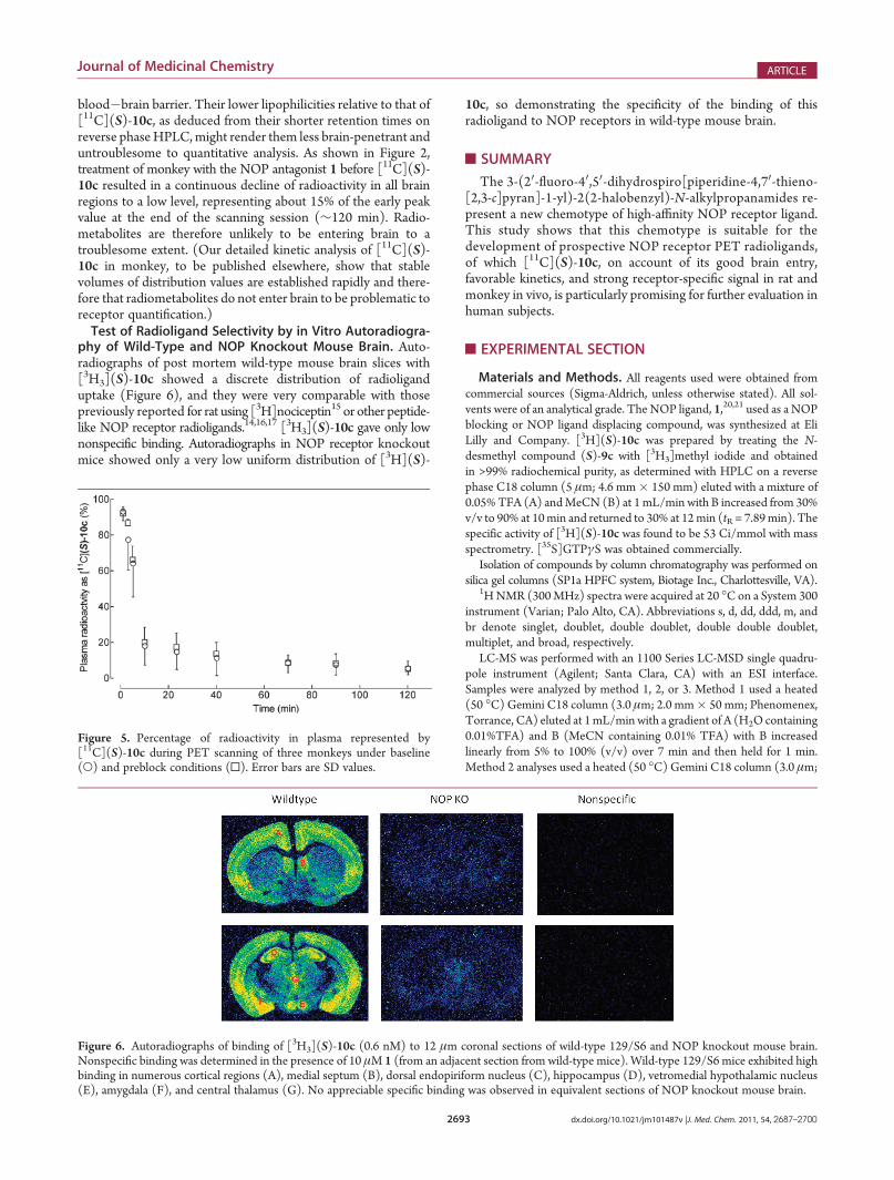
blood�brain barrier. Their lower lipophilicities relative to that of[11C](S)-10c, as deduced from their shorter retention times onreverse phaseHPLC,might render them less brain-penetrant anduntroublesome to quantitative analysis. As shown in Figure 2,treatment of monkey with the NOP antagonist 1 before [11C](S)-10c resulted in a continuous decline of radioactivity in all brainregions to a low level, representing about 15% of the early peakvalue at the end of the scanning session (∼120 min). Radio-metabolites are therefore unlikely to be entering brain to atroublesome extent. (Our detailed kinetic analysis of [11C](S)-10c in monkey, to be published elsewhere, show that stablevolumes of distribution values are established rapidly and there-fore that radiometabolites do not enter brain to be problematic toreceptor quantification.)Test of Radioligand Selectivity by in Vitro Autoradiogra-
phy of Wild-Type and NOP Knockout Mouse Brain. Auto-radiographs of post mortem wild-type mouse brain slices with[3H3](S)-10c showed a discrete distribution of radioliganduptake (Figure 6), and they were very comparable with thosepreviously reported for rat using [3H]nociceptin15 or other peptide-like NOP receptor radioligands.14,16,17 [3H3](S)-10c gave only lownonspecific binding. Autoradiographs in NOP receptor knockoutmice showed only a very low uniform distribution of [3H](S)-
10c, so demonstrating the specificity of the binding of thisradioligand to NOP receptors in wild-type mouse brain.
’SUMMARY
The 3-(20-fluoro-40,50-dihydrospiro[piperidine-4,70-thieno-[2,3-c]pyran]-1-yl)-2(2-halobenzyl)-N-alkylpropanamides re-present a new chemotype of high-affinity NOP receptor ligand.This study shows that this chemotype is suitable for thedevelopment of prospective NOP receptor PET radioligands,of which [11C](S)-10c, on account of its good brain entry,favorable kinetics, and strong receptor-specific signal in rat andmonkey in vivo, is particularly promising for further evaluation inhuman subjects.
’EXPERIMENTAL SECTION
Materials and Methods. All reagents used were obtained fromcommercial sources (Sigma-Aldrich, unless otherwise stated). All sol-vents were of an analytical grade. The NOP ligand, 1,20,21 used as a NOPblocking or NOP ligand displacing compound, was synthesized at EliLilly and Company. [3H](S)-10c was prepared by treating the N-desmethyl compound (S)-9c with [3H3]methyl iodide and obtainedin >99% radiochemical purity, as determined with HPLC on a reversephase C18 column (5 μm; 4.6 mm� 150 mm) eluted with a mixture of0.05% TFA (A) andMeCN (B) at 1mL/min with B increased from 30%v/v to 90% at 10min and returned to 30% at 12min (tR = 7.89min). Thespecific activity of [3H](S)-10c was found to be 53 Ci/mmol with massspectrometry. [35S]GTPγS was obtained commercially.
Isolation of compounds by column chromatography was performed onsilica gel columns (SP1a HPFC system, Biotage Inc., Charlottesville, VA).
1HNMR (300MHz) spectra were acquired at 20 �C on a System 300instrument (Varian; Palo Alto, CA). Abbreviations s, d, dd, ddd, m, andbr denote singlet, doublet, double doublet, double double doublet,multiplet, and broad, respectively.
LC-MS was performed with an 1100 Series LC-MSD single quadru-pole instrument (Agilent; Santa Clara, CA) with an ESI interface.Samples were analyzed by method 1, 2, or 3. Method 1 used a heated(50 �C) Gemini C18 column (3.0 μm; 2.0 mm� 50 mm; Phenomenex,Torrance, CA) eluted at 1mL/min with a gradient of A (H2O containing0.01%TFA) and B (MeCN containing 0.01% TFA) with B increasedlinearly from 5% to 100% (v/v) over 7 min and then held for 1 min.Method 2 analyses used a heated (50 �C) Gemini C18 column (3.0 μm;
Figure 6. Autoradiographs of binding of [3H3](S)-10c (0.6 nM) to 12 μm coronal sections of wild-type 129/S6 and NOP knockout mouse brain.Nonspecific binding was determined in the presence of 10 μM 1 (from an adjacent section from wild-type mice). Wild-type 129/S6 mice exhibited highbinding in numerous cortical regions (A), medial septum (B), dorsal endopiriform nucleus (C), hippocampus (D), vetromedial hypothalamic nucleus(E), amygdala (F), and central thalamus (G). No appreciable specific binding was observed in equivalent sections of NOP knockout mouse brain.
Figure 5. Percentage of radioactivity in plasma represented by[11C](S)-10c during PET scanning of three monkeys under baseline(O) and preblock conditions (0). Error bars are SD values.

2694 dx.doi.org/10.1021/jm101487v |J. Med. Chem. 2011, 54, 2687–2700
Journal of Medicinal Chemistry ARTICLE
2.0 mm � 50 mm) eluted at 1 mL/min with a gradient of A (aqNH4HCO3; 10 mM) and B (MeCN), with B increased linearly from10% to 100% (v/v) over 7 min and then held for 1 min. Method 3 usedan XBridge C18 column (3.5 μm; 2.1 mm � 50 mm; Waters, Milford,MA) eluted at 1 mL/min with a gradient of A (aq NH4HCO3; 10 mM,pH 9) and B (MeCN), with B increased linearly from 10% to 100%(v/v) over 7 min and then held for 1 min. Ions betweenm/z 80 and 800were captured after electrospray ionization of the eluted test sample. Thepurities of synthesized ligands were all found to beg95%, by methods 1,2, or 3 as specified later.
High-resolution mass spectrometry (HRMS) was performed on anTOF/Q-TOF instrument (Agilent). Data were acquired in dual ESImode (þ and�) in the range of 95�1700 amu. The sample (∼0.1 mg/mL in MeCN/H2O, 1:1 v/v; 2 μL) was injected in loop mode onto aZorbax SB-C8 column (3.5 μm; 3.0 mm � 150 mm; Agilent) eluted at0.5 mL/min with a mixture of 0.1% formic acid in water (A) and 0.1%formic acid in MeCN (B) with B increasing from 5% to 100% over15 min and then held at 100% for 8 min. Eluate was monitored with adiode array detector operating in the range 190�700 nm.
Melting points (mp) were determined on a DSC Q100 V9.8instrument (Universal) with a ramp of 5 �C/min up to 300 �C, andwere uncorrected.
Optical rotations were measured with a 341 polarimeter (Perkin-Elmer) at 20 �C and at 589 nm (sodium lamp).
γ-Radioactivity from carbon-11 was measured using a calibrated dosecalibrator (Atomlab 300; Biodex Medical Systems; Shirley, NY). Radio-activity measurements were corrected for physical decay. All radio-chemistry with carbon-11 was performed in a lead-shielded hot-cell forpersonnel protection from radiation.Statistics. Values are given as means( the standard deviation of the
mean, unless otherwise stated.Animals. Male Sprague�Dawley rats (Harlan Sprague�Dawley,
Indianapolis, IN), weighing between 230 and 280 g, were used for in vivoexperiments. All rats were housed in rooms using a 12 h light/dark cycleand had ad libitum access to normal rat chow and water until thebeginning of the 3 h experimental protocol. All experiments with ratswere performed in accord with the National Research Council Guideunder protocols approved by the Animal Care andUse Committee of EliLilly and Company.
All rhesus (Macaca mulatta) monkeys were handled in accordancewith the Guide for the Care and Use of Laboratory Animals28 and theNational Institute of Health Animal Care and Use Committee.Chemistry. Spiro[4,5-dihydrothieno[2,3-c]pyran-7,40-piperidine]
(2). 2-(Thiophen-3-yl)ethanol (15.4 g, 120 mmol) was added to asolution of N-t-butoxycarbonyl-4-piperidone (20 g, 100 mmol) inCH2Cl2 (200 mL) at room temperature (rt). TFA (38 mL) was addeddropwise to the mixture over 30 min, while the internal temperature waskept between 23 and 30 �C. The mixture was then stirred at rt for 20 h.Solvent was then evaporated off, and the residue was poured into NaOH(1M; 200 mL). The mixture was stirred for 15 min and the pH adjustedto about 11 with 50% aq NaOH. Then the mixture was extracted withCH2Cl2. The organic layers were separated, dried over Na2SO4, andevaporated. The crude residue was purified on silica gel (CH2Cl2/MeOH; 20:1 v/v) to afford 19.9 g (95%) of 2 as a solid. LC-MS (ESI)m/z 210 (MþH) þ, purity 100%, tR = 1.68 min, method 2. 1H NMR (300MHz, CDCl3): δ 7.14 (d, J = 4.8 Hz, 1H), 6.77 (d, J = 5.1 Hz, 1H), 3.93(t, J = 5.4 Hz, 2H), 3.09 (td, J = 3.0 Hz, 12.3 Hz, 2H), 2.98�2.92 (m,2H), 2.70 (t, J = 5.7 Hz, 2H), 2.01�1.99 (m, 2H), 1.86 (td, J = 4.5 Hz,12.3 Hz, 2H).t-Butyl Spiro[4,5-dihydrothieno[2,3-c]pyran-7,40-piperidine]-1-car-
boxylate (3). Triethylamine (TEA, 0.97 g, 9.56 mmol) was added to amixture of 2 (1.0 g, 4.78 mmol) and di-t-butyl dicarbonate (1.25 g,5.74 mmol) in dry dichloromethane (20 mL) and stirred at rt for 5 h.The reaction mixture was then washed with 1 M HCl and brine, dried
over Na2SO4, filtered, and concentrated in vacuo to afford 3 as a yellowoil (1.476 g, 100%). LC-MS (ESI): m/z 210 (M þ H � 100)þ, purity100%, tR = 1.82 min, method 1. 1H NMR (300MHz, CDCl3) δ 7.14 (d,J = 4.8 Hz, 1H), 6.77 (d, J = 5.1 Hz, 1H), 4.05�3.91 (m, 4H), 3.21�3.05(m, 2H), 2.70 (t, J = 5.4 Hz, 2H), 2.00�1.96 (m, 2H), 1.80 (td, J = 5.1Hz, 12.9 Hz, 2H), 1.52 (s, 9H).
t-Butyl 2-Fluorospiro[4,5-dihydrothieno[2,3-c]pyran-7,40-piperidine]-10-carboxylate (4). n-BuLi (1.6 M; 10.7 mmol) in hexane (6.7 mL) wasadded dropwise to a solution of 3 (1.102 g, 3.57 mmol) in dry THF(15 mL) at �78 �C under N2. The reaction mixture was stirredat �78 �C for 1 h, and then a solution of NFSI (2.811 g, 8.93 mmol)in dry THF (30 mL) was added at �78 �C. The mixture was thenwarmed to rt and stirred overnight. The reaction was quenched withsaturated NH4Cl solution and the mixture extracted with EtOAc. Theorganic layer was washed with brine, dried over Na2SO4, and concen-trated in vacuo. The residue was purified by column chromatography(petroleum ether/CH2Cl2; 2:1 v/v), and then HPLC on a DiscoveryC18 column (5 μm; 21.2 mm � 150 mm; Waters Corp.) eluted with0.005% TFA in water/MeCN as mobile phase at 24 mL/min to afford 4(tR = 7.8�8.3 min) as a yellow oil (510 mg, 43.7%). LC-MS (ESI): m/z228 (MþH� 100)þ, purity 100%, tR = 2.90 min, method 2. 1H NMR(300 MHz, CDCl3) δ 6.13 (d, J = 1.5 Hz, 1H), 4.03�3.90 (m, 4H),3.17�3.02 (m, 2H), 2.56 (t, J = 5.1 Hz, 2H), 2.02�1.93 (m, 2H),1.72�1.60 (m, 2H), 1.47 (s, 9H).
2-Fluorospiro[4,5-dihydrothieno[2,3-c]pyran-7,40-piperidine] (5).HCl (4M; 13.6 mmol) in dioxane (3.4 mL) was added to a solution of 4(510 mg, 1.56 mmol) in CH2Cl2 (3 mL). The reaction mixture wasstirred for 3 h, diluted with CH2Cl2, and then washed with water andbrine. The organic phase was dried over Na2SO4, filtered, and concen-trated in vacuo to afford 5 as a yellow solid (278 mg, 78%). LC-MS(ESI): m/z 228 (M þ H)þ, purity 93.8%, tR = 1.99 min, method 2. 1HNMR (300 MHz, CDCl3) δ 6.12 (s, 1H), 3.92 (t, J = 5.7 Hz, 2H), 3.70(s, 1H), 3.04�2.89 (m, 4H), 2.54 (t, J = 5.7 Hz, 2H), 2.02�1.97 (m,2H), 1.72 (td, J = 13.2 Hz, 4.8 Hz, 2H).
t-Butyl 3-(2-Fluorospiro[4,5-dihydrothieno[2,3-c]pyran-7,40-piperidine]-10-yl)propanoate (6). A mixture of 5 (278 mg, 1.22 mmol), t-butylacrylate (313 mg, 2.45 mmol), and TEA (367 mg, 3.67 mmol) in THF(10 mL) was heated to 65 �C, and stirred for 20 h. The reaction mixturewas then cooled to rt and concentrated in vacuo. The residue wasdissolved in CH2Cl2 and washed with 1.0 M HCl (aq) and brine. Theorganic layer was dried over Na2SO4, filtered, and concentrated in vacuo.The residue was purified by TLC (petroleum ether/EtOAc; 1:1 v/v) toafford 6 as a yellow oil (285 mg, 65.8%). LC-MS (ESI): m/z 356 (MþH)þ, purity 100%, tR = 2.30 min, method 2. 1H NMR (300 MHz,CDCl3) δ 6.11 (d, J = 2.1 Hz, 1H), 3.91 (t, J = 5.4 Hz, 2H), 2.73�2.68(m, 4H), 2.54 (t, J = 5.4 Hz, 2H), 2.46�2.41 (m, 4H), 2.04�1.98 (m,2H), 1.81 (td, J = 13.5, 4.5 Hz, 2H), 1.46 (s, 9H).
t-Butyl 2-[(2-Fluorophenyl)methyl]-3-(2-fluorospiro[4,5-dihydrothieno-[2,3-c]pyran-7,40-piperidine]-10-yl)propanoate (7a). A 1 M solution ofLiN(TMS)2 (2.70 mmol) in THF (2.70 mL) was added dropwise to astirred solution of 6 (318 mg, 0.90 mmol) in dry THF (20 mL)at �78 �C. The mixture was stirred for 1 h at the same temperature.1,3-Dimethyl-tetrahydropyrimidin-2(1H)-one (92 mg, 0.72 mmol) indry THF (10 mL) was added at �78 �C and stirred for 30 min. To theresulting mixture was added 1-(bromomethyl)-2-fluorobenzene (141mg, 0.75 mmol) in dry THF (10 mL) at�78 �C. Stirring was continuedfor 1 h, and then the temperature was allowed to rise to 0 �C, where itwas kept for 1 h. The reaction mixture was quenched with saturatedNH4Cl solution and twice extracted with EtOAc. The organic layerswere combined and washed with brine, dried over Na2SO4, and concen-trated. The residue was purified by TLC (petroleum ether/EtOAc; 8:1v/v) to afford 7a as a yellow oil (341mg, 82.8%). LC-MS (ESI):m/z 464(MþH)þ, purity 91.6%, tR = 2.63 min, method 2. 1H NMR (300MHz,CDCl3) δ 7.22�7.14 (m, 2H), 7.06�6.90 (m, 2H), 6.11 (d, J = 1.5 Hz,
2695 dx.doi.org/10.1021/jm101487v |J. Med. Chem. 2011, 54, 2687–2700
Journal of Medicinal Chemistry ARTICLE
1H), 3.90 (t, J = 5.1 Hz, 2H), 2.98�2.30 (m, 11H), 2.03�1.91 (m, 2H),1.80�1.65 (m, 2H), 1.34 (s, 9H).t-Butyl 3-(2-Fluorospiro[4,5-dihydrothieno[2,3-c]pyran-7,40-piperidine]-
10-yl)propanoate (7b).Compound 6 (0.83 g, 2.33) was dissolved in dryTHF (15mL) and then cooled to�78 �C. The solution was purged withnitrogen three times. LiN(TMS)2 (7.0 mL, 7.00 mmol) was addeddropwise to the reaction at �78 �C, and the mixture was stirred at thistemperature for 1.5 h. A solution of 1,3-dimethyl-3,4,5,6-tetrahydro-2(1H)-pyrimidinone (0.24 g, 1.87 mmol) in THF (5 mL) was addeddropwise, and the reaction mixture was stirred for 0.5 h at�78 �C. Thena solution of 1-(bromomethyl)-2-chlorobenzene (0.72 g, 3.50 mmol) inTHF (5mL) was added. Themixture was stirred at�78 �C for 0.5 h andthen at 0 �C for 0.5 h. The reaction was quenched by adding saturated aqNH4Cl. The aqueous phase was extracted with EtOAc and thendiscarded. The organic phase was dried over Na2SO4 and concentrated.TLC (petroleum ether/EtOAc; 10:1 v/v) of the crude product gave 7bas a solid (1.1 g, 98% yield). LC-MS (ESI): m/z: 480 (MþH)þ, purity95.2%, tR = 2.77 min, method 2. 1H NMR (300 MHz, CDCl3): δ7.34�7.13 (m, 4H), 6.10 (s, 1H), 3.89 (t, J = 5.4Hz, 2H), 3.05�2.29 (m,11H), 2.01�1.90 (m, 2H), 1.80�1.65 (m, 2H), 1.34 (s, 9H).2-[(2-Fluorophenyl)methyl]-3-(2-fluorospiro[4,5-dihydrothieno-
[2,3-c]pyran-7,40-piperidine]-10-yl)propanoic Acid (8a).Compound 7a(1.53 g, 3.29 mmol) was dissolved in dry CH2Cl2 (5 mL), and TFA(10 mL) was added. The mixture was stirred at rt for 16 h. Solvent wasremoved to give 8a as a yellow oil (1.33 g). The crude material was usedin subsequent steps without further purification. LC-MS (ESI): 408(M þ H)þ, purity 94.5%, tR = 1.74 min, method 2.2-[(2-Chlorophenyl)methyl]-3-(2-fluorospiro[4,5-dihydrothieno-
[2,3-c]pyran-7,40-piperidine]-10-yl)propanoic Acid (8b). Compound7b (1.10 g, 2.29 mmol) was dissolved in dry CH2Cl2 (30 mL), andTFA (3mL)was added. Themixture was stirred at rt for 5 h. The solventwas removed to give crude 8b (0.97 g), which was used in subsequentsteps without further purification. LC-MS (ESI): 424 (MþH)þ, purity98%. tR = 2.07 min, method 2.(2S)-2-[(2-Fluorophenyl)methyl]-3-(2-fluorospiro[4,5-dihydrothieno
[2,3-c]pyran-7,40-piperidine]-10-yl)-N-isopropyl-propanamide ((S)-9a).Di-isopropylethylamine (DIPEA; 1.29 g, 9.984mmol) was added toa solution of 8a (0.68 g, 1.677 mmol) in CH2Cl2 (20 mL), followed by2-propanamine (0.39 g, 6.6 mmol), 1-ethyl-3(dimethylaminopropyl)-carbodiimide (EDCI) hydrochloride (0.64 g, 3.34 mmol), and 1-hydro-xybenzotriazole (HOBt, 0.45 g, 3.33 mmol). The reaction mixture wasstirred at rt overnight, diluted with CH2Cl2 (50 mL), washed withNaHCO3 (aq) and brine, dried overNa2SO4, and evaporated to dryness.The residue was purified with TLC (CH2Cl2/MeOH; 25:1 v/v) toafford9a (0.65mg, 88%). LC-MSESIm/z: 449 (Mþ 1)þ, purity 98%, tR =5.05 min, method 2 . Chiral HPLC of 9a on a Chiralpak AD column: (10μm; 4.6 mm � 250 mm) eluted with hexane containing dimethylethy-lamine (DMEA; 0.2%) and EtOH (5%) at 1 mL/min, gave (S)-9a as asolid (268mg): tR = 7.34min;mp: 139.0�139.9 �C; [R]D =�7.8� (c 8.2MeOH). 1H NMR (300 MHz, MeOH-d4): δ 7.23�7.18 (m, 2H),7.08�7.00 (m, 2H), 6.22 (s, 1H), 3.92�3.83 (m, 3H), 2.89�2.65 (m,6H), 2.54�2.32 (m, 5H), 1.99�1.94 (m, 2H), 1.81�1.70 (m, 2H), 1.18(d, J = 6.6 Hz, 3H), 1.00 (d, J = 6.6 Hz, 3H). LC-MS (ESI): m/z 449.2(Mþ 1)þ, purity 100%, tR= 4.21min, method 3. HRMS (ESI): calcd forC24H30F2N2O2S þ Hþ, 448.2069, found 448.2073.(2R)-2-[(2-Fluorophenyl)methyl]-3-(2-fluorospiro[4,5-dihydrothieno
[2,3-c]pyran-7,40-piperidine]-10-yl)-N-isopropyl-propanamide ((R)-9a). The chiral HPLC of 9, described above, also gave (R)-9a as a solid(271 mg): tR = 9.26 min; mp: 137.3�139.1 �C; [R]D = þ8.2� (c 11.1MeOH). 1H NMR and LC-MS as for (S)-9, purity 100%, tR = 4.21 min,method 3. HRMS (ESI) calcd for C24H30F2N2O2S þ Hþ, 448.2069,found 448.2075.(2S)-2-[(2-Chlorophenyl)methyl]-3-(2-fluorospiro[4,5-dihydrothieno
[2,3-c]pyran-7,40-piperidine]-10-yl)propanamide ((S)-9b).DIPEA (0.89 g,
6.85 mmol) was added to a solution of 8b (0.48 g, 1.14 mmol) inCH2Cl2 (20 mL), followed by NH4Cl (0.18 g, 3.34 mmol), EDCIhydrochloride (0.44 g, 2.28 mmol), and HOBt (0.31 g, 2.28 mmol). Thereaction mixture was stirred at rt overnight, diluted with CH2Cl2(50 mL), washed with NaHCO3 (aq) and brine, dried over Na2SO4,and evaporated to dryness. The residue was purified with TLC(CH2Cl2/MeOH, 5:1 v/v) to afford 9b (250 mg, 52%). Chiral HPLCof 9b on a Daicel AD-H column (5 μm; 21.2 mm� 250 mm) eluted at12 mL/min with A/B (90:10 v/v) (A, 0.1% diethylamine (DEA) inhexane; B, 0.1%DEA in EtOH) with eluate monitored for absorbance at214 nm gave (S)-9b (80 mg; tR = 13.29 min); mp: 127.5�130.1 �C;[R]D = �12.7� (c 10.0 MeOH). LC-MS (ESI): m/z: 423 (M þ H)þ,purity: 99%, tR = 4.59 min, method 2. 1H NMR (300 MHz, CDCl3) δ7.64 (brs, 1H), 7.35�7.24 (m, 2H), 7.21�7.12 (m, 2H), 6.11 (d, J = 1.2Hz, 1H), 5.25 (brs, 1H), 3.86 (t, J = 5.4 Hz, 2H), 3.35�3.26 (m, 1H),2.89�2.61 (m, 5H), 2.54�2.44 (m, 3H), 2.41�2.36 (m, 1H),2.24�2.12 (m,1H), 2.01�1.92 (m, 2H), 1.81�1.60 (m, 2H). HRMS(ESI): calcd for C21H24ClFN2O2S þ Hþ, 423.1304, found 423.1306.
(2R)-2-[(2-Chlorophenyl)methyl]-3-(2-fluorospiro[4,5-dihydrothieno[2,3-c]pyran-7,40-piperidine]-10-yl)propanamide ((R)-9b). The chiralHPLCof 9b (90mg), described above, also gave (R)-9b (tR = 15.74min;90 mg); mp: 126.1�129.4 �C; [R]D =þ12� (c 10.0 MeOH). 1H NMRand LC-MS, as for (S)-9b, purity 100%, tR = 4.59 min, method 2. HRMS(ESI): calcd for C21H24ClFN2O2S þ Hþ, 423.1304, found 423.1303.
(2S)-2-[(2-Fluorophenyl)methyl]-3-(2-fluorospiro[4,5-dihydrothieno[2,3-c]pyran-7,40-piperidine]-10-yl)propanamide ((S)-9c). Di-isopro-pylethylamine (DIPEA) (1.41 g, 10.9 mmol) was added to a solutionof crude 8a (0.74 g, 1.82mmol) in CH2Cl2 (20mL), followed byNH4Cl(0.29 g, 5.54 mmol), EDCI hydrochloride (0.70 g, 3.63 mmol), andHOBt (0.49 g, 3.63 mmol). The reaction mixture was stirred at rtovernight. The mixture was then diluted with CH2Cl2 (50 mL), washedwith NaHCO3 (aq) and brine, dried over Na2SO4, and evaporated todryness. The crude material was purified with TLC (CH2Cl2/MeOH;25:1 v/v) to afford 9c as a light-yellow oil (0.5 g, 60% yield). ChiralHPLC of 9c on a Daicel OJ-H column: (5 μm; 21.2 mm � 250 mm);eluted at 15 mL/min with A/B (80: 20 v/v) (A, 0.1% DEA/hexane; B,0.1% DEA/alcohol) with eluate monitored for absorbance at 214 nmgave (S)-9c (0.230 g; tR = 9.6 min); mp: 118.2�120.0 �C; [R]D =þ2.6�(c 7.09 MeOH). 1H NMR (300 MHz, CDCl3): δ 7.92 (brs, 1H),7.25�7.14 (m, 2H), 7.10�6.95 (m, 2H), 6.10 (d, J = 1.5 Hz, 1H), 5.32(brs, 1H), 3.88 (t, J = 5.4 Hz, 2H), 3.22 (d, J = 3.9 Hz, 1H), 2.82�2.58(m, 5H), 2.57�2.44 (m, 3H), 2.36 (d, J = 3.0 Hz, 1H), 2.17 (t, J = 12.0Hz, 1H), 2.01 (d, J = 13.5 Hz, 2H), 1.82�1.55 (m, 2H). LC-MS (ESI):m/z: 407 (Mþ 1)þ, purity: 99%, tR = 4.34min, method 2. HRMS (ESI):calcd for C21H24F2N2O2S þ Hþ, 407.1599; found, 407.1602.
(2R)-2-[(2-Fluorophenyl)methyl]-3-(2-fluorospiro[4,5-dihydrothieno[2,3-c]pyran-7,40-piperidine]-10-yl)propanamide ((R)-9c). The chiralHPLC of 9c, described above, also gave (R)-9c (0.21 g; tR = 8.2 min);mp: 117.4�119.9 �C; [R]D =�2.9� (c 7.29 MeOH). 1H NMR and LC-MS (ESI) as for (S)-9c; purity 100%, tR = 4.3 min, method 2. HRMS(ESI): calcd for C21H24F2N2O2S þ Hþ, 407.1599, found 407.1600.
(2S)-2-[(2-Fluorophenyl)methyl]-3-(2-fluorospiro[4,5-dihydro-thieno[2,3-c]pyran-7,40-piperidine]-10-yl)-N-isopropyl-N-methyl-pro-panamide ((S)-10a). DIPEA (0.74 g, 5.73 mmol) was added to asolution of 8a (0.75 g, 1.84 mmol) in CH2Cl2 (50 mL), followed byN-methyl 2-propanamine, (0.280 g, 3.83 mmol), EDCI hydrochloride(0.71 g, 3.7 mmol), and HOBt (0.50 g, 3.70 mmol). The reactionmixture was stirred at rt overnight, diluted with CH2Cl2 (30 mL),washed with NaHCO3 (aq) and brine, dried over Na2SO4, andevaporated to dryness. The residue was purified by chromatographyto afford 10a as an oil (0.654 g, 76.8%). Chiral HPLC on a Chiralpak ADcolumn (10 μm; 4.6 mm � 250 mm) eluted with 10% hexane in 0.2%DMEA in EtOH at 1 mL/min gave (S)-10a as an oil (170 mg;tR = 5.03 min); [R]D = �16.5� (c 7.9 MeOH). 1H NMR (DMSO-d6,

2696 dx.doi.org/10.1021/jm101487v |J. Med. Chem. 2011, 54, 2687–2700
Journal of Medicinal Chemistry ARTICLE
300 MHz) δ (amide rotamers are observed) 7.27�7.14 (m, 2H),7.11�7.05 (m, 2H), 6.44 (s, 1H), 4.64 and 4.03 (d J = 6 Hz, 1H),3.83 (m, 2H), 3.25 (m, 1H), 2.83 (m, 1H), 2.59 (s, 3H), 2.72�2.23 (m,9H), 1.92�1.81 (m, 2H), 1.61�1.52 (m, 2H), 1.15 and 0.99 (dd, J = 6Hz, 3H), 0.85 and 0.75 (dd, J = 6 Hz, 3H). LC-MS (ESI):m/z 463.2 (Mþ H)þ, purity 100%, tR = 4.74, method 3. HRMS (ESI): calcd forC25H32F2N2O2S þ Hþ, 463.2225, found 463.2228.(2R)-2-[(2-Fluorophenyl)methyl]-3-(2-fluorospiro[4,5-dihydrothieno
[2,3-c]pyran-7,40-piperidine]-10-yl)-N-isopropyl-N-methyl-propanamide((R)-10a). The chiral HPLC of 10a described above also gave (R)-10aas an oil (191 mg; tR = 6.40 min); [R]D = þ16.0� (c 7.9 MeOH). 1HNMR and LC-MS (ESI) as for (S)-10a: purity 100%, tR = 4.75 min,method 3. HRMS (ESI): calcd for C25H32F2N2O2S þ Hþ, 463.2225,found 463.2227.(2S)-2-[(2-Chlorophenyl)methyl]-3-(2-fluorospiro[4,5-dihydrothieno
[2,3-c]pyran-7,40-piperidine]-10-yl)-N-methyl-propanamide ((S)-10b).DIPEA (0.89 g, 6.85 mmol) was added to a solution of 8b (0.48 g, 1.14mmol) in CH2Cl2 (20 mL), followed by methanamine hydrochloride(0.23 g, 3.43 mmol), EDCI hydrochloride (0.44 g, 2.28 mmol), andHOBt (0.31 g, 2.28 mmol). The reaction mixture was stirred at rtovernight, diluted with CH2Cl2 (50 mL), washed with NaHCO3 (aq)and brine, dried over Na2SO4, and evaporated to dryness. The crudematerial was purified with TLC (CH2Cl2/MeOH; 25:1 v/v) to afford10c as an off-white solid (0.37 g, 75% yield). Chiral HPLC of 10b on aDaicel AD-H column (5 μm; 21.2 mm � 250 mm) eluted with A/B(90:10 v/v) (A, 0.1% DEA in hexane; B, 0.1% DEA in EtOH) at 12 mL/min with eluate monitored for absorbance at 214 nm gave (S)-10b (100mg, tR = 5.63 min); mp: 161.1�165.4 �C; [R]D = �23.2� (c 6.67MeOH). 1H NMR (300 MHz, CDCl3): δ 7.36�7.32 (m, 1H),7.30�7.23 (m, 1H), 7.21�7.11 (m, 2H), 6.12 (d, J = 1.5 Hz, 1H),3.88 (t, J = 5.7 Hz, 2H), 3.26 (d, J = 5.7 Hz, 1H), 2.88�2.61 (m, 8H),2.24 (t, J = 11.7 Hz, 1H), 2.04�1.94 (m, 2H), 1.82�1.66 (m, 3H). LC-MS (ESI): m/z: 437(M þ 1)þ, purity 100%, tR = 4.38 min, method 2.HRMS (ESI): calcd for C22H26ClFN2O2S þ Hþ, 437.1460, found437.1464.(2R)-2-[(2-Chlorophenyl)methyl]-3-(2-fluorospiro[4,5-dihydrothieno
[2,3-c]pyran-7,40-piperidine]-10-yl)-N-methyl-propanamide ((R)-10b).The chiral HPLC of 10b, described above also gave (R)-10b (120 mg,tR = 7.93 min); mp: 165.6�167.8 �C; [R]D =þ24� (c 10.0 MeOH). 1HNMR and LC-MS as for (S)-10b: purity 100%, tR = 4.38 min, method 2.HRMS (ESI): calcd for C22H26ClFN2O2S þ Hþ, 437.1460, found,437.1468.(2S)-2-[(2-Fluorophenyl)methyl]-3-(2-fluorospiro[4,5-dihydrothieno
[2,3-c]pyran-7,40-piperidine]-10-yl)-N-methyl-propanamide ((S)-10c).DIPEA (1.37 g, 10.6 mmol) was added to a solution of 8a (0.54 g, 1.33mmol) in CH2Cl2 (20 mL), followed by methanamine hydrochloride(0.27 g, 4.00 mmol), EDCI hydrochloride (0.51 g, 2.66 mmol), andHOBt (0.41 g, 2.68 mmol). The reaction mixture was stirred at rtovernight, diluted with CH2Cl2 (50 mL), washed with NaHCO3 (aq)and brine, dried over Na2SO4, and evaporated to dryness. The residuewas purified with TLC (CH2Cl2/MeOH; 25:1 v/v) to afford 10c as anoff-white solid (0.41 g, 73% yield). Chiral HPLC of 10c on a Daicel AD-Hcolumn: (5 μm; 21.2 mm � 250 mm) eluted at 15 mL/min with A/B(90:10 v/v) (A, 0.1% DEA/hexane; B, 0.1% DEA/alcohol) with eluatemonitored for absorbance at 214 nm gave (S)-10c as a white solid (0.18g, tR = 10.47 min); mp: 99.5�101.8 �C; [R]D =�7.0� (c 10.0 MeOH).1H NMR (300 MHz, CDCl3) δ 7.54 (br, s, 1H), 7.26�7.14 (m, 2H),7.08- 6.95 (m, 2H), 6.11 (s, 1H), 3.86 (t, J = 5.4 Hz, 2H), 3.25�3.13 (m,1H), 2.82�2.58 (m, 8H), 2.53 (t, J = 5.4 Hz, 3H), 2.47�2.09 (m, 2H),1.99 (d, J = 14.4 Hz, 2H), 1.87�1.52 (m, 2H). LC-MS (ESI) m/z: 421(M þ 1)þ, purity: 100%, tR = 4.48 min, method 2. HRMS (ESI): calcdfor C22H26F2N2O2S þ Hþ: 421.1756; found 421.1756.(2R)-2-[(2-Fluorophenyl)methyl]-3-(2-fluorospiro[4,5-dihydrothieno
[2,3-c]pyran-7,40-piperidine]-10-yl)-N-methyl-propanamide ((R)-10c).
The chiral HPLC of 10c described above also gave (R)-10c, as a whitesolid (0.17 g; tR = 16.98 min); [R]D =þ7.5� (c 10.0 MeOH). 1H NMRand LC-MS, as for (S)-10c; purity: 100%, tR = 4,48 min, method 2.HRMS (ESI): calcd for C22H26F2N2O2S þ Hþ, 421.1756, found421.1762.In Vitro NOP Receptor Binding. Radioligand binding assays are
commonly used to determine the affinity (Ki) of a compound forbinding to a particular receptor or target protein. A filtration-based[3H]nociceptin binding assay was developed, based on previous assayformats,29 with minor modifications. Assay incubations were performedin deep-well 96-well plates with [3H]nociceptin (final assay concentra-tion 0.2 nM) and 5�10 μg of membrane protein (isolated from CHOcells expressing cloned human NOP receptors) in a final volume of0.5 mL of HEPES buffer (20 mM; pH 7.4) containing, 5 mM MgCl2,1 mM EGTA, 100 mM NaCl, and 0.1% bovine serum albumin (BSA).Incubations were performed for 60 min at rt, which had been foundpreviously to be optimal and terminated by filtration through glass fiberfilters (Wallac filtermat A; pretreated with 0.3% polyethyleneimine for1 h) on a cell harvester (Tomtec Inc., Hamden, CT). The filters werewashed thrice with 5 mL of ice-cold Tris 3HCl buffer (50 mM, pH 7.4).Filtermats were then dried and imbedded with Meltilex scintillant A andthe radioactivity counted in a Microbeta scintillation counter (Wallac).Specific binding was determined by displacement with 100 nM un-labeled nociceptin. Curves were plotted as the percent of specific bindingand IC50 values were determined using a sigmoidal dose response curvewith variable slope using GraphPad Prism (GraphPad Software; SanDiego, CA). Ki values were calculated from the IC50 by the equation ofCheng and Prusoff,30 whereKi = IC50� (1þ (L/Kd))
�1. L is free ligandconcentration, and Kd is the equilibrium dissociation constant of[3H]nociceptin at cloned human NOP receptors expressed in CHOcells and was separately determined from a receptor saturation study as0.16 ( 0.02 nM (n = 4)In Vitro Functional Blockade of NOP Receptor Agonist-
Mediated G-Protein Activation�[35S]GTPγS Binding. Ago-nist-mediated stimulation of G-protein coupled receptors results inthe activation of membrane associated GRβγ�protein heterotrimercomplexes and represents the first step in the transduction of extra-cellular signals to the modification of intracellular pathways. The firststep in activation of receptor-mediated activation of GRβγ�proteinsheterotrimer is the exchange of GR subunit bound guanosine dipho-sphate (GDP) for guanosine triphosphate (GTP). The binding of GTPto the GR subunit causes dissociation of the heterotrimer subunits, Gβand Gγ, resulting in the modulation of several intracellular signalingcascades. Receptor-mediated G-protein activation can be measured usingthe nonhydrolyzable radiolabeled analogue of GTP, [35S]GTPγS.Thus, the NOP receptor antagonist affinity (Kb) of test ligands wasmeasured in membranes expressing cloned human NOP receptorswith a [35S]GTPγS binding assay, according to previously describedprotocols with minor modifications.31,32
Assays were conducted in deep-well 96-well plates in a 200μL volumewith the following buffer composition: 100 mM NaCl, 20 mM HEPES,5 mM MgCl2, 1 mM EDTA, 0.1% BSA, 3 μM GDP, 0.5 nM[35S]GTPγS. NOP receptor membrane suspension was added at aconcentration of 20 μg protein per well, and >90% receptor stimulationwas achieved with 300 nM nociceptin. In this assay, nociceptin is a fullagonist (99.8( 7.06% relative efficacy; n = 14) with an EC50 of 2.4 nMusing a four-parameter fit. Wheat germ agglutinin-coated SPA(scintillation proxity assay) beads (Amersham, Arlington Hts., IL) wereadded at 1 mg per well to detect membrane-bound [35S]GTPγS. Plateswere sealed and incubated for 2 h at rt and then placed at 4 �C overnightto allow the SPA beads to settle. Plates were then counted for radio-activity in aMicrobeta instrument (Wallac). Specific [35S]GTPγS bindingwas determined as the difference in cpm observed in the absence andpresence of 10 μM unlabeled GTPγS. Data were plotted as the percent

2697 dx.doi.org/10.1021/jm101487v |J. Med. Chem. 2011, 54, 2687–2700
Journal of Medicinal Chemistry ARTICLE
of specific [35S]GTPγS bound from which IC50 values were determinedusing a sigmoidal dose response curve with variable slope and GraphPadPrism software (GraphPad Software). Antagonist affinity (Kb) wasestimated according to Delapp et al.31 using a modification of theequation of Cheng and Prusoff30 where Kb = IC50� (1þ (L/EC50))
�1.Characterization of Test Ligands in Rat In Vivo. Doses of the
ligands, 10a�c, to be given to rats, were selected to be low but still allowaccurate measurement by ex vivo LC-MS/MS. Thus, each test ligand (1mg) was dissolved in β-cyclodextrin solution (25%; 1 mL) and thendiluted to a final ligand concentration of 6 μg/mL. Each of thesesolutions was administered intravenously to separate groups of threeor four rats, with each injection given in a volume of 0.5 mL/kg via thelateral tail vein. Rats were sacrificed at 10, 20, 40, or 60 min after ligandinjection. Samples of hypothalamic and striatal tissue were dissected out,weighed, and placed in conical centrifuge tubes on ice. Four volumes(w/v) of acetonitrile containing 0.1% formic acid were added to eachtube. These samples were then homogenized using an ultrasonic probeand centrifuged at 3.75g for 16 min. Supernatant liquid (100 μL) wasdiluted by adding sterile water (100�900 μL) in HPLC injection vialsfor subsequent LC-MS/MS analysis.
Ligands were analyzed with a model 1200 HPLC apparatus (AgilentTechnologies, Palo Alto, CA) linked to an API 4000 mass spectrometer(Applied Biosystems, Foster City, CA, USA). The liquid chromatogra-phy was performed on a C18 column (2.1 mm � 50 mm; Agilent; partno. 971700-907) eluted with 0.1% formic acid in water/acetonitrile(40:60 by vol for (S)-10a and (S)-10b, and 30:70 by vol for (S)-10c) at0.25 mL/min (tR = 1.7, 1.4, and 3.0 min for (S)-10a�c, respectively).The ligands were detected by monitoring the precursor to product iontransitions, namely, 463.8 to 240.0 for (S)-10a, 438.325 to 240 for (S)-10b, and 421.154 to 239.8 for (S)-10c. Standards were prepared byadding known quantities of analyte to samples of brain tissue fromnontreated rats and processed as described above. To normalize foramount of ligand injected and animal weight, the concentration ofinjected ligand found in brain region tissue, was transformed to %SUV(standardized uptake value), as follows:
%SUV ¼ ðligand amount measured in tissue;
ng=mL=injected ligand dose; μg=kgÞ � 100
Levels of ligand in hypothalamus, a region with high NOP receptorexpression,15,16 were taken to represent total ligand binding (specific andnonspecific). Striatal levels were used to represent nonspecific binding.It should be noted, however, that striatum does have low NOP receptorexpression.15,16 Thus, striatum is a pseudo receptor-null region ratherthan a true null region. As such, the difference between the ligandconcentration measured in the total binding region, the hypothalamus,and that measured in the pseudonull region, the striatum, is an under-estimation of the true NOP receptor-specific binding.Radiochemistry. Production of [11C]Carbon Dioxide.No-carrier-
added (NCA) [11C]carbon dioxide (∼1.5 Ci) was produced with aPETtrace cyclotron (GEMedical Systems;Milwaukee,WI) according tothe 14N(p,R)11C reaction33 by irradiation of nitrogen gas (initialpressure 160 psi; 75 mL volume) containing 1% oxygen with a protonbeam (16.5 MeV, 45 μA) for 20 min.Production of [11C]Methyl Iodide. NCA [11C]methyl iodide was
produced from NCA [11C]carbon dioxide via reduction to[11C]methane followed by vapor phase iodination.34 Thus, at the endof the proton irradiation, [11C]carbon dioxide was delivered to aPETtrace MeI process module (GE Medical Systems, model KAB301-31001) inside a hot-cell through stainless tubing (OD 1/8 in, ID 1/16in) over 2 min, trapped on molecular sieve (13X), and reduced to[11C]methane over nickel at 360 �C. The [11C]methane was recircu-lated over iodine at 720 �C to generate [11C]methyl iodide, which wastrapped on Porapak Q held in the recirculation path.
Labeling of Ligands [11C](S)-10a�c and [11C](R)-10c with Car-bon-11. Each ligand was labeled with carbon-11, purified, and formu-lated for intravenous injection, by the following general procedure.Precursor N-desmethyl compound (0.5 mg) was added to a V-vialcontaining anhydrous DMSO (0.4 mL) and finely ground KOH (4mg).[11C]Methyl iodide, produced from a 20 min cyclotron irradiation, wasreleased from the Porapak trap at 180 �C into a stream of nitrogen, andbubbled into the DMSO at 17 mL/min until the radioactivity in the vialmaximized. The sealed mixture was then heated at 80 �C for 5 min, afterwhich the crude reaction mixture was diluted with water (500 μL). Theproduct was injected onto a Luna C18 column (10 μm, 10 mm � 250mm; Phenomenex) eluted with MeCN (A)/0.1M HCOONH4 (B)(70:30 v/v) at 6.5 mL/min for [11C](S)-10a, with A/B (60:40 v/v) at6.5 mL/min for [11C](S)-10b, and with A/B (50:50 v/v) at 8.5 mL/minfor 5 min, and then 6 mL/min, for [11C](S)-10c or [11C](R)-10c.Eluate was monitored for absorbance at 235 nm (Beckman 166 UVdetector) and for γ-radiation with a semiconductor pin-diode detector(Bioscan). The radioligand peak (tR = 11.20, 10.1, 11.83, and 11.83 minfor [[11C](S)-10a�c and [11C](R)-10c, respectively) was collected andmobile phase removed under high vacuum at 80 �C for 1 min. Ethanol(1.5 mL) was added to dissolve the radioactive residue and thesolution then diluted with saline (13.5 mL). Finally, this solution wassterilized by filtration through a sterile Millex-MP filter (MilliporeCorp.) to provide radioligand ready for intravenous injection, pend-ing HPLC analysis.
Each radioligand was analyzed for radiochemical purity, specificactivity, and chemical purity with reverse phase HPLC (see SupportingInformation for elution conditions and retention times). Eluates weremonitored for radioactivity (pin diode detector; Bioscan) and absor-bance at 235 nm. The response of the absorbance detector was calibratedfor mass of radioligand to allow determination of the mass of carrier inradioligand injectate for subsequent calculation of specific radioactivity(as Ci of radioligand/μmol of carrier). Samples were injected alone forthe radiochemical purity, chemical purity, and specific activity measure-ments and then coinjected with the reference nonradioactive compoundto check for coelution and thus radiotracer identity. The identity of eachproduct was verified by LC-MS/MS of carrier in the radioligandpreparation on a Finnigan LCQ DECA instrument.
The chiral purity of [11C](S)-10c was confirmed by HPLC on aChiralpak AD column (4.6 mm� 250 mm) eluted at 2.0 mL/min withmobile phase composed of 0.1% TEA in hexane/0.1% TEA in isopropylalcohol (96:4 v/v). [11C](S)-10c and (S)-10c had retention times of10.7 min and the enantiomer (R)-10c a retention time of 12.35 min (seeSupporting Information).Computation and Measurement of Lipophilicity. The
cLogD values for ligands (S)-10a�c at pH 7.4 were computed withPallas 3.7 software (CompuDrug). The log D value of (S)-10c (at pH7.4) was measured with [11C](S)-10c by a previously describedmethod.35,36 log D values at pH 7.4 were also measured for enantiomersof ligands 10a�c by a shake flask method.PET Experiments in Monkeys. For each scanning session, the
subject monkey was immobilized with ketamine and maintainedunder anesthesia with 1�3% isoflurane in oxygen. An intravenousperfusion line, filled with saline (0.9% w/v), was used for bolusinjection of high purity radioligand (>99.9% radiochemical purity).PET serial dynamic images of brain were obtained for up to 90 minon either an Advance (GE Medical Systems, WI) or Focus 220(Siemens) PET camera.
Decay-corrected time�activity curves (TACs) were obtained for 16irregular volumes of interest (VOIs) in brain (anterior cingulate,amygdala, basal frontal cortex, caudate, cerebellum hippocampus, hy-pothalamus, insula, lateral temporal cortex, medial temporal cortex,occipital cortex, parietal cortex, posterior cingulate, prefrontal cortex,putamen, and thalamus). Radioactivity levels in VOIs were normalized
2698 dx.doi.org/10.1021/jm101487v |J. Med. Chem. 2011, 54, 2687–2700
Journal of Medicinal Chemistry ARTICLE
for injected dose and monkey weight by expression as a % standardizeduptake value (%SUV):
%SUV ¼ % injected dose per mL� body weight in g
The following scans were performed with [11C](S)-10c. Baselinescans: Five male monkeys (9.5�12.9 kg) received brain scans at baselineafter a bolus intravenous injection of radioligand (4.92�6.60 mCi; doseof carrier (S)-10c, 0.127�0.355 nmol/kg), which was given at 10 minfollowing intravenous injection of vehicle for preblocking agent. Preblockscans: These were performed on the same day at about 3 h after thebaseline scan in each of the five monkeys. In each case, the NOPpreblock agent 1 (1 mg/kg, iv) was administered over 2 at 10 min beforeradioligand (4.92�6.70 mCi; dose of carrier, 0.190�0.337 nmol/kg).Displacement scans: Onemonkey (11.47 kg) was scanned with [11C](S)-10c (6.20 mCi; dose of carrier (S)-10c, 0.457 nmol/kg) with displacer 1(1 mg/kg, iv) administered over 2 at 40 min from the start of the scan.One monkey (7.2 kg) was scanned with [11C](S)-10c (6.78 mCi; doseof carrier (S)-10c, 0.794 nmol/kg) with naloxone (5 mg/kg, iv)administered over 2 at 37 min from the start of the scan. Baseline andpreblock scans were also performed with [11C](R)-10c in one monkey(wt, 9.9 kg; dose, 6.53 and 6.72 mCi; carrier, 0.496 and 0.366 nmol/kg),[11C](S)-10a in two monkeys (wt, 11.5 and 12 kg; doses, 4.78�5.93mCi; carrier, 0.0912�0.329 nmol/kg) and [11C](S)-10b in threemonkeys (wt, 9.1�10.1 kg; doses, 5.33�5.92 mCi; carrier, 0.123�0.411 nmol/kg). A total of six monkeys were used for all PETexperiments, with one monkey studied with each of the three radi-oligands (S)-10a�c under baseline and preblock conditions to provide awithin-subject comparison.Stability of [11C](S)-10c in Monkey Whole Blood and
Plasma in Vitro and in Buffer. [11C](S)-10c was incubated for30min in wholemonkey blood (0.5 mL). A blood sample (0.20mL)wasremoved and added to water (0.3 mL) to lyse the cells. A sample(0.45 mL) was then removed, added to acetonitrile (0.7 mL), andcentrifuged. Then the supernatant liquid was analyzed by reverse phaseradio-HPLC on a Novapak C18 column (4 μm; 100 mm � 800 mm;Waters Corp.) housed in a radial compression module (RCM 100) andeluted with MeOH/H2O/Et3N (80:20:0.1 by vol) at 2.5 mL/min. Thestability of [11C](S)-10c to incubation in sodium phosphate buffer (0.15M, pH 7.4) for 2.5 h at rt was also assessed by reverse phase HPLC.Plasma Protein Binding of [11C](S)-10c.37 The radioligand was
added to human plasma, pooled from two subjects, and placed at the topof an “Amicon” Centrifree filter unit (200 μL/unit) and filtered by ultracentrifugation at 5000g. Then the filtrate (50 μL), remainder of filtrateand all components of the filter units were counted for radioactivity toallow calculation of fP. The procedure was also perfomed with plasmapooled from two monkeys. Each measurement was made thrice. Valuesdo not take account of nonspecific binding to the filter housing.Emergence of Radiometabolites of [11C](S)-10c inMonkey
Plasma In Vivo. During each of six PET scans (three baseline andthree preblock) with [11C](S)-10c, blood samples were drawn periodi-cally from the monkey femoral artery and collected in heparin-treatedVacutainer tubes. The samples were centrifuged and the plasmaseparated. A sample of plasma (0.45 mL) was mixed with acetonitrile(0.7 mL) and centrifuged. The supernatant liquid was analyzed withradio-HPLC on a Novapak C18 column (4 μm; 8 mm � 100 mm)eluted at 2.5 mL/min with MeOH/H2O/Et2N (80:20:0.1 by vol). Thetime-courses for percentages of radioactivity in plasma represented by[11C](S)-10c and its radiometabolites were calculated.38
[3H3](S)-10c Autoradiography.Male littermates of NOP knock-out and 129/S6 wild-type mice were generated by heterozygous matings(kindly provided by Dr. John Pintar, UMDNJ, Piscataway, NJ). Animalswere euthanized by decapitation, and the brains were removed andfrozen on dry ice. Twelve μm frozen coronal sections from bothgenotypes were thaw-mounted onto chrome alum/gelatin coated slides
and stored at�70 �Cuntil used. Sections were preincubated at 25 �C for15 min in Tris-HCl buffer (50 mM) containing, 5 mM MgCl2(Mallinckrodt), 1 mMEDTA (EM Science), and 0.1% BSA. Incubationswere conducted in the same buffer containing 0.6 nM [3H3](S)-10c at25 �C for 120 min. Nonspecific binding was determined on adjacentsections in the presence of the NOP receptor antagonist 1 at aconcentration of 10 μM. Sections were washed twice on ice for10 min in preincubation buffer containing no BSA, followed by a rinsein ice-cold distilled water. Slides were dried under a cool stream of airand opposed to a phosphorimaging plate (BAS-IP TR 2025, Fuji, Tokyo,Japan) for 4 days. The plate was scanned using a BAS-5000 imager(Fuji). Quantification was performed with MCID 7.0 imaging software(Interfocus Imaging, UK). 3H-Microscales (Amersham) were exposedwith the slides and used for quantification of binding.
’ASSOCIATED CONTENT
bS Supporting Information. X-ray analysis of (S)-9c and(R)-9a. Supplementary PET scans of [11C](S)-10c and[11C](R)-9a. HPLC of separation of [11C]-(S)-10c. HPLCanalyses of radioligands [11C](S)-10a�c. This material is avail-able free of charge via the Internet at http://pubs.acs.org.
’AUTHOR INFORMATION
Corresponding Author*Phone: 301 594 5986. Fax: 301 480 5112. E-mail: [email protected].
’ACKNOWLEDGMENT
This study was supported by the Intramural Research Programof the National Institutes of Health (NIH), specifically theNational Institute of Mental Health (NIMH). We thank theNIH PET Department for radioisotope production, Dr. TalakadG. Lohith, Dr. Harushige Ozaki, Leah Dickstein, and Robert L.Gladding for assistance with PET imaging, Dr. Fabrice G.Sim�eon, and Cheryl L. Morse for contribution to radiochemistry,and Dr. H. Umesha Shetty for LC-MS measurements.
’ABBREVIATIONS USED
AUC, area-under-the-curve; Boc, t-butyloxycarbonyl; BSA, bo-vine serum albumin; DEA, diethylamine; DIPEA, di-isopropy-lethylamine; DMEA, dimethylethylamine; DMSO, dimethylsulfoxide; EDCI, 1-ethyl-3(dimethylaminopropyl)-carbodiimide;fP, plasma free fraction; GDP, guanosine diphosphate; GTP,guanosine triphosphate; HOBt, 1-hydroxybenzotriazole;MRI,magnetic resonance imaging; NCA, no-carrier-added; NFSI, N-fluoro-N-(phenylsulfonyl)benzenesulfonamide; NOP, nocicep-tin/orphanin FQ peptide; PET, positron emission tomography;rt, room temperature; SPA, scintillation proxity assay; SUV, stan-dardized uptake value; TAC, time�activity curve; TEA, triethy-lamine; THF, tetrahydrofuran; TFA, trifluoracetic acid; VOI,volume of interest
’REFERENCES
(1) Meunier, J. C.; Mollereau, C.; Toll, L.; Suaudeau, C.; Moisand,C.; Alvinerie, P.; Butour, J. L.; Guillemot, J. C.; Ferrara, P.;Monsarrat, B.;Mazarguil, H.; Vassart, G.; Parmentier, M.; Costentin, J. Isolation andstructure of the endogenous agonist of opioid-like ORL 1 receptor.Nature 1995, 377, 532–535.
2699 dx.doi.org/10.1021/jm101487v |J. Med. Chem. 2011, 54, 2687–2700
Journal of Medicinal Chemistry ARTICLE
(2) Reinschied, R. K.; Nothacker, H.-P.; Bourson, A.; Ardati, A.;Henningsen, R. A.; Bunzow, J. R.; Grandy, D. K.; Langen, H.; Monsma,F, Jr.; Civelli, O. Orphanin FQ: a neuropeptide that activates an opioid-like G-protein coupled receptor. Science 1995, 270, 792–794.(3) Mogli, J. S.; Pasternak, G. W. The molecular and behavioral
pharmacology of the orphanin FQ/nociceptin peptide and receptorfamily. Pharmacol. Rev. 2001, 53, 381–415.(4) Lambert, D. G. The nociceptin/orphanin FQ receptor: a target
with broad therapeutic potential. Nature Rev. Drug Discovery 2008,7, 694–710.(5) Murphy, N. P. The nociceptin/orphamin FQ system as a target
for treating alcoholism. CNS Neurol. Disord.: Drug Targets 2010,9, 87–93.(6) Phelps, M. E. Positron emission tomography provides molecular
imaging of biological processes. Proc. Natl. Acad. Sci. U.S.A. 2000,97, 9226–9233.(7) Wong, D. F.; Gr€under, G.; Bra�si�c, J. R. Brain imaging research:
does the science serve clinical practice? Int. Rev. Psychiatry 2007,19, 541–558.(8) Gibson, R. E.; Burns, H. D.; Hamill, T. G.; Eng, W. S.; Francis,
B. E.; Ryan, C. Non-invasive radiotracer imaging as a tool for drugdevelopment. Curr. Radiopharm. Des. 2000, 6, 973–989.(9) Lee, C. M.; Farde, L. Using positron emission tomography
to facilitate CNS drug development. Trends Pharmacol. Sci. 2006,27, 310–316.(10) Pike, V. W. Positron-emitting radioligands for studies in vivo—
probes for human psychopharmacology. J. Psychopharmacol. 1993,7, 139–158.(11) Laruelle, M.; Slifstein, M.; Huang, Y. Relationships between
radiotracer properties and image quality in molecular imaging of thebrain with positron emission tomography. Mol. Imaging Biol. 2003,5, 363–375.(12) Patel, S.; Gibson, R. In vivo site-directed radiotracers: a mini-
review. Nucl. Med. Biol. 2008, 35, 805–815.(13) Pike, V.W. PETRadiotracers: crossing the blood�brain barrier
and surviving metabolism. Trends Pharmacol. Sci. 2009, 30, 431–440.(14) Neal, C. R., Jr.; Mansour, A.; Reinscheid, R.; Nothacker, H. P.;
Civelli, O.; Akil, H.; Watson, S. J., Jr. Opioid receptor-like (ORL1)receptor distribution in the rat central nervous system: comparison ofORL receptor mRNA expression with 125I-(14Tyr)-orphanin FQ bind-ing. J. Comp. Neurol. 1999, 412, 563–605.(15) Florin, S.; Meunier, J. C.; Costentin, J. Autoradiographic
localization of [3H]nociceptin binding sites in the rat brain. Brain Res.2000, 880, 11–16.(16) Letchworth, S. R.; Mathis, J. P.; Rossi, G. C.; Bodnar, R. J.;
Pasternak, G. W. Autoradiographic localization of 125I[Tyr14]orphaninFG/nociceptin and 125I[Tyr10]orphanin FQ/nociceptin(1�11) bind-ing sites in rat brain. J. Comp. Neurol. 2000, 423, 319–329.(17) Bojnik, E.; Farkas, J.; Magyar, A.; T€omb€oly, C.; G€uc-l€u, U.;
G€und€uz, O.; Borsodi, A.; Corbani, M.; Benyhe, S. Selective and highaffinity labeling of neuronal and recombinant nociceptin receptors withthe hexapeptide radioprobe [3H]Ac-RYYRIK-ol. Neurochem. Int. 2009,55, 458–466.(18) Bridge, K. E.; Wainwright, A.; Reilly, K.; Oliver, K. R. Autoradio-
graphic localization of 125I[Tyr14] nocicepetin/orphanin FQ bindingsites in macaque primate CNS. Neuroscience 2003, 118, 513–523.(19) Largent-Milnes, T. M.; Vanderah, T. W. Recently patented and
promising ORL-1 ligands: where have we been and where are we going?Expert Opin. Ther. Pat. 2010, 20, 291–305.(20) Spagnola, B.; Carra, G.; Fantin, M.; Fischetti, C.; Hebbes, C.;
McDonald, J.; Barnes, T. A.; Rizzi, A.; Trapella, C.; Fanton, G.; Morari,M.; Lambert, D. G.; Regoli, D.; Calo, G. Pharmacological characteriza-tion of the nociceptin/orphanin FQ receptor antagonist SB-612111[(�)-cis-1-methyl-7-[[4-(2,6-dichlorophenyl)piperidin-1-yl]methyl]-6,7,8,9-tetrahydro-5H-benzocyclohepten-5-ol]: in vitro studies. J. Pharmacol.Exp. Ther. 2007, 321, 961–967.(21) Rizzi, A.; Gavioli, E. C.; Marzola, G.; Spagnola, B.; Zucchini, S.;
Ciccocioppo, R.; Trapella, C.; Regoli, D.; Calo, G. Pharmacological
characterization of the nociceptin/orphanin FQ receptor antagonistSB-612111 [(�)-cis-1-methyl-7-[[4-(2,6-dichlorophenyl)piperidin-1-yl]methyl]-6,7,8,9-tetrahydro-5H-benzocyclohepten-5-ol]: in vivo stud-ies. J. Pharmacol. Exp. Ther. 2007, 321, 968–974.
(22) Chernet, E.; Martin, L. J.; Li, D.; Need, A. B.; Barth, V. N.; Rash,K. S.; Phebus, L. A. Use of LC/MS to assess brain tracer distribution inpreclinical in vivo receptor occupancy studies: dopamine D2, serotonin2A and NK-1 receptors as examples. Life Sci. 2005, 78, 340–346.
(23) Barth, V. N.; Chernet, E.; Martin, L. J.; Rash, K. S.; Morin, M.;Phebus, L. A. Comparison of rat dopamine D2 receptor occupancy for aseries of antipsychotic drugs measured using radiolabeled or nonlabeledraclopride tracer. Life Sci. 2006, 78, 3007–3012.
(24) Waterhouse, R. N. Determination of lipophilicity and its use asa predictor of blood�brain barrier penetration of molecular imagingagents. Mol. Imaging Biol. 2003, 5, 376–389.
(25) Jones, A. K. P.; Luthra, S. K.; Mazi�ere, B.; Pike, V. W.; Loc’h, C.;Crouzel, C.; Syrota, A.; Jones, T. Regional cerebral opioid receoptrosstudies with [11C]diprenorphine in normal volunteers. J. Neurosci.Methods 1988, 23, 121–129.
(26) Galynker, I.; Schlyer, D. J.; Dewey, S. L.; Fowler, J. S.; Logan, J.;Gatley, S. J.; MacGregor, R. R.; Ferrieri, R. A.; Holland, M. J.; Brodie, J.;Simom, E.; Wolf, A. P. Opioid receptor imaging and displacementstudies with [6-O-[11C]methyl]buprenorphine in baboon brain. Nucl.Med. Biol. 1996, 23, 325–331.
(27) Zoghbi, S. S.; Anderson, K. B.; Jenko, K. J.; Innis, R. B.; Pike,V. W. On the quantitative relationship between radiotracer lipophilicityand plasma free fraction. NeuroImage 2010, 52 (Suppl 1), S221.
(28) Clark, J. D.; Baldwin, R. L.; Bayne, K. A.; Brown, M. J.; Gebhart,G. F.; Gonder, J. C.; Gwathmey, J. K.; Keeling, M. E.; Kohn, D. F.; Robb,J. W.; Smith, O. A.; Steggerda, J.-A. D.; VandeBer, J. L.Guide for the Careand Use of Laboratory Animals; National Academy Press: WashingtonDC, 1996.
(29) Ardati, A.; Henningsen, R. A.; Higelin, J.; Reinscheid, R. K.;Civelli, O.; Monsma, F. J., Jr. Interaction of [3H]orphanin FQ and 125I-Tyr14-orphanin FQ with the orphanin FQ receptor: kinetics andmodulation by cations and guanine nucleotides. Mol. Pharmacol. 1997,51, 816–824.
(30) Cheng, Y.; Prusoff, W. H. Relationship between the inhibitionconstant (KI) and the concentration of inhibitor which causes 50%inhibition (I50) of an enzymatic reaction. Biochem. Pharmacol. 1973,22, 3099–3108.
(31) Delapp, N. W.; McKinzie, J. H.; Sawyer, B. D.; Vandergriff, A.;Falcone, J.; McClure, D.; Felder, C. C. Determination of[35S]guanosine-50-O-(3-thio)triphosphate binding mediated by choli-nergic muscarinic receptors in membranes from Chinese hamster ovarycells and rat striatum using an anti-G protein scintillation proximityassay. J. Pharmacol. Exp. Ther. 1999, 289, 946–955.
(32) Ozaki, S.; Kawamoto, H.; Itoh, Y.; Miyaji, M.; Azuma, T.;Ichikawa, D.; Nambu, H.; Iguchi, T.; Iwasawa, Y.; Ohta, H. In vitro andin vivo pharmacological characterization of J-113397, a potent andselective non-peptidyl ORL1 receptor antagonist. Eur. J. Pharmacol.2000, 402, 45–53.
(33) Christman, D. R.; Finn, R. D.; Karlstr€om, K.; Wolf, A. P. Theproduction of ultra high specific activity 11C-labeled hydrogen cyanide,carbon dioxide, carbon monoxide and methane via the 14N(p,R)11Creaction. Int. J. Appl. Radiat. Isot. 1975, 26, 435–442.
(34) Larsen, P.; Ulin, J.; Dahlstr€om, K.; Jensen, M. Synthesis of[11C]iodomethane by iodination of [11C]methane. Appl. Radiat. Isot.1997, 48, 153–157.
(35) Zoghbi, S. S.; Baldwin, R. M.; Seibyl, J. P.; Charney, D. S.; Innis,R. B. A radiotracer technique for determining apparent pKa of receptor-binding ligands. J. Label. Compd. Radiopharm. 1997, 41, 136.
(36) Briard, E.; Zoghbi, S. S.; Imaizumi, M.; Gourley, J. P.; Hong, J.;Cropley, V.; Fujita,M.; Innis, R. B.; Pike, V.W. Synthesis and evaluation oftwo sensitive 11C-labeled aryloxyanilide ligands for imaging brain periph-eral benzodiazepine receptors in vivo. J. Med. Chem. 2008, 51, 17–30.
(37) Gandelman,M. S.; Baldwin, R.M.; Zoghbi, S. S.; Zea-Ponce, Y.;Innis, R. B. Evaluation of ultrafiltration for the free-fraction determination

2700 dx.doi.org/10.1021/jm101487v |J. Med. Chem. 2011, 54, 2687–2700
Journal of Medicinal Chemistry ARTICLE
of single photon emission computed tomography (SPECT) radio-tracers: β-CIT, IBF, and iomazenil. J. Pharm. Sci. 1994, 83,1014–1019.(38) Zoghbi, S. S.; Shetty, U. H.; Ichise,M.; Fujita,M.; Imaizumi,M.;
Liow, J. S.; Shah, J.; Musachio, J. L.; Pike, V.W.; Innis, R. B. PET imagingof the dopamine transporter with [18F]FECNT: a polar radiometaboliteconfounds brain radioligand measurements. J. Nucl. Med. 2006,47, 520–527.
![Selective and high affinity labeling of neuronal and recombinant nociceptin receptors with the hexapeptide radioprobe [3H]Ac-RYYRIK-ol](https://static.fdokumen.com/doc/165x107/633741c44554fe9f0c05ba75/selective-and-high-affinity-labeling-of-neuronal-and-recombinant-nociceptin-receptors.jpg)


![SR 16435 [1-(1-(Bicyclo[3.3.1]nonan-9-yl)piperidin-4-yl)indolin-2-one], a Novel Mixed Nociceptin/Orphanin FQ/ -Opioid Receptor Partial Agonist: Analgesic and Rewarding Properties](https://static.fdokumen.com/doc/165x107/63450fdddf19c083b107e372/sr-16435-1-1-bicyclo331nonan-9-ylpiperidin-4-ylindolin-2-one-a-novel.jpg)

