Fluorine-18-Labeled Benzamide Analogues for Imaging the σ 2 Receptor Status of Solid Tumors with...
11
Fluorine-18-Labeled Benzamide Analogues for Imaging the σ 2 Receptor Status of Solid Tumors with Positron Emission Tomography Zhude Tu, ² Jinbin Xu, ² Lynne A. Jones, ² Shihong Li, ² Craig Dumstorff, ² Suwanna Vangveravong, ² Delphine L. Chen, ² Kenneth T. Wheeler, ‡ Michael J. Welch, ² and Robert H. Mach* ,² DiVision of Radiological Sciences, Washington UniVersity School of Medicine, 510 South Kingshighway BouleVard, St. Louis, Missouri 63110, and Department of Radiology, Wake Forest UniVersity School of Medicine, Winston-Salem, North Carolina 27157 ReceiVed December 29, 2006 A series of fluorine-containing benzamide analogs was synthesized and evaluated as candidate ligands for positron emission tomography (PET) imaging of the sigma-2 (σ 2 ) receptor status of solid tumors. Four compounds having a moderate to high affinity for σ 2 receptors and a moderate to low affinity for sigma-1 (σ 1 ) receptors were radiolabeled with fluorine-18 via displacement of the corresponding mesylate precursor with [ 18 F]fluoride. Biodistribution studies in female Balb/c mice bearing EMT-6 tumor allografts demonstrated that all four F-18-labeled compounds had a high tumor uptake (2.5-3.7% ID/g) and acceptable tumor/ normal tissue ratios at 1 and 2 h post-i.v. injection. An analysis of the chemistry and biodistribution data suggested that N-(4-(6,7-dimethoxy-3,4-dihydroisoquinolin-2(1H)-yl)butyl)-2-(2-[ 18 F]-fluoroethoxy)-5-me- thylbenzamide ([ 18 F]3c) and N-(4-(6,7-dimethoxy-3,4-dihydroisoquinolin-2(1H)-yl)butyl)-2-(2-[ 18 F]-fluo- roethoxy)-5-iodo-3-methoxybenzamide ([ 18 F]3f) are acceptable compounds for imaging the σ 2 receptor status of solid tumors. Introduction Sigma receptors were originally thought to be a subtype of the opioid receptors. Subsequent studies revealed that sigma receptors were a distinct class of receptors that are expressed in many normal tissues, including liver, kidneys, endocrine glands, and the central nervous system (CNS). 1 It has been well-established that there are at least two types of sigma receptors, sigma-1 (σ 1 ) and sigma-2 (σ 2 ) . 2,3 The σ 1 receptor has been cloned from tissues of guinea pig, rat, mouse, and man 4 and displays a 30% sequence homology with a yeast sterol enzyme isomer. 5 The σ 2 receptor has not been cloned, but evidence suggests that this receptor is linked to potassium channels and intracellular calcium release in NCB- 20 cells. 1,6,7 An overexpression of σ 2 receptors has been reported in a variety of human and murine tumors. 8-10 The observation that the density of σ 2 receptors is greater than that of σ 1 receptors in a wide panel of tumor cells grown under various cell culture conditions 5 suggested that the σ 2 receptor may be a biomarker of tumor cell proliferation. In a series of studies designed to test this hypothesis, the density of σ 2 receptors was found to be 10-fold higher in proliferating versus quiescent mouse mammary adenocarcinoma cells, both in vitro 8,11 and in vivo. 12 This higher σ 2 receptor density in proliferating tumor cells did not depend on other biological or physiological factors. 11,12 Consequently, these data suggest that radioligands possessing a high affinity and selectivity for σ 2 receptors have the potential to not only detect solid tumors, but also to measure their proliferative status using noninvasive imaging techniques such as positron emission tomography (PET) and single photon emission computed tomography (SPECT). 13 The search for σ 2 selective ligands has led to the identification of a number compounds having modest to high selectivity for σ 2 versus σ 1 receptors (Figure 1). These include CB-184 (10), CB-64D (11), BIMU-1 (12), 13,14 and PB-167 (13) 15-17 , as well as the conformationally flexible benzamide analogs developed in our laboratory (14-16). 18,19 We previously reported the evaluation of several 11 C, 76 Br, and 125/123 I radiolabeled con- formationally flexible benzamide analogs using EMT-6 tumor- bearing female Balb/c mice. 20-22 The initial in vivo studies of 5-methyl-2-[ 11 C]-methoxy-N-[2-(6,7-dimethoxy-3,4-dihydro-1H- isoquinolin-2-yl)-butyl]-benzamide and 5-[ 76 Br]-bromo-2,3- dimethoxy-N-[2-(6,7-dimethoxy-3,4-dihydro-1H-isoquinolin-2- yl)-butyl]-benzamide indicated that these compounds were potential radiopharmaceuticals for imaging solid tumors and their proliferative status with PET. However, the radionuclide properties of 76 Br and 11 C make them less than ideal for PET imaging when compared to the radionuclide properties of 18 F. For example, studies comparing the nuclide properties of 18 F and 76 Br indicate that radiotracers containing 18 F give higher quality PET images due to the relatively high energy of the positron emitted by 76 Br that often produces “blurred” images. 23 Although this is not an issue with 11 C-lableled radiotracers, the longer half-life of 18 F(t 1/2 ) 109.8 min) compared to 11 C(t 1/2 ) 20.4 min) places fewer time constraints on tracer synthesis and permits longer scan sessions that usually result in higher tumor/normal tissue ratios for the 18 F-labeled radiotracers. Therefore, the development of an 18 F-labeled radiotracer is important for imaging the σ 2 receptor status of human solid tumors by PET. In this paper, we report the synthesis and in vitro/in vivo characterization of several fluorinated conformationally flexible benzamide analogs having a moderate to high affinity and selectivity for σ 2 receptors. These 18 F-labeled compounds were prepared and evaluated in Balb/c female mice bearing EMT-6 mammary tumor allografts to assess their potential as PET radioligands for imaging the σ 2 receptor status of human solid tumors. * To whom correspondence should be addressed. Robert H. Mach, Ph.D., Division of Radiological Sciences, Washington University School of Medicine, Campus Box 8225, 510 S. Kingshighway Blvd., St. Louis, Missouri 63110. Tel.: 314-362-8538. Fax: 314-362-0039. E-mail: [email protected]. ² Washington University School of Medicine. ‡ Wake Forest University School of Medicine. 3194 J. Med. Chem. 2007, 50, 3194-3204 10.1021/jm0614883 CCC: $37.00 © 2007 American Chemical Society Published on Web 06/19/2007
Transcript of Fluorine-18-Labeled Benzamide Analogues for Imaging the σ 2 Receptor Status of Solid Tumors with...

Fluorine-18-Labeled Benzamide Analogues for Imaging theσ2 Receptor Status of Solid Tumorswith Positron Emission Tomography
Zhude Tu,† Jinbin Xu,† Lynne A. Jones,† Shihong Li,† Craig Dumstorff,† Suwanna Vangveravong,† Delphine L. Chen,†
Kenneth T. Wheeler,‡ Michael J. Welch,† and Robert H. Mach*,†
DiVision of Radiological Sciences, Washington UniVersity School of Medicine, 510 South Kingshighway BouleVard, St. Louis, Missouri 63110,and Department of Radiology, Wake Forest UniVersity School of Medicine, Winston-Salem, North Carolina 27157
ReceiVed December 29, 2006
A series of fluorine-containing benzamide analogs was synthesized and evaluated as candidate ligands forpositron emission tomography (PET) imaging of the sigma-2 (σ2) receptor status of solid tumors. Fourcompounds having a moderate to high affinity forσ2 receptors and a moderate to low affinity for sigma-1(σ1) receptors were radiolabeled with fluorine-18 via displacement of the corresponding mesylate precursorwith [18F]fluoride. Biodistribution studies in female Balb/c mice bearing EMT-6 tumor allografts demonstratedthat all four F-18-labeled compounds had a high tumor uptake (2.5-3.7% ID/g) and acceptable tumor/normal tissue ratios at 1 and 2 h post-i.v. injection. An analysis of the chemistry and biodistribution datasuggested thatN-(4-(6,7-dimethoxy-3,4-dihydroisoquinolin-2(1H)-yl)butyl)-2-(2-[18F]-fluoroethoxy)-5-me-thylbenzamide ([18F]3c) and N-(4-(6,7-dimethoxy-3,4-dihydroisoquinolin-2(1H)-yl)butyl)-2-(2-[18F]-fluo-roethoxy)-5-iodo-3-methoxybenzamide ([18F]3f) are acceptable compounds for imaging theσ2 receptor statusof solid tumors.
Introduction
Sigma receptors were originally thought to be a subtypeof the opioid receptors. Subsequent studies revealed thatsigma receptors were a distinct class of receptors that areexpressed in many normal tissues, including liver, kidneys,endocrine glands, and the central nervous system (CNS).1 Ithas been well-established that there are at least two types ofsigma receptors, sigma-1 (σ1) and sigma-2 (σ2).
2,3 The σ1
receptor has been cloned from tissues of guinea pig, rat,mouse, and man4 and displays a 30% sequence homology witha yeast sterol enzyme isomer.5 The σ2 receptor has not beencloned, but evidence suggests that this receptor is linked topotassium channels and intracellular calcium release in NCB-20 cells.1,6,7
An overexpression ofσ2 receptors has been reported in avariety of human and murine tumors.8-10 The observation thatthe density ofσ2 receptors is greater than that ofσ1 receptorsin a wide panel of tumor cells grown under various cell cultureconditions5 suggested that theσ2 receptor may be a biomarkerof tumor cell proliferation. In a series of studies designed totest this hypothesis, the density ofσ2 receptors was found to be10-fold higher in proliferating versus quiescent mouse mammaryadenocarcinoma cells, both in vitro8,11and in vivo.12 This higherσ2 receptor density in proliferating tumor cells did not dependon other biological or physiological factors.11,12Consequently,these data suggest that radioligands possessing a high affinityand selectivity forσ2 receptors have the potential to not onlydetect solid tumors, but also to measure their proliferative statususing noninvasive imaging techniques such as positron emissiontomography (PET) and single photon emission computedtomography (SPECT).13
The search forσ2 selective ligands has led to the identificationof a number compounds having modest to high selectivity forσ2 versusσ1 receptors (Figure 1). These include CB-184 (10),CB-64D (11), BIMU-1 (12),13,14and PB-167 (13)15-17, as wellas the conformationally flexible benzamide analogs developedin our laboratory (14-16).18,19 We previously reported theevaluation of several11C, 76Br, and 125/123I radiolabeled con-formationally flexible benzamide analogs using EMT-6 tumor-bearing female Balb/c mice.20-22 The initial in vivo studies of5-methyl-2-[11C]-methoxy-N-[2-(6,7-dimethoxy-3,4-dihydro-1H-isoquinolin-2-yl)-butyl]-benzamide and 5-[76Br]-bromo-2,3-dimethoxy-N-[2-(6,7-dimethoxy-3,4-dihydro-1H-isoquinolin-2-yl)-butyl]-benzamide indicated that these compounds werepotential radiopharmaceuticals for imaging solid tumors andtheir proliferative status with PET. However, the radionuclideproperties of76Br and11C make them less than ideal for PETimaging when compared to the radionuclide properties of18F.For example, studies comparing the nuclide properties of18Fand 76Br indicate that radiotracers containing18F give higherquality PET images due to the relatively high energy of thepositron emitted by76Br that often produces “blurred” images.23
Although this is not an issue with11C-lableled radiotracers, thelonger half-life of18F (t1/2 ) 109.8 min) compared to11C (t1/2
) 20.4 min) places fewer time constraints on tracer synthesisand permits longer scan sessions that usually result in highertumor/normal tissue ratios for the18F-labeled radiotracers.Therefore, the development of an18F-labeled radiotracer isimportant for imaging theσ2 receptor status of human solidtumors by PET.
In this paper, we report the synthesis and in vitro/in vivocharacterization of several fluorinated conformationally flexiblebenzamide analogs having a moderate to high affinity andselectivity forσ2 receptors. These18F-labeled compounds wereprepared and evaluated in Balb/c female mice bearing EMT-6mammary tumor allografts to assess their potential as PETradioligands for imaging theσ2 receptor status of human solidtumors.
* To whom correspondence should be addressed. Robert H. Mach, Ph.D.,Division of Radiological Sciences, Washington University School ofMedicine, Campus Box 8225, 510 S. Kingshighway Blvd., St. Louis,Missouri 63110. Tel.: 314-362-8538. Fax: 314-362-0039. E-mail:[email protected].
† Washington University School of Medicine.‡ Wake Forest University School of Medicine.
3194 J. Med. Chem.2007,50, 3194-3204
10.1021/jm0614883 CCC: $37.00 © 2007 American Chemical SocietyPublished on Web 06/19/2007
Results and Discussion
Chemistry. The design strategy for generatingσ2-selectiveligands suitable for18F-labeling involved replacing the orthomethoxy group of the11C-labeled benzamide analogs21 with a2-fluoroethyl group, as shown in Scheme 1. Condensation of1a and1b with a substituted salicylic acid gave the correspond-ing substituted 2-hydroxybenzamide analogs,2a-e. Alkylationof the ortho hydroxyl group with 2-bromo-1-fluoroethane usingpotassium carbonate as a base produced3a-e in moderate tohigh yield. Compound3f was prepared by iodination of thecorresponding tin precursor,3g, which was prepared from3busing standard stannylation reaction conditions. Compounds3a-f were then converted into either the hydrochloride or theoxalic acid salts for the in vitroσ1 and σ2 receptor bindingassays.
Based on the results of the in vitro binding studies with theunlabeled compounds,3c-f were radiolabeled with18F, asshown in Schemes 2-4. Scheme 2 outlines the synthesis of themesylate precursors required for the radiolabeling procedure.Alkylation of the ortho hydroxyl group of compounds2c-ewith 1-bromoethyl acetate followed by hydrolysis of the acetategroup produced the corresponding 2-hydroxyethoxy analogs,4c-e, in good yield. Compounds4c-e were then converted tothe corresponding mesylates,5c-e, by treatment with meth-anesulfonyl chloride in dichloromethane using triethylamine asan acid scavenger.
Synthesis of the precursor for the corresponding 5-iodoanalog,5f, is shown in Scheme 3. Esterification of 5-bromo-2-methoxy salicylic acid followed by alkylation of the orthohydroxyl group with 1-bromoethyl acetate, then hydrolysis ofthe acetate and benzoate esters produced the corresponding2-hydroxyethyl analog,9. Condensation of9 with the amine,1b, gave the amide,4f, which was converted to the correspond-ing mesylate,5f, using the conditions described above for theanalogs5c-e.
The synthesis of[18F]3c, [18F]3d, [18F]3e, and [18F]3f wasaccomplished by treating the mesylate precursors,5c-f, with[18F]fluoride/potassium carbonate and Kryptofix 222, usingdimethyl sulfoxide (DMSO) as the solvent. The reaction mixturewas irradiated for 30-40 s in a microwave oven, and the crudeproduct was separated from the unreacted [18F]fluoride using aC-18 reverse phase Sep-Pak cartridge and methanol as theeluent. The crude product was then purified by high-performanceliquid chromatography (HPLC) using a C-18 reverse phasecolumn. The entire procedure required∼2 h, and the radio-chemical yield, corrected for decay to the start of synthesis,was 20∼30%. The specific activities ranged from 1500 to 2500Ci/mmol.
In Vitro Binding Studies. In vitro binding studies wereconducted to measure the affinity of the target compounds forσ1 and σ2 receptors. The binding assays used [3H](+)-penta-zocine for theσ1 receptors and [3H]1,3-di(2-tolyl)guanidine ([3H]-
Figure 1. Structure and properties of severalσ2 selective ligands.
18F-Labeled Benzamide Analogues for Imaging Journal of Medicinal Chemistry, 2007, Vol. 50, No. 143195

DTG) in the presence of 100 nM (+)-pentazocine for theσ2
receptors. TheKi values were determine from Scatchardplots. The results of the binding assays for compounds3a-fare shown in Table 1. Increasing the length of the spacer
group from two carbons (3a) to four carbons (3c) results in a69-fold increase in the affinity forσ1 receptors, a 15-foldincrease in the affinity forσ2 receptors, and a 0.5 unitincrease in the log D value; a measure of the lipophilicity ofthe compounds (Table 1). Although four of the five compounds(3c-f) with a four-carbon spacer had higher affinities forσ1 receptors (theirKi values ranged from 330 to 2150 nM)than the compound (3a) with a two-carbon spacer (Ki ) 22,-750 nM), the affinities of3c-f for σ2 receptors increasedproportionately more (theirKi values ranged from 0.26 to6.95 nM), leading to substantial increases in theirσ2/σ1 ratios(Table 1).
It is not clear why compound3b had a relatively low affinityfor both the σ1 and σ2 receptors, especially consideringcompound3f was found to have the highest affinity forσ2
Scheme 1a
a Reagents and conditions: (a) RCOOH, BOP/CH2Cl2 or DCC/CH2Cl2, rt, 18 hrs; (b) BrCH2CH2F, K2CO3/acetone reflux, over 48 h; (c) [Sn(C4H9)3]2,Pd(PPh3)4(0)/toluene at 110°C; (d) I2/CH2Cl2, room temperature.
Table 1. Affinity ( Ki) of the Benzamide Analogs6a-e for the σ1 andσ2 Receptors Assayed In Vitro
Ki value (nM)
σ1 σ2 σ1/σ2 ratioLog Da
(pH ) 7.4)
3a 22 750( 3410 102( 4 222 2.543b 15 300( 2305 386( 93 40 3.563c 330( 25 6.95( 1.63 48 3.063d 1076( 88 0.65( 0.22 1656 3.893e 1300( 225 1.06( 0.30 1230 4.133f 2150( 410 0.26( 0.07 8190 3.46
a Calculated using the program ACD/log D.
3196 Journal of Medicinal Chemistry, 2007, Vol. 50, No. 14 Tu et al.

receptors of the six compounds evaluated. The differencebetween3b and 3f only involves substituting an iodine atomfor a bromine atom in the meta position, and this samesubstitution resulted in virtually no change in theσ2 receptoraffinity when it was made in the analogs lacking the 3-methoxygroup (compare3d to 3e).
Finally, theσ2/σ1 ratios for compounds3c-f varied from 48to 8190. The excellentσ2 receptor affinities and moderate to
high σ2/σ1 ratios for the compounds3c-f suggests that theircorresponding18F-labeled analogs may be useful radiotracersfor imaging theσ2 receptor status of solid tumors with PET.Also, the log D values for these compounds, a measure of theirlipophilicity, are within the range that should lead to a highuptake in solid tumors.21
In Vivo Evaluation. The results of the biodistribution studiesin female Balb/c mice bearing EMT-6 tumors are shown in
Scheme 2a
a Reagents and conditions: (a) BrCH2CH2OAc/acetone, K2CO3, reflux 18 h; (b) NaOH/H2O, CH3OH, room temperature; (c) methanesulfonyl chloride,CH2Cl2, triethylamine, room temperature.
Scheme 3a
a Reagents and conditions: (a) methanol/98% H2SO4 reflux, 18 h; (b) BrCH2CH2OAc/acetone, K2CO3, reflux, 18 h; (c) [Sn(C4H9)3]2, Pd(PPh3)4(0)/toluene at 110°C; (d) I2/CH2Cl2, room temperature; (e) NaOH/H2O, CH3OH, room temperature; (f)1b, BOP/CH2Cl2 or DCC/CH2Cl2, room temperature,18 h; (g) methanesulfonyl chloride, CH2Cl2, triethylamine, room temperature, 18 h.
18F-Labeled Benzamide Analogues for Imaging Journal of Medicinal Chemistry, 2007, Vol. 50, No. 143197
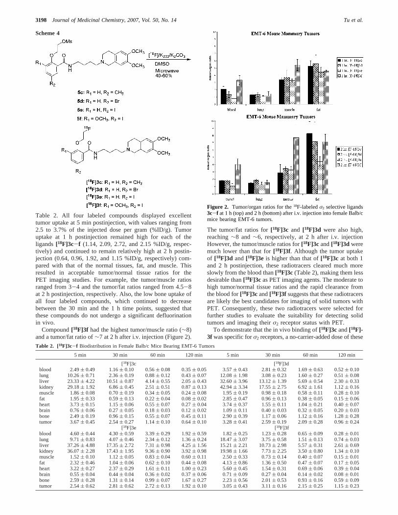
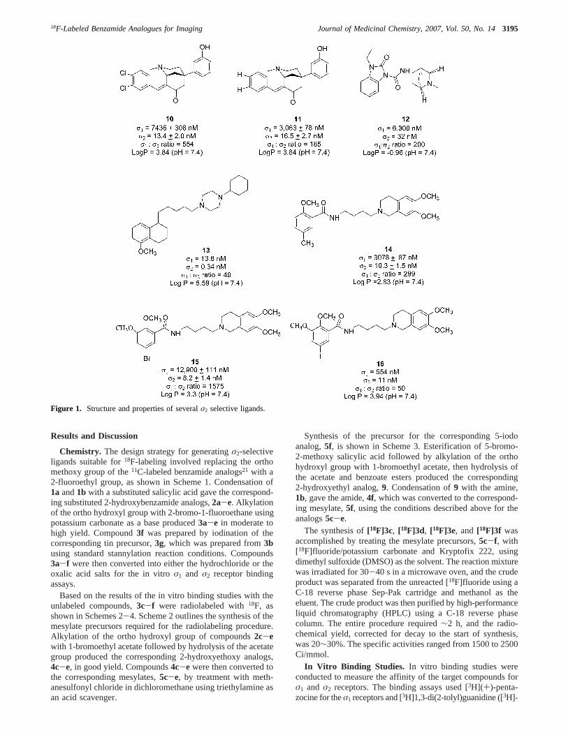
Table 2. All four labeled compounds displayed excellenttumor uptake at 5 min postinjection, with values ranging from2.5 to 3.7% of the injected dose per gram (%ID/g). Tumoruptake at 1 h postinjection remained high for each of theligands [18F]3c-f (1.14, 2.09, 2.72, and 2.15 %ID/g, respec-tively) and continued to remain relatively high at 2 h postin-jection (0.64, 0.96, 1.92, and 1.15 %ID/g, respectively) com-pared with that of the normal tissues, fat, and muscle. Thisresulted in acceptable tumor/normal tissue ratios for thePET imaging studies. For example, the tumor/muscle ratiosranged from 3-4 and the tumor/fat ratios ranged from 4.5-8at 2 h postinjection, respectively. Also, the low bone uptake ofall four labeled compounds, which continued to decreasebetween the 30 min and the 1 h time points, suggested thatthese compounds do not undergo a significant defluorinationin vivo.
Compound[18F]3f had the highest tumor/muscle ratio (∼8)and a tumor/fat ratio of∼7 at 2 h after i.v. injection (Figure 2).
The tumor/fat ratios for[18F]3c and [18F]3d were also high,reaching∼8 and∼6, respectively, at 2 h after i.v. injectionHowever, the tumor/muscle ratios for[18F]3c and[18F]3d weremuch lower than that for[18F]3f. Although the tumor uptakeof [18F]3d and [18F]3e is higher than that of[18F]3c at both 1and 2 h postinjection, these radiotracers cleared much moreslowly from the blood than[18F]3c (Table 2), making them lessdesirable than[18F]3c as PET imaging agents. The moderate tohigh tumor/normal tissue ratios and the rapid clearance fromthe blood for[18F]3c and[18F]3f suggests that these radiotracersare likely the best candidates for imaging of solid tumors withPET. Consequently, these two radiotracers were selected forfurther studies to evaluate the suitability for detecting solidtumors and imaging theirσ2 receptor status with PET.
To demonstrate that the in vivo binding of[18F]3c and[18F]-3f was specific forσ2 receptors, a no-carrier-added dose of these
Table 2. [18F]3c-f Biodistribution in Female Balb/c Mice Bearing EMT-6 Tumors
5 min 30 min 60 min 120 min 5 min 30 min 60 min 120 min
[18F]3c [18F]3dblood 2.49( 0.49 1.16( 0.10 0.56( 0.08 0.35( 0.05 3.57( 0.43 2.81( 0.32 1.69( 0.63 0.52( 0.10lung 10.26( 0.71 2.36( 0.19 0.88( 0.12 0.43( 0.07 12.08( 1.98 3.08( 0.23 1.60( 0.27 0.51( 0.08liver 23.33( 4.22 10.51( 0.87 4.14( 0.55 2.05( 0.43 32.60( 3.96 13.12( 1.39 5.69( 0.54 2.30( 0.33kidney 29.18( 1.92 6.86( 0.45 2.51( 0.51 0.87( 0.13 42.94( 3.34 17.55( 2.75 6.92( 1.61 1.12( 0.16muscle 1.86( 0.08 0.70( 0.19 0.34( 0.05 0.24( 0.08 1.95( 0.19 0.98( 0.18 0.58( 0.11 0.28( 0.10fat 1.95( 0.33 0.59( 0.13 0.22( 0.04 0.08( 0.02 2.85( 0.47 0.96( 0.13 0.38( 0.05 0.15( 0.06heart 3.73( 0.15 1.15( 0.06 0.55( 0.07 0.27( 0.04 3.74( 0.37 1.55( 0.11 1.04( 0.21 0.40( 0.07brain 0.76( 0.06 0.27( 0.05 0.18( 0.03 0.12( 0.02 1.09( 0.11 0.40( 0.03 0.32( 0.05 0.20( 0.03bone 2.49( 0.19 0.96( 0.15 0.55( 0.07 0.45( 0.11 2.90( 0.39 1.17( 0.06 1.12( 0.16 1.28( 0.28tumor 3.67( 0.45 2.54( 0.27 1.14( 0.10 0.64( 0.10 3.28( 0.41 2.59( 0.19 2.09( 0.28 0.96( 0.24
[18F]3e [18F]3fblood 4.60( 0.44 4.30( 0.59 3.39( 0.29 1.92( 0.59 1.82( 0.25 1.23( 0.28 0.65( 0.09 0.28( 0.01lung 9.71( 0.83 4.07( 0.46 2.34( 0.12 1.36( 0.24 18.47( 3.07 3.75( 0.58 1.51( 0.13 0.74( 0.03liver 37.26( 4.88 17.35( 2.72 7.31( 0.98 4.25( 1.56 15.21( 2.21 10.73( 2.98 5.57( 0.31 2.61( 0.69kidney 36.07( 2.28 17.43( 1.95 9.36( 0.90 3.92( 0.98 19.98( 1.66 7.73( 2.25 3.50( 0.80 1.34( 0.10muscle 1.52( 0.10 1.12( 0.05 0.83( 0.04 0.60( 0.11 2.50( 0.33 0.73( 0.14 0.40( 0.07 0.15( 0.01fat 2.32( 0.46 1.04( 0.06 0.62( 0.10 0.44( 0.08 4.13( 0.86 1.36( 0.50 0.47( 0.07 0.17( 0.05heart 3.22( 0.27 2.37( 0.29 1.61( 0.11 1.00( 0.23 5.60( 0.45 1.54( 0.31 0.69( 0.06 0.39( 0.04brain 0.55( 0.04 0.44( 0.04 0.36( 0.02 0.37( 0.06 0.71( 0.09 0.27( 0.04 0.14( 0.02 0.08( 0.01bone 2.59( 0.28 1.31( 0.14 0.99( 0.07 1.67( 0.27 2.23( 0.56 2.01( 0.53 0.93( 0.16 0.59( 0.09tumor 2.54( 0.62 2.81( 0.62 2.72( 0.13 1.92( 0.10 3.05( 0.43 3.11( 0.16 2.15( 0.25 1.15( 0.23
Scheme 4
Figure 2. Tumor/organ ratios for the18F-labeledσ2 selective ligands3c-f at 1 h (top) and 2 h (bottom) after i.v. injection into female Balb/cmice bearing EMT-6 tumors.
3198 Journal of Medicinal Chemistry, 2007, Vol. 50, No. 14 Tu et al.

radiotracers was coinjected into EMT-6 tumor-bearing mice withN-(4-fluorobenzyl)piperidinyl-4-(3-bromophenyl) acetamide(YUN-143a), a sigma ligand displaying a high affinity for bothσ1 andσ2 receptors. Previous studies from our group have shownthat 1 mg/kg YUN-143 is highly effective at blockingσ1 andσ2 receptors in vivo.21,23,26,27Coinjection of YUN-143 with either[18F]3c or [18F]3f resulted in a significant decrease (∼50%) inthe tumor/muscle and tumor/fat ratios at 1 h postinjection (Figure3). These data indicate that both[18F]3c and [18F]3f bindselectively toσ2 receptors in vivo.
To confirm the feasibility of using these radioligands as PETimaging agents for determining theσ2 receptor status of solidtumors, a CT/PET study using either[18F]3c or [18F]3f in femaleBalb/c mice bearing EMT-6 tumors was performed on amicroPET-F220 (CTI-Concorde Microsystems, Inc.) and aMicroCAT-II system. All mice were imaged at 1.0 h after i.v.injection of the radiotracer. The EMT-6 tumors were readilyidentifiable using either radioligand, indicating that they are bothacceptable agents for detecting solid tumors and imaging theirσ2 receptor status with PET (Figure 4). Nevertheless, furtherstudies will still be required before selecting one of thesecompounds for translation to the clinic.
Conclusion
In the present study, we synthesized several novel confor-mationally flexible benzamide analogues having a moderate tohigh binding affinity and selectivity forσ2 receptors (Table 1).Four of these compounds were selected as candidates fordeveloping18F-labeled PET probes to image theσ2 receptorstatus of solid tumors.[18F]3c, [18F]3d, [18F]3e, and[18F]3f weresuccessfully synthesized and evaluated as potential radiotracersfor imaging EMT-6 tumors in female Balb/c mice. Of the four18F-labeled analogues,[18F]3c and[18F]3f had the best biodis-tribution kinetics and tumor/normal tissue ratios. Blockingstudies confirmed that the uptake of[18F]3c and[18F]3f wasσ2
receptor mediated. Our initial CT/PET studies indicate that[18F]-3c and[18F]3f are acceptable agents for detecting solid tumorsand imaging theirσ2 receptor status with PET. Further evaluationof these two radioligands is still required before selecting oneof them for translation to the clinic.
Experimental Section
Materials. All reagents were purchased from commercialsuppliers and used without further purification unless otherwisestated. Tetrahydrofuran (THF) was distilled from sodium hydrideimmediately prior to use. Anhydrous toluene was distilled fromsodium/toluene shortly before use.
General.All anhydrous reactions were carried out in oven-driedglassware under an inert nitrogen atmosphere unless otherwisestated. When the reactions involved extraction with dichloromethane(CH2Cl2), chloroform (CHCl3), ethyl acetate (EtOAc), or ethyl ether(Et2O), the organic solutions were dried with anhydrous Na2SO4
and concentrated with a rotary evaporator under reduced pressure.Flash column chromatography was conducted using silica gel 60a,“40 Micron Flash” (32-63um; Scientific Adsorbents, Inc.). Meltingpoints were determined using the MEL-TEMP 3.0 apparatus andleft uncorrected.1H NMR spectra were recorded at 300 MHz on aVarian Mercury-VX spectrometer with CDCl3 as solvent andtetramethylsilane (TMS) as the internal standard. All chemical shiftvalues are reported in ppm (δ). Elemental analyses (C, H, N) weredetermined by Atlantic Microlab, Inc.
Procedure A: General Method for Synthesis of the Substi-tuted 2-Hydroxybenzoic Acid Amides, 2a-e.N-[2-(6,7-Dimethoxy-3,4-dihydro-1H-isoquinolin-2-yl)-ethyl]-2-hydroxy-5-methyl-benzamide (2a).1,3-dicyclohexycarbodimide (432.6 mg, 2.1 mmol)and 1-hydroxybenzotriazole (283.8 mg, 2.10 mmol) were added toan ice-water bath cooled solution of1a21 (472.0 mg, 2.0 mmol)and 2-hydroxy-5-methyl-benzoic acid (152 mg, 2.0 mmol) in 30mL of dichloromethane. After the reaction mixture was stirredovernight, analysis of the products using thin layer chromatographywith 20% methanol and 80% ethyl ether as the mobile phaseindicated that the reaction was complete. After completion of thereaction, another 50 mL of dichloromethane was added to themixture. The organic solution was then washed with an aqueoussaturated NaHCO3 solution and brine, sequentially. The organicsolution was dried with anhydrous sodium sulfate. After removalof the solvent, the crude product was purified by column chroma-tography using 20% methanol and 80% ethyl ether as the mobilephase. The yield of2a was 37.1%. The1H NMR spectrum (300MHz, CDCl3) of the purified product was 2.25 (s, 3H), 2.75-2.95(m, 6H), 3.58-3.65 (m, 4H), 3.82-3.83 (s, 6H), 6.48-6.51 (s, 1H),6.62-6.63 (s, 1H), 6.82-6.85 (d, 1H), 7.02 (s, 1H), 7.08 (s, 1H),7.20 (d, 1H). LCMSm/z 371.2 (M + H).
5-Bromo-N-[4-(6,7-dimethoxy-3,4-dihydro-1H-isoquinolin-2-yl)-butyl]-2-hydroxy-3-methoxy-benzamide (2b).Compound2bwas prepared from 5-bromo-2-hydroxy-3-methoxy-benzoic acid and1b as described above for2a. The yield of2b was 16.7%. The1HNMR spectrum (300 MHz, CDCl3) of the purified product was1.73-1.76 (m, 4H), 2.57-2.59 (m, 2H), 2.76-2.81 (m, 4H), 3.45-3.47 (m, 2H), 3.58-3.61 (m, 2H), 3.82 (s, 3H), 3.86 (s, 3H), 3.88(s, 3H), 6.48-6.51 (t, 1H), 6.56-6.59 (t, 1H), 6.97-7.00 (m, 1H),7.07-7.10 (m, 1H). LCMSm/z 493.10 (M+ H).
a Abbreviations: YUN-143, N-(4-fluorobenzyl)piperidinyl-4-(3-bro-mophenyl) acetamide; CT, computed tomography.
Figure 3. Comparison of the tumor/fat and tumor/muscle ratios for[18F]3c or [18F]3f when there is no carrier added and when theσ1 andσ2 receptors are blocked with 1 mg/kg of YUN-143. All values wereobtained 1 h after injection of the radiotracer.
Figure 4. MicroPET and microCT images of EMT-6 tumors in femaleBalb/c mice. All MicroPET images were acquired 1 h after i.v. injectionof either [18F]3c or [18F]3f.
18F-Labeled Benzamide Analogues for Imaging Journal of Medicinal Chemistry, 2007, Vol. 50, No. 143199
N-[4-(6,7-Dimethoxy-3,4-dihydro-1H-isoquinolin-2-yl)-butyl]-2-hydroxy-5-methyl-benzamide (2c).Compound2cwas preparedfrom 2-hydroxy-5-methyl-benzoic acid and1b as described abovefor 2a. The yield of 2c was 45%. The1H NMR spectrum (300MHz, CDCl3) of the purified product was 1.75 (m, 4H), 2.12 (s,3H), 2.58 (m, 2H), 2.75-2.77 (m, 2H), 2.82-2.84 (m, 2H), 3.40-3.50 (m, 2H), 3.58 (s, 2H), 3.83 (s, 3H), 3.85 (s, 3H), 6.50 (s, 1H),6.60 (s, 1H), 6.85- 6.88 (d, 1H), 7.06 (s, 1H), 7.13-7.16 (d, 2H),7.61 (s, 1H). LCMSm/z 399.20 (M+ H).
N-[4-(6,7-Dimethoxy-3,4-dihydro-1H-isoquinolin-2-yl)-butyl]-2-hydroxy-5-bromo-benzamide (2d).Compound2d was preparedfrom 5-bromo-2-hydroxy-benzoic acid and1b as described abovefor 2a. The yield of2d was 28.0%. The1H NMR spectrum (300MHz, CDCl3) of the purified product was 1.71-1.81 (m, 4H),2.55-2.85 (m, 6H), 3.44-3.48 (m, 2H), 3.58-3.60 (m, 2H), 3.82(s, 3H), 3.89 (s, 3H), 6.50 (s, 1H), 6.59 (s, 1H), 6.82-6.84 (d,1H), 7.30-7.40 (d, 1H), 7.52 (d, 1H), 8.30 (s,1H). Anal. (C22H27-BrN2O4‚1.25H2O) C, H, N.
N-[4-(6,7-Dimethoxy-3,4-dihydro-1H-isoquinolin-2-yl)-butyl]-2-hydroxy-5-iodo-benzamide (2e).Compound2e was preparedform 2-hydroxy-5-iodo-benzoic acid and1b as described above for2a. The yield of2ewas 27.0%. The1H NMR spectrum (300 MHz,CDCl3) of the purified product was 1.69-1.81 (m, 4H), 2.54-2.65 (m, 2H), 2.75-2.83 (m, 2H), 3.44-3.48 (m, 2H), 3.58 (s,2H), 3.82 (s, 3H), 3.85 (s, 3H), 6.50 (s, 1H), 6.58 (s, 1H), 6.70-6.74 (d, 1H), 7.54-7.55 (d, 1H), 7.65-7.67 (d, 1H), 8.20 (s, 1H).Anal. (C22H27IN2O4‚0.75H2O) C, H, N.
Procedure B: General Method for the Synthesis of theSubstituted 2-(2-Fluoroethoxy) Benzoic Acid Amides, 3a-e.[N-(6,7-Dimethoxy-3,4-dihydro-1H-isoquinolin-2-yl)-ethyl]-2-(2-fluoro-ethoxy)-5-methyl-benzamide (3a).Potassium carbonate(792.5 mg, 4.88 mmol) was added to a solution of2a (278 mg,0.75 mmol) and 2-bromo-1-fluoroethane (620 mg, 4.88 mmol) inacetone (60 mL). The reaction mixture was refluxed for 48 h untilthe reaction was complete as determined by thin layer chromatog-raphy with 5% methanol and 95% ethyl ether as the mobile phase.The solvent was evaporated, 30 mL of water was added to the flask,and then the mixture was extracted with dichloromethane (25 mL× 3). After the organic layer was dried with anhydrous sodiumsulfate, the crude product was purified by column chromatographyusing 5% methanol and 95% ethyl ether as the mobile phase. Theyield of 3a was 90%. The1H NMR spectrum (300 MHz, CDCl3)of the purified product was 2.33 (s, 3H), 2.64-2.80 (m, 6H), 3.63-3.75 (m, 4H), 3.84 (s, 3H), 3.86 (s, 3H), 4.16 (m, 1H), 4.21 (m,1H), 4.41 (m, 1H), 4.60 (m, 1H), 6.55 (s, 1H), 6.61 (s, 1H), 6.78-6.81 (d, 1H), 7.20 (d, 1H), 8.00 (s, 1H), 8.28 (s, 1H). LCMSm/z417.22 (M + H). For the in vitro binding experiments, the freebase was converted into the hydrochloride salt; mp 159-161 °C.Anal. (C23H30ClFN2O4) C, H, N.
5-Bromo-N-[4-(6,7-dimethoxy-3,4-dihydro-1H-isoquinolin-2-yl)-butyl]-2-(2-fluoro-ethoxy)-3-methoxy-benzamide (3b).Com-pound3b was prepared from2b as described for3a above. Theyield of 3a was 50%. The1H NMR spectrum (300 MHz, CDCl3)of the purified product was 1.64 (m, 4H), 2.48-2.52 (t, 3H), 2.64-2.66 (t, 3H), 2.74-2.78 (t, 3H), 3.42-3.55 (m, 4H), 3.79-3.87(m, 9H), 4.19-4.22 (t, 2H), 4.29-4.32 (t, 2H), 4.58-4.61 (t, 2H),4.74-4.77 (t, 2H), 6.47 (s, 1H), 6.54 (s, 1H), 7.07 (d, 1H), 7.78(d, 1H), 8.10 (s, 1H). For the in vitro binding experiments, the freebase was converted into the oxalic acid salt; mp 127-129 °C.LCMS m/z 590.30 (M+ Li). Anal. (C26H33BrFN2O7) C, H, N.
N-[6,7-Dimethoxy-3,4-dihydro-1H-isoquinolin-2-yl-butyl]-2-(2-fluoro-ethoxy)-5-methyl-benzamide (3c).Compound3c wasprepared from2c as described above for3a. The yield of3c was67%. The1H NMR spectrum (300 MHz, CDCl3) of the purifiedproduct was 1.67-2.00 (m, 4H), 2.33 (s, 3H), 2.51-2.56(t, 3H), 2.67-2.72 (t, 3H), 3H), 2.78-2.82 (t, 3H), 3.48-3.54 (m,4H), 3.82 (s, 3H), 3,83 (s, 3H), 4.21-4.24 (t, 1H), 4.30-4.33 (t,1H), 4.68-4.71 (t, 1H), 4.84-4.87 (t, 1H), 6.49 (s, 1H), 6.57 (s,1H), 6.79-6.82 (d, 1H), 7.20 (m, 1H), 7.96 (s, 1H), 7.99-7.80 (d,1H). LCMS m/z 445.25 (M + H). For the in vitro binding
experiments, the free base was converted into the oxalic acid salt;mp 131-133 °C. Anal. (C26H34FN2O2) C, H, N.
N-[4-(6,7-Dimethoxy-3,4-dihydro-1H-isoquinolin-2-yl)-butyl]-2-(2-fluoro-ethoxy)-5-bromo-benzamide (3d).Compound3d wasprepared from2d as described above for3a. The yield of3d was38.90%. The1H NMR spectrum (300 MHz, CDCl3) of the purifiedproduct was 1.57-1.80 (m, 4H), 2.62-2.66 (m, 3H), 2.78-2.82(m, 3H), 3.48-3.51 (m, 2H), 3.60-3.64 (m, 2H), 3.82 (s, 3H),3.83 (s, 3H), 4.21-4.25 (t, 1H), 4.31-4.35 (t, 1H), 4.70-4.74 (t,1H), 4.86-4.90 (t, 1H), 6.49 (s, 1H), 6.57 (s, 1H), 6.78-6.82 (d,1H), 7.48-7.52 (d, 1H), 7.96 (s, 1H), 8.26 (d, 1H). LCMSm/z509.1 (M+ H). For the in vitro binding experiments, the free basewas converted into the oxalic acid salt; mp 119-121 °C. Anal.(C25H31BrFN2O6) C, H, N.
N-[4-(6,7-Dimethoxy-3,4-dihydro-1H-isoquinolin-2-yl)-butyl]-2-(2-fluoro-ethoxy)-5-iodo-benzamide (3e).Compound3e wasprepared from2e as described above for3a. The yield of3e was41.4%. The1H NMR spectrum (300 MHz, CDCl3) of the purifiedproduct was 1.61-1.72 (m, 4H), 2.46-2.52 (m, 2H), 2.67-2.71(m, 2H), 2.76-2.78 (m, 2H), 3.41-3.47 (m, 2H), 3.52 (t, 2H),3.80 (s, 3H), 3.83 (s, 3H), 4.19-4.21 (m, 1H), 4.27-4.31 (m, 1H),4.67-4.71 (m, 1H), 4.83-4.86 (m, 1H), 6.46 (s, 1H), 6.55 (s, 1H),6.62-6.66 (d, 1H), 7.62-7.67 (m, 1H), 7.87 (s, 1H), 8.40-8.41(d, 1H). LCMS m/z 557.13 (M + H). For the in vitro bindingexperiments, the free base was converted into the oxalic acid salt;mp 121-123 °C. Anal. (C25H31FIN2O6) C, H, N.
Procedure C: General Method for Converting the Substi-tuted 5-Bromo-benzoic Acid Derivatives into their Substituted5-Tributylstannanyl Benzoic Acid Derivatives. N-[4-(6,7-Dimethoxy-3,4-dihydro-1H-isoquinolin-2-yl)-butyl]-2-(2-fluoro-ethoxy)-3-methoxy-5-tributylstannanyl-benzamide (3g).Nitrogenwas bubbled for 5-10 min through a solution of3b (200 mg, 0.371mmol) in 20 mL of fresh distilled toluene. The whole system wascovered with aluminum foil. Tetrakis(triphenylphosphine palladium-(0) [(PPh3)4Pd(0); 42 mg, 0.036 mmol] and bis(tributytin) ([Sn-(C4H9)3]2; 575 mg, 0.99 mmol) was added to the reaction mixtureand heated overnight at 110°C with an oil bath. Thin layerchromatography with 45% hexane, 45% ethyl ether, and 10%methanol as the mobile phase was used to assess when the reactionwas complete. After quenching the reaction, the crude product waspurified on a silica gel column to isolate the tin intermediate,3g.The yield of 3g was 64%. The1H NMR spectrum (300 MHz,CDCl3) of the purified product was 0.87-1.58 (m, 27H), 1.61-1.69 (m, 4H), 2.56 (s, 2H), 2.72 (s, 2H), 2.81-2.82 (s, 2H), 3.47-3.49 (d, 2H), 3.57 (s, 2H), 3.83 (s, 6H), 3.88 (s, 3H), 4.25 (d, 1H),4.37 (s, 1H), 4.62-4.65 (s, 1H), 4.78-4.81 (s, 1H), 6.51 (s, 1H),6.58 (s, 1H), 7.81 (s, 1H), 8.07 (s, 1H). LCMSm/z 747.60 (M-H).
Procedure D: General Method for Converting the TinPrecursor of the Benzoic Acid Derivatives into their Corre-sponding Iodine Substituted Benzoic Acid Derivatives. (N-[4-(6,7-Dimethoxy-3,4-dihydro-1H-isoquinolin-2-yl)-butyl]-2-(2-fluoro-ethoxy)-3-methoxy-5-iodo-benzamide (3f).A solution ofiodine in CHCl3 (5 mL, 0.5 M) was added dropwise to a solutionof the tin precursor,3g (258 mg, 0.34 mmol), in 20 mL of CH2Cl2until the color of the solution persisted. The reaction was stirred atroom temperature for 30 min, and a solution of 5% aqueousNaHSO3 was added until the solution was colorless. The mixturewas extracted with CH2Cl2, and the organic layers were washedwith brine before being dried with Na2SO4. The organic layers werethen concentrated under vacuum and purified using a silica gelcolumn with 15% methanol and 85% ether as the mobile phase toisolate3f. The yield of3f was 36%. The1H NMR spectrum (300MHz, CDCl3) of the purified product was 1.60-1.80 (m, 2H),1.80-2.10 (m, 4H), 3.19-3.2 (m, 2H), 3.40-3.50 (m, 2H), 3.68(m, 2H), 3.83 (m, 2H), 3.92 (s, 3H), 3.95 (s, 3H), 3.99 (s, 3H),4.26 (t, 1H), 4.37 (s, 1H), 4.65 (s, 1H), 4.80 (s, 1H), 6.50 (s, 1H),6.58 (s, 1H), 7.28 (d, 1H), 8.02 (d, 1H), 8.21 (s, 1H). LCMSm/z587.14 (M + H). For the in vitro binding experiments, the freebase was converted into the oxalic acid salt; mp 125-127 °C.
3200 Journal of Medicinal Chemistry, 2007, Vol. 50, No. 14 Tu et al.

Procedure E: General Method for Converting the Substi-tuted 2-Hydroxy Benzoic Acid Derivatives into their Substituted2-(2-Hydroxy-ethoxy)-benzoic Acid Amides.N-[4-6,7-Dimethoxy-3,4-dihydro-1H-isoquinolin-2-yl)-butyl]-2-(2-hydroxy-ethoxy)-5-methyl-benzamide (4c).Anhydrous potassium carbonate (546.0mg, 3.26 mmol) was added to a solution of2c (200.0 mg, 0.5 mmol)and 2-bromoethyl acetate (547.0 mg, 3.27 mmol) in 60 mL ofacetone. The reaction mixture was refluxed for 48 h under nitrogen.After 48 h, thin layer chromatography with 15% methanol and 85%ether as the mobile phase indicated that the reaction was complete.After evaporating the solvent, the residue was dissolved in 30 mLof water and extracted with ethyl acetate (20× 3 mL). Then theorganic component was washed with brine, dried with anhydroussodium sulfate, and concentrated, and the final product was purifiedon a silica gel column to isolate the 2-{2-[4-(6,7-dimethoxy-3,4-dihydro-1H-isoquinolin-2-yl)-butylcarbamoyl]-4-methyl-phenoxy}-ethyl ester. The yield of this intermediate was 82.2%. The1H NMRspectrum (300 MHz, CDCl3) of the purified product was 1.70 (m,4H), 2.01 (s, 3H), 2.33 (s, 3H), 2.56 (m, 2H), 2.71-2.73 (m, 2H),2.81 (m, 2H), 3.50-3.52 (m, 2H), 3.55 (s, 2H), 3.83 (s, 3H), 3.84(s, 3H), 4.23 (t, 2H), 4.50 (t, 2H), 6.50 (s, 1H), 6.58 (s, 1H), 6.78-6.82 (d, 1H), 7.18-7.25 (d, 1H), 7.95 (s, 1H), 8.02 (s, 1H).
NaOH (30 mg, 0.75mmol) was added to a solution of thisintermediate (182 mg, 0.375mmol) in 20 mL of methanol and 10mL of water. The reaction mixture was stirred overnight until thereaction was complete. Then 0.375 mL of 2 N HCl was added toneutralize the solution. After evaporating the solvent, the residuewas dissolved in 60 mL of ethyl acetate. The solution was washedfirst with water, then brine, and finally dried with anhydrous sodiumsulfate. After evaporating the solvent, the crude product was purifiedon a silica gel column. The yield of4c was 96%. The1H NMRspectrum (300 MHz, CDCl3) of the purified product was 1.68-1.85 (m, 4H), 2.39 (s, 3H), 2.45 (s, 1H), 2.51-2.61 (m, 2H), 2.80-2.87 (m, 4H), 3.45-3.61 (m, 4H), 3.76-3.80 (t, 2H), 3.83 (s, 3H),3.85 (s, 3H), 3.83 (s, 3H), 4.05-4.08 (t, 2H), 6.49 (s, 1H), 6.60 (s,1H), 6.79-6.83 (d, 1H), 7.15-7.19 (d, 2H), 7.93 (s, 1H), 8.30 (s,1H).
5-Bromo-N-[4-(6,7-dimethoxy-3,4-dihydro-1H-isoquinolin-2-yl)-butyl]-2-(2-hydroxy-ethoxy)-benzamide (4d).Compound4dwas prepared from2d as described above for4c. The yield of4dwas 70%. The1H NMR spectrum (300 MHz, CDCl3) of the purifiedproduct was 1.65-1.90 (m, 4H), 2.66-2.70 (m, 2H), 2.92 (m, 4H),3.51-3.54 (m, 2H), 3.72 (m, 2H), 3.72-3.81 (t, 2H), 3.83 (s, 3H),3.85 (s, 3H), 4.05-4.09 (t, 2H), 6.51 (s, 1H), 6.60 (s, 1H), 6.77-6.81 (d, 1H), 7.44-7.48 (d, 2H), 8.17 (s, 1H), 8.30 (s, 1H).
N-[4-(6,7-Dimethoxy-3,4-dihydro-1H-isoquinolin-2-yl)-butyl]-2-(2-hydroxy-ethoxy)-5-iodo-benzamide (4e).Compound4ewasprepared from2e as described above for4c. The yield of4e was77%. The1H NMR spectrum (300 MHz, CDCl3) of the purifiedproduct was 1.60-1.90 (m, 4H), 2.63-2.66 (m, 3H), 2.90 (s, 2H),3.52-3.55 (m, 2H), 3.70 (m, 2H), 3.79-3.82 (t, 2H), 3.83 (s, 3H),3.85 (s, 3H), 4.08-4.10 (t, 2H), 6.50 (s, 1H), 6.60 (s, 1H), 6.67-6.70 (d, 1H), 7.60-7.70 (d, 1H), 8.30 (s, 1H), 8.40-8.41 (d, 1H).
Procedure F: General Method for Converting the Sub-stituted 2-Hydroxy-ethoxy Benzoic Acid Amides to theirMethanesulfonic Acid Esters. 2-(2-(4-(6,7-Dimethoxy-3,4-dihy-droisoquinolin-2(1H)-yl)butylcarbamoyl)-4-methylphenoxy)-ethyl Methanesulfonate (5c).Methanesulfonic chloride (120 mg,1.04 mmol) was added to an ice-water cooled solution of4c (354mg, 0.8 mmol) and triethylamine (242 mg, 2.4 mmol) in 30 mL ofdichloromethane. The reaction mixture was stirred for 3 h untilthin layer chromatography using 5% methanol and 95% dichlo-romethane as the mobile phase indicated that the reaction wascomplete. After 3 h, 20 mL of dichloromethane was added, thesolution was washed with first a saturated sodium carbonate aqueoussolution (20 mL × 3) and then brine, and finally dried withanhydrous sodium sulfate. After evaporating the solvent, the crudeproduct was purified on a silica gel column to isolate5c. The yieldof 5c was 81.6%. The1H NMR spectrum (300 MHz, CDCl3) ofthe purified product was 2.07-2.10 (m, 4H), 2.71 (s, 3H), 2.80-3.00 (m, 2H), 3.08-3.20 (m, 4H), 3.40 (m, 3H), 3.80-4.00 (m,
4H), 4.20 (s, 3H), 4.22 (s, 3H), 4.60-4.68 (t, 2H), 4.95-4.97 (t,2H), 6.88 (s, 1H), 6.95 (s, 1H), 7.15-7.18 (d, 1H), 7.50-7.60 (d,1H), 8.20 (s, 1H), 8.19 (s, 1H). Anal. (C26H36N2O7S) C, H, N.
2-(4-Bromo-2-(4-(6,7-dimethoxy-3,4-dihydroisoquinolin-2(1H)-yl)butylcarbamoyl)phenoxy)ethyl Methanesulfonate (5d).Com-pound5d was prepared from4d as described above for5c. Theyield of 5d was 77%. The1H NMR spectrum (300 MHz, CDCl3)of the purified product was 1.72-1.75 (m, 4H), 2.61-2.68 (m, 2H),2.85 (s, 3H), 3.05 (s, 2H), 3.42-3.58 (m, 4H), 3.68 (s, 2H), 3.82(s, 3H), 3.83 (s, 3H), 4.29 (t, 2H), 4.60 (t, 2H), 6.50 (s, 1H), 6.57(s, 1H), 6.70-6.77 (d, 1H), 7.45-7.55 (d, 1H), 7.95 (s, 1H), 8.20-8.22 (d, 1H). LCMSm/z 585.10 (M+ H).
2-(2-(4-(6,7-Dimethoxy-3,4-dihydroisoquinolin-2(1H)-yl)bu-tylcarbamoyl)-4-iodophenoxy)ethyl Methanesulfonate (5e).Com-pound5e was prepared from4e as described above for5c. Theyield of 5e was 80%. The1H NMR spectrum (300 MHz, CDCl3)of the purified product was 1.60-1.80 (m, 4H), 2.52-2.55 (m, 2H),2.67-2.70 (m, 2H), 2.70-2.78 (m, 2H), 3.02 (s, 3H), 3.46-3.52(m, 4H), 3.82 (s, 3H), 3.83 (s, 3H), 4.23-4.26 (t, 2H), 4.56-4.60(t, 2H), 6.48 (s, 1H), 6.55 (s, 1H), 6.60-6.64 (d, 1H), 7.64-7.68(d, 1H), 7.85 (s, 1H), 8.38-8.39 (d, 1H). LCMSm/z 633.10 (M+H).
2-(2-Acetoxy-ethoxy)-5-bromo-3-methoxy-benzoic Acid Meth-yl Ester (7). Initially, 1.0 mL of 98% concentrated sulfuric acidwas added to a solution of 5-bromo-2-hydroxy-3-methoxy-benzoicacid, 6 (1.0 g, 4.0 mmol), in 50 mL of methanol. The reactionmixture was refluxed overnight until thin layer chromatographyusing 20% ethyl acetate and 80% hexane as the mobile phaseindicated that the reaction was complete. After evaporating themethanol, the residue was dissolved in 60 mL of ethyl acetate andwashed with a saturated NaHCO3 aqueous solution and then brine.After drying with anhydrous sodium sulfate, the solution wasconcentrated and the crude product was purified on a silica gelcolumn to isolate the intermediate, 5-bromo-2-hydroxy-3-methoxy-benzoic acid methyl ester. The yield of this intermediate was 94%.The1H NMR spectrum (300 MHz, CDCl3) of the purified productwas 3.88 (s, 3H), 3.95 (s, 3H), 7.09 (d, 1H), 7.55 (t, 1H), 10.96 (d,1H).
Potassium carbonate (2.90 g, 21.0 mmol) was added to a solutionof the above intermediate (0.84 g, 3.23 mmol) and 2-bromoethylacetate (3.5 g, 20.96 mmol) in 60 mL of acetone. The reactionmixture was refluxed for 72 h until thin layer chromatography using20% ethyl acetate and 80% hexane as the mobile phase indicatedthat the reaction was complete. After evaporating the solvent, theresidue was dissolved in 30 mL of water and then extracted withethyl acetate (25 mL× 3). The organic solution was dried withanhydrous sodium sulfate, resuspended, and purified on a silicagel column. The yield of7 was 78%. The1H NMR spectrum (300MHz, CDCl3) of the purified product was 2.10 (s, 3H), 3.86 (s,3H), 3.88 (s, 3H), 4.21-4.24 (t, 2H), 4.38-4.41 (t, 3H), 7.14 (s,1H), 7.45 (s, 1H).
2-(2-Acetoxy-ethoxy)-5-iodo-3-methoxy-benzoic Acid MethylEster (8). Nitrogen was bubbled for 5-10 min through a solutionof of 2-(2-acetoxy-ethoxy)-5-bromo-3-methoxy-benzoic acid methylester,7 (270 mg, 0.778 mmol), in 20 mL of freshly distilled toluene.The reaction system was covered with aluminum foil. Tetrakis-(triphenylphosphine) palladium(0) [(PPh3)4Pd(0); 100 mg, 0.087mmol] and bis(tributlytin) ([Sn(C4H9)3]2; 899 mg, 1.55 mmol) wereadded to the reaction mixture and heated overnight at 110°C in anoil-bath while stirring. After quenching, thin layer chromatographyusing 15% ethyl acetate and 85% hexane as the mobile phaseindicated that the reaction was complete. The product was thenpurified on a silica gel column to isolate the tin precursor, 2-(2-acetoxy-ethoxy)-3-methoxy-5-tributylstannanyl-benzoic acid methylester. The yield of the tin precursor was 37.3%. The1H NMRspectrum (300 MHz, CDCl3) of the purified product was 0.8-1.75(m, 27H), 2.10 (s, 3H), 3.87 (s, 3H), 3.89 (s, 3H), 4.24-4.27 (t,2H), 4.38-4.41 (t, 2H), 7.09-7.30 (s 1H), 7.35 (s 1H).
A solution of iodine in CHCl3 (5 mL, 0.5 M) was added dropwiseto a solution of above tin precursor (680 mg, 1.22 mmol) in 20mL of CH2Cl2 until the color of the solution persisted. Then the
18F-Labeled Benzamide Analogues for Imaging Journal of Medicinal Chemistry, 2007, Vol. 50, No. 143201

reaction was stirred at room temperature for 30 min, and a quenchsolution of 5% aqueous NaHSO3 was added until the solutionbecame colorless. The mixture was extracted with CH2Cl2, and theorganic layers were washed with brine and dried by Na2SO4. Theorganic layers were then condensed under vacuum and purifiedusing a silica gel column with 15% ethyl acetate and 85% hexaneas the mobile phase. The yield of8 was 90%. The1H NMRspectrum (300 MHz, CDCl3) of the purified product was 2.10 (s,3H), 3.85 (s, 3H), 3.88 (s, 3H), 4.21-4.25 (t, 2H), 4.37-4.41 (t,2H), 7.29-7.30 (s 1H), 7.63 (s 1H).
2-(2-Hydroxy-ethoxy)-5-iodo-3-methoxy-benzoic Acid (9).Com-pound9 was prepared from8 as described in Procedure E. Theyield of 9 was 81%. The1H NMR spectrum (300 MHz, CDCl3) ofthe purified product was 3.89 (s, 3H), 3.93-3.96 (t, 2H), 4.33-4.36 (t, 2H), 7.08 (s, 1H), 7.62 (s, 1H).
N-[4-(6,7-Dimethoxy-3,4-dihydro-1H-isoquinolin-2-yl)-butyl]-2-(2-hydroxy-ethoxy)-5-iodo-3-methoxy-benzamide (4f).Com-pound4f was prepared from9 and1b as described in ProcedureA. The yield of 4f was 29%. The1H NMR spectrum (300 MHz,CDCl3) of the purified product was 1.72-1.75 (m, 4H), 2.56 (m,2H), 2.75-2.77 (m, 2H), 2.81-2.83 (m, 2H), 3.49-3.51 (m, 2H),3.55 (s, 2H), 3.56-3.60 (t, 2H), 3.82 (s, 3H), 3.83 (s, 3H), 3.85 (s,3H), 4.06-4.10 (t, 2H), 6.47 (s, 1H), 6.57 (s, 1H), 6.90 (s, 1H),7.57 (s, 1H), 7.70-7.80 (s, 1H).
2-(2-(4-(6,7-Dimethoxy-3,4-dihydroisoquinolin-2(1H)-yl)bu-tylcarbamoyl)-4-iodo-6-methoxyphenoxy)ethyl Methanesulfonate(5f). Compound5f was prepared from4f as described in ProcedureF. The yield of5f was 61%. The1H NMR spectrum (300 MHz,CDCl3) of the purified product was 1.72 (m, 4H), 2.55 (s, 2H),2.70 (d, 2H), 22.76 (d, 2H), 3.05 (s, 3H), 3.85 (m, 9H), 4.26 (m,2H), 4.49 (m, 2H), 6.49 (s, 1H), 6.55 (s, 1H), 6.93 (d, 1H), 7.51(m, 1H), 8.02 (s, 1H). LCMSm/z 663.20 (M+ H). Anal. (C25H32-FIN2O5) Calcd: C, 51.20; H, 5.50; N, 4.78. Found: C, 36.61; H,4.34; N, 3.12.
Radiochemistry. Production of [18F]Fluoride. [18F]Fluoridewas produced in our institution by proton irradiation of enriched18O water (95%) [reaction:18O(p, n)18F] using either a JSW BC-16/8 (Japan Steel Works) or a CS-15 cyclotron (Cyclotron Corp)
Procedure G: General Method for Labeling the Substituted2-(2-Fluoroethoxy) Benzoic Acid Amide Analogs with18F. [18F]-(N-[4-(6,7-Dimethoxy-3,4-dihydro-1H-isoquinolin-2-yl)-butyl]-2-(2-fluoro-ethoxy)-3-methoxy-5-iodo-benzamide ([18F]3f). [18F]-fluoride (100-150 mCi) was added to a 10-mL Pyrex screw captube containing 5-6 mg of Kryptofix 222 and 0.75 mg of K2CO3.Using HPLC grade acetonitrile (3× 1.0 mL), the water wasazeotropically evaporated from this mixture at 110°C under astream of argon. After all of the water was removed, a solution ofthe precursor,5f (1.5-2.0 mg), in DMSO (0.2 mL) was added tothe reaction vessel containing the18F/Kryptofix mixture. A 3 mmglass bead was added to the reaction vessel to ensure a morehomogeneous heat distribution when the sample was irradiated withmicrowaves, and the vessel was capped firmly on a speciallydesigned remotely operated capping station. After vortexing, thereaction mixture was irradiated with microwaves for 30-40 s atmedium power (60 W) until the thin layer chromatography scannerwith a 25% of methanol and 75% dichloromethane mobile phaseindicated that the incorporation yield was 40-60%.
After adding 6 mL of water and shaking, the solution was loadedon a C-18 reverse phase Waters Oasis cartridge (HLB-6 cm3) thathad previously been rinsed with a solution of 5% methanol in water(5-8 mL). The sample was then rinsed three times with 6 mL ofwater to eliminate the unreacted fluoride. The retained activity waseluted with 5-8 mL of acetonitrile. After evaporating the aceto-nitrile to a volume of<0.5 mL, the sample was loaded on a C-18Alltech econosil semipreparative HPLC column (250× 10 um).The product was eluted with 29% acetonitrile and 71% 0.1 Mammonium formate buffer at a flow rate of 4.5 mL/min. Theretention time of the[18F]3f was∼33 min. The solution containingthe [18F]3f was concentrated and resuspended in saline, and a 100µL aliquot was sent for quality control analysis before using it in
the biodistribution and imaging studies. The entire procedurerequired∼2 h.
Quality control analysis was performed on an analytical HPLCsystem that consisted of an Alltech econosil reversed phase C-18column (250× 4.6 mm) with a mobile phase of 35% acetonitrileand 65% 0.1 M ammonium formate buffer at pH 4.0-4.5. At aflow rate of 1.2 mL/min, the[18F]3f eluted at 13.2 min with aradiochemical purity of>99%. The labeling yield was∼30%(decay corrected), and the specific activity was>2000 Ci/mmol.
[18F](N-[4-(6,7-Dimethoxy-3,4-dihydro-1H-isoquinolin-2-yl)-butyl]-2-(2-fluoro-ethoxy)-5-methyl-benzamide ([18F]3c). Com-pound[18F]3c was prepared from5cas described above for[18F]3f,with the following exceptions. The semipreparative HPLC mobilephase was 39% methanol and 61% 0.1 M formate buffer. At a flowrate of 3.5 mL/min, the[18F]3c eluted at ∼33 min with aradiochemical purity of>99%. The labeling yield was∼35%(decay corrected), and the specific activity was>1500 Ci/mmol.The entire procedure took∼2 h.
To check that the chemical characteristics of[18F]3c wereidentical to the cold standard,3c, both compounds were run on theanalytical HPLC system with a mobile phase of 52% methanol and48% 0.1 M formate buffer. At a flow rate of 1.5 mL/min, the twocompounds coeluted with a retention time of 4.7 min.
[18F](N-[4-(6,7-Dimethoxy-3,4-dihydro-1H-isoquinolin-2-yl)-butyl]-2-(2-fluoro-ethoxy)-5-bromo-benzamide ([18F]3d). Com-pound[18F]3d was prepared from5d as described above for[18F]3fwith the following exceptions. The semipreparative HPLC mobilephase was 13% of THF and 87% 0.1 M formate buffer. At a flowrate of 3.5 mL/min, the[18F]3d eluted at ∼20 min with aradiochemical purity of>98%. The labeling yield was∼30%(decay corrected), and the specific activity was>1500 Ci/mmol.The entire procedure took∼2 h.
To check that the chemical characteristics of[18F]3d wereidentical to the cold standard,3d, both compounds were run onthe analytical HPLC system with a mobile phase of 38% acetonitileand 62% 0.1 M formate buffer. At a flow rate of 1.5 mL/min, thetwo compounds coeluted with a retention time of∼8 min.
[18F](N-[4-(6,7-Dimethoxy-3,4-dihydro-1H-isoquinolin-2-yl)-butyl]-2-(2-fluoro-ethoxy)-5-Iodo-benzamide ([18F]3e). Com-pound[18F]3ewas prepared from5eas described above for[18F]3f,with the following exceptions. The semipreparative HPLC mobilephase was 15% THF and 85% 0.1 M formate buffer. At a flowrate of 6.0 mL/min, the[18F]3e eluted at ∼35 min with aradiochemical purity of>99%. The labeling yield was∼30%(decay corrected), and the specific activity was>1500 Ci/mmol.The entire procedure took∼2 h.
To check that the chemical characteristics of[18F]3e wereidentical to the cold standard,3e, both compounds were run on theanalytical HPLC system with a mobile phase of 17% tetrahydro-furan and 83% 0.1 M ammonium formate buffer. At a flow rate of2.0 mL/min, the two compounds coeluted with a retention time of15.2 min.
Characterization of the Potential σ2 Selective Ligands InVitro and In Vivo. Sigma Receptor Binding Assays.The novelsigma ligands were dissolved inN,N-dimethylformamide (DMF),DMSO, or ethanol and then diluted in 50 mM Tris-HCl buffercontaining 150 mM NaCl and 100 mM EDTA at pH 7.4 prior toperforming theσ1 andσ2 receptor binding assays The proceduresfor isolating the membrane homogenates and performing theσ1
and σ2 receptor binding assays have been described in detailpreviously.21
Briefly, the σ1 receptor binding assays were conducted in 96-well plates using guinea pig brain membrane homogenates (∼300µg protein) and∼5 nM [3H](+)-pentazocine (34.9 Ci/mmol, Perkin-Elmer, Boston, MA). The total incubation time was 90 min at roomtemperature. Nonspecific binding was determined from samples thatcontained 10µM of cold haloperidol. After 90 min, the reactionwas terminated by the addition of 150µL of ice-cold wash buffer(10 mM Tris-HCl, 150 mM NaCl, pH 7.4) using a 96 channeltransfer pipet (Fisher Scientific, Pittsburgh, PA). The samples wereharvested and filtered rapidly through a 96-well fiberglass filter
3202 Journal of Medicinal Chemistry, 2007, Vol. 50, No. 14 Tu et al.
plate (Millipore, Billerica, MA) that had been presoaked with 100µL of 50 mM Tris-HCl buffer at pH 8.0 for 1 h. Each filter waswashed three times with 200µL of ice-cold wash buffer, and thefilter was counted in a Wallac 1450 MicroBeta liquid scintillationcounter (Perkin-Elmer, Boston, MA).
The σ2 receptor binding assays were conducted using rat livermembrane homogenates (∼300 µg protein) and∼5 nM [3H]DTG(58.1 Ci/mmol, Perkin-Elmer, Boston, MA) in the presence of 1µM (+)-pentazocine to blockσ1 sites. The incubation time was120 min at room temperature. Nonspecific binding was determinedfrom samples that contained 10µM of cold haloperidol. All otherprocedures were identical to those described for theσ1 receptorbinding assay above.
Data from the competitive inhibition experiments were modeledusing nonlinear regression analysis to determine the concentrationthat inhibits 50% of the specific binding of the radioligand (IC50
value). Competitive curves were best fit to a one-site fit and gavepseudo-Hill coefficients of 0.6-1.0.Ki values were calculated usingthe method of Cheng and Prusoff24 and are presented as the mean( 1 SEM. For these calculations, we used aKd value of 7.89 nMfor [3H](+)-pentazocine and guinea pig brain; for [3H]DTG andrat liver, we used 30.73 nM20
Biodistribution Studies. All animal experiments were conductedin compliance with the Guidelines for the Care and Use of ResearchAnimals established by Washington University’s Animal StudiesCommittee. EMT-6 mouse mammary adenocarcinoma cells (5×10⁵ cells in 100µL of phosphate-buffered saline) were implantedsubcutaneously in the scapular region of female Balb/c mice (∼2months old and 17-22 g; Charles River Laboratories). Thebiodistribution studies were initiated 7-10 days after implantationwhen the tumor size was∼0.2 cm3 (∼200 mg).
For the biodistribution studies, 10-20 µCi of [18F]3c, [18F]3d,[18F]3e, or [18F]3f in 100-150 µL of saline was injected via thetail vein into EMT-6 tumor-bearing female Balb/c mice. Groupsof at least four mice were used for each time point. At 5, 30, 60,and 120 min after injection, the mice were euthanized, and samplesof blood, lung, liver, kidney, muscle, fat, heart, brain, bone, andtumor were removed, weighed, and counted in a Beckman Gamma8000 well counter. After counting, the percentage of the injecteddose per gram of tissue (%ID/g) was calculated. The tumor/organratios were calculated by dividing the %ID/g of the tumor by the%ID/g of each organ.
Blocking studies in tumor-bearing mice were conducted bycoinjecting 1 mg/kg of cold YUN-143 with[18F]3c or [18F]3f. Yun-143 has a high affinity for bothσ1 andσ2 receptors and is routinelyused in our laboratory for sigma receptor blocking studies.13,14,25
All mice were sacrificed 60 min after injection of the radiotracer,and the tumor/organ ratios were determined as described above.
Imaging Studies.Each mouse was imaged on both a microPET-F220 (CTI-Concorde Microsystems, Inc.) and a MicroCAT IISystem (ImTek, Inc.). For the microPET studies, each mouse wasinjected with∼0.25 mCi of either[18F]3c or [18F]3f via the tailvein and imaged 1 h later. MicroCT images were also obtainedand coregistered with the PET images to determine the exactanatomical location of the radiotracers.
Acknowledgment. This work was supported by GrantCA-102869 awarded by the National Institutes of Health andGrant DAMD17-01-1-0446 awarded by the Department ofDefense Breast Cancer Research Program of the U.S. ArmyMedical Research and Materiel Command Office. The authorsgratefully thank William H. Margenau and Robert Dennettfor their excellent technical assistance. Mass spectrometrywas provided by the Washington University MassSpectrometry Resource, an NIH Research Resource (Grant No.P41RR0954).
Supporting Information Available: Analytical data on all newcompounds. This material is available free of charge via the Internetat http://pubs.acs.org.
References
(1) Walker, J. M.; Bowen, W. D.; Walker, F. O.; Matsumoto, R. E.; DeCosta, B.; Rice, K. R. Sigma receptors: Biology and function.Pharmacol ReV. 1990, 42, 355-402.
(2) Hellewell, S. B.; Bowen, W. D. A sigma-like binding site in ratpheochromocytoma (PC12) cells: Decreased affinity for (+)-benzomorphans and lower molecular weight suggest a different sigmareceptor form from that of guinea pig brain.Brain Res.1990, 527,244-253.
(3) Quirion R.; Bowen W. D.; Itzhak Y.; et al. A proposal for theclassification of sigma binding sites.Trends Pharmacol. Sci.1992,13, 85-86.
(4) Guitart, X.; Codony, X.; Monroy, X. Sigma receptors: Biologyand therapeutic potential.Psychopharmacology2004, 174, 1920-1929.
(5) Hanner, M.; Moebius, F. F.; Flandorfer, A.; Knaus, H. G.; Striessnig,J.; Kempner, E.; et al. Purification, molecular cloning, and expressionof the mammalian sigma1-binding site.Proc. Natl. Acad. Sci. U.S.A.1996, 93, 8072-8077.
(6) Hellewell, S. B.; Bruce, A.; Feinstein, G.; Orringer, J.; Williams,W.; Bowen, W. D. Rat liver and kidney contain high densitiesof r1 and r2 receptors: Characterization by ligand binding andphotoaffinity labeling.Eur. J. Pharmacol., Mol. Pharmacol Sect.1994, 268, 9-18.
(7) Vilner, B. J.; John, C. S.; Bowen, W. D. Sigma-1 and sigma-2receptors are expressed in a wide variety of human and rodent tumorcell lines.Cancer Res. 1995, 55, 408-413.
(8) Bem, W. T.; Thomas, G. E.; Mammone, J. Y.; Homan, S. M.; Levy,B. K; Johnson, F. E.; et al. Overexpression of r receptors in nonneuralhuman tumors.Cancer Res. 1991, 51, 6558-6562.
(9) Vilner B. J.; Bowen W. D. Characterization of sigma-like bindingproperties of NB41A3, S-20Y, and N1E-115 neuroblastomas, C6gliomas, and NG108-15 neuroblastoma-glioma hybrid cells: Furtherevidence for sigma-2 receptors. InMultiple sigma and PCP receptorligands: mechanisms for neuromodulation and neuroprotection?;Kamenka, J. M., Domino, E. F., Eds.; 7 NPP Books: Ann Arbor,MI, 1992; pp 341-353.
(10) Mach, R. H.; Smith, C. R.; Al Nabulsi, I.; Whirrett, B. R.;Childers, S. R.; Wheeler, K. T. Sigma-2 receptors as potentialbiomarkers of proliferation in breast cancer.Cancer Res.1997, 57,156-161.
(11) Al-Nabulsi, I.; Mach, R. H.; Wang, L. M.; Wallen, C. A.; Keng, P.C.; Sten, K.; et al. Effect of ploidy, recruitment, environmental factors,and tamoxifen treatment on the expression of sigma-2 receptors inproliferating and quiescent tumor cells.Br. J. Cancer1999, 81, 925-933.
(12) Wheeler, K. T.; Wang, L. M.; Wallen, C. A.; Childers, S. R.;Cline, J. M.; Keng, P. C.; et al. Sigma-2 receptors as a biomarkerof proliferation in solid tumors.Br. J. Cancer2000, 82, 1223-1232.
(13) Bowen, W. D.; Bertha, C. M.; Vilner, B. J.; Rice, K. C. CB-64Dand CB-184: Ligands with highσ2 receptor affinity and subtypeselectivity.Eur. J. Pharmacol. 1995, 278, 257-260.
(14) Bonhaus, D. W.; Loury, D. N.; Jakeman, L. B.; To, Z.; DeSouza,A.; Eglen, R. M.; et al. [3H]BIMU-1, a 5-hydroxy-tryptamine3receptor ligand in NG-108 cells, selectively labels sigma-2 bindingsites in guinea pig brain hippocampus.J. Pharmacol. Exp. Ther.1993,267, 961.
(15) Colabufo, N. A.; Berardi, F.; Contino, M.; Fazio, F.; Matarrese, M.;Moresco, R. M.; Niso, M.; Perrone, R.; Tortorella, V. Distributionof sigma receptors in EMT-6 cells: Preliminary biological evaluationof PB167 and potential for in-vivo PET. J. Pharm. Pharmacol.2005,57 (11), 1453-1459.
(16) Kassiou, M.; Dannals, R. F.; Liu, X.; Wong, D. F.; Ravert, H. T.;Scheffel, U. A. Synthesis and in vivo evaluation of a new PETradioligand for studying sigma-2 receptors.Bioorg. Med. Chem.2005,13 (11), 3623-3626.
(17) Berardi, F.; Ferorelli, S.; Abate, C.; Colabufo, N. A.; Contino, M.;Perrone, P.; Tortorella, V. 4-(Tetralin-1-yl)- and 4-(naphthalen-1-yl)alkyl derivatives of 1-cyclohexylpiperazine as receptor ligands withagonistσ2 activity. J. Med. Chem.2004, 47 (9), 2308-2317.
(18) Mach, R. H.; Huang, R. H.; Freeman, R. A.; Wu, L.; Blair, S.;Luedtke, R. R. Synthesis of 2-(5-bromo-2,3-dimethoxyphenyl)-5-(aminomethyl)-1H-pyrrole analogues and their binding affinities fordopamine D2, D3, and D4 receptors.Bioorg. Med. Chem.2003, 11,225.
(19) Huang, Y.; Luedtke, R. R.; Freeman, R. A.; Wu, L. and Mach,R. H. Synthesis and structure-activity relationships ofnaphthamides as dopamine D3 receptor ligands.J. Med. Chem.2001,44, 1815.
18F-Labeled Benzamide Analogues for Imaging Journal of Medicinal Chemistry, 2007, Vol. 50, No. 143203

(20) Tu, Z.; Dence, C. S.; Ponde, D. E.; Jones, L.; Wheeler, K. T.; Welch,M. J.; Mach R. H. Carbon-11 labeled sigma2 receptor ligands forimaging breast cancer.Nucl. Med. Biol.2005, 32 (5), 423-430.
(21) Xu, J.; Tu, Z.; Jones, L. A.; Vangveravong, S.; Wheeler, K. T.;Mach, R. H. [3H]N-[4-(3,4-dihydro-6,7-dimethoxyisoquinolin-2(1H)-yl)butyl]-2-methoxy-5-methylbenzamide: a novel sigma-2 receptorprobe.Eur. J. Pharmacol.2005, 25 (1-3), 8-17.
(22) Hou, C.; Tu, Z.; Mach, R.; Kung, H. F; Kung, M. P. Characterizationof a novel iodinated sigma-2 receptor ligand as a cell proliferationmarker.Nucl. Med. Biol.2006, 33 (2), 203-209.
(23) Laforest, R.; Rowland, D. J.; Welch, M. J. MicroPET imaging ofnon-conventional isotopes.IEEE Trans. Nucl. Sci.2002, 49 (5),2119-2126.
(24) Cheng, Y. C.; Prusoff, W. H. Relationship between the inhibitionconstant (K1) and the concentration of inhibitor which causes 50percent inhibition (I50) of an enzymatic reaction.Biochem. Pharmacol.1973, 22, 3099-3108.
(25) Mach, R. H.; Huang, Y.; Buchheimer, N.; et al. [18F]N-(4′-fluorobenzyl)-4-(3-bromophenyl) acetamide for imaging the sigmareceptor status of tumors: Comparison with [18F]FDG and [125I]-IUDR. Nucl. Med. Biol.2001, 28, 451-458.
(26) Rowland, D. J.; Tu, Z.; Xu, J.; Ponde, D.; Mach, R. H.; Welch, M.J. Synthesis and evaluation of two high-affinity 76Br-labeled sigma-2receptor ligands.J. Nucl. Med.2006, 47, 1041-1048.
JM0614883
3204 Journal of Medicinal Chemistry, 2007, Vol. 50, No. 14 Tu et al.


![Prime Submodules and Local Gabriel Correspondence in σ[ M ]](https://static.fdokumen.com/doc/165x107/632520837fd2bfd0cb034c8c/prime-submodules-and-local-gabriel-correspondence-in-s-m-.jpg)