Fluorine analysis of human dentin surrounding resin composite after fluoride application by...
23
Instructions for use Title Fluorine analysis of human dentin surrounding resin composite after fluoride application by μ-PIGE/PIXE analysis Author(s) Okuyama, Katsushi; Komatsu, Hisanori; Yamamoto, Hiroko; Pereira, Patricia N. R.; Bedran-Russo, Ana K.; Nomachi, Masaharu; Sato, Takahiro; Sano, Hidehiko Citation Nuclear Instruments and Methods in Physics Research Section B: Beam Interactions with Materials and Atoms, 269(20): 2269-2273 Issue Date 2011-10-15 Doc URL http://hdl.handle.net/2115/47762 Right Type article (author version) Additional Information Hokkaido University Collection of Scholarly and Academic Papers : HUSCAP
Transcript of Fluorine analysis of human dentin surrounding resin composite after fluoride application by...

Instructions for use
Title Fluorine analysis of human dentin surrounding resin compositeafter fluoride application by μ-PIGE/PIXE analysis
Author(s)Okuyama, Katsushi; Komatsu, Hisanori; Yamamoto, Hiroko;Pereira, Patricia N. R.; Bedran-Russo, Ana K.; Nomachi,Masaharu; Sato, Takahiro; Sano, Hidehiko
CitationNuclear Instruments and Methods in Physics Research SectionB: Beam Interactions with Materials and Atoms, 269(20):2269-2273
Issue Date 2011-10-15
Doc URL http://hdl.handle.net/2115/47762
Right
Type article (author version)
AdditionalInformation
Hokkaido University Collection of Scholarly and Academic Papers : HUSCAP
Fluorine analysis of human dentin surrounding resin composite after fluoride
application by µ-PIGE/PIXE analysis
Katsushi Okuyamaa,b
, Hisanori Komatsua, Hiroko Yamamoto
c, Patricia N R Pereira
b,
Ana K. Bedran-Russod, Masaharu Nomachi
e, Takahiro Sato
f, Hidehiko Sano
a
a Graduate School of Dental Medicine, Hokkaido University, Department of Restorative
Dentistry, Kita-13, Nishi-7, Kita-ku, Hokkaido, Sapporo 060-8586, Japan;
b School of Dentistry, University of North Carolina, Department of Operative Dentistry,
302 Brauer, CB#7450, Chapel Hill, North Carolina 27599-7450, United States;
c Graduate School of Dentistry, Osaka University, 1-8 Yamada-Oka, Osaka, Suita
565-0871, Japan;
d University of Illinois at Chicago, College of Dentistry, Department of Restorative
Dentistry, 801 S. Paulina St., Chicago, Illinois 60612, United States;
e Graduate School of Science, Osaka University, 1-1 Machikaneyama, Osaka, Toyonaka
560-0043, Japan;
f TARRI, JAEA, Advanced Radiation Technology, 1233 Watanuki-machi, Gunma,
Takasaki 370-1292, Japan.
Abstract
The use of fluoride for the prevention of caries is based on the transformation of
hydroxylapatite to fluoroapatite in the presence of fluoride ions, thereby strengthening
tooth structure. Adhesion of dentin and resin composite (tooth-colored restoration

material) requires a dentin bonding system, since resin composite is not able to adhere
to dentin directly. Demineralization of dentin by acid etching is an important step in
the dentin bonding system; however, demineralization also introduces weaknesses in
tooth structure. If the demineralized dentin could be strengthened by the application of
fluoride, then the dentin-resin composite bond strength might also improve. To test
this hypothesis, the present study evaluated the influence of fluoride applications on the
strength of the dentin–resin composite bond by (1) tensile strength testing analyses, (2)
SEM analyses of tooth structure, and (3) detection of calcium (Ca) and fluorine (F)
distribution patterns by micro proton-induced X-ray emission (μ-PIXE) and micro
proton-induced gamma emission (μ-PIGE) analyses, conducted at the Takasaki Ion
Accelerators for Advanced Radiation Application (TIARA) at the Takasaki Advanced
Radiation Research Institute (TARRI).
In this study, the dentin in extracted human molars was exposed by grinding and
the dentin was etched with 35% phosphoric acid. Fluoride was applied at two
concentrations, 0.022% (100 ppmF) and 2.21% (10,000 ppmF) NaF solution, for two
time periods, 30 s and 60 s, prior to bonding the resin composite with the treated dentin.
Controls were prepared in the same manner, but without the fluoride application.
Bond strength was measured with a micro-tensile testing unit, and the fluorine and
calcium distributions at the interface between dentin and resin composite were detected
by µ-PIGE and µ-PIXE analysis, respectively.
Results indicate that the 10,000 ppmF applications resulted in higher bond
strengths than observed in either the 100 ppmF applications or the control group. In
addition, PIGE analyses showed high concentrations of fluorine in the hybrid bonding
layer of the 10,000 ppmF samples, suggesting that the fluorine contributes to the
strength of the dentin–resin composite bond. Detection of fluoroapatite within the
hybrid bonding layer suggests that bond strength involves remineralization processes.
INTRODUCTION
The use of fluoride for the prevention of caries is based on the transformation of
hydroxylapatite to fluoroapatite in the presence of fluorine, and the resulting
strengthening of tooth matrix [1]. Fluoride supplements have been used for many
years, applied by a variety of methods including drinking water fluoridation, fluoride

mouth rinses, topical applications to tooth surfaces, and the use of fluoride dentifrices
[2-4]. Several studies have reported that fluoride applications cause remineralization
of dentin surrounding cavity walls [5-8], as well as the inhibition of secondary caries
[5-10]. Thus, fluoride contributes to dental hygiene by improving tooth structure and
by inhibiting primary and secondary caries.
Adhesion of resin composite (tooth-colored restoration compounds) to dentin
requires special bonding techniques because resin composite does not directly adhere to
dentin. Bonding is facilitated by the demineralization of dentin via acid etching,
leaving the collagen matrix of the tooth. After demineralization, a dilute resin with
adhesive qualities is applied to the dentin surface. The adhesive resin penetrates the
collagen matrix of the dentin, forming an intercalated layer of resin–collagen in the
demineralized area. This layer is called the “hybrid layer” [11]. Itota et al. [12]
reported that the application of fluoride to demineralized dentin surrounding artificial
caries improved the bond strength between dentin and resin composite. However, the
existence of fluorine within the hybrid interface between dentin and resin composite
was not reported.

The goal of this study was to evaluate the influence of fluoride applications on
the bond strength between dentin and resin composite. Proton-induced gamma
emission (PIGE) analysis permits the detection of fluorine uptake into the enamel
surrounding fluoride-containing dental compounds during the pH cycling process [13,
14]. A PIGE technique for teeth sample was developed at the Japan Atomic Energy
Research Institute (JAERI) [15-18] to detect fluorine in dentin and in resin composites;
the F distributions observed for a range of conditions surrounding were previously
reported [19-23]. Analyses were conducted at the Takasaki Ion Accelerators for
Advanced Radiation Application (TIARA) at the Takasaki Advanced Radiation
Research Institute (TARRI).
MATERIALS AND METHODS
1. Analysis of bond strength between dentin and resin composite
Analyses of bond strength were performed on extracted human molars, stored at
4°C in 0.5% thymol in distilled water. The occlusal enamel and pulp tissue were
removed, and the occlusal surface was ground with 600 grit SiC paper under running

water to expose dentin. All dentin surfaces were etched with 35% phosphoric acid gel
(Scotchbond Etchant, 3M ESPE) for 15 seconds and then rinsed with water for 10
seconds. The samples were immersed in 0.022% (100 ppmF) or 2.21% (10,000 ppmF)
NaF solution for 30 or 60 seconds, and then rinsed with distilled water for 10 seconds.
The dentin was then blot dried with absorbent paper, leaving a moist surface. After
fluoride treatment, Scotchbond Etchant (3M ESPE) was applied twice to the dentin
surface with a micro-brush, gently air-dried (oil-free air), and then light-cured for 10
seconds using a light curing unit (Astralis 5; Vivadent Ets, Schaan, Liechtenstein).
Finally, three layers of a Z100 resin composite (3M ESPE) were built up to a height of
4–5 mm; each layer was light-cured for 40 seconds prior to the application of the next
layer. Specimens were stored in distilled water at 37°C for 24 hours prior to testing.
Controls were prepared in the same manner, but without the fluoride treatment.
Samples were sectioned in a direction parallel to the long axis of the tooth into
4–6 slabs, (0.70 ± 0.05) mm thick, using a low speed diamond saw (Isomet; Buehler,
Lake Bluff, IL, USA) under water irrigation. The slices were trimmed with a diamond
bur under water, yielding a surface area of (1.0 ± 0.1) mm2 at the interface between
dentin and resin composite. Specimens were fixed to a Ciucchi’s jig with
cyanoacrylate cement (Loctite Super Glue; Henkel, Avon, OH, USA) and tested for
tensile strength using a desktop micro-tensile testing unit (EZ-Test; Shimadzu Co.,
Kyoto, Japan) at a cross-head speed of 1 mm/min. The micro-tensile bond strength
(MTBS), expressed in megapascals (MPa), was calculated as the maximum load at
failure (kgf) divided by the cross-sectional area of the surface (mm2).
2. SEM observations and measurements of hybrid layer thickness
Hybrid layer thickness was determined by scanning electron microscope (SEM)
observations, for an additional set of tooth samples. First, the dentin surface was
exposed and ground flat. After acid etching, some of the specimens were stored in
fluoride solution, followed by the application of the bonding agent on the dentin surface.
Next, resin composite was built up on the dentin surface, followed by sectioning in a
direction parallel to the long axis of the tooth. After trimming, the specimens were
embedded in epoxy resin, polished with 1,200 grit SiC paper, and then polished with a
diamond paste with 1 µm particle size. The polished specimens were treated with 10%

phosphoric acid for 3–5 seconds, followed by immersion in 5% sodium hypochlorite for
5 minutes to enhance the hybrid layer. The specimens were gold sputter-coated and
examined by SEM (JOEL-6300). The thickness of the hybrid layer was determined as
the distance between resin tags, measured from SEM photos (Fig. 1). All MTBS and
hybrid layer thickness data were subject to one-way ANOVA analysis; multiple
comparisons were performed by Fisher’s PLSD test (p < 0.05).
3. Fluorine analyses at the interface between dentin and resin composite
As previously reported [14, 15], a 1.7 MeV proton beam, accelerated by the TIARA
single-ended accelerator at TARRI, was captured from an ion micro-beam apparatus.
Specimens were attached directly to the window at the end of the micro-beam system
[18] and bombarded in ambient air conditions. The beam spot diameter was about 1
µm, and the beam current, about 100 pA. The maximum scanned area was 1 mm × 1
mm.
Fluorine concentrations were measured with PIGE by the nuclear reaction 19
F(p,
αγ)16
O; gamma-rays were detected with a 4 inch (10.16 cm) NaI detector located 5
mm behind the sample. Calcium concentrations were measured with proton-induced
X-ray emission (PIXE), which was simultaneously detected with a Si(Li) detector
placed in vacuum [18]. Beam intensity was determined by the X-ray yield from a
copper foil [19]. Samples for F and Ca analyses were prepared in the same manner as
for SEM observations prior to embedding the specimens in epoxy resin. Fluorine and
Ca concentrations were measured in 400 µm × 400 µm sampling areas, with data
converted to a graphical resolution of 128 × 128 pixels [18]. We used the analysis
program “PIXEana” for conversion PIXE raw data to concentration [18].
RESULTS
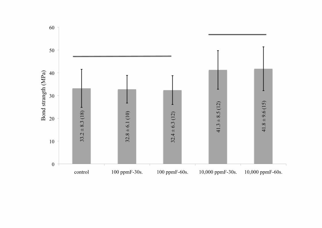
MTBS results are presented in Fig. 2: 10,000 ppmF samples (10,000 ppmF-30
seconds, 10,000 ppmF-60 seconds) showed higher bond strengths than samples exposed
to lower F concentrations. The thickness of the hybrid layer showed no significant
correlations with either fluoride concentration or the duration of fluoride immersion
(Fig. 3).
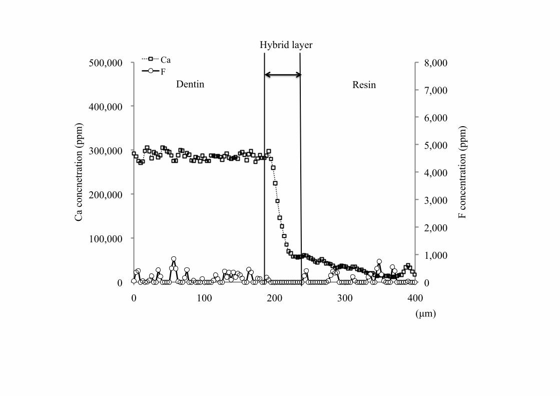
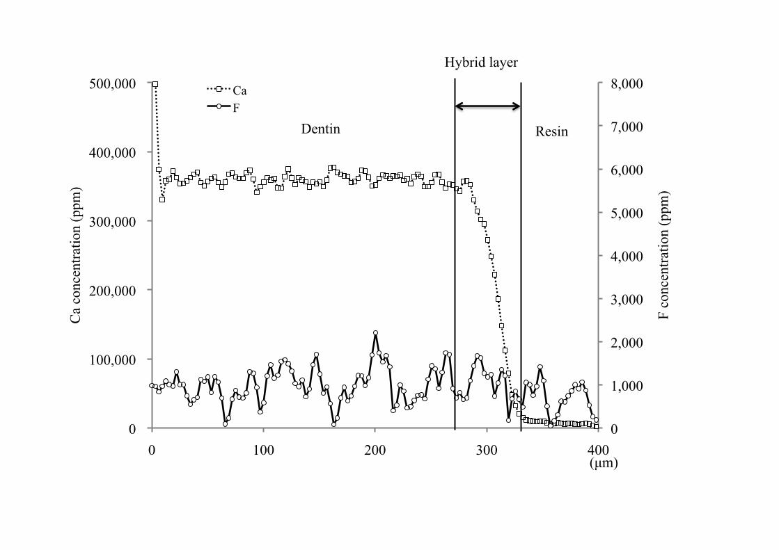
Fluorine and Ca distributions in the dentin, hybrid, and resin composite layers

are presented in Figs. 4–6. Calcium concentrations were highest in dentin,
intermediate in the hybrid layer, and lowest in the resin composite. Fluorine
distributions varied with the concentration of applied fluoride. Fluorine was nearly
absent in the control group (Fig. 4). In the 100 ppmF samples, F concentrations were
low in all areas (dentin, hybrid layer, and resin composite) (Fig. 5). In the 10,000
ppmF samples, fluorine concentrations in the dentin and resin composite were the same
as in the 100 ppmF samples. In the hybrid layer, however, fluorine concentrations in
the 10,000 ppmF samples were higher than those in the 100ppmF samples (Fig. 6).
DISCUSSION
The bonding of dentin and resin composite requires initial demineralization of
the dentin surface by phosphoric acid. Acid etching removes the smear layer and
demineralizes the underlying dentin matrix, exposing the tubule apertures, the collagen
fibrils, and the interfibrillar spaces. In the bonding process, adhesive resin monomers
diffuse into the etched dentin, and polymerization of these monomers results in the
formation of a hybrid layer.
Fluoride applications increase the adhesion of resin composites to dentin.
However, the increased adhesion is apparently unrelated to the thickness of the hybrid
layer, which showed no significant correlation to the concentration of applied fluoride.
The concentration of applied fluoride also had no effect on the structure of the hybrid
layer or on the infiltration patterns of resin into the demineralized area.
Strong bonding between dentin and resin composite depends on the thorough
penetration of adhesive into the dentin substrate [24], facilitated by demineralization by
phosphoric acid etching. The application of fluoride to demineralized dentin does not
appear to impede the penetration of adhesive.
Applications of high concentrations of fluoride (10,000 ppmF) resulted in higher
bond strengths than those observed in control groups. Bond strength is apparently
unrelated to fluorine levels in the dentin, which remained low in both low- and
high-concentration fluoride applications. It is possible that fluorine in dentin is
removed by post-fluoride rinsing, but the details of this process are unknown.
High bond strengths between dentin and resin are correlated with high
concentrations of fluorine in the hybrid layer. Because high concentration of fluorine
was detected into hybrid layer, we guessed deposits of fluoroapatite in the collagen
network of the hybrid layer; this remineralization of dentin occurs by precipitation onto
residual crystals [25]. Thus, the hybrid layer might be reinforced by the
remineralization process. Our results indicate that bond strengths between dentin and
resin depend on the threshold levels of applied fluoride, since the MTBS was
significantly greater in 10,000 ppmF samples than in both the 100 ppmF and control
samples.
Itota et al. [12] reported that fluoride ions were detected on artificially
demineralized dentin (caries dentin); the fluoride was introduced without a
post-application rinse, by means of EDS. However, their research indicated that
dentin-resin bond strength did not increase when the fluoride application was followed
by water rinsing; our results do not conform with their findings, probably because of
differences in (1) the substrate used for the bonding tests, and (2) the type of etching
that preceded the application of adhesive. Itota et al. applied a self-etching primer to
artificial caries, whereas in our research protocol, a more aggressive total-etching
technique (using phosphoric acid) was applied to undeteriorated dentin. Because

phosphoric acid has a lower pH than self-etching primer, phosphoric acid penetrates the
dentin more deeply and causes greater demineralization of dentin as compared with
self-etching primer. Thus, the total-etching technique affords a greater region for
adhesive bonding, as well as structural strengthening due the crystallization of
fluoroapatite within the hybrid bonding layer.
CONCLUSION
The results of this study indicate that high concentrations of topical fluoride applied to
demineralized dentin after etching improve the bond strength between dentin and resin
composite. µ-PIGE analyses detected enhanced fluorine levels on the interface
between dentin and resin composite (hybrid layer), suggesting that fluorine
(fluoroapatite) enhances structural integrity and bond strength. The research clarifies
the relationships between topical fluoride applications, fluorine uptake in dentin and
resin composite compounds, and the resulting strengthening of dentin–resin composite
bonds.
ACKNOWLEGEMENTS
We would like to thank the members of the department of Advanced Radiation
Technology, TARRI, JAEA, for their operation of the accelerator facility.
References
[1] B. Øgaard, J. Dent. Res. 69 Spec (1990) 813.
[2] P. F. DePaola, P. M. Soparkar, C. Triol, A. R. Volpe, L. Garcia, J. Duffy, and B.
Vaughan, Am. J. Dent. 6 Spec (1993) S7.
[3] B. Øgaard, L. Seppa, and G. Rolla, Adv. Dent. Res. 8 (1994) 190.
[4] K. W. Stephen, Adv. Dent. Res. 8 (1994) 185.
[5] M. Nagamine, T. Itota, Y. Torii, M. Irie, M. Staninec, and K. Inoue, Am. J. Dent. 10
(1997) 173.
[6] T. Itota, S. Nakabo, Y. Iwai, N. Konishi, M. Nagamine, Y. Torii, and M. Yoshiyama,
Oper. Dent. 26 (2001) 445.
[7] T. Itota, S. Nakabo, Y. Iwai, N. Konishi, M. Nagamine, and Y. Torii, J. Oral. Rehabil.
29 (2002) 523.
[8] K. Okuyama, T. Nakata, P. N. R. Pereira, C. Kawamoto, H. Komatsu, and H. Sano,
Oper. Dent. 31 (2006) 135.
[9] L. E. Tam, G. P. Chan, and D. Yim, Oper. Dent. 22 (1997) 4.
[10] P. N. R. Pereira, S. Inokoshi, and J. Tagami, J. Dent. 26 (1998) 505.
[11] N. Nakabayashi, K. Kojima, and E. Masuhara, J. Biomed. Mater. Res. 16 (1982)
265.
[12] T. Itota, Y. Torii, S. Nakabo, and M. Yoshiyama, J. Prosthet. Dent. 88 (2002) 503.
[13] H. Komatsu, H. Yamamoto, M. Nomachi, K. Yasuda, Y. Matsuda, Y. Murata, T.
Kijimura, H. Sano, T. Sakai, and T. Kamiya, Nucl. Instrum. Methods Phys. Res. B 260
(2007) 201.
[14] H. Komatsu, H. Yamamoto, M. Nomachi, K. Yasuda, Y. Matsuda, M. Kinugawa, T.
Kijimura, H. Sano, T. Satou, S. Oikawa, and T. Kamiya, Nucl. Instrum. Methods Phys.
Res. B 267 (2009) 2136.
[15] M. Nomachi, K. Yasuda, H. Yamamoto, Y. Iwami, S. Ebisu, Y. Sugaya, T. Kamiya,
T. Sakai, M. Fukuda, and Y. Naitoh, TIARA Ann. Rep. 1998, JAERI-Review 99-025
(1999) 226.
[16] T. Sakai, Y. Naitoh, T. Kamiya, S. Matuyama, K. Gotoh, S. Yokota, H. Yamasaki,
and K. Ishii, Biol. Trace Elem. Res. 71–72 (1999) 77.
[17] M. Nomachi, K. Yasuda, H. Yamamoto, Y. Iwami, S. Ebisu, T. Kamiya, T. Sakai,
and M. Oikawa, TIARA Ann. Rep. 1999, JAERI-Review 2000-024 (2000) 242.
[18] T. Sakai, T. Kamiya, M. Oikawa, T. Sato, A. Tanaka, and K. Ishii, Nucl. Instrum.
Methods Phys. Res. B 190 (2002) 271.
[19] M. Nomachi, Y. Sugaya, M. Yoshifuku, K. Yasuda, H. Yamamoto, Y. Iwami, S.
Ebisu, T. Kamiya, T. Sakai, and M. Oikawa, TIARA Ann. Rep. 2000, JAERI-Review
2001-039 (2001) 244.
[20] H. Yamamoto, M. Nomachi, Y. Sugaya, M. Yoshifuku, K. Yasuda, Y. Iwami, S.

Ebisu, T. Kamiya, T. Sakai, and M. Oikawa, TIARA Ann. Rep. 2001, JAERI-Review
2002-035 (2002) 258.
[21] H. Yamamoto, M. Nomachi, K. Yasuda, Y. Iwami, S. Ebisu, T. Kamiya, and T.
Sakai, Nucl. Instrum. Methods Phys. Res. B 210 (2003) 388.
[22] H. Yamamoto, M. Nomachi, K. Yasuda, Y. Iwami, S. Ebisu, T. Sakai, and M.
Fukuda, Nucl. Instrum. Methods Phys. Res. B 231 (2005) 300.
[23] H. Yamamoto, M. Nomachi, K. Yasuda, Y. Iwami, S. Ebisu, H. Komatsu, T. Sakai,
and T. Kamiya, Nucl. Instrum. Methods Phys. Res. B 260 (2007) 194.
[24] S. Inokoshi, H. Hosoda, C. Harnirattisai, and Y. Shimada, Oper. Dent. 18 (1993) 8.
[25] B. Klont, and J. M. Ten Cate, Caries Res. 25 (1991) 39.

A
C
(a
D
A
D
(b
33.2
± 8
.3 (1
8)
32.8
± 6
.1 (1
0)
32.4
± 6
.3 (1
2)
41.3
± 8
.5 (1
2)
41.8
± 9
.6 (1
5)
0
10
20
30
40
50
60
control 100 ppmF-30s. 100 ppmF-60s. 10,000 ppmF-30s. 10,000 ppmF-60s.
Bon
d st
rang
th (M
Pa)

3.0
± 0.
6 (6
)
2.9
± 0.
5 (6
)
3.0
± 0.
3 (6
)
3.3
± 0.
6 (6
)
3.3
± 0.
4 (6
)
0
1
2
3
4
5
control 100ppmF-30s. 100ppmF-60s. 10,000ppmF-30s. 10,000ppmF-60s.
Hyb
rid la
yer t
hick
ness
(µm
)
0
1,000
2,000
3,000
4,000
5,000
6,000
7,000
8,000
0
100,000
200,000
300,000
400,000
500,000
0 100 200 300 400
F co
ncen
tratio
n (p
pm)
Ca
conc
netra
tion
(ppm
)
(µm)
Ca F
Resin Dentin
Hybrid layer
0
1,000
2,000
3,000
4,000
5,000
6,000
7,000
8,000
0
100,000
200,000
300,000
400,000
500,000
0 100 200 300 400
F co
ncen
tratio
n (p
pm)
Ca
conc
entra
tion
(ppm
)
(µm)
Ca F
Dentin Resin
Hybrid layer

0
1,000
2,000
3,000
4,000
5,000
6,000
7,000
8,000
0
100,000
200,000
300,000
400,000
500,000
0 100 200 300 400
F co
ncen
tratio
n (p
pm)
Ca
conc
entra
tion
(ppm
)
(µm)
Ca F Dentin Resin
Hybrid layer