Efficacy of treatment with glycosaminoglycans on experimental collagen-induced arthritis in rats
10
R122 Introduction Rheumatoid arthritis (RA) is a common human autoim- mune disease characterised by chronic inflammation of the synovial joints and by subsequent progressive, erosive destruction of articular tissue [1]. This disease affects about 1% of the human population. The aetiology and pathogenesis of this disease are not yet fully understood but it seems likely that an autoimmune-mediated attack on the joints has a crucial role in the pathogenesis of RA [2]. Collagen-induced arthritis (CIA) in Lewis rats is a widely used experimental animal model of inflammatory polyarthri- tis with clinical and pathological features similar to those of human RA that are dependent on both humoral and cel- lular immunity to the immunising antigen [3]. It has been suggested previously that the generation of free radicals and other reactive oxygen species (ROS) such as singlet oxygen and hypochlorous acid might have a role in the pathogenesis of degenerative joint disease [4]. ROS are highly reactive transient chemical species with the potential to initiate cellular damage in joint tissues. These reactive molecules are formed during normal aerobic metabolism in cells and after the activation CIA = collagen-induced arthritis; C4S = chondroitin-4-sulphate; GAG = glycosaminoglycan; GSH = reduced glutathione; HYA = hyaluronic acid; RA = rheumatoid arthritis; ROS = reactive oxygen species; SOD = superoxide dismutase; TNF-α = tumour necrosis factor-α. Arthritis Research & Therapy Vol 5 No 3 Campo et al. Research article Efficacy of treatment with glycosaminoglycans on experimental collagen-induced arthritis in rats Giuseppe M Campo 1 , Angela Avenoso 1 , Salvatore Campo 1 , Alida M Ferlazzo 2 , Domenica Altavilla 3 and Alberto Calatroni 1 1 Department of Biochemical, Physiological and Nutritional Sciences, School of Medicine, University of Messina, Messina, Italy 2 Department of Morphology, Biochemistry, Physiology and Animal Production, School of Veterinary Medicine, University of Messina, Messina, Italy 3 Department of Experimental and Clinical Medicine and Pharmacology, School of Medicine, University of Messina, Messina, Italy Corresponding author: Giuseppe M Campo (e-mail: [email protected]) Received: 14 Aug 2002 Revisions requested: 10 Sep 2002 Revisions received: 20 Dec 2002 Accepted: 12 Feb 2003 Published: 6 Mar 2003 Arthritis Res Ther 2003, 5:R122-R131 (DOI 10.1186/ar748) © 2003 Campo et al., licensee BioMed Central Ltd (Print ISSN 1478-6354; Online ISSN 1478-6362). This is an Open Access article: verbatim copying and redistribution of this article are permitted in all media for any purpose, provided this notice is preserved along with the article's original URL. Abstract To evaluate the antioxidant activity of the glycosaminoglycans hyaluronic acid (HYA) and chondroitin-4-sulphate (C4S), we used a rat model of collagen-induced arthritis (CIA). Arthritis was induced in Lewis rats by multiple intradermal injections of 250 µl of emulsion containing bovine type II collagen in complete Freund’s adjuvant at the base of the tail and into three to five other sites on the back. Rats were challenged again with the same antigen preparation 7 days later. Disease developed about 11 days after the second immunization. The effects of treatment in the rats were monitored by biochemical parameters and by macroscopic and histological evaluations in blood, synovial tissue and articular cartilage. Arthritis produced the following symptoms: severe periarticular erythema, edema and inflammation in the hindpaws; membrane peroxidation in the cartilage of the joints; endogenous antioxidant wasting; high tumour necrosis factor-α (TNF-α) plasma levels; and synovial neutrophil accumulation. Treatment with HYA and C4S, starting at the onset of arthritis for 10 days, limited the erosive action of the disease in the articular joints of knee and paw, reduced lipid peroxidation, restored the endogenous antioxidants reduced glutathione (GSH) and superoxide dismutase, decreased plasma TNF-α levels, and limited synovial neutrophil infiltration. These data confirm that erosive destruction of the joint cartilage in CIA is due at least in part to free radicals released by activated neutrophils and produced by other biochemical pathways. The beneficial effects obtained with the treatment suggest that HYA and C4S could be considered natural endogenous macromolecules to limit erosive damage in CIA or as a useful tool with which to study the involvement of free radicals in rheumatoid arthritis. Keywords: antioxidants, collagen-induces arthritis, free radicals, glycosaminoglycans, lipid peroxidation Open Access
-
Upload
magaraibleo -
Category
Documents
-
view
1 -
download
0
Transcript of Efficacy of treatment with glycosaminoglycans on experimental collagen-induced arthritis in rats
R122
IntroductionRheumatoid arthritis (RA) is a common human autoim-mune disease characterised by chronic inflammation ofthe synovial joints and by subsequent progressive, erosivedestruction of articular tissue [1]. This disease affectsabout 1% of the human population. The aetiology andpathogenesis of this disease are not yet fully understoodbut it seems likely that an autoimmune-mediated attack onthe joints has a crucial role in the pathogenesis of RA [2].
Collagen-induced arthritis (CIA) in Lewis rats is a widelyused experimental animal model of inflammatory polyarthri-
tis with clinical and pathological features similar to thoseof human RA that are dependent on both humoral and cel-lular immunity to the immunising antigen [3].
It has been suggested previously that the generation offree radicals and other reactive oxygen species (ROS)such as singlet oxygen and hypochlorous acid might havea role in the pathogenesis of degenerative joint disease[4]. ROS are highly reactive transient chemical specieswith the potential to initiate cellular damage in jointtissues. These reactive molecules are formed duringnormal aerobic metabolism in cells and after the activation
CIA = collagen-induced arthritis; C4S = chondroitin-4-sulphate; GAG = glycosaminoglycan; GSH = reduced glutathione; HYA = hyaluronic acid;RA = rheumatoid arthritis; ROS = reactive oxygen species; SOD = superoxide dismutase; TNF-α = tumour necrosis factor-α.
Arthritis Research & Therapy Vol 5 No 3 Campo et al.
Research articleEfficacy of treatment with glycosaminoglycans on experimentalcollagen-induced arthritis in ratsGiuseppe M Campo1, Angela Avenoso1, Salvatore Campo1, Alida M Ferlazzo2,Domenica Altavilla3 and Alberto Calatroni1
1Department of Biochemical, Physiological and Nutritional Sciences, School of Medicine, University of Messina, Messina, Italy2Department of Morphology, Biochemistry, Physiology and Animal Production, School of Veterinary Medicine, University of Messina, Messina, Italy3Department of Experimental and Clinical Medicine and Pharmacology, School of Medicine, University of Messina, Messina, Italy
Corresponding author: Giuseppe M Campo (e-mail: [email protected])
Received: 14 Aug 2002 Revisions requested: 10 Sep 2002 Revisions received: 20 Dec 2002 Accepted: 12 Feb 2003 Published: 6 Mar 2003
Arthritis Res Ther 2003, 5:R122-R131 (DOI 10.1186/ar748)© 2003 Campo et al., licensee BioMed Central Ltd (Print ISSN 1478-6354; Online ISSN 1478-6362). This is an Open Access article: verbatimcopying and redistribution of this article are permitted in all media for any purpose, provided this notice is preserved along with the article's originalURL.
Abstract
To evaluate the antioxidant activity of the glycosaminoglycanshyaluronic acid (HYA) and chondroitin-4-sulphate (C4S), weused a rat model of collagen-induced arthritis (CIA). Arthritiswas induced in Lewis rats by multiple intradermal injections of250 µl of emulsion containing bovine type II collagen incomplete Freund’s adjuvant at the base of the tail and intothree to five other sites on the back. Rats were challengedagain with the same antigen preparation 7 days later. Diseasedeveloped about 11 days after the second immunization. Theeffects of treatment in the rats were monitored by biochemicalparameters and by macroscopic and histological evaluations inblood, synovial tissue and articular cartilage. Arthritis producedthe following symptoms: severe periarticular erythema, edemaand inflammation in the hindpaws; membrane peroxidation inthe cartilage of the joints; endogenous antioxidant wasting;
high tumour necrosis factor-α (TNF-α) plasma levels; andsynovial neutrophil accumulation. Treatment with HYA andC4S, starting at the onset of arthritis for 10 days, limited theerosive action of the disease in the articular joints of knee andpaw, reduced lipid peroxidation, restored the endogenousantioxidants reduced glutathione (GSH) and superoxidedismutase, decreased plasma TNF-α levels, and limitedsynovial neutrophil infiltration. These data confirm that erosivedestruction of the joint cartilage in CIA is due at least in part tofree radicals released by activated neutrophils and producedby other biochemical pathways. The beneficial effects obtainedwith the treatment suggest that HYA and C4S could beconsidered natural endogenous macromolecules to limiterosive damage in CIA or as a useful tool with which to studythe involvement of free radicals in rheumatoid arthritis.
Keywords: antioxidants, collagen-induces arthritis, free radicals, glycosaminoglycans, lipid peroxidation
Open Access

Available online http://arthritis-research.com/content/5/3/R122
R123
of phagocytes during infection or inflammation; a conse-quence of the uncontrolled production of free radicals isdamage to biomolecules leading to altered function anddisease [5]. There are many pieces of evidence, bothdirect and indirect, implicating radicals in the pathogene-sis of inflammatory synovitis, such as the capacity ofseveral cells that are present in the inflamed joint(macrophages, neutrophils, lymphocytes and endothelialcells) to produce free radicals when isolated and stimu-lated [6]. Cells are normally protected from ROS-induceddamage by a variety of endogenous scavenging proteins,enzymes and chemical compounds that constitute theendogenous antioxidant systems [7]. It has been reportedthat ROS destroy antioxidant systems (in fact the enzy-matic and/or non-enzymatic antioxidant systems areimpaired in RA) and that RA patients are thus exposed tooxidant stress and lipid peroxidation because of thereduced antioxidant defence system [8,9].
Glycosaminoglycans (GAGs), a large family of hetero-geneous polysaccharides, are linear sulphate-substitutedpolymers composed of alternating hexuronic acid and hex-osamine units that are important in all living organisms [10].Their structure and degree of heterogeneity seem to behighly specific; the ability of several proteins to bind GAGsmight reflect functional relationships and is likely to beexploited physiologically in a variety of ways. Several reportshave shown that during the progression of RA the physio-logical levels of blood GAGs are increased [11–13]. Theobvious explanation is that GAGs originate from the metab-olism of the joint cartilage damaged by erosion. Neverthe-less, the exact meaning of their increase is still unclear.
Molecules able to limit the generation and the effects ofROS exert a protective action in a variety of experimentalinflammatory diseases, including CIA [14–17]. Manyinvestigators have described the antioxidant properties ofsome GAGs (mainly for hyaluronic acid [HYA] and chon-droitin-4-sulphate [C4S]) in experimental models, bothin vitro and in vivo [18–22].
Starting from these findings, the aim of the present studywas to assess the possible ability of HYA and C4S in lim-iting inflammation and joint cartilage erosion in an experi-mental model of CIA in Lewis rats.
Materials and methodsAnimalsMale Lewis rats 6–7 weeks old, with a mean weight of175–200 g, were used in our study. Rats, purchased fromCharles River (Calco, Italy), were maintained underclimate-controlled conditions in a 12 hours light : 12 hoursdark cycle. The animals were fed with standard rodentchow and water ad libitum. The health status of the animalcolony was monitored in accordance with the guidelinesfrom the Italian Veterinary Board. Rats were subdivided
into the following groups: (1) control (n = 7); (2) CIA plusvehicle (n = 7); (3) CIA plus HYA (25 mg/kg) (n = 7); (4)CIA plus C4S (25 mg/kg) (n = 7); (5) CIA plus HYA plusC4S (each at 25 mg/kg) (n = 7).
MaterialsHyaluronic acid from human umbilical cord, chondroitin sul-phate A from bovine trachea and bovine type II collagenwere purchased from Sigma–Aldrich Srl (Milan, Italy); com-plete Freund’s adjuvant was obtained from Difco Laborato-ries (Detroit, MI, USA). All other reagents were purchasedfrom Fluka (division of Sigma–Aldrich Srl, Milan, Italy).
Induction of arthritisCIA was induced in rats as described previously [23], bymultiple intradermal injections, at the base of the tail andinto three to five other sites on the back, of 250 µg ofbovine type II collagen in 125 µl of 0.1 M acetic acid emul-sified in an equal volume of complete Freund’s adjuvantcontaining 2 mg/ml Mycobacterium tuberculosis H37 RA[23]. Rats were challenged again with the same antigenpreparation 7 days later. Before injection, animals wereanaesthetised with ether and injections were performedwith a 15 gauge needle. Disease developed about11 days after the second immunisation.
Treatment with GAGsAt day 11, animals were randomised to receive treatmentslisted in the Animals section, timed to coincide approxi-mately with the onset of arthritis pathology. GAGs weredissolved in physiological saline (0.9% NaCl) and adminis-tered daily, in a volume of 1 ml/kg body weight, intraperi-toneally once a day until the 20th day.
Plasma GAG evaluationEvaluation of plasma galactosamine and glucosamine wasperformed at day 21 to estimate indirectly the concentrationof HYA and C4S in the blood of animals after the intraperi-toneal treatment. Samples of blood (1.5ml) were drawn, atthe end of the experiment, from a tail vessel. The blood wascollected in polyethylene tubes with the previous addition of75µl of heparin solution (4000 IU).The plasma samplesobtained after centrifugation for 10 min at 3000g and 4°Cwere frozen at –80°C until assay. On the day of analysis,GAGs were first isolated and purified and then hydrolysedinto their constituent monosaccharides [24]. These aminosugars (glucosamine and galactosamine) were thenassayed by a specific HPLC method [24].
Arthritis assessmentsEvaluation of joint inflammation was performed by ablinded independent observer with no knowledge of thetreatment protocol. The severity of the arthritis in eachpaw was quantified daily by a clinical score measurement[25] from 0 to 4 as follows: 0, no macroscopic signs ofarthritis; 1, swelling of one group of joints (namely, wrist or

Arthritis Research & Therapy Vol 5 No 3 Campo et al.
R124
ankle joints); 2, two groups of swollen joints; 3, threegroups of swollen joints; 4; swelling of the entire paw. Themaximum score for each rat was 16. Clinical severity wasalso assessed by the quantification of the paw volumechanges. Measurements were performed with a dialgauge caliper. Changes in body weight were monitored todetermine the rate of the increment in each rat.
Histological analysisRats were killed at day 21 by ether narcosis; hind limbswere removed and fixed in 10% buffered formalin. Thelimbs were decalcified in 5% formic acid, processed forparaffin embedding, sectioned at 5 µm thickness, and sub-sequently stained with haematoxylin–eosin for examinationunder a light microscope [26]. Sections were examinedfor the presence of hyperplasia of the synovium, pannusformation, and destruction of the joint space.
Lipid peroxidation analysisDetermination of malonaldehyde in the articular tissue wasperformed to estimate the extent of lipid peroxidation inthe damaged cartilage. At the end of the experiment hindlimbs were removed and maintained at 0°C, then the jointcartilage was quickly separated from the bone and muscu-lar tissue and frozen at –80°C until assay. On the day ofanalysis, after thawing, cartilage samples were washed inice-cold 20 mM Tris-HCl, pH 7.4, and blotted onabsorbent paper. Each sample was then minced in ice-cold 20 mM Tris-HCl, pH 7.4 containing 1 mg/ml butylatedhydroxytoluene and homogenised in a 1 : 10 (w/v) ratiowith an Ultra-Turrax homogeniser. After centrifugation for10 min at 3000g and 4°C, the clear supernatant was usedfor biochemical assay. Analysis was performed with a col-orimetric commercial kit (Lipid peroxidation assay kit, cat.no. 437634; Calbiochem, La Jolla, CA, USA). In brief,0.65 ml of 10.3 mM N-methyl-2-phenylindole in acetonitrilewas added to 0.2 ml of homogenate supernatant. Aftervortex-mixing for 3–4 s and the addition of 0.15 ml of 37%HCl, samples were well mixed, closed with a tight stopperand incubated for 60 min at 45°C. The samples were thencooled on ice and the absorbance was measured spec-trophotometrically at 586 nm. A calibration curve of anaccurately prepared standard malonaldehyde solution wasalso run for quantification. The malonaldehyde concentra-tion was expressed as nmol/mg of protein.
Determination of GSHSamples of joint cartilage obtained at the end of the experi-ment were frozen at –80°C until GSH assay. The analysiswas performed with a spectrophotometric method [27]. Inbrief, tissue samples were homogenised in a solution con-taining 5% trichloroacetic acid and 5mM EDTA at 4°C.Then each sample was centrifuged for 10 min at 15,000gand 4°C. An aliquot of homogenate supernatant (0.4ml)was added to a dark polyethylene tube containing 1.6ml of0.4M Tris-EDTA buffer, pH 8.9. After vortex-mixing, 40 µl of
10mM dithiobisnitrobenzoic acid in methanol was added.The samples were vortex-mixed again and the absorbanceat 412nm was read after 5 min. The values of unknownsamples were drawn from a standard curve plotted byassaying different known concentrations of GSH. Theamount of GSH was expressed as µmol/g of protein.
Evaluation of superoxide dismutase (SOD)Samples of joint cartilage were washed with 0.9% NaClcontaining 0.16mg/ml heparin and homogenised with aUltra-Turrax homogeniser in ice-cold 0.25M sucrose con-taining 1mM diethylenetriamine pentaacetic acid (1 :1,w/v). Each sample was then centrifuged for 20 min at20,000g and 4°C. The supernatant was aspirated and thetotal SOD activity was assayed spectrophotometrically at505nm with a commercial kit (Ransod assay kit, cat. no.Sd 125; Randox Laboratories, Crumlin, UK). In brief, 50 µlof each diluted sample (1 :10 [w/v] in 0.01M potassiumphosphate buffer, pH 7.0) was mixed with 1.7ml of solutioncontaining 0.05mM xanthine and 0.025mM iodonitrotetra-zolium chloride. After being mixed for 5s, 250 µl of xanthineoxidase (80U/l) was added. The time between reading theinitial and final absorbances was 3 min. A standard curve ofSOD solution (from 2 to 32U/ml) was run for quantifica-tion. All standard and diluted sample rates were convertedinto a percentage of the buffer diluent rate and subtractedfrom 100% to give a percentage inhibition. Sample SODactivities were obtained from a plotted curve of the per-centage inhibition for each standard. SOD values wereexpressed as units/mg of protein.
Plasma tumour necrosis factor-α (TNF-α) assayPlasma TNF-α concentration was determined with anELISA commercial kit (Rat TNF-α ELISA kit ultra sensitive,cat. no. 22079; NBS Biologicals Ltd, Huntingdon, UK). Atthe end of the experiment, samples of blood (0.5 ml) weredrawn from a tail vessel. The blood was collected in poly-ethylene tubes with the previous addition of 25 µl ofheparin solution (4000 IU).The plasma samples obtainedafter centrifugation for 10 min at 3000g and 4°C werefrozen at –80°C until assay. In brief, 100 µl of standards,samples and controls were added to each well of thecoated microplate. After 3 hours of incubation at 24°C themicroplate was decanted and the liquid discarded. Then,100 µl of biotinylated anti-TNF-α antibody was added toeach well. After 45 min of incubation at 24°C and a furtherelimination of the liquid from the wells, 100 µl of Strepta-vidin–horseradish peroxidase conjugate was added. Afterincubation for a further 45 min and a washing of the wells,100 µl of chromogen were added. The absorbance ofeach well was read spectrophotometrically at 450 nm.TNF-α values were expressed as ng/ml.
Articular neutrophil accumulationMyeloperoxidase activity was analysed as an index of neu-trophil infiltration in the synovial tissue, because it is
closely correlated with the number of neutrophils presentin the tissue [28]. We measured myeloperoxidase in thesynovial tissue of joints by a specific assay for this enzyme[29]. In brief, synovial tissue samples were separated fromrat joints and were first homogenised in a solution contain-ing 20 mM potassium phosphate buffer, pH 7.0, to 1 : 10(w/v) and then centrifuged for 30 min at 20,000g and 4°C.The supernatant of each sample was discarded and theresulting pellet was added to a buffer solution consistingof 0.5% hexadecyltrimethylammonium bromide dissolvedin 50 mM potassium phosphate buffer, pH 6, containing50 µl of protease and phosphatase inhibitor cocktails.Samples were then sonicated for 1 min and centrifugedfor 30 min at 20,000g and 4°C. An aliquot of the super-natant was allowed to react with a solution of o-dianisidinedihydrochloride (0.167 mg/ml) and 0.0005% hydrogenperoxide. The rate of change in absorbance was mea-sured spectrophotometrically at 405 nm. Myeloperoxidaseactivity has been defined as the concentration of enzymedegrading 1 µmol of peroxide/min at 37°C and wasexpressed as U/g of protein.
Statistical analysisData are expressed as means ± SD. The differencebetween the means of two groups was evaluated with anANOVA and was considered significant at P < 0.05.
Statement of animal careThe studies reported in this manuscript have been per-formed in accordance with the declaration of Helsinki andwith the Guide for the Care and Use of Laboratory Animals.
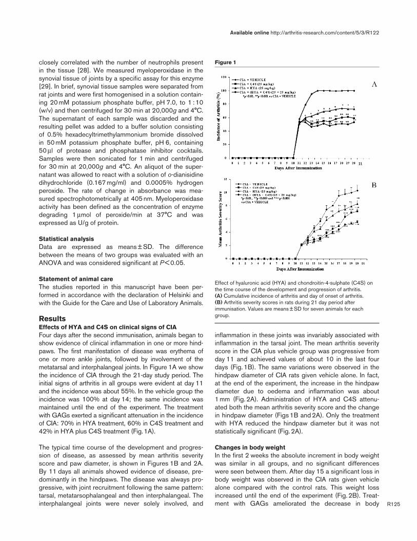
ResultsEffects of HYA and C4S on clinical signs of CIAFour days after the second immunisation, animals began toshow evidence of clinical inflammation in one or more hind-paws. The first manifestation of disease was erythema ofone or more ankle joints, followed by involvement of themetatarsal and interphalangeal joints. In Figure 1A we showthe incidence of CIA through the 21-day study period. Theinitial signs of arthritis in all groups were evident at day 11and the incidence was about 55%. In the vehicle group theincidence was 100% at day 14; the same incidence wasmaintained until the end of the experiment. The treatmentwith GAGs exerted a significant attenuation in the incidenceof CIA: 70% in HYA treatment, 60% in C4S treatment and42% in HYA plus C4S treatment (Fig.1A).
The typical time course of the development and progres-sion of disease, as assessed by mean arthritis severityscore and paw diameter, is shown in Figures 1B and 2A.By 11 days all animals showed evidence of disease, pre-dominantly in the hindpaws. The disease was always pro-gressive, with joint recruitment following the same pattern:tarsal, metatarsophalangeal and then interphalangeal. Theinterphalangeal joints were never solely involved, and
inflammation in these joints was invariably associated withinflammation in the tarsal joint. The mean arthritis severityscore in the CIA plus vehicle group was progressive fromday 11 and achieved values of about 10 in the last fourdays (Fig. 1B). The same variations were observed in thehindpaw diameter of CIA rats given vehicle alone. In fact,at the end of the experiment, the increase in the hindpawdiameter due to oedema and inflammation was about1 mm (Fig. 2A). Administration of HYA and C4S attenu-ated both the mean arthritis severity score and the changein hindpaw diameter (Figs 1B and 2A). Only the treatmentwith HYA reduced the hindpaw diameter but it was notstatistically significant (Fig. 2A).
Changes in body weightIn the first 2 weeks the absolute increment in body weightwas similar in all groups, and no significant differenceswere seen between them. After day 15 a significant loss inbody weight was observed in the CIA rats given vehiclealone compared with the control rats. This weight lossincreased until the end of the experiment (Fig. 2B). Treat-ment with GAGs ameliorated the decrease in body
Available online http://arthritis-research.com/content/5/3/R122
R125
Figure 1
Effect of hyaluronic acid (HYA) and chondroitin-4-sulphate (C4S) onthe time course of the development and progression of arthritis. (A) Cumulative incidence of arthritis and day of onset of arthritis. (B) Arthritis severity scores in rats during 21 day period afterimmunisation. Values are means ± SD for seven animals for eachgroup.

weight; the greatest effect was achieved after treatmentwith both polymers (Fig. 2B).
HistologyRepresentative joint histopathology of the experimentalgroups is shown in Figure 3. A characteristic of arthriticjoints in rats with CIA is synovial hyperplasia, pannus for-mation, exudation of cells into the joint space, and erosionof bone and cartilage. A massive influx of inflammatorycells, synovial hyperplasia, and accumulation of abundantmonomorphonuclear and polymorphonuclear cells in thejoint space are evident (Fig. 3B) compared with a normalcontrol group (Fig. 3A). By comparison, rats treated withHYA plus C4S revealed minimal evidence of inflammationor joint destruction. In fact the synovial membrane in thejoints was like normal synovium, with few signs of synovialhyperplasia or other characteristics of inflammation(Fig. 3E). A reduced degree of arthritis severity was alsoobserved in the rats that received only HYA or C4S(Fig. 3C,D).
Plasma GAG levelsBecause HYA contains the amino sugar glucosamine as acomponent, whereas C4S contains galactosamine,Table 1 reports the concentrations of GAGs, expressed interms of glucosamine and galactosamine, in rats thatunderwent CIA after 10 days of treatment with HYA, C4Sor both substances. In the control group, galactosaminelevels were 9.81 ± 1.11 mg/l while glucosamine levelswere 10.35 ± 1.71 mg/l; these values were consideredphysiological. However, in CIA rats treated with vehiclealone there was a significant increase in both aminosugars. The increase was even more significant in thethree groups administered with HYA, C4S or both GAGs.The intraperitoneal administration of HYA increased glu-cosamine levels by about 31%; treatment with C4Sincreased galactosamine levels by about 30%. The admin-istration of both GAGs increased both glucosamine andgalactosamine (30% and 22% respectively; Table 1).
Arthritis Research & Therapy Vol 5 No 3 Campo et al.
R126
Figure 2
Effect of hyaluronic acid (HYA) and chondroitin-4-sulphate (C4S) ontime course of change in paw diameter (A) and body weight increment(B) in rats with collagen-induced arthritis during the 21 days afterimmunisation. Values are means±SD for seven animals for each group.
Table 1
Effect of hyaluronic acid (HYA) and chondroitin-4-sulphate (C4S) administration on plasma levels of galactosamine andglucosamine in rats subjected to collagen-induced arthritis (CIA), at the end of the experiment (day 21)
Concentration in plasma (mg/l) Increase (%)
Experimental group Galactosamine Glucosamine Galactosamine Glucosamine
Control 9.81 ± 1.11 10.35 ± 1.71
CIA + vehicle 12.58 ± 1.45* 14.79 ± 2.01*
CIA + HYA 14.68 ± 2.74 19.34 ± 3.27† +17 +31
CIA + C4S 16.34 ± 3.18† 14.97 ± 3.16 +30 +1
CIA + HYA + C4S 15.39 ± 1.86† 19.24 ± 3.36† +22 +30
Where added, the concentrations of HYA and C4S were each 25 mg/kg. Values are means ± SD for seven rats for each group.Significance: *P < 0.01 versus control; †P < 0.05 versus CIA + vehicle.

Assessment of malonaldehyde (MAL)Determination of malonaldehyde in the articular cartilagewas performed to estimate free-radical damage to biologi-cal membranes (Fig. 4). Low levels of malonaldehyde wereseen in the control group at the end of the experiment(day 21); these values were considered normal. In con-trast, a significant increase in malonaldehyde productionwas found in the joints of CIA rats given vehicle alone.Treatment with GAGs decreased malonaldehyde concen-trations by inhibiting lipid peroxidation in the cartilagetissue. Treatment with HYA was at the limit of significance,whereas the maximum effect was observed by administer-ing HYA plus C4S (Fig. 4).
GSH assayThe concentration of GSH was evaluated to estimateendogenous defences against hydrogen peroxide forma-tion. Figure 5A shows the changes in GSH content evalu-ated in the joint articular cartilage (day 21) in theexperimental groups. In normal control rats GSH levelsranged between 5.0 and 7.0 µmol/g of protein. In contrast,a marked decrease in GSH concentrations was found inthe joint articular cartilage of CIA rats given vehicle alone.Treatment with each of the two polymers significantlyinhibited the decrease in GSH levels. In this case, too, themaximum effect was obtained in the group treated withboth polymers.
SOD activitySOD activity was evaluated to estimate endogenousdefences against superoxide anions. Figure 5B sum-marises the articular cartilage content of SOD in theexperimental groups. In control animals normal SOD activ-ities ranged between 10.0 and 15.0 U/mg of protein. Incontrast, a significant decrease in this antioxidant wasseen in CIA rats treated only with vehicle. As previously,chronic administration of the two GAGs significantlylimited the decline in SOD.
TNF-αα levelsFigure 6 reports the changes in TNF-α concentrationassayed in plasma of rats at the end of the experiment. Inthe control group the normal levels of TNF-α rangedbetween 20.0 and 40.0 pg/ml. A marked increase inTNF-α concentration was found in the plasma of CIA ratsgiven vehicle alone. Administration of GAGs significantlyinhibited the increase in the cytokine in the three othergroups.
Available online http://arthritis-research.com/content/5/3/R122
R127
Figure 4
Articular cartilage malonaldehyde (MAL) content of joints of animalswith RA treated with hyaluronic acid (HYA), chondroitin-4-sulphate(C4S) or both glycosaminoglycans. Values are means ± SD for sevenanimals for each group.
Figure 3
Representative joint histopathology of the groups with collagen-induced arthritis (CIA) administered vehicle alone and administeredhyaluronic acid (HYA), chondroitin-4-sulphate (C4S) or bothglycosaminoglycans, at the end of the experiment. (A) control; (B) CIAplus vehicle; (C) CIA plus HYA; (D) CIA plus C4S; (E) CIA plus HYAplus C4S. (Original magnification ×100.)

Myeloperoxidase analysisVery low myeloperoxidase activity was measured in controlrats (Fig.7). In contrast, elevated myeloperoxidase levelswere measured in the vehicle-administered CIA group.However, treatment with HYA and C4S decreased neu-trophil accumulation by reducing myeloperoxidase activity inthe synovial tissue of the joints. The decrease in myeloperoxi-dase activity was similar in all GAG-treated groups (Fig.7).
DiscussionFree radicals have long been implicated as mediators oftissue damage in RA patients [30]. Correspondingly, it hasbeen shown that affected articulations are infiltrated byblood-derived cells, mainly neutrophils, macrophages anddendritic cells [31]. In response to activation, these cellsare responsible for the generation of ROS [32,33], whichare released in large amounts into the surrounding tissue.When the endogenous antioxidant defences are over-come, the resulting production of free radicals inducesimpairment and destruction of the affected joint con-stituents such as synovial fluid, cartilage and other articu-lar constituents [30]. One of several approaches toreduce oxidative stress is treatment with antioxidant com-pounds as therapeutic agents [34–38].
Acid GAGs are present in blood, usually in proteoglycanform. The main GAG of normal human plasma is C4S,which is mostly in a low-sulphate form [39,40]. Keratansulphate, heparan sulphate and HYA are the other GAGstructures usually detected in human plasma [39,40]. Inanimals, the total amounts of GAGs in plasma [41] aresimilar to those measured in humans [42]. Nevertheless,significantly increased plasma concentrations of GAGshave been observed in a wide range of diseases
Arthritis Research & Therapy Vol 5 No 3 Campo et al.
R128
Figure 5
Articular cartilage antioxidant content of joints of animals with RAtreated with hyaluronic acid (HYA), chondroitin-4-sulphate (C4S) orboth glycosaminoglycans. (A) Articular reduced glutathione (GSH)levels. (B) Articular superoxide dismutase (SOD) activity. Values aremeans ± SD for seven animals for each group.
Figure 6
Plasma TNF-α (TNF-α) concentrations assayed in rats with RA treatedwith hyaluronic acid (HYA), chondroitin-4-sulphate (C4S) or bothglycosaminoglycans. Values are means ± SD for seven animals foreach group.
Figure 7
Articular myeloperoxidase (MPO) content of joints of animals with RAtreated with hyaluronic acid (HYA), chondroitin-4-sulphate (C4S) orboth glycosaminoglycans. Values are means ± SD for seven animals foreach group.

[39,43–46] including RA in humans [47,48] and experi-mental arthritis in rat and mouse [49,50]. These changesin circulating GAGs in RA are thought to represent prod-ucts of the connective tissue metabolism, and some circu-lating GAG structures are probably degradation productsoriginating from articular cartilage [51]. However, the highlevels of HYA and other GAGs found in RA [48] could notbe explained only by the erosion of articular cartilage. Infact, the biological role, the real sites of origin and themetabolic fate of these amino-sugar-containing polysac-charides are not clearly understood.
Another approach to explaining the increased presence ofGAG in the plasma comes from evidence that these mole-cules might function as carriers or modulators betweenadjacent cells [52]. It has been shown that GAGs can alterthe binding with selectins because the latter bind severalglycoproteins [53]. HYA and other GAGs could decreasecytokine gene expression directly [54] or indirectly bybinding the CD44 receptor [55]. GAGs might also stimu-late or inhibit cell proliferation in different mesothelioma celllines [56]. In addition, some GAGs possess antioxidantactivity capable of reducing free radicals and inhibiting lipidperoxidation [57–59]. The use of these molecules as thera-peutic agents has shown some positive outcomes both inhumans and in experimental animal models [36,60–64].The present study was designed to evaluate the effect ofchronic treatment with HYA and C4S in a rat model of CIA.The choice of these two compounds from other GAGs wasmade on the basis of the evidence that they show the bestantioxidant activity [36,60–64].
The data obtained in the groups treated with HYA, C4S orboth GAGs were positive in all parameters considered.Nevertheless, treatment with HYA alone showed less pro-tection than that with C4S, and often the data were notsignificant (Fig. 2A,B) or at the limit of significance(Figs 3C, 4, 5A,B and 7). In contrast, the administration ofboth compounds showed maximal effect in limiting theCIA damage in all parameters. The beneficial effect ofHYA and C4S was made evident by measuring the inci-dence of CIA. Similar results were obtained for the meanarthritis severity score. HYA and C4S were also able toreduce the hindpaw diameter and the body weightdecrease observed in the CIA rats receiving vehicle alone.
Lipid peroxidation is considered a critical mechanism of theinjury that occurs during RA [9,65]. The evidence support-ing these biochemical changes is based on the analysis ofa large number of intermediate products [66]. An indicativemethod, extensively used, of evaluating lipid peroxidation isanalysis of tissue malonaldehyde [66]. The large amount ofmalonaldehyde found in the CIA plus vehicle group atday 21 is consistent with the occurrence of damage medi-ated by free radicals. Treatment with the two GAGs pro-duced a significant attenuation of cartilage injury.
The production of oxygen free radicals that occurs withthe development of arthritis in the articular cartilage leadsto decreased GSH and SOD levels as a consequence oftheir consumption during oxidative stress and cellular lysis[67,68]. This decrease contributes to increased cellulardamage by favouring attack by free radicals. HYA andC4S blunted the depletion of GSH and SOD, probably bycompeting in scavenging for free radicals, and as a resulthelped to preserve the integrity of cellular membranes inthe injured cartilage.
The myeloperoxidase results demonstrated that a strongdecrease in infiltration of polymorphonuclear cellsoccurred in the synovial tissue of joints. This decrease andthe other biochemical parameters were evaluated by histo-logical analysis, confirming the protective effects of thetwo polymers. We suggest that the decrease in neutrophilaccumulation induced by GAGs might be due to the inhi-bition of lipid peroxidation and the consequent decrease inthe chemotactic reduction of peroxide [66].
Several areas of investigation have indirectly implicatedTNF-α as a contributor to cellular damage in CIA. The highlevels of this cytokine can be interpreted as a progressionof cartilage cell injury [69]. The antioxidant activity ofGAGs might have lowered plasma TNF-α concentrationsand consequently mitigated articular cell damage.
Which, then, is the mechanism by which HYA and C4Sprotect the cartilage against free radical attack? HYA andC4S are linear polymers formed by alternating hexuronicacid and hexosamine units. HYA is non-sulphated com-pound whereas C4S is sulphated in position 4 of theamino sugar. One plausible explanation for the antioxidantactivity of HYA and C4S is the presence in their structureof a carboxylic group that might bind transition metalssuch as Cu2+ or Fe2+ [59,70], which are in turn responsi-ble for the initiation of Fenton’s reaction. In this reactionthe oxidation of Fe2+ or Cu2+ to Fe3+ or Cu3+ leads to theformation of the detrimental hydroxyl radical (OH•) fromhydrogen peroxide. In this way these molecules mightfunction as metal chelators like the antioxidant deferoxam-ine or the calcium chelator EDTA. Another antioxidantmechanism might be the direct scavenging effect of HYAand C4S on free-radical molecules, especially the OH•
radical or other Fenton’s reaction intermediates such asthe superoxide anion [19,61].
These hypotheses could explain the increased levels ofGAGs during RA. In fact, elevated circulating levels ofGAGs might be a biological response to the production offree radicals. The aim of our work was to increase thephysiological levels of HYA and C4S by administeringthese compounds endogenously. Several studies havepreviously reported an increase in blood and tissue distrib-ution of HYA and C4S after their administration in rats
Available online http://arthritis-research.com/content/5/3/R122
R129

Arthritis Research & Therapy Vol 5 No 3 Campo et al.
R130
[71,72]. We suggest that, after intraperitoneal administra-tion, these polymers might be absorbed by the lymphaticsystem and blood vessels and then they may accumulateat the sites of production of free radicals. In addition, theamounts of HYA and C4S might cause an increase in totalnegative charge with a consequent inhibition of lympho-cyte interactions with the target cell surface. In this wayGAGs could exert a positive anti-inflammatory effect.
Treatment was performed intraperitoneally and not by theoral route, as previously reported by other investigators[64,73]. We suggest that this different mode of adminis-tration, as demonstrated by our results, is preferable to theoral route because a large amount of the two polymerscan reach the inflamed cartilage and they are availablelocally to neutralise transition metals or other reactivespecies.
ConclusionsThe evidence of benefits obtained in this study show thatGAGs are neither a specific drug nor an alternative toactual therapies for RA, but they represent a small step inour understanding of this complex pathology. The hypothe-ses about the mechanism of action in our model, reportedabove, need several further investigations for confirmation.GAGs might then be a useful tool in the study of theinvolvement of free radicals in CIA or in assessing othermodels of damage induced by free radicals.
Competing interestsNone declared.
AcknowledgementsWe gratefully acknowledge the expert technical assistance of EneaLetterio. This study was supported by a grant from PRA (ResearchAthenaeum Project) of MURST, Italy.
References1. Feldmann M, Brennan FM, Maini RN: Rheumatoid arthritis. Cell
1996, 85:307-310.2. Jawaheer D, Thomson W, MacGregor AJ, Carthy D, Davidson J,
Dyer PA, Silman AJ, Ollier WE: ‘Homozygosity’ for the HLA-DRshared epitope contributes the highest risk for rheumatoidarthritis concordance in identical twins. Arthritis Rheum 1994,37:681-6866.
3. Wooley PH, Luthra HS, Stuart JM, David CS: Type II collagen-induced arthritis in mice. I. Major histocompatibility complex (Iregion) linkage and antibody correlates. J Exp Med 1981, 154:688-700.
4. Blake DR, Merry P, Unsworth J, Kidd BL, Outhwaite JM, Ballard R,Morris CJ, Gray L, Lunec J: Hypoxic-reperfusion injury in theinflamed human joint. Lancet 1989, i:289-293.
5. Lunec J: Free radicals: their involvement in disease processes.Ann Clin Biochem 1990, 27:173-182.
6. Dormandy TL: Free-radical pathology and medicine. A review. JR Coll Physicians Lond 1989, 23:221-227.
7. Halliwell B, Gutteridge JM: The antioxidants of human extracel-lular fluids. Arch Biochem Biophys 1990, 280:1-8.
8. Heliovaara M, Knekt P, Aho K, Aaran RK, Alfthan G, Aromaa A:Serum antioxidants and risk of rheumatoid arthritis. AnnRheum Dis 1994, 53:51-53.
9. Gambhir JK, Lali P, Jain AK: Correlation between blood antioxi-dant levels and lipid peroxidation in rheumatoid arthritis. ClinBiochem 1997, 30:351-355.
10. Iozzo RV: Matrix proteoglycans: from molecular design to cel-lular function. Annu Rev Biochem 1998, 67:609-652.
11. Friman C, Eronen I, Videman T: Plasma glycosaminoglycans inexperimental osteoarthritis caused by immobilization. JRheumatol 1982, 9:292-294.
12. Reddy GK, Dhar SC: Metabolism of glycosaminoglycans intissue of arthritic rat. Mol Cell Biochem 1991, 106:117-124.
13. Caterson B, Flannery CR, Hughes CE, Little CB: Mechanismsinvolved in cartilage proteoglycan catabolism. Matrix Biol2000, 19:333-344.
14. Campo GM, Squadrito F, Altavilla D, Squadrito Giov, Avenoso A,Canale P, Ioculano M, Sperandeo A, Caputi AP: Protection ofischemic and reperfused rat myocardium by the nonglucocor-ticoid 21-aminosteroid U-74389G, a new inhibitor of lipid per-oxidation. J Pharmacol Exp Ther 1996, 277:333-340.
15. Kroger H, Miesel R, Dietrich A, Ohde M, Altrichter S, Braun C, Ock-enfels H: Suppression of type II collagen-induced arthritis by N-acetyl-L-cysteine in mice. Gen Pharmacol 1997, 29:671-674.
16. Campo GM, Squadrito F, Campo S, Altavilla D, Quartarone C,Ceccarelli S, Ferlito M, Avenoso A, Squadrito G, Saitta A, CaputiAP: Beneficial effect of Raxofelast, an hydrophilic vitamin Eanalogue, in the rat heart after ischemia and reperfusioninjury. J Mol Cell Cardiol 1998, 30:1493-1503.
17. Campo GM, Squadrito F, Ceccarelli S, Calò M, Avenoso A,Campo S, Squadrito G, Altavilla D: Reduction of Carbon tetra-chloride-induced rat liver injury by IRFI 042, a novel dualvitamin E-like antioxidant. Free Radic Res 2001, 34:379-393.
18. Sato H, Takahashi T, Ide H, Fukushima T, Tabata M, Sekine F,Kobayashi K, Negishi M, Niwa Y: Antioxidant activity of synovialfluid, hyaluronic acid, and two subcomponents of hyaluronicacid. Synovial fluid scavenging effect is enhanced in rheuma-toid arthritis patients. Arthritis Rheum 1988, 31:63-71.
19. Presti D, Scott JE: Hyaluronan-mediated protective effectagainst cell damage caused by enzymatically producedhydroxyl (OH•) radicals is dependent on hyaluronan molecularmass. Cell Biochem Funct 1994, 12:281-288.
20. Cortivo R, Brun P, Cardarelli L, O’Regan M, Radice M, Abatan-gelo G: Antioxidant effects of hyaluronan and its alpha-methyl-prednisolone derivative in chondrocyte and cartilagecultures. Semin Arthritis Rheum 1996, 26:492-501.
21. Albertini R, De Luca G, Passi A, Moratti R, Abuja PM: Chon-droitin-4-sulfate protects high-density lipoprotein againstcopper-dependent oxidation. Arch Biochem Biophys 1999,365:143-149.
22. Sela S, Shurtz-Swirski R, Shapiro G, Nasser L, Hamzi M, ShashaSM, Kristal B: Oxidative stress during hemodialysis: effect ofheparin. Kidney Int 2001, 78:S159-S163.
23. Cremer M: Type II collagen-induced arthritis in rats. In Hand-book of Animal Models for the Rheumatic Diseases, vol 1. Editedby Greenwald RA, Diamond HS. Boca Raton, FL: CRC Press;1988:17-27.
24. Campo GM, Campo S, Ferlazzo AM, Vinci R, Calatroni A:Improved high-performance liquid chromatographic methodto estimate aminosugars and its application to glycosamino-glycan determination in plasma and serum. J Chromatogr B2001, 765:151-160.
25. Larsson P, Kleinau S, Holmdahl R, Klareskog L: Homologoustype II collagen-induced arthritis in rats. Characterization ofthe disease and demonstration of clinically distinct forms ofarthritis in two strains of rats after immunization with thesame collagen preparation. Arthritis Rheum 1990, 33:693-701.
26. Durie FH, Fava RA, Foy TM, Aruffo A, Ledbetter JA, Noelle RJ:Prevention of collagen-induced arthritis with an antibody togp39, the ligand for CD40. Science 1993, 261:1328-1330.
27. Ellman GL: Tissue sulphydryl groups. Arch Biochem Biophys1959, 82:70-77.
28. Lefkowitz DL, Gelderman MP, Fuhrmann SR, Grahams StarnesJD, Lefkowitz SS, Bollen A, Moguilevsky N. Neutrophil myeloper-oxidase-macrophage interactions perpetuate chronic inflam-mation associated with experimental arthritis. Clin Immun1999, 91:145-155.
29. Mullane KM, Kraemer R, Smith B: Myeloperoxidase activity as aquantitative assessment of neutrophil infiltration into ischaemicmyocardium. J Pharmacol Methods 1985, 14:157-167.
30. Bauerova K, Bezek A: Role of reactive oxygen and nitrogenspecies in etiopathogenesis of rheumatoid arthritis. GenPhysiol Biophys 1999, 18:15-20.

31. VanderBorght A, Geusens P, Raus J, Stinissen P: The autoim-mune pathogenesis of rheumatoid arthritis: role of autoreac-tive T cells and new immunotherapies. Semin Arthritis Rheum2001, 31:160-175.
32. Knight JA: Free radicals, antioxidants, and the immune system.Ann Clin Lab Sci 2000, 30:145-158.
33. Babior BM: Phagocytes and oxidative stress. Am J Med 2000,109:33-44.
34. Haqqi TM, Anthony DD, Gupta S, Ahmad N, Lee MS, Kumars GK,Mukhtar H: Prevention of collagen-induced arthritis in mice bya polyphenolic fraction from green tea. Proc Natl Acad SciUSA 1999, 96:4524-4529.
35. Corvo ML, Boerman OC, Oyen WJG, Van Bloois LV, Cruz MEM,Crommelin DJA, Storm G: Intravenous administration of super-oxide dismutase entrapped in long circulating liposomes. II. Invivo fate in a rat model of adjuvant arthritis. Biochim BiophysActa 1999, 1419:325-334.
36. Sakai A, Hirano T, Okazaki R, Okimoto N, Tanaka K, Nakamura T:Large-dose ascorbic acid administration suppresses thedevelopment of arthritis in adjuvant-infected rats. Arch OrthopTrauma Surg 1999, 119:121-126.
37. Darlington LG, Stone TW: Antioxidants and fatty acids in theamelioration of rheumatoid arthritis and related disorders. BrJ Nutr 2001, 85:251-269.
38. Ostrakhovitch EA, Afanas’ev IB: Oxidative stress in rheumatoidarthritis leukocytes: suppression by rutin and other antioxi-dants and chelators. Biochem Pharmacol 2001, 62:743-746.
39. Calatroni A, Donnelly PV, Di Ferrante N: The glycosaminogly-cans of human plasma. J Clin Invest 1969, 48:332-343.
40. Staprans I, Felts JM: Isolation and characterization of gly-cosaminoglycans in human plasma. J Clin Invest 1985, 76:1984-1991.
41. Ferlazzo AM, Campo S, Vinci R, Ferrlazzo A, Calatroni A: Concen-tration and composition of serum and plasma glycosamino-glycans in domestic animal species. Comp Biochem Physiol1997, 118B:935-942.
42. Calatroni A, Vinci R, Ferlazzo AM: Characteristics of the interac-tions between acid glycosaminoglycans and proteins innormal human plasma as revealed by the behaviour of theprotein-polysaccharide complexes in ultrafiltration and chro-matographic procedures. Clin Chim Acta 1992, 206:167-180.
43. Friman C, Nordstrom D, Eronen I: Plasma glycosaminoglycansin systemic lupus erythematosus. J Rheumatol 1987, 14:1132-1134.
44. Laurent TC, Laurent UBG, Fraser JRE: Serum hyaluronan as adisease marker. Ann Med 1996, 28:241-253.
45. Radhakrishnamurthy B, Tracy RE, Dalferes ER Jr, Berenson GS:Proteoglycans in human coronary arteriosclerotic lesions. ExpMol Pathol 1998, 65:1-8.
46. Gambaro G, van der Woude FJ: Glycosaminoglycans: use intreatment of diabetic nephropathy. J Am Soc Nephrol 2000,11:359-368.
47. Engstrom-Laurent A, Hallgren R: Circulating hyaluronate inrheumatoid arthritis: relationship to inflammatory activity andthe effect of corticosteroid therapy. Ann Rheum Dis 1985, 44:83-88.
48. Roughley PJ: Articular cartilage and changes in arthritis: non-collagenous proteins and proteoglycans in the extracellularmatrix of cartilage. Arthritis Res 2001, 3:342-347.
49. Bjork J, Kleinau S, Tengblad A, Smedegard G: Elevated levels ofserum hyaluronate and correlation with disease activity inexperimental models of arthritis. Arthritis Rheum 1989, 32:306-311.
50. Van der Kraan PM, de Lange J, Vitters EL, van Beuningen HM, vanOsch GJ, van Lent PL, van den Berg WB: Analysis of changes inproteoglycan content in murine articular cartilage using imageanalysis. Osteoarthritis Cartilage 1994, 2:207-214.
51. Thonar EJ, Lenz ME, Klintworth GK, Caterson B, Pachman LM,Glickman P, Katz R, Huff J, Kuettner KE: Quantification ofkeratan sulfate in blood as a marker of cartilage catabolism.Arthritis Rheum 1985, 28:1367-1376.
52. Ruoslahti E, Yamaguchi Y: Proteoglycans as modulators ofgrowth factor activities. Cell 1991, 64:867-869.
53. Vestweber D, Blanks J: Mechanisms that regulate the function ofthe selectins and their ligands. Physiol Rev 1999, 79:181-213.
54. Haslinger B, Mandl-Weber S, Sellmayer A, Sitter T: Hyaluronanfragments induce the synthesis of MCP-1 and IL-8 in cultured
human peritoneal mesothelial cells. Cell Tissue Res 2001,305:79-86.
55. Sconocchia G, Campagnano L, Adorno D, Iacona A, CococcettaNY, Boffo V, Amadori S, Casciani CU: CD44 ligation on periph-eral blood polymorphonuclear cells induces interleukin-6 pro-duction. Blood 2001, 97:3621-3627.
56. Syrokou A, Tzanakakis G, Tsegenidis T, Hjerpe A, Karamanos NK:Effects of glycosaminoglycans on proliferation of epithelialand fibroblast human malignant mesothelioma cells: a struc-ture-function relationship. Cell Prolif 1999, 32:85-99.
57. Foschi D, Castoldi L, Radaelli E, Abelli P, Calderini G, Rastrelli A,Mariscotti C, Marazzi M, Trabucchi E: Hyaluronic acid preventsoxygen free-radical damage to granulation tissue: a study inrats. Int J Tissue React 1990, 12:333-339.
58. Arai H, Kashiwagi S, Nagasaka Y, Uchida K, Hoshii Y, NakamuraK: Oxidative modification of apolipoprotein E in human very-low-density lipoprotein and its inhibition by glycosaminogly-cans. Arch Biochem Biophys 1999, 367:1-8.
59. Albertini R, Passi A, Abuja PM, De Luca G: The effect of gly-cosaminoglycans and proteoglycans on lipid peroxidation. IntJ Mol Med 2000, 6:129-136.
60. Graf J, Neusel E, Schneider E, Niethard FU: Intra-articular treat-ment with hyaluronic acid in osteoarthritis of the knee joint: acontrolled clinical trial versus mucopolysaccharide polysulfu-ric acid ester. Clin Exp Rheumatol 1993, 11:367-372.
61. Breborowicz A, Wieczorowska K, Martis L, Oreopoulos DG: Gly-cosaminoglycan chondroitin sulphate prevents loss of ultrafil-tration during peritoneal dialysis in rats. Nephron 1994, 67:346-350.
62. Shankland WE: The effects of glucosamine and chondroitinsulfate on osteoarthritis of the TMJ: a preliminary report of 50patients. Cranio 1998, 16:230-235.
63. Leffler CT, Philippi AF, Leffler SG, Mosure JC, Kim PD: Glu-cosamine, chondroitin, and manganese ascorbate for degen-erative joint disease of the knee or low back: a randomized,double-blind, placebo-controlled pilot study. Mil Med 1999,164:85-91.
64. Beren J, Hill SL, Diener-West M, Rose NR: Effect of pre-loadingoral glucosamine HCl/chondroitin sulfate/manganese ascor-bate combination on experimental arthritis in rats. Exp BiolMed 2001, 226:144-151.
65. Jira W, Spiteller G, Richter A: Increased levels of lipid oxidationproducts in low density lipoproteins of patients suffering fromrheumatoid arthritis. Chem Phys Lipids 1997, 87:81-89.
66. Wills ED: Evaluation of lipid peroxidation in lipid and biologicalmembranes. In Biochemical Toxicology; a Practical Approach.Edited by Snell K, Mullock B. Oxford: IRL Press; 1987:127-152.
67. Kizilntuc A, Cogalgil S, Cerrahoglu L: Carnitine and antioxidantslevels in patients with rheumatoid arthritis. Scand J Rheumatol1998, 27:441-445.
68. Hassan MQ, Hadi RA, Al-Rawi ZS, Padron VA, Stohs SJ: Theglutathione defense system in the pathogenesis of rheuma-toid arthritis. J Appl Toxicol 2001, 21:69-73.
69. Feldmann M, Brennan FM, Foxwell BM, Maini RN: The role ofTNF-alpha and IL-1 in rheumatoid arthritis. Curr Dir Autoim-mun 2001, 3:188-199.
70. Volpi N, Tarugi P: Influence of chondroitin sulfate chargedensity, sulfate group position, and molecular mass on Cu++-mediated oxidation of human low-density lipoproteins: effectof normal human plasma-derived chondroitin sulfate. JBiochem (Tokyo) 1999, 125:297-304.
71. Palmieri L, Conte A, Giovannini L, Lualdi P, Ronca G: Metabolicfate of exogenous chondroitin sulfate in the experimentalanimal. Arzneimittelforschung 1990, 40:319-323.
72. Gustafson S, Bjorkman T: Circulating hyaluronan, chondroitinsulphate and dextran sulphate bind to a liver receptor thatdoes not recognize heparin. Glycoconj J 1997, 14:561-568.
73. Omata T, Itokazu Y, Inoue N, Segawa Y: Effects of chondroitinsulfate-C on articular cartilage destruction in murine collagen-induced arthritis. Arzneimittelforschung 2000, 50:148-153.
CorrespondenceGiuseppe M Campo PhD, Department of Biochemical, Physiologicaland Nutritional Sciences, School of Medicine, University of Messina,Policlinico Universitario, Torre Biologica, 5º piano, Via C. Valeria,98100 Messina, Italy. Tel: +39 90 221 3334; fax: +39 90 221 3330;e-mail: [email protected]
Available online http://arthritis-research.com/content/5/3/R122
R131