Obesity-Associated Differentially Methylated Regions in Colon ...
Carcinogenesisvol.20 no.3 pp.373–382, 1999
ACCELERATED PAPER
Effect of hMSH6 cDNA expression on the phenotype of mismatchrepair-deficient colon cancer cell line HCT15
Teresa Lettieri, Giancarlo Marra, Gabriele Aquilina 1,Margherita Bignami 1, Nigel E.A.Crompton2,Fabio Palombo3 and Josef Jiricny4
Institute of Medical Radiobiology, August Forel-Strasse 7, 8008 Zu¨rich,Switzerland,1Istituto Superiore di Sanita`, Viale Regina Elena 299, 00161Rome, Italy, 2Tumour Therapy Evaluation Laboratory, Paul Scherrer-Institute, 5232 Villigen, Switzerland and3Istituto di Ricerche di BiologiaMolecolare ‘P.Angeletti’, Via Pontina km 30 600, I-00040 Pomezia, Italy4To whom correspondence should be addressedEmail: [email protected]
Mismatch recognition in human cells is mediated primarilyby a heterodimer of hMSH2 and hMSH6. Cells mutatedin both alleles of the hMSH6 gene are deficient in thecorrection of base/base mispairs and insertion/deletionloops of one nucleotide and thus exhibit a strong mutatorphenotype, evidenced by elevated mutation rates and micro-satellite instability, as well as by tolerance to methylatingagents. The decrease in replication fidelity associated witha loss of mismatch correction implies that with eachdivision, these cells are likely to acquire new mutationsthroughout their genomes. Should such secondarymutations occur in genes linked to replication fidelity orinvolved in the maintenance of genomic stability, theymight contribute to the observed mutator phenotype. Thehuman colon tumour line HCT15 represents one such case.Although it carries inactivating mutations in both hMSH6alleles, it has also been shown to contain a missensemutation in the coding sequence of the proofreading domainof the polymerase-δgene. In an attempt to find out whetherthe phenotype of HCT15 cells was indeed brought aboutsolely by the lack of hMSH6, we stably transfected themwith a vector carrying the wild-type hMSH6 cDNA. Ourresults show that although the levels of transgenic hMSH6were low, expression of the wild-type protein resulted in asubstantial restoration of mismatch binding, mismatchrepair capacity and the stability of mononucleotide repeats,as well as in the reduction of mutation rates. Althoughmethylation tolerance of the hMSH6-expressing cells wasnot markedly affected, the G2 cell cycle checkpoint, absentin N-methyl-N9-nitro- N-nitrosoguanidine-treated controlcells, was restored.
Introduction
Mutations in genes encoding mismatch repair proteins havebeen found in both sporadic and hereditary colon cancers (1–4). Post-replicative mismatch correction increases the fidelity
Abbreviations: BrdU, 5-bromo-29-deoxyuridine; DMEM, Dulbecco’s modi-fied Eagle’s medium; DMSO, dimethylsulphoxide; DTT, dithiothreitol;HNPCC, hereditary non-polyposis colon cancer; HU, hydroxyurea; IDL,insertion/deletion loop; MGMT,O6-methylguanine methyltransferase; MNNG,N-methyl-N9-nitro-N-nitrosoguanidine; MNU,N-methyl-N-nitrosourea; PBS,phosphate-buffered saline; PMSF, phenylmethylsulphonyl fluoride; pol,polymerase; RER, replication error in repeats; 6-TG, 6-thioguanine.
© Oxford University Press 373
of DNA replication by up to three orders of magnitude in allorganisms studied to date (5). Cells deficient in mismatchrepair display elevated spontaneous mutation frequencies,instability of repeated sequence motifs (e.g. microsatellites),increased gene conversion and recombination levels (4,6,7)and tolerance to certain types of DNA-modifying drugs (8).Our understanding of this repair pathway in human cells hasbeen greatly facilitated by the fact that the process is highlyconserved throughout evolution. Thus, genetic and biochemicalstudies of mismatch repair in lower organisms, in particularEscherichia coli and Saccharomyces cerevisiae, have beenextremely informative. Moreover, the different phenotypes ofhuman tumour-derived mismatch repair-deficient cells helpedto elucidate the roles of the individual mismatch repair proteinsin the correction process. Biochemical studies carried out withextracts of these cells were instrumental in the discovery that,while in E.coli the initial steps of mismatch correction aremediated by the homodimeric MutS and MutL proteins,recognition of base/base mispairs and small insertion/deletionloops (IDLs) in human extracts is mediated primarily byhMutSα, an abundant heterodimer of two MutS homologues,hMSH2 and hMSH6 (the latter is also known as GTBPor p160) (9–11). The resulting protein–DNA complex issubsequently bound by a second heterodimer, hMutLα, com-posed of hMLH1 and hPMS2 (12). The system also appearsto have some built-in redundancy, as a second heterodimer,hMutSβ, composed of hMSH2 and hMSH3, participates in thecorrection of IDLs, but not base/base mispairs (13–16).
There is little doubt that inheritance of mutations in mismatchrepair genes, especially inhMLH1 and hMSH2, predisposesto cancer of the colon, endometrium and ovary, as witnessedby the high frequency of these types of tumours in hereditarynon-polyposis colon cancer (HNPCC) families (2). Indeed, thediscovery that tumour tissue from HNPCC patients exhibitedmicrosatellite instability, a phenotype referred to as replicationerror in repeats (RER1) (17), instigated the search for defectsin DNA replication or post-replicative repair. There is alsoample evidence that the RER1 phenotype, elevated mutationfrequency, mismatch repair deficiency and tolerance to methyl-ating agents are linked with mutations in mismatch repairgenes (3,4,18). However, unambiguous evidence that thesemutations are entirely responsible for the observed phenotypeis still lacking. This is due to the fact that mismatch repair-deficient cells have a propensity to acquire new mutationsduring each replication cycle. The possibility that suchmutations could have arisen in other genes involved in themaintenance of genomic integrity could therefore not beexcluded. A case in point is the human colon tumour lineHCT15. These cells carry a mutatedhMSH6gene, with oneallele having been inactivated by a single nucleotide deletionmutation in codon 222 which results in a change from leucineto a termination codon and the other allele having a complexdeletion/substitution mutation (GATAGA→T) at codon 1103,which causes protein synthesis to terminate three amino acid
by guest on September 20, 2016
http://carcin.oxfordjournals.org/D
ownloaded from

T.Lettieri et al.
residues downstream (19). HCT15 cells are, correspondingly,defective in the correction of base/base mismatches and smallIDLs, as measured by anin vitro assay (9), and have a strongmutator phenotype (20,21). They are also tolerant to highconcentrations of methylating agents (22). However, an earlierstudy also identified a mutation in the polymerase (pol)-δgene, where a G→A transition mutation alters a conservedamino acid residue that is located in the 39→59proofreadingexonuclease domain of the protein (23). As mutations in theproofreading exonuclease subunits of other polymerases areknown to possess extremely strong mutator phenotypes (24),there exists the possibility that the pol-δmutation contributesto the phenotype of HCT15 cells. One way to answer thisquestion is to express the wild-type hMSH6 protein in thesecells and to see whether the mutator phenotype of the trans-fected cells has been corrected. We now report that expressionof wild-type hMSH6 in HCT15 cells resulted in a substantialcorrection of the defects in mismatch binding and base/base mispair correction and restored stability to microsatellitesequences. Although expressed only at low levels, wild-typehMSH6 also brought about a modest reduction in mutationrate at theHPRT locus. The sensitivity of the hMSH6-expressing HCT15 cells to methylating agents remained largelyunaltered, but the G2 cell cycle checkpoint, which is generallyabsent from mismatch repair-deficient cells following treatmentwith methylating agents (25), appears to have been restored.
Materials and methods
The reagents used in these experiments were obtained from the followingsources: pCDNA-3 from Invitrogen (San Diego, CA); G418 (Geneticin) fromGibco BRL Life Technologies (Gaithersburg, MD).N-methyl-N-nitrosourea(MNU; Sigma, St Louis, MO) was dissolved in dimethylsulphoxide (DMSO)and diluted in phosphate-buffered saline (PBS)/20 mM HEPES, pH 7.4, tothe required concentration immediately before use. A stock solution ofO6-benzylguanine (a kind gift of J.Thomale, University of Essen, Essen,Germany) was prepared in DMSO and stored at –20°C. The Megaprime DNAlabelling system was from Amersham International (Little Chalfont, UK);Dulbecco’s modified Eagle’s medium (DMEM) was from Gibco; the HCT15cell line was kindly provided by Dr Thomas Kunkel (NIEHS, NC). Standardlaboratory procedures were carried out according to Sambrooket al. (26).
Construction of the hMSH6cDNA expression vector
The full-lengthhMSH6cDNA (10,27) was excised from plasmid pBluescripthMSH6 (10) with BamHI and XhoI. The 4283 bp fragment was ligatedbetween theBamHI andXhoI sites of pCDNA-3. The resulting plasmid,pCDNA/hMSH6, and the control, insert-less pCDNA-3 vector were purifiedby isopycnic centrifugation on a CsCl gradient and transfected into HCT15cells. The insert in the H-5 clone was then sequenced to ensure that nomutations were present in thehMSH6coding region.
Transfection of HCT15 cells and selection of stable clones
The HCT15 cells were grown in DMEM supplemented with 10% fetal bovineserum (Gibco), in a humidified 5% CO2 atmosphere at 37°C. The DNAtransfection of HCT15 cells was performed by the calcium phosphateprecipitation technique. Briefly, logarithmically growing cells were sub-cultured at a density of 13106 cells/10 cm dish and 24 h later 20µg plasmidDNA/calcium phosphate precipitate were left on the cells overnight. The cellswere washed and incubated for 2 days in fresh culture medium, whereuponselection was initiated by the addition of G418 to the growth medium at900 µg/ml. After 3 weeks, the resistant colonies were collected in cloningrings and propagated into mass cultures. Three weeks later, the colonies wereanalysed for hMSH6 expression. The clones stably expressing hMSH6 weremaintained in a medium containing 900µg/ml G418. One clone, namedH-5, expressing the highest level of hMSH6 (see below), was selected forfurther study. A second clone, named H-c, stably transfected with the emptyexpression vector, was used as a control.
Preparation of nuclear extracts
Nuclear extracts were prepared according to the method of Dignamet al.(28). The harvested cells were washed twice with PBS, collected and dissolvedin 1 vol ice-cold buffer A [10 mM HEPES, pH 7.9, 10 mM KCl, 1.5 mM
374
MgCl2, 1 mM dithiothreitol (DTT), 1 mM phenylmethylsulphonyl fluoride(PMSF)]. After 15 min incubation on ice, the cells were homogenized with15 strokes in a glass micro-Dounce homogenizer pre-rinsed with buffer A.The homogenate was centrifuged in an Eppendorf microfuge for 20 s at12 000 r.p.m. The nuclear pellets were resuspended in two-thirds vol ice-coldbuffer C (20 mM HEPES, pH 7.9, 420 mM NaCl, 1.5 mM MgCl2, 0.2 mMEDTA, 20% glycerol, 1 mM DTT, 1 mM PMSF) and incubated for 30 minat 4°C on a rotating wheel. The samples were then centrifuged for 5 min at12 000 r.p.m. and the supernatant was collected, snap frozen in small aliquotsin liquid nitrogen and stored at –80°C.
Band shift assays
The band shift assays were carried out as described previously (29). Briefly,32P-labelled 34mer oligonucleotide duplexes, either perfectly matched (G/C)or containing a single G/T mismatch (G/T), were incubated with 15µg nuclearextract in the presence of 25 mM HEPES–KOH, pH 8.0, 0.5 mM EDTA,10% (v/v) glycerol, 0.5 mM DTT and 1µg poly(dI·dC) competitor DNA(Boehringer Mannheim, Mannheim, Germany) in a total volume of 20µl.The mixture was allowed to stand at room temperature for 20 min. Fivemicroliters of the mixture were loaded onto a 6% non-denaturing polyacryl-amide gel run in 13 TAE buffer. Electrophoresis was carried out at 150 Vuntil the bromophenol blue dye, loaded in an adjacent well, migrated ~7 cm.The dried gels were autoradiographed at –80°C.
Immunoblotting (western) analysis
Aliquots of 25–100µg nuclear extract were loaded on 7.5% SDS–poly-acrylamide gels. After electrophoresis, the proteins were transferred to anitrocellulose membrane (Schleicher & Schuell) by electroblotting at 30 Vovernight at 4°C in 25 mM Tris, 192 mM glycine and 20% methanol. Themembrane was blocked with 5% low fat milk in TBST (100 mM Tris–HCl,pH 8, 150 mM NaCl, 0.05% Tween 20) for 1 h at room temperature. Themembrane was then incubated for 1 h at room temperature with mousemonoclonal anti-hMSH6 (15) and anti-hMSH2 (AB-2; Calbiochem) antibodiesat a final concentration of 0.8 and 0.2µg/ml, respectively. After three washeswith TBST, the membrane was incubated with alkaline phosphatase-conjugatedanti-mouse IgG (1:4000; Sigma) for 1 h at room temperature and developedaccording to the manufacturer’s (Sigma) instructions.
Northern blot analysis
Total RNA was extracted from logarithmically growing HeLa, H-c and H-5cells with TRIzol reagent (Gibco BRL). Aliquots of 30µg were then suspendedin formamide loading buffer and electrophoresed on a 1% formaldehyde–agarose gel. The positions of the 28S and 18S rRNAs were determined byinspection on a UV transilluminator and the gel was then blotted for 16 h in203 SSC on a Genescreen nylon membrane (DuPont/NEN, Keene, NH). Thefilter was rinsed in 23 SSC, baked under vacuum for 1 h at 80°C andcrosslinked for 30 s in a Stratalinker (Stratagene, La Jolla, CA). Prehybridiza-tion was performed in 53 SSPE, 50% formamide, 53 Denhardt’s solution,1% SDS, 10% dextran sulphate and 100 mg/ml denatured salmon sperm DNAfor 2 h at 42°C. Hybridization was carried out in the same buffer containing13106 c.p.m./ml 32P-labelled random primed 2 kb gel-purifiedhMSH6fragment for 16 h at 42°C. The membrane was washed once at roomtemperature in 23SSPE (10 min), once at 65°C in 23 SSPE, 2% SDS(10 min) and the filter was then air dried and autoradiographed.
RT–PCR analysis of hMSH6 expression in HeLa, H-c and H-5 cells
hMSH6cDNA was synthesized by reverse transcription, starting from 4µgtotal RNA. Following denaturation by heating for 5 min at 75°C, a mixturecontaining 100 pmol oligo d(T)12–18 primer (Pharmacia Biotech, Uppsala,Sweden) and 1000 U Moloney murine leukaemia virus reverse transcriptase(M-MLV 200 U/µl; Promega, Madison, WI) in 20µl 10 mM DTT, 50 mMTris–HCl, pH 8.3, 75 mM KCl, 3 mM MgCl2 and 0.2 mM each deoxynucleosidetriphosphate (Pharmacia Biotech) was added and the reaction was incubatedfor 1 h at 37°C. The enzyme was inactivated by heating for 5 min at 95°C.The PCR primers pT7 (TAATACGACTCACTATAGGG), C15r (AACTGTA-CATGAACACGGA) and Nick5 (CGGGATCCGATGTCGCGACAGAGC-ACC; Figure 1a) were then used to amplify thehMSH6transcripts. PCR wasperformed using 35 cycles of 95°C for 1 min, 50°C for 45 s and 72°C for90 s in 13 buffer (Stratagene) with 100 ng HeLa cDNA or with 50 ng H-cor H-5 cDNA, 5% DMSO, 0.2 mM each deoxynucleoside triphosphate,1 mM each primer and 2.5 U Taq polymerase (Taq-plus Precision PCRSystem; Stratagene). The products were separated by electrophoresis in anagarose gel containing ethidium bromide and visualized on a UV trans-illuminator (Figure 1c).
Mutation rate analysis
The H-c and H-5 cells were plated at low density (100 cells/dish) to ensurethe absence of pre-existing mutants. The cultures were grown to between53104 and 1.53106 cells/dish. All cells in each replicate were plated into
by guest on September 20, 2016
http://carcin.oxfordjournals.org/D
ownloaded from
hMSH6 cDNA and mismatch repair phenotype
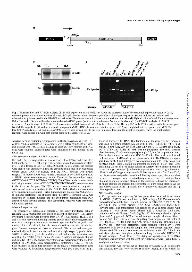
Fig. 1. Northern blot and RT–PCR analysis of hMSH6 expression in H-5 cells. (a) Schematic representation of the retroviral expression vector. P CMV,enhancer/promoter cassette of cytomegalovirus; BGHpA, bovine growth hormone polyadenylation signal sequence. Arrows indicate the position andorientation of primers used in the RT–PCR experiments. The dashed arrow indicates the transcription start site. (b) Hybridization of total RNA extracted fromHeLa, H-c and H-5 cells with either a radiolabelledhMSH6probe (top) or with a referenceβ-actin probe (bottom). (c) RT–PCR analysis of hMSH6expression. Amplification ofhMSH6cDNA reverse transcribed from total mRNA isolated from HeLa, H-c and H-5 cells. PCR reactions with the primer pairNick5/C15r amplified both endogenous and transgenichMSH6cDNA. In contrast, only transgenic cDNA was amplified with the primer pair pT7/C15r[see (a)]. Plasmids pCDNA and pCDNA/hMSH6 were used as controls. In the two right-most lanes are the negative controls, where the amplificationreactions were carried out with both primer pairs in the absence of DNA.
selective medium containing 6-thioguanine (6-TG; Sigma) at a densityø53105
cells/10 cm dish. Colonies were grown for 2 weeks before fixing with methanoland staining with 10% Giemsa in aqueous solution. Only colonies with.50cells were counted. Mutation rates were calculated by the method of themean (20).
DNA sequence analysis of HPRTmutationsH-c and H-5 cells were plated at a density of 100 cells/dish and grown to afinal number of 53105 cells. The replica cultures were trypsinized and platedin 6-TG at a density of 1253103 cells/10 cm dish. After 2 weeks, the colonieswere picked with cloning cylinders and grown to confluence in 24-well tissueculture plates. RNA was isolated from the HPRT– mutants with TRIzolreagent. The mutant RNAs were reverse transcribed as described above usinga HPRT primer complementary to the 39-end of the non-coding region(GTTTCCAAACTCAACTTGAACTCTC). The cDNA products were ampli-fied using a second primer (CCGGCTTCCTCCTCCTGAGC) complementaryto the 59-end of the gene. The PCR products were purified and sequencedwith nested primers according to the ABI PRISM dRhodamine terminatorcycle sequencing reaction kit (Perkin Elmer/Applied Biosystems, Wilton, CT).In mutants displaying loss of exons from the cDNAs, genomic DNA wasextracted by standard methods and the exon–intron boundaries were PCRamplified with specific primers. The sequencing reactions were performedwith nested primers.
Mismatch repair assaysThe efficiency of cytoplasmic extracts of the stably transfected cells inrepairing DNA mismatches was tested as described previously (15). Briefly,cytoplasmic extracts were prepared from 53108 HeLa, parental HCT15, H-cand H-5 cells harvested in the logarithmic growth phase. Following resuspen-sion in an appropriate volume of ice-cold hypotonic buffer to yield a celldensity of 13108 cells/ml, the cells were allowed to swell for 10 min in aglass Dounce homogenizer (Kontes, Vineland, NJ) on ice and then lysedmechanically with four or more strokes with a tight (type B) pestle. When.80% of cells were lysed, the nuclei were pelleted and the supernatant wascentrifuged at 12 000g for 10 min at 4°C, frozen in liquid nitrogen in aliquotsand stored at –80°C. The protein concentration was determined by the Bradfordmethod (30). M13mp2 DNA heteroduplexes containing a G/G, G/T or T/Gbase mispairs in the coding sequence of thelacZ α-complementation genewere obtained by hybridizing single-stranded viral (1) DNA with the (–)
375
strand of linearized RF DNA. One femtomole of the respective heteroduplexwas used in a repair reaction (25µl) with 30 mM HEPES, pH 7.8, 7 mMMgCl2, 4 mM ATP, 200µM each CTP, GTP and UTP, 100µM each dATP,dGTP, dTTP and dCTP, 40 mM creatine phosphate, 100 fmol creatinephosphokinase, 15 mM sodium phosphate, pH 7.5, and 50µg protein extract.The incubation was carried out at 37°C for 20 min. The repair was directedto the (–) strand of M13mp2 by the presence of a nick. The DNA heteroduplexwas then purified and introduced by electroporation intoEscherichia coliNR9162 (mutSstrain), plated on minimal medium in a soft agar layercontaining 0.5 ml of a log phase culture of CSH50 (theα-complementationstrain), 0.5 µg isopropyl-β-D-thiogalactopyranoside and 2µg 5-bromo-4-chloro-3-indoyl-β-D-galactopyranoside. Following incubation for 16 h at 37°C,the plaques were assigned to one of the following phenotypes: blue, colourlessor mixed. If no repair occurred, mixed plaques were observed containing bothblue and colourless progeny. Repair of the substrate reduced the percentageof mixed plaques and increased the percentage of pure colour plaques. As thenick directs repair to the (–) strand, the (1) phenotype increases and the (–)phenotype decreases.Microsatellite analysisA 121 bp region encompassing 26 deoxyadenosines localized in an intronof hMSH2 (BAT26) was amplified by PCR using 4,7,29,7’-tetrachloro-6-carboxyfluorescein-labelled forward primer 59-TGACTACTTTTGACTT-CAGCC-39 and reverse primer 59-AACCATTCAACATTTTTAACCC-39(300 nM each). The amplification was carried out in 25µl total volume withGenAmp 13 PCR buffer (Perkin Elmer), 1.25 U AmpliTaq Gold DNApolymerase (Perkin Elmer), 1.5 mM MgCl2, 100µM deoxynucleotide triphos-phates and 5 ng genomic DNA extracted from each single cell clone. After 1cycle of 94°C for 9 min to activate AmpliTaq DNA polymerase, 30 cycles of94°C for 45 s, 51°C for 1 min and 70°C for 30 s and a final elongation stepat 70°C for 7 min were performed. Mock reactions without DNA wereperformed with every twentieth sample and were always negative. Afterdilution, the PCR products were denatured with formamide at 92°C for 2 minand electrophoresed through a polymer capillary in an ABI PRISM 310Genetic Analyzer (Perkin Elmer). The sizing and quantitation of the DNAfragments was performed with the GenScan Analysis software (Perkin Elmer).Methylation tolerance studiesThis experiment was carried out as described previously (31). To measurecell survival, 100 cells were treated 18 h after seeding in 6 cm dishes for
by guest on September 20, 2016
http://carcin.oxfordjournals.org/D
ownloaded from
T.Lettieri et al.
30 min at 37°C with various concentrations of MNU in PBS/20 mM HEPES,pH 7.4. Cells were then washed, fed with complete medium and, 1–2 weekslater, the surviving colonies were fixed with methanol, stained with Giemsa andcounted. In the experiments performed in the presence ofO6-benzylguanine, thedrug was added to the medium at a final concentration of 25µM 2 h prior tothe MNU treatment and kept in the medium for an additional 72 h.
Cell cycle analysis
Cell synchronization in early S phase was accomplished by treatment withhydroxyurea (HU; Sigma). Confluent cells arrested in the resting phase werereplated in fresh complete medium along with 2 mM HU to allow G1 traverse.Fourteen hours later, by which time all cells had accumulated in early S phase,the HU was removed by two washes with prewarmed serum-free medium. Thecells were then treated with 0 or 5µM N-methyl-N9-nitro-N-nitrosoguanidine(MNNG; Sigma) in serum-free medium for 45 min at 37°C and 5% CO2.After treatment, they were washed once with 10% serum-containing mediumand returned to drug-free 10% serum-containing medium for 2, 4.5, 7, 12, 15and 19 h. At each time point, the cells were incubated with 10µM 5-bromo-29-deoxyuridine (BrdU; Sigma) for 30 min before harvesting. They were thenwashed with PBS, fixed in 70% ethanol and stored at 4°C. Nuclear preparationand dual labelling of DNA by propidium iodide (Sigma) and anti-BrdU–fluorescein conjugate (Boehringer Mannheim, Basel, Switzerland) were per-formed as described (25). Cell cycle analysis was performed using a BectonDickinson (Lincoln Park, NJ) FACscan flow cytometer and Cell Questsoftware. The data analysis is based on two independent experiments.
Results
Stable transfection of HCT15 cells with a retroviral vectorcarrying the hMSH6 cDNA results in the expression ofhMSH6 mRNAThe expression vehicle pCDNA-3 is an integrating retroviralvector containing a selectable marker gene that confers resist-ance to G418. ThehMSH6 cDNA was cloned between thecytomegalovirus enhancer/promoter cassette and a polyadenyl-ation signal sequence of the human growth hormone gene.The correct orientation of the insert was ensured by directionalcloning betweenBamHI andXhoI sites of the vector (Figure 1a;see also Materials and methods).
The hMSH6expression vector pCDNA/hMSH6 was trans-fected into logarithmically growing HCT15 cells by calciumphosphate precipitation. Following long-term selection forG418 resistance, a total of 10 clones were obtained, whichwere selected and grown into mass cultures under constantselective pressure. These clones were tested for the presenceof integratedhMSH6cDNA by Southern blotting, as well asfor expression of the protein by band shift and immunoblottingassays. Of these 10 clones, two, H-2 and H-5, containeddetectable amounts ofhMSH6cDNA. We selected clone H-5for further analysis. Southern blot experiments indicated thatH-5 carries about three copies of the integrated vector DNA(data not shown).
Because the H-5 clone was resistant to G418, the selectablemarker gene was clearly expressed from the transfected vector.We therefore decided to test whether this clone also expressedhMSH6mRNA. To this end, total RNA isolated from HeLa,H-c (control, HCT15 cells stably transfected with the emptypCDNA-3 vector) and H-5 cells was size fractionated on anagarose gel, transferred to a nitrocellulose membrane andhybridized with a radiolabelledhMSH6probe as described inMaterials and methods. The relative quantities of the specificsignals were normalized with respect toβ-actin. As shown inFigure 1b, the amount ofhMSH6 mRNA in the H-5 clonecorresponded to ~50% of that seen in HeLa cells. In contrast,the H-c clone expressed only low amounts ofhMSH6mRNA.It is possible that thehMSH6 promoter is epigeneticallyinactivated in the H-c cells. However, we consider it morelikely that the mRNA is selectively degraded due to the
376
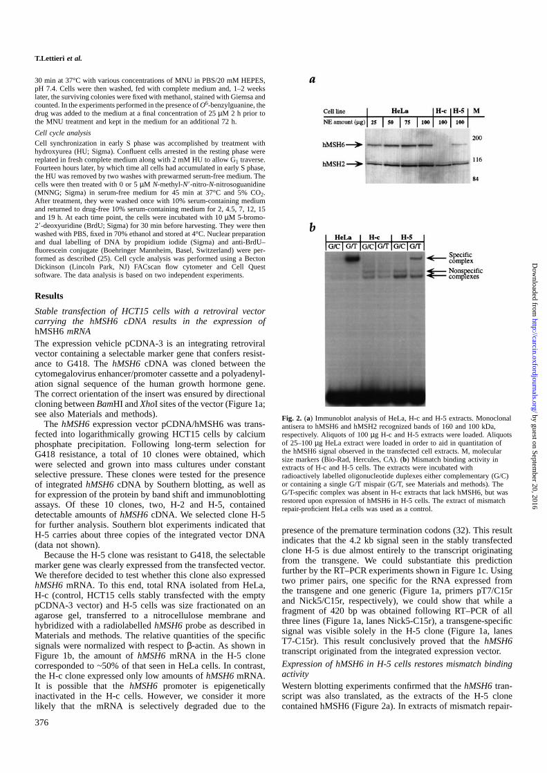
Fig. 2. (a) Immunoblot analysis of HeLa, H-c and H-5 extracts. Monoclonalantisera to hMSH6 and hMSH2 recognized bands of 160 and 100 kDa,respectively. Aliquots of 100µg H-c and H-5 extracts were loaded. Aliquotsof 25–100µg HeLa extract were loaded in order to aid in quantitation ofthe hMSH6 signal observed in the transfected cell extracts. M, molecularsize markers (Bio-Rad, Hercules, CA). (b) Mismatch binding activity inextracts of H-c and H-5 cells. The extracts were incubated withradioactively labelled oligonucleotide duplexes either complementary (G/C)or containing a single G/T mispair (G/T, see Materials and methods). TheG/T-specific complex was absent in H-c extracts that lack hMSH6, but wasrestored upon expression of hMSH6 in H-5 cells. The extract of mismatchrepair-proficient HeLa cells was used as a control.
presence of the premature termination codons (32). This resultindicates that the 4.2 kb signal seen in the stably transfectedclone H-5 is due almost entirely to the transcript originatingfrom the transgene. We could substantiate this predictionfurther by the RT–PCR experiments shown in Figure 1c. Usingtwo primer pairs, one specific for the RNA expressed fromthe transgene and one generic (Figure 1a, primers pT7/C15rand Nick5/C15r, respectively), we could show that while afragment of 420 bp was obtained following RT–PCR of allthree lines (Figure 1a, lanes Nick5-C15r), a transgene-specificsignal was visible solely in the H-5 clone (Figure 1a, lanesT7-C15r). This result conclusively proved that thehMSH6transcript originated from the integrated expression vector.
Expression of hMSH6 in H-5 cells restores mismatch bindingactivityWestern blotting experiments confirmed that thehMSH6tran-script was also translated, as the extracts of the H-5 clonecontained hMSH6 (Figure 2a). In extracts of mismatch repair-
by guest on September 20, 2016
http://carcin.oxfordjournals.org/D
ownloaded from
hMSH6 cDNA and mismatch repair phenotype
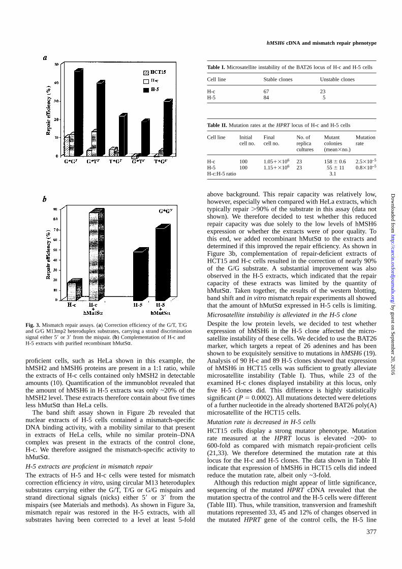
Fig. 3. Mismatch repair assays. (a) Correction efficiency of the G/T, T/Gand G/G M13mp2 heteroduplex substrates, carrying a strand discriminationsignal either 59 or 39 from the mispair. (b) Complementation of H-c andH-5 extracts with purified recombinant hMutSα.
proficient cells, such as HeLa shown in this example, thehMSH2 and hMSH6 proteins are present in a 1:1 ratio, whilethe extracts of H-c cells contained only hMSH2 in detectableamounts (10). Quantification of the immunoblot revealed thatthe amount of hMSH6 in H-5 extracts was only ~20% of thehMSH2 level. These extracts therefore contain about five timesless hMutSαthan HeLa cells.
The band shift assay shown in Figure 2b revealed thatnuclear extracts of H-5 cells contained a mismatch-specificDNA binding activity, with a mobility similar to that presentin extracts of HeLa cells, while no similar protein–DNAcomplex was present in the extracts of the control clone,H-c. We therefore assigned the mismatch-specific activity tohMutSα.
H-5 extracts are proficient in mismatch repairThe extracts of H-5 and H-c cells were tested for mismatchcorrection efficiencyin vitro, using circular M13 heteroduplexsubstrates carrying either the G/T, T/G or G/G mispairs andstrand directional signals (nicks) either 59or 39 from themispairs (see Materials and methods). As shown in Figure 3a,mismatch repair was restored in the H-5 extracts, with allsubstrates having been corrected to a level at least 5-fold
377
Table I. Microsatellite instability of the BAT26 locus of H-c and H-5 cells
Cell line Stable clones Unstable clones
H-c 67 23H-5 84 5
Table II. Mutation rates at theHPRT locus of H-c and H-5 cells
Cell line Initial Final No. of Mutant Mutationcell no. cell no. replica colonies rate
cultures (mean3no.)
H-c 100 1.0513106 23 1586 0.6 2.5310–5
H-5 100 1.1513106 23 556 11 0.8310–5
H-c:H-5 ratio 3.1
above background. This repair capacity was relatively low,however, especially when compared with HeLa extracts, whichtypically repair.90% of the substrate in this assay (data notshown). We therefore decided to test whether this reducedrepair capacity was due solely to the low levels of hMSH6expression or whether the extracts were of poor quality. Tothis end, we added recombinant hMutSαto the extracts anddetermined if this improved the repair efficiency. As shown inFigure 3b, complementation of repair-deficient extracts ofHCT15 and H-c cells resulted in the correction of nearly 90%of the G/G substrate. A substantial improvement was alsoobserved in the H-5 extracts, which indicated that the repaircapacity of these extracts was limited by the quantity ofhMutSα. Taken together, the results of the western blotting,band shift andin vitro mismatch repair experiments all showedthat the amount of hMutSαexpressed in H-5 cells is limiting.
Microsatellite instability is alleviated in the H-5 cloneDespite the low protein levels, we decided to test whetherexpression of hMSH6 in the H-5 clone affected the micro-satellite instability of these cells. We decided to use the BAT26marker, which targets a repeat of 26 adenines and has beenshown to be exquisitely sensitive to mutations inhMSH6(19).Analysis of 90 H-c and 89 H-5 clones showed that expressionof hMSH6 in HCT15 cells was sufficient to greatly alleviatemicrosatellite instability (Table I). Thus, while 23 of theexamined H-c clones displayed instability at this locus, onlyfive H-5 clones did. This difference is highly statisticallysignificant (P5 0.0002). All mutations detected were deletionsof a further nucleotide in the already shortened BAT26 poly(A)microsatellite of the HCT15 cells.
Mutation rate is decreased in H-5 cellsHCT15 cells display a strong mutator phenotype. Mutationrate measured at theHPRT locus is elevated ~200- to600-fold as compared with mismatch repair-proficient cells(21,33). We therefore determined the mutation rate at thislocus for the H-c and H-5 clones. The data shown in Table IIindicate that expression of hMSH6 in HCT15 cells did indeedreduce the mutation rate, albeit only ~3-fold.
Although this reduction might appear of little significance,sequencing of the mutatedHPRT cDNA revealed that themutation spectra of the control and the H-5 cells were different(Table III). Thus, while transition, transversion and frameshiftmutations represented 33, 45 and 12% of changes observed inthe mutatedHPRT gene of the control cells, the H-5 line
by guest on September 20, 2016
http://carcin.oxfordjournals.org/D
ownloaded from
T.Lettieri et al.
Table III. Spontaneous mutation spectra of theHPRT locus of H-c and H-5cells
H-c H-5
Mutations characterized 24 26Transitions 8 (33%) 4 (15%)
A:T→G:C 4 2G:C→A:T 4 2
Transversions 11 (45%) 17 (65%)G:C→T:A 8 14G:C→C:G 0 1T: A→A:T 3 0A:T→C:G 0 2
Frameshifts 3 (12%) 1 (4%)Deletions 0 4 (15%)Undefined mutations 2 (8%) 0
Table IV. Progression of the untreated or MNNG-treated H-c and H-5 cellsthrough the cell cycle after release from an HU block
Cell cycle Time post-release H-c H-c/MNNG H-5 H-5/MNNGphase (h) (%) (%) (%) (%)
G1 2 1.7 12.5 6.1 16.74.5 4.1 8.7 1.8 7.77 6.1 10.1 1.9 3.79 10.4 4.6 8.9 5.8
12 12.1 13.2 12.9 4.515 41.1 36.1 36.1 9.719 22.9 24.6 12.8 22.1
S 2 97.8 86.8 93.3 78.54.5 93.9 88.2 97.4 87.37 64.4 67.9 72.2 91.39 34.5 42.3 37.5 79.6
12 12.1 30.1 9.1 76.915 53.1 43.3 54.9 55.119 71.4 72.5 80.4 46.6
G2/M 2 0.5 0.7 0.5 4.84.5 1.9 3.1 0.7 4.97 29.6 22.1 25.9 4.99 55.1 53.1 53.6 14.6
12 75.8 56.7 78.1 18.615 5.9 20.5 8.9 35.219 5.7 2.8 6.7 31.4
displayed primarily transversions and deletions of multiplebases. Transitions and frameshifts, which arise from uncorrec-ted purine/pyrimidine mispairs and insertion/deletion loops,respectively, were less frequent. It is interesting to note inthis respect that the latter premutagenic lesions are the bestsubstrates for hMutSαin in vitro mismatch binding assays(34), as well as being the most efficiently corrected mispairtypesin vivo (35). It is therefore likely that although the levelof hMutSα in the H-5 cells is low, it is nonetheless sufficientto mediate the correction of a significant proportion of thosesubstrates which it binds with the highest affinity (see alsobelow).
H-5 cells are not significantly sensitized to killing bymethylating agentsTreatment of cells with simple methylating agents introducesnumerous modifications into DNA. However, it could be shownthat the lesion primarily responsible for the cytotoxicity of thesesubstances isO6-methylguanine. Resistance to methylatingagents is commonly associated with overexpression ofO6-methylguanine methyltransferase (MGMT), which reverses thedamage by removing methyl groups from modified guanines
378
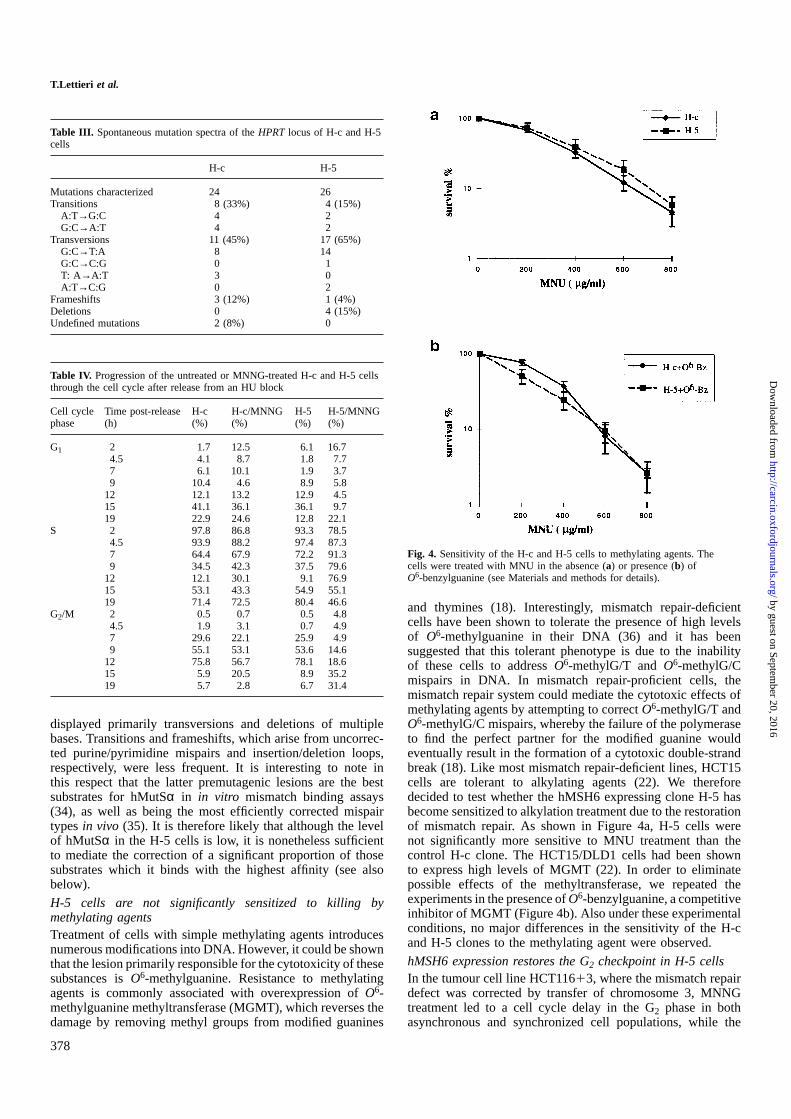
Fig. 4. Sensitivity of the H-c and H-5 cells to methylating agents. Thecells were treated with MNU in the absence (a) or presence (b) ofO6-benzylguanine (see Materials and methods for details).
and thymines (18). Interestingly, mismatch repair-deficientcells have been shown to tolerate the presence of high levelsof O6-methylguanine in their DNA (36) and it has beensuggested that this tolerant phenotype is due to the inabilityof these cells to addressO6-methylG/T andO6-methylG/Cmispairs in DNA. In mismatch repair-proficient cells, themismatch repair system could mediate the cytotoxic effects ofmethylating agents by attempting to correctO6-methylG/T andO6-methylG/C mispairs, whereby the failure of the polymeraseto find the perfect partner for the modified guanine wouldeventually result in the formation of a cytotoxic double-strandbreak (18). Like most mismatch repair-deficient lines, HCT15cells are tolerant to alkylating agents (22). We thereforedecided to test whether the hMSH6 expressing clone H-5 hasbecome sensitized to alkylation treatment due to the restorationof mismatch repair. As shown in Figure 4a, H-5 cells werenot significantly more sensitive to MNU treatment than thecontrol H-c clone. The HCT15/DLD1 cells had been shownto express high levels of MGMT (22). In order to eliminatepossible effects of the methyltransferase, we repeated theexperiments in the presence ofO6-benzylguanine, a competitiveinhibitor of MGMT (Figure 4b). Also under these experimentalconditions, no major differences in the sensitivity of the H-cand H-5 clones to the methylating agent were observed.
hMSH6 expression restores the G2 checkpoint in H-5 cellsIn the tumour cell line HCT11613, where the mismatch repairdefect was corrected by transfer of chromosome 3, MNNGtreatment led to a cell cycle delay in the G2 phase in bothasynchronous and synchronized cell populations, while the
by guest on September 20, 2016
http://carcin.oxfordjournals.org/D
ownloaded from
hMSH6 cDNA and mismatch repair phenotype
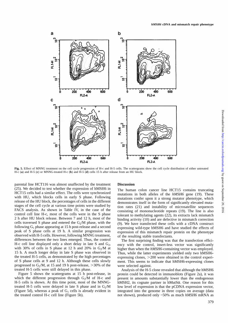
Fig. 5. Effect of MNNG treatment on the cell cycle progression of H-c and H-5 cells. The scattergrams show the cell cycle distribution of either untreatedH-c (a) and H-5 (c) or MNNG-treated H-c (b) and H-5 (d) cells 15 h after release from an HU block.
parental line HCT116 was almost unaffected by the treatment(25). We decided to test whether the expression of hMSH6 inHCT15 cells had a similar effect. The cells were synchronizedwith HU, which blocks cells in early S phase. Followingrelease of the HU block, the percentages of cells in the differentstages of the cell cycle at various time points were studied byFACS analysis. As shown in Table IV, in the case of thecontrol cell line H-c, most of the cells were in the S phase2 h after HU block release. Between 7 and 12 h, most of thecells traversed S phase and entered the G2/M phase, with thefollowing G1 phase appearing at 15 h post-release and a secondpeak of S phase cells at 19 h. A similar progression wasobserved with H-5 cells. However, following MNNG treatment,differences between the two lines emerged. Thus, the controlH-c cell line displayed only a short delay in late S and G2,with 30% of cells in S phase at 12 h and 20% in G2/M at15 h. A much longer delay in late S phase was observed inthe treated H-5 cells, as demonstrated by the high percentagesof S phase cells at 9 and 12 h. Although these cells slowlyprogressed to G2/M, at 15 and 19 h post-release,.30% of thetreated H-5 cells were still delayed in this phase.
Figure 5 shows the scattergrams at 15 h post-release, inwhich the different progression through G2/M of H-c andH-5 cells is shown. At this time point, most of the MNNG-treated H-5 cells were delayed in late S phase and in G2/M(Figure 5d), whereas a peak of G1 cells is already evident inthe treated control H-c cell line (Figure 5b).
379
Discussion
The human colon cancer line HCT15 contains truncatingmutations in both alleles of thehMSH6 gene (19). Thesemutations confer upon it a strong mutator phenotype, whichdemonstrates itself in the form of significantly elevated muta-tion rates (21) and instability of microsatellite sequencesconsisting of mononucleotide repeats (19). The line is alsotolerant to methylating agents (22), its extracts lack mismatchbinding activity (10) and are defective in mismatch correction(9). We have transfected these cells with a cDNA constructexpressing wild-type hMSH6 and have studied the effects ofexpression of this mismatch repair protein on the phenotypeof the resulting stable transfectants.
The first surprising finding was that the transfection effici-ency with the control, insert-less vector was significantlyhigher than when thehMSH6-containing vector was employed.Thus, while the latter experiments yielded only two hMSH6-expressing clones,.200 were obtained in the control experi-ment. This seems to indicate that hMSH6-expressing cloneswere selected against.
Analysis of the H-5 clone revealed that although the hMSH6protein could be detected in immunoblots (Figure 2a), it waspresent in amounts substantially lower than the endogenoushMSH2, its cognate partner in hMutSα. One reason for thislow level of expression is that the pCDNA expression vector,integrated into the genome in three copies on average (datanot shown), produced only ~50% as muchhMSH6mRNA as
by guest on September 20, 2016
http://carcin.oxfordjournals.org/D
ownloaded from
T.Lettieri et al.
was detected in the control HeLa cells (Figure 1b, comparethe specific signal with the intensity of theβ-actin internalcontrol). However, this should not be critical in itself, asimmortalized lymphocytes from HNPCC patients with onemutated mismatch repair allele have been reported to begenerally proficient in mismatch repair (37). The secondexplanation could be that the hMSH6 protein, even thoughexpressed relatively efficiently, is subject to proteolyticdegradation or misfolding prior to associating with hMSH2.Indeed, hMSH6 is known to be proteolytically labile. Inextracts of cells mutated in hMSH2 (e.g. LoVo and HEC59),hMSH6 is present at barely detectable levels (10), suggestingthat it is degraded in the absence of its partner hMSH2. Ourprevious studies showed that expression of hMSH6 withouthMSH2 in a baculovirus system also resulted in a significantdegradation of the former protein (13). Moreover, mixing ofhMSH2 and hMSH6 expressed separately gave only lowamounts of active hMutSα, while co-expression of the twoproteins yielded the biologically active mismatch bindingfactor in high amounts (29), which suggests that the twopolypeptides probably interact during folding. In their naturalenvironment, the two genes are co-localized on chromosome2p16 and it is likely that their expression is co-regulated,thus ensuring effective heterodimerization. This hypothesis issupported by the reports that transfer of chromosome 2 intoHCT15 cells (33) resulted in a substantial correction of themutator phenotype. The third possibility concerns the fact thathMSH6 competes for hMSH2 with another MutS homologue,hMSH3. Under normal circumstances, hMSH3 appears to beexpressed at low levels and therefore the relative ratio of thehMutSαand hMutSβheterodimers favours the former speciesby a substantial margin (16). However, due to the absence ofhMSH6 in HCT15 cells, hMSH3 should be able to interactwith hMSH2 more successfully. Indeed, hMutSβ levels inthese cells have been reported to be elevated ~3-fold (16).Although expression of hMSH6 would be expected to decreasehMutSβ levels, this apparently failed to happen: the relativequantities of hMSH2 and hMSH3 in extracts of H-5 cells weresimilar to those detected in the control H-c line, at least asjudged by immunoblotting (data not shown). We must thereforeconsider the possibility that expression ofhMSH6mRNA fromthe pCDNA vector did not fully satisfy the criteria necessaryfor successful translation of the protein and for its successiveheterodimerization with hMSH2.
Although present at low levels, the amount of active hMutSαin the extracts of H-5 cells was nonetheless sufficient tosubstantially restore mismatch binding activity (Figure 2b) andmismatch repair (Figure 3a). Most importantly, expression ofwild-type hMSH6 in these cells greatly alleviated microsatelliteinstability at the poly(A) marker BAT26 (Table I). It wastherefore somewhat surprising to discover that mutation rates,measured at theHPRT locus, were reduced only 3-fold(Table II) and that sensitivity of the hMSH6-expressing cloneH-5 to MNU was not significantly different from the H-ccontrol (Figure 4). How could this apparent discrepancy beexplained? In our opinion, the underlying reason for theseinconsistencies is the low level of hMutSαexpression, coupledwith the substrate specificity of this heterodimeric mismatchrecognition factor.In vitro binding studies, carried out withpurified native (38) and recombinant (13) hMutSαdemon-strated that the preferred substrate is an IDL of one extrahelicalnucleotide, followed by the G/T mispair. Flanking sequencesalso appear to play an important role in the efficiency of
380
substrate recognition, whereby IDLs appear to be most effici-ently recognized in the context of repeated motifs (39). As thedissociation constant of hMutSαwith respect to the G/Toligonucleotide substrate is in the low nanomolar range (29,40),we were able to detect mismatch binding activity in the extractsof H-5 cells despite low protein amounts (Figure 2b). Asimilar argument applies for thein vitro mismatch correctionexperiments, which were carried out with substrates efficientlyrecognized by hMutSα(Figure 4). Even lower concentrationsof hMutSα are required for efficient recognition of singlenucleotide IDLs, which are the underlying cause of microsatel-lite instability in hMSH6-deficient cells. Correspondingly, thepoly(A) tract of the BAT26 marker was stabilized in H-5 cells.
In contrast to microsatellites, spontaneous mutations arisingin a gene such asHPRT are highly heterogeneous, includingall types of base/base mispairs and frameshifts. Some of thesewill be good substrates for hMutSαand these might beexpected to be corrected even in the presence of low levels ofthe factor. Other mispairs and, possibly, also frameshifts outsidemicrosatellites might not be bound by hMutSαsufficientlywell in order to initiate the mismatch correction process. Asshown in Table III, transition mutations, which result fromG/T or A/C mispairs, were down by 50% in H-5 cells, whiletransversions, due to either G/A, or C/T mismatches, bothknown to be poorly correctedin vivo (35), were elevated.
Restoration of sensitivity to methylating agents such asMNU or MNNG would also be expected to be affected byhMutSα concentration. The cytotoxic effects of methylatingagents are probably mediated by two distinct pathways.One of these is most likely linked to abortive attempts ofthe mismatch repair system to correctO6-methylG/T andO6-methylG/C mispairs arising during replication of methyl-ated DNA (18), whereby two concurrent mismatch repairevents in close proximity may cause a double-strand break.Recentin vitro studies demonstrated that these mispairs areindeed recognized by hMutSα(41). However, as its affinityfor O6-methylG/T andO6-methylG/C is substantially lowerthan for the G/T mispair, the majority of these methylatedlesions would most likely remain unrecognized unless the cellsexpressed high concentrations of the mismatch binding factor.As this is clearly not the case for the H-5 line described here,its sensitivity to alkylating agents is similar to the controlH-c line. The second response to DNA alkylation, or indeedto DNA damage in general, is the deployment of a cell cyclecheckpoint, which allows the cell to repair the damage beforeproceeding with DNA replication and/or mitosis. Such a delayin cell cycle progression was documented in the mismatchrepair-deficient cell line HCT116, which failed to arrest follow-ing treatment with methylating agents (25), but in which thisG2 delay was restored when the repair defect was correctedby the transfer of chromosome 3, which carries a wild-typecopy of the hMLH1 gene. The ability of mismatch repairproteins to signal to the apoptotic machinery was furtherdocumented for the closely related cell lines MT1 and TK6(42). Treatment of the latter, mismatch repair-proficient cellswith alkylating agents resulted in the elevation of p53 and p21levels. No similar increase was observed in MT1 cells, whichare mutated inhMSH6. As treatment of both lines withetoposide, an inhibitor of topoisomerase II, activated p53 andp21 in both lines, we concluded that p53 was wild-type inMT1 cells and that their failure to activate a p53 responsefollowing alkylation treatment was due to the absence ofhMutSα-mediated damage detection. If we accept that mis-
by guest on September 20, 2016
http://carcin.oxfordjournals.org/D
ownloaded from

hMSH6 cDNA and mismatch repair phenotype
match repair proteins are involved in DNA damage signallingto the apoptotic machinery or to the cell cycle checkpointcontrol, this effect could be mediated by only a few molecules,which might stall the replication fork by remaining bound atO6-methylG/T orO6-methylG/C mispairs and thus bring abouta cell cycle arrest or delay. This hypothesis is supported bythe data presented in Table IV and Figure 5, where the stablytransfected H-5 line can be seen to be delayed in the lateS/G2 stage of the cell cycle following MNNG treatment, whilethe control H-c clone progressed with only a short delay.
Mismatch repair deficiency was linked with a multitude ofphenotypic traits, ranging from microsatellite instability totolerance to alkylating agents. Earlier studies demonstratedthat transfer of chromosome 3 into hMLH1-deficient lineHCT116 (43) or of chromosome 2 into HCT15 and DLD1cells (33), both lacking hMSH6, could bring about an almosttotal reversion of the mutator phenotype. However, the caveatof these studies was that whole chromosomes carry many genesand thus that the correction effect could not be unambiguouslyassigned to a single gene. Recently, Risingeret al. (44)succeeded in overexpressinghPMS2cDNA in HEC-1-A cellsand succeeded in restoring the repair of IDLs but not of base/base mismatches. This partial phenotypic correction could beexplained by the discovery that these cells also harbour, inaddition to the mutatedhPMS2, a mutatedhMSH6gene. Wenow show that expression of hMSH6 in the HCT15 cell linealso resulted in only a partial reversion of its numerousphenotypic traits. Unlike in the case of HEC-1-A cells,however, this incomplete correction was most likely not causedby the presence of a mutation in a second mismatch repairgene. Rather, due to the low level of expression of thetransgenic protein, the observed effects were only small insome instances. However, the reversion trend unambiguouslyconfirmed the association of thehMSH6mismatch repair genemalfunction with mononucleotide repeat instability, elevatedmutation rate and the G2 cell cycle checkpoint.
AcknowledgementsWe are indebted to Tom Kunkel for the generous gift of the HCT15 line andof the M13 mp2 phage, to Paola Delmastro for the anti-hMSH6 monoclonalantibodies and to Primo Scha¨r for helpful discussions and for critical readingof the manuscript. This work was supported in part by the Swiss NationalScience Foundation and by the Julius Mu¨ller-Stiftung.
References1.Marra,G. and Boland,C.R. (1996) DNA repair and colorectal cancer.
Gastroenterol. Clin. North Am.,25, 755–772.2.Jiricny,J. (1996) Mismatch repair and cancer.Cancer Surv., 28, 47–68.3.Kolodner,R.D. (1995) Mismatch repair: mechanisms and relationship to
cancer susceptibility.Trends Biochem. Sci., 20, 397–401.4.Modrich,P. and Lahue,R. (1996) Mismatch repair in replication fidelity,
genetic recombination, and cancer biology.Annu. Rev. Biochem., 65,101–133.
5.Modrich,P. (1991) Mechanisms and biological effects of mismatch repair.Annu. Rev. Genet.,25, 229–253.
6.Ciotta,C., Ceccotti,S., Aquilina,G., Humbert,O., Palombo,F., Jiricny,J.and Bignami,M. (1998) Increased somatic recombination in methylationtolerant human cells with defective DNA mismatch repair.J. Mol. Biol.,276, 705–719.
7.Perucho,M. (1996) Cancer of the microsatellite mutator phenotype.Biol.Chem.,377, 675–684.
8.Fink,D., Aebi,S. and Howell,S.B. (1998) The role of DNA mismatch repairin drug resistance.Clin. Cancer Res.,4, 1–6.
9.Drummond,J.T., Li,G.M., Longley,M.J. and Modrich,P. (1995) Isolation ofan hMSH2-p160 heterodimer that restores DNA mismatch repair to tumorcells. Science,268, 1909–1912.
10.Palombo,F., Gallinari,P., Iaccarino,I., Lettieri,T., Hughes,M., D’Arrigo,A.,
381
Truong,O., Hsuan,J.J. and Jiricny,J. (1995) GTBP, a 160-kilodalton proteinessential for mismatch-binding activity in human cells.Science,268,1912–1914.
11.Acharya,S., Wilson,T., Gradia,S., Kane,M.F., Guerrette,S., Marsischky,G.T.and Fishel,R. (1996) hMSH2 forms specific mispair-binding complexeswith hMSH3 and hMSH6.Proc. Natl Acad. Sci. USA, 93, 13629–13634.
12.Li,G.M. and Modrich,P. (1995) Restoration of mismatch repair to nuclearextracts of H6 colorectal tumor cells by a heterodimer of human MutLhomologs.Proc. Natl Acad. Sci. USA,92, 1950–1954.
13.Palombo,F., Iaccarino,I., Nakajima,E., Ikejima,M., Shimada,T. andJiricny,J. (1996) hMutSβ, a heterodimer of hMSH2 and hMSH3, binds toinsertion/deletion loops in DNA.Curr. Biol., 6, 1181–1184.
14.Risinger,J.I., Umar,A., Boyd,J., Berchuck,A., Kunkel,T.A. and Barrett,J.C.(1996) Mutation of MSH3 in endometrial cancer and evidence for itsfunctional role in heteroduplex repair.Nature Genet., 14, 102–105.
15.Marra,G., Iaccarino,I., Lettieri,T., Roscilli,G., Delmastro,P. and Jiricny,J.(1998) Mismatch repair deficiency associated with overexpression of theMSH3 gene.Proc. Natl Acad. Sci. USA,95, 8568–8573.
16.Genschel,J., Littman,S.J., Drummond,J.T. and Modrich,P. (1998) Isolationof MutSbeta from human cells and comparison of the mismatch repairspecificities of MutSbeta and MutSalpha.J. Biol. Chem.,273, 19895–19901.
17. Ionov,Y., Peinado,M.A., Malkhosyan,S., Shibata,D. and Perucho,M. (1993)Ubiquitous somatic mutations in simple repeated sequences reveal a newmechanism for colonic carcinogenesis. Nature, 363, 558–561.
18.Karran,P. and Bignami,M. (1994) DNA damage tolerance, mismatch repairand genome instability.BioEssays,16, 833–839.
19.Papadopoulos,N., Nicolaides,N.C., Liu,B.et al. (1995) Mutations of GTBPin genetically unstable cells.Science,268, 1915–1917.
20.Bhattacharyya,N.P., Skandalis,A., Ganesh,A., Groden,J. and Meuth,M.(1994) Mutator phenotypes in human colorectal carcinoma cell lines.Proc.Natl Acad. Sci. USA, 91, 6319–6323.
21.Bhattacharyya,N.P. and Meuth,M. (1995) Molecular analysis of mutationsin mutator colorectal carcinoma cell lines.Hum. Mol. Genet.,4, 2057–2064.
22.Branch,P., Hampson,R. and Karran,P. (1995) DNA mismatch bindingdefects, DNA damage tolerance, and mutator phenotypes in humancolorectal carcinoma cell lines.Cancer Res.,55, 2304–2309.
23.da Costa,L.T., Liu,B., el Deiry,W.et al. (1995) Polymerase delta variantsin RER colorectal tumours.Nature Genet.,9, 10–11.
24.Schaaper,R.M. (1988) Mechanisms of mutagenesis in theEscherichia colimutator mutD5: role of DNA mismatch repair.Proc. Natl Acad. Sci. USA,85, 8126–8130.
25.Hawn,M.T., Umar,A., Carethers,J.M., Marra,G., Kunkel,T.A., Boland,C.R.and Koi,M. (1995) Evidence for a connection between the mismatch repairsystem and the G2 cell cycle checkpoint.Cancer Res.,55, 3721–3725.
26.Sambrook,J., Fritsch,E. and Maniatis,T. (1989)Molecular Cloning: ALaboratory Manual, 2nd Edn. Cold Spring Harbor Laboratory Press, ColdSpring Harbor, NY.
27.Nicolaides,N.C., Palombo,F., Kinzler,K.W., Vogelstein,B. and Jiricny,J.(1996) Molecular cloning of the N-terminus of GTBP.Genomics,31,395–397.
28.Dignam,J.D., Martin,P.L., Shastry,B.S. and Roeder,R.G. (1983) Eukaryoticgene transcription with purified components.Methods Enzymol., 101,582–598.
29. Iaccarino,I., Marra,G., Palombo,F. and Jiricny,J. (1998) hMSH2 andhMSH6 play distinct roles in mismatch binding and contribute differentlyto the ATPase activity of hMutS-alpha.EMBO J.,17, 2677–2686.
30.Bradford,M.M. (1976) A rapid and sensitive method for the quantitationof microgram quantities of protein utilizing the principle of protein–dyebinding.Anal. Biochem.,72, 248–254.
31.Aquilina,G., Ceccotti,S., Martinelli,S., Hampson,R. and Bignami,M. (1998)N-(2-chloroethyl)-N9-cyclohexyl-N-nitrosourea sensitivity in mismatchrepair-defective human cells.Cancer Res.,58, 135–141.
32.Maquat,L.E. (1995) When cells stop making sense: effects of nonsensecodons on RNA metabolism in vertebrate cells.RNA,1, 453–465.
33.Umar,A., Koi,M., Risinger,J.I., Glaab,W.E., Tindall,K.R., Kolodner,R.D.,Boland,C.R., Barrett,J.C. and Kunkel,T.A. (1997) Correction ofhypermutability, N-methyl-N9-nitro-N-nitrosoguanidine resistance, anddefective DNA mismatch repair by introducing chromosome 2 into humantumor cells with mutations in MSH2 and MSH6.Cancer Res.,57,3949–3955.
34.Holmes,J.Jr, Clark,S. and Modrich,P. (1990) Strand-specific mismatchcorrection in nuclear extracts of human andDrosophila melanogastercelllines.Proc. Natl Acad. Sci. USA, 87, 5837–5841.
35.Brown,T.C. and Jiricny,J. (1988) Different base/base mispairs are correctedwith different efficiencies and specificities in monkey kidney cells.Cell,54, 705–711.
by guest on September 20, 2016
http://carcin.oxfordjournals.org/D
ownloaded from
T.Lettieri et al.
36.Branch,P., Aquilina,G., Bignami,M. and Karran,P. (1993) Defectivemismatch binding and a mutator phenotype in cells tolerant to DNAdamage. Nature,362, 652–654.
37.Parsons,R., Li,G.M., Longley,M., Modrich,P., Liu,B., Berk,T.,Hamilton,S.R., Kinzler,K.W. and Vogelstein,B. (1995) Mismatch repairdeficiency in phenotypically normal human cells.Science,268, 738–740.
38.Hughes,M.J. and Jiricny,J. (1992) The purification of a human mismatch-binding protein and identification of its associated ATPase and helicaseactivities.J. Biol. Chem.,267, 23876–23882.
39.Macpherson,P., Humbert,O. and Karran,P. (1998) Frameshift mismatchrecognition by the human MutS alpha complex.Mutat. Res.,408, 55–66.
40.Gradia,S., Acharya,S. and Fishel,R. (1997) The human mismatchrecognition complex hMSH2–hMSH6 functions as a novel molecularswitch.Cell, 91, 995–1005.
41.Duckett,D.R., Drummond,J.T., Murchie,A.I., Reardon,J.T., Sancar,A.,Lilley,D.M. and Modrich,P. (1996) Human MutSalpha recognizes damaged
382
DNA base pairs containing O6-methylguanine, O4-methylthymine, or thecisplatin–d(GpG) adduct.Proc. Natl Acad. Sci. USA,93, 6443–6447.
42.D’Atri,S., Tentori,L., Lacal,P.M., Graziani,G., Pagani,E., Benincasa,E.,Zambruno,G., Bonmassar,E. and Jiricny,J. (1998) Involvement of themismatch repair system in temozolomide-induced apoptosis.Mol.Pharmacol.,54, 334–341.
43.Koi,M., Umar,A., Chauhan,D.P., Cherian,S.P., Carethers,J.M., Kunkel,T.A.and Boland,C.R. (1994) Human chromosome 3 corrects mismatch repairdeficiency and microsatellite instability and reducesN-methyl-N9-nitro-N-nitrosoguanidine tolerance in colon tumor cells with homozygous hMLH1mutation.Cancer Res.,54, 4308–4312.
44.Risinger,J.I., Umar,A., Glaab,W.E., Tindall,K.R., Kunkel,T.A. andBarrett,J.C. (1998) Single gene complementation of the hPMS2 defect inHEC-1-A endometrial carcinoma cells.Cancer Res.,58, 2978–2981.
Received September 23, 1998; revised and accepted November 12, 1998
by guest on September 20, 2016
http://carcin.oxfordjournals.org/D
ownloaded from
Copyright © 2022 FDOKUMEN