Early Increases in Superantigen-Specific Foxp3+ Regulatory T Cells during Mouse Mammary Tumor Virus...
11
Published Ahead of Print 21 May 2008. 2008, 82(15):7422. DOI: 10.1128/JVI.00102-08. J. Virol. Isabel Piazzon Héctor Costa, Susan R. Ross, Irene Nepomnaschy and Cecilia Courreges, Gabriela Camicia, Daniela Lorenzo, Gabriel Cabrera, Dalia Burzyn, Juliana Mundiñano, M. Mammary Tumor Virus Infection Regulatory T Cells during Mouse + Foxp3 Early Increases in Superantigen-Specific http://jvi.asm.org/content/82/15/7422 Updated information and services can be found at: These include: SUPPLEMENTAL MATERIAL Supplemental material REFERENCES http://jvi.asm.org/content/82/15/7422#ref-list-1 at: This article cites 52 articles, 31 of which can be accessed free CONTENT ALERTS more» articles cite this article), Receive: RSS Feeds, eTOCs, free email alerts (when new http://journals.asm.org/site/misc/reprints.xhtml Information about commercial reprint orders: http://journals.asm.org/site/subscriptions/ To subscribe to to another ASM Journal go to: on June 7, 2014 by guest http://jvi.asm.org/ Downloaded from on June 7, 2014 by guest http://jvi.asm.org/ Downloaded from
Transcript of Early Increases in Superantigen-Specific Foxp3+ Regulatory T Cells during Mouse Mammary Tumor Virus...
Published Ahead of Print 21 May 2008. 2008, 82(15):7422. DOI: 10.1128/JVI.00102-08. J. Virol.
Isabel PiazzonHéctor Costa, Susan R. Ross, Irene Nepomnaschy andCecilia Courreges, Gabriela Camicia, Daniela Lorenzo, Gabriel Cabrera, Dalia Burzyn, Juliana Mundiñano, M. Mammary Tumor Virus Infection
Regulatory T Cells during Mouse+Foxp3Early Increases in Superantigen-Specific
http://jvi.asm.org/content/82/15/7422Updated information and services can be found at:
These include:
SUPPLEMENTAL MATERIAL Supplemental material
REFERENCEShttp://jvi.asm.org/content/82/15/7422#ref-list-1at:
This article cites 52 articles, 31 of which can be accessed free
CONTENT ALERTS more»articles cite this article),
Receive: RSS Feeds, eTOCs, free email alerts (when new
http://journals.asm.org/site/misc/reprints.xhtmlInformation about commercial reprint orders: http://journals.asm.org/site/subscriptions/To subscribe to to another ASM Journal go to:
on June 7, 2014 by guesthttp://jvi.asm
.org/D
ownloaded from
on June 7, 2014 by guest
http://jvi.asm.org/
Dow
nloaded from
JOURNAL OF VIROLOGY, Aug. 2008, p. 7422–7431 Vol. 82, No. 150022-538X/08/$08.00�0 doi:10.1128/JVI.00102-08Copyright © 2008, American Society for Microbiology. All Rights Reserved.
Early Increases in Superantigen-Specific Foxp3� Regulatory T Cellsduring Mouse Mammary Tumor Virus Infection�†
Gabriel Cabrera,1‡ Dalia Burzyn,1‡ Juliana Mundinano,1 M. Cecilia Courreges,2 Gabriela Camicia,1Daniela Lorenzo,1 Hector Costa,1 Susan R. Ross,2 Irene Nepomnaschy,1 and Isabel Piazzon1*
Instituto de Leucemia Experimental, Consejo Nacional de Investigaciones Cientıficas y Tecnicas, Division Medicina Experimental,Instituto de Investigaciones Hematologicas, Academia Nacional de Medicina, Buenos Aires, Argentina,1 and Department of
Microbiology and Abramson Family Cancer Center, University of Pennsylvania, Philadelphia, Pennsylvania2
Received 15 January 2008/Accepted 12 May 2008
Mouse mammary tumor virus (MMTV) is a milk-borne betaretrovirus that has developed strategies toexploit and subvert the host immune system. Here, we show in a natural model of MMTV infection that thevirus causes early and progressive increases in superantigen (SAg)-specific Foxp3� regulatory T cells (Treg) inPeyer’s patches (PP). These increases were shown to be dependent on the presence of dendritic cells. CD4�
CD25� T cells from the PP of infected mice preferentially suppress the proliferative response of T cells toSAg-expressing antigen-presenting cells ex vivo. We investigated the influence of the depletion of CD25� cellsat different stages of the infection. When CD25� cells were depleted before MMTV infection, an increase in thenumber of PP SAg-cognate Foxp3� T cells was found at day 6 of infection. Since the SAg response is associatedwith viral amplification, the possibility exists that Treg cells attenuate the increase in viral load at the beginningof the infection. In contrast, depletion of CD25� cells once the initial SAg response has developed caused alower viral load, suggesting that at later stages Treg cells may favor viral persistence. Thus, our resultsindicated that Treg cells play an important and complex role during MMTV infection.
Mouse mammary tumor virus (MMTV) is a betaretrovirustransmitted through milk that causes mammary tumors in mice(7, 32). The MMTV infection model has provided a valuabletool to study how a pathogen can take advantage of the hostimmune system, and several strategies of virus-host exploita-tion have been described for this virus (reviewed in references1 and 13). In neonatal Peyer’s patches (PP), MMTV is thoughtto infect antigen presenting cells (APCs), which then present avirus-encoded superantigen (SAg) to T cells expressing SAg-specific T-cell receptor V� chains (9). The resulting interactionis critical since it leads to the amplification of infection inlymphoid cells and induces the proliferation of infected B cells(21). Infected lymphocytes then carry MMTV to the mammarygland, allowing the virus to be passed to the next generation (1,13). In addition, it has been demonstrated that MMTV inter-acts with Toll-like receptor 4 (38), and we have shown that thisinteraction induces recruitment of dendritic cells (DCs) to thePP and increases the expression of the MMTV cell entry re-ceptor on DCs in vivo (8). We have also reported that DCs arecapable of producing infectious virus that can be transmitted toother cell types (11). It has also been reported that MMTV cansubvert the host immune system by inducing Toll-like receptor4-dependent secretion of interleukin-10 by splenic B cells (24).
However, the involvement of Foxp3� regulatory T (Treg) cellsduring MMTV infection has not been studied.
Since the CD4� CD25� T-cell population with regulatorycapacity was characterized in 1995 (42), numerous reports havepostulated the existence of different subsets of regulatory cells(43). The transcription factor Foxp3 is currently considered themost selective marker for CD4� CD25� Treg cells (27, 53).CD4� CD25� Foxp3� cells develop either in the thymus or inthe periphery and are called “natural” or “adaptive” Treg cells,respectively. Natural Treg cells migrate from the thymus andconstitute 5 to 10% of peripheral CD4� T cells in mice, whileadaptive Treg cells are converted from CD4� CD25� Foxp3� Tcells under appropriate conditions (43). To date, there is a lackof specific markers to distinguish between these two popula-tions. The T-cell receptor specificity of Treg cells has beencontroversial since their initial characterization. At first it wasassumed that these cells recognized self-antigens, but accumu-lating evidence suggests that Treg cells are involved in re-sponses to foreign antigens and may function to keep immuneresponses in check after initial recognition of such antigens (2,6, 28, 45). In support of this, it has been recently reported thatnon-self antigens are the cognate specificities of Foxp3� Treg
cells (36). The specificity of Treg cells with regard to theirsuppressor-effector function is another debated issue. Whileinitial reports postulated that Treg cells activated through theirT-cell receptor suppressed immune responses in a nonspecificmanner, recent studies suggest that these cells may suppresswith specificity both in vivo and in vitro (23, 34, 35, 49).
Recent reports have suggested that Treg-cell induction maybe a strategy used by some pathogens to establish and maintaininfection (6, 14, 30, 31, 40, 46). As MMTV is known to exploitthe immune system, the aim of this study was to investigatewhether MMTV causes early alterations in PP CD4� Foxp3�
* Corresponding author. Mailing address: Instituto de LeucemiaExperimental-Consejo Nacional de Investigaciones Cientıficas y Tec-nicas, Division Medicina Experimental, Instituto de InvestigacionesHematologicas, Academia Nacional de Medicina, Pacheco de Melo3081 (1425), Buenos Aires, Argentina. Phone: 54 11 4805 3411, ext.287. Fax: 54 11 4803 9475. E-mail: [email protected].
‡ G. C. and D. B. contributed equally to this work.† Supplemental material for this article may be found at http://jvi
.asm.org/.� Published ahead of print on 21 May 2008.
7422
on June 7, 2014 by guesthttp://jvi.asm
.org/D
ownloaded from

Treg populations and to determine the role of these cells duringmilk-borne MMTV infection.
MATERIALS AND METHODS
Mice. The following strains of mice were used: BALB/cJ (H-2d Mtv7�) mice;MMTV(LA)-infected BALB/cJ mice (20) carrying three different MMTV exog-enous viruses, BALB6, BALB2, and BALB14, encoding SAgs with major spec-ificities for V�6, V�2, and V�14 T cells, respectively; MMTV(BALB6)-infectedBALB/cJ mice; C3H/HeN (H-2k Mtv-7�) mice; and AKR/J (H-2k Mtv-7�) mice.Mice were bred in the animal facility of the Instituto de Leucemia Experimental,Consejo Nacional de Investigaciones Cientıficas y Tecnicas, Instituto de Inves-tigaciones Hematologicas, Academia Nacional de Medicina, Buenos Aires, Ar-gentina. CD11c-DTR mice [C.FVB-Tg (Itgax-DTR/EGFP) 57Lan/J] (25), orig-inally obtained from Dan Littman, were maintained by crossing with BALB/cJmice at the University of Pennsylvania, Philadelphia, PA. The mice were housedaccording to the policies of the University of Pennsylvania and Academia Na-cional de Medicina (NIH Guide for the Care and Use of Laboratory Animals)(33). Mtv-7� and Mtv-7� mice were generated by crossing (BALB/cJ � AKR/J)F1 � BALB/cJ mice, and their phenotype was determined by fluorescence-activated cell sorting (FACS) analyzing the presence or absence of V�6� T cells.These mice were nursed on MMTV(BALB6)-infected females.
Experimental model. Eight-day-old mice were foster nursed on MMTV-in-fected mothers for the days indicated in the text or in the figures. The mice weresacrificed, their PP were dissected, and single-cell suspensions were prepared byhomogenization through a stainless steel mesh. The cells were washed once withRPMI medium–10% fetal bovine serum. FACS analysis was performed as de-scribed below.
DC depletion and infection of CD11c-DTR mice. For systemic DC depletion,CD11c-DTR transgenic mice were injected intraperitoneally with 4 ng/g of bodyweight diphtheria toxin (DT) (Sigma Chemical Co., St. Louis, MO) 1 day priorto injection with virus.
Virus preparation. MMTV(LA) (20) was isolated from tumor tissue, as pre-viously described (19). Dilutions of purified virus were tested for B-cell andSAg-mediated T-cell activation in vivo, as previously described (20). The maxi-mum dilution of virus giving significant activation in the draining versus non-draining lymph node was used in all the experiments. Fifty microliters of dilutedMMTV(LA) virus stock was injected into the right-hind footpad of mice; the lefthind footpad served as the uninfected control.
Flow cytometry. Cells were incubated with Fc-� block antibody (anti-mouseCD16/32; Pharmingen, San Diego, CA) to prevent nonspecific binding of anti-bodies to Fc-� receptors. Cells (1 � 106) were stained with the following mono-clonal antibodies (Pharmingen) and subjected to FACS analysis: fluoresceinisothiocyanate (FITC) or Cy-chrome 5-conjugated anti-CD4 (clone H129.19),allophycocyanin-conjugated anti-CD4 (clone RM4-5), FITC or peridinin chlo-rophyll protein-conjugated anti-CD25 (clones 7D4 and PC61, respectively),FITC-conjugated anti-V�6 (clone RR4-7), and FITC-conjugated anti-V�10(clone B21.5). Intracellular staining of Foxp3 was performed using phyco-erythrin-conjugated anti-Foxp3 and the Foxp3 staining buffer set (e-Bioscience)according to the manufacturer’s protocol. Cells were acquired on a FACScan orFACSaria cytometer (Becton Dickinson, Mountain View, CA). Data were ana-lyzed by using CELLQUEST software (Becton Dickinson ImmunocytometrySystems).
MLR. Mixed lymphocyte reactions (MLR) were performed in round-bottom96-well culture plates (Corning Costar, Cambridge, MA) by coculturing thefollowing cells purified by magnetic cell sorting beads (Miltenyi Biotec, Ger-many) according to the manufacturer’s protocol for 4 days: 2 � 105 BALB/cJCD4� CD25� cells (responders) labeled with 5,6-carboxifluorescein diacetatesuccinimidyl ester (CFSE) (Molecular probes, Eugene, OR) and 2 � 105 mito-mycin C-treated mononuclear spleen cells from AKR/J or C3H/HeN mice (stim-ulators). CD4� CD25� or CD4� CD25� cells obtained from PP of infected oruninfected pups were treated with mitomycin C (16), and different numbers ofcells (0.5 � 105, 1.0 � 105, or 1.5 � 105) were added to the MLR mixtures fromthe beginning. Proliferation of CD4� responder cells was analyzed by CFSEdilution.
Depletion of CD25� cells. It has been previously shown that depletion of CD25cells by anti-CD25 antibody (clone PC61) was short term and reversible (31). Fordepletion before MMTV infection, 2-day-old BALB/cJ mice were inoculatedintraperitoneally (i.p.) for three successive days with 0.1 mg of anti-CD25 anti-body (clone PC61); i.p. inoculation with isotype-matched rat monoclonal anti-body was used as a control. At day 8 after birth, these mice were foster nursedfor 15 days on MMTV(LA)-infected mothers.
For depletion during MMTV infection, 8-day old mice were foster nursed on
MMTV(LA)-infected mothers for 6 days, and then half of the littermates wereinjected i.p. daily with 0.1 mg of anti-CD25 antibody from day 6 to day 8 ofinfection. These mice were used to compare MMTV infection levels betweendepleted and nondepleted 31-day-old mice and to cross with CD25-depletedlittermates to obtain a second generation of mice. Half of the second generationof mice was depleted of CD25� T cells from day 6 to day 8 of infection, andMMTV infection levels were compared between depleted and nondepleted 31-day-old mice. The efficiency of depletion was assessed by FACS using anti-CD25antibody (clone 7D4). A 90% reduction in CD4� CD25� cells was achieved withthese protocols.
Radioactive PCR. DNA was isolated from spleens and mesenteric lymphnodes of 23- to 31-day-old mice. Semiquantitative radioactive PCR analysis wascarried out as follows. (i) For MMTV(LA) amplification, the PCR programconsisted of 1 min at 94°C, 40 s at 60°C, and 1 min at 72°C for 38 cycles in PCRbuffer containing 250 to 500 ng of DNA, 0.5 U Taq polymerase, 0.5 �Ci of[�-32P]dATP, 10 mM Tris-HCl, 50 mM KCl, 1.5 mM MgCl2, a 0.1 �M concen-tration of each primer, and a 0.2 mM concentration of each deoxynucleotide ina final volume of 50 �l. (ii) For �-actin amplification, the PCR program consistedof 1 min at 94°C, 60 s at 56°C, and 1 min at 72°C for 33 cycles in the same PCRbuffer used for MMTV(LA) amplification. These conditions gave linear DNAamplification. The reactions were exponential up to 35 cycles for �-actin and 40cycles for the virus before the beginning of the plateau (see supplemental ma-terial). The following primers were used: to detect virus, 5�-AATTCGGAGAACTCGACCTTCC-3� and 5�-CCCCCATGAGTATATTTGA-3�; to detect themouse �-actin, 5�-TCATGAAGTGTGACGTTGACATC-3� and 5�-CCTAGAAGCATTTGCGGTGCAACGATG-3�. The PCR products were analyzed by gelelectrophoresis in 30% acrylamide gels. The bands corresponding to virus and�-actin amplification were quantified using Scion Image software.
Statistical analysis. Levels of significance were determined using a two-tailedStudent’s t test, and a confidence level of greater than 95% (P 0.05) was usedto establish significance.
RESULTS
MMTV infection induces early increases in PP Foxp3� Treg
cells. In order to analyze alterations in the population of Treg
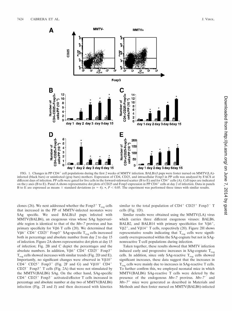
cells in neonatal PP at early times after milk-borne MMTVinfection, the expression of CD25 and Foxp3 in CD4� T cellswas studied. FACS analysis indicated that the percentage (datanot shown) and absolute number of PP CD4� CD25� Foxp3�
Treg cells increased progressively in MMTV(LA)-infected miceduring the first 2 weeks of infection (Fig. 1A and B). In addi-tion, FACS analysis showed that CD4� CD25� Foxp3� T cells,which have been described either as proliferating Treg cells(18) or as a Treg cell reservoir (52), increased with trendssimilar to CD4� CD25� Foxp3� Treg cells (Fig. 1C). Of inter-est, CD4� CD25� Foxp3� activated/effector T cells showed asignificant increase in the percentage (data not shown) andabsolute number at day 2 of infection and then diminishedmarkedly from day 3 onwards (Fig. 1D). Finally, the CD4�
CD25� Foxp3� T-cell population also increased in percentage(data not shown) and absolute number during the first 2 weeksof infection (Fig. 1E), likely due to the recruitment of naiveCD4� T cells to PP that takes place during MMTV infection,as we have previously reported (12).
These results indicate that MMTV infection induced pro-gressive increases in both the CD4� CD25� Foxp3� andCD4� CD25� Foxp3� populations of Treg cells at least duringthe first 2 weeks of infection. In contrast, CD4� CD25�
Foxp3� activated/effector T cells significantly increased at day2 of infection but then decreased and were maintained at alower level from day 3 onward.
MMTV infection induces increases in SAg-specific Treg
cells. The early response to the MMTV SAg results in anincrease in the percentage of SAg-reactive V�-specific T-cell
VOL. 82, 2008 REGULATORY T CELLS DURING MMTV INFECTION 7423
on June 7, 2014 by guesthttp://jvi.asm
.org/D
ownloaded from
clones (26). We next addressed whether the Foxp3� Treg cellsthat increased in the PP of MMTV-infected neonates wereSAg specific. We used BALB/cJ pups infected withMMTV(BALB6), an exogenous virus whose SAg hypervari-able region is identical to that of the Mtv-7 provirus and hasprimary specificity for V�6 T cells (20). We determined thatV�6� CD4� CD25� Foxp3� SAg-specific Treg cells increasedboth in percentage and absolute number from day 2 to day 15of infection. Figure 2A shows representative dot plots at day 15of infection; Fig. 2B and C depict the percentages and theabsolute numbers. In addition, V�6� CD4� CD25� Foxp3�
Treg cells showed increases with similar trends (Fig. 2D and E).Importantly, no significant changes were observed in V�10�
CD4� CD25� Foxp3� (Fig. 2F and G) and V�10� CD4�
CD25� Foxp3� T cells (Fig. 2A) that were not stimulated bythe MMTV(BALB6) SAg. On the other hand, SAg-specificCD4� CD25� Foxp3� activated/effector T cells increased inpercentage and absolute number at day two of MMTV(BALB6)infection (Fig. 2I and J) and then decreased with kinetics
similar to the total population of CD4� CD25� Foxp3� Tcells (Fig. 1D).
Similar results were obtained using the MMTV(LA) viruswhich carries three different exogenous viruses: BALB6,BALB2, and BALB14 with primary specificities for V�6�,V�2�, and V�14� T cells, respectively (20). Figure 2H showsrepresentative results indicating that Treg cells were signifi-cantly overrepresented within the SAg-cognate but not in SAg-nonreactive T-cell populations during infection.
Taken together, these results showed that MMTV infectioninduced early and progressive increases in SAg-cognate Treg
cells. In addition, since only SAg-reactive Treg cells showedsignificant increases, these data suggest that the increases inTreg cells were mainly due to increases in SAg-reactive T cells.To further confirm this, we employed neonatal mice in whichMMTV(BALB6) SAg-reactive T cells were deleted by thepresence of the endogenous Mtv-7 provirus. Mtv-7� andMtv-7� mice were generated as described in Materials andMethods and then foster nursed on MMTV(BALB6)-infected
FIG. 1. Changes in PP CD4� cell populations during the first 2 weeks of MMTV infection. BALB/cJ pups were foster nursed on MMTV(LA)-infected (black bars) or uninfected (gray bars) mothers. Expression of CD4, CD25, and intracellular Foxp3 in PP cells was analyzed by FACS atdifferent days of infection. PP cells were gated for live cells in the forward-sideward scatter (B to E) and for CD4� cells (A). Cell types are indicatedon the y axes (B to E). Panel A shows representative dot plots of CD25 and Foxp3 expression in PP CD4� cells at day 2 of infection. Data in panelsB to E are expressed as means standard deviations (n � 4). *, P 0.05. The experiment was performed three times with similar results.
7424 CABRERA ET AL. J. VIROL.
on June 7, 2014 by guesthttp://jvi.asm
.org/D
ownloaded from

mothers for 6 days. Mtv-7� mice showed increases in the per-centage and the absolute number of CD4� CD25� Foxp3�
and CD4� CD25� Foxp3� Treg cells, whereas Mtv-7� miceshowed no significant increases in these cell populations (Fig.3). These results confirm that the increases in Treg cells that
occurred during MMTV infection were mainly due to increasesin SAg-reactive T cells.
Treg-cell increases are dependent on the presence of DCs.DCs play an important role in MMTV infection (8, 11). Werecently showed that the SAg response is abrogated in the
FIG. 2. SAg specificity of PP Foxp3� Treg cells during the first 2 weeks after MMTV infection. BALB/cJ pups were foster nursed onMMTV(BALB6)-infected (A to G and I and J) or MMTV(LA)-infected (H) (black bars) or uninfected (gray bars) mothers. Expression of V�6,CD4, CD25, and intracellular Foxp3 in PP cells was analyzed by FACS at different days after infection. PP cells were gated for live cells in theforward-sideward scatter (B to G and I and J) and for V�� CD4� cells (A and H). Cell types are indicated along the y axes (B to G and I andJ). (A) Representative dot plots of CD25 and Foxp3 in PP V�6� CD4� and V�10� CD4� cells at day 15 of infection. (H) Percentage of Foxp3�
cells within V�6� CD4�, V�14� CD4�, and V�10� CD4� cells at day 15 of infection. Data are expressed as the means standard deviations(n � 4). *, P 0.05. The experiments were performed three times with similar results.
VOL. 82, 2008 REGULATORY T CELLS DURING MMTV INFECTION 7425
on June 7, 2014 by guesthttp://jvi.asm
.org/D
ownloaded from

absence of these cells (11). To investigate whether DCs wereinvolved in the MMTV-induced increase in Foxp3� Treg cells,we used an in vivo model of inducible DC ablation, the CD11c-DTR mice (25). Administration of DT to CD11c-DTR miceproduces a selective and temporal depletion of CD11c� cells,generating conditional DC knockouts (25). Although ablationwas successfully accomplished in adult mice, our attempts toadapt the model to neonatal mice failed because the DT waslethal for the transgenic pups, even in low doses. Therefore, wecarried out these experiments in adult mice. We first testedwhether MMTV also affected the CD4� CD25� Foxp3� cellsubset in an experimental model of footpad infection. WhenMMTV(LA) was injected subcutaneously into wild-type adultanimals, the draining popliteal lymph node showed an increasein the percentages and absolute numbers of CD4� CD25�
Foxp3� and V�6� CD4� CD25� Foxp3� Treg cells, resem-bling the one observed in the PP (not shown). CD11c-DTRmice were injected i.p. with DT (day �1) and then withMMTV in the footpad (day 0), and the numbers of CD4�
CD25� Foxp3� and V�6� CD4� CD25� Foxp3� cells wereanalyzed in the draining and nondraining popliteal lymphnodes at 2 and 6 days after infection. DC depletion abolishedincreases in both the CD4� CD25� Foxp3� Treg cells and theSAg-specific Treg cells (Fig. 4), thus demonstrating that theMMTV-induced increase in Treg cells was DC dependent.
CD4� CD25� PP cells from infected mice preferentiallysuppress the proliferative response of T cells to SAg-express-ing APCs. MLR assays are commonly used to test the suppres-sive activity of Treg cells. For the MLR assays, we purifiedCD4� CD25� cells from the PP of BALB/cJ mice foster
nursed for 6 days on MMTV(LA)-infected mothers in order toevaluate their ability to suppress responses ex vivo. We per-formed the MLR assays by coculturing CFSE-labeledBALB/cJ CD4� CD25� cells (responders) with mitomycin C-treated mononuclear spleen cells from AKR/J mice (stimula-tors). CD4� CD25� or CD4� CD25� cells obtained from thePP of infected or uninfected pups were added to the MLRmixture from the beginning of the culture. Proliferation ofCD4� responder cells was analyzed by CFSE dilution. Asshown in Fig. 5, PP CD4� CD25� cells from infected micestrongly inhibited proliferation in the MLR assay using AKR/JAPCs and suppressed the proliferative response more effi-ciently than CD4�CD25� cells from uninfected littermates. Inaddition, the percentage of inhibition increased in a dose-dependent manner (data not shown). These results indicatethat CD4� CD25� cells obtained from the PP of infected micehad a strong immunosuppressive activity when AKR/J APCswere used in the MLR assays. In addition, the suppressivecapacity of these cells was greater than that of CD4� CD25�
cells from uninfected mice.
FIG. 3. Increases in PP Treg cells are dependent on the presence ofSAg-reactive T cells. Mtv-7� and Mtv-7� mice generated by crossing(BALB/cJ � AKR/J)F1 � BALB/cJ mice were nursed on MMTV(BALB6)-infected or uninfected mothers for 6 days. The expression ofCD4, CD25, and intracellular Foxp3 in PP cells was analyzed by FACS.PP cells were gated for live cells in the forward-sideward scatter. Celltypes are indicated along the y axes. Data are presented as the means standard deviations (n � 5). *, P 0.05. The experiment was per-formed four times with similar results. FIG. 4. Increases in Treg cells are dependent on the presence of
DCs. CD11c-DTR adult mice were injected i.p. with DT (day �1) andwith MMTV(LA) in the footpad (day 0). After 2 or 6 days the expres-sion levels of V�6, CD4, CD25, and Foxp3 were analyzed by FACS inthe draining and nondraining (ND) popliteal lymph nodes. Lymphnode cells were gated for mononuclear cells in the forward-sidewardscatter. (A) Number of CD4� CD25� Foxp3� cells in draining popli-teal lymph nodes of mice treated with DT or not treated with DT andin nondraining popliteal lymph nodes (ND). (B) Number of V�6�
CD4� CD25� Foxp3� cells in untreated (No DT) DT-treated lymphnodes and in nondraining (ND) lymph nodes. Data are presented asthe means standard deviations (n � 3). *, P 0,05. One experimentout of two is shown.
7426 CABRERA ET AL. J. VIROL.
on June 7, 2014 by guesthttp://jvi.asm
.org/D
ownloaded from
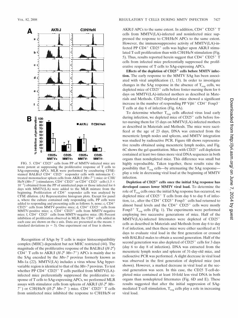
Recognition of SAgs by T cells is major histocompatibilitycomplex (MHC) dependent but not MHC restricted (44). Themagnitude of the proliferative response of the BALB/cJ (H-2d)CD4� T cells to AKR/J (H-2k Mtv-7�) APCs is mainly due tothe SAg encoded by the Mtv-7 provirus formerly known asMls-1a (22). MMTV(LA) includes a virus whose SAg hyper-variable region is identical to that of the Mtv-7 provirus. To testwhether PP CD4� CD25� T cells purified from MMTV(LA)-infected mice preferentially suppressed the proliferative re-sponse of T cells to SAg-expressing APCs, we performed MLRassays with stimulator cells from spleens of AKR/J (H-2k Mtv-7�) or C3H/HeN (H-2k Mtv-7�) mice. CD4� CD25� T cellsfrom uninfected mice inhibited the response to C3H/HeN or
AKR/J APCs to the same extent. In addition, CD4� CD25� Tcells from MMTV(LA)-infected and noninfected mice sup-pressed the response to C3H/HeN APCs to the same extent.However, the immunosuppressive activity of MMTV(LA)-in-fected PP CD4� CD25� cells was higher upon AKR/J stimu-lated T-cell proliferation than with C3H/HeN stimulation (Fig.5). Thus, results reported herein suggest that CD4� CD25� Tcells from infected mice preferentially suppressed the prolif-erative response of T cells to SAg-expressing APCs.
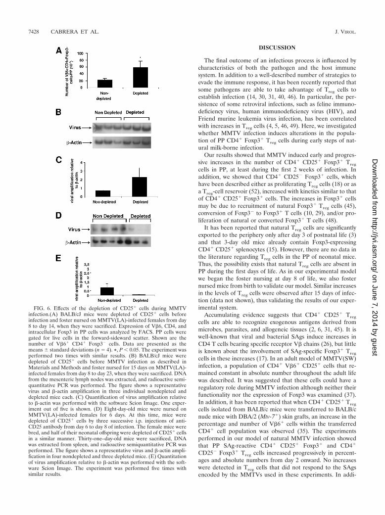
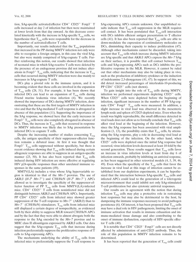
Effects of the depletion of CD25� cells before MMTV infec-tion. The early response to the MMTV SAg has been associ-ated with viral amplification (1, 13). In order to investigatechanges in the SAg response in the absence of Treg cells, wedepleted mice of CD25� cells before foster-nursing them for 6days on MMTV(LA)-infected mothers as described in Mate-rials and Methods. CD25-depleted mice showed a significantincrease in the number of responding PP V�6� CD4� Foxp3�
T cells at day 6 of infection (Fig. 6A).To determine whether Treg cells affected virus load early
during infection, we depleted mice of CD25� cells before fos-ter-nursing them for 15 days on MMTV(LA)-infected mothersas described in Materials and Methods. The mice were sacri-ficed at the age of 23 days, DNA was extracted from themesenteric lymph nodes and spleens, and MMTV integrationwas studied by radioactive PCR. Figure 6B shows representa-tive results obtained using mesenteric lymph nodes, and Fig.6C shows the gel quantitation. Mice with CD25� cell depletioncontained at least two times more viral DNA sequences in bothorgans than nondepleted mice. This difference was small buthighly reproducible. Taken together, these results raise thepossibility that Treg cells—by attenuating the SAg response—play a role in decreasing viral load at the beginning of MMTVinfection.
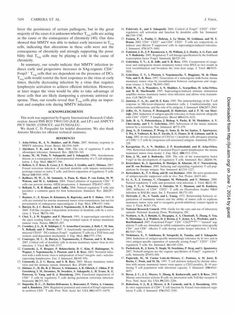
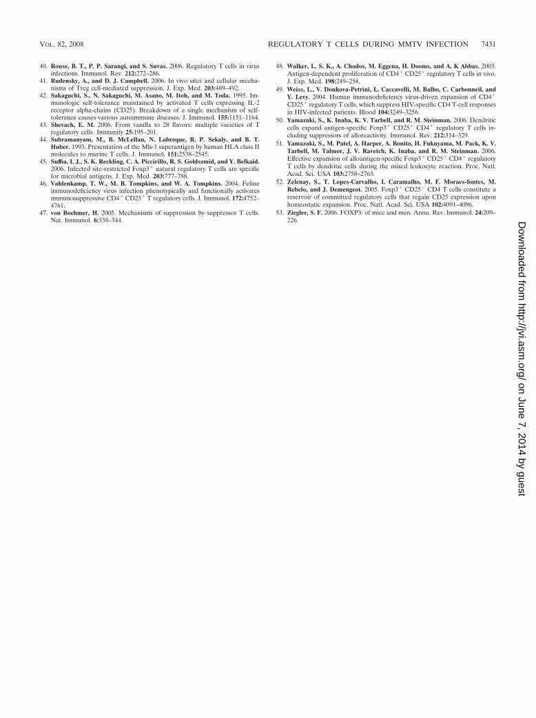
Depletion of CD25� cells once the initial SAg response hasdeveloped causes lower MMTV viral load. To determine therole of Treg cells once the initial SAg response has occurred, wedepleted mice of CD25� T cells from day 6 to day 8 of infec-tion, i.e., after the CD4� CD25� Foxp3� cells had returned toalmost basal levels and the CD4� CD25� cells were mostlyFoxp3� Treg cells (Fig. 1). The experiments were performedemploying two successive generations of mice. Half of theMMTV(LA)-infected littermates were depleted of CD25�
cells as described in Materials and Methods from day 6 to day8 of infection, and then these mice were either sacrificed at 31days to evaluate viral load in the first generation or crossedwith BALB/cJ males to obtain a second generation. Half of thesecond generation was also depleted of CD25� cells for 3 days(day 6 to day 8 of infection). DNA was extracted from themesenteric lymph nodes and spleens of 31-day-old mice, andradioactive PCR was performed. A slight decrease in viral loadwas observed in the first generation of depleted mice (notshown). However, a marked decrease in viral load at the sec-ond generation was seen. In this case, the CD25 T-cell-de-pleted mice contained at least 10-fold less viral DNA in bothorgans than nondepleted littermates (Fig. 6D and E). Theseresults suggested that after the initial suppression of SAg-mediated T-cell stimulation, Treg cells play a role in increasingviral load.
FIG. 5. CD4� CD25� cells from PP of MMTV-infected mice aremore potent at suppressing the proliferative response of T cells toSAg-expressing APCs. MLR were performed by coculturing CFSE-stained BALB/cJ CD4� CD25� responder cells with mitomycin C-treated mononuclear spleen cells from AKR/J (Mtv-7�) mice or C3H/HeN (Mtv-7�) stimulators. CD4� CD25� or CD4� CD25� cells (1.5 �10�5) obtained from the PP of uninfected pups or those infected for 6days with MMTV(LA) were added to the MLR mixture from thebeginning. Proliferation of CD4� responder cells was analyzed byCFSE dilution. (A) Representative histogram plots. Except for panela, where the culture contained only responding cells, PP cells wereadded to responding and presenting cells as follows: b, none; c, CD4�
CD25� cells from MMTV-positive mice; d, CD4� CD25� cells fromMMTV-positive mice; e, CD4� CD25� cells from MMTV-negativemice; f, CD4� CD25� cells from MMTV-negative mice. (B) Percentinhibition of proliferation observed in MLR; the CD4� cells added ineach case are shown on the x axis. Data are presented as the means standard deviations (n � 3). One experiment out of four is shown.
VOL. 82, 2008 REGULATORY T CELLS DURING MMTV INFECTION 7427
on June 7, 2014 by guesthttp://jvi.asm
.org/D
ownloaded from
DISCUSSION
The final outcome of an infectious process is influenced bycharacteristics of both the pathogen and the host immunesystem. In addition to a well-described number of strategies toevade the immune response, it has been recently reported thatsome pathogens are able to take advantage of Treg cells toestablish infection (14, 30, 31, 40, 46). In particular, the per-sistence of some retroviral infections, such as feline immuno-deficiency virus, human immunodeficiency virus (HIV), andFriend murine leukemia virus infection, has been correlatedwith increases in Treg cells (4, 5, 46, 49). Here, we investigatedwhether MMTV infection induces alterations in the popula-tion of PP CD4� Foxp3� Treg cells during early steps of nat-ural milk-borne infection.
Our results showed that MMTV induced early and progres-sive increases in the number of CD4� CD25� Foxp3� Treg
cells in PP, at least during the first 2 weeks of infection. Inaddition, we showed that CD4� CD25� Foxp3� cells, whichhave been described either as proliferating Treg cells (18) or asa Treg-cell reservoir (52), increased with kinetics similar to thatof CD4� CD25� Foxp3� cells. The increases in Foxp3� cellsmay be due to recruitment of natural Foxp3� Treg cells (45),conversion of Foxp3� to Foxp3� T cells (10, 29), and/or pro-liferation of natural or converted Foxp3� T cells (48).
It has been reported that natural Treg cells are significantlyexported to the periphery only after day 3 of postnatal life (3)and that 3-day old mice already contain Foxp3-expressingCD4� CD25� splenocytes (15). However, there are no data inthe literature regarding Treg cells in the PP of neonatal mice.Thus, the possibility exists that natural Treg cells are absent inPP during the first days of life. As in our experimental modelwe began the foster nursing at day 8 of life, we also fosternursed mice from birth to validate our model. Similar increasesin the levels of Treg cells were observed after 15 days of infec-tion (data not shown), thus validating the results of our exper-imental system.
Accumulating evidence suggests that CD4� CD25� Treg
cells are able to recognize exogenous antigens derived frommicrobes, parasites, and allogeneic tissues (2, 6, 31, 45). It iswell-known that viral and bacterial SAgs induce increases inCD4 T cells bearing specific receptor V� chains (26), but littleis known about the involvement of SAg-specific Foxp3� Treg
cells in these increases (17). In an adult model of MMTV(SW)infection, a population of CD4� V�6� CD25� cells that re-mained constant in absolute number throughout the adult lifewas described. It was suggested that these cells could have aregulatory role during MMTV infection although neither theirfunctionality nor the expression of Foxp3 was examined (37).In addition, it has been reported that when CD4� CD25� Treg
cells isolated from BALB/c mice were transferred to BALB/cnude mice with DBA/2 (Mtv-7�) skin grafts, an increase in thepercentage and number of V�6� cells within the transferredCD4� cell population was observed (35). The experimentsperformed in our model of natural MMTV infection showedthat PP SAg-reactive CD4� CD25� Foxp3� and CD4�
CD25� Foxp3� Treg cells increased progressively in percent-ages and absolute numbers from day 2 onward. No increaseswere detected in Treg cells that did not respond to the SAgsencoded by the MMTVs used in these experiments. In addi-
FIG. 6. Effects of the depletion of CD25� cells during MMTVinfection.(A) BALB/cJ mice were depleted of CD25� cells beforeinfection and foster nursed on MMTV(LA)-infected females from day8 to day 14, when they were sacrificed. Expression of V�6, CD4, andintracellular Foxp3 in PP cells was analyzed by FACS. PP cells weregated for live cells in the forward-sideward scatter. Shown are thenumber of V�6� CD4� Foxp3� cells. Data are presented as themeans standard deviations (n � 4). *, P 0.05. The experiment wasperformed two times with similar results. (B) BALB/cJ mice weredepleted of CD25� cells before MMTV infection as described inMaterials and Methods and foster nursed for 15 days on MMTV(LA)-infected females from day 8 to day 23, when they were sacrificed. DNAfrom the mesenteric lymph nodes was extracted, and radioactive semi-quantitative PCR was performed. The figure shows a representativevirus and �-actin amplification in three individual nondepleted anddepleted mice each. (C) Quantification of virus amplification relativeto �-actin was performed with the software Scion Image. One exper-iment out of five is shown. (D) Eight-day-old mice were nursed onMMTV(LA)-infected females for 6 days. At this time, mice weredepleted of CD25� cells by three successive i.p. injections of anti-CD25 antibody from day 6 to day 8 of infection. The female mice werebred, and half of their neonatal offspring were depleted of CD25� cellsin a similar manner. Thirty-one–day-old mice were sacrificed, DNAwas extracted from spleen, and radioactive semiquantitative PCR wasperformed. The figure shows a representative virus and �-actin ampli-fication in four nondepleted and three depleted mice. (E) Quantitationof virus amplification relative to �-actin was performed with the soft-ware Scion Image. The experiment was performed five times withsimilar results.
7428 CABRERA ET AL. J. VIROL.
on June 7, 2014 by guesthttp://jvi.asm
.org/D
ownloaded from
tion, SAg-specific activated/effector CD4� CD25� Foxp3� Tcells increased at day 2 of infection but then were maintainedat lower levels from that day onward. As this decrease corre-lated kinetically with the increase in SAg-specific Treg cells, wehypothesize that Treg cells were responsible for the decrease inthe number of activated/effector T cells.
Importantly, our results indicated that the Treg populationsthat increased in the PP during MMTV infection not only wereable to recognize a foreign antigen, in this case the viral SAg,but also were mainly comprised of SAg-cognate T cells. Fur-ther reinforcing this notion, our results showed that infectionof neonatal mice in which SAg-reactive T cells were deleted bythe presence of an endogenous provirus did not induce alter-ations in Foxp3� Treg cells, indicating that the increase in Treg
cells that occurred during MMTV infection were due mainly toincreases in SAg-cognate T cells.
DCs play a pivotal role in the immune system, and it isbecoming evident that these cells are involved in the expansionof Treg cells (28, 51). For example, it has been shown thatinfected DCs can lead to an expansion of Treg cells duringFriend murine leukemia virus infection (4). We recentlyshowed the importance of DCs during MMTV infection, dem-onstrating that these are the first targets of MMTV infection invivo and that the SAg-mediated T-cell response is abrogated inabsence of this population (11). Supporting the importance ofthe SAg response, we showed here that the early increases inFoxp3� Treg cells were also completely abrogated in absence ofDCs. Thus, the increase in Treg cells that occurred in responseto MMTV infection was likely due to SAg presentation byinfected DCs to cognate T cells.
Despite the increasing number of studies concerning Treg
cells, the antigen specificity of their suppressor-effector func-tion remains a debated issue. At first it was thought thatFoxp3� Treg cells suppressed without specificity, but there isrecent evidence showing that Treg cells induced during certainallogeneic responses are able to suppress in an antigen-specificmanner (23, 50). It has also been reported that Treg cellsinduced during HIV infection are more effective at regulatingHIV p24-specific responses than other unrelated immune re-sponses in the same patients (49).
MMTV(LA) includes a virus whose SAg hypervariable re-gion is identical to that of the Mtv-7 provirus. The use ofAKR/J (H-2k Mtv-7�) and C3H/HeN (H-2k Mtv-7�) APCsallowed us to investigate the specificity of the suppressor-ef-fector function of PP Treg cells from MMTV(LA)-infectedmice. CD4� CD25� T cells from noninfected mice did notdistinguish between AKR/J and C3H/HeN APCs. Importantly,PP CD4� CD25� cells from infected mice showed greatersuppression of the T-cell response to Mtv-7� (AKR/J) than toMtv-7� (C3H/HeN) stimulators. Treg cells from infected micestill displayed a certain degree of nonspecificity, as evidencedboth by their ability to suppress MLR toward C3H/HeN APCsand by the fact that they were able to almost abrogate both theresponse to the SAg encoded by the Mtv-7 provirus and toMHC class II alloantigens expressed by AKR/J cells. Our datasuggest that the SAg-cognate Treg cells that increase duringinfection preferentially suppress the proliferative response of Tcells to SAg-expressing APCs.
The mechanisms underlying the ability of Treg cells frominfected mice to preferentially suppress the T-cell response to
SAg-expressing APCs remain unknown. Our unpublished re-sults indicate that Treg cell suppression in the MLR requirescell contact. It has been postulated that Treg-cell interactionwith DCs inhibits efficient antigen presentation to T effectorcells (41). It has also been reported that Treg cells are able todown-modulate the expression of costimulatory molecules onDCs, diminishing their capacity to induce proliferation (47).Although other mechanisms cannot be discarded, taking intoaccount that Treg cells which increase during MMTV infectionare SAg specific and that AKR/J APCs express the Mtv-7 SAgon their surface, it is possible that cell contact between Treg
cells and SAg-expressing APCs such as DCs inhibits the pro-liferative response in the MLR. However, it cannot be ruledout that other mechanisms may also be taking place in vivo,such as the production of inhibitory cytokines or the inductionof indoleamine 2,3-dioxygenase (41, 47). In support of this, wedid detect increased percentages of interleukin-10-producingPP CD4� CD25� cells (not shown).
To gain insight into the role of Treg cells during MMTV,infection we performed experiments depleting CD25� cells.When CD25� cells were depleted before the beginning ofinfection, significant increases in the number of PP SAg-cog-nate CD4� Foxp3� Treg cells were measured. In addition, aslight increase in viral load was observed in the mesentericlymph node and spleen of CD25-depleted mice. Even when theresult was highly reproducible, the small difference detected inviral loads does not allow us to formally conclude that Treg cellsaffect viral load at the beginning of the infection. However,since the SAg response has been associated with viral ampli-fication (1, 13), the possibility exists that Treg cells, by attenu-ating the SAg response, play a role in decreasing viral load atthe beginning of the infection. On the other hand, whenCD25� cells were depleted once the initial SAg response hadoccurred, virus infection levels decreased at least 10-fold by thesecond generation. These results suggest that Treg cells favoran increase in virus infection levels at least from day 6 ofinfection onwards, probably by inhibiting an antiviral response,as has been suggested in other retroviral models (4, 5, 39, 46,49). Even when the specificity of the Treg cells that favor theincrease in viral load at this stage of infection cannot be es-tablished from our depletion experiments, it can be hypothe-sized that the interaction between SAg-specific Treg cells andinfected APCs could lead to the generation of a tolerogenicmicroenvironment that could inhibit not only SAg-stimulatedT-cell proliferation but also cytotoxic antiviral responses.
Our results are in agreement with the notion that duringinfection Treg cells may play a protective role in restraininghost responses but may also be detrimental to the host, bydampening the immune responses necessary to avoid pathogenpersistence (6). Of interest, it has been proposed that Treg cellsmay have a dual role in HIV pathogenesis, limiting the onset ofimmune activation that results in immune exhaustion and im-mune-mediated tissue damage and also contributing to theonset of immune dysfunction, especially of HIV-specific effec-tor T cells (40).
It is notable that CD4� CD25� Foxp3� cells are not directlyaffected by administration of anti-CD25 antibody. Thus, therole of this population in MMTV infection remains to bedetermined.
It has been reported that the generation of Treg cells could
VOL. 82, 2008 REGULATORY T CELLS DURING MMTV INFECTION 7429
on June 7, 2014 by guesthttp://jvi.asm
.org/D
ownloaded from
favor the persistence of certain pathogens, but in the greatmajority of the cases it is unknown whether Treg cells are actingas the cause or the consequence of chronicity (40). Our datashowed that MMTV was able to induce early increases in Treg
cells, indicating that alterations in these cells were not theconsequence of chronicity and strongly supporting the possi-bility that Treg cells may be playing a role in the cause ofchronicity.
In summary, our results indicate that MMTV infection in-duces early and progressive increases in SAg-cognate CD4�
Foxp3� Treg cells that are dependent on the presence of DCs.Treg cells would restrict the host responses to the virus at earlytimes, thereby decreasing infection by a virus that requireslymphocyte activation to achieve efficient infection. However,at later stages the virus would be able to take advantage ofthese cells that are likely dampening a cytotoxic antiviral re-sponse. Thus, our results reveal that Treg cells play an impor-tant and complex role during MMTV infection.
ACKNOWLEDGMENTS
This work was supported by Fogarty International Research Collab-oration Award PHS RO3 TW011103 (S.R.R. and I.P.) and ANPCYTPICT 506305, CONICET, and FUNDALEU.
We thank C. D. Pasqualini for helpful discussions. We also thankAntonio Morales for efficient technical assistance.
REFERENCES
1. Acha-Orbea, H., A. N. Shakhov, and D. Finke. 2007. Immune response toMMTV infection. Front. Biosci. 12:1594–1609.
2. Aluvihare, V. R., and A. G. Betz. 2006. The role of regulatory T cells inalloantigen tolerance. Immunol. Rev. 212:330–343.
3. Asano, M., M. Toda, N. Sakaguchi, and S. Sakaguchi. 1996. Autoimmunedisease as a consequence of developmental abnormality of a T cell subpopu-lation. J. Exp. Med. 184:387–396.
4. Balkow, S., F. Krux, K. Loser, J. U. Becker, S. Grabbe, and U. Dittmer. 2007.Friend retrovirus infection of myeloid dendritic cells impairs maturation,prolongs contact to naive T cells, and favors expansion of regulatory T cells.Blood 110:3949–3958.
5. Beilharz, M. W., L. M. Sammels, A. Paun, K. Shaw, P. van Eeden, M. W.Watson, and M. L. Ashdown. 2004. Timed ablation of regulatory CD4� Tcells can prevent murine AIDS progression. J. Immunol. 172:4917–4925.
6. Belkaid, Y., R. B. Blank, and I. Suffia. 2006. Natural regulatory T cells andparasites: a common quest for host homeostasis. Immunol. Rev. 212:287–300.
7. Beutner, U., E. Kraus, D. Kitamura, K. Rajewsky, and B. T. Huber. 1994. Bcells are essential for murine mammary tumor virus transmission, but not forpresentation of endogenous superantigens. J. Exp. Med. 179:1457–1466.
8. Burzyn, D., J. C. Rassa, D. Kim, I. Nepomnaschy, S. R. Ross, and I. Piazzon.2004. Toll-like receptor 4-dependent activation of dendritic cells by a retro-virus. J. Virol. 78:576–584.
9. Choi, Y., J. W. Kappler, and P. Marrack. 1991. A superantigen encoded inthe open reading frame of the 3� long terminal repeat of mouse mammarytumour virus. Nature 350:203–207.
10. Coombes, J. L., K. R. Siddiqui, C. V. Arancibia-Carcamo, J. Hall, C. M. Sun,Y. Belkaid, and F. Powrie. 2007. A functionally specialized population ofmucosal CD103� DCs induces Foxp3� regulatory T cells via a TGF-beta andretinoic acid-dependent mechanism. J. Exp. Med. 204:1757–1764.
11. Courreges, M. C., D. Burzyn, I. Nepomnaschy, I. Piazzon, and S. R. Ross.2007. Critical role of dendritic cells in mouse mammary tumor virus in vivoinfection. J. Virol. 81:3769–3777.
12. Czarneski, J., P. Berguer, P. Bekinschtein, D. C. Kim, P. Hakimpour, N.Wagner, I. Nepomnaschy, I. Piazzon, and S. R. Ross. 2002. Neonatal infec-tion with a milk-borne virus is independent of beta7 integrin- and L-selectin-expressing lymphocytes. Eur. J. Immunol. 32:945–956.
13. Czarneski, J., J. C. Rassa, and S. R. Ross. 2003. Mouse mammary tumorvirus and the immune system. Immunol. Res. 27:469–480.
14. Dittmer, U., H. He, R. J. Messer, S. Schimmer, A. R. Olbrich, C. Ohlen, P. D.Greenberg, I. M. Stromnes, M. Iwashiro, S. Sakaguchi, L. H. Evans, K. E.Peterson, G. Yang, and K. J. Hasenkrug. 2004. Functional impairment ofCD8� T cells by regulatory T cells during persistent retrovirus infection.Immunity 20:293–303.
15. Dujardin, H. C., O. Burlen-Defranoux, L. Boucontet, P. Vieira, A. Cumano,and A. Bandeira. 2004. Regulatory potential and control of Foxp3 expressionin newborn CD4� T cells. Proc. Natl. Acad. Sci. USA 101:14473–14478.
16. Fehervari, Z., and S. Sakaguchi. 2004. Control of Foxp3� CD25� CD4�
regulatory cell activation and function by dendritic cells. Int. Immunol.16:1769–1780.
17. Feunou, P., L. Poulin, C. Habran, A. Le Moine, M. Goldman, and M. Y.Braun. 2003. CD4� CD25� and CD4� CD25� T cells act, respectively, asinducer and effector T suppressor cells in superantigen-induced tolerance.J. Immunol. 171:3475–3484.
18. Fontenot, J. D., J. P. Rasmussen, L. M. William, J. L. Dooley, A. G. Farr, andA. Y. Rudensky. 2005. Regulatory T cell lineage specification by the forkheadtranscription factor Foxp3. Immunity 22:329–341.
19. Golovkina, T. V., A. B. Jaffe, and S. R. Ross. 1994. Coexpression of exoge-nous and endogenous mouse mammary tumor virus RNA in vivo results inviral recombination and broadens the virus host range. J. Virol. 68:5019–5026.
20. Golovkina, T. V., I. Piazzon, I. Nepomnaschy, V. Buggiano, M. de OlanoVela, and S. R. Ross. 1997. Generation of a tumorigenic milk-borne mousemammary tumor virus by recombination between endogenous and exoge-nous viruses. J. Virol. 71:3895–3903.
21. Held, W., G. A. Waanders, A. N. Shakhov, L. Scarpellino, H. Acha-Orbea,and H. R. MacDonald. 1993. Superantigen-induced immune stimulationamplifies mouse mammary tumor virus infection and allows virus transmis-sion. Cell 74:529–540.
22. Janeway, C. A., Jr., and M. E. Katz. 1985. The immunobiology of the T cellresponse to Mls-locus-disparate stimulator cells. I. Unidirectionality, newstrain combinations, and the role of Ia antigens. J. Immunol. 134:2057–2063.
23. Joffre, O., N. Gorsse, P. Romagnoli, D. Hudrisier, and J. P. M. van Meer-wijk. 2004. Induction of antigen-specific tolerance to bone marrow allograftswith CD4� CD25� T lymphocytes. Blood 103:4216–4221.
24. Jude, B. A., Y. Pobezinskaya, J. Bishop, S. Parke, R. M. Medzhitov, A. V.Chervonsky, and T. V. Golovkina. 2003. Subversion of the innate immunesystem by a retrovirus. Nat. Immunol. 4:573–578.
25. Jung, S., D. Unutmaz, P. Wong, G. Sano, K. De los Santos, T. Sparwasser,S. Wu, S. Vuthoori, K. Ko, F. Zavala, E. G. Pamer, D. R. Littman, and R. A.Lang. 2002. In vivo depletion of CD11c(�) dendritic cells abrogates primingof CD8(�) T cells by exogenous cell-associated antigens. Immunity 17:211–220.
26. Karapetian, O., A. N. Shakhov, J. P. Kraehenbuhl, and H. Acha-Orbea.1994. Retroviral infection of neonatal Peyer’s patch lymphocytes: the mousemammary tumor virus model. J. Exp. Med. 180:1511–1516.
27. Kim, J. M., and A. Rudensky. 2006. The role of the transcription factorFoxp3 in the development of regulatory T cells. Immunol. Rev. 212:86–98.
28. Kretschmer, K., I. Apostolou, D. Hawiger, K. Khazaie, M. C. Nussenzweig,and H. von Boehmer. 2005. Inducing and expanding regulatory T cell pop-ulations by foreign antigen. Nat. Immunol. 6:1219–1227.
29. Kretschmer, K., T. S. Heng, and H. von Boehmer. 2006. De novo productionof antigen-specific suppressor cells in vivo. Nat. Protoc. 1:653–661.
30. Li, S., E. J. Gowans, C. Chougnet, M. Plebanski, and U. Dittmer. 2008.Natural regulatory T cells and persistent viral infection. J. Virol. 82:21–30.
31. Long, T. T., S. Nakazawa, S. Onizuka, M. C. Huaman, and H. Kanbara.2003. Influence of CD4� CD25� T cells on Plasmodium berghei NK65infection in BALB/c mice. Int. J. Parasitol. 33:175–183.
32. McGrath, C. M., S. Nandi, and L. Young. 1972. Relationship between or-ganization of mammary tumors and the ability of tumor cells to replicatemammary tumor virus and to recognize growth-inhibitory contact signals invitro. J. Virol. 9:367–376.
33. National Research Council. 1996. Guide for the care and use of laboratoryanimals. National Academy Press, Washington, DC.
34. Nesburn, A. B., I. Bettahi, G. Dasgupta, A. A. Chentoufi, X. Zhang, S. You,N. Morishige, A. J. Wahlert, D. J. Brown, J. V. Jester, S. L. Wechsler, and L.BenMohamed. 2007. Functional Foxp3� CD4� CD25(Bright�) “natural” reg-ulatory T cells are abundant in rabbit conjunctiva and suppress virus-specificCD4� and CD8� effector T cells during ocular herpes infection. J. Virol.81:7647–7661.
35. Nishimura, E., T. Sakihama, R. Setoguchi, K. Tanaka, and S. Sakaguchi.2004. Induction of antigen-specific immunologic tolerance by in vivo and invitro antigen-specific expansion of naturally arising Foxp3� CD25� CD4�
regulatory T cells. Int. Immunol. 16:1189–1201.36. Pacholczyk, R., J. Kern, N. Singh, M. Iwashima, P. Kraj, and L. Ignatowicz.
2007. Nonself-antigens are the cognate specificities of Foxp3� regulatory Tcells. Immunity 27:493–504.
37. Papiernik, M., M. Carmo Leite-de-Moraes, C. Pontoux, A. M. Joret, B.Rocha, C. Penit, and M. Dy. 1997. T cell deletion induced by chronic infec-tion with mouse mammary tumor virus spares a CD25-positive, IL-10 pro-ducing T cell population with infectious capacity. J. Immunol. 158:4642–4653.
38. Rassa, J. C., J. L. Meyers, Y. Zhang, R. Kudaravalli, and S. R Ross. 2002.Murine retroviruses activate B cells via interaction with Toll-like receptor 4.Proc. Natl. Acad. Sci. USA 99:2281–2286.
39. Robertson, S. J., R. J. Messer, A. B. Carmody, and K. J. Hasenkrug. 2006.In vitro suppression of CD8� T cell function by Friend virus-induced regu-latory T cells. J. Immunol. 176:3342–3349.
7430 CABRERA ET AL. J. VIROL.
on June 7, 2014 by guesthttp://jvi.asm
.org/D
ownloaded from
40. Rouse, B. T., P. P. Sarangi, and S. Suvas. 2006. Regulatory T cells in virusinfections. Immunol. Rev. 212:272–286.
41. Rudensky, A., and D. J. Campbell. 2006. In vivo sites and cellular mecha-nisms of Treg cell-mediated suppression. J. Exp. Med. 203:489–492.
42. Sakaguchi, S., N. Sakaguchi, M. Asano, M. Itoh, and M. Toda. 1995. Im-munologic self-tolerance maintained by activated T cells expressing IL-2receptor alpha-chains (CD25). Breakdown of a single mechanism of self-tolerance causes various autoimmune diseases. J. Immunol. 155:1151–1164.
43. Shevach, E. M. 2006. From vanilla to 28 flavors: multiple varieties of Tregulatory cells. Immunity 25:195–201.
44. Subramanyam, M., B. McLellan, N. Labreque, R. P. Sekaly, and B. T.Huber. 1993. Presentation of the Mls-1 superantigen by human HLA class IImolecules to murine T cells. J. Immunol. 151:2538–2545.
45. Suffia, I. J., S. K. Reckling, C. A. Piccirillo, R. S. Goldszmid, and Y. Belkaid.2006. Infected site-restricted Foxp3� natural regulatory T cells are specificfor microbial antigens. J. Exp. Med. 203:777–788.
46. Vahlenkamp, T. W., M. B. Tompkins, and W. A. Tompkins. 2004. Felineimmunodeficiency virus infection phenotypically and functionally activatesimmunosuppressive CD4� CD25� T regulatory cells. J. Immunol. 172:4752–4761.
47. von Boehmer, H. 2005. Mechanisms of suppression by suppressor T cells.Nat. Immunol. 6:338–344.
48. Walker, L. S. K., A. Chodos, M. Eggena, H. Dooms, and A. K Abbas. 2003.Antigen-dependent proliferation of CD4� CD25� regulatory T cells in vivo.J. Exp. Med. 198:249–258.
49. Weiss, L., V. Donkova-Petrini, L. Caccavelli, M. Balbo, C. Carbonneil, andY. Levy. 2004. Human immunodeficiency virus-driven expansion of CD4�
CD25� regulatory T cells, which suppress HIV-specific CD4 T-cell responsesin HIV-infected patients. Blood 104:3249–3256.
50. Yamazaki, S., K. Inaba, K. V. Tarbell, and R. M. Steinman. 2006. Dendriticcells expand antigen-specific Foxp3� CD25� CD4� regulatory T cells in-cluding suppressors of alloreactivity. Immunol. Rev. 212:314–329.
51. Yamazaki, S., M. Patel, A. Harper, A. Bonito, H. Fukuyama, M. Pack, K. V.Tarbell, M. Talmor, J. V. Ravetch, K. Inaba, and R. M. Steinman. 2006.Effective expansion of alloantigen-specific Foxp3� CD25� CD4� regulatoryT cells by dendritic cells during the mixed leukocyte reaction. Proc. Natl.Acad. Sci. USA 103:2758–2763.
52. Zelenay, S., T. Lopes-Carvalho, I. Caramalho, M. F. Moraes-fontes, M.Rebelo, and J. Demengeot. 2005. Foxp3� CD25� CD4 T cells constitute areservoir of committed regulatory cells that regain CD25 expression uponhomeostatic expansion. Proc. Natl. Acad. Sci. USA 102:4091–4096.
53. Ziegler, S. F. 2006. FOXP3: of mice and men. Annu. Rev. Immunol. 24:209–226.
VOL. 82, 2008 REGULATORY T CELLS DURING MMTV INFECTION 7431
on June 7, 2014 by guesthttp://jvi.asm
.org/D
ownloaded from