Modulation of IL17 and Foxp3 Expression in the Prevention of Autoimmune Arthritis in Mice
8
Modulation of IL-17 and Foxp3 Expression in the Prevention of Autoimmune Arthritis in Mice Joana Duarte 1,2 , Ana Agua-Doce 1,2 , Vanessa G. Oliveira 1,2 , Joa ˜ o Eurico Fonseca 1,3 , Luis Graca 1,2 * 1 Instituto de Medicina Molecular, Faculdade de Medicina, University of Lisbon, Lisbon, Portugal, 2 Instituto Gulbenkian de Cie ˆ ncia, Oeiras, Portugal, 3 Rheumatology Department, Hospital de Santa Maria, Lisbon, Portugal Abstract Background: Rheumatoid Arthritis (RA) is a chronic immune mediated disease associated with deregulation of many cell types. It has been reported that different T cell subsets have opposite effects in disease pathogenesis, in particular Th17 and Treg cells. Methodology and Findings: We investigated whether non-depleting anti-CD4 monoclonal antibodies, which have been reported as pro-tolerogenic, can lead to protection from chronic autoimmune arthritis in SKG mice – a recently described animal model of RA – by influencing the Th17/Treg balance. We found that non-depleting anti-CD4 prevented the onset of chronic autoimmune arthritis in SKG mice. Moreover, treated mice were protected from the induction of arthritis up to 60 days following anti-CD4 treatment, while remaining able to mount CD4-dependent immune responses to unrelated antigens. The antibody treatment also prevented disease progression in arthritic mice, although without leading to remission. Protection from arthritis was associated with an increased ratio of Foxp3, and decreased IL-17 producing T cells in the synovia. In vitro assays under Th17-polarizing conditions showed CD4-blockade prevents Th17 polarization, while favoring Foxp3 induction. Conclusions: Non-depleting anti-CD4 can therefore induce long-term protection from chronic autoimmune arthritis in SKG mice through reciprocal changes in the frequency of Treg and Th17 cells in peripheral tissues, thus shifting the balance towards immune tolerance. Citation: Duarte J, Agua-Doce A, Oliveira VG, Fonseca JE, Graca L (2010) Modulation of IL-17 and Foxp3 Expression in the Prevention of Autoimmune Arthritis in Mice. PLoS ONE 5(5): e10558. doi:10.1371/journal.pone.0010558 Editor: Derya Unutmaz, New York University, United States of America Received February 26, 2010; Accepted April 16, 2010; Published May 10, 2010 Copyright: ß 2010 Duarte et al. This is an open-access article distributed under the terms of the Creative Commons Attribution License, which permits unrestricted use, distribution, and reproduction in any medium, provided the original author and source are credited. Funding: This work was funded by SUDOE, grant number IMMUNONET-SOE1/1P1/E014, and supported by a research grant from Fundac ¸a ˜o para a Cie ˆncia e Tecnologia (FCT), Portugal (FCT/POCI/SAU-MMO/55974/2004). JD, AA-D, and VGO are funded with scholarships from FCT (SFRH/BD/23631/2005, SFRH/BD/49093/ 2008, and SFRH/BPD/22575/2005). The funders had no role in study design, data collection and analysis, decision to publish, or preparation of the manuscript. Competing Interests: The authors have declared that no competing interests exist. * E-mail: [email protected] Introduction Rheumatoid arthritis (RA) is a common chronic autoimmune inflammatory disease characterized by destruction of the synovial joints, leading to progressive disability, increased co-morbidity and premature mortality [1,2]. Both genetic and environmental factors are known to contribute to the development of the disease [3]. RA is characterized by a complex immune mediated response with the participation of many cell types including CD4 + T cells [4,5,6], such as the IL-17 producing Th17 subset, which have been shown to play an important role in the pathogenesis of the disease [7,8,9]. The participation of CD4 + T cells in the pathogenesis of RA, namely by influencing other key cellular mediators of the disease (such as B cells or macrophages), has prompted the development of therapeutic strategies targeting this lymphocyte population [10,11,12,13]. Monoclonal antibodies (MAbs) targeting key T cell molecules (such as co-receptor and co-stimulation) have been suggested as drugs capable of achieving long-term protection from the disease, with the potential of leading to immune tolerance, following a short treatment [14]. Indeed long-term transplantation tolerance can be induced in mice following CD4 or co-stimulation blockade [15,16,17]. The most commonly used mouse models for autoimmune arthritis – such as collagen-induced arthritis – have been instrumental in the development of new therapies, such as the blockade of key cytokines, such as TNF. However, arthritis in these mice is self-limited and, as such, pre-clinical studies of putative tolerogenic regimens aiming for long-term effects have been hampered by the lack of suitable animal models of chronic autoimmune arthritis that are not TCR transgenic. SKG mice, harboring a mutation in ZAP-70 rendering T cells more resistant to activation and thus interfering with appropriate negative selection in the thymus, have been recently described as developing chronic autoimmune arthritis with several character- istics resembling RA [18]. Arthritis in SKG mice has a centripetal course starting with small finger joints, eventually leading to histological changes and bone destruction similar to RA [19]. The incidence and severity of the disease is greater in females, with most mice developing rheumatoid factor (RF), and some animals displaying extra-articular lesions similar to rheumatoid nodules and pneumonitis [18]. Although CD4 + T cells, and its Th17 subset, are important in the pathogenesis of arthritis in SKG mice, other cell populations, such as B cells, participate in the disease as suggested by the production of RF in these animals [18]. PLoS ONE | www.plosone.org 1 May 2010 | Volume 5 | Issue 5 | e10558
-
Upload
independent -
Category
Documents
-
view
2 -
download
0
Transcript of Modulation of IL17 and Foxp3 Expression in the Prevention of Autoimmune Arthritis in Mice
Modulation of IL-17 and Foxp3 Expression in thePrevention of Autoimmune Arthritis in MiceJoana Duarte1,2, Ana Agua-Doce1,2, Vanessa G. Oliveira1,2, Joao Eurico Fonseca1,3, Luis Graca1,2*
1 Instituto de Medicina Molecular, Faculdade de Medicina, University of Lisbon, Lisbon, Portugal, 2 Instituto Gulbenkian de Ciencia, Oeiras, Portugal, 3 Rheumatology
Department, Hospital de Santa Maria, Lisbon, Portugal
Abstract
Background: Rheumatoid Arthritis (RA) is a chronic immune mediated disease associated with deregulation of many celltypes. It has been reported that different T cell subsets have opposite effects in disease pathogenesis, in particular Th17 andTreg cells.
Methodology and Findings: We investigated whether non-depleting anti-CD4 monoclonal antibodies, which have beenreported as pro-tolerogenic, can lead to protection from chronic autoimmune arthritis in SKG mice – a recently describedanimal model of RA – by influencing the Th17/Treg balance. We found that non-depleting anti-CD4 prevented the onset ofchronic autoimmune arthritis in SKG mice. Moreover, treated mice were protected from the induction of arthritis up to 60days following anti-CD4 treatment, while remaining able to mount CD4-dependent immune responses to unrelatedantigens. The antibody treatment also prevented disease progression in arthritic mice, although without leading toremission. Protection from arthritis was associated with an increased ratio of Foxp3, and decreased IL-17 producing T cells inthe synovia. In vitro assays under Th17-polarizing conditions showed CD4-blockade prevents Th17 polarization, whilefavoring Foxp3 induction.
Conclusions: Non-depleting anti-CD4 can therefore induce long-term protection from chronic autoimmune arthritis in SKGmice through reciprocal changes in the frequency of Treg and Th17 cells in peripheral tissues, thus shifting the balancetowards immune tolerance.
Citation: Duarte J, Agua-Doce A, Oliveira VG, Fonseca JE, Graca L (2010) Modulation of IL-17 and Foxp3 Expression in the Prevention of Autoimmune Arthritis inMice. PLoS ONE 5(5): e10558. doi:10.1371/journal.pone.0010558
Editor: Derya Unutmaz, New York University, United States of America
Received February 26, 2010; Accepted April 16, 2010; Published May 10, 2010
Copyright: � 2010 Duarte et al. This is an open-access article distributed under the terms of the Creative Commons Attribution License, which permitsunrestricted use, distribution, and reproduction in any medium, provided the original author and source are credited.
Funding: This work was funded by SUDOE, grant number IMMUNONET-SOE1/1P1/E014, and supported by a research grant from Fundacao para a Ciencia eTecnologia (FCT), Portugal (FCT/POCI/SAU-MMO/55974/2004). JD, AA-D, and VGO are funded with scholarships from FCT (SFRH/BD/23631/2005, SFRH/BD/49093/2008, and SFRH/BPD/22575/2005). The funders had no role in study design, data collection and analysis, decision to publish, or preparation of the manuscript.
Competing Interests: The authors have declared that no competing interests exist.
* E-mail: [email protected]
Introduction
Rheumatoid arthritis (RA) is a common chronic autoimmune
inflammatory disease characterized by destruction of the synovial
joints, leading to progressive disability, increased co-morbidity and
premature mortality [1,2]. Both genetic and environmental factors
are known to contribute to the development of the disease [3]. RA
is characterized by a complex immune mediated response with the
participation of many cell types including CD4+ T cells [4,5,6],
such as the IL-17 producing Th17 subset, which have been shown
to play an important role in the pathogenesis of the disease [7,8,9].
The participation of CD4+ T cells in the pathogenesis of RA,
namely by influencing other key cellular mediators of the disease
(such as B cells or macrophages), has prompted the development of
therapeutic strategies targeting this lymphocyte population
[10,11,12,13]. Monoclonal antibodies (MAbs) targeting key T cell
molecules (such as co-receptor and co-stimulation) have been
suggested as drugs capable of achieving long-term protection from
the disease, with the potential of leading to immune tolerance,
following a short treatment [14]. Indeed long-term transplantation
tolerance can be induced in mice following CD4 or co-stimulation
blockade [15,16,17].
The most commonly used mouse models for autoimmune
arthritis – such as collagen-induced arthritis – have been
instrumental in the development of new therapies, such as the
blockade of key cytokines, such as TNF. However, arthritis in
these mice is self-limited and, as such, pre-clinical studies of
putative tolerogenic regimens aiming for long-term effects have
been hampered by the lack of suitable animal models of chronic
autoimmune arthritis that are not TCR transgenic.
SKG mice, harboring a mutation in ZAP-70 rendering T cells
more resistant to activation and thus interfering with appropriate
negative selection in the thymus, have been recently described as
developing chronic autoimmune arthritis with several character-
istics resembling RA [18]. Arthritis in SKG mice has a centripetal
course starting with small finger joints, eventually leading to
histological changes and bone destruction similar to RA [19]. The
incidence and severity of the disease is greater in females, with
most mice developing rheumatoid factor (RF), and some animals
displaying extra-articular lesions similar to rheumatoid nodules
and pneumonitis [18]. Although CD4+ T cells, and its Th17
subset, are important in the pathogenesis of arthritis in SKG mice,
other cell populations, such as B cells, participate in the disease as
suggested by the production of RF in these animals [18].
PLoS ONE | www.plosone.org 1 May 2010 | Volume 5 | Issue 5 | e10558

Our data reports the long-term protection from chronic
autoimmune arthritis following a short course of non-depleting
anti-CD4 MAb in SKG mice, associated with decreased IL-17 and
increased Foxp3 expression in the synovial tissue. Furthermore,
the non-depleting nature of the therapeutic MAb preserves the
immune competence of treated mice.
Methods
Ethics StatementAll experiments involving animals were approved by the Animal
User and Ethical Committees at the Instituto Gulbenkian de
Ciencia, according with directives from Direccao Geral Veter-
inaria (PORT 1005/92). Mice were bred and maintained under
specific pathogen free (SPF) conditions.
MiceBALB/c, DO11.10.RAG1-/-, and SKG mice (generously
provided by Professor Shimon Sakaguchi, Kyoto, Japan). Experi-
mental animals were between 8–10 weeks of age and sex matched.
Autoimmune arthritis induction and anti-CD4 treatmentBALB/c and SKG mice were injected intraperitoneally (i.p.)
with a single shot of 3mg curdlan per mouse. Treated mice were
injected with 1 mg anti-CD4 on days 0 (the day of curdlan
injection), 2 and 4. Mice treated at disease onset, were injected
with 3 shots of 1 mg anti-CD4 every other day, from the day they
have reached a clinical score of 0.5.
Clinical assessment of arthritisJoint swelling was monitored in blinded cages by two
independent observers and scored as described elsewhere [18]:
0, no joint swelling; 0.1, swelling of one finger joint; 0.5, mild
swelling of wrist, ankle, or base of tail; and 1.0, severe swelling of
wrist, ankle or base of tail. Scores for all joints were totaled for
each mouse (with a maximum score of 5 corresponding to severe
swelling of the four paws and base of tail).
Antibodies and reagentsCurdlan (Wako, Japan) and Zymosan-A (Sigma-Aldrich, USA)
were dissolved in sterile PBS at 15 mg/ml and 20 mg/ml,
respectively. Non-depleting anti-CD4 (YTS177) and the isotype
control anti-dog CD4 (YKIX302) MAbs were produced in our
laboratory using Integra CL1000 flasks (IBS, Chur, Switzerland),
purified by 50% ammonium sulfate precipitation, dialyzed against
PBS, and purity checked by native and SDS gel electrophoresis.
The hybridomas were generously provided by Professor Herman
Waldmann (Oxford, UK).
Cytokine analysisEvaluation of serum cytokines was performed using the mouse
inflammation cytometric bead array (BD Biosciences, San Diego,
CA), with beads specific for IL-6, TNF, IFN-c, monocyte
chemoattractant protein-1 (MCP-1), and IL-10. IL-17 was
quantified by ELISA using a kit from R&D Systems. Cytokine
concentrations were measured in duplicates, and compared with
standard curves, according to manufacturer instructions.
Quantification of rheumatoid factorsSerum levels of rheumatoid factor were measured by ELISA as
previously described [18], using 5 mg/ml mouse IgG to coat the
plates, and 1 mg/ml anti-mouse-IgM-HRP for detection (Southern
Biotech, Birmingham, USA).
Determination of ovalbumin-specific immune responsesOn day 30 following anti-CD4 treatment, or treatment with an
isotype control, the mice were immunized with two injections two
weeks apart, of 20 mg ovalbumin (OVA, grade V; Sigma, St Louis,
USA), in 2.0 mg of endotoxin-free aluminum hydroxide (alum,
Alu-gel-S, Serva, Heidelberg, Germany), and sacrificed one week
after the last immunization. Non-immunized (naıve) mice were
maintained as negative controls. The serum concentration of
OVA-specific immunoglobulins was determined by ELISA using
an OVA-specific IgG1 kit (SouthernBiotech, Birmingham, USA)
with anti-OVA IgG1 standard from Serotec (Oxford, UK).
HistologyAnkle and metatarso-phalangical (MTP) joints were collected
and cryopreserved in OCT (Sakura, NL). Cryosections were
stained with hematoxilin-eosin according to standard procedures.
Flow cytometryCells were stained for flow cytometric analysis with the following
fluorochrome-labeled monoclonal antibodies: CD3 (145-2C11),
CD4 (RM4-5), CD25 (PC61.5), and Foxp3 (FJK-16s) from
eBiosciences or BD Biosciences. Intracellular cytokines were
investigated in lymphocytes activated for three hours in PMA-
ionomycin in the presence of Brefeldin-A. Monoclonal antibodies
specific for IFN-c (XMG1.2), IL-17A (eBio17B7), and IL-10
(JES5-16E3) were used (all from eBiosciences).
Th17-polarization assaysOVA-specific CD4+ T cells from DO11.10.RAG1-/- mice were
purified by magnetic separation with CD4 (L3T4) microbeads
(Miltenyi Biotec, Germany). Cell purities were between 92–96%. The
T cells were cultured for 5 days with bone marrow derived DCs,
0.1 mM OVA peptide (New England Peptide LLC, USA), and
10 mg/ml anti-CD4 (YTS177) in IMDM 5% FBS (Invitrogen), 1%
Pen/Strep (GibCo), 0.1% b-Mercaptoethanol (GibCo), 1 ng/ml
TGF-b (R&D systems), 20 ng/ml IL-6 (R&D systems), 10 ng/ml IL-
1b (Ebiosciences), and 10 mg/ml anti-INFc (R46A2). At the end of
the culture the cells were harvested, and processed for flow cytometry.
RNA extraction and real time PCRTotal RNA was extracted from synovial tissue, dissected from
ankle joints, using lysis buffer, and following the tissue RNA kit
instructions (Omega bio-tek, USA). Foxp3 and IL-17 were
quantified by real time PCR, performed on the ABI PrismH7000 sequence detection system (Applied Biosystems, USA). The
relative mRNA levels of the target genes were normalized against
CD3. CD3, Foxp3 and IL-17 primers are described elsewhere
[20,21,22].
StatisticsStatistical significance was determined using the two-tailed non-
parametric Student’s t test (Mann-Whitney U). P values ,0.05
were deemed significant.
Results
SKG mice develop chronic autoimmune arthritis uponsystemic curdlan immunization
Although initial reports have suggested that SKG mice develop
chronic autoimmune arthritis spontaneously [18], it was later
confirmed that disease induction requires exposure to yeast wall
extract (zymosan) or purified b-glucans, like curdlan or laminarin,
acting through the pattern recognition receptor Dectin-1 [23].
Th17/Treg Balance in Arthritis
PLoS ONE | www.plosone.org 2 May 2010 | Volume 5 | Issue 5 | e10558

We confirmed that a single intraperitoneal injection of 2 mg
Zymosan (not shown) or 3 mg Curdlan is sufficient to induce
chronic disease in SKG mice, but not in wild-type controls
(Figure 1A). Both male and female mice developed arthritis,
although the disease was more severe in females (Figure 1B). Based
on these data we decided to use curdlan in female SKG mice in
subsequent experiments.
Non-depleting anti-CD4 treatment prevents the onset ofautoimmune arthritis
To assess whether non-depleting anti-CD4 MAbs, suggested in
previous studies as having tolerogenic potential, can lead to long-
term beneficial effects in chronic autoimmune arthritis, female
SKG mice were treated with non-depleting anti-CD4 together
with curdlan. Anti-CD4 treatment was effective in preventing the
development of autoimmune arthritis (n = 5, P,0.001, Figure 2A).
Since arthritic SKG mice are known to produce high titres of RF
(IgM anti-IgG), we quantified serum RF. We found that serum RF
were below the limit of detection in all mice treated with anti-CD4
(Figure 2B).
Furthermore, animals treated with anti-CD4 not only remained
without clinical manifestations of the disease, but their joints were
free from inflammatory cell infiltrates. Indeed, histological sections
of ankle and MTP joints from anti-CD4 treated mice showed
normal joint tissues, without inflammatory infiltration or bone
erosions, compared to the curdlan induced arthritic mice
(Figure 2C).
We also evaluated the presence of pro-inflammatory cytokines
in arthritic mice compared to anti-CD4 treated animals. We
observed a significant decrease in the concentration of IL-6, TNF,
MCP-1 and IFN-c in sera from anti-CD4 treated mice, suggesting
that a treatment targeting T cells can have an impact on additional
cell types producing those cytokines (Figure 2D). It should be
noted that IL-6, which has been associated with Th17 differen-
tiation, is known to be critical for the pathogenesis of chronic
autoimmune arthritis in SKG mice [24], and was not detectable in
any of the anti-CD4 treated animals. Despite the reported
association of IL-17 with arthritis in SKG mice, this cytokine
was not detected in sera from arthritic animals, remaining below
detection level in all groups (Figure 2E). This observation is in
accordance with the proposed local effect of this inflammatory
mediator.
Previous studies have suggested a protective role for IL-10 in
this murine model of autoimmune arthritis [25]. However, we did
not find any significant difference in the serum levels of IL-10,
between arthritic and anti-CD4 treated mice (n = 10, P.0.05,
Figure 2E).
Non-depleting anti-CD4 alters the balance of Treg/Th17cells in the synovial tissue and draining LNs
It has been reported that the anti-CD4 MAb used in our study
(YTS177) has a non-depleting isotype [26]. We confirmed the
non-depleting nature of the MAb as neither the splenic frequency
(Figure 3A and B) nor the total number (not shown) of CD4+ T
cells were reduced in animals treated with anti-CD4.
Transplantation tolerance achieved with non-depleting anti-
CD4 has been associated with the induction of Treg cells, both in
the spleen and within tolerated transplants [27,28,29]. We
investigated if changes in Treg frequency could be seen in the
spleen of anti-CD4 treated mice. We found that there was no
change in T cell subpopulations of anti-CD4-treated animals,
namely Foxp3+ Treg cells, with a similar frequency of these cells in
mice exposed to curdlan in the presence or absence of anti-CD4
treatment (Figure 3A and B). In addition, the frequency of IL-17,
IFN-c or IL-10-producing T cells, identified by intracellular
cytokine staining, remained constant in all groups of animals, and
below 2% of the CD4+ T cells (not shown).
As our data show that anti-CD4 treatment can prevent joint
inflammation, even though T cell subpopulations in secondary
lymphoid organs appear to be unaffected, we investigated whether
anti-CD4 treatment was leading to alterations in the T cell
subpopulations within the synovial tissue of protected animals.
Given the technical limitations for the direct enumeration of
individual T cells from mouse synovial tissue, we used the
expression of Foxp3 and IL-17 mRNA as surrogate markers for
the relative frequency of, respectively, Tregs and Th17 cells. For
this purpose Foxp3 and IL-17 mRNA expression was measured by
quantitative real time PCR and normalized to CD3 expression
(thus controlling for different numbers of infiltrating T cells in
different samples, as the arthritic synovium contains greater
numbers of T cells). This method, of using CD3 expression for
normalization of different numbers of infiltrating T cells in tissues
with few lymphocytes has been established for other tissues with
small numbers of T cells, such as skin grafts in mice [22]. We
observed that while IL-17 expression among synovial T cells (i.e.
IL-17/CD3 mRNA ratio) was reduced in mice treated with anti-
CD4 and consequently protected from inflammatory manifesta-
tions of the disease, the expression of Foxp3 among synovial T
cells was increased in the same conditions (n = 4, P,0.05,
Figure 3C). Thus, the Foxp3/Th17 ratio in the synovial tissue is
substantially shifted following anti-CD4 treatment. In addition, the
analysis of popliteal LNs, draining affected joints, showed a
reduction in the frequency of IL-17+ T cells in anti-CD4-treated
mice (n = 8, P,0.05) while the frequency of Foxp3+ cells remained
similar in both groups of animals (Figure 3D).
CD4-blockade prevents in-vitro Th17 polarization whilefavoring Foxp3 expression
Although non-depleting anti-CD4 has been widely studied for
the induction of transplantation tolerance its exact mechanism of
action has not been fully characterized. In experiments performed
with TCR-transgenic mice devoid of Foxp3+ Treg cells, it was
previously shown that CD4-blockade could directly lead to
peripheral conversion of T cells towards Foxp3+ Treg cells thus
achieving transplantation tolerance [22]. However, it was never
assessed whether CD4-blockade could directly interfere with Th17
polarization, or whether a reduction in Th17 cells (as we observed
in vivo) would be secondary to Treg-mediated suppression.
Figure 1. SKG mice develop chronic autoimmune arthritis uponinduction with curdlan. (A) Arthritis incidence in female SKG andBALB/c mice after a single i.p. injection of 3 mg curdlan (n = 6). Curdlanwas able to induce chronic arthritis in SKG but not in BALB/c mice. Eachpoint represents the median per group. (B) Clinical score of 5 month-oldmale (M) and female (F) SKG mice 90 days following disease inductionwith curdlan. Data are from two independent experiments.doi:10.1371/journal.pone.0010558.g001
Th17/Treg Balance in Arthritis
PLoS ONE | www.plosone.org 3 May 2010 | Volume 5 | Issue 5 | e10558
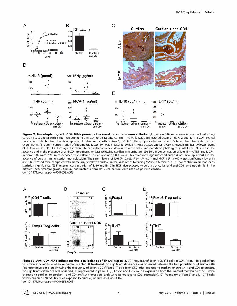
Figure 2. Non-depleting anti-CD4 MAb prevents the onset of autoimmune arthritis. (A) Female SKG mice were immunized with 3mgcurdlan i.p. together with 1 mg non-depleting anti-CD4 or an isotype control. The MAb was administered again on days 2 and 4. Anti-CD4 treatedmice were protected from the development of autoimmune arthritis (n = 6, P,0.001). Data, represented as mean 6 SEM, are from two independentexperiments. (B) Serum concentration of rheumatoid factor (RF) was measured by ELISA. Mice treated with anti-CD4 showed significantly lower levelsof RF (n = 6, P,0.001) (C) Histological sections stained with eosin-hematoxilin from the ankle and metatarso-phalangical joints from SKG mice in theabsence and in the presence of anti-CD4 treatment, 90 days following curdlan immunization. (D) Serum concentration of IL-6, IFN-c, TNF and MCP-1in naive SKG mice, SKG mice exposed to curdlan, or curlan and anti-CD4. Naive SKG mice were age matched and did not develop arthritis in theabsence of curdlan immunization (no induction). The serum levels of IL-6 (P,0.05), IFN-c (P,0.01) and MCP-1 (P,0.01) were significantly lower inanti-CD4 treated mice compared with animals injected with curldan in the absence of tolerizing MAbs. Differences in TNF concentration did not reachstatistical significance. (E) The serum concentration of IL-10 and IL-17 in SKG mice exposed to curdlan, or curlan and anti-CD4 remained similar in thedifferent experimental groups. Culture supernatants from Th17 cell culture were used as positive control.doi:10.1371/journal.pone.0010558.g002
Figure 3. Anti-CD4 MAb influences the local balance of Th17/Treg cells. (A) Frequency of splenic CD4+ T cells or CD4+Foxp3+ Treg cells fromSKG mice exposed to curdlan, or curdlan + anti-CD4 treatment. No significant difference was observed between the two populations of animals. (B)Representative dot plots showing the frequency of splenic CD4+Foxp3+ T cells from SKG mice exposed to curdlan, or curdlan + anti-CD4 treatment.No significant difference was observed, as represented in panel A. (C) Foxp3 and IL-17 mRNA expression from the synovial membrane of SKG miceexposed to curdlan, or curdlan + anti-CD4 (mRNA expression levels were normalized to CD3 expression). (D) Frequency of Foxp3+ and IL-17+ T cellswithin draining LNs of SKG mice exposed to curdlan, or curdlan + anti-CD4.doi:10.1371/journal.pone.0010558.g003
Th17/Treg Balance in Arthritis
PLoS ONE | www.plosone.org 4 May 2010 | Volume 5 | Issue 5 | e10558

To address this issue we investigated whether CD4-blockade
could prevent in vitro Th17 polarization. We used OVA-specific
TCR-transgenic CD4+ T cells sorted from DO11.10.RAG-/- mice.
We stimulated these cells in vitro, for five days, in the presence of
DCs loaded with the appropriate peptide and under culture
conditions promoting optimal Th17 polarization (in presence of
TGF-b, IL-6, IL-1b, and anti-IFN-c)[30]. We observed that
addition of anti-CD4 led to a significant reduction of Th17 cells
(n = 6, P,0.05, Figure 4). In spite of the presence of cytokines that
promote Th17 polarization (cytokines that are known to prevent
Foxp3 induction) the addition of anti-CD4 resulted in a significant
increase in the frequency of Foxp3+ T cells (n = 6, P,0.05,
Figure 4).
Non-depleting anti-CD4 induces long- term protectionfrom autoimmune arthritis
To assess whether protection from arthritis induced with anti-
CD4 treatment had a long-term effect, SKG mice initially injected
with curdlan and non-depleting anti-CD4 were challenged with
curdlan 60 days following the initial treatment. Animals exposed to
curdlan in the presence of the putative tolerogenic anti-CD4 MAb
at day 0 were protected from the induction of arthritis following
curdlan administration at day 60, unlike the age-matched controls
that did not receive any treatment at day 0 (n = 6, P,0.05,
Figure 5A).
However, it should be noted, that some anti-CD4 treated mice
developed mild manifestations of arthritis following curdlan
challenge at day 60, although without progressing to the severe
manifestations of the disease observed in control groups.
Non-depleting anti-CD4 prevents progression ofestablished autoimmune arthritis
Given the fact that non-depleting anti-CD4 treatment prevents
the onset of autoimmune arthritis, we tested the effectiveness of a
similar course of anti-CD4 for the treatment of established arthritis
in SKG mice. Female SKG mice were immunized with curdlan
and when the clinical score reached 0.5 the arthritic mice were
randomly included in a group treated with anti-CD4 MAb or a
control group. Mice treated with non-depleting anti-CD4 showed
a long-term benefit with slower disease progression and less severe
clinical scores (n = 5, P,0.05, Figure 5B). However, remission was
only achieved in a minority of the treated animals.
Anti-CD4 treated mice remain immunocompetentA concern of immunomodulatory or tolerogenic therapeutic
strategies is their long-term impact on the overall immune
response. We therefore assessed the immune competence of anti-
CD4-treated BALB/c mice (same genetic background as the SKG
mice, but without the ZAP70 point mutation) to mount CD4+ T
cell-dependent immune responses towards an unrelated antigen.
For this purpose, mice treated with anti-CD4 were sensitized with
20 mg OVA-alum 30 days following anti-CD4 treatment. The
quantification of OVA-specific immunoglobulins in the serum was
determined one week following sensitization. Our data show that
the concentration of OVA-specific immunoglobulins was similar in
immunized mice, regardless of previous anti-CD4 treatment, and
considerable higher than in naive controls (n = 5, P,0.05,
Figure 5C). Moreover, we confirmed the same results in SKG
mice, subjected to the same protocol (n = 2, not shown). Of note, if
OVA was administered at the time of anti-CD4 treatment in
BALB/c mice, the animals became unable to produce OVA-
specific immunoglobulins following subsequent OVA immuniza-
tion (n = 6, P,0,05, Figure 5D), thus proving that OVA-specific
IgG production is CD4-dependent, and that tolerance is only
imposed over the antigens present at the time of tolerance
induction.
Discussion
Our data show that a short course of non-depleting anti-CD4
can lead to long-term protection from the development of
autoimmune arthritis, in a murine model of chronic disease. We
have shown that our non-depleting antibody (clone YTS177) is not
affecting T cells in the spleen, but seems to be preventing
autoimmune arthritis, acting locally at the site of inflammation. It
is likely that by targeting CD4+ T cells, the pathogenic cycle of
events leading to synovial inflammation and progressive joint
destruction is abrogated, as other cellular players are not recruited
towards the articular tissue to cause inflammation. This impact of
CD4-blockade on other cell types is well illustrated by the marked
decrease of pro-inflammatory cytokines produced by dendritic
cells (DCs) and macrophages in anti-CD4 treated animals. In
addition, given Th17 cells have been described as involved in the
production of autoantibodies in experimental autoimmune
arthritis [31,32], our observation that anti-CD4-treated mice do
not produce RF is in line with the hypothesis that by maintaining
pathogenic T cell clones under control the B cells will not receive
the necessary stimuli for the production of autoantibodies. We
cannot exclude, at this time, a direct effect of non-depleting anti-
CD4 MAbs on innate cells expressing CD4, namely natural killer
T (NKT) cells that have been reported as being able to provide
‘‘help’’ to B cells, and to influence autoimmune arthritis [33].
In our experiments, we found a limited efficacy for tolerance
induction once the animals became overtly arthritic. Although we
Figure 4. CD4-blockade prevents Th17 polarization. (A) SortedTCR-transgenic cells were stimulated in vitro with peptide-loaded DCsunder culture conditions known to preferentially polarize Th17 cells,with the addition of recombinant TGF-b, IL-6, IL-1b, and anti-IFN-c. After5 days of culture we observed a significant reduction of IL-17+ cells inthe presence of anti-CD4 (n = 6, P,0.05). In contrast, anti-CD4 additionled to an increased frequency of Foxp3+ T cells (n = 6, P,0.05). (B)Representative dot plots from the two different culture conditions. Anindependent experiment was performed with a peptide dose of 0.3 mMwith similar results.doi:10.1371/journal.pone.0010558.g004
Th17/Treg Balance in Arthritis
PLoS ONE | www.plosone.org 5 May 2010 | Volume 5 | Issue 5 | e10558
do not have a complete explanation for this observation, it is
possible that such resistance to tolerance induction may be due to
the participation of other cell types, besides CD4+ T cells, at that
late time. In fact, in transplantation it is known that in pre-
sensitized animals a population of Asialo GM1+ CD8+ T cells can
create a barrier for tolerance induction with MAbs [34]. Thus, it
may be possible to enhance the efficacy of tolerance induction in
overt arthritis with reagents targeting other cell types, namely
CD8+ T cells and B cells.
It remains to be established whether the long-term protection
from arthritis afforded following anti-CD4 treatment, even after a
new curdlan challenge at a later time, can be explained by the
development of regulatory mechanisms that have been described
in other animal models of anti-CD4 induced immune tolerance
[28,35]. In fact, it is now established that CD4+ T cell activation in
the presence of TGF-b and IL-6 favors T cell conversion towards
arthritogenic Th17 cells, while activation in the same environment
devoid of IL-6 shifts the differentiation from Th17 towards Foxp3+
Treg cells [30]. SKG autoimmune arthritis development is known
to be dependent on Th17 [36].
Our data suggests that protection from arthritis induced with
anti-CD4 is associated with an overall decrease of infiltrating T
cells in synovial tissue and, within those T cells in the synovium,
with an increase in the frequency of synovial Foxp3+ Treg cells. As
a consequence, the tissue is endowed with local changes that are
likely to prevent the onset of arthritis directly within the local
environment where inflammation would occur, even following a
later exposure to curdlan at a time (day 60) where anti-CD4 MAbs
are no longer present. Moreover, the reciprocal decrease of IL-17
expression at the same time that the expression of Foxp3 increases,
in the joints of anti-CD4 treated mice, supports the hypothesis that
indeed the balance between Treg and Th17 can determine the
decision between prevention or onset of autoimmune arthritis.
The observation that IL-6 decreases in the serum of anti-CD4
treated mice is also in agreement with this hypothesis. It should be
noted, however, that the number of T cells is markedly reduced in
the synovium of treated mice, as can be seen in the histological
sections (and confirmed with greater CD3 expression in the
synovia by RT-PCR – thus the need to use CD3, rather than a
housekeeping gene, to normalize our gene expression studies). It
was recently reported that IL-17A can be produced by mast cells
in rheumatoid arthritis synovia [37]. Although we did not
investigate this possible source of IL-17, the overall quantification
of IL-17 transcripts was significantly reduced in anti-CD4 treated
animals, where T cells were also less abundant. As a consequence,
the shift in the Treg/Th17 balance in the synovial tissue is likely to
be also influenced by tissue accessibility to different types of
effector T cells.
We have also shown that CD4-blockade can directly inhibit T
cell polarization towards an IL-17-producing Th17 phenotype,
even when the most appropriate cytokine environment is
provided. In addition, our data show that even in the presence
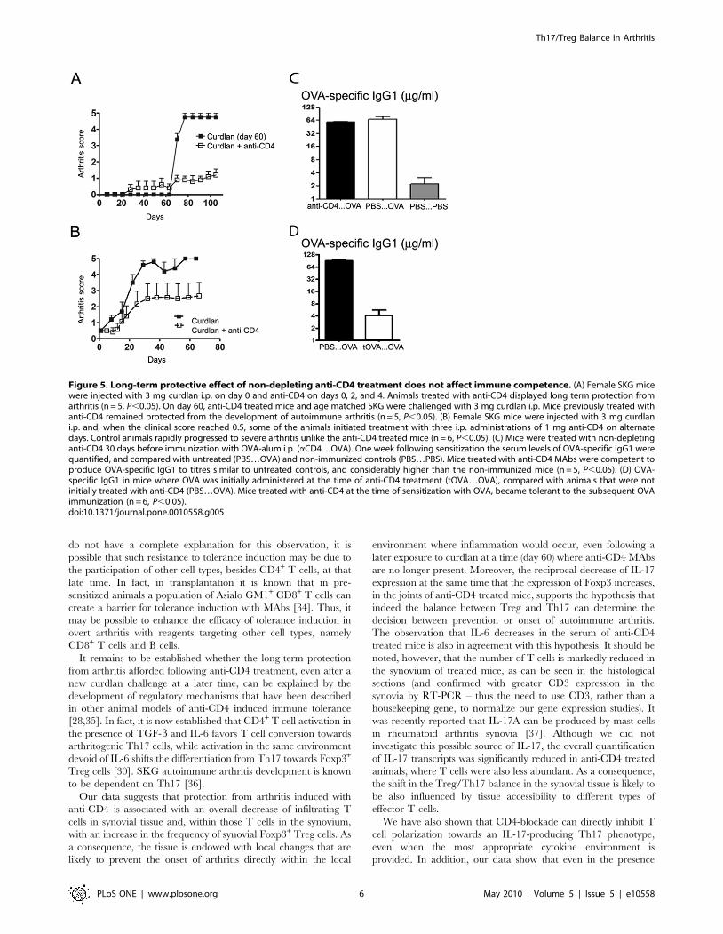
Figure 5. Long-term protective effect of non-depleting anti-CD4 treatment does not affect immune competence. (A) Female SKG micewere injected with 3 mg curdlan i.p. on day 0 and anti-CD4 on days 0, 2, and 4. Animals treated with anti-CD4 displayed long term protection fromarthritis (n = 5, P,0.05). On day 60, anti-CD4 treated mice and age matched SKG were challenged with 3 mg curdlan i.p. Mice previously treated withanti-CD4 remained protected from the development of autoimmune arthritis (n = 5, P,0.05). (B) Female SKG mice were injected with 3 mg curdlani.p. and, when the clinical score reached 0.5, some of the animals initiated treatment with three i.p. administrations of 1 mg anti-CD4 on alternatedays. Control animals rapidly progressed to severe arthritis unlike the anti-CD4 treated mice (n = 6, P,0.05). (C) Mice were treated with non-depletinganti-CD4 30 days before immunization with OVA-alum i.p. (aCD4…OVA). One week following sensitization the serum levels of OVA-specific IgG1 werequantified, and compared with untreated (PBS…OVA) and non-immunized controls (PBS…PBS). Mice treated with anti-CD4 MAbs were competent toproduce OVA-specific IgG1 to titres similar to untreated controls, and considerably higher than the non-immunized mice (n = 5, P,0.05). (D) OVA-specific IgG1 in mice where OVA was initially administered at the time of anti-CD4 treatment (tOVA…OVA), compared with animals that were notinitially treated with anti-CD4 (PBS…OVA). Mice treated with anti-CD4 at the time of sensitization with OVA, became tolerant to the subsequent OVAimmunization (n = 6, P,0.05).doi:10.1371/journal.pone.0010558.g005
Th17/Treg Balance in Arthritis
PLoS ONE | www.plosone.org 6 May 2010 | Volume 5 | Issue 5 | e10558

of those cytokines known to inhibit Foxp3 induction (namely IL-6),
CD4-blockade can facilitate the peripheral conversion of Foxp3+
Treg cells. Taken together, our data complements previous studies
on the mechanism of tolerance induction in transplantation with
anti-CD4 (where peripheral induction of Foxp3+ Treg cells have
been shown critical), by demonstrating that CD4-blockade can
also lead to a direct inhibition of Th17 polarization – a critical
factor for the arthritis pathogenesis, namely in SKG mice [36].
Given the essential role of CD4+ T cells in the pathogenesis of
RA, both directly and by recruiting and activating other
participating cell types (such as B cells, DCs and macrophages),
the therapeutic targeting of CD4+ lymphocytes has been
extensively pursued [38]. Depleting and non-depleting anti-CD4
MAbs have been tested in several animal models of autoimmune
arthritis [39,40,41,42], and in clinical trials with RA patients
[43,44,45]. In spite of promising results in pre-clinical studies, the
therapeutic effectiveness of anti-CD4 in clinical trials was modest
and short-term, possibly due to transient immunosuppression and
not tolerance. In retrospect, those unimpressive results are not too
surprising due to technical details related with dosing and MAb
characteristics. In fact the immunogenicity of the mouse or
chimeric MAb used was well documented as leading to their rapid
clearance and consequent loss of efficient CD4 blockade [46]. The
reduction of the number of CD4+ T cells was also associated with a
concern of possible increased susceptibility to infection.
Our data show that non-depleting anti-CD4 can be effective in
preventing chronic autoimmune arthritis while preserving immune
competence, as treated mice remain able to mount a CD4-
dependent immune response against a different antigen (OVA).
OVA immunization is a well established protocol for the induction
of CD4-dependent production of OVA-specific immunoglobulins.
Moreover, administration of anti-CD4 together with OVA
prevents the effectiveness of subsequent immunizations with the
same antigen. Taken together, these data suggest that anti-CD4
prevents immune responses towards antigens present at the time of
treatment, without hampering immune responses to unrelated
antigens introduced in the organism at a later time, therefore
preserving immunocompetence. It should be noted that these
types of studies, on the long-term immunocompetence of treated
mice have been difficult to perform in animal models that, unlike
SKG mice, do not develop a chronic form of arthritis.
In summary, we show that therapeutic strategies leading to
synovial accumulation of Treg cells and reduction of Th17 are
capable of protecting from the onset of autoimmune arthritis, as
well as to prevent long-term disease progression, without leading
to overall immune suppression.
Acknowledgments
We thank Margarida Souto Carneiro and Marta Monteiro for helpful
suggestions. We are grateful to Professor Shimon Sakaguchi (Kyoto, Japan)
for providing the SKG mice, and to Professor Herman Waldmann
(Oxford, UK) for providing reagents used in this study.
Author Contributions
Conceived and designed the experiments: JD JEF LG. Performed the
experiments: JD AAD VO. Analyzed the data: JD AAD VO JEF LG.
Wrote the paper: JD JEF LG.
References
1. Chehata JC, Hassell AB, Clarke SA, Mattey DL, Jones MA, et al. (2001)
Mortality in rheumatoid arthritis: relationship to single and composite measures
of disease activity. Rheumatology (Oxford) 40: 447–452.
2. Gabriel SE, Crowson CS, Kremers HM, Doran MF, Turesson C, et al. (2003)
Survival in rheumatoid arthritis: a population-based analysis of trends over 40
years. Arthritis Rheum 48: 54–58.
3. Firestein GS (2003) Evolving concepts of rheumatoid arthritis. Nature 423:
356–361.
4. Banerjee S, Webber C, Poole AR (1992) The induction of arthritis in mice by the
cartilage proteoglycan aggrecan: roles of CD4+ and CD8+ T cells. Cell
Immunol 144: 347–357.
5. Breedveld FC, Dynesius-Trentham R, de Sousa M, Trentham DE (1989)
Collagen arthritis in the rat is initiated by CD4+ T cells and can be amplified by
iron. Cell Immunol 121: 1–12.
6. Van Boxel JA, Paget SA (1975) Predominantly T-cell infiltrate in rheumatoid
synovial membranes. N Engl J Med 293: 517–520.
7. Chabaud M, Durand JM, Buchs N, Fossiez F, Page G, et al. (1999) Human
interleukin-17: A T cell-derived proinflammatory cytokine produced by the
rheumatoid synovium. Arthritis Rheum 42: 963–970.
8. Kotake S, Udagawa N, Takahashi N, Matsuzaki K, Itoh K, et al. (1999) IL-17 in
synovial fluids from patients with rheumatoid arthritis is a potent stimulator of
osteoclastogenesis. J Clin Invest 103: 1345–1352.
9. Ziolkowska M, Koc A, Luszczykiewicz G, Ksiezopolska-Pietrzak K, Klimczak E,
et al. (2000) High levels of IL-17 in rheumatoid arthritis patients: IL-15 triggers
in vitro IL-17 production via cyclosporin A-sensitive mechanism. J Immunol
164: 2832–2838.
10. Choy EH, Connolly DJ, Rapson N, Jeal S, Brown JC, et al. (2000)
Pharmacokinetic, pharmacodynamic and clinical effects of a humanized IgG1
anti-CD4 monoclonal antibody in the peripheral blood and synovial fluid of
rheumatoid arthritis patients. Rheumatology (Oxford) 39: 1139–1146.
11. Graca L, Waldmann H (2006) Reprogramming the immune system using
antibodies. Methods Mol Biol 333: 247–268.
12. Mason U, Aldrich J, Breedveld F, Davis CB, Elliott M, et al. (2002) CD4
coating, but not CD4 depletion, is a predictor of efficacy with primatized
monoclonal anti-CD4 treatment of active rheumatoid arthritis. J Rheumatol 29:
220–229.
13. Schulze-Koops H, Burkhardt H, Kalden JR (1999) What we have learned from
trials of immunomodulatory agents in rheumatoid arthritis: Future directions.
Drugs Today (Barc) 35: 327–351.
14. Isaacs JD (2007) T cell immunomodulation–the Holy Grail of therapeutic
tolerance. Curr Opin Pharmacol 7: 418–425.
15. Graca L, Honey K, Adams E, Cobbold SP, Waldmann H (2000) Cutting edge:
anti-CD154 therapeutic antibodies induce infectious transplantation tolerance.
J Immunol 165: 4783–4786.
16. Graca L, Le Moine A, Cobbold SP, Waldmann H (2003) Antibody-induced
transplantation tolerance: the role of dominant regulation. Immunol Res 28:
181–191.
17. Karim M, Bushell AR, Wood KJ (2002) Regulatory T cells in transplantation.
Curr Opin Immunol 14: 584–591.
18. Sakaguchi N, Takahashi T, Hata H, Nomura T, Tagami T, et al. (2003) Altered
thymic T-cell selection due to a mutation of the ZAP-70 gene causes
autoimmune arthritis in mice. Nature 426: 454–460.
19. Caetano-Lopes J, Henriques R, Canhao H, Duarte J, Amaral PM, et al. (2009)
Chronic arthritis directly induces quantitative and qualitative bone disturbances
leading to compromised biomechanical properties. Clin Exp Rheumatol 27:
475–482.
20. Hori S, Nomura T, Sakaguchi S (2003) Control of regulatory T cell development
by the transcription factor Foxp3. Science 299: 1057–1061.
21. Wu Q, Martin RJ, Rino JG, Breed R, Torres RM, et al. (2007) IL-23-dependent
IL-17 production is essential in neutrophil recruitment and activity in mouse
lung defense against respiratory Mycoplasma pneumoniae infection. Microbes
Infect 9: 78–86.
22. Cobbold SP, Castejon R, Adams E, Zelenika D, Graca L, et al. (2004) Induction
of foxP3+ regulatory T cells in the periphery of T cell receptor transgenic mice
tolerized to transplants. J Immunol 172: 6003–6010.
23. Yoshitomi H, Sakaguchi N, Kobayashi K, Brown GD, Tagami T, et al. (2005) A
role for fungal {beta}-glucans and their receptor Dectin-1 in the induction of
autoimmune arthritis in genetically susceptible mice. J Exp Med 201: 949–960.
24. Hirota K, Yoshitomi H, Hashimoto M, Maeda S, Teradaira S, et al. (2007)
Preferential recruitment of CCR6-expressing Th17 cells to inflamed joints via
CCL20 in rheumatoid arthritis and its animal model. J Exp Med 204:
2803–2812.
25. Hata H, Sakaguchi N, Yoshitomi H, Iwakura Y, Sekikawa K, et al. (2004)
Distinct contribution of IL-6, TNF-alpha, IL-1, and IL-10 to T cell-mediated
spontaneous autoimmune arthritis in mice. J Clin Invest 114: 582–588.
26. Qin SX, Cobbold S, Benjamin R, Waldmann H (1989) Induction of classical
transplantation tolerance in the adult. J Exp Med 169: 779–794.
27. Graca L, Thompson S, Lin CY, Adams E, Cobbold SP, et al. (2002) Both
CD4(+)CD25(+) and CD4(+)CD25(-) regulatory cells mediate dominant
transplantation tolerance. J Immunol 168: 5558–5565.
28. Graca L, Cobbold SP, Waldmann H (2002) Identification of regulatory T cells in
tolerated allografts. J Exp Med 195: 1641–1646.
Th17/Treg Balance in Arthritis
PLoS ONE | www.plosone.org 7 May 2010 | Volume 5 | Issue 5 | e10558
29. Kingsley CI, Karim M, Bushell AR, Wood KJ (2002) CD25+CD4+ regulatory T
cells prevent graft rejection: CTLA-4- and IL-10-dependent immunoregulationof alloresponses. J Immunol 168: 1080–1086.
30. Veldhoen M, Hocking RJ, Atkins CJ, Locksley RM, Stockinger B (2006)
TGFbeta in the context of an inflammatory cytokine milieu supports de novodifferentiation of IL-17-producing T cells. Immunity 24: 179–189.
31. Hsu HC, Yang P, Wang J, Wu Q, Myers R, et al. (2008) Interleukin 17-producing T helper cells and interleukin 17 orchestrate autoreactive germinal
center development in autoimmune BXD2 mice. Nat Immunol 9: 166–175.
32. Jacobs JP, Wu HJ, Benoist C, Mathis D (2009) IL-17-producing T cells canaugment autoantibody-induced arthritis. Proc Natl Acad Sci U S A 106:
21789–21794.33. Coppieters K, Dewint P, Van Beneden K, Jacques P, Seeuws S, et al. (2007)
NKT cells: manipulable managers of joint inflammation. Rheumatology(Oxford) 46: 565–571.
34. Trambley J, Bingaman AW, Lin A, Elwood ET, Waitze SY, et al. (1999) Asialo
GM1(+) CD8(+) T cells play a critical role in costimulation blockade-resistantallograft rejection. J Clin Invest 104: 1715–1722.
35. Graca L, Le Moine A, Lin CY, Fairchild PJ, Cobbold SP, et al. (2004) Donor-specific transplantation tolerance: the paradoxical behavior of CD4+CD25+ T
cells. Proc Natl Acad Sci U S A 101: 10122–10126.
36. Hirota K, Hashimoto M, Yoshitomi H, Tanaka S, Nomura T, et al. (2007) T cellself-reactivity forms a cytokine milieu for spontaneous development of IL-17+Th cells that cause autoimmune arthritis. J Exp Med 204: 41–47.
37. Hueber AJ, Asquith DL, Miller AM, Reilly J, Kerr S, et al. Mast cells express IL-
17A in rheumatoid arthritis synovium J Immunol 184: 3336–3340.
38. Isaacs JD (2008) Therapeutic T-cell manipulation in rheumatoid arthritis: past,
present and future. Rheumatology (Oxford) 47: 1461–1468.
39. Chu CQ, Londei M (1996) Induction of Th2 cytokines and control of collagen-
induced arthritis by nondepleting anti-CD4 Abs. J Immunol 157: 2685–2689.
40. Mauri C, Chu CQ, Woodrow D, Mori L, Londei M (1997) Treatment of a
newly established transgenic model of chronic arthritis with nondepleting anti-
CD4 monoclonal antibody. J Immunol 159: 5032–5041.
41. Ranges GE, Fortin S, Barger MT, Sriram S, Cooper SM (1988) In vivo
modulation of murine collagen induced arthritis. Int Rev Immunol 4: 83–90.
42. Ranges GE, Sriram S, Cooper SM (1985) Prevention of type II collagen-induced
arthritis by in vivo treatment with anti-L3T4. J Exp Med 162: 1105–1110.
43. Herzog C, Walker C, Pichler W, Aeschlimann A, Wassmer P, et al. (1987)
Monoclonal anti-CD4 in arthritis. Lancet 2: 1461–1462.
44. Horneff G, Burmester GR, Emmrich F, Kalden JR (1991) Treatment of
rheumatoid arthritis with an anti-CD4 monoclonal antibody. Arthritis Rheum
34: 129–140.
45. Reiter C, Kakavand B, Rieber EP, Schattenkirchner M, Riethmuller G, et al.
(1991) Treatment of rheumatoid arthritis with monoclonal CD4 antibody M-
T151. Clinical results and immunopharmacologic effects in an open study,
including repeated administration. Arthritis Rheum 34: 525–536.
46. Horneff G, Winkler T, Kalden JR, Emmrich F, Burmester GR (1991) Human
anti-mouse antibody response induced by anti-CD4 monoclonal antibody
therapy in patients with rheumatoid arthritis. Clin Immunol Immunopathol 59:
89–103.
Th17/Treg Balance in Arthritis
PLoS ONE | www.plosone.org 8 May 2010 | Volume 5 | Issue 5 | e10558