Allele-Specific Cytokine Responses at the HLA-C Locus: Implications for Psoriasis
Upload
rockefellerCategory
view
2download
0
Effective treatment of psoriasis with etanercept is linked tosuppression of IL-17 signaling, not “immediate-response” TNFgenes
Lisa C. Zaba, MD, PhD1, Mayte Suárez-Fariñas, PhD1, Judilyn Fuentes-Duculan, MD, KristineNograles, MD, Emma Guttman-Yassky, MD, MSc, Irma Cardinale, MS, Michelle A. Lowes,MB.BS, PhD2, and James G. Krueger, MD, PhD2
AbstractBackground—TNF inhibitors have revolutionized the treatment of psoriasis vulgaris, as well aspsoriatic and rheumatoid arthritis, and Crohns disease. Despite our understanding that these agentsblock TNF, their complex mechanism of action in disease resolution is still unclear.
Objective—To globally analyze the genomic effects of TNF inhibition in psoriasis patients, andcompare genomic profiles of patients who responded or did not respond to treatment.
Methods—In a clinical trial using etanercept TNF inhibitor to treat psoriasis vulgaris (n=15),Affymetrix gene arrays were used to analyze gene profiles in lesional skin at multiple time-pointsduring drug treatment (baseline, and weeks 1, 2, 4 and 12) compared to non-lesional skin. Patientswere stratified as responders (n=11) or non-responders (n=4) based on histological disease resolution.Cluster analysis was used to define gene sets that were modulated with similar magnitude and velocityover time.
Results—In responders, 4 clusters of down-regulated genes and 3 clusters of up-regulated geneswere identified. Genes down-modulated most rapidly reflected direct inhibition of myeloid lineageimmune genes. Up-regulated genes included stable dendritic cell population genes CD1c and CD207(Langerin). Comparison of responders and non-responders revealed rapid down-modulation of innateIL-1β and IL-8 “sepsis cascade” cytokines in both groups, but only responders down-regulated IL-17pathway genes to baseline levels.
Conclusion—While both responders and non-responders to etanercept inactivated “sepsiscascade” cytokines, response to etanercept is dependent on inactivation of myeloid dendritic cellgenes and inactivation of Th17 immune response.
Capsule Summary—Cutaneous genes regulated during psoriasis treatment by etanercept providea global view of response in disease tissue. Only responders down-regulated IL-17 pathway genes.
© 2009 American Academy of Allergy, Asthma and Immunology. Published by Mosby, Inc. All rights reserved.Corresponding author James G. Krueger The Rockefeller University Laboratory for Investigative Dermatology 1230 York Avenue,Box 178 New York, NY 10065 Ph: 212-327-7730 Fax: 212-327-8353 [email protected] .1,2contributed equally to this workCommunicating author Michelle A. Lowes The Rockefeller University Laboratory for Investigative Dermatology 1230 York Avenue,Box 178 New York, NY 10065 Ph: 212-327-7576 Fax: 212-327-8353 [email protected]'s Disclaimer: This is a PDF file of an unedited manuscript that has been accepted for publication. As a service to our customerswe are providing this early version of the manuscript. The manuscript will undergo copyediting, typesetting, and review of the resultingproof before it is published in its final citable form. Please note that during the production process errors may be discovered which couldaffect the content, and all legal disclaimers that apply to the journal pertain.
NIH Public AccessAuthor ManuscriptJ Allergy Clin Immunol. Author manuscript; available in PMC 2010 November 1.
Published in final edited form as:J Allergy Clin Immunol. 2009 November ; 124(5): 1022–10.e1-395. doi:10.1016/j.jaci.2009.08.046.
NIH
-PA Author Manuscript
NIH
-PA Author Manuscript
NIH
-PA Author Manuscript
KeywordsTNF; psoriasis; etanercept; gene; Th17; Th1
IntroductionTNF was initially identified in 1976 as the soluble, LPS-dependent serum factor that killedmurine fibrosarcoma cells but not normal fibroblasts 1. Later, TNF was characterized as amacrophage-derived product that rapidly induced expression of IL-1, IL-6, and IL-8, leadingto recruitment of innate inflammatory leukocytes within hours of an infectious insult 2, 3. Thiscytokine sequence, which can be termed the “sepsis cascade model,” 4 has largely dominatedthinking about the function of TNF in chronic inflammatory diseases. The initial rationale fortesting TNF antagonists in rheumatoid arthritis was based on demonstrated increases in allsepsis cascade cytokines in synovial fluid of affected joints, as well as abundant neutrophilsproducing collagen-destroying matrix metalloproteases 5. Indeed, genomic analysis ofperipheral blood during anti-TNF treatment for rheumatoid arthritis showed a down-modulation of several of these acute-response genes during response (CCL4, IL-8, IL-1β) 6.
Extension of these findings led to clinical trials of TNF antagonists in inflammatory boweldisease and psoriasis with subsequent therapeutic success and FDA approval 7, 8. There hasbeen a strong temptation to link pathogenesis of human inflammatory diseases, and especiallythose successfully treated with TNF antagonists such as psoriasis and rheumatoid arthritis, todirect effects of sepsis cascade cytokines on innate immunity. However, despite the success ofTNF blocking agents, psoriasis vulgaris has some elements of pathogenesis that are not wellexplained by sepsis cascade cytokines and neutrophil activation. First, there is a strong T cellcomponent in psoriasis, including reports of T cell clonality 9-11, and there is considerableclinical benefit provided by putative T cell targeted therapeutics 12, 13. Second, psoriasis hasbeen induced in several transplanted skin models without apparent involvement of neutrophilsin converted grafts 14. Third, recent success in treating psoriasis with antibodies to the p40cytokine subunit shared between IL-12 and IL-23 implies a central role for adaptive immunity,specifically Th1 and/or Th17 T cells, in the pathogenesis of this disease 15-19. We sought tounderstand how TNF is linked to pathogenic actions of T cells in psoriasis.
In the present study we used Affymetrix arrays to study gene expression in psoriasis lesionsat several time points during treatment with etanercept. Responding and non-respondingpatents show equivalent suppression of innate, sepsis cytokines (IL-1β, IL-8), but onlyresponding patients demonstrate strong modulation of downstream genes linked to IL-17signaling. Hence, mechanisms for response success and failure appear to be distinct in psoriasisversus rheumatoid arthritis. These studies define a new way to judge the extent of overalldisease reversal based on the degree to which broad disease-related gene sets are suppressedby a specific treatment, and may enable the design of even more effective and simple anti-psoriatic therapeutics.
Materials and MethodsMaterial and Methods, and statistical analysis are described in greater detail in SupplementalMaterials and Methods (SMM, Online Repository (OR)).
Patient studies and classificationTwenty adult patients with moderate to severe psoriasis were treated with etanercept 50mg s.c.twice weekly for 12 weeks. Clinical trial number NCT00116181. Clinical and histological
Zaba et al. Page 2
J Allergy Clin Immunol. Author manuscript; available in PMC 2010 November 1.
NIH
-PA Author Manuscript
NIH
-PA Author Manuscript
NIH
-PA Author Manuscript

response of patients in this trial was previously published 20. Gene array was performed on 15sequential patients.
Immunohistochemistry and immunofluorescenceFrozen skin sections were stained for in a standard manner (SMM, section 1). A one-tailedstudents t-test was used to compare baseline and week 2 cell counts.
Tissue gene expression by microarrayRNA was extracted from skin biopsies using the RNeasy Mini Kit (Qiagen). For eachAffymetrix gene chip, 2μg total RNA was reverse-transcribed, amplified, and labeled 21.Microarray data has been deposited in Geo-Omnibus (GSE 11903).
Statistical AnalysisThe analysis was conducted using R software (www.R-project.org) with bioconductorpackages (www.bioconductor.org). Briefly, expression measures were obtained usingGCRMA, and probe sets with a SD>0.15, and expression greater than 4 in at least 5 samples,were kept for further analysis (SMM, section 2). Statistical methods used in the analyses ofdata are summarized in OR Table 1.
Differential expression criteriaA mixed-effect linear model was used to model gene expression in order to account for thetime-course design of the clinical trial. To define “etanercept-modulated genes”, a moderatedF statistic was used to determine if any of the expression values across time differed fromlesional baseline at any time-point. An F test was used to determine genes where the timeprofile was different between responders and non-responders. The p-values of both tests wereadjusted for multiple hypothesis using Benjamini-Hochberg method.
Consensus clusteringTo find distinct profiles among etanercept-modulated genes, as well as between respondersand non-responders, we performed hierarchical average linkage clustering with distancemeasure equal to 1-Pearsons’s correlation coefficient. The optimal number of discoveredclusters was assessed using Consensus Clustering algorithm available in GenePattern software.
RT-PCRRT-PCR was conducted for IL-1β and IL-8 as previously described 20.
Response profile of cytokine keratinocyte “pathways”We recently defined sets of keratinocyte genes induced by specific cytokines, termed IL-17,TNF and IFNγ keratinocyte “pathways”, respectively 22. A linear model was used to definethe response profile of these pathways during etanercept treatment . Gene Set EnrichmentAnalysis (GSEA) approach 23 was used to compare the response profile of the keratinocytepathways in responders and non-responders (OR Figure 1). GSEA is a method to minecorrelations of any gene set (eg keratinocyte “pathways”) with a specific phenotype (egresponse) rather than single gene approach.
ResultsPatient population
RNA was extracted for gene array from skin collected at each biopsy time point (non-lesionalbaseline, lesional baseline, weeks 1, 2, 4, and 12). Quantifiers of disease resolution included
Zaba et al. Page 3
J Allergy Clin Immunol. Author manuscript; available in PMC 2010 November 1.
NIH
-PA Author Manuscript
NIH
-PA Author Manuscript
NIH
-PA Author Manuscript
epidermal thickness (a linear measurement in mm), Ki67 (a marker of cell proliferation), andK16 gene expression (indicating aberrant keratinocyte differentiation). Patients were stratifiedas “responders” (n=11) or “non-responders” (n=4) based on whether or not they achievedhistologic disease resolution by week 12 of etanercept treatment (decreased epidermalthickening, normalization of Ki67 and K16) 20.
Disease genes were temporally modulated by etanercept in a “reverse cascade”Genes with expression values that changed during etanercept treatment (Fold change [FCH]> 2 for any time-point, false discovery rate [FDR] <0.05) were selected as “etanercept-modulated genes”. We identified 978 probe-sets that were down-regulated during the 12 weeksof treatment with etanercept, and 999 probe-sets that were up-regulated (OR Tables 2, 3,respectively). We used consensus clustering to identify 4 clusters that were down-regulated(OR Figure 2a), and 3 clusters that were up-regulated (OR Figure 2b), with distinct kinetics.This gave a cascade of genes that were modulated by etanercept, and as this was in a reversefashion as inflammation was quenched, thus we termed this modulation by etanercept a“reverse cascade”.
Genes down-modulated by etanerceptGenes down-modulated by etanercept clustered into 4 groups (Figure 1). Gene expression ateach time-point was normalized to non-lesional baseline expression, and average expressionof genes within each cluster was plotted over time (Figure 1a) with corresponding heat maps(Figure 1b). The set of genes that were maximally suppressed by 1 week of treatment (Figure1, cluster 1) are likely to be genes that are directly modulated by TNF. However, with moretime, many additional down-regulated genes were identified and these expanded toprogressively larger sets of regulated genes over time (Figure 1, clusters 2-4).
Cluster 1 genes (31 probe sets) were down-modulated most rapidly and defined as“immediately” down-modulated genes, cluster 2 with 168 probe sets was “early,” cluster 3with 616 probe sets was “mid”, and cluster 4 with 163 probe sets was “late”. All down-regulatedgenes are listed by cluster in OR Table 2. Rapidly down-modulated cluster 1 genes includedthose involved in leukocyte chemotaxis IL-8, CCL4 (MIP-1β), CCL3 (MIP-1α), FPR1 andplasminogen activator of urokinase receptor (PLAUR). This cluster also contained severalgenes involved in anti-apoptosis, such as BCL2A1; cell cycle genes AURKA, NCAPG, CDC6,and SPC25; and keratinocyte genes DSC2, SPRR3 and heparin-binding epidermal growthfactor-like protein (HBEGF). There were also several genes involved in lipid metabolism, suchas LIPG, LDLR, LRP8 and APOBEC3A. Two cytokines included in this cluster were IL-1βand IL-19. IL-1β is an acute-phase cytokine produced by many cell types, particularlymonocytes. The regulation of IL-1β by TNF is well appreciated, and hence rapid down-modulation of IL-1β by TNF-inhibition is to be expected. IL-19 is a recently discoveredcytokine belonging to the IL-10 family of cytokines, and is produced by monocytes as well asepithelial and endothelial cells during inflammation 24. Although some of these genes areknown TNF early response genes, many of them have not been previously identified as TNFearly response genes in human skin.
Genes most rapidly down-modulated by etanercept were enriched with myeloid specificgenes
In order to determine which leukocyte lineages were most rapidly inhibited by etanercepttreatment, we used the expression values of myeloid cells (CD33+ and CD14+), T cells(CD4+ and CD8+), and skin from the Novartis normal tissue compendium 25. OR Figure 3ashows the expression of immediately down-regulated genes (cluster 1) in this data set. Myeloidcells, not T cells, expressed genes that were rapidly down-modulated with etanercept. T cell
Zaba et al. Page 4
J Allergy Clin Immunol. Author manuscript; available in PMC 2010 November 1.
NIH
-PA Author Manuscript
NIH
-PA Author Manuscript
NIH
-PA Author Manuscript
specific genes were modulated later, in cluster 2 (OR Figure 4), suggesting that TNF inhibitiondirectly modulates myeloid-lineage gene products, which subsequently effect T cells.
There were two particularly interesting myeloid-specific genes contained within the rapidlydown-modulated cluster 1 that we confirmed using double label immunofluorescence: HBEGF(OR Figure 3b) and PLAUR (OR Figure 3c). There was a large increase in HBEGF withinmyeloid CD11c+ dermal DCs (co-expression giving a yellow color) in lesional compared tonon-lesional skin. At week 2 of etanercept treatment, HBEGF and CD11c expression weredecreased, and by week 12, expression of both markers normalized to non-lesional levels.PLAUR, another down-modulated “immediate” cluster 1 myeloid-specific gene, was alsorapidly decreased at the protein level by week 2 of etanercept treatment. PLAUR is a keymolecule in the regulation of cell-surface plasminogen activation, and may be involved ininflammation 26.
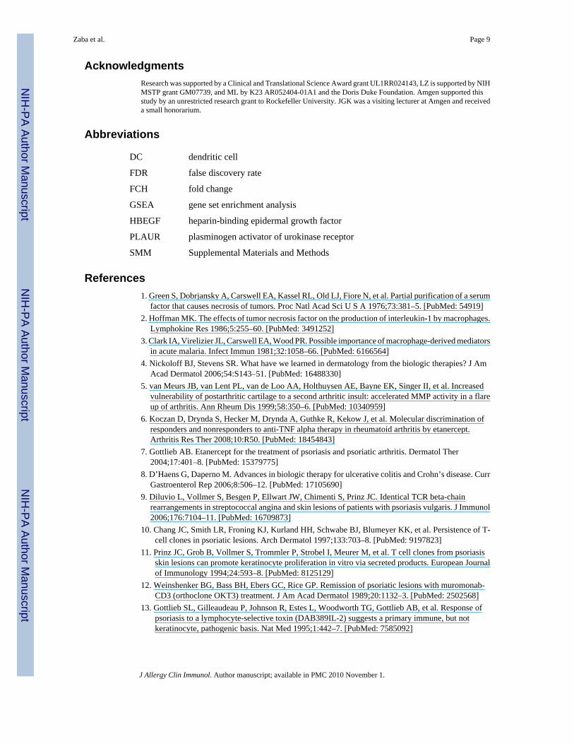
Resident DC genes were up-regulated during etanercept treatmentEtanercept up-regulated 999 probe-sets (816 genes) in responding patients, and these wereclustered into three groups. The three clusters are shown in Figure 2a (“up early”, “up mid”,and “up late”) and genes in each cluster are listed in OR Table 3. Inspection of genes containedwithin the most rapidly up-regulated clusters 1 and 2 revealed several dendritic cell (DC) genes:myeloid DC genes CD1c and CD1e were contained within cluster 1, and Langerhans cell genesCD207 (Langerin) and CD1a were both in cluster 2. In normal human skin there are two residentDC populations: dermal DCs that co-express the myeloid lineage marker CD11c and CD1c,and epidermal Langerhans cells (Langerin and CD1a+) 27.Therefore, the genomic up-regulation of CD1c and CD207 could represent a normalization of these stable resident DCpopulations with treatment. We confirmed this on a cellular level by performing cell countson sections from biopsies collected at lesional baseline and week 2 of etanercept treatment(Figure 2b). CD1c+ resident myeloid DCs were increased by approximately three-fold (mean7 cells at baseline and 23 cells at week 2; p=0.03), and CD207+ cells were increasedapproximately two-fold (mean 39 and 72 cells, respectively; p=0.001). However, week 12counts of CD1c and CD207+ cells (mean 8 and 64 cells, respectively) were not significantlydifferent from baseline lesional skin. In contrast, there was a large influx of inflammatoryCD11c+ cells that do not co-express CD1c in psoriasis lesions (we have termed “inflammatory”myeloid DCs) 28, and these cells were decreased rapidly with etanercept treatment by week 220. Therefore, although etanercept decreases inflammatory myeloid DCs after two weeks oftreatment, stable resident myeloid DCs and Langerhans cells were temporarily increased.
Cluster 3 was the largest group of up-regulated genes containing 933 probe-sets. The genes inthis group include cutaneous structural proteins such as laminin (α4 and β4), collagen type Vand VI, heparin sulfate proteoglycan 2, fibronectin, and keratins such as 15, 18 and 19. Up-regulation of these genes during effective treatment suggests normalization of regenerativehyperplasia.
Both responders and non-responders rapidly down-modulated TNF early response genesIL-1β and IL-8
It is possible that some patients do not respond to etanercept treatment because complete TNF-blockade is not achieved. We tested this hypothesis by determining the genomic behavior ofTNF immediate response genes IL-1β and IL-8 in both responders and non-responders (Figure3). We determined that IL-1β and IL-8 are rapidly down-modulated with etanercept treatment,irrespective of the patient’s ultimate phenotypic response, suggesting that non-response cannotbe explained by inadequate dosing. PCR analysis of IL-1β and IL-8 confirms that thesecytokines are similarly down-regulated in responders and non-responders (Figure 3b). We havepreviously published RT-PCR data for these two cytokines, for responders (n=11) 20. For the
Zaba et al. Page 5
J Allergy Clin Immunol. Author manuscript; available in PMC 2010 November 1.
NIH
-PA Author Manuscript
NIH
-PA Author Manuscript
NIH
-PA Author Manuscript
non-responders (n=4), there was also a significant reduction in these two cytokines by week12. There was no significant difference in the time profiles of either of these cytokines betweenthe responders and non-responders (IL-1β: F-statistic 1.57, p=0.177; IL-8: F-statistic 1.47,p=0.209).
Differences between responders and non-respondersIn order to determine the key genomic differences over time between responders and non-responders, we performed consensus clustering on probe-sets where the time-profile differedfor responders versus non-responders (FCH >2, FDR<0.2 using the moderated F-statistic). Aseven cluster structure was identified, with 4 clusters containing more than 6 probe sets (ORFigure 5; Figure 4a). These genes are listed in OR Table 4.
Cluster 1 contained genes that were strongly down-modulated over time in responders but notin non-responders. Inspection of the cluster 1 heat-map revealed an interesting pattern (Figure4b). In non-responders, a group of probe-sets (black box) were down-regulated at week 0compared with non-lesional skin (green), but up-regulated compared to non-lesional skin (red)in responders. Patients who responded to etanercept down-regulated these genes duringtreatment, while those who did not respond had increased expression of these genes by weekfour. Most notably, this group contains CCL20, a T cell and DC chemoattractant involved inthe trafficking of CCR6+ Th17 T cells into inflamed psoriatic lesions 29. High expression ofthese “black box” genes in lesional skin may be predictive of response to etanercept. Alsocontained in cluster 1 was STAT3, a transcription factor that induces Th17 polarization. Thus,key IL-17 pathway genes were strongly down-modulated in responders, but not in non-responders.
Cluster 2 contained genes that were up-regulated in responders, and after a slight increase inthe first week, these genes were down-regulated in non-responders. Included in this list aremany cell cycle proteins and keratinocyte genes, likely representing the normalization of KCproliferation and differentiation in responders, but not in non-responders.
IL-17 pathway genes are strongly down-modulated in etanercept responders, but not in non-responders
To further evaluate the difference between responders and non-responders in keratinocytepathways, we performed GSEA analysis to determine how genes contained within eachcytokine pathway were modulated over time in responders and non-responders (Figure 5).GSEA analysis demonstrated that the IL-17, TNF, IL-17 plus TNF (genes regulated both byTNF and IL-17), and IFNγ pathways were significantly different in responders and non-responders (OR Table 5). IL-17 pathway genes were highly expressed in both responders andnon-responders but returned to baseline non-lesional levels by week 12 only in the responders(Figure 5a). The TNF pathway genes were reduced to baseline in both responders and non-responders (Figure 5b). This supports our interpretation that there is adequate dosing ofetanercept. The combination of TNF and IL-17 had a pattern closer to IL-17, again suggestingthat resolution of disease is associated with reduction in IL-17 genes (Figure 5c). In fact, theIL-17 plus TNF pathway genes exhibit the most different profile in responders and non-responders. IFNγ pathway genes were down-regulated in both responders and non-respondersbut with different kinetics (responders were more gradual) and non-responders did not reachbaseline non-lesional levels completely (Figure 5d). In summary, IL-17 regulated genes wereexpressed at higher levels than TNF and IFNγ genes in psoriatic lesional skin, and IL-17 geneswere only down-modulated in responders.
Zaba et al. Page 6
J Allergy Clin Immunol. Author manuscript; available in PMC 2010 November 1.
NIH
-PA Author Manuscript
NIH
-PA Author Manuscript
NIH
-PA Author Manuscript

DiscussionThe original impetus for testing anti-TNF drugs in rheumatoid arthritis was born fromestablished elevation of TNF regulated “sepsis cascade” cytokines IL-1β and IL-8 in inflamedsynovial fluid. The same principle was applied to patients with psoriatic arthritis. Interestingly,however, it was soon noted that the kinetics of disease resolution differed between the jointand the skin component of psoriatic arthritis. In most patients, joint disease improved within2 weeks while the skin inflammation did not maximally resolve for greater than 12 weeks 30.This in itself presented the possibility that the arthritic and skin disease components had adifferent mechanism of action, with TNF as a common upstream cytokine.
In rheumatoid arthritis, suppression of the sepsis cascade does indeed govern response toetanercept TNF inhibition: responders down-regulate IL-1β and IL-8 in PBMCs by 72 hoursand non-responders do not down-regulate these cytokines 6. Our current study however,suggests that a more complex model is operating in psoriasis skin lesions. Both responders andnon-responders rapidly down-regulate IL-1β and IL-8, indicating that down-regulation ofsepsis cascade cytokines is not sufficient for psoriasis lesion resolution. Therefore, the rapidresponse of arthritis to TNF inhibition may be explained by disease directly regulated by TNFand TNF early response genes. Psoriasis however, requires a more complex model of diseasethat is only indirectly modulated by TNF.
Recent studies in genetics have shown that alleles of genes encoding p19 and p40 subunits ofIL-23, and IL-23R confer risk for development of psoriasis 31. This pathway controls thedevelopment of Th17 T cells, which may contribute IL-17 and IL-22 to an inflammatory skinenvironment. The importance of this pathway is also supported by therapeutic response ofpsoriasis to monoclonal antibodies that inhibit IL-12 and IL-23, producing major improvementin 70-80% of cases 32. Since TNF inhibitors can also confer nearly equivalent therapeuticbenefit, there may be some overlap in inflammatory circuits regulated by TNF and p40cytokines. One possible intersection between TNF and Th17 T cells has been suggested fromour prior study of the effect of etanercept effect on synthesis of IL-23, IL-17 and IL-22, namelythat TNF may serve as a DC activator for IL-23 production, which then promotes activationof Th17 T cells.
The present study further expands the analysis of the effect of etanercept on inflammatorypathways. Broad sets of genes distinctly regulated by TNF, IL-17, and IFNγ can be measuredduring a many week period of disease improvement induced by etanercept. Since the set ofinflammatory genes regulated by etanercept increases over time, our results suggest additionalindirect effects of TNF on more complex cellular or molecular circuits. For example, TNF hasbeen shown to be a “co-stimulus” for T cell activation as well as a critical molecule for DCdevelopment and maturation 33.
We considered two hypotheses for the actions of TNF in psoriasis, and therefore the effects ofetancercept on gene expression. Under the first model, TNF would act as a direct activator ofNFκB (classical TNF transcription factor) and therefore genes primarily regulated by thistranscription factor would normalize as the activation stimulus (TNF) was attenuated byetanercept. Under the second hypothesis, we considered that TNF has additional, broaderdownstream or indirect actions, e.g., as an activator of DCs and as a T cell co-stimulus. Inthis hypothesis, etanercept might regulate many different transcription factor pathways overtime and TNF inhibition might produce different kinetics for normalization of genes driven bydiverse transcription factors in multiple cell types, and the involution response might beconsidered as a reverse “cascade” over time. Hence an effect of TNF blockade may beprogressive collapse of cellular elements needed to sustain skin inflammation, a “domino”effect.
Zaba et al. Page 7
J Allergy Clin Immunol. Author manuscript; available in PMC 2010 November 1.
NIH
-PA Author Manuscript
NIH
-PA Author Manuscript
NIH
-PA Author Manuscript

The earliest effects of TNF blockade in responders, as we show in this manuscript, are thedown-regulation of myeloid lineage cells, and the rapid up-modulation of stable DC lineagegenes CD1c and CD207 (Langerin). This suggests that both resident CD1c and Langerhanscells may either not play an active role in TNF-mediated inflammation, or may be “protective.”Previous work by our group has shown rapid down-regulation of CD11c+ inflammatory DCcell counts and their inflammatory products iNOS and IL-23 (p19 and p40 subunit) by RT-PCR 20. Later down-modulation of IL-17 pathway genes, but not TNF or IFNγ pathway genes,are predictive of response to etanercept, thus solidifying the importance of IL-17 in TNF-dependent psoriatic inflammation.
OR Figure 6a outlines how TNF-dependant DC activation might be primarily blocked byetanercept in responding patients. Patients with psoriasis who do not respond to TNF-inhibitionmust have TNF-independent inflammatory pathways leading to the psoriatic phenotype (ORFigure 6b). Three pathway models could explain why non-responders do not down-regulatethe IL-17 pathway. In non-responders, DCs may be activated independently of TNF by eithercytokines (for example IFN); direct T cell-induced DC maturation (for example CD40L); ornon-responders may have DC-independent activation of Th17 cells by cytokines other thanIL-23, such as IL-6, TGFβ and IL-15 34, 35. Clearly, further studies will be required todetermine how the IL-17 pathway is sustained in non-responders.
In conclusion, our study characterizes a sequence of gene expression changes in inflamed skinlesions as these resolve with etanercept treatment. Genes that are regulated immediately afterstarting etanercept are candidates for “direct” TNF gene targets, and expand the roster ofpotential TNF regulated genes in skin inflammation. The progressively larger set of genes thatincrease over time suggest that many cellular activation circuits, and especially those linkedto DCs and adaptive immunity, are impacted by blocking TNF in a therapeutic context. Theresolution of psoriasis by etenercept thus appears to be linked more to modulation of adaptiveimmune circuits, rather than innate immune circuits, although both pathways are modulatedby this TNF antagonist.
Key Messages
• Cluster analysis on psoriasis skin lesions over time during treatment with the TNFinhibiting drug etanercept demonstrated a “domino-effect” model of diseaseinvolution.
• Many down-modulated TNF early response genes were myeloid lineage genes,while up-regulated genes include resident dendritic cell genes.
• In psoriasis, both etanercept responders and non-responders rapidly down-modulated “sepsis cascade” cytokines IL-1β and IL-8. This contrasts withrheumatoid arthritis patients in which only responders down-regulated IL-1β andIL-8.
• In psoriasis, only responders down-regulated IL-17 pathway cytokines to baselinevalues after 12 weeks, suggesting that ultimate Th17 suppression is necessary forresponse to TNF inhibiting drugs.
Supplementary MaterialRefer to Web version on PubMed Central for supplementary material.
Zaba et al. Page 8
J Allergy Clin Immunol. Author manuscript; available in PMC 2010 November 1.
NIH
-PA Author Manuscript
NIH
-PA Author Manuscript
NIH
-PA Author Manuscript
AcknowledgmentsResearch was supported by a Clinical and Translational Science Award grant UL1RR024143, LZ is supported by NIHMSTP grant GM07739, and ML by K23 AR052404-01A1 and the Doris Duke Foundation. Amgen supported thisstudy by an unrestricted research grant to Rockefeller University. JGK was a visiting lecturer at Amgen and receiveda small honorarium.
Abbreviations
DC dendritic cell
FDR false discovery rate
FCH fold change
GSEA gene set enrichment analysis
HBEGF heparin-binding epidermal growth factor
PLAUR plasminogen activator of urokinase receptor
SMM Supplemental Materials and Methods
References1. Green S, Dobrjansky A, Carswell EA, Kassel RL, Old LJ, Fiore N, et al. Partial purification of a serum
factor that causes necrosis of tumors. Proc Natl Acad Sci U S A 1976;73:381–5. [PubMed: 54919]2. Hoffman MK. The effects of tumor necrosis factor on the production of interleukin-1 by macrophages.
Lymphokine Res 1986;5:255–60. [PubMed: 3491252]3. Clark IA, Virelizier JL, Carswell EA, Wood PR. Possible importance of macrophage-derived mediators
in acute malaria. Infect Immun 1981;32:1058–66. [PubMed: 6166564]4. Nickoloff BJ, Stevens SR. What have we learned in dermatology from the biologic therapies? J Am
Acad Dermatol 2006;54:S143–51. [PubMed: 16488330]5. van Meurs JB, van Lent PL, van de Loo AA, Holthuysen AE, Bayne EK, Singer II, et al. Increased
vulnerability of postarthritic cartilage to a second arthritic insult: accelerated MMP activity in a flareup of arthritis. Ann Rheum Dis 1999;58:350–6. [PubMed: 10340959]
6. Koczan D, Drynda S, Hecker M, Drynda A, Guthke R, Kekow J, et al. Molecular discrimination ofresponders and nonresponders to anti-TNF alpha therapy in rheumatoid arthritis by etanercept.Arthritis Res Ther 2008;10:R50. [PubMed: 18454843]
7. Gottlieb AB. Etanercept for the treatment of psoriasis and psoriatic arthritis. Dermatol Ther2004;17:401–8. [PubMed: 15379775]
8. D’Haens G, Daperno M. Advances in biologic therapy for ulcerative colitis and Crohn’s disease. CurrGastroenterol Rep 2006;8:506–12. [PubMed: 17105690]
9. Diluvio L, Vollmer S, Besgen P, Ellwart JW, Chimenti S, Prinz JC. Identical TCR beta-chainrearrangements in streptococcal angina and skin lesions of patients with psoriasis vulgaris. J Immunol2006;176:7104–11. [PubMed: 16709873]
10. Chang JC, Smith LR, Froning KJ, Kurland HH, Schwabe BJ, Blumeyer KK, et al. Persistence of T-cell clones in psoriatic lesions. Arch Dermatol 1997;133:703–8. [PubMed: 9197823]
11. Prinz JC, Grob B, Vollmer S, Trommler P, Strobel I, Meurer M, et al. T cell clones from psoriasisskin lesions can promote keratinocyte proliferation in vitro via secreted products. European Journalof Immunology 1994;24:593–8. [PubMed: 8125129]
12. Weinshenker BG, Bass BH, Ebers GC, Rice GP. Remission of psoriatic lesions with muromonab-CD3 (orthoclone OKT3) treatment. J Am Acad Dermatol 1989;20:1132–3. [PubMed: 2502568]
13. Gottlieb SL, Gilleaudeau P, Johnson R, Estes L, Woodworth TG, Gottlieb AB, et al. Response ofpsoriasis to a lymphocyte-selective toxin (DAB389IL-2) suggests a primary immune, but notkeratinocyte, pathogenic basis. Nat Med 1995;1:442–7. [PubMed: 7585092]
Zaba et al. Page 9
J Allergy Clin Immunol. Author manuscript; available in PMC 2010 November 1.
NIH
-PA Author Manuscript
NIH
-PA Author Manuscript
NIH
-PA Author Manuscript

14. Nestle FO, Nickoloff BJ. From classical mouse models of psoriasis to a spontaneous xenograft modelfeaturing use of AGR mice. Ernst Schering Res Found Workshop 2005:203–12. [PubMed: 15526944]
15. Krueger GG, Langley RG, Leonardi C, Yeilding N, Guzzo C, Wang Y, et al. A humaninterleukin-12/23 monoclonal antibody for the treatment of psoriasis. N Engl J Med 2007;356:580–92. [PubMed: 17287478]
16. Gottlieb AB, Cooper KD, McCormick TS, Toichi E, Everitt DE, Frederick B, et al. A phase 1, double-blind, placebo-controlled study evaluating single subcutaneous administrations of a humaninterleukin-12/23 monoclonal antibody in subjects with plaque psoriasis. Curr Med Res Opin2007;23:1081–92. [PubMed: 17519075]
17. Yen D, Cheung J, Scheerens H, Poulet F, McClanahan T, McKenzie B, et al. IL-23 is essential for Tcell-mediated colitis and promotes inflammation via IL-17 and IL-6. J Clin Invest 2006;116:1310–6. [PubMed: 16670770]
18. Toichi E, Torres G, McCormick TS, Chang T, Mascelli MA, Kauffman CL, et al. An Anti-IL-12p40Antibody Down-Regulates Type 1 Cytokines, Chemokines, and IL-12/IL-23 in Psoriasis. J Immunol2006;177:4917–26. [PubMed: 16982934]
19. McKenzie BS, Kastelein RA, Cua DJ. Understanding the IL-23-IL-17 immune pathway. TrendsImmunol 2006;27:17–23. [PubMed: 16290228]
20. Zaba LC, Cardinale I, Gilleaudeau P, Sullivan-Whalen M, Farinas M Suarez, Fuentes-Duculan J, etal. Amelioration of epidermal hyperplasia by TNF inhibition is associated with reduced Th17responses. J Exp Med 2007;204:3183–94. [PubMed: 18039949]
21. Haider AS, Lowes MA, Suarez-Farinas M, Zaba LC, Cardinale I, Khatcherian A, et al. Identificationof Cellular Pathways of “Type 1,” Th17 T Cells, and TNF- and Inducible Nitric Oxide Synthase-Producing Dendritic Cells in Autoimmune Inflammation through Pharmacogenomic Study ofCyclosporine A in Psoriasis. J Immunol 2008;180:1913–20. [PubMed: 18209089]
22. Nograles K, Zaba LC, Guttman-Yassky E, Fuentes-Duculan J, Farinas M Suarez, Cardinale I, et al.Th17 cytokines IL-17 and IL-22 modulate distinct inflammatory and keratinocyte-responsepathways. Br J Dermatol 2008;159(5):1092. 2008. [PubMed: 18684158]
23. Subramanian A, Tamayo P, Mootha VK, Mukherjee S, Ebert BL, Gillette MA, et al. Gene setenrichment analysis: a knowledge-based approach for interpreting genome-wide expression profiles.Proc Natl Acad Sci U S A 2005;102:15545–50. [PubMed: 16199517]
24. Commins S, Steinke JW, Borish L. The extended IL-10 superfamily: IL-10, IL-19, IL-20, IL-22,IL-24, IL-26, IL-28, and IL-29. J Allergy Clin Immunol 2008;121:1108–11. [PubMed: 18405958]
25. Su AI, Wiltshire T, Batalov S, Lapp H, Ching KA, Block D, et al. A gene atlas of the mouse andhuman protein-encoding transcriptomes. Proc Natl Acad Sci U S A 2004;101:6062–7. [PubMed:15075390]
26. Kasperska-Zajac A, Rogala B. Circulating levels of urokinase-type plasminogen activator (uPA) andits soluble receptor (suPAR) in patients with atopic eczema/dermatitis syndrome. Inflammation2005;29:90–3. [PubMed: 16858643]
27. Zaba LC, Fuentes-Duculan J, Steinman RM, Krueger JG, Lowes MA. Normal human dermis containsdistinct populations of CD11c+BDCA-1+ dendritic cells and CD163+FXIIIA+ macrophages. J ClinInvest 2007;117:2517–25. [PubMed: 17786242]
28. Zaba LC, Fuentes-Duculan J, Eungdamrong NJ, Abello MV, Novitskaya I, Pierson KC, et al. Psoriasisis characterized by accumulation of immunostimulatory and Th1/Th17 cell-polarizing myeloiddendritic cells. J Invest Dermatol 2009;129:79–88. [PubMed: 18633443]
29. Yamazaki T, Yang XO, Chung Y, Fukunaga A, Nurieva R, Pappu B, et al. CCR6 regulates themigration of inflammatory and regulatory T cells. J Immunol 2008;181:8391–401. [PubMed:19050256]
30. Krueger G, Callis K. Potential of tumor necrosis factor inhibitors in psoriasis and psoriatic arthritis.Arch Dermatol 2004;140:218–25. [PubMed: 14967799]
31. Nair RP, Ruether A, Stuart PE, Jenisch S, Tejasvi T, Hiremagalore R, et al. Polymorphisms of theIL12B and IL23R genes are associated with psoriasis. J Invest Dermatol 2008;128:1653–61.[PubMed: 18219280]
32. Papp KA, Langley RG, Lebwohl M, Krueger GG, Szapary P, Yeilding N, et al. Efficacy and safetyof ustekinumab, a human interleukin-12/23 monoclonal antibody, in patients with psoriasis: 52-week
Zaba et al. Page 10
J Allergy Clin Immunol. Author manuscript; available in PMC 2010 November 1.
NIH
-PA Author Manuscript
NIH
-PA Author Manuscript
NIH
-PA Author Manuscript

results from a randomised, double-blind, placebo-controlled trial (PHOENIX 2). Lancet2008;371:1675–84. [PubMed: 18486740]
33. Chomarat P, Dantin C, Bennett L, Banchereau J, Palucka AK. TNF skews monocyte differentiationfrom macrophages to dendritic cells. J Immunol 2003;171:2262–9. [PubMed: 12928370]
34. Basso AS, Cheroutre H, Mucida D. More stories on Th17 cells. Cell Res 2009;19:399–411. [PubMed:19255592]
35. Ortega C, Fernandez AS, Carrillo JM, Romero P, Molina IJ, Moreno JC, et al. IL-17-producing CD8+ T lymphocytes from psoriasis skin plaques are cytotoxic effector cells that secrete Th17-relatedcytokines. J Leukoc Biol. 2009
Zaba et al. Page 11
J Allergy Clin Immunol. Author manuscript; available in PMC 2010 November 1.
NIH
-PA Author Manuscript
NIH
-PA Author Manuscript
NIH
-PA Author Manuscript

Figure 1. Consensus clustering grouped genes down-regulated by etanercept into 4 clusters(a) Consensus clustering of genes down-modulated in patients who responded to etanercepttreatment (n=11) identified 4 gene clusters that were down-modulated at different time pointsduring the course of etanercept treatment: “immediate”, “early”, “mid”, and “late” clusters.Gene expression in lesional skin at weeks 0, 1, 2, 4 and 12 was normalized to non-lesional geneexpression, and shown as average cluster gene expression +/− SEM. (b) Heat map of eachdown-modulated gene cluster.
Zaba et al. Page 12
J Allergy Clin Immunol. Author manuscript; available in PMC 2010 November 1.
NIH
-PA Author Manuscript
NIH
-PA Author Manuscript
NIH
-PA Author Manuscript
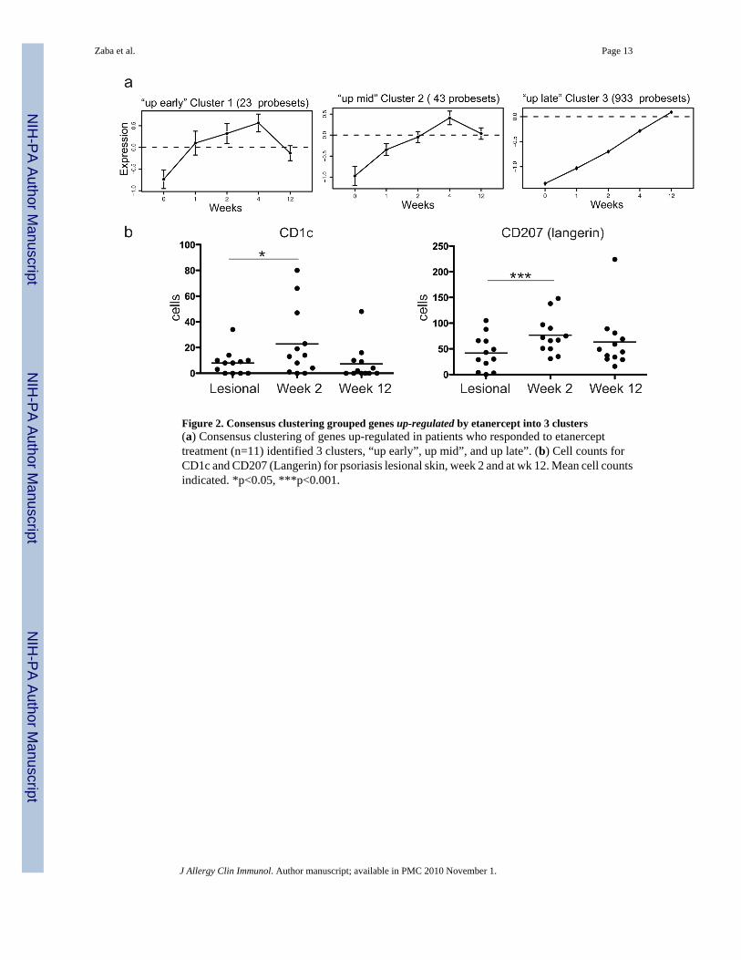
Figure 2. Consensus clustering grouped genes up-regulated by etanercept into 3 clusters(a) Consensus clustering of genes up-regulated in patients who responded to etanercepttreatment (n=11) identified 3 clusters, “up early”, up mid”, and up late”. (b) Cell counts forCD1c and CD207 (Langerin) for psoriasis lesional skin, week 2 and at wk 12. Mean cell countsindicated. *p<0.05, ***p<0.001.
Zaba et al. Page 13
J Allergy Clin Immunol. Author manuscript; available in PMC 2010 November 1.
NIH
-PA Author Manuscript
NIH
-PA Author Manuscript
NIH
-PA Author Manuscript
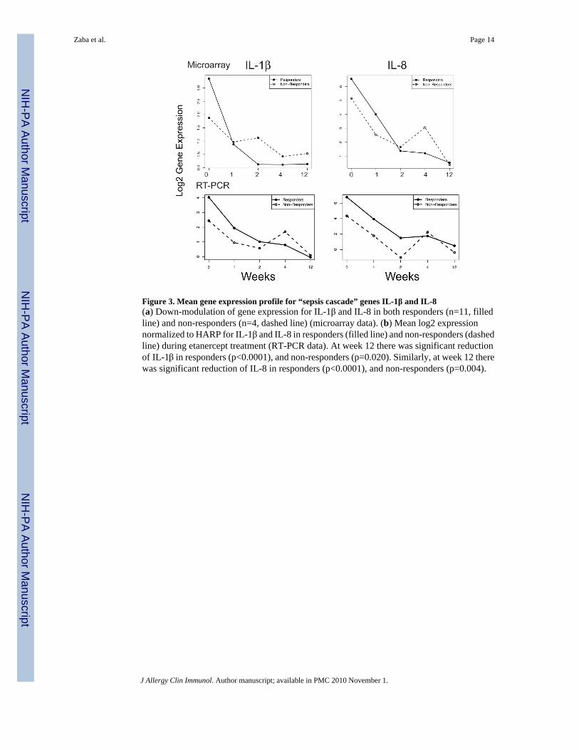
Figure 3. Mean gene expression profile for “sepsis cascade” genes IL-1β and IL-8(a) Down-modulation of gene expression for IL-1β and IL-8 in both responders (n=11, filledline) and non-responders (n=4, dashed line) (microarray data). (b) Mean log2 expressionnormalized to HARP for IL-1β and IL-8 in responders (filled line) and non-responders (dashedline) during etanercept treatment (RT-PCR data). At week 12 there was significant reductionof IL-1β in responders (p<0.0001), and non-responders (p=0.020). Similarly, at week 12 therewas significant reduction of IL-8 in responders (p<0.0001), and non-responders (p=0.004).
Zaba et al. Page 14
J Allergy Clin Immunol. Author manuscript; available in PMC 2010 November 1.
NIH
-PA Author Manuscript
NIH
-PA Author Manuscript
NIH
-PA Author Manuscript

Figure 4. Consensus clustering grouped genes that were different in responders and non-responders(a) Consensus clustering of genes that were different in responders and non-responders groupedgenes into 4 clusters. (b) Heat map of cluster 1. There was a group of genes that were down-regulated at baseline in non-responders but not responders (black box).
Zaba et al. Page 15
J Allergy Clin Immunol. Author manuscript; available in PMC 2010 November 1.
NIH
-PA Author Manuscript
NIH
-PA Author Manuscript
NIH
-PA Author Manuscript
Figure 5. Profiles of “cytokine pathways” in etanercept gene sets in responders and non-ersponders,using GSEA analysisKeratinocyte cytokine “pathways” were created by culturing primary human keratinocyteswith either IL-17, TNF, or IFNγ and defining cytokine pathway gene sets (“IL-17”, “TNFspecific”, “IL-17 & TNF”, and “IFNγ”) 22. (a, b, c, d) Genes modulated by etanercept overtime were analyzed by GSEA. Average normalized expression (+/− 95% confidence interval)was plotted over time. Responders showed significant down-modulation of IL-17 pathwaygenes compared to non-responders.
Zaba et al. Page 16
J Allergy Clin Immunol. Author manuscript; available in PMC 2010 November 1.
NIH
-PA Author Manuscript
NIH
-PA Author Manuscript
NIH
-PA Author Manuscript
Copyright © 2022 FDOKUMEN