Epidermal HLA-DR and the enhancement of cutaneous reactivity to superantigenic toxins in psoriasis
9
Introduction Psoriasis is a chronic inflammatory skin disorder affect- ing 1–2% of the general population. The characteristic lesion of psoriasis is a sharply demarcated erythematous papule or plaque containing hyperproliferating ker- atinocytes as well as infiltrating neutrophils, mono- cytes, and T lymphocytes (1). Although psoriasis is con- sidered an autoimmune disease, increasing evidence suggests an important role for bacteria in its initiation and/or propagation. Colonization and infection with Staphylococcus and Streptococcus have been reported to exacerbate psoriasis (2, 3). In this regard, Staphylococcus aureus has been found on the skin of more than half the patients with chronic plaque psoriasis (2). We have pre- viously identified patients with psoriasis vulgaris who have experienced exacerbations of their disease in asso- ciation with staphylococcal skin infections (4). The most convincing clinical and experimental association between bacterial infection and psoriasis, however, is in patients with acute guttate (eruptive) psoriasis (3, 5). Given the strong association between bacterial infec- tion and psoriasis, intensive studies have sought to dis- cern the mechanisms by which bacteria participate in the pathogenesis of this common skin disease. Recent stud- ies have demonstrated that streptococcal pyrogenic exo- toxins (SPEs) and staphylococcal enterotoxins can act as superantigens (6, 7), providing plausible mechanisms by which these bacteria could cause an inflammatory skin lesion containing activated T cells and monocytes. The term superantigen (SAg) was coined to describe a family of microbial proteins that are potent stimulators of T cells and macrophages (6, 7). When bound to MHC class II molecules, SAg’s stimulate T cells expressing particu- lar T-cell receptor (TCR) Vβ gene segments (8). In addi- tion to this ability to activate large numbers of T cells, in vitro studies have reported that SAg’s can also activate and stimulate cytokine production from MHC class II–expressing cells, including activated keratinocytes (9–11), independent of T cells. The latter effect is trans- duced through the MHC class II molecule (11). The Journal of Clinical Investigation | November 1999 | Volume 104 | Number 9 1181 Epidermal HLA-DR and the enhancement of cutaneous reactivity to superantigenic toxins in psoriasis Jeffrey B. Travers, 1 Qutayba A. Hamid, 2 David A. Norris, 3 Christine Kuhn, 1 Ralph C. Giorno, 3 Patrick M. Schlievert, 4 Evan R. Farmer, 1 and Donald Y.M. Leung 3,5 1 Departments of Dermatology and Pharmacology, Indiana University Medical Center, Indianapolis, Indiana 46202, USA 2 Meakins-Christie Laboratories and Departments of Pathology, McGill University, Montreal, Quebec H2X 2P2, Canada 3 Departments of Pediatrics, Dermatology, and Medicine, University of Colorado Health Sciences Center, Denver, Colorado 80262, USA 4 Department of Microbiology, University of Minnesota, Minneapolis, Minnesota 55455, USA 5 Division of Pediatric Allergy and Immunology, The National Jewish Medical and Research Center, Denver, Colorado 80206, USA Address correspondence to: Donald Y.M. Leung, The National Jewish Medical and Research Center, 1400 Jackson Street, Room K926, Denver, Colorado 80206, USA. Phone: (303) 398-1379; Fax: (303) 270-2182; E-mail: [email protected]. Received for publication March 16, 1999, and accepted in revised form September 10, 1999. Streptococcal and staphylococcal superantigens (SAg’s) have been implicated in the pathogenesis of inflammatory skin diseases, but the mechanisms by which these toxins act are unknown. The pres- ent study assessed the ability of nanogram quantities of topically applied purified toxic shock syn- drome toxin-1 (TSST-1), staphylococcal enterotoxin type B, and streptococcal pyrogenic enterotox- in types A and C to induce inflammatory reactions in clinically uninvolved skin of normal controls and subjects with psoriasis, atopic dermatitis, and lichen planus. These SAg’s triggered a significantly greater inflammatory skin response in psoriatics than in normal control subjects or in subjects with atopic dermatitis or lichen planus. Surprisingly, skin biopsies did not exhibit the T-cell receptor Vβ stimulatory properties predicted for SAg-induced skin reactions. By 6 hours after patch testing with SAg’s, TNF-α mRNA had increased in the epidermis (but not the dermis) in biopsies from psoriatics, compared with controls. Immunohistochemical studies revealed significantly higher HLA-DR expres- sion in keratinocytes from psoriatics than from controls. However, a mutant TSST-1 protein that fails to bind HLA-DR did not elicit an inflammatory skin reaction. These results indicate that ker- atinocyte expression of HLA-DR enhances inflammatory skin responses to SAg’s. They may also account for previous studies failing to demonstrate selective expansion of T-cell receptor Vβs in pso- riatics colonized with SAg-producing Staphylococcus aureus, and they identify a novel T cell–inde- pendent mechanism by which SAg’s contribute to the pathogenesis of inflammatory skin diseases. J. Clin. Invest. 104:1181–1189 (1999).
-
Upload
independent -
Category
Documents
-
view
3 -
download
0
Transcript of Epidermal HLA-DR and the enhancement of cutaneous reactivity to superantigenic toxins in psoriasis

IntroductionPsoriasis is a chronic inflammatory skin disorder affect-ing 1–2% of the general population. The characteristiclesion of psoriasis is a sharply demarcated erythematouspapule or plaque containing hyperproliferating ker-atinocytes as well as infiltrating neutrophils, mono-cytes, and T lymphocytes (1). Although psoriasis is con-sidered an autoimmune disease, increasing evidencesuggests an important role for bacteria in its initiationand/or propagation. Colonization and infection withStaphylococcus and Streptococcus have been reported toexacerbate psoriasis (2, 3). In this regard, Staphylococcusaureus has been found on the skin of more than half thepatients with chronic plaque psoriasis (2). We have pre-viously identified patients with psoriasis vulgaris whohave experienced exacerbations of their disease in asso-ciation with staphylococcal skin infections (4). Themost convincing clinical and experimental associationbetween bacterial infection and psoriasis, however, is inpatients with acute guttate (eruptive) psoriasis (3, 5).
Given the strong association between bacterial infec-tion and psoriasis, intensive studies have sought to dis-cern the mechanisms by which bacteria participate in thepathogenesis of this common skin disease. Recent stud-ies have demonstrated that streptococcal pyrogenic exo-toxins (SPEs) and staphylococcal enterotoxins can act assuperantigens (6, 7), providing plausible mechanisms bywhich these bacteria could cause an inflammatory skinlesion containing activated T cells and monocytes. Theterm superantigen (SAg) was coined to describe a familyof microbial proteins that are potent stimulators of Tcells and macrophages (6, 7). When bound to MHC classII molecules, SAg’s stimulate T cells expressing particu-lar T-cell receptor (TCR) Vβ gene segments (8). In addi-tion to this ability to activate large numbers of T cells, invitro studies have reported that SAg’s can also activateand stimulate cytokine production from MHC classII–expressing cells, including activated keratinocytes(9–11), independent of T cells. The latter effect is trans-duced through the MHC class II molecule (11).
The Journal of Clinical Investigation | November 1999 | Volume 104 | Number 9 1181
Epidermal HLA-DR and the enhancement of cutaneousreactivity to superantigenic toxins in psoriasis
Jeffrey B. Travers,1 Qutayba A. Hamid,2 David A. Norris,3 Christine Kuhn,1
Ralph C. Giorno,3 Patrick M. Schlievert,4 Evan R. Farmer,1 and Donald Y.M. Leung3,5
1Departments of Dermatology and Pharmacology, Indiana University Medical Center, Indianapolis, Indiana 46202, USA2Meakins-Christie Laboratories and Departments of Pathology, McGill University, Montreal, Quebec H2X 2P2, Canada3Departments of Pediatrics, Dermatology, and Medicine, University of Colorado Health Sciences Center, Denver, Colorado 80262, USA
4Department of Microbiology, University of Minnesota, Minneapolis, Minnesota 55455, USA5Division of Pediatric Allergy and Immunology, The National Jewish Medical and Research Center, Denver, Colorado 80206, USA
Address correspondence to: Donald Y.M. Leung, The National Jewish Medical and Research Center, 1400 Jackson Street, Room K926, Denver, Colorado 80206, USA. Phone: (303) 398-1379; Fax: (303) 270-2182; E-mail: [email protected].
Received for publication March 16, 1999, and accepted in revised form September 10, 1999.
Streptococcal and staphylococcal superantigens (SAg’s) have been implicated in the pathogenesis ofinflammatory skin diseases, but the mechanisms by which these toxins act are unknown. The pres-ent study assessed the ability of nanogram quantities of topically applied purified toxic shock syn-drome toxin-1 (TSST-1), staphylococcal enterotoxin type B, and streptococcal pyrogenic enterotox-in types A and C to induce inflammatory reactions in clinically uninvolved skin of normal controlsand subjects with psoriasis, atopic dermatitis, and lichen planus. These SAg’s triggered a significantlygreater inflammatory skin response in psoriatics than in normal control subjects or in subjects withatopic dermatitis or lichen planus. Surprisingly, skin biopsies did not exhibit the T-cell receptor Vβstimulatory properties predicted for SAg-induced skin reactions. By 6 hours after patch testing withSAg’s, TNF-α mRNA had increased in the epidermis (but not the dermis) in biopsies from psoriatics,compared with controls. Immunohistochemical studies revealed significantly higher HLA-DR expres-sion in keratinocytes from psoriatics than from controls. However, a mutant TSST-1 protein thatfails to bind HLA-DR did not elicit an inflammatory skin reaction. These results indicate that ker-atinocyte expression of HLA-DR enhances inflammatory skin responses to SAg’s. They may alsoaccount for previous studies failing to demonstrate selective expansion of T-cell receptor Vβs in pso-riatics colonized with SAg-producing Staphylococcus aureus, and they identify a novel T cell–inde-pendent mechanism by which SAg’s contribute to the pathogenesis of inflammatory skin diseases.
J. Clin. Invest. 104:1181–1189 (1999).
The association between SAg’s and psoriasis has beenstrengthened by recent reports culturing streptococcalpyrogenic exotoxin serotype C–producing (SPEC- orscarlet fever type C–producing) group A streptococcusfrom the oropharynx of patients with acute guttate pso-riasis and demonstrating increased numbers of Vβ2-expressing T cells in their lesional skin (5, 12). In addi-tion, recent studies from 2 separate groups ofinvestigators indicate that normal-appearing skin frompsoriatic patients grafted onto immunodeficient micecan be induced to develop into psoriatic lesions byrepeated injection with autologous SAg-treatedimmunocytes (13, 14). Together, these findings suggestthat SAg stimulation can initiate psoriasis. To date,however, there have been no in vivo studies in humansdirectly examining the effects of SAg’s on the unin-volved skin of psoriatic patients. The objective of thepresent study was to evaluate the reactivity of psoriaticskin to topically applied bacterial SAg’s and determinethe mechanisms by which they induce skin inflamma-tion in vivo in psoriasis.
MethodsPatients. Fifty-seven adult patients were enrolled intothis study. Twenty-six patients with type I (15) psoria-sis (age range, 23–52 years; mean, 35 years); 6 patientswith atopic dermatitis (age range, 21–28; mean, 25years), diagnosed according to the Hanifin and Rajkacriteria (16); and 5 patients with biopsy-proven lichenplanus (age range, 24–56 years; mean, 43 years) alsoparticipated in this study. Twenty-one subjects (agerange, 22–52 years; mean, 32 years) without a personalor family history of skin disease or respiratory allergywere enrolled into the study to serve as normal con-trols. Patients refrained from using topical medicationsto the arm undergoing patch testing and from usingoral antihistamines for at least 2 weeks before patchtesting. None of the patients was on any systemicimmunosuppressive drugs, including corticosteroidsor cyclosporin. The protocols involving human sub-jects were approved by the institutional review boardsof both the University of Colorado Health SciencesCenter and the Indiana University School of Medicine.Informed consent was obtained from all subjectsbefore performing all studies.
Patch-testing protocol. Staphylococcal and streptococcalexotoxins were purified by Patrick M. Schlievert asdescribed previously (17). In selected experiments, amutant toxic shock syndrome toxin-1 (TSST-1) pro-tein, G31S/S32P, produced by site-directed mutagene-sis was also used. G31S/S32P has previously beendemonstrated to lack HLA-DR binding (18, 19).
A total of 20 µL of 1 µg/mL solutions of highly puri-fied TSST-1, staphylococcal enterotoxin B (SEB), strep-tococcal pyrogenic exotoxins A and C (SPEA, SPEC) orG31S/S32P in PBS, or PBS alone were placed in 8-mm-diameter Finn chambers (Epitest Ltd., Oy, Finland) andapplied to uninvolved skin on the volar forearm. Mostexperiments used tape-stripping a 2 × 12 cm section ofuninvolved skin 100 times with cellophane tape (3MInc., Minneapolis, Minnesota, USA) before applicationof the patches. Twenty microliters of 1% (vol/vol) sodi-um lauryl sulfate (Sigma Chemical Co., St. Louis, Mis-souri, USA) was used as an irritant control. At 48 hours,the patch tests were removed and clinically assessed.The following clinical measurement system was usedto rate the reactions: 0 = no reaction; 1 = nonpalpablemacular erythema; 2 = palpable erythema; and 3 = pal-pable erythema extending beyond the test chamber.After clinical assessment, 4-mm punch biopsies wereobtained from the reaction sites, with part fixed inbuffered formalin, routinely processed for 4-µm paraf-fin-embedded sections, and stained with hema-toxylin/eosin. The other half was snap-frozen in OCTfor immunohistochemical studies.
Quantitation of skin inflammatory cell infiltrate. To quan-titate mononuclear inflammatory cells in the dermisof biopsy specimens, an eyepiece counting grid (Olym-pus Optical Co. Tokyo, Japan) was used at ×200, whichprovided an area of 0.25 mm2. The grid was placed atthe left side of the tissue with the top of the gridtouching the dermal-epidermal junction. All themononuclear cells in the grid were counted. Threeadjacent 0.25-mm2 squares were counted lateral to thefirst to assay a total area of 1.0 mm2 encompassing thepapillary and part of the reticular dermis. Similarly,the grid was placed at the right side of the tissue, and3 more sections were counted. The counts were aver-aged and then multiplied by 4 to give the number ofmononuclear cells per square millimeter. Duplicate
1182 The Journal of Clinical Investigation | November 1999 | Volume 104 | Number 9
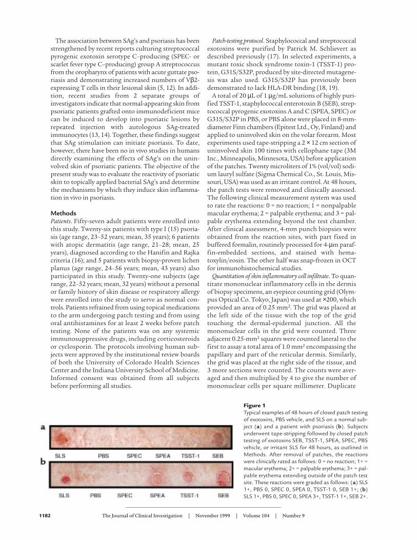
Figure 1Typical examples of 48 hours of closed patch testingof exotoxins, PBS vehicle, and SLS on a normal sub-ject (a) and a patient with psoriasis (b). Subjectsunderwent tape-stripping followed by closed patchtesting of exotoxins SEB, TSST-1, SPEA, SPEC, PBSvehicle, or irritant SLS for 48 hours, as outlined inMethods. After removal of patches, the reactionswere clinically rated as follows: 0 = no reaction; 1+ =macular erythema; 2+ = palpable erythema; 3+ = pal-pable erythema extending outside of the patch testsite. These reactions were graded as follows: (a) SLS1+, PBS 0, SPEC 0, SPEA 0, TSST-1 0, SEB 1+; (b)SLS 1+, PBS 0, SPEC 0, SPEA 3+, TSST-1 1+, SEB 2+.
slides from each biopsy were counted and averaged. Allcell counts were done by one of the authors (E.R.Farmer) under blinded conditions.
In situ hybridization studies. In these experiments, skinbiopsy specimens were fixed immediately in freshly pre-pared 4% paraformaldehyde/PBS solution for 2 hours,washed in 15% sucrose/PBS 3 times, embedded in OCTcompound (Tissue-Tek; Miles Inc., Elkhart, Indiana,USA), and snap-frozen in isopentane cooled in liquidnitrogen. Cryostat sections 5 µm thick were cut on 0.1%poly-L-lysine–coated slides, air-dried overnight at 37°C,and stored at –80°C until used. In situ hybridizationstudies for IL-4, IFN-γ, and TNF-α mRNA were per-formed as described previously using digoxigenin-labeled cRNA probes (20, 21). The following controlexperiments were also performed: (a) omitting the probein the in situ hybridization protocol and using a sensedig-labeled riboprobe; (b) pretreatment of slides withRNase A before in situ hybridization; (c) use of an unre-lated antisense digoxigenin-labeled RNA probe (humanatrial natriuretic polypeptide); and (d) omitting thedigoxigenin-alkaline-phosphatase conjugate.
Immunohistochemistry studies. Cryostat sections pre-pared as described for in situ hybridization studies wereused for analysis of HLA-DR expression. Immunoreac-tivity was assessed by using the alkaline phosphataseantialkaline phosphatase technique and by using eitheran anti–HLA-DR, anti-CD25, anti-CD4, or anti-CD8mAb (DAKO Canada Inc.; Mississauga, Ontario, Cana-da) as described previously (22). The reaction wasviewed with fast red alkaline-phosphatase substrate. Ascontrols, sections were processed in the absence of theprimary antibody. System and specificity controls werealso included in each staining run, using human ton-sils, obtained at routine tonsillectomy operations, andmouse IgG2a myeloma proteins as a negative control.
For studies analyzing TCR Vβ expression, 4-µm cryo-stat sections were cut, dehydrated in acetone for 10 min-utes, and air-dried. Sections were incubated 30–60 min-utes at room temperature with the respective mouseanti-human Vβ mAb (Immunotech, Westbrook, Maine,
USA), anti-CD3 mAb, or mouse im-munoglobulin isotype control (BectonDickinson Immunocytometry Systems,San Jose, California, USA). Sections werethen washed and stained by the labeledavidin biotin method using peroxidase-labeled streptavidin (DAKO) as des-cribed previously (5). Afterward, sectionswere counterstained with hematoxylinand mounted. Control antibodies weretested on step sections of the same tissuespecimens. The negative staining con-trols were isotype-specific mouseimmunoglobulins with irrelevant speci-ficity. Normal human tonsil sectionswere used as positive controls for Vβ T-cell staining and CD antibodies.
Quantitation and statistical analysis. Thereadings of all histological, immunohistochemical, andin situ hybridization studies were conducted in blindedfashion without knowledge of the subject’s identity ordisease condition. Epidermal TNF-α mRNA and HLA-DR immunoreactivity was expressed as percentage ofepidermal cells that express TNF-α mRNA or HLA-DRprotein. Subepidermal mRNA expression of TNF-α and
The Journal of Clinical Investigation | November 1999 | Volume 104 | Number 9 1183
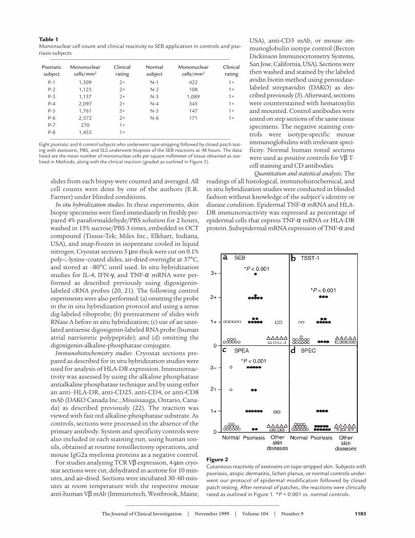
Table 1Mononuclear cell count and clinical reactivity to SEB application in controls and pso-riasis subjects
Psoriatic Mononuclear Clinical Normal Mononuclear Clinicalsubject cells/mm2 rating subject cells/mm2 rating
P-1 1,509 2+ N-1 422 1+P-2 1,123 2+ N-2 108 1+P-3 1,137 2+ N-3 1,089 1+P-4 2,097 2+ N-4 345 1+P-5 1,761 3+ N-5 147 1+P-6 2,372 2+ N-6 171 1+P-7 270 1+P-8 1,455 1+
Eight psoriatic and 6 control subjects who underwent tape-stripping followed by closed patch test-ing with exotoxins, PBS, and SLS underwent biopsies of the SEB reactions at 48 hours. The datalisted are the mean number of mononuclear cells per square millimeter of tissue obtained as out-lined in Methods, along with the clinical reaction (graded as outlined in Figure 2).
Figure 2Cutaneous reactivity of exotoxins on tape-stripped skin. Subjects withpsoriasis, atopic dermatitis, lichen planus, or normal controls under-went our protocol of epidermal modification followed by closedpatch testing. After removal of patches, the reactions were clinicallyrated as outlined in Figure 1. *P < 0.001 vs. normal controls.
immunoreactivity for HLA-DR were presented as num-ber of cells per square millimeter. Statistical analysesused the unpaired t test (Instat Program; GraphPad Soft-ware for Science Inc., San Diego, California, USA) toassess clinical differences in exotoxin reactivities betweensubject groups and Wilcoxon rank order test to assessdifferences between mononuclear cell counts in biopsyspecimens from psoriatics and control subjects.
ResultsClosed patch testing of SAg toxins. Initially, 6 normal and 4psoriatic patients underwent closed patch testing with20 ng TSST-1, SEB, SPEA, and SPEC on uninvolvedvolar forearm skin. Forty-eight–hour closed patch test-ing did not induce cutaneous reactions to any of the tox-ins, yet all experienced a reaction to the irritant (1%) SLS.These preliminary studies suggested that a single appli-cation of this concentration of toxins to intact skin wasnot sufficient to elicit an inflammatory skin reaction.
Tape-stripping is a standardized method used toinduce epidermal activation and to increase cutaneousabsorption by removal of the stratum corneum (23). Todetermine whether epidermal modification wasrequired for nanogram amounts of SAg to induce skininflammatory responses, volar forearm skin was treat-ed with tape-stripping before application of the toxinsas described in Methods. Tape-stripping was repeated100 times, as it has been shown by Nickoloff and Naiduthat this protocol resulted in the maximal amount ofkeratinocyte activation in normal skin (23).
Using this protocol of epidermal modification and48 hours of closed patch testing, positive reactions tothe SAg’s and SLS were noted. No clinical reactions
were seen to the PBS vehicle. The SAg reactions con-sisted of erythematous macules or plaques and wereusually asymptomatic. The SLS reactions consisted oferythematous thin plaques, but unlike the SAg-induced reactions, were uniformly associated withmild-moderate pruritus. The reactions to SAg’s dif-fered greatly among the subjects, however, even themost vigorous (3+) reactions resolved within 1 week.No systemic symptoms, such as fever, headache, dizzi-ness, or presence/worsening of skin rash, were notedin subjects undergoing these protocols.
Although skin reactions were elicited in both psori-atic and normal subjects, psoriatics reacted to moreSAg’s and with greater intensity than normal subjects.Examples of reactions seen in normal and psoriaticsubjects to the various exotoxins are shown in Figure 1.Figure 2 compares the intensity of clinical reactionsexhibited by psoriatic and control subjects. Statistical-ly significant (P < 0.001) increased clinical reactions toSEB, TSST-1, and SPEA were seen in psoriatic versuscontrol populations. Only 4 of 15 psoriatic subjects,but 0 of 15 control subjects reacted to SPEC (not sig-nificant). Subjects with atopic dermatitis (6 subjects)or the papulosquamous disease lichen planus (5 sub-jects) reacted in a similar fashion to normal controls(Figure 2). The cutaneous reactivity to exotoxins corre-lated with the extent of psoriasis (as assessed by per-centage of body surface area involvement) as shown inFigure 3. As depicted in Figure 3, 5 of 15 (4 of highest
1184 The Journal of Clinical Investigation | November 1999 | Volume 104 | Number 9
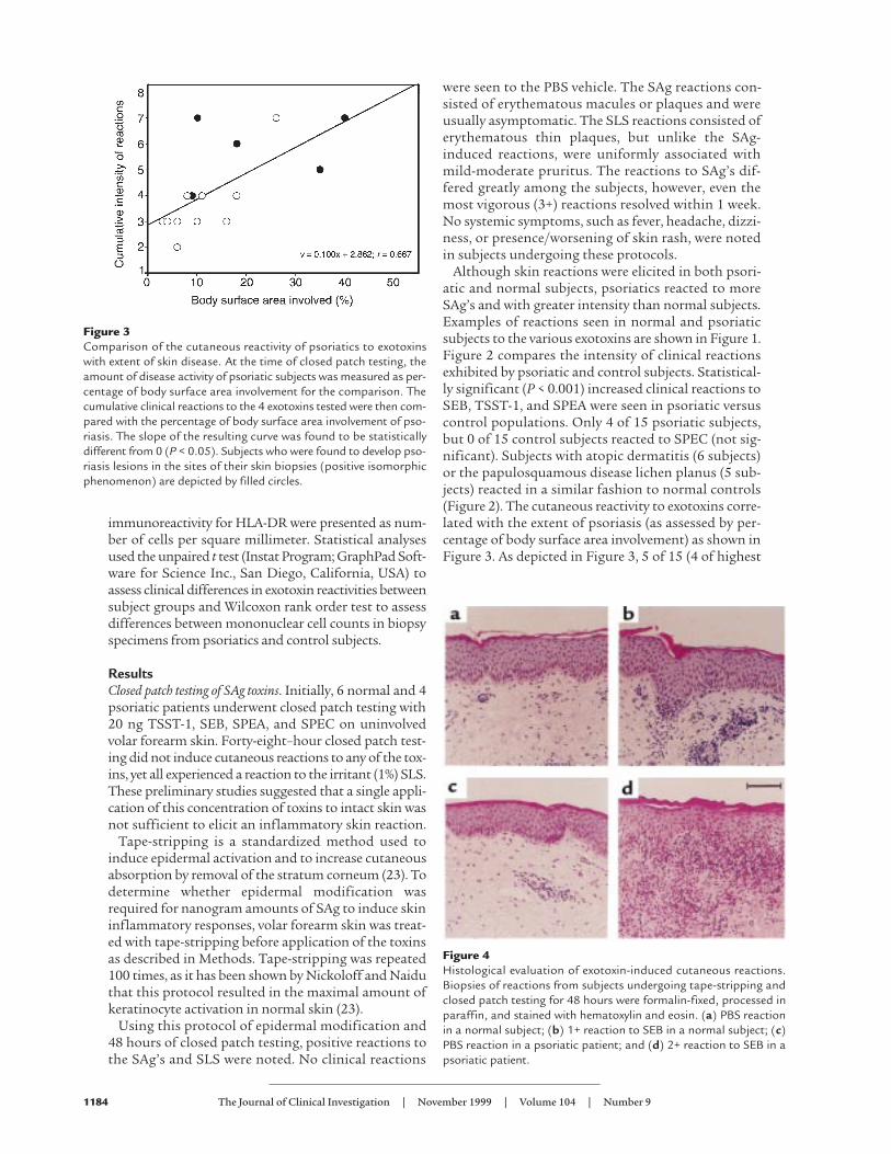
Figure 3Comparison of the cutaneous reactivity of psoriatics to exotoxinswith extent of skin disease. At the time of closed patch testing, theamount of disease activity of psoriatic subjects was measured as per-centage of body surface area involvement for the comparison. Thecumulative clinical reactions to the 4 exotoxins tested were then com-pared with the percentage of body surface area involvement of pso-riasis. The slope of the resulting curve was found to be statisticallydifferent from 0 (P < 0.05). Subjects who were found to develop pso-riasis lesions in the sites of their skin biopsies (positive isomorphicphenomenon) are depicted by filled circles.
Figure 4Histological evaluation of exotoxin-induced cutaneous reactions.Biopsies of reactions from subjects undergoing tape-stripping andclosed patch testing for 48 hours were formalin-fixed, processed inparaffin, and stained with hematoxylin and eosin. (a) PBS reactionin a normal subject; (b) 1+ reaction to SEB in a normal subject; (c)PBS reaction in a psoriatic patient; and (d) 2+ reaction to SEB in apsoriatic patient.
responders) psoriatic subjects developed psoriaticlesions in the skin biopsy sites. Unlike the SAg-inducedcutaneous reactions, those induced by SLS were simi-lar among subjects with psoriasis, atopic dermatitis,lichen planus, and normal controls.
Histological evaluation of SAg-induced reactions. At 48hours when the patches were removed to assess theclinical reactions, skin biopsies were obtained fromsome psoriatic and normal subjects for both routinehistology and immunohistochemistry. As shown inFigure 4, the reactions consisted of epidermal and der-mal spongiosis, with mononuclear (lymphocytic andmonocytic) cell infiltrates, both in the papillary dermisand the epidermis. Neither eosinophils nor neutrophilswere seen in significant numbers in the biopsy speci-mens. In several of the biopsies taken from psoriaticpatients, mild epidermal acanthosis was noted. Intra-papillary thinning or neutrophilic microabscesses,characteristic of psoriasis lesions, were not seen. Forpositive SEB reactions in 8 psoriatic and the 6 normalsubjects, the numbers of inflammatory cells were exam-ined. As shown in Table 1, statistically significant (P =0.045 by the Wilcoxon rank order test) increased num-bers of mononuclear cells were seen in the psoriatic ver-sus control populations.
Characterization of T cells in SAg-induced reactions. Becauseselective expansion of TCR Vβ is a hallmark of super-antigenic stimulation of T cells (5, 8), the reactions wereassessed for the presence of TCR Vβ expansion byimmunohistochemistry. Staining of skin specimens
from 6 subjects with positive (48 hours) exotoxin-induced reactions with mAb’s directed to TCR Vβ 2(TSST-1 and SPEC reactive), 3 (SEB reactive), 8.1 (SPEAreactive), 12 (SEB and SPEA reactive), and 17 (SEB reac-tive) failed to demonstrate selective TCR Vβ expansionthat correlated with the predicted stimulatory propertiesof the SAg (data not shown). These findings are in con-trast to our ability to demonstrate marked expansion ofVβ2+ T cells in the skin lesions of guttate psoriasis (5).
We also examined the T-cell infiltrate in skin biopsiesof SEB- and PBS-stimulated sites from psoriasispatients for CD25 (as a marker of T-cell activation),CD4, and CD8 expression. SEB-stimulated sites com-pared with PBS-stimulated sites contained increasednumbers of T cells per square millimeter expressingCD25 (54 ± 15 vs. 10 ± 2; P < 0.05), CD4 (160 ± 10 vs. 71± 20; P < 0.05), and CD8 (57 ± 7 vs. 23 ± 6; P < 0.05).SEB-stimulated skin sites also expressed increased IFN-γ mRNA (33 ± 4 vs. 8 ± 4 cells/mm2; P < 0.05) expres-sion, but not increased IL-4 mRNA (11 ± 2 vs. 10 ± 6cells/mm2; P = not significant) expression.
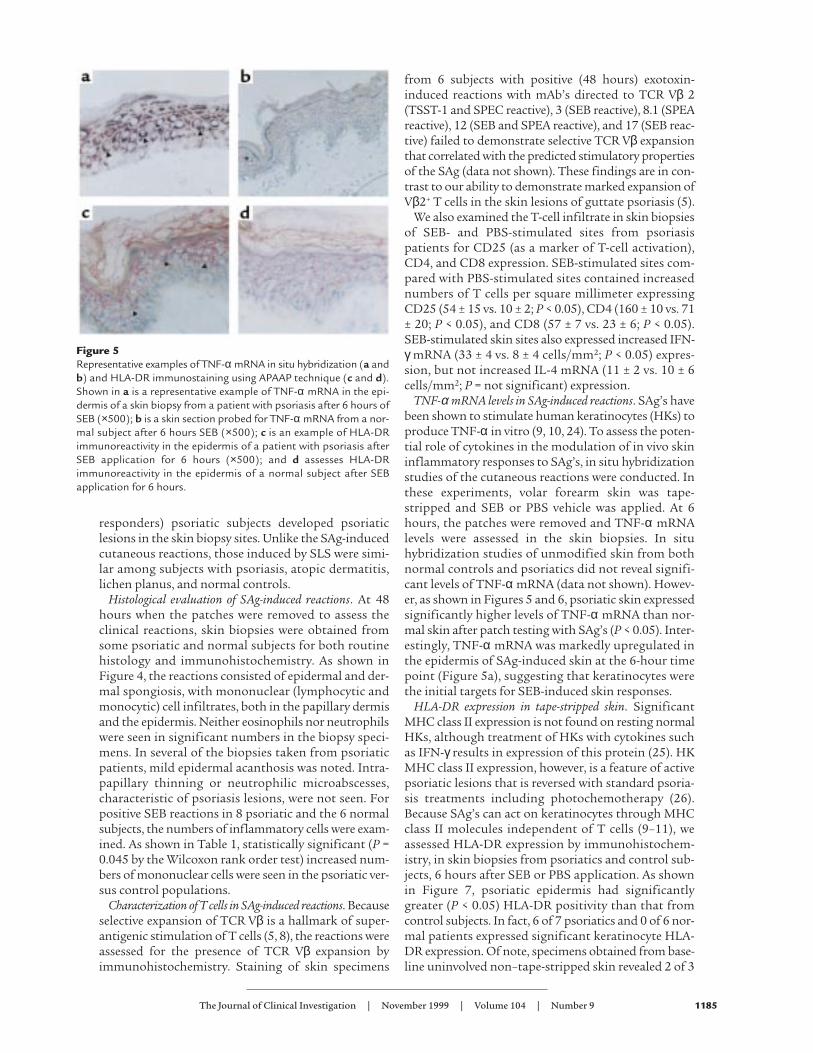
TNF-α mRNA levels in SAg-induced reactions. SAg’s havebeen shown to stimulate human keratinocytes (HKs) toproduce TNF-α in vitro (9, 10, 24). To assess the poten-tial role of cytokines in the modulation of in vivo skininflammatory responses to SAg’s, in situ hybridizationstudies of the cutaneous reactions were conducted. Inthese experiments, volar forearm skin was tape-stripped and SEB or PBS vehicle was applied. At 6hours, the patches were removed and TNF-α mRNAlevels were assessed in the skin biopsies. In situhybridization studies of unmodified skin from bothnormal controls and psoriatics did not reveal signifi-cant levels of TNF-α mRNA (data not shown). Howev-er, as shown in Figures 5 and 6, psoriatic skin expressedsignificantly higher levels of TNF-α mRNA than nor-mal skin after patch testing with SAg’s (P < 0.05). Inter-estingly, TNF-α mRNA was markedly upregulated inthe epidermis of SAg-induced skin at the 6-hour timepoint (Figure 5a), suggesting that keratinocytes werethe initial targets for SEB-induced skin responses.
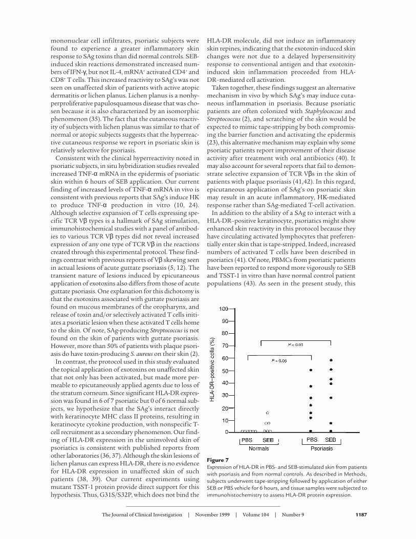
HLA-DR expression in tape-stripped skin. SignificantMHC class II expression is not found on resting normalHKs, although treatment of HKs with cytokines suchas IFN-γ results in expression of this protein (25). HKMHC class II expression, however, is a feature of activepsoriatic lesions that is reversed with standard psoria-sis treatments including photochemotherapy (26).Because SAg’s can act on keratinocytes through MHCclass II molecules independent of T cells (9–11), weassessed HLA-DR expression by immunohistochem-istry, in skin biopsies from psoriatics and control sub-jects, 6 hours after SEB or PBS application. As shownin Figure 7, psoriatic epidermis had significantlygreater (P < 0.05) HLA-DR positivity than that fromcontrol subjects. In fact, 6 of 7 psoriatics and 0 of 6 nor-mal patients expressed significant keratinocyte HLA-DR expression. Of note, specimens obtained from base-line uninvolved non–tape-stripped skin revealed 2 of 3
The Journal of Clinical Investigation | November 1999 | Volume 104 | Number 9 1185
Figure 5Representative examples of TNF-α mRNA in situ hybridization (a andb) and HLA-DR immunostaining using APAAP technique (c and d).Shown in a is a representative example of TNF-α mRNA in the epi-dermis of a skin biopsy from a patient with psoriasis after 6 hours ofSEB (×500); b is a skin section probed for TNF-α mRNA from a nor-mal subject after 6 hours SEB (×500); c is an example of HLA-DRimmunoreactivity in the epidermis of a patient with psoriasis afterSEB application for 6 hours (×500); and d assesses HLA-DRimmunoreactivity in the epidermis of a normal subject after SEBapplication for 6 hours.
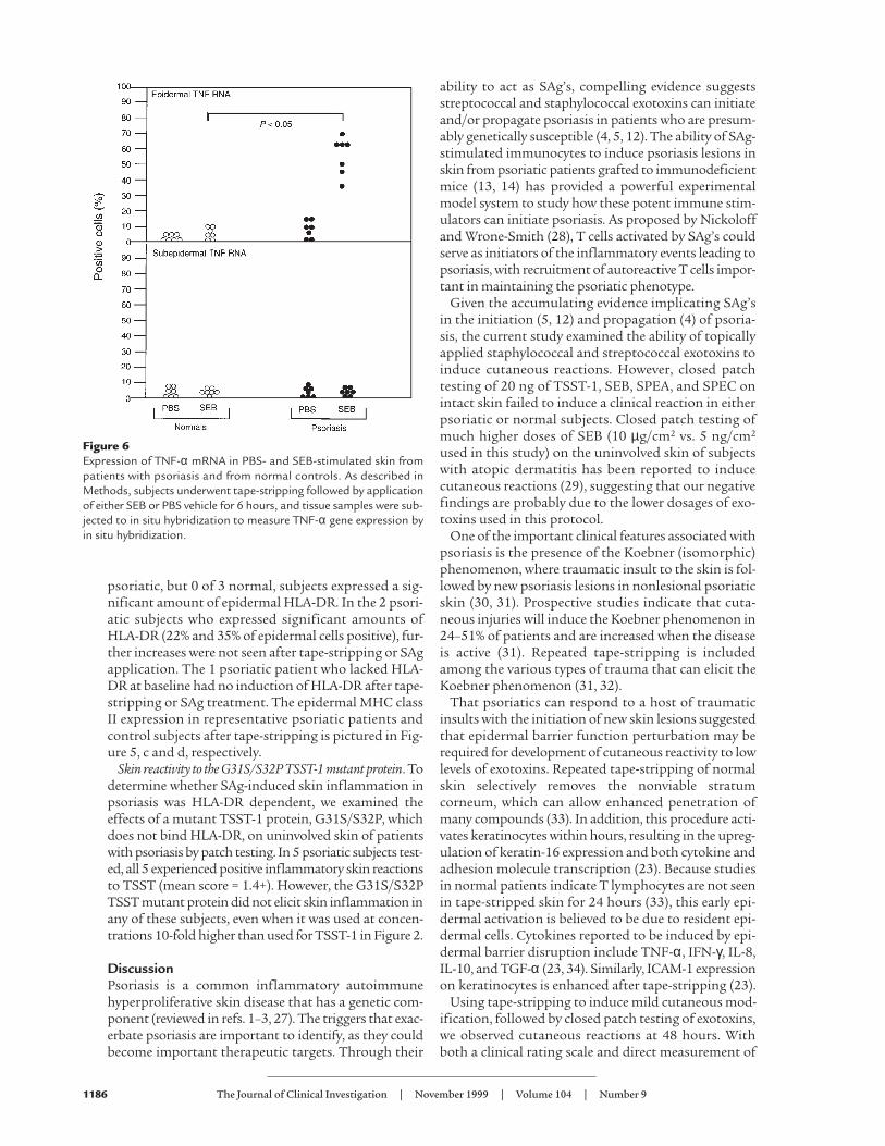
psoriatic, but 0 of 3 normal, subjects expressed a sig-nificant amount of epidermal HLA-DR. In the 2 psori-atic subjects who expressed significant amounts ofHLA-DR (22% and 35% of epidermal cells positive), fur-ther increases were not seen after tape-stripping or SAgapplication. The 1 psoriatic patient who lacked HLA-DR at baseline had no induction of HLA-DR after tape-stripping or SAg treatment. The epidermal MHC classII expression in representative psoriatic patients andcontrol subjects after tape-stripping is pictured in Fig-ure 5, c and d, respectively.
Skin reactivity to the G31S/S32P TSST-1 mutant protein. Todetermine whether SAg-induced skin inflammation inpsoriasis was HLA-DR dependent, we examined theeffects of a mutant TSST-1 protein, G31S/S32P, whichdoes not bind HLA-DR, on uninvolved skin of patientswith psoriasis by patch testing. In 5 psoriatic subjects test-ed, all 5 experienced positive inflammatory skin reactionsto TSST (mean score = 1.4+). However, the G31S/S32PTSST mutant protein did not elicit skin inflammation inany of these subjects, even when it was used at concen-trations 10-fold higher than used for TSST-1 in Figure 2.
DiscussionPsoriasis is a common inflammatory autoimmunehyperproliferative skin disease that has a genetic com-ponent (reviewed in refs. 1–3, 27). The triggers that exac-erbate psoriasis are important to identify, as they couldbecome important therapeutic targets. Through their
ability to act as SAg’s, compelling evidence suggestsstreptococcal and staphylococcal exotoxins can initiateand/or propagate psoriasis in patients who are presum-ably genetically susceptible (4, 5, 12). The ability of SAg-stimulated immunocytes to induce psoriasis lesions inskin from psoriatic patients grafted to immunodeficientmice (13, 14) has provided a powerful experimentalmodel system to study how these potent immune stim-ulators can initiate psoriasis. As proposed by Nickoloffand Wrone-Smith (28), T cells activated by SAg’s couldserve as initiators of the inflammatory events leading topsoriasis, with recruitment of autoreactive T cells impor-tant in maintaining the psoriatic phenotype.
Given the accumulating evidence implicating SAg’sin the initiation (5, 12) and propagation (4) of psoria-sis, the current study examined the ability of topicallyapplied staphylococcal and streptococcal exotoxins toinduce cutaneous reactions. However, closed patchtesting of 20 ng of TSST-1, SEB, SPEA, and SPEC onintact skin failed to induce a clinical reaction in eitherpsoriatic or normal subjects. Closed patch testing ofmuch higher doses of SEB (10 µg/cm2 vs. 5 ng/cm2
used in this study) on the uninvolved skin of subjectswith atopic dermatitis has been reported to inducecutaneous reactions (29), suggesting that our negativefindings are probably due to the lower dosages of exo-toxins used in this protocol.
One of the important clinical features associated withpsoriasis is the presence of the Koebner (isomorphic)phenomenon, where traumatic insult to the skin is fol-lowed by new psoriasis lesions in nonlesional psoriaticskin (30, 31). Prospective studies indicate that cuta-neous injuries will induce the Koebner phenomenon in24–51% of patients and are increased when the diseaseis active (31). Repeated tape-stripping is includedamong the various types of trauma that can elicit theKoebner phenomenon (31, 32).
That psoriatics can respond to a host of traumaticinsults with the initiation of new skin lesions suggestedthat epidermal barrier function perturbation may berequired for development of cutaneous reactivity to lowlevels of exotoxins. Repeated tape-stripping of normalskin selectively removes the nonviable stratumcorneum, which can allow enhanced penetration ofmany compounds (33). In addition, this procedure acti-vates keratinocytes within hours, resulting in the upreg-ulation of keratin-16 expression and both cytokine andadhesion molecule transcription (23). Because studiesin normal patients indicate T lymphocytes are not seenin tape-stripped skin for 24 hours (33), this early epi-dermal activation is believed to be due to resident epi-dermal cells. Cytokines reported to be induced by epi-dermal barrier disruption include TNF-α, IFN-γ, IL-8,IL-10, and TGF-α (23, 34). Similarly, ICAM-1 expressionon keratinocytes is enhanced after tape-stripping (23).
Using tape-stripping to induce mild cutaneous mod-ification, followed by closed patch testing of exotoxins,we observed cutaneous reactions at 48 hours. Withboth a clinical rating scale and direct measurement of
1186 The Journal of Clinical Investigation | November 1999 | Volume 104 | Number 9
Figure 6Expression of TNF-α mRNA in PBS- and SEB-stimulated skin frompatients with psoriasis and from normal controls. As described inMethods, subjects underwent tape-stripping followed by applicationof either SEB or PBS vehicle for 6 hours, and tissue samples were sub-jected to in situ hybridization to measure TNF-α gene expression byin situ hybridization.
mononuclear cell infiltrates, psoriatic subjects werefound to experience a greater inflammatory skinresponse to SAg toxins than did normal controls. SEB-induced skin reactions demonstrated increased num-bers of IFN-γ, but not IL-4, mRNA+ activated CD4+ andCD8+ T cells. This increased reactivity to SAg’s was notseen on unaffected skin of patients with active atopicdermatitis or lichen planus. Lichen planus is a nonhy-perproliferative papulosquamous disease that was cho-sen because it is also characterized by an isomorphicphenomenon (35). The fact that the cutaneous reactiv-ity of subjects with lichen planus was similar to that ofnormal or atopic subjects suggests that the hyperreac-tive cutaneous response we report in psoriatic skin isrelatively selective for psoriasis.
Consistent with the clinical hyperreactivity noted inpsoriatic subjects, in situ hybridization studies revealedincreased TNF-α mRNA in the epidermis of psoriaticskin within 6 hours of SEB application. Our currentfinding of increased levels of TNF-α mRNA in vivo isconsistent with previous reports that SAg’s induce HKto produce TNF-α production in vitro (10, 24).Although selective expansion of T cells expressing spe-cific TCR Vβ types is a hallmark of SAg stimulation,immunohistochemical studies with a panel of antibod-ies to various TCR Vβ types did not reveal increasedexpression of any one type of TCR Vβ in the reactionscreated through this experimental protocol. These find-ings contrast with previous reports of Vβ skewing seenin actual lesions of acute guttate psoriasis (5, 12). Thetransient nature of lesions induced by epicutaneousapplication of exotoxins also differs from those of acuteguttate psoriasis. One explanation for this dichotomy isthat the exotoxins associated with guttate psoriasis arefound on mucous membranes of the oropharynx, andrelease of toxin and/or selectively activated T cells initi-ates a psoriatic lesion when these activated T cells hometo the skin. Of note, SAg-producing Streptococcus is notfound on the skin of patients with guttate psoriasis.However, more than 50% of patients with plaque psori-asis do have toxin-producing S. aureus on their skin (2).
In contrast, the protocol used in this study evaluatedthe topical application of exotoxins on unaffected skinthat not only has been activated, but made more per-meable to epicutaneously applied agents due to loss ofthe stratum corneum. Since significant HLA-DR expres-sion was found in 6 of 7 psoriatic but 0 of 6 normal sub-jects, we hypothesize that the SAg’s interact directlywith keratinocyte MHC class II proteins, resulting inkeratinocyte cytokine production, with nonspecific T-cell recruitment as a secondary phenomenon. Our find-ing of HLA-DR expression in the uninvolved skin ofpsoriatics is consistent with published reports fromother laboratories (36, 37). Although the skin lesions oflichen planus can express HLA-DR, there is no evidencefor HLA-DR expression in unaffected skin of suchpatients (38, 39). Our current experiments usingmutant TSST-1 protein provide direct support for thishypothesis. Thus, G31S/S32P, which does not bind the
HLA-DR molecule, did not induce an inflammatoryskin repines, indicating that the exotoxin-induced skinchanges were not due to a delayed hypersensitivityresponse to conventional antigen and that exotoxin-induced skin inflammation proceeded from HLA-DR–mediated cell activation.
Taken together, these findings suggest an alternativemechanism in vivo by which SAg’s may induce cuta-neous inflammation in psoriasis. Because psoriaticpatients are often colonized with Staphylococcus andStreptococcus (2), and scratching of the skin would beexpected to mimic tape-stripping by both compromis-ing the barrier function and activating the epidermis(23), this alternative mechanism may explain why somepsoriatic patients report improvement of their diseaseactivity after treatment with oral antibiotics (40). Itmay also account for several reports that fail to demon-strate selective expansion of TCR Vβs in the skin ofpatients with plaque psoriasis (41,42). In this regard,epicutaneous application of SAg’s on psoriatic skinmay result in an acute inflammatory, HK-mediatedresponse rather than SAg-mediated T-cell activation.
In addition to the ability of a SAg to interact with aHLA-DR–positive keratinocyte, psoriatics might showenhanced skin reactivity in this protocol because theyhave circulating activated lymphocytes that preferen-tially enter skin that is tape-stripped. Indeed, increasednumbers of activated T cells have been described inpsoriatics (41). Of note, PBMCs from psoriatic patientshave been reported to respond more vigorously to SEBand TSST-1 in vitro than have normal control patientpopulations (43). As seen in the present study, this
The Journal of Clinical Investigation | November 1999 | Volume 104 | Number 9 1187
Figure 7Expression of HLA-DR in PBS- and SEB-stimulated skin from patientswith psoriasis and from normal controls. As described in Methods,subjects underwent tape-stripping followed by application of eitherSEB or PBS vehicle for 6 hours, and tissue samples were subjected toimmunohistochemistry to assess HLA-DR protein expression.
increased proliferative response to SAg’s correlatedwith extent of disease activity (44). The increasedcytokine gene and protein expression in psoriatic skintreated with SEB suggests an intrinsic psoriasis epider-mal phenotype. This increased response to SEB and theearly time course of response (6 hours) suggests an epi-dermal phenotype that favors stimulation in psoriasis.
As reported here and by Strange et al. (29), the abilityto react to the epicutaneous application of exotoxins isnot exclusive to psoriatics. However, the ability oftraumatized skin to react to these SAg’s appears to bemore pronounced in subjects with psoriasis. As hasbeen described with the Koebner phenomenon, thisreactivity tends to correlate with disease activity (30,31). Of note, 4 of the 5 psoriatic subjects who werefound to have the highest cutaneous reactivity to theexotoxins also exhibited the Koebner phenomenonafter skin biopsy. Taken together, these findings alsosuggest that epidermal modification is required for theoptimal induction of SAg-induced skin inflammationin psoriasis. Epidermal modification results in theincreased exposure of activated MHC class II–express-ing keratinocytes to exotoxins; interaction of the SAgwith the MHC class II–positive cells then results in theproduction of proinflammatory cytokines. The obser-vation that atopic dermatitis skin reacted to SAg’s in amanner similar to normal skin may be accounted forby the lack of HLA-DR expression on atopic skin (45).
The further characterization of this cutaneous hyper-reactivity might provide exciting new directions for thefuture unraveling of the genetic and immunologicalmechanisms involved in psoriasis. Dissection of bothgenetic and immunological mechanisms involved inthis common skin disease should have important con-sequences for its treatment.
AcknowledgmentsSupported in part by research grants from the PublicHealth Services (HL-36577, AR-41256, HL-37260, AI-31545, K081993, and HL-36611); a grant from the Divi-sion of Research Resources of the General ClinicalResearch Center (5 MO1 RR00051); and the Universityof Colorado Cancer Center. The authors thank LorenGolitz and John Aeling for technical assistance and his-tological interpretation of the cutaneous reactions; Tsu-Yi Chuang for statistical help; and Maureen Sandoval forkind assistance in the preparation of this manuscript.
1. Nickoloff, B.J. 1991. The cytokine network in psoriasis. Arch. Dermatol.127:871–884.
2. Leung, D.Y.M., et al. 1993. Presence of IgE antibodies to staphylococcalexotoxins on the skin of patients with atopic dermatitis. Evidence for anew group of allergens. J. Clin. Invest. 92:1374–1380.
3. Henderson, C.A., and Highet, A.S. 1988. Acute psoriasis associated withLancefield Group C and Group G cutaneous streptococcal infections.Br. J. Dermatol. 118:559–561.
4. Leung, D.Y.M., Walsh, P., Giorno, R., and Norris, D.A. 1993. A potentialrole for superantigens in the pathogenesis of psoriasis. J. Invest. Derma-tol. 100:225–228.
5. Leung, D.Y.M., et al. 1995. Evidence for a streptococcal superantigen-driven process in acute guttate psoriasis. J. Clin. Invest. 96:2106–2112.
6. Kappler, J., et al. 1989. V beta-specific stimulation of human T cells bystaphylococcal toxins. Science. 244:811–813.
7. Choi, Y.W., et al. 1989. Interaction of Staphylococcus aureus toxin “super-antigens” with human T cells. Proc. Natl. Acad. Sci. USA. 86:8941–8945.
8. Kotzin, B.L., Leung, D.Y.M., Kappler, J., and Marrack, P. 1993. Super-antigens and their potential role in human disease. Adv. Immunol.54:99–166.
9. Nickoloff, B.J., et al. 1993. Accessory cell function of keratinocytes for super-antigens. Dependence on lymphocyte function-associated antigen-1/inter-cellular adhesion molecule-1 interaction. J. Immunol. 150:2148–2159.
10. Tokura, Y., et al. 1994. Superantigenic staphylococcal exotoxins induceT-cell proliferation in the presence of Langerhans cells or class II-bear-ing keratinocytes and stimulate keratinocytes to produce T-cell–activat-ing cytokines. J. Invest. Dermatol. 102:31–38.
11. Rich, R.R., Mollick, J.A., and Cook, R.G. 1989. Superantigens: interac-tion of staphylococcal enterotoxins with MHC class II molecules. Trans.Am. Clin. Climatol. Assoc. 101:195–204.
12. Lewis, H.M., et al. 1993. Restricted T-cell receptor V beta gene usage inthe skin of patients with guttate and chronic plaque psoriasis. Br. J. Der-matol. 129:514–520.
13. Boehncke, W.H., Dressel, D., Zollner, T.M., and Kaufmann, R. 1996.Pulling the trigger on psoriasis [letter]. Nature. 379:777.
14. Wrone-Smith, T., and Nickoloff, B.J. 1996. Dermal injection of immuno-cytes induces psoriasis. J. Clin. Invest. 98:1878–1887.
15. Henseler, T., and Christophers, E. 1985. Psoriasis of early and late onset:characterization of two types of psoriasis vulgaris. J. Am. Acad. Dermatol.13:450–456.
16. Hanifin, J.M., and Rajka, G. 1980. Diagnostic features of atopic der-matitis. Acta Derm. Venereol. Suppl. (Stockh.) 92:44–47.
17. Blomster-Hautamaa, D.A., and Schlievert, P.M. 1988. Preparation oftoxic shock syndrome toxin-1. Methods Enzymol. 165:37–43.
18. Murray, D.L., et al. 1996. Localization of biologically important regionson toxic shock syndrome toxin 1. Infect. Immun. 64:371–374.
19. Hurley, J.M., et al. 1995. Identification of class II major histocompati-bility complex and T cell receptor binding sites in the superantigen toxicshock syndrome toxin 1. J. Exp. Med. 181:2229–2235.
20. Ying, S., et al. 1994. T lymphocytes and mast cells express messengerRNA for interleukin-4 in the nasal mucosa in allergen-induced rhinitis.Immunology. 82:200–206.
21. Hamid, Q., Boguniewicz, M., and Leung, D.Y.M. 1994. Differential insitu cytokine gene expression in acute versus chronic atopic dermatitis.J. Clin. Invest. 94:870–876.
22. Frew, A.J., and Kay, A.B. 1988. The relationship between infiltratingCD4+ lymphocytes, activated eosinophils, and the magnitude of theallergen-induced late phase cutaneous reaction in man. J. Immunol.141:4158–4164.
23. Nickoloff, B.J., and Naidu, Y. 1994. Perturbation of epidermal barrierfunction correlates with initiation of cytokine cascade in human skin. J.Am. Acad. Dermatol. 30:535–546.
24. Ezepchuk, Y.V., et al. 1996. Staphylococcal toxins and protein A differ-entially induce cytotoxicity and release of tumor necrosis factor–alphafrom human keratinocytes. J. Invest. Dermatol. 107:603–609.
25. Wikner, N.E., et al. 1986. Study of HLA-DR synthesis in cultured humankeratinocytes. J. Invest. Dermatol. 87:559–564.
26. Vallat, V.P., et al. 1994. PUVA bath therapy strongly suppresses immuno-logical and epidermal activation in psoriasis: a possible cellular basis forremittive therapy. J. Exp. Med. 180:283–296.
27. Christophers, E., and Sterry, W. 1993. Psoriasis. In Dermatology in generalmedicine. T.B. Fitzpatrick, A.Z. Eisen, K. Wolff, and K.F. Austen, editors.McGraw-Hill, New York, NY. 489–514.
28. Nickoloff, B.J., and Wrone-Smith, T. 1998. Superantigens, autoantigens,and pathogenic T cells in psoriasis [letter]. J. Invest. Dermatol. 110:459–460.
29. Strange, P., Skov, L., Lisby, S., Nielsen, P.L., and Baadsgaard, O. 1996.Staphylococcal enterotoxin B applied on intact normal and intact atopicskin induces dermatitis. Arch. Dermatol. 132:27–33.
30. Koebner, J. 1877. Zur aetiologie der psoriasis. Vjscher. Derm. 4:203–207. 31. Eddy, D., Ascheim, E., and Farber, E.M. 1964. Experimental analysis of
isomorphic (Koebner) response in psoriasis. Arch. Dermatol. 89:579–588.32. Heng, M.C., Kloss, S.G., Kuehn, C.S., and Chase, D.G. 1985. The
sequence of events in psoriatic plaque formation after tape-stripping. Br.J. Dermatol. 112:517–532.
33. Pinkus, H. 1951. Examination of epidermis by the strip method II. Bio-metric data on regeneration of the human epidermis. J. Invest. Dermatol.19:431–447.
34. Wood, L.C., Jackson, S.M., Elias, P.M., Grunfeld, C., and Feingold, K.R.1992. Cutaneous barrier perturbation stimulates cytokine productionin the epidermis of mice. J. Clin. Invest. 90:482–487.
35. Arndt, K. 1993. Lichen planus. In Dermatology in general medicine. T.B.Fitzpatrick, A.Z. Eisen, K. Wolff, and K.F. Austen, editors. McGraw-Hill.New York, NY. 1134–1144.
36. Esgleyes-Ribot, T., Chandraratna, R.A., Lew-Kaya, D.A., Sefton, J., andDuvic, M. 1994. Response of psoriasis to a new topical retinoid, AGN190168. J. Am. Acad. Dermatol. 30:581–590.
1188 The Journal of Clinical Investigation | November 1999 | Volume 104 | Number 9
37. Paukkonen, K., Naukkarinen, A., and Horsmanheimo, M. 1995. Thedevelopment of manifest psoriatic lesions is linked with the appearanceof ICAM-1 positivity on keratinocytes. Arch. Dermatol. Res. 287:165–170.
38. Farthing, P.M., Matear, P., and Cruchley, A.T. 1992. Langerhans cell dis-tribution and keratinocyte expression of HLADR in oral lichen planus.J. Oral Pathol. Med. 21:451–455.
39. Volc-Platzer, B., Groh, V., and Wolff, K. 1987. Differential expression ofclass II alloantigens by keratinocytes in disease. J. Invest. Dermatol.89:64–68.
40. Rosenberg, E.W., et al. 1986. Use of rifampin with penicillin and eryth-romycin in the treatment of psoriasis. Preliminary report. J. Am. Acad.Dermatol. 14:761–764.
41. Schmitt-Egenolf, M., Boehncke, W.H., Christophers, E., Stander, M., andSterry, W. 1991. Type I and type II psoriasis show a similar usage of T-
cell receptor variable regions. J. Invest. Dermatol. 97:1053–1056.42. Boehncke, W.H., et al. 1995. T-cell-receptor repertoire in chronic plaque-
stage psoriasis is restricted and lacks enrichment of superantigen-asso-ciated V beta regions. J. Invest. Dermatol. 104:725–728.
43. Jeffes, E.W., III, et al. 1995. Elevated numbers of proliferating mononu-clear cells in the peripheral blood of psoriatic patients correlate with dis-ease severity. J. Invest. Dermatol. 105:733–738.
44. Yokote, R., Tokura, Y., Furukawa, F., and Takigawa, M. 1995. Suscepti-ble responsiveness to bacterial superantigens in peripheral bloodmononuclear cells from patients with psoriasis. Arch. Dermatol. Res.287:443–447.
45. Barker, J.N., and MacDonald, D.M. 1987. Epidermal class II human lym-phocyte antigen expression in atopic dermatitis: a comparison with exper-imental allergic contact dermatitis. J. Am. Acad. Dermatol. 16:1175–1179.
The Journal of Clinical Investigation | November 1999 | Volume 104 | Number 9 1189