Dopamine Replacement Modulates Oscillatory Coupling Between Premotor and Motor Cortical Areas in...
11
Dopamine Replacement Modulates Oscillatory Coupling Between Premotor and Motor Cortical Areas in Parkinson’s Disease Damian Marc Herz 1,2 , Esther Florin 1,3,4 , Mark Schram Christensen 2,5,6 , Christiane Reck 1 , Michael Thomas Barbe 1,3 , Maike Karoline Tscheuschler 1 , Marc Tittgemeyer 7 , Hartwig Roman Siebner 2 and Lars Timmermann 1 1 Department of Neurology, University Hospital Cologne, Cologne, Germany, 2 Danish Research Centre for Magnetic Resonance, Copenhagen University Hospital Hvidovre, Hvidovre, Denmark, 3 Cognitive Neurology Section, Institute of Neurosciences and Medicine (INM-3), Research Centre Juelich, Juelich, Germany, 4 McConnell Brain Imaging Centre, Montreal Neurological Institute, McGill University, Montreal, QC, Canada, 5 Department of Nutrition, Exercise and Sports, 6 Department of Neuroscience and Pharmacology, University of Copenhagen, Copenhagen, Denmark and 7 Max Planck Institute for Neurological Research, Cologne, Germany Address correspondence to Damian Marc Herz, Danish Research Centre for Magnetic Resonance, Centre for Functional and Diagnostic Imaging and Research, Copenhagen University Hospital Hvidovre, Kettegaard Allé 30, 2650 Hvidovre, Denmark. Email: [email protected] Efficient neural communication between premotor and motor cortical areas is critical for manual motor control. Here, we used high- density electroencephalography to study cortical connectivity in patients with Parkinson’s disease (PD) and age-matched healthy controls while they performed repetitive movements of the right index finger at maximal repetition rate. Multiple source beamformer analysis and dynamic causal modeling were used to assess oscil- latory coupling between the lateral premotor cortex (lPM), sup- plementary motor area (SMA), and primary motor cortex (M1) in the contralateral hemisphere. Elderly healthy controls showed task- related modulation in connections from lPM to SMA and M1, mainly within the γ-band (>30 Hz). Nonmedicated PD patients also showed task-related γ-γ coupling from lPM to M1, but γ coupling from lPM to SMA was absent. Levodopa reinstated physiological γ-γ coupling from lPM to SMA and significantly strengthened coupling in the feed- back connection from M1 to lPM expressed as β-β as well as θ-β coupling. Enhancement in cross-frequency θ-β coupling from M1 to lPM was correlated with levodopa-induced improvement in motor function. The results show that PD is associated with an altered neural communication between premotor and motor cortical areas, which can be modulated by dopamine replacement. Keywords: dynamic causal modeling (DCM), effective connectivity, electroencephalography (EEG), neural communication Introduction Parkinson’s disease (PD) is a progressive neurodegenerative disease characterized by slowness of movement (akinesia), ri- gidity, tremor at rest and postural instability (Lang and Lozano 1998a,b). The core pathophysiological mechanism is degener- ation of dopaminergic neurons in substantia nigra pars com- pacta (SNc), which is thought to cause abnormal modulation of cortico-basal ganglia-thalamo-cortical pathways (Hammond et al. 2007). While in healthy people, phasic movements are modulated in the γ-band (Pfurtscheller et al. 2003; Miller et al. 2007), pathological firing patterns in the β-band (13–30 Hz) in the subthalamic nucleus (STN) have been linked to akinesia and rigidity in PD patients (Marsden et al. 2001; Kuhn et al. 2008). In PD patients receiving therapeutic deep brain stimu- lation (DBS), modulation of STN activity leads to changes in oscillatory activity and coupling in motor cortical areas (Devos et al. 2004; Silberstein et al. 2005). Further, it has been shown that oscillatory activity in distinct connections between the cortex and basal ganglia is specifically linked to different fre- quency bands (Hirschmann et al. 2011, Litvak et al. 2011a, Timmermann and Fink 2011). These findings are in good agreement with the concept that altered firing patterns in basal ganglia lead to abnormal activation of cortical motor areas im- pairing their respective function (Timmermann et al. 2003; Redgrave et al. 2010). Accordingly, functional magnetic resonance imaging (fMRI) and positron emission tomography (PET) studies have pro- vided converging evidence for abnormal activation of core motor regions comprising the supplementary motor area (SMA), lateral premotor cortex (lPM), and primary motor cortex (M1) underlying motor impairment in PD (Stoessl et al. 2011; Rowe and Siebner 2012). While a decreased activity in SMA and an increased activity in lPM during motor tasks has repeatedly been reported when patients were off medication (Sabatini et al. 2000; Haslinger et al. 2001; Wu et al. 2010), normal activity patterns can—at least partially—be restored by dopaminergic medication (Haslinger et al. 2001; Buhmann et al. 2003; Rowe et al. 2010). However, the observed neural activations vary significantly depending on the specific para- digm and the amount of attention that is assigned to the actions (Rowe et al. 2002a, b; Lau et al. 2004). Recent neuroi- maging studies suggest that a functional disconnection between prefrontal, premotor, and motor areas might account for the observed hypoactivation of different neural regions (Rowe et al. 2010; Wu et al. 2011). However, little is known about the electrophysiological dynamics underlying altered communication between premotor and motor areas in PD. We hypothesized that during fast repetitive finger movements, neural communication between mesial and lateral premotor areas and the motor cortex would be expressed as abnormal oscillatory coupling in patients OFF medication. Furthermore, we expected that levodopa would at least partially normalize premotor-motor cortical connectivity in PD. To specifically test this, we used dynamic causal modeling (DCM) of movement-related cortico-cortical oscillatory coup- ling (Chen et al. 2008) to investigate effective connectivity between core motor regions in the precentral cortex, namely M1, SMA, and lPM (Picard and Strick 2001). Electroencephalo- graphy (EEG) was recorded while PD patients and healthy age-matched individuals performed fast self-paced extension- © The Author 2013. Published by Oxford University Press. This is an Open Access article distributed under the terms of the Creative Commons Attribution Non-Commercial License (http://creativecommons.org/licenses/by-nc/3.0/), which permits non-commercial re-use, distribution, and reproduction in any medium, provided the original work is properly cited. For commercial re-use, please contact journals. [email protected] Cerebral Cortex doi:10.1093/cercor/bht140 Cerebral Cortex Advance Access published June 2, 2013 by guest on June 7, 2013 http://cercor.oxfordjournals.org/ Downloaded from
Transcript of Dopamine Replacement Modulates Oscillatory Coupling Between Premotor and Motor Cortical Areas in...
Dopamine Replacement Modulates Oscillatory Coupling Between Premotor and MotorCortical Areas in Parkinson’s Disease
Damian Marc Herz1,2, Esther Florin1,3,4, Mark Schram Christensen2,5,6, Christiane Reck1, Michael Thomas Barbe1,3,Maike Karoline Tscheuschler1, Marc Tittgemeyer7, Hartwig Roman Siebner2 and Lars Timmermann1
1Department of Neurology, University Hospital Cologne, Cologne, Germany, 2Danish Research Centre for Magnetic Resonance,Copenhagen University Hospital Hvidovre, Hvidovre, Denmark, 3Cognitive Neurology Section, Institute of Neurosciences andMedicine (INM-3), Research Centre Juelich, Juelich, Germany, 4McConnell Brain Imaging Centre, Montreal Neurological Institute,McGill University, Montreal, QC, Canada, 5Department of Nutrition, Exercise and Sports, 6Department of Neuroscience andPharmacology, University of Copenhagen, Copenhagen, Denmark and 7Max Planck Institute for Neurological Research,Cologne, Germany
Address correspondence to Damian Marc Herz, Danish Research Centre for Magnetic Resonance, Centre for Functional and Diagnostic Imaging andResearch, Copenhagen University Hospital Hvidovre, Kettegaard Allé 30, 2650 Hvidovre, Denmark. Email: [email protected]
Efficient neural communication between premotor and motor corticalareas is critical for manual motor control. Here, we used high-density electroencephalography to study cortical connectivity inpatients with Parkinson’s disease (PD) and age-matched healthycontrols while they performed repetitive movements of the rightindex finger at maximal repetition rate. Multiple source beamformeranalysis and dynamic causal modeling were used to assess oscil-latory coupling between the lateral premotor cortex (lPM), sup-plementary motor area (SMA), and primary motor cortex (M1) in thecontralateral hemisphere. Elderly healthy controls showed task-related modulation in connections from lPM to SMA and M1, mainlywithin the γ-band (>30 Hz). Nonmedicated PD patients also showedtask-related γ-γ coupling from lPM to M1, but γ coupling from lPM toSMA was absent. Levodopa reinstated physiological γ-γ couplingfrom lPM to SMA and significantly strengthened coupling in the feed-back connection from M1 to lPM expressed as β-β as well as θ-βcoupling. Enhancement in cross-frequency θ-β coupling from M1 tolPM was correlated with levodopa-induced improvement in motorfunction. The results show that PD is associated with an alteredneural communication between premotor and motor cortical areas,which can be modulated by dopamine replacement.
Keywords: dynamic causal modeling (DCM), effective connectivity,electroencephalography (EEG), neural communication
Introduction
Parkinson’s disease (PD) is a progressive neurodegenerativedisease characterized by slowness of movement (akinesia), ri-gidity, tremor at rest and postural instability (Lang and Lozano1998a,b). The core pathophysiological mechanism is degener-ation of dopaminergic neurons in substantia nigra pars com-pacta (SNc), which is thought to cause abnormal modulationof cortico-basal ganglia-thalamo-cortical pathways (Hammondet al. 2007). While in healthy people, phasic movements aremodulated in the γ-band (Pfurtscheller et al. 2003; Miller et al.2007), pathological firing patterns in the β-band (13–30 Hz) inthe subthalamic nucleus (STN) have been linked to akinesiaand rigidity in PD patients (Marsden et al. 2001; Kuhn et al.2008). In PD patients receiving therapeutic deep brain stimu-lation (DBS), modulation of STN activity leads to changes inoscillatory activity and coupling in motor cortical areas (Devoset al. 2004; Silberstein et al. 2005). Further, it has been shown
that oscillatory activity in distinct connections between thecortex and basal ganglia is specifically linked to different fre-quency bands (Hirschmann et al. 2011, Litvak et al. 2011a,Timmermann and Fink 2011). These findings are in goodagreement with the concept that altered firing patterns in basalganglia lead to abnormal activation of cortical motor areas im-pairing their respective function (Timmermann et al. 2003;Redgrave et al. 2010).
Accordingly, functional magnetic resonance imaging (fMRI)and positron emission tomography (PET) studies have pro-vided converging evidence for abnormal activation of coremotor regions comprising the supplementary motor area(SMA), lateral premotor cortex (lPM), and primary motorcortex (M1) underlying motor impairment in PD (Stoessl et al.2011; Rowe and Siebner 2012). While a decreased activity inSMA and an increased activity in lPM during motor tasks hasrepeatedly been reported when patients were off medication(Sabatini et al. 2000; Haslinger et al. 2001; Wu et al. 2010),normal activity patterns can—at least partially—be restored bydopaminergic medication (Haslinger et al. 2001; Buhmannet al. 2003; Rowe et al. 2010). However, the observed neuralactivations vary significantly depending on the specific para-digm and the amount of attention that is assigned to theactions (Rowe et al. 2002a, b; Lau et al. 2004). Recent neuroi-maging studies suggest that a functional disconnectionbetween prefrontal, premotor, and motor areas might accountfor the observed hypoactivation of different neural regions(Rowe et al. 2010; Wu et al. 2011). However, little is knownabout the electrophysiological dynamics underlying alteredcommunication between premotor and motor areas in PD. Wehypothesized that during fast repetitive finger movements,neural communication between mesial and lateral premotorareas and the motor cortex would be expressed as abnormaloscillatory coupling in patients OFF medication. Furthermore,we expected that levodopa would at least partially normalizepremotor-motor cortical connectivity in PD.
To specifically test this, we used dynamic causal modeling(DCM) of movement-related cortico-cortical oscillatory coup-ling (Chen et al. 2008) to investigate effective connectivitybetween core motor regions in the precentral cortex, namelyM1, SMA, and lPM (Picard and Strick 2001). Electroencephalo-graphy (EEG) was recorded while PD patients and healthyage-matched individuals performed fast self-paced extension-
© The Author 2013. Published by Oxford University Press.This is an Open Access article distributed under the terms of the Creative Commons Attribution Non-Commercial License (http://creativecommons.org/licenses/by-nc/3.0/),which permits non-commercial re-use, distribution, and reproduction in any medium, provided the original work is properly cited. For commercial re-use, please contact [email protected]
Cerebral Cortexdoi:10.1093/cercor/bht140
Cerebral Cortex Advance Access published June 2, 2013 by guest on June 7, 2013
http://cercor.oxfordjournals.org/D
ownloaded from

flexion movements of the right index finger both before andafter application of levodopa. This enabled us to assess physio-logical and disease-related changes in cortico-cortical com-munication between premotor and motor areas and toinvestigate how dopamine replacement modulates cortico-cortical oscillatory coupling in PD.
Participants and Methods
ParticipantsThirteen patients with clinical diagnosis of idiopathic PDwithout dementia and 13 healthy individuals participated inthe study. Exclusion criteria were as follows: age ≥80 years,neurological disease other than PD, abnormal MRI, and treat-ment with DBS. Two PD patients and 1 control participantwere later excluded (see “source analysis”), leaving 11 patients(3 females; age 60.5 ± 9.4 years, mean ± SD) and 12 healthycontrol participants (6 females; age 63.8 ± 7.2 years). Clinicaldetails are summarized in Table 1. All participants were right-handed as revealed by self-report. In accordance with thedeclaration of Helsinki, all participants gave their written in-formed consent to the study, which was approved by the localethics committee of the Faculty of Medicine at the University ofCologne (study-nr: 08 067).
Experimental ConditionsSubjects were seated in a comfortable chair with their eyesclosed and asked to perform repetitive fast extension-flexionmovements of the right index finger in the metacarpophalan-geal joint. Participants were instructed to perform the move-ments at maximal repetition rate, while the hand was restingon a desk. The movement range was ∼30° in the horizontalplane. Each trial lasted for 10 s and was repeated 20 times. Toavoid fatigue, we included small breaks (5–10 s) betweentrials. We also included a baseline condition without move-ment, where subjects had to keep still with their eyes closed(rest condition) for ∼5 min. Two examiners monitored the taskperformance during the motor task and controlled that partici-pants did not fall asleep during the rest condition. Additionally,all participants performed a second motor task. The resultsrelated to the additional task will be reported separately,because it addresses a different experimental question, andtherefore, different DCMs were constructed and compared. Allpatients were tested in the morning in the practical OFF state at
least 12 h after withdrawal of their dopaminergic medication.Immediately prior to the experiment, a movement disordersspecialist (MTB) assessed the Unified Parkinson’s diseaserating scale III (UPDRS-III) (Fahn et al. 1987). After completingthe testing in the OFF state, patients received 200 mg offast-released soluble levodopa (Madopar LT®, La Roche, Basel,Switzerland) and motor improvement was assessed consecu-tively every 15 min until a marked improvement of akinesiaand rigidity was observed (at least 15% difference betweenUPDRS-ON and UPDRS-OFF). We then conducted EEG record-ings as previously in the OFF state. PD patients did notperform any motor tasks during the break to avoid interferenceeffects. One patient developed severe dyskinesias after appli-cation of levodopa and was therefore not tested in the ONstate. The healthy participants performed the experimentsonly once without application of levodopa.
Data Acquisition and PreprocessingBefore the EEG experiment, T1-weighted structural magneticresonance images (MRI) of the whole brain were acquired on a3-Tesla Trio scanner (Siemens, Erlangen, Germany) using a3D-modified driven equilibrium Fourier transform sequence(repetition-time = 1930 ms, echo-time = 5.8 ms, flip-angle =18°, slice thickness = 1.25 mm) for the control group and on a1.5-Tesla Intera scanner (Philips, Amsterdam, the Netherlands)using a 3D-turbo field echo sequence (repetition-time = 20 ms,echo-time = 4.6 ms, flip-angle = 25°, slice thickness = 2 mm) forpatients. In 4 control subjects and 2 patients, MR images couldnot be acquired because of claustrophobia. The MR imageswere transformed to Talairach-space in Brainvoyager software(Brain Innovation, Maastricht, the Netherlands) and a mesh ofthe head was generated for electrode co-registration. If nostructural MRI was available, we used a standard brain tem-plate for electrode co-registration and source analysis.
One hundred twenty-two electrodes were mounted on thehead using an elastic cap in a spherical array (Easy-Cap,Herrsching, Germany). Optimal positioning of EEG electrodeswas ensured using an ultrasound localization system (CMS20,Zebris, Isny, Germany) before starting the EEG recordings.EEG data were recorded with a 122-channel EEG system(Braintronics, Almere, the Netherlands) after assuring that im-pedances of all electrodes were ≤10 kΩ. EEG signals were am-plified, band-pass filtered from 0.87 to 344 Hz and digitized ata sample rate of 1024 Hz. EEG data preprocessing was carriedout on a personal computer using the brain electrical sourceanalysis (BESA) software (BESA, Graefelfing, Germany).Default electrode positions delivered by the manufacturer(Easy-Cap) were co-registered to the individual MRI for eachsubject. In a next step, the data were average-referenced andartifact-corrected. A channel was classified as noisy, if the am-plitude was larger than 120 μV, smaller than 0.07 μV, orshowed a higher gradient than 75 μV to adjacent channels,which corresponds to the BESA default settings. Correction foreye-movement artifacts was carried out using the BESA eye-movement correction tool. The voltage threshold for horizontaland vertical eye movements was set at 150 and 250 μV, respect-ively (Ille et al. 2002). Additionally, the whole EEG recordingwas visually inspected for artifacts. Noisy trials were removedand excluded from the analysis. Noisy channels were extrapo-lated or interpolated (spherical spline interpolation) in case ofa sufficient number of adjacent channels.
Table 1Patient clinical details
Case Age/sex Disease duration(years)
Parkinsonism UPDRS (OFF/ON) LEDD(mg/day)
1 60 f 6 Left 32/26 9102 58 f 4 Right 24/15 8603 64 f 1 Right 17/8 2404 50 m 2 Left 11/6 1505 46 m 5 Left 31/15 2606 67 m 20 Left 38/31 10407 71 m 11 Right 57/44 16008 53 m 13 Left 22/9 9509 75 m 9 Left 34/16 65010 69 m 10 Left 30/15 100011 53 m 7 Right 14/12 965
m, male; f, female; LEDD, levodopa-equivalent daily dose; LEDDs were calculated according to(Tomlinson et al. 2010).
2 Oscillatory Coupling in Parkinson’s Disease • Herz et al.
by guest on June 7, 2013http://cercor.oxfordjournals.org/
Dow
nloaded from
We simultaneously recorded activity of the right first dorsalinterosseus (FDI) muscle using surface electromyography(EMG) electrodes (AMBU, Ølstykke, Denmark) to assess taskperformance. EMG signals were amplified and digitized at asample rate of 1024 Hz. Analysis of EMG data was carried outusing MATLAB 7.10.2 (MathWorks, Natick, MA, USA) and com-prised 2 steps. In a first step, we analyzed the peak frequencyin the EMG for each participant to assess the repetition fre-quency of the finger movements. The bandwidth was set to 1–7 Hz to include the typical frequency range of fast repetitivemovements of the index finger (Aoki et al. 2003) and to avoidthe dominant α-peak observed in EEG data. In a second step,we analyzed the power of the EMG data. Frequencies weredivided into the θ- (4–7 Hz), α- (8–12 Hz), β- (13–30 Hz), andγ-band (31–48 Hz) (Timmermann et al. 2007) and normalizedto the mean power. The aim of this analysis step was to assessputative group differences in EMG activity that could confoundthe EEG spectra. Tests for differences in EMG power and peakfrequency between patients in the ON and OFF state andbetween patients and healthy controls were conducted usingthe Wilcoxon signed-rank test and Mann–Whitney U-test,respectively, and corrected for multiple comparisons usingfalse-discovery rate.
Source AnalysisDetails of the analysis procedure can be found in (Herz et al.2012). In short, we defined a core motor cortical network of in-terest based on studies using fMRI (Sabatini et al. 2000; Haslin-ger et al. 2001; Rowe et al. 2010; Wu et al. 2010). This corenetwork comprised M1, lPM, and SMA in the left hemispherecontralateral to the moving hand. To confirm that activitywithin the network was consistently present in our data, wedefined a fronto-parietal cortical area comprising these regions(x: 8–−32, y: 0–−50) based on coordinates from Haslingeret al. (Haslinger et al. 2001) and conducted source analysisusing BESA’s multiple source beamformer (BESA, Graefelfing,Germany). We used the rest condition as baseline, that is, thepower in the target time–frequency interval was referenced tothe corresponding interval in the baseline condition. This ap-proach allows detecting sources that are specifically related tomovement, but can also lead to removal of sources that aremodulated in frequencies that are related to resting EEGactivity, particularly in the α-frequency band. Sources, as de-tected during beamformer analysis, were fitted on individualMRI and the corresponding stereotactic coordinates were regis-tered in Talairach space with a 10-mm range to account for thelow spatial resolution of EEG source analysis. This was donefor the first 5 sources detected by the MSBF based on thenumber of sources in the study conducted by Haslinger et al.(2001). To avoid modeling of data unrelated to the motor task,we excluded 2 of the originally included 13 patients and 1 of13 control subjects, because they failed to show activation inthe predefined area.
Dynamic Causal ModelingIn the remaining 11 PD patients and 12 control subjects, weperformed connectivity analysis of the EEG data using DCM ofinduced responses (Chen et al. 2008) as implemented in SPM8(Update revision number: 4290; Wellcome Trust Centre forNeuroimaging, London, UK). DCM of induced responsesenables modeling of spectral responses as the response of a set
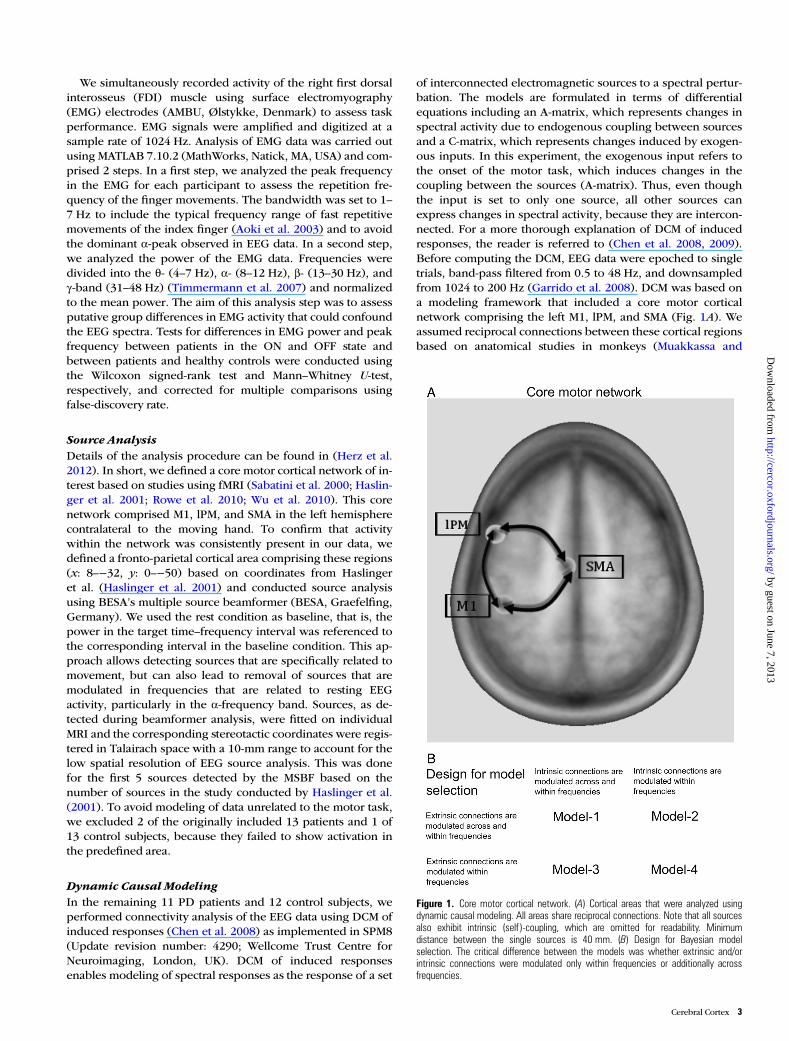
of interconnected electromagnetic sources to a spectral pertur-bation. The models are formulated in terms of differentialequations including an A-matrix, which represents changes inspectral activity due to endogenous coupling between sourcesand a C-matrix, which represents changes induced by exogen-ous inputs. In this experiment, the exogenous input refers tothe onset of the motor task, which induces changes in thecoupling between the sources (A-matrix). Thus, even thoughthe input is set to only one source, all other sources canexpress changes in spectral activity, because they are intercon-nected. For a more thorough explanation of DCM of inducedresponses, the reader is referred to (Chen et al. 2008, 2009).Before computing the DCM, EEG data were epoched to singletrials, band-pass filtered from 0.5 to 48 Hz, and downsampledfrom 1024 to 200 Hz (Garrido et al. 2008). DCM was based ona modeling framework that included a core motor corticalnetwork comprising the left M1, lPM, and SMA (Fig. 1A). Weassumed reciprocal connections between these cortical regionsbased on anatomical studies in monkeys (Muakkassa and
Figure 1. Core motor cortical network. (A) Cortical areas that were analyzed usingdynamic causal modeling. All areas share reciprocal connections. Note that all sourcesalso exhibit intrinsic (self )-coupling, which are omitted for readability. Minimumdistance between the single sources is 40 mm. (B) Design for Bayesian modelselection. The critical difference between the models was whether extrinsic and/orintrinsic connections were modulated only within frequencies or additionally acrossfrequencies.
Cerebral Cortex 3
by guest on June 7, 2013http://cercor.oxfordjournals.org/
Dow
nloaded from

Strick 1979; Barbas and Pandya 1987; Fang et al. 2005). Weused the identical coordinates as in our previous study (Herzet al. 2012) based on an fMRI study by Haslinger et al. (2001).The input induced by the experimental manipulation (onset ofthe motor task) was set to the lPM. Several considerationsprompted us to include only a single input at the onset of thecontinuous movement rather than defining several inputs foreach “submovement.” First, the repetitive movements rep-resent a continuous motor pattern that is generated and con-trolled as an entity (Gerloff et al. 1998; Siebner et al. 2001),which cannot be partitioned into distinct segments (Kennerleyet al. 2004). Second, we refrained from explicitly modeling dis-crete sensorimotor input generated by the movement itself,because the central motor control adopts a continuous “whole-field control” rather than monitoring particular aspects of eachsubmovement during repetitive movements with a high rep-etition rate (Siebner et al. 2001). And finally, a wide range ofprevious studies have used constant intervals of continuousmovements exceeding the duration of one “submovement” foranalyzing cortical connectivity patterns (Gross et al. 2002; Tim-mermann et al. 2003; Serrien et al. 2004; Pollok et al. 2005;Lalo et al. 2008). This approach was confirmed by inspectingthe predictions of time frequency plots, which revealed thatadequate predictions of spectral responses were not restrictedto the beginning of a trial but could be modeled for the wholetrial duration (see Results section).
Four models were compared using DCM (Fig. 1B). The criti-cal difference between the models was whether extrinsic con-nections and/or intrinsic connections were modulated onlywithin the same frequency or within and across frequencies.Extrinsic connections refer to connections between areas,while intrinsic connections refer to “self-connections” of eachsource (i.e., how a connection influences itself ). For each par-ticipant, the dimensionality of spectra was reduced to 4 fre-quency modes derived from a singular value decomposition ofthe spectra (Chen et al. 2009). The bandwidth for computingspectral densities was chosen from 4 to 40 Hz to account forθ-activity (4–7 Hz) that has been linked to large-scale inte-gration during cognitive and motor events (Canolty and Knight2010) and to avoid a potential 50-Hz electric current artifact.The time-window was set to −100 to 2000 ms with respect totask onset, which has been shown to be adequate in previousstudies to reduce the amount of data for computation (Chenet al. 2010, Herz et al. 2012). We did not include the restingcondition in the model, because in DCM resting data are purenoise that is best modeled by a flat line. Parameters of eachmodel and each participant were estimated by minimizing therelative entropy defined via the data and model outcome usingan expectation maximization algorithm (Chen et al. 2008). InDCM of induced responses, DCM does not model data featuresin sensor space. Instead, after inversion of the electromagneticmodel, the power of the neural source is modeled (Litvak et al.2011b). As the skull acts as a low-pass filter on intracerebralactivity, power spectra in EEG recordings are dominated byactivity in low-frequency bands (Schaul 1998). Thus, modeloptimization will more strongly rely on low-frequency power,because most variance of the data is explained by the low-frequency components of the frequency modes. This generallyapplies to DCM of neurophysiological data, but is less pro-nounced when using MEG or intracerebral recordings com-pared with EEG measurements. The different models werethen compared with regards to their accuracy in explaining the
data taking into account complexity of the model (Penny et al.2004). Here, we compared the different models using Bayesianmodel selection for random effects (Stephan et al. 2009). Themodel with the highest posterior exceedance probability, thatis, the model with the highest relative probability comparedwith any other model considered, was used to make inferenceon coupling parameters. The prior odds ratio assumed that allmodels were equally likely. We opted for comparing only a fewmodels and focusing on changes in oscillatory couplingbetween left M1, lPM, and SMA, because the main goal of thisstudy was to assess changes in task-dependent modulationwithin a core motor network in PD.
ANOVAwas used to test the significance of oscillatory coup-ling within the most likely model. To test whether changes ineffective connectivity could be confounded by power changesof the cortical sources, we compared time–frequency spectrabetween groups using ANOVA. We report significant couplingat a statistical threshold of P < 0.05 familywise error (FWE)-corrected at the cluster level. In an exploratory analysis, we ex-tracted for each participant individual coupling values fromconnections that were significantly modulated during the task.Coupling values were averaged over the respective significantfrequencies (e.g., γ-γ coupling from lPM to SMA). Positivevalues indicate that a source region exerts a positive influenceon a target region (i.e., increases power in the target region)during the task, while negative values indicate a negative influ-ence (i.e., a decrease in power). We then calculated the Spear-man rank correlation coefficient (2-tailed) to test whetherindividual differences in coupling showed a linear relationshipwith differences in motor impairment (UPDRS-III scores), ap-plying Bonferroni-correction for multiple comparisons. Alldata are given as mean ± standard deviation, if not specifiedotherwise.
Results
After a short training session of 5 min, all subjects were able toperform the repetitive extension-flexion task properly withtheir right index finger without showing signs of fatiguethroughout the experiment. Two patients showed a predomi-nantly left-sided resting tremor, which was present OFF andON medication. No tremor of the right hand was observedduring task performance. Application of levodopa alleviatedmotor symptoms in all patients as reflected by a consistent de-crease in the UPDRS score (OFF state: 28.18 ± 12.9 vs. ONstate: 17.91 ± 11.41; P≤ 0.001 paired samples t-test).
EMG Recordings During the RepetitiveExtension-Flexion TaskPatients OFF medication performed the task significantlyslower than the control group with a mean repetition fre-quency of 3.47 Hz ± 0.96 compared with 4.7 Hz ± 1.12(P < 0.05). There was a significant improvement in repetitionfrequency after levodopa application (ON state: 4.56 Hz ± 1.26vs. OFF state: 3.47 Hz ± 0.96; P < 0.05) with an increase in rep-etition frequency in 9 of 11 patients (Fig. 2). No significantdifference in repetition frequency was found when comparingPD patients ON medication and healthy controls.
There were no significant differences between groups(PD-OFF, PD-ON, Control group) when comparing EMGpower for the θ-, α-, β-, and γ-bands.
4 Oscillatory Coupling in Parkinson’s Disease • Herz et al.
by guest on June 7, 2013http://cercor.oxfordjournals.org/
Dow
nloaded from

Bayesian Model Selection andModel FitBayesian model selection for random effects strongly favored amodel postulating cross- and within-frequency coupling inboth extrinsic and intrinsic (self-) connections (model 1) in allgroups (Fig. 3). Exceedance probability was almost 1 in thecontrol group and patients ON medication and ∼0.87 inpatients OFF medication, highly outranking all other models.We therefore based our statistical inferences on coupling par-ameters as revealed by model 1. Figure 4 shows that model 1satisfyingly predicted the observed spectral responses differ-ently for the 3 considered regions over the whole 2 s period.The model explained ∼95% of the original spectral variance(Control: 93.4% ± 4, mean ± SD; PD-OFF: 95.1% ± 2.7%;PD-ON: 96.3% ± 1.9). On the group level, there were no signifi-cant differences in time–frequency spectra between thehealthy controls, PD patients in the OFF state and PD patients
in the ON state in any of the regions, indicating that the ob-served differences in effective connectivity were not con-founded by differences in power.
Task-Induced Changes in Effective ConnectivityAn overview of connections showing significant couplingduring the task is given in Table 2.
In the control group, task-induced changes in effective con-nectivity occurred in connections from lPM to both SMA andM1 (Fig. 5 left). Increased task-related coupling from lPM toM1 was prominent in the γ-band (γ-γ coupling), while couplingfrom lPM to SMA was modulated in the γ- and β-band (Fig. 6left). No other neural connection within the tested DCMshowed a task-related modulation in oscillatory coupling inhealthy controls.
Figure 2. Movement frequency during the motor task. (A) Individual EMG data of the first dorsal interosseus muscle of a representative PD patient before (OFF) and afterapplication of levodopa (ON). Note that movement frequency increased by ∼1 Hz after application of levodopa. (B) Results of the group comparison of movement frequency. Errorbars indicate standard deviation. *P< 0.05; ns, not significant.
Figure 3. Results of Bayesian model selection for random effects. Model 1, which considers coupling both within and across frequencies in extrinsic and intrinsic(self )-connections, explains the data best in all groups.
Cerebral Cortex 5
by guest on June 7, 2013http://cercor.oxfordjournals.org/
Dow
nloaded from
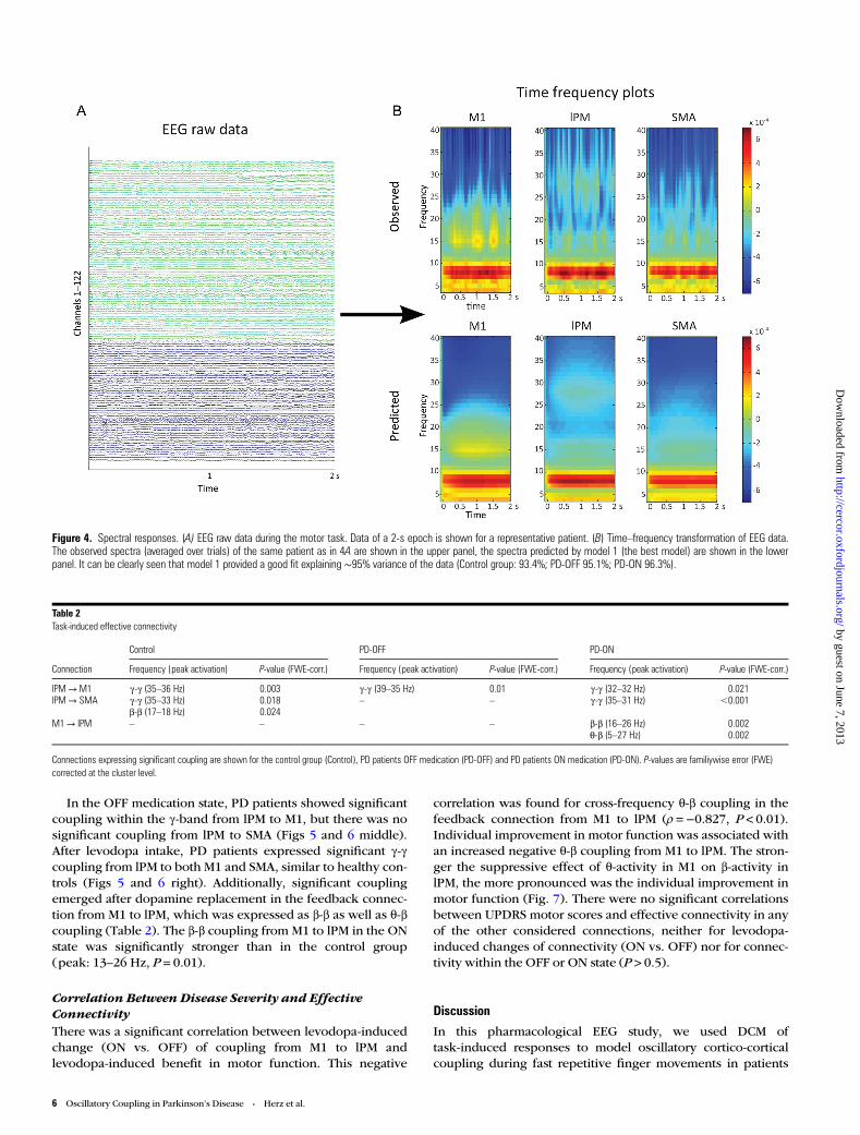
In the OFF medication state, PD patients showed significantcoupling within the γ-band from lPM to M1, but there was nosignificant coupling from lPM to SMA (Figs 5 and 6 middle).After levodopa intake, PD patients expressed significant γ-γcoupling from lPM to both M1 and SMA, similar to healthy con-trols (Figs 5 and 6 right). Additionally, significant couplingemerged after dopamine replacement in the feedback connec-tion from M1 to lPM, which was expressed as β-β as well as θ-βcoupling (Table 2). The β-β coupling from M1 to lPM in the ONstate was significantly stronger than in the control group(peak: 13–26 Hz, P = 0.01).
Correlation Between Disease Severity and EffectiveConnectivityThere was a significant correlation between levodopa-inducedchange (ON vs. OFF) of coupling from M1 to lPM andlevodopa-induced benefit in motor function. This negative
correlation was found for cross-frequency θ-β coupling in thefeedback connection from M1 to lPM (ρ =−0.827, P < 0.01).Individual improvement in motor function was associated withan increased negative θ-β coupling from M1 to lPM. The stron-ger the suppressive effect of θ-activity in M1 on β-activity inlPM, the more pronounced was the individual improvement inmotor function (Fig. 7). There were no significant correlationsbetween UPDRS motor scores and effective connectivity in anyof the other considered connections, neither for levodopa-induced changes of connectivity (ON vs. OFF) nor for connec-tivity within the OFF or ON state (P > 0.5).
Discussion
In this pharmacological EEG study, we used DCM oftask-induced responses to model oscillatory cortico-corticalcoupling during fast repetitive finger movements in patients
Figure 4. Spectral responses. (A) EEG raw data during the motor task. Data of a 2-s epoch is shown for a representative patient. (B) Time–frequency transformation of EEG data.The observed spectra (averaged over trials) of the same patient as in 4A are shown in the upper panel, the spectra predicted by model 1 (the best model) are shown in the lowerpanel. It can be clearly seen that model 1 provided a good fit explaining ∼95% variance of the data (Control group: 93.4%; PD-OFF 95.1%; PD-ON 96.3%).
Table 2Task-induced effective connectivity
Control PD-OFF PD-ON
Connection Frequency (peak activation) P-value (FWE-corr.) Frequency (peak activation) P-value (FWE-corr.) Frequency (peak activation) P-value (FWE-corr.)
lPM→M1 γ-γ (35–36 Hz) 0.003 γ-γ (39–35 Hz) 0.01 γ-γ (32–32 Hz) 0.021lPM→ SMA γ-γ (35–33 Hz) 0.018 – – γ-γ (35–31 Hz) <0.001
β-β (17–18 Hz) 0.024M1→ lPM – – – – β-β (16–26 Hz) 0.002
θ-β (5–27 Hz) 0.002
Connections expressing significant coupling are shown for the control group (Control), PD patients OFF medication (PD-OFF) and PD patients ON medication (PD-ON). P-values are familiywise error (FWE)corrected at the cluster level.
6 Oscillatory Coupling in Parkinson’s Disease • Herz et al.
by guest on June 7, 2013http://cercor.oxfordjournals.org/
Dow
nloaded from

with PD. Our study sheds significant new light on the impactof PD on motor-related cortical oscillatory coupling. Connec-tivity analysis revealed a functional disconnection of mesial
premotor cortex with reduced γ-γ coupling from lPM to SMA.This mesial to lateral coupling in the premotor cortex was re-stored by dopamine replacement along with strengthening β-βas well as θ-β coupling in the feedback connection from M1 tolPM. Critically, the beneficial effect of levodopa on motor func-tion was closely related to enhanced cross-frequency θ-β coup-ling from M1 to lPM.
Connectivity Pattern in Healthy Elderly ControlsIn healthy elderly individuals, fast repetitive finger movementsinduced a change in effective connectivity in cortico-corticalconnections from left lPM to M1 and SMA, presumably reflect-ing feed-forward computations that facilitate the regular andfast repetition of the finger movements. The connections fromlPM to M1 and SMAwere predominantly modulated within theγ-band, but also within lower frequency bands. The increase inγ coupling from lPM to SMA and from lPM to M1 during fast
Figure 5. Spatial distribution of task-induced effective connectivity. The frequencies inwhich the connections are modulated are listed in Table 2.
Figure 6. Frequency–frequency matrices. The matrices (4–40 Hz) of connections from lPM to M1 (first row) and from lPM to SMA (second row) are illustrated. The left columnshows matrices from the healthy control group, the middle column shows matrices from PD patients OFF medication, and the right column shows matrices from PD patients ONmedication.
Figure 7. Correlation analysis. (A) The feedback connection from M1 to lPM expressed significant coupling in PD patients ON but not OFF medication. (B) Frequency–frequencymatrix of the connection from M1 to lPM in the ON state showing significant β-β and θ-β couplings. C: Negative correlation between θ-β coupling from M1 to lPM and motorimpairment (ρ=−0.827, P<0.01). Negative coupling values indicate that θ-activity in M1 suppresses β-activity in lPM, while positive values indicate that θ-activity in M1enhances β-activity in lPM. The stronger β-activity in lPM was suppressed, the more pronounced was the improvement in motor function.
Cerebral Cortex 7
by guest on June 7, 2013http://cercor.oxfordjournals.org/
Dow
nloaded from

repetitive finger movements replicates the coupling patternthat we found in young healthy participants performing thesame task (Herz et al. 2012). Together, the findings show astable association between premotor-to-motor γ coupling andfast repetitive finger movements across a wide age range. Thisis in good accordance with previous studies, which consist-ently reported γ-band activity in participants performingphasic movements (Sheer et al. 1966; Pfurtscheller et al. 1997,2003; Miller et al. 2007). We defined coordinates of themodeled sources based on a study using fMRI (Haslinger et al.2001), which has a much higher spatial resolution than EEGstudies. As there is little localizing information in electromag-netic signals, slight changes of source localization would notvery much alter the data features (Chen et al. 2010). However,one has to bear in mind that EEG and fMRI recordings measuredifferent aspects of neural activation. Scheeringa et al. (2011)have demonstrated that the blood-oxygen-level-dependentsignal measured in fMRI studies correlates differently with dis-tinct frequency bands measured in electrophysiological record-ings. This incongruency between fMRI and EEG studies has tobe considered when interpreting the results of the current studywhere EEG analysis was informed by findings from fMRIstudies. Another critical point is whether the “correct” numberof sources was included in the model. Importantly, includingmore sources in the model would not affect the conclusionabout effective connectivity drawn in this study, because effec-tive connectivity can be polysynaptic and could therefore bemediated by unmodeled sources. The main concern would bethat choosing too few sources would leave variance in the datafeatures unexplained. This, however, is unlikely given the highproportion of variance explained by our DCM (on average 95%).
Abnormal Oscillatory Coupling Between PremotorandMotor Cortex in PDHealthy individuals and patients with PD shared motornetwork properties with respect to the spatial distribution ofoscillatory coupling. During fast repetitive finger movements,PD patients irrespective of medication state as well as healthycontrols showed γ-γ coupling from lPM to M1. However, DCManalysis revealed impaired cortico-cortical coupling as PDpatients in the OFF state lacked the physiological γ-γ couplingfrom lPM to SMA that was present in healthy participants. Inhealthy individuals, γ-activity is associated with the prep-aration and execution of phasic movements, whereas β-activityin the motor system is thought to be related to the maintenanceof a stable sensorimotor state (Engel and Fries 2010). In PDpatients, decreased activity in the γ-band and increased activityin the β-band during movement execution has been assignedan antikinetic effect (Brown and Marsden 1998; Schnitzler andGross 2005). Application of levodopa can restore γ-activity incortical areas, which may be induced through efferent connec-tions from the STN (Williams et al. 2002; Litvak et al. 2012).However, β-activity cannot be viewed as pathological per se.Isometric contractions are related to cortico-muscular (Saleniusand Hari 2003; Schoffelen et al. 2008) and cortico-corticalcoupling (Herz et al. 2012) in the β-band. A recent study ident-ified modulation of β-activity during encoding of incrementalforce generation (Florin et al. 2013). Thus, modulation ofneural activity in different frequency bands is state dependentand has to be interpreted with caution depending on thespecific task.
Our results extend previous work by showing that PDpatients are capable of expressing γ-activity in cortical pre-motor areas during phasic finger movements even in the OFFstate. However, γ-activity in the lPM is not efficiently coupledwith the mesial premotor cortex in the absence of levodopa.Critically, dopamine replacement reinstated a normal amountof γ-γ coupling from lPM to SMA. It has been shown that dis-tinct cortico-cortical and cortico-subcortical connectionsexpress their functional coupling at specific frequencies (Sil-berstein et al. 2005; Hirschmann et al. 2011; Litvak et al.2011a). Here, we show that in PD, the expression of alteredfrequency-specific coupling in cortico-cortical pathways isstate dependent and can be modulated by dopamine replace-ment. The patients in this study were tested in a “relative OFFstate” after 12 h withdrawal of dopaminergic medication. Thisapproach is common in neuroimaging studies of PD, but itdoes not allow studying a “real OFF state,” because long-termadaptation in neural networks has probably been induced bychronic treatment. Future electrophysiological studies in denovo PD patients need to assess the effect of dopamine repla-cement on “drug naïve” neural networks in PD.
Several neuroimaging studies have provided evidence forabnormal cortical activity and connectivity in PD. In the OFFstate, PD patients consistently showed hypoactivation of SMAboth at rest (Skidmore et al. 2013) and during movements (Sa-batini et al. 2000; Haslinger et al. 2001; Buhmann et al. 2003;Wu et al. 2010). Additionally, increased activation of lPM hasrepeatedly been reported in the OFF state (Sabatini et al. 2000;Haslinger et al. 2001; Rowe et al. 2010; Wu et al. 2010). In arecent fMRI study, DCM revealed that the selection of actionsenhanced coupling between prefrontal cortex (PFC) and therostral SMA in medicated PD patients (Rowe et al. 2010). Thesame PD patients showed increased coupling between PFCand lPM but not rostral SMA after medication withdrawal(Rowe et al. 2010). Although we did not model prefrontalsources in the current model and the fMRI study by Rowe et al.used a more cognitively demanding action selection task, theswitch from a more lateral information flow (via a PFC-lPMconnection) in the OFF state to a more latero-medial infor-mation flow (via a PFC-rostral SMA connection) in the ON statewas similar to the finding that γ-γ coupling from lPM to SMAwas only observed in the ON state. Extending the finding byRowe et al. (2010), we show that dysfunctional cortico-corticalcommunication is expressed as aberrant oscillatory coupling inPD, comprising a functional lateral to medial disconnection inthe premotor cortex. The alterations in coupling patterns arelikely to be task dependent. Here, we chose a highly auto-mated motor task, which required the fast release of sequentialagonist-antagonist movements (i.e., fast self-paced extension-flexion movements). This task was not demanding in terms ofcognitive control of actions, but was highly challenging interms of motor execution. This enabled us to focus our analysison a core premotor-motor network. Future studies need toexamine to what extent PFC can modulate the altered com-munication between premotor and motor areas, for instance,by using motor tasks requiring a higher amount of attention.
It is important to note that we cannot draw conclusion aboutthe causality between absence of γ-γ coupling from lPM toSMA and impaired motor function. As PD patients OFF medi-cation performed the motor task more slowly than healthy par-ticipants and PD patients ON medication, the observed changein effective connectivity might be due to differences in
8 Oscillatory Coupling in Parkinson’s Disease • Herz et al.
by guest on June 7, 2013http://cercor.oxfordjournals.org/
Dow
nloaded from
performance. This question can be addressed by applyingmotor tasks that are more rigorously controlled for equal taskperformance, even though withdrawal of medication in PDpatients will always lead to at least subtle changes in motorperformance. Another shortcoming of this study is that PDpatients always performed the motor task in the OFF state fol-lowed by a second recording in the ON state, while healthysubjects only performed the task once. We chose a simplemotor task that is highly overlearned and automatic andadditionally included a 5-min training session before record-ings to make sure that all participants were able to perform thetask adequately. This makes learning effects underlying theobserved differences in effective connectivity highly unlikely.However, we cannot discount possible unspecific effects oftask order.
While dopaminergic medication reinstated physiological γ-γcoupling from lPM to SMA, it did not normalize the task-relatedcortical coupling pattern. Healthy subjects expressed both γ-γand β-β coupling from lPM to M1, but PD patients ON medi-cation only showed significant γ-γ coupling from lPM to M1during the task. This is in line with previous studies showingthat cortical activity is modified but not normalized by levodo-pa intake (Haslinger et al. 2001, Palmer et al. 2010, Tropiniet al. 2011). This is particularly interesting, because there wasno difference in task performance between PD patients ONmedication and healthy controls in the current study. Thesefindings suggest that clinical improvement induced by dopa-mine replacement is not mediated by restoration of physiologi-cal neural networks, but that improved motor function mightbe related to abnormal neural activation patterns. This notionis further supported by the finding that feedback couplingfrom M1 to lPM was only expressed in PD patients ON medi-cation, but not in healthy participants.
Enhanced Feedback Coupling from Primary MotorCortex to Lateral Premotor CortexIn the current study, levodopa increased within- and cross-frequency coupling from M1 to lPM, which was closely associ-ated with levodopa-induced improvement in motor function.This finding suggests that levodopa modulates feedback con-nections in PD patients performing fast repetitive finger move-ments.
Although the minimum distance between cortical sourceswas 4 cm to account for the low spatial resolution of EEG, theoscillatory activity recorded over M1 is likely to contain activityfrom adjacent parts of dorsal premotor and somatosensorycortex, which are located in close proximity to the primarymotor hand area (Geyer et al. 1996; Geyer et al. 2000; Sriniva-san et al. 2006). The increase in feedback connections from M1after application of levodopa might therefore also convey the“efference copy” signals of the executed movements, whichconstitute a central role in the control of movements (Sperry1950; Ghasia et al. 2008). The basal ganglia with the direct andindirect pathway provide a dual system for center (excitatory)-surround (inhibitory) mechanism to focus its effect on selectedcortical neurons (Mink 1996). It has been argued that thiscenter-surround mechanism is used to focus the output to aspecific group of muscles required for performing a specifictask (Mink 2003). This operation is made possible throughopening the sensory channel for the expected sensory feed-back afferents during movement. Thus, one of the important
functions of basal ganglia seems to be the gating of sensoryinput for motor control. Even though levodopa leads to an alle-viation of motor impairment in PD, it does not restore physio-logical motor function. Hence, PD patients might rely strongeron feedback and efferent copy signals than healthy individuals.Sensory feedback originating from receptors in skin, muscle,and joints plays a pivotal role during the execution of move-ments (Lemon and Porter 1976; Riddle and Baker 2005; Baker2007; Patino et al. 2008). In the current study, we found astrong inverse correlation between cross-frequency θ-β coup-ling from M1 to lPM and individual motor improvement afterlevodopa intake. The more β-activity in lPM was suppressed byθ-activity in M1, the stronger was the individual benefit inmotor function. Activity in the θ-band has been linked to arange of different cognitive functions, for example, workingmemory (Jensen and Tesche 2002) or spatial navigation(Caplan et al. 2001), which demand integration of informationfrom various brain regions. In the current study, activity in theθ-band also corresponded roughly to the repetition frequencyof the fast finger movements at ∼4 Hz. Interestingly, recentstudies have shown that θ-oscillations are involved in sensori-motor integration in humans (Caplan et al. 2003; Cruikshanket al. 2012). Thus, even though previous studies have focusedmainly on activity in higher frequency bands during move-ment, θ-oscillations might play a central role in the humanmotor system. Oscillations in the β-band, on the other hand,are thought to have an antikinetic effect during phasic move-ments (Brown and Marsden 1998). We therefore hypothesizethat negative feedback coupling between the θ- and β-band ob-served in this study might indicate improved integration of theafferent feedback with the efference copy signal leading to asuppression of antikinetic β-oscillations in the premotorcortex. An improved integration of sensory feedback mightthus constitute a dopamine-dependent compensatory mechan-ism in patients with PD.
Funding
This work was supported by the German Research Foundation[Clinical Research Group 219 to L.T.]; the Koeln FortuneProgram/Faculty of Medicine, University of Cologne [179/2007to D.M.H.]; the Danish Medical Research Council [FSS09-072163 to D.M.H.]; and Lundbeckfonden [Grant of Excel-lence “ContAct” R59 A5399 to H.R.S.]. Funding to pay theOpen Access publication charges for this article was providedby the German Research Foundation (Clinical Research Group219).
NotesConflict of Interest: None declared.
ReferencesAoki T, Francis PR, Kinoshita H. 2003. Differences in the abilities of
individual fingers during the performance of fast, repetitivetapping movements. Exp Brain Res. 152:270–280.
Baker SN. 2007. Oscillatory interactions between sensorimotor cortexand the periphery. Curr Opin Neurobiol. 17:649–655.
Barbas H, Pandya DN. 1987. Architecture and frontal cortical connec-tions of the premotor cortex (area 6) in the rhesus monkey. J CompNeurol. 256:211–228.
Cerebral Cortex 9
by guest on June 7, 2013http://cercor.oxfordjournals.org/
Dow
nloaded from
Brown P, Marsden CD. 1998. What do the basal ganglia do? Lancet.351:1801–1804.
Buhmann C, Glauche V, Sturenburg HJ, Oechsner M, Weiller C, BuchelC. 2003. Pharmacologically modulated fMRI—cortical responsive-ness to levodopa in drug-naive hemiparkinsonian patients. Brain.126:451–461.
Canolty RT, Knight RT. 2010. The functional role of cross-frequencycoupling. Trends Cogn Sci. 14:506–515.
Caplan JB, Madsen JR, Raghavachari S, Kahana MJ. 2001. Distinct pat-terns of brain oscillations underlie two basic parameters of humanmaze learning. J Neurophysiol. 86:368–380.
Caplan JB, Madsen JR, Schulze-Bonhage A, Aschenbrenner-Scheibe R,Newman EL, Kahana MJ. 2003. Human theta oscillations related tosensorimotor integration and spatial learning. J Neurosci.23:4726–4736.
Chen CC, Henson RN, Stephan KE, Kilner JM, Friston KJ. 2009.Forward and backward connections in the brain: a DCM study offunctional asymmetries. Neuroimage. 45:453–462.
Chen CC, Kiebel SJ, Friston KJ. 2008. Dynamic causal modelling ofinduced responses. Neuroimage. 41:1293–1312.
Chen CC, Kilner JM, Friston KJ, Kiebel SJ, Jolly RK, Ward NS. 2010.Nonlinear coupling in the human motor system. J Neurosci.30:8393–8399.
Cruikshank LC, Singhal A, Hueppelsheuser M, Caplan JB. 2012. Thetaoscillations reflect a putative neural mechanism for human sensori-motor integration. J Neurophysiol. 107:65–77.
Devos D, Labyt E, Derambure P, Bourriez JL, Cassim F, Reyns N, BlondS, Guieu JD, Destee A, Defebvre L. 2004. Subthalamic nucleusstimulation modulates motor cortex oscillatory activity in Parkin-son’s disease. Brain. 127:408–419.
Engel AK, Fries P. 2010. Beta-band oscillations—signalling the statusquo? Curr Opin Neurobiol. 20:156–165.
Fahn S, Elton RL, UPDRS Development Committee. 1987. Unified Par-kinson’s disease rating scale. MacMillan Healthcare Information. In:Fahn S, Marsden CD, Goldstein M, Calne DB, editors. Recent devel-opments in Parkinson’s disease. Vol. 2, pp. 153–163. Florham Park,NJ: Macmillan Health Care Information.
Fang PC, Stepniewska I, Kaas JH. 2005. Ipsilateral cortical connections ofmotor, premotor, frontal eye, and posterior parietal fields in a prosi-mian primate, Otolemur garnetti. J Comp Neurol. 490:305–333.
Florin E, Dafsari HS, Reck C, Barbe MT, Pauls KA, Maarouf M, Sturm V,Fink GR, Timmermann L. 2013. Modulation of local field potentialpower of the subthalamic nucleus during isometric force gener-ation in patients with Parkinson’s disease. Neuroscience.240:106–116.
Garrido MI, Friston KJ, Kiebel SJ, Stephan KE, Baldeweg T, Kilner JM.2008. The functional anatomy of the MMN: a DCM study of theroving paradigm. Neuroimage. 42:936–944.
Gerloff C, Uenishi N, Nagamine T, Kunieda T, Hallett M, Shibasaki H.1998. Cortical activation during fast repetitive finger movements inhumans: steady-state movement-related magnetic fields and their cor-tical generators. Electroencephalogr Clin Neurophysiol. 109:444–453.
Geyer S, Ledberg A, Schleicher A, Kinomura S, Schormann T, Burgel U,Klingberg T, Larsson J, Zilles K, Roland PE. 1996. Two differentareas within the primary motor cortex of man. Nature.382:805–807.
Geyer S, Matelli M, Luppino G, Zilles K. 2000. Functional neuroanat-omy of the primate isocortical motor system. Anat Embryol (Berl).202:443–474.
Ghasia FF, Meng H, Angelaki DE. 2008. Neural correlates of forwardand inverse models for eye movements: evidence from three-dimensional kinematics. J Neurosci. 28:5082–5087.
Gross J, Timmermann L, Kujala J, Dirks M, Schmitz F, Salmelin R,Schnitzler A. 2002. The neural basis of intermittent motor control inhumans. Proc Natl Acad Sci USA. 99:2299–2302.
Hammond C, Bergman H, Brown P. 2007. Pathological synchroniza-tion in Parkinson’s disease: networks, models and treatments.Trends Neurosci. 30:357–364.
Haslinger B, Erhard P, Kampfe N, Boecker H, Rummeny E, SchwaigerM, Conrad B, Ceballos-Baumann AO. 2001. Event-related functional
magnetic resonance imaging in Parkinson’s disease before andafter levodopa. Brain. 124:558–570.
Herz DM, Christensen MS, Reck C, Florin E, Barbe MT, Stahlhut C,Pauls KA, Tittgemeyer M, Siebner HR, Timmermann L. 2012. Task-specific modulation of effective connectivity during two simple un-imanual motor tasks: a 122-channel EEG study. Neuroimage.59:3187–3193.
Hirschmann J, Ozkurt TE, Butz M, Homburger M, Elben S, HartmannCJ, Vesper J, Wojtecki L, Schnitzler A. 2011. Distinct oscillatorySTN-cortical loops revealed by simultaneous MEG and local fieldpotential recordings in patients with Parkinson’s disease. Neuro-image. 55:1159–1168.
Ille N, Berg P, Scherg M. 2002. Artifact correction of the ongoing EEGusing spatial filters based on artifact and brain signal topographies.J Clin Neurophysiol. 19:113–124.
Jensen O, Tesche CD. 2002. Frontal theta activity in humans increaseswith memory load in a working memory task. Eur J Neurosci.15:1395–1399.
Kennerley SW, Sakai K, Rushworth MF. 2004. Organization of actionsequences and the role of the pre-SMA. J Neurophysiol. 91:978–993.
Kuhn AA, Kempf F, Brucke C, Gaynor Doyle L, Martinez-Torres I, Po-gosyan A, Trottenberg T, Kupsch A, Schneider GH, Hariz MI et al.2008. High-frequency stimulation of the subthalamic nucleus sup-presses oscillatory beta activity in patients with Parkinson’s diseasein parallel with improvement in motor performance. J Neurosci.28:6165–6173.
Lalo E, Thobois S, Sharott A, Polo G, Mertens P, Pogosyan A, Brown P.2008. Patterns of bidirectional communication between cortex andbasal ganglia during movement in patients with Parkinson disease.J Neurosci. 28:3008–3016.
Lang AE, Lozano AM. 1998b. Parkinson’s disease. First of two parts. NEngl J Med. 339:1044–1053.
Lang AE, Lozano AM. 1998a. Parkinson’s disease. Second of two parts.N Engl J Med. 339:1130–1143.
Lau HC, Rogers RD, Ramnani N, Passingham RE. 2004. Willed actionand attention to the selection of action. Neuroimage.21:1407–1415.
Lemon RN, Porter R. 1976. Afferent input to movement-related precen-tral neurones in conscious monkeys. Proc R Soc Lond B Biol Sci.194:313–339.
Litvak V, Eusebio A, Jha A, Oostenveld R, Barnes G, Foltynie T, Limou-sin P, Zrinzo L, Hariz MI, Friston K et al. 2012. Movement-relatedchanges in local and long-range synchronization in Parkinson’sdisease revealed by simultaneous magnetoencephalography andintracranial recordings. J Neurosci. 32:10541–10553.
Litvak V, Jha A, Eusebio A, Oostenveld R, Foltynie T, Limousin P,Zrinzo L, Hariz MI, Friston K, Brown P. 2011a. Resting oscillatorycortico-subthalamic connectivity in patients with Parkinson’sdisease. Brain. 134:359–374.
Litvak V, Mattout J, Kiebel S, Phillips C, Henson R, Kilner J, Barnes G,Oostenveld R, Daunizeau J, Flandin G et al. 2011b. EEG and MEGdata analysis in SPM8. Comput Intell Neurosci. 2011:852961.
Marsden JF, Limousin-Dowsey P, Ashby P, Pollak P, Brown P. 2001.Subthalamic nucleus, sensorimotor cortex and muscle interrelation-ships in Parkinson’s disease. Brain. 124:378–388.
Miller KJ, Leuthardt EC, Schalk G, Rao RP, Anderson NR, Moran DW,Miller JW, Ojemann JG. 2007. Spectral changes in cortical surfacepotentials during motor movement. J Neurosci. 27:2424–2432.
Mink JW. 1996. The basal ganglia: focused selection and inhibition ofcompeting motor programs. Prog Neurobiol. 50:381–425.
Mink JW. 2003. The Basal Ganglia and involuntary movements: im-paired inhibition of competing motor patterns. Arch Neurol.60:1365–1368.
Muakkassa KF, Strick PL. 1979. Frontal lobe inputs to primate motorcortex: evidence for four somatotopically organized ‘premotor’areas. Brain Res. 177:176–182.
Palmer SJ, Lee PW, Wang ZJ, Au WL, Mckeown MJ. 2010. theta, betaBut not alpha-band EEG connectivity has implications for dual taskperformance in Parkinson’s disease. Parkinsonism Relat Disord.16:393–397.
10 Oscillatory Coupling in Parkinson’s Disease • Herz et al.
by guest on June 7, 2013http://cercor.oxfordjournals.org/
Dow
nloaded from

Patino L, Omlor W, Chakarov V, Hepp-Reymond MC, Kristeva R. 2008.Absence of gamma-range corticomuscular coherence duringdynamic force in a deafferented patient. J Neurophysiol.99:1906–1916.
Penny WD, Stephan KE, Mechelli A, Friston KJ. 2004. Comparingdynamic causal models. Neuroimage. 22:1157–1172.
Pfurtscheller G, Graimann B, Huggins JE, Levine SP, Schuh LA. 2003.Spatiotemporal patterns of beta desynchronization and gamma syn-chronization in corticographic data during self-paced movement.Clin Neurophysiol. 114:1226–1236.
Pfurtscheller G, Neuper C, Andrew C, Edlinger G. 1997. Foot and handarea mu rhythms. Int J Psychophysiol. 26:121–135.
Picard N, Strick PL. 2001. Imaging the premotor areas. Curr Opin Neu-robiol. 11:663–672.
Pollok B, Gross J, Muller K, Aschersleben G, Schnitzler A. 2005. Thecerebral oscillatory network associated with auditorily paced fingermovements. Neuroimage. 24:646–655.
Redgrave P, Rodriguez M, Smith Y, Rodriguez-Oroz MC, Lehericy S,Bergman H, Agid Y, Delong MR, Obeso JA. 2010. Goal-directedand habitual control in the basal ganglia: implications for Parkin-son’s disease. Nat Rev Neurosci. 11:760–772.
Riddle CN, Baker SN. 2005. Manipulation of peripheral neural feed-back loops alters human corticomuscular coherence. J Physiol.566:625–639.
Rowe J, Friston K, Frackowiak R, Passingham R. 2002a. Attention toaction: specific modulation of corticocortical interactions inhumans. Neuroimage. 17:988–998.
Rowe J, Stephan KE, Friston K, Frackowiak R, Lees A, Passingham R.2002b. Attention to action in Parkinson’s disease: impaired effec-tive connectivity among frontal cortical regions. Brain.125:276–289.
Rowe JB, Hughes LE, Barker RA, Owen AM. 2010. Dynamic causalmodelling of effective connectivity from fMRI: are results reprodu-cible and sensitive to Parkinson’s disease and its treatment? Neuro-image. 52:1015–1026.
Rowe JB, Siebner HR. 2012. The motor system and its disorders. Neu-roimage. 61:464–477.
Sabatini U, Boulanouar K, Fabre N, Martin F, Carel C, Colonnese C,Bozzao L, Berry I, Montastruc JL, Chollet F et al. 2000. Corticalmotor reorganization in akinetic patients with Parkinson’s disease:a functional MRI study. Brain. 123(Pt 2):394–403.
Salenius S, Hari R. 2003. Synchronous cortical oscillatory activityduring motor action. Curr Opin Neurobiol. 13:678–684.
Schaul N. 1998. The fundamental neural mechanisms of electroence-phalography. Electroencephalogr Clin Neurophysiol. 106:101–107.
Scheeringa R, Fries P, Petersson KM, Oostenveld R, Grothe I, NorrisDG, Hagoort P, Bastiaansen MC. 2011. Neuronal dynamics under-lying high- and low-frequency EEG oscillations contribute indepen-dently to the human BOLD signal. Neuron. 69:572–583.
Schnitzler A, Gross J. 2005. Normal and pathological oscillatory com-munication in the brain. Nat Rev Neurosci. 6:285–296.
Schoffelen JM, Oostenveld R, Fries P. 2008. Imaging the human motorsystem’s beta-band synchronization during isometric contraction.Neuroimage. 41:437–447.
Serrien DJ, Pogosyan AH, Brown P. 2004. Cortico-cortical coupling pat-terns during dual task performance. Exp Brain Res. 157:79–84.
Sheer DE, Grandstaff NW, Benignus VA. 1966. Behavior and 40-c-sec.electrical activity in the brain. Psychol Rep. 19:1333–1334.
Siebner HR, Limmer C, Peinemann A, Bartenstein P, Drzezga A,Conrad B. 2001. Brain correlates of fast and slow handwriting inhumans: a PET-performance correlation analysis. Eur J Neurosci.75:249–261.
Silberstein P, Pogosyan A, Kuhn AA, Hotton G, Tisch S, Kupsch A,Dowsey-Limousin P, Hariz MI, Brown P. 2005. Cortico-corticalcoupling in Parkinson’s disease and its modulation by therapy.Brain. 128:1277–1291.
Skidmore FM, Yang M, Baxter L, Von Deneen KM, Collingwood J, HeG, White K, Korenkevych D, Savenkov A, Heilman KM et al. 2013.Reliability analysis of the resting state can sensitively and specifi-cally identify the presence of Parkinson disease. Neuroimage.240:106–116.
Sperry RW. 1950. Neural basis of the spontaneous optokineticresponse produced by visual inversion. J Comp Physiol Psychol.43:482–489.
Srinivasan R, Winter WR, Nunez PL. 2006. Source analysis of EEG oscil-lations using high-resolution EEG and MEG. Prog Brain Res.159:29–42.
Stephan KE, Penny WD, Daunizeau J, Moran RJ, Friston KJ. 2009.Bayesian model selection for group studies. Neuroimage.46:1004–1017.
Stoessl AJ, Martin WW, Mckeown MJ, Sossi V. 2011. Advances inimaging in Parkinson’s disease. Lancet Neurol. 10:987–1001.
Timmermann L, Fink GR. 2011. Pathological network activity in Par-kinson’s disease: from neural activity and connectivity to causality?Brain. 134:332–334.
Timmermann L, Florin E, Reck C. 2007. Pathological cerebral oscil-latory activity in Parkinson’s disease: a critical review on methods,data and hypotheses. Expert Rev Med Devices. 4:651–661.
Timmermann L, Gross J, Dirks M, Volkmann J, Freund HJ, Schnitzler A.2003. The cerebral oscillatory network of parkinsonian restingtremor. Brain. 126:199–212.
Tomlinson CL, Stowe R, Patel S, Rick C, Gray R, Clarke CE. 2010. Sys-tematic review of levodopa dose equivalency reporting in Parkin-son’s disease. Mov Disord. 25:2649–2653.
Tropini G, Chiang J, Wang ZJ, Ty E, Mckeown MJ. 2011. Altered direc-tional connectivity in Parkinson’s disease during performance of avisually guided task. Neuroimage. 56:2144–2156.
Williams D, Tijssen M, Van Bruggen G, Bosch A, Insola A, Di LazzaroV, Mazzone P, Oliviero A, Quartarone A, Speelman H et al. 2002.Dopamine-dependent changes in the functional connectivitybetween basal ganglia and cerebral cortex in humans. Brain.125:1558–1569.
Wu T, Wang L, Hallett M, Chen Y, Li K, Chan P. 2011. Effective connec-tivity of brain networks during self-initiated movement in Parkin-son’s disease. Neuroimage. 55:204–215.
Wu T, Wang L, Hallett M, Li K, Chan P. 2010. Neural correlates of bi-manual anti-phase and in-phase movements in Parkinson’s disease.Brain. 133:2394–2409.
Cerebral Cortex 11
by guest on June 7, 2013http://cercor.oxfordjournals.org/
Dow
nloaded from