Distribution of Particulate Matter and Tissue Remodeling in the Human Lung
7
Environmental Health Perspectives • VOLUME 108 | NUMBER 11 | November 2000 1063 Distribution of Particulate Matter and Tissue Remodeling in the Human Lung Kent E. Pinkerton, 1 Francis H.Y. Green, 2 Cathy Saiki, 1 Val Vallyathan, 3 Charles G. Plopper, 1 Venu Gopal, 4 Daniel Hung, 1 Emily B. Bahne, 1 Susan S. Lin, 1 Margaret G. Ménache, 5 and Marc B. Schenker 1 1 Department of Anatomy, Physiology, and Cell Biology, School of Veterinary Medicine and Department of Epidemiology and Preventive Medicine, School of Medicine, University of California, Davis, California, USA; 2 Department of Pathology and Laboratory Medicine, Faculty of Medicine, University of Calgary, Calgary, Alberta, Canada; 3 National Institute for Occupational Safety and Health, Morgantown, West Virginia, USA; 4 Fresno Medical Coroner’s Office, Fresno, California, USA; 5 Department of Pediatrics, School of Medicine, University of New Mexico, Albuquerque, New Mexico, USA Exposure to airborne particles is a common event. Most particles that are respired are readily removed by mucociliary clearance aided by macrophage phagocytosis (1). A smaller fraction is retained within lung tis- sues or redistributed to regional lymph nodes (2). The deposition and clearance of particles within the respiratory system occurs in an inhomogeneous manner. The fate of particles is not well established, and there is little information on the distribution and retention of particles under conditions of ambient exposure. One purpose of this study was to design and implement an approach that would allow the assessment of particle retention as well as histologic analy- sis of response at different levels of the lung. This paper describes the methodology for preparing tissue and sampling airways and gas-exchange regions along precisely defined airway paths. The environment of the central valley of California places individuals at increased risk of exposure to particles. This region encom- passes a rich farming area as well as extensive urban development. The interface between rural and urban environments creates a unique setting for exposure to both natural and anthropogenic sources of airborne particulate matter. The predominantly dry farming techniques of the central valley result in high levels of airborne dust from oper- ations such as field preparation and harvest- ing of row crops and tree fruits ( 3,4 ). Combustion exhaust particles may also arise from equipment used in agricultural operations. Urban sprawl also generates an abundance of combustion and secondary photochemical gases and airborne particles. We have had the opportunity over the past 5 years to examine lung specimens from deceased young Hispanic males through the Fresno County Coroner’s Office. Approximately 50% of these individ- uals are known to have been farmworkers; the remaining individuals were employed in nonfarming operations. These individuals were healthy and considered to be free of pulmonary disease, having died from nonres- piratory causes. Our objective was to examine the rela- tionship between retained carbonaceous and mineral dust in the lungs and the remodel- ing of the small airways along the same air- way paths of each individual. In this paper, we report evidence that both carbonaceous and mineral dust are primarily distributed to the terminal and respiratory bronchioles and that there is anatomical remodeling within these same sites. Methods Population. Left lungs from 42 autopsies of Hispanic males were collected at the Fresno County (California) Coroner’s Office from June 1994 to June 1995. Demographic infor- mation, limited smoking, and occupational histories were obtained from the medical examiner and the coroner’s files. The study subjects ranged in age from 18 to 73 years and had died suddenly or unexpectedly. An autopsy was performed at the coroner’s office to determine the cause and manner of death as dictated by state statute. Complete autop- sies were performed within a period of 12–24 hr after death. The project was reviewed and approved by the Human Subjects Review Committee of the University of California, Davis (UC-Davis). Preparation. The left lung of each deceased individual was cannulated through the left mainstem bronchus and inflation- fixed with 2% glutaraldehyde at a hydrostatic pressure of 30 cm of water for a period of 2 hr from a constant-pressure gravity apparatus. The lungs were cut in the sagittal plane to include the mainstem bronchus, hilar struc- tures, and the medial aspect of both the upper and lower lobes of the left lung. Each speci- men was subsequently stored in fixative and shipped to UC-Davis. Upon arrival, we pho- tographed each lung from the cut sagittal sur- face as well as a medial view (Figure 1A,B). Address correspondence to K.E. Pinkerton, Department of Anatomy, Physiology, and Cell Biology, University of California, 1321 Haring Hall, Davis, CA 95616-5270 USA. Telephone: (530) 752-8334. Fax: (530) 752-7690. E-mail: [email protected] We thank the Fresno County Coroner’s Office staff and M. Brown, R. Beckman, M. Orenstein, and S. Grimes at the University of California-Davis for material coordination. We appreciate the dedi- cated assistance of the following individuals in the completion of this study: A. Karkhanis, M. Cudmore, M. Figueroa, A. Hernandez, J. Liao, A. Maskin, W. Nezami, J. Peake, T. Yang, S. Wheeler, and N. Willits. This study was supported by EPA Star grant R826246 and NIH grants ES05707 and RR00169, NIOSH U07/CCU906162, and the Alberta Lung Association. Received 1 February 2000; accepted 7 July 2000. Articles We examined the relationship between intrapulmonary particle distribution of carbonaceous and mineral dusts and remodeling of the airways along anatomically distinct airway paths in the lungs of Hispanic males from the central valley of California. Lung autopsy specimens from the Fresno County Coroner’s Office were prepared by intratracheal instillation of 2% glutaraldehyde at 30 cm H 2 O pressure. Two distinct airway paths into the apico-posterior and apico-anterior portions of the left upper lung lobe were followed. Tissue samples for histologic analysis were generally taken from the intrapulmonary second, fourth, sixth, and ninth airway generations. Parenchymal tissues beyond the 12th airway generation of each airway path were also analyzed. There was little evidence of visible particle accumulation in the larger conducting airways (generations 2–6), except in bronchial-associated lymphoid tissues and within peribronchial connective tissue. In contrast, terminal and respiratory bronchioles arising from each pathway revealed varying degrees of wall thickening and remodeling. Walls with marked thickening contained moderate to heavy amounts of carbonaceous and mineral dusts. Wall thickening was associated with increases in col- lagen and interstitial inflammatory cells, including dust-laden macrophages. These changes were significantly greater in first-generation respiratory bronchioles compared to second- and third- generation respiratory bronchioles. These findings suggest that accumulation of carbonaceous and mineral dust in the lungs is significantly affected by lung anatomy with the greatest retention in centers of lung acini. Furthermore, there is significant remodeling of this transitional zone in humans exposed to ambient particulate matter. Key words: asthma, California, fibrosis, lung pathology, PM 2.5 , PM 10 , particulate matter, pigmentation. Environ Health Perspect 108:1063–1069 (2000). [Online 23 October 2000] http://ehpnet1.niehs.nih.gov/docs/2000/108p1063-1069pinkerton/abstract.html
-
Upload
independent -
Category
Documents
-
view
1 -
download
0
Transcript of Distribution of Particulate Matter and Tissue Remodeling in the Human Lung

Environmental Health Perspectives • VOLUME 108 | NUMBER 11 | November 2000 1063
Distribution of Particulate Matter and Tissue Remodeling in the Human Lung
Kent E. Pinkerton,1 Francis H.Y. Green,2 Cathy Saiki,1 Val Vallyathan,3 Charles G. Plopper,1 Venu Gopal,4
Daniel Hung,1 Emily B. Bahne,1 Susan S. Lin,1 Margaret G. Ménache,5 and Marc B. Schenker1
1Department of Anatomy, Physiology, and Cell Biology, School of Veterinary Medicine and Department of Epidemiology and PreventiveMedicine, School of Medicine, University of California, Davis, California, USA; 2Department of Pathology and Laboratory Medicine,Faculty of Medicine, University of Calgary, Calgary, Alberta, Canada; 3National Institute for Occupational Safety and Health,Morgantown, West Virginia, USA; 4Fresno Medical Coroner’s Office, Fresno, California, USA; 5Department of Pediatrics, School ofMedicine, University of New Mexico, Albuquerque, New Mexico, USA
Exposure to airborne particles is a commonevent. Most particles that are respired arereadily removed by mucociliary clearanceaided by macrophage phagocytosis (1). Asmaller fraction is retained within lung tis-sues or redistributed to regional lymphnodes (2). The deposition and clearance ofparticles within the respiratory system occursin an inhomogeneous manner. The fate ofparticles is not well established, and there islittle information on the distribution andretention of particles under conditions ofambient exposure. One purpose of thisstudy was to design and implement anapproach that would allow the assessment ofparticle retention as well as histologic analy-sis of response at different levels of the lung.This paper describes the methodology forpreparing tissue and sampling airways andgas-exchange regions along precisely definedairway paths.
The environment of the central valley ofCalifornia places individuals at increased riskof exposure to particles. This region encom-passes a rich farming area as well as extensiveurban development. The interface betweenrural and urban environments creates aunique setting for exposure to both naturaland anthropogenic sources of airborne
particulate matter. The predominantly dryfarming techniques of the central valley resultin high levels of airborne dust from oper-ations such as field preparation and harvest-ing of row crops and tree fruits (3,4).Combustion exhaust particles may alsoarise from equipment used in agriculturaloperations. Urban sprawl also generates anabundance of combustion and secondaryphotochemical gases and airborne particles.
We have had the opportunity over thepast 5 years to examine lung specimensfrom deceased young Hispanic malesthrough the Fresno County Coroner’sOffice. Approximately 50% of these individ-uals are known to have been farmworkers;the remaining individuals were employed innonfarming operations. These individualswere healthy and considered to be free ofpulmonary disease, having died from nonres-piratory causes.
Our objective was to examine the rela-tionship between retained carbonaceous andmineral dust in the lungs and the remodel-ing of the small airways along the same air-way paths of each individual. In this paper,we report evidence that both carbonaceousand mineral dust are primarily distributedto the terminal and respiratory bronchioles
and that there is anatomical remodelingwithin these same sites.
Methods
Population. Left lungs from 42 autopsies ofHispanic males were collected at the FresnoCounty (California) Coroner’s Office fromJune 1994 to June 1995. Demographic infor-mation, limited smoking, and occupationalhistories were obtained from the medicalexaminer and the coroner’s files. The studysubjects ranged in age from 18 to 73 yearsand had died suddenly or unexpectedly. Anautopsy was performed at the coroner’s officeto determine the cause and manner of deathas dictated by state statute. Complete autop-sies were performed within a period of 12–24hr after death. The project was reviewed andapproved by the Human Subjects ReviewCommittee of the University of California,Davis (UC-Davis).
Preparation. The left lung of eachdeceased individual was cannulated throughthe left mainstem bronchus and inflation-fixed with 2% glutaraldehyde at a hydrostaticpressure of 30 cm of water for a period of 2 hrfrom a constant-pressure gravity apparatus.The lungs were cut in the sagittal plane toinclude the mainstem bronchus, hilar struc-tures, and the medial aspect of both the upperand lower lobes of the left lung. Each speci-men was subsequently stored in fixative andshipped to UC-Davis. Upon arrival, we pho-tographed each lung from the cut sagittal sur-face as well as a medial view (Figure 1A,B).
Address correspondence to K.E. Pinkerton,Department of Anatomy, Physiology, and CellBiology, University of California, 1321 HaringHall, Davis, CA 95616-5270 USA. Telephone:(530) 752-8334. Fax: (530) 752-7690. E-mail:[email protected]
We thank the Fresno County Coroner’s Officestaff and M. Brown, R. Beckman, M. Orenstein,and S. Grimes at the University of California-Davisfor material coordination. We appreciate the dedi-cated assistance of the following individuals in thecompletion of this study: A. Karkhanis, M.Cudmore, M. Figueroa, A. Hernandez, J. Liao, A.Maskin, W. Nezami, J. Peake, T. Yang, S. Wheeler,and N. Willits.
This study was supported by EPA Star grantR826246 and NIH grants ES05707 and RR00169,NIOSH U07/CCU906162, and the Alberta LungAssociation.
Received 1 February 2000; accepted 7 July 2000.
Articles
We examined the relationship between intrapulmonary particle distribution of carbonaceous andmineral dusts and remodeling of the airways along anatomically distinct airway paths in the lungsof Hispanic males from the central valley of California. Lung autopsy specimens from the FresnoCounty Coroner’s Office were prepared by intratracheal instillation of 2% glutaraldehyde at 30cm H2O pressure. Two distinct airway paths into the apico-posterior and apico-anterior portionsof the left upper lung lobe were followed. Tissue samples for histologic analysis were generallytaken from the intrapulmonary second, fourth, sixth, and ninth airway generations. Parenchymaltissues beyond the 12th airway generation of each airway path were also analyzed. There was littleevidence of visible particle accumulation in the larger conducting airways (generations 2–6),except in bronchial-associated lymphoid tissues and within peribronchial connective tissue. Incontrast, terminal and respiratory bronchioles arising from each pathway revealed varying degreesof wall thickening and remodeling. Walls with marked thickening contained moderate to heavyamounts of carbonaceous and mineral dusts. Wall thickening was associated with increases in col-lagen and interstitial inflammatory cells, including dust-laden macrophages. These changes weresignificantly greater in first-generation respiratory bronchioles compared to second- and third-generation respiratory bronchioles. These findings suggest that accumulation of carbonaceous andmineral dust in the lungs is significantly affected by lung anatomy with the greatest retention incenters of lung acini. Furthermore, there is significant remodeling of this transitional zone inhumans exposed to ambient particulate matter. Key words: asthma, California, fibrosis, lungpathology, PM2.5, PM10, particulate matter, pigmentation. Environ Health Perspect108:1063–1069 (2000). [Online 23 October 2000]http://ehpnet1.niehs.nih.gov/docs/2000/108p1063-1069pinkerton/abstract.html
We also examined each lung and documentedselected gross features on a standard form.These included pleural pigmentation andthickening, the appearance and size of the tra-cheobronchial lymph nodes, hemorrhage, andfibrosis. Emphysema was graded on grossphotographs of the whole lung by the methodof Thurlbeck (5). The airways were examinedfor mucous plugs or aspirated material withinthe lumen.
Airway microdissection. Beginning at theleft mainstem bronchus, the airways weremicrodissected using razor blades, scissors,and a dissecting microscope along two path-ways leading to the apico-posterior and apico-anterior portions of the left upper lobe(Figure 1C,D). Details of the dissectionapproach and the airway recordings and num-bering system have been previously described(6). We performed the microdissections in anidentical, systematic way for all 42 cases. Thedimensions and orientation (length, diameter,and branching angle) were measured in allgenerations along the two paths (Figure 2).The microdissection created complementaryhalves of each airway (Figure 1C). These
were examined with a dissecting microscopefor visible pigment distribution, and tissuesamples for histologic analysis were generallytaken from the second, fourth, sixth, andninth airway generations. We also sampledparenchymal tissues and associated terminaland respiratory bronchioles were also sam-pled beyond the 12th airway generation ofeach airway path that was microdissected.
Pathologic evaluation. Samples of airwaysof varying size and airway generation, togeth-er with samples of pulmonary parenchyma,were taken from representative areas of theupper and lower lobes for the overall evalua-tion of pathologic changes. Each tissue blockwas embedded in paraffin (Fischer Scientific,Fair Lawn, NJ), and 5-µm thick sectionswere cut using a rotary microtome andstained with hematoxylin and eosin. We alsoused sirius red and elastic trichrome stains toconfirm the presence of collagen and smoothmuscle within tissue sections.
We applied standard diagnostic criteriafor the recognition of pneumoconioticlesions (7–10). The following histologic fea-tures were evaluated using a semiquantitative
scale: macules, defined as collections of dust-laden macrophages in a size range of 0.1–0.6mm within the walls of respiratory bronchi-oles and adjacent alveoli; nodules, defined asfibrotic lesions up to 1 cm in size with round,irregular, or serpiginous borders and contain-ing dust-laden macrophages; and interstitialfibrosis, defined as diffuse or irregular fibrosisof alveolar septa and/or alveolar ducts.
We evaluated tracheobronchial lymphnodes for the presence of fibrosis and dust.Chronic bronchitis and asthma were evaluat-ed using standard pathologic criteria (7).Small airways disease, subdivided into miner-al dust-associated small airways disease (11)and smoking-related small airways disease(12,13), was identified, and exposure to ciga-rette smoke in the recent past was assessed onthe basis of accumulation of characteristicsmokers’ macrophages within the respiratorybronchioles and adjacent alveoli (14,15).
Structural remodeling of small airways.The microdissection technique and prelimi-nary analyses of tissue sections indicated thatsmall airways were the primary retention sitefor particles. For the semiquantitative
Articles • Pinkerton et al.
1064 VOLUME 108 | NUMBER 11 | November 2000 • Environmental Health Perspectives
Figure 1. Medial surface of the left human lung (A) and the cut, sagittal surfaceof the same lung (B). Several small apical blebs (arrow) were noted in thislung. Anatomically distinct airway paths beginning at the left mainstembronchus are followed to the apico-posterior and apico-anterior regions of theleft upper lobe (C). A magnified view of the microdissected airways (D). Scalebar in (A) = 5 cm.
Figure 2. Stick diagram of the airway branching pattern from the left upper lobeof a 45-year-old farm laborer (employed for 15 years) who had lived for 17 yearsin Fresno County. He was a nonsmoker. Beginning at the left mainstembronchus, airway paths leading to the apico-posterior and apico-anteriorregions of the lungs were followed by microdissection. Letters in circles indicate sites taken for embedment and histologic examination. A, second-generation airway (left mainstem bronchus); B, third-generation airway; C,fifth-generation airway; D,E,F, first-generation respiratory bronchioles beyondthe tenth-generation airway; G, hilar lymph node.
Apico-posteriorregion
Apico-anteriorregion
Trachea
To rightlung To lingula
Todiaphragmatic
lobe
1 cm
G
A B
C
F
DE
40°
30°

evaluation of small airways, six samples percase were taken from the left upper lobe toinclude the ventilatory zones of the airwaypaths that had been microdissected. Serialsections were stained with hematoxylin andeosin or elastic trichrome for connective tis-sue (collagen and elastic fibers), and smoothmuscle. The following airways were evaluat-ed: membranous (terminal) bronchioles,first-generation respiratory bronchioles, sec-ond-generation respiratory bronchioles, andthird-generation respiratory bronchioles. Ineach serial section, we graded all membra-nous bronchioles cut in a plane perpendicularto the longitudinal profile of the airway. Allorientations of respiratory bronchioles wereincluded; however, the generation of eachairway level was clearly identified based onposition from the terminal bronchiole and/orfirst-generation respiratory bronchiole. Theorientation (longitudinal, oblique, or cross-section) was recorded. We evaluated theairways for fibrosis, muscle hypertrophy,inflammation, visible and polarizable pig-ment, and intraluminal macrophages. Becausethe degree of epithelial loss from autolysis wasvariable, we did not attempt semiquantitativeanalysis of this feature. Each of the featureswas graded from 0 to 3, where 0 representedno evidence of that feature, and 1–3 repre-sented increasing grades of severity. The cate-gories of severity were based on preliminaryreview of the material to assess the range ofchange present. Typical examples of eachgrade were photographed for periodic review.Variability of the readings and interobserverreproducibility were established by havingthree readers (F.H.Y.G., K.E.P., and V.V.)each independently evaluate the same 30 his-tologic slides on two different occasions.
Statistical analysis. We analyzed 42 casesfor this study. To compare the degree (score)of histologic change within specific levels ofthe lungs, particularly for the first-, second-,and third-generation respiratory bronchioles,
we used the Friedman test. To increase thepower of this analysis, only cases with threeor more complete sets of respiratory bronchi-oles consisting of contiguous first-, second-,and third-generation bronchioles were used.Of the 42 cases, 28 cases met this criteria.We used linear regression analysis to comparescores of histologic features with scores of theabundance of particles (i.e., carbonaceoussubstances and crystalline dust). Spearmancorrelation coefficients were also used toexamine the relationship between particleburden and histologic change within eachregion of the lungs. A p-value < 0.05 wasconsidered to be statistically significant. Weevaluated interobserver variables for the grad-ing of respiratory bronchioles with the kappastatistic (16).
Results
The demographic profile of the study popu-lation is shown in Table 1. All cases wereHispanic males; 34% had lived in FresnoCounty up to 10 years, 37% for 11–20 years,20% for 21–30 years, and 9% had lived inthe region for more than 30 years. In general,subjects were young, with a median age of 33years (range 18–73 years). The majority ofsubjects worked in farming or had otherblue-collar occupations. Approximately halfof the subjects were current smokers at thetime of death (Table 1). Cause of death bythe International Classification of Diseases,Revision 9, Clinical Modification (ICD-9CM)is shown in Table 2. As these were coroner’scases, the majority of deaths were sudden orunexplained. Deaths due to vehicular acci-dents, cardiovascular disease, homicide, andsuicide predominated.
Table 3 shows the major pathologicabnormalities in the lungs of this popula-tion. On gross examination, with the excep-tion of hemorrhage, almost no lungs showedvisible evidence of disease. Only one case, a44-year-old smoker, showed grossly visibleemphysema in the form of several small api-cal subpleural blebs (Figure 1A,B). Therewas variable visible pigmentation, mostlyalong lymphatics in the subpleural interlobu-lar septa, but also in some cases in the centriacinar zones and along the bron-chopulmonary airways. Black pigment wasalso seen within tracheobronchial lymphnodes, which were variably enlarged.
A majority of normal-appearing lungs ongross examination displayed subtle but recog-nizable abnormalities at the microscopic level(Table 3). Approximately half of the subjectsshowed histologic evidence of smoking-relat-ed lung injury; i.e. chronic bronchitis andsmall airways disease. Smoking-related smallairway disease and mineral dust-associatedsmall airways diseases were seen in 45% and26% of cases, respectively. Pneumoconiosiswas observed (macules) in 4 subjects (10%),lymph node fibrosis associated with mineraldust in 16 (38%), and asthma in 14 (33%)of the 42 subjects (Table 3).
A typical microdissection pathway isshown schematically in Figure 2. The leftmainstem bronchus (second generation air-way) as well as the third, fifth, ninth, andtwelfth airway generations were sampled.The number of airway generations toparenchymal regions sampled ranged from 8to 15. Parenchymal tissues arising from theapico-posterior and apico-anterior regionswere examined for membranous and respira-tory bronchioles. Microscopic evaluation ofthe microdissected airways revealed in manyinstances that the epithelial lining layer ofthe airway had sloughed. In spite of this loss,the interstitial wall of the airway as well assubmucosal glands were preserved.
Carbonaceous pigment was localized inand around lymphatic vessels in the adventi-tial portions of the airways and within theattendant lymph nodes. However, only rarelywere carbonaceous materials or birefringentparticles identified immediately beneath theepithelial lining of the airways or within thebronchial wall, and in these circumstancesthe amounts were small (Figure 3A–C). Incontrast, at the level of the membranous andrespiratory bronchioles, the presence of car-bonaceous materials as well as birefrigent par-ticles was noted in the majority of cases(Figure 3D–F). These particles were particu-larly abundant and heavy in the adventitialwall of membranous bronchioles (Figure 4)and within the walls of first-generation respi-ratory bronchioles. The pigment and birefrin-gent particles in the walls of the respiratorybronchiole were primarily seen around the
Articles • Particulate matter and the human lung
Environmental Health Perspectives • VOLUME 108 | NUMBER 11 | November 2000 1065
Table 1. Demographic characteristics of popula-tion.
Characteristic Number or frequency
Population 42Smokers 19 (45%)Nonsmokers 23 (55%)Median age 33 (24–53)
(25th–75th quartiles)
Table 2. Cause of death.
Cause of death ICD-9CM code range Frequency
Heart and cardiovascular disease/natural causes E420–429 8 (19%)All vehicle accidents E800–E848 16 (38%)Accidental poisonings by psychotropic agents E854 3 (7%)Accidental drowning and submersion E910 1 (2%)Agricultural machine accident E919 1 (2%)Suicide and self-inflicted injury E950–E959 4 (10%)Homicide and injury inflicted by others E960–E969 6 (15%)Unknown cause — 3 (7%)
Table 3. Lung pathology in 42 residents of FresnoCounty, California.
Specific feature Prevalence
Mineral dust small airways 11 (26%)disease
Smoking-related small 19 (45%)airways disease
Pneumoconiosis (macules 4 (10%)and/or nodules)
Interstitial fibrosis 4 (10%)Lymph node fibrosis 16 (38%)Chronic bronchitis 21 (50%)Asthma 14 (33%)Emphysema 1 (2%)

attendant pulmonary artery, but also extend-ed into the peribronchiolar tissues and adja-cent alveolar septal walls. In first-generationrespiratory bronchioles, dust retention wasgreatest in the nonalveolarized portion of thewall (Figure 3D–F). Heavy particle distribu-tion was also observed within bronchiallymph nodes (Figure 3G). Here the dust waslocated within reticular cells lining the sinus-es. Fibrosis, associated with visible and bire-fringent particles, was observed in 38% of thecase population (Figure 4).
The distribution of particles and histo-logic features of the membranous and respi-ratory bronchioles were further analyzed inrespiratory bronchioles, in longitudinal pro-file, which could be identified as either first,second, or third generation (Figure 5). Of the42 original cases, 28 contained a minimumof 3 sets of respiratory bronchioles, each con-sisting of first-, second-, and third-generationsegments. Based on this sample power, ananalysis was done to score for changes withineach of these three generations of respiratorybronchioles from each case for the followinghistologic features: smooth muscle hypertro-phy, fibrosis, alveolar macrophage frequency,degree of inflammatory change within theinterstitial wall, amount of carbonaceous pig-ment within each respiratory bronchiole gen-eration, and the amount of birefringent dustparticles within each of these generations.Membranous bronchioles within the sametissue sections were evaluated for the samefeatures. For all histologic analysis and scor-ing, there was reasonable agreement betweenthe three observers, with interobserver vari-ability ranging from 64.0% to 73.0%, forcomplete agreement with kappa of0.42–0.57.
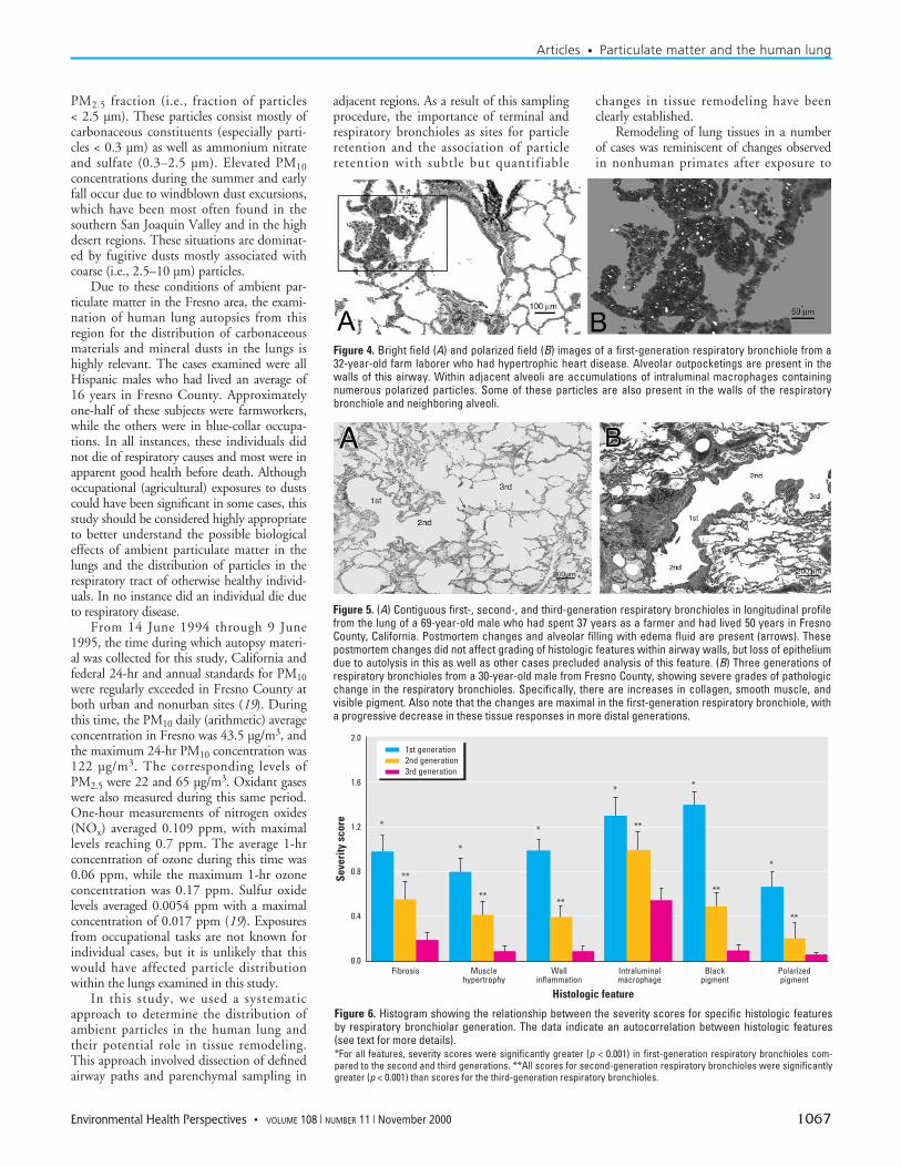
The results of the histologic and particleanalysis of respiratory bronchioles by order ofgeneration are given in Figure 6. There was ahighly significant difference (p < 0.001) in thedegree of histologic changes for all featuresexamined in first-generation respiratorybronchioles, compared to second-generationrespiratory bronchioles, as well as changes insecond-generation compared to third-genera-tion respiratory bronchioles (p < 0.001). Theamount of pigment in terminal bronchioleswas significantly correlated with the amountof pigment in respiratory bronchioles, partic-ularly for mineral dusts (linear regressionanalysis, R2 = 0.66, p < 0.05). Multiple vari-able linear regression analysis of the relation-ship between pigment and airway fibrosisrevealed that pigment was powerful predictorof the degree of fibrosis in the terminal bron-chioles and all three generations of respiratorybronchioles. Scatterplots of fibrosis versus pig-mentation scores for first-, second-, and third-generation respiratory bronchioles are shownin Figure 7.
DiscussionSituated in the heart of the San JoaquinValley, Fresno has some of the highest inhal-able particle concentrations (particulate mat-ter ≤ 10 µm; PM10) in the United States,often exceeding the National Ambient Air
Quality Standard of 150 µg/m3 averagedover 24 hr. The physicochemical characteris-tics and seasonal variability of PM at Fresnohave been described in detail by Chow et al.(17,18). During the winter months, the high-est PM10 concentrations have a dominant
Articles • Pinkerton et al.
1066 VOLUME 108 | NUMBER 11 | November 2000 • Environmental Health Perspectives
Figure 3. Light micrographs of the second- (A), third- (B), and fifth- (C) generation airways, first-genera-tion respiratory bronchioles (D,E,F), and bronchial lymph node (G) from the same lung. The location ofeach anatomical site is illustrated in Figure 2. Particles were not evident within the interstitial wall of thesecond-, third-, or fifth-generation airways. However, significant amounts of black pigment were notedwithin the first-generation respiratory bronchioles (arrows; D,E,F). Scoring for black pigment was 1 in (D),2 in (E), and 3 in (F) (see text).
PM2.5 fraction (i.e., fraction of particles< 2.5 µm). These particles consist mostly ofcarbonaceous constituents (especially parti-cles < 0.3 µm) as well as ammonium nitrateand sulfate (0.3–2.5 µm). Elevated PM10concentrations during the summer and earlyfall occur due to windblown dust excursions,which have been most often found in thesouthern San Joaquin Valley and in the highdesert regions. These situations are dominat-ed by fugitive dusts mostly associated withcoarse (i.e., 2.5–10 µm) particles.
Due to these conditions of ambient par-ticulate matter in the Fresno area, the exami-nation of human lung autopsies from thisregion for the distribution of carbonaceousmaterials and mineral dusts in the lungs ishighly relevant. The cases examined were allHispanic males who had lived an average of16 years in Fresno County. Approximatelyone-half of these subjects were farmworkers,while the others were in blue-collar occupa-tions. In all instances, these individuals didnot die of respiratory causes and most were inapparent good health before death. Althoughoccupational (agricultural) exposures to dustscould have been significant in some cases, thisstudy should be considered highly appropriateto better understand the possible biologicaleffects of ambient particulate matter in thelungs and the distribution of particles in therespiratory tract of otherwise healthy individ-uals. In no instance did an individual die dueto respiratory disease.
From 14 June 1994 through 9 June1995, the time during which autopsy materi-al was collected for this study, California andfederal 24-hr and annual standards for PM10were regularly exceeded in Fresno County atboth urban and nonurban sites (19). Duringthis time, the PM10 daily (arithmetic) averageconcentration in Fresno was 43.5 µg/m3, andthe maximum 24-hr PM10 concentration was122 µg/m3. The corresponding levels ofPM2.5 were 22 and 65 µg/m3. Oxidant gaseswere also measured during this same period.One-hour measurements of nitrogen oxides(NOx) averaged 0.109 ppm, with maximallevels reaching 0.7 ppm. The average 1-hrconcentration of ozone during this time was0.06 ppm, while the maximum 1-hr ozoneconcentration was 0.17 ppm. Sulfur oxidelevels averaged 0.0054 ppm with a maximalconcentration of 0.017 ppm (19). Exposuresfrom occupational tasks are not known forindividual cases, but it is unlikely that thiswould have affected particle distributionwithin the lungs examined in this study.
In this study, we used a systematicapproach to determine the distribution ofambient particles in the human lung andtheir potential role in tissue remodeling.This approach involved dissection of definedairway paths and parenchymal sampling in
adjacent regions. As a result of this samplingprocedure, the importance of terminal andrespiratory bronchioles as sites for particleretention and the association of particleretention with subtle but quantifiable
changes in tissue remodeling have beenclearly established.
Remodeling of lung tissues in a numberof cases was reminiscent of changes observedin nonhuman primates after exposure to
Articles • Particulate matter and the human lung
Environmental Health Perspectives • VOLUME 108 | NUMBER 11 | November 2000 1067
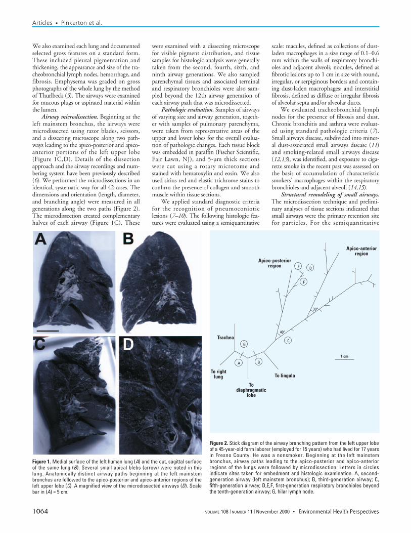
Figure 5. (A) Contiguous first-, second-, and third-generation respiratory bronchioles in longitudinal profilefrom the lung of a 69-year-old male who had spent 37 years as a farmer and had lived 50 years in FresnoCounty, California. Postmortem changes and alveolar filling with edema fluid are present (arrows). Thesepostmortem changes did not affect grading of histologic features within airway walls, but loss of epitheliumdue to autolysis in this as well as other cases precluded analysis of this feature. (B) Three generations ofrespiratory bronchioles from a 30-year-old male from Fresno County, showing severe grades of pathologicchange in the respiratory bronchioles. Specifically, there are increases in collagen, smooth muscle, andvisible pigment. Also note that the changes are maximal in the first-generation respiratory bronchiole, witha progressive decrease in these tissue responses in more distal generations.
Figure 4. Bright field (A) and polarized field (B) images of a first-generation respiratory bronchiole from a32-year-old farm laborer who had hypertrophic heart disease. Alveolar outpocketings are present in thewalls of this airway. Within adjacent alveoli are accumulations of intraluminal macrophages containingnumerous polarized particles. Some of these particles are also present in the walls of the respiratorybronchiole and neighboring alveoli.
Figure 6. Histogram showing the relationship between the severity scores for specific histologic featuresby respiratory bronchiolar generation. The data indicate an autocorrelation between histologic features(see text for more details). *For all features, severity scores were significantly greater (p < 0.001) in first-generation respiratory bronchioles com-pared to the second and third generations. **All scores for second-generation respiratory bronchioles were significantlygreater (p < 0.001) than scores for the third-generation respiratory bronchioles.
2.0
1.6
1.2
0.8
0.4
0.0
Seve
rity
sco
re
1st generation2nd generation3rd generation
*
*
*
* *
*
Fibrosis Musclehypertrophy
Wallinflammation
Intraluminalmacrophage
Blackpigment
Polarizedpigment
Histologic feature
**
****
**
**
**

ambient concentrations of ozone (20), theprimary oxidant gas of photochemical air pol-lution. Predominant pathological effects werealso confined to the epithelial and interstitialtissue compartments of the respiratory bron-chioles, forming the transitional zone betweenthe conducting airways and gas-exchangeregions of the lungs. The importance of thissite as a target for particle-induced injury iswell established in occupational settings (21).The centriacinar region is the primary site ofinjury in coal worker’s pneumoconiosis (8),asbestosis (9), and silica- and silicate-inducedinjury (10). Silicate pneumoconiosis has alsobeen found in limited numbers of farmwork-ers from the central valley of California (22).It has also been known for many years thatcigarette smoking induces injury in thisregion in young smokers and that respiratorybronchiolitis precedes the functionally moreimportant disease, centriacinar emphysema,often by several decades (14,15). However, itis not generally recognized that subtle lesionscan occur in the centriacinar zone of the lungin individuals exposed to ambient particles.Our results show a continuum of changes inthe respiratory bronchioles, being most severein individuals who smoke, but also present toa lesser degree in the population in general.These lesions have physiologic effects that canonly be detected by nonroutine tests for smallairway function (15). They may progress toclinically significant disease status (15).
Analytical electron microscopic studies byChurg and colleagues (23) have revealed thatthe concentration of particles in respiratorybronchioles can be 25–100 times greater thanthe concentration of particles in the mainstembronchus (23). This finding is in keepingwith our own observations. Churg et al. (23)also noted high particle concentrations at thebifurcations of airway generations 4 and 5.We did not observe large accumulations ofvisible dust at these sites, but this may be dueto the fact that our technique identified onlyparticles in tissue sections rather than particlenumber recovered by complete tissue diges-tion. Churg and Brauer (24) have also studiedthe size of particles in the pulmonary
parenchyma (which includes the respiratorybronchiolar region) and have shown that thegeometric mean particle diameter was 0.38µm. Conversion of the projected diameters toaerodynamic diameters revealed that 96% ofthe particles had aerodynamic diameters < 2.5µm. These data, taken in conjunction withour own observations, indicate that it is thefiner particles (< 2.5 µm diameter) that areresponsible for tissue remodeling.
Our data also show marked differencesbetween particle retention and remodelingin the first-, second-, and third-generationrespiratory bronchioles. The major site ofimpact and injury appeared to be the termi-nal bronchiole and adjacent first-generationrespiratory bronchioles, with progressivedecrease in both retention of particles andinjury in the second- and third-generationrespiratory bronchioles. This region of thelung is anatomically distinct, as the respira-tory bronchioles have some of the sameproperties of conducting airways as well asserving a gas-exchange function (25).Furthermore, this region is the point ofentry and exit to the ventilatory unit; gasesand airborne particles entering and leavingthe acinus have to pass through this con-stricted zone. Particles deposited within theacinus are also cleared through this portalwithin macrophages. Thus, the region isunusually vulnerable to the effects of gasesand particles impacting the airway wall andto the influence of macrophages and neu-trophils containing ingested particles.
Models of particle (aerosol) depositionhave demonstrated the importance of respi-ratory bronchiole and alveolar duct struc-tures in particle deposition (26–29). It is alsoa region with highly specialized cells, includ-ing alveolar epithelial cells (type I and II),ciliated cells, nonciliated bronchiolar epithe-lial (Clara) cells, neuroendocrine cells, andothers which may be particularly vulnerableto damage associated with particle retention.In this region, chaotic mixing may be theimportant mechanism of particle depositionand dispersion. The anatomical makeup ofthis region suggests that there is potential for
clearance overload with enhanced depositionand subsequent retention of particles in thiszone. This appears to be the mechanism fordevelopment of the macular lesions of coalworker’s pneumoconiosis (7).
The relative absence of retained particu-late matter in the larger conducting airwaysprobably reflects more rapid clearance of par-ticles deposited in these regions. Churg andcolleagues (30) have shown that particles tendto be deposited and retained at airway bifur-cations, and that these are also the sites inwhich cancers are more likely to develop. Thecentriacinar zone in the human is not general-ly recognized as being a primary site for thegenesis of lung cancers, although in recentdecades there has been a marked increase inperipheral adenocarcinomas (31), suggestingthat this region may need to be reevaluated asa potential site for the origin of lung cancers.This is also the region where lung cancersdevelop in rodents exposed to a variety ofnonfibrous, nongenotoxic particulates (32).
In summary, we have shown that a com-bination of microdissection, histology, andsemiquantitative evaluation of tissue changesis a valid approach for evaluating the effectsof airborne particles on the human lung.Furthermore, we show that the principalsites of deposition of ambient particles andassociated tissue remodeling are the terminalbronchioles and first-generation respiratorybronchioles. This transitional zone hasunique anatomical and physiological fea-tures. The significance of lesions at this siteis not currently known: however, extrapola-tion from studies of occupational groups andcigarette smokers would suggest that theremay be long-term health effects.
REFERENCES AND NOTES
1. Lippmann M, Yeates DB, Albert RE. Deposition, retentionand clearance of inhaled particles. Br J Ind Med37:337–362 (1980).
2. Oberdorster G. Lung clearance of inhaled insoluble andsoluble particles. J Aerosol Med 1:289–330 (1988).
3. Neiuwenhuijsen MJ, Kruize H, Shenker MB. Exposure todust and its particle size distribution in California agricul-ture. Am J Ind Hyg Assoc J 58:34–38 (1998).
4. Neiuwenhuijsen MJ, Shenker MB. Determinants of
Articles • Pinkerton et al.
1068 VOLUME 108 | NUMBER 11 | November 2000 • Environmental Health Perspectives
Figure 7. Scatterplot of regression analysis for fibrosis score to black pigment score of first- (A), second- (B), and third- (C) generation respiratory bronchioles. Apositive correlation is seen for all three airways, but it is greatest for first- and second-generation respiratory bronchioles.
3
2.5
2
1.5
1
.5
0
0 .5 1 1.5 2 2.5 3 0 .5 1 1.5 2 2.5 3 0 .5 1 1.5 2 2.5 3
3
2.5
2
1.5
1
.5
0
3
2.5
2
1.5
1
.5
0
A B C
Black pigment score Black pigment score Black pigment score
Fibr
osis
sco
re
Fibr
osis
sco
re
Fibr
osis
sco
re
r2 = 0.55 r2 = 0.53 r2 = 0.31
personal dust exposure during field crop operations inCalifornia agriculture. Am J Ind Hyg Assoc J 59:9–13 (1998).
5. Thurlbeck WM, Ryder RC, Sternby N. A comparativestudy of the severity of emphysema in necropsy popula-tions in three different countries. Am Rev Resp Dis109(2):239–248 (1974).
6. Plopper C. Structural methods for studying bronchiolarepithelial cells. In: Models of Lung Disease: Microscopyand Structural Methods (Gil J, ed). New York:MarcelDekker, Inc. 1990;537–559.
7. Green FHY, Churg A. Pathologic features of occupationallung disease. In: Pathology of Occupational LungDisease (Churg A, Green FHY, eds). 2nd ed. Baltimore,MD:Williams & Wilkins, 1998;209–234.
8. Kleinerman J, Green F, Harley RA, Lapp L, Laqueur L,Laqueur W, Naeye RL, Taylor G, Wiot J, Wyatt J.Pathology standards for coal worker’s pneumoconiosis.(A report of the Pneumoconiosis Committee of theCollege of American Pathologists.) Arch Pathol Lab Med101:375–431 (1979).
9. Craighead JE, Abraham JL, Churg A, Green FHY,Kleinerman J, Pratt PC, Seemayer TA, Vallyathan V,Weill H. The pathology of asbestos-associated diseasesof the lungs and pleural cavities: diagnostic criteria andproposed grading scheme. Arch Pathol Lab Med106(1):544–596 (1982).
10. Craighead JE, Kleinerman J, Abraham JL, Gibbs AR,Green FHY, Harley RA, Ruttner JR, Vallyathan V, JulianoEB. Diseases associated with exposure to silica andnon-fibrous silicate minerals. Arch Pathol Lab Med112:673–720 (1988).
11. Churg A. Small airways disease associated with mineraldust exposure. Semin Respir Med 13:140–148 (1992).
12. Cosio MG, Hale KA, Niewoehner DE. Morphologic andmorphometric effects of prolonged cigarette smoking onthe small airways. Am Rev Resp Dis 122(2):265–271 (1980).
13. Adesina AM, Vallyathan V, McQuillen EN, Weaver SO,Craighead JE. Bronchiolar inflammation and fibrosis asso-ciated with smoking. A morphologic cross-sectional popu-lation analysis. Am Rev Respir Dis 143(1):144–149 (1991).
14. Niewoehner DE, Kleinerman J, Rice DB. Pathologicchanges in the peripheral airways of young cigarettesmokers. N Engl J Med 291(15):755–758 (1974).
15. Wright JL, Cagle P, Churg A, Colby TV, Myers J. State ofthe art: diseases of the small airways. Am Rev Respir Dis146:240–262 (1992).
16. Fisher LD, van Belle G. Categorical data: contingencytables. In: Biostatistics: A Methodology for the HealthSciences. New York:John Wiley & Sons, Inc., 1993;256–259.
17. Chow J, Watson J, Lowenthal D, Solomon P, Magliano K,Ziman S, Richards L. PM10 source apportionment inCalifornia’s San Joaquin Valley. Atmos Environ26A:3335–3354 (1992).
18. Chow J, Watson J, Lowenthal D, Solomon P, Magliano K,Ziman S, Richards L. PM10 and PM2.5 compositions inCalifornia’s San Joaquin Valley. Aerosol Sci Tech18:105–128 (1993).
19. Alexis A, Gaffney P, Garcia C, Nystrom M, Rood R. The1999 California Almanac of Emissions and Air Quality.Sacramento, CA:California Environmental ProtectionAgency/Air Resources Board 1999; 271.
20. Harkema J, Plopper C, Hyde D, St. George J, Wilson D,Dungworth D. Response of macaque bronchiolar epithe-lium to ambient concentrations of ozone. Am J Pathol143:857–866 (1993).
21. Green FHY, Churg A. Occupational asthma, byssinosis,extrinsic allergic alveolitis and related conditions. In:Pathology of Occupational Lung Disease (Churg A,Green FHY, eds). 2nd ed. Baltimore, MD:Williams &Wilkins, 1998;403–450.
22. Sherwin RP, Barman ML, Abraham JL. Silicate pneumo-coniosis of farm workers. Lab Invest 40:576–582 (1979).
23. Churg A, Brauer M, Vedal S, Stevens B. Ambient mineralparticles in small airways of the normal human lung. JEnviron Med 1:39–45 (1999).
24. Churg A, Brauer M. Human lung parenchyma retainsPM2.5. Am J Respir Crit Care Med 155:2109–2111 (1997).
25. Plopper CG, Ten Have-Opbroek A. Anatomical and histo-logical classification of the bronchioles. In: Diseases ofBronchioles (Epler G, ed). New York:Raven Press, Ltd.1994;15–25.
26. Tsuda S, Iwasaki M, Yoshida M, Shirasu Y. Inhalationchamber with size discriminator for liquid aerosols.Fundam Appl Toxicol 4:378–387 (1984).
27. Federspiel WJ, Fredberg JJ. Axial dispersion in respira-tory bronchioles and alveolar ducts. J Appl Physiol 64(6):2614–2621 (1988).
28. Darquenne C, Paiva M. Two- and three-dimensional sim-ulations of aerosol transport and deposition in alveolarzone of human lung. J Appl Physiol 4:1401–1414 (1996).
29. Tsuda A, Henry FS, Butler JP. Chaotic mixing of alveolat-ed duct flow in rhythmically expanding pulmonary acinus.J Appl Physiol 79:1055–1063 (1995).
30. Churg A, Wright JL, Stevens B. Exogenous mineral parti-cles in the human bronchial mucosa and lung parenchy-ma. Nonsmokers in the general population. Exp Lung Res16:159–175 (1990).
31. Thun MJ, Lalley CA, Flannery JT, Callee EE, FlandersWD, Health CW Jr. Cigarette smoking and changes inthe histopathology of lung cancer. J Natl Cancer Inst89:1580–1586 (1997).
32. Hahn FF. Carcinogenic responses of the respiratorytract. In: Comprehensive Toxicology (Sipes IG, McQueenCA, Gandolfi AJ, eds). Toxicology of the RespiratorySystem, Vol 8 (Roth RA, ed). New York:Pergamon,1997;187–201.
Articles • Particulate matter and the human lung
Environmental Health Perspectives • VOLUME 108 | NUMBER 11 | November 2000 1069