Dissociating Neural Correlates of Cognitive Components in Mental Calculation
10
Mental calculation is a complex cognitive operation that is composed of a set of distinct functional processes. Using functional magnetic resonance imaging (fMRI), we mapped brain activity in healthy subjects performing arithmetical tasks and control tasks evoking a comparable load on visuo-constructive, linguistic, attentional and mnemonic functions. During calculation, as well as non-mathematical tasks, similar cortical networks consisting of bilateral prefrontal, premotor and parietal regions were activated, suggesting that most of these cortical areas do not exclusively represent modules for calculation but support more general cognitive operations that are instrumental but not specific to mental arithmetic. Significant differences between calculation and the non-mathematical tasks were found in parietal sub-regions, where non-arithmetic number or letter substitution tasks preferentially activated the superior parietal lobules whereas calculation predominantly elicited activation of the left dorsal angular gyrus and the medial parietal cortices. We interpret the latter activations to reflect sub-processes of mental calculation that are related to the processing of numerical representations during exact calculation and to arithmetical fact retrieval. Finally, we found that more complex calculation tasks involving the application of calculation rules increased activity in left inferior frontal areas that are known to subserve linguistic and working memory functions. Taken together, these findings help to embed the specific cognitive operation of calculation into a neural framework that provides the required set of instrumental components. This result may further inform the cognitive modeling of calculation and adds to the understanding of neuropsychological deficit patterns in patients. Introduction Since Henschen coined the term ‘acalculia’ in his classical overview of calculation impairments in brain-damaged patients (Henschen, 1919), there has been a large amount of work on both characterizing the deficits in performing calculations and on relating these to sites of lesions in neurological patients. While disturbances of calculation may occasionally be found in relative isolation from other cognitive impairments (Warrington, 1982), they are usually associated with deficits in wider functional contexts than the performance of arithmetical operations alone (Collignon et al., 1977). Neuropsychological assessment has guided researchers to define different types of acalculia. In particular, specific disturbances of arithmetical skills, called pure anarithmetia, were distinguished from other functional impairments (alexia, agraphia, visuospatial disorders and constructional apraxia) that affect calculation without a genuine deficit in number processing and calculation (Levin et al., 1993). In other words, linguistic and visuo-spatial malfunctions have inevitable repercussions on calculation, but are not specific to it. Mirroring the diversity of clinical manifestations, the conceptual models developed for calculation assume a composite multi-level process. In their abstract–modular model of number processing McCloskey et al. distinguish input mechanisms subserving the recognition of numbers and operation symbols from output mechanisms for writing or uttering numbers (McCloskey et al., 1985). In the case of written numerical material these mechanisms involve visuo-spatial and visuo-constructive computations and linguistic processes. To perform arithmetical tasks a third component is postulated that stores basic number facts and rules, as well as procedures for more complex arithmetical operations. A different cognit- ive model of number processing is the ‘triple code theory’ (Dehaene, 1992), which proposes two representational forms for input and output devoid of semantic information (visual arabic and auditory verbal code) and a third, analogical ‘magnitude’ or ‘quantity’ representation. While an asemantic route is sufficient for reading or writing arabic numerals, mental calculation requires access to numerical meaning via a semantic route. Both models share in common the notion that calculation relies on components of a more general nature that are also required for other cognitive functions. These include sensory feature input processing, visuo-constructive capacity, symbolic operational and particularly linguistic processing, as well as memory and attention. Parallelling the diversity of functions recruited for the execution of mental arithmetic, deficits in calculation have been associated with a variety of lesion locations in patients. From the early work of Henschen onwards, a key role has repeatedly, but not exclusively, been assigned to the left angular gyrus (Boller and Grafman, 1985). However, instead of pinpointing a specific area as a ‘calculation center’, an overview of lesion studies allows merely a vague conclusion, namely that left hemispheric lesions are more likely than right hemispheric lesions to affect calcu- lation and posterior damage more likely than anterior damage (Dahmen et al., 1982; Grafman et al., 1982). These findings suggest not only that several distinct and spatially segregated areas subserve calculation, but also that the neuronal processes performed in these areas support a broad set of cognitive oper- ations and are not confined to calculation. So far, only a few functional magnetic resonance imaging (fMRI) studies have addressed the neural correlates of mental arithmetic (Burbaud et al., 1995, 1999; Rueckert et al., 1996; Chochon et al., 1999; Dehaene et al., 1999; Rickard et al., 2000). Together, they as well as mostly earlier studies using positron emission tomography (Roland and Friberg, 1985; Dehaene et al., 1996; Sakurai et al., 1996; Pesenti et al., 2000) describe a distributed network that subserves the performance of calcu- lation tasks, including prefrontal, premotor and parietal cortices. However, the exact interpretation of the observed activations appears difficult for at least two reasons. Firstly, the experi- mental conditions and statistical comparisons in most of these studies non-selectively highlighted structures partaking in calculation as well as lower levels of numerical processing. Dissociating Neural Correlates of Cognitive Components in Mental Calculation O. Gruber 1,2 , P. Indefrey 3 , H. Steinmetz 4 and A. Kleinschmidt 4 1 Max-Planck-Institute of Cognitive Neuroscience, Leipzig, Germany, 2 Institute of Medicine, Research Center Jülich, Jülich, Germany, 3 Max-Planck-Institute for Psycholinguistics, Nijmegen, The Netherlands and 4 Department of Neurology, University of Frankfurt, Frankfurt, Germany Cerebral Cortex Apr 2001;11:350–359; 1047–3211/01/$4.00 © Oxford University Press 2001. All rights reserved.
-
Upload
uni-duesseldorf -
Category
Documents
-
view
2 -
download
0
Transcript of Dissociating Neural Correlates of Cognitive Components in Mental Calculation
Mental calculation is a complex cognitive operation that iscomposed of a set of distinct functional processes. Using functionalmagnetic resonance imaging (fMRI), we mapped brain activity inhealthy subjects performing arithmetical tasks and control tasksevoking a comparable load on visuo-constructive, linguistic,attentional and mnemonic functions. During calculation, as well asnon-mathematical tasks, similar cortical networks consisting ofbilateral prefrontal, premotor and parietal regions were activated,suggesting that most of these cortical areas do not exclusivelyrepresent modules for calculation but support more generalcognitive operations that are instrumental but not specific to mentalarithmetic. Significant differences between calculation and thenon-mathematical tasks were found in parietal sub-regions, wherenon-arithmetic number or letter substitution tasks preferentiallyactivated the superior parietal lobules whereas calculationpredominantly elicited activation of the left dorsal angular gyrus andthe medial parietal cortices. We interpret the latter activations toreflect sub-processes of mental calculation that are related to theprocessing of numerical representations during exact calculationand to arithmetical fact retrieval. Finally, we found that morecomplex calculation tasks involving the application of calculationrules increased activity in left inferior frontal areas that are known tosubserve linguistic and working memory functions. Taken together,these findings help to embed the specific cognitive operation ofcalculation into a neural framework that provides the required setof instrumental components. This result may further inform thecognitive modeling of calculation and adds to the understanding ofneuropsychological deficit patterns in patients.
IntroductionSince Henschen coined the term ‘acalculia’ in his classical
overview of calculation impairments in brain-damaged patients
(Henschen, 1919), there has been a large amount of work on
both characterizing the deficits in performing calculations and
on relating these to sites of lesions in neurological patients.
While disturbances of calculation may occasionally be found in
relative isolation from other cognitive impairments (Warrington,
1982), they are usually associated with deficits in wider
functional contexts than the performance of arithmetical
operations alone (Collignon et al., 1977). Neuropsychological
assessment has guided researchers to define different types of
acalculia. In particular, specific disturbances of arithmetical
skills, called pure anarithmetia, were distinguished from other
functional impairments (alexia, agraphia, visuospatial disorders
and constructional apraxia) that affect calculation without a
genuine deficit in number processing and calculation (Levin et
al., 1993). In other words, linguistic and visuo-spatial
malfunctions have inevitable repercussions on calculation, but
are not specific to it.
Mirroring the diversity of clinical manifestations, the
conceptual models developed for calculation assume a
composite multi-level process. In their abstract–modular model
of number processing McCloskey et al. distinguish input
mechanisms subserving the recognition of numbers and
operation symbols from output mechanisms for writing or
uttering numbers (McCloskey et al., 1985). In the case of written
numerical material these mechanisms involve visuo-spatial and
visuo-constructive computations and linguistic processes. To
perform arithmetical tasks a third component is postulated that
stores basic number facts and rules, as well as procedures
for more complex arithmetical operations. A different cognit-
ive model of number processing is the ‘triple code theory’
(Dehaene, 1992), which proposes two representational forms
for input and output devoid of semantic information (visual
arabic and auditory verbal code) and a third, analogical
‘magnitude’ or ‘quantity’ representation. While an asemantic
route is sufficient for reading or writing arabic numerals, mental
calculation requires access to numerical meaning via a semantic
route. Both models share in common the notion that calculation
relies on components of a more general nature that are also
required for other cognitive functions. These include sensory
feature input processing, visuo-constructive capacity, symbolic
operational and particularly linguistic processing, as well as
memory and attention.
Parallelling the diversity of functions recruited for the
execution of mental arithmetic, deficits in calculation have been
associated with a variety of lesion locations in patients. From the
early work of Henschen onwards, a key role has repeatedly, but
not exclusively, been assigned to the left angular gyrus (Boller
and Grafman, 1985). However, instead of pinpointing a specific
area as a ‘calculation center’, an overview of lesion studies allows
merely a vague conclusion, namely that left hemispheric lesions
are more likely than right hemispheric lesions to affect calcu-
lation and posterior damage more likely than anterior damage
(Dahmen et al., 1982; Grafman et al., 1982). These findings
suggest not only that several distinct and spatially segregated
areas subserve calculation, but also that the neuronal processes
performed in these areas support a broad set of cognitive oper-
ations and are not confined to calculation.
So far, only a few functional magnetic resonance imaging
(fMRI) studies have addressed the neural correlates of mental
arithmetic (Burbaud et al., 1995, 1999; Rueckert et al., 1996;
Chochon et al., 1999; Dehaene et al., 1999; Rickard et al., 2000).
Together, they as well as mostly earlier studies using positron
emission tomography (Roland and Friberg, 1985; Dehaene et
al., 1996; Sakurai et al., 1996; Pesenti et al., 2000) describe a
distributed network that subserves the performance of calcu-
lation tasks, including prefrontal, premotor and parietal cortices.
However, the exact interpretation of the observed activations
appears difficult for at least two reasons. Firstly, the experi-
mental conditions and statistical comparisons in most of these
studies non-selectively highlighted structures partaking in
calculation as well as lower levels of numerical processing.
Dissociating Neural Correlates of CognitiveComponents in Mental Calculation
O. Gruber1,2, P. Indefrey3, H. Steinmetz4 and A. Kleinschmidt4
1Max-Planck-Institute of Cognitive Neuroscience, Leipzig,
Germany, 2Institute of Medicine, Research Center Jülich,
Jülich, Germany, 3Max-Planck-Institute for Psycholinguistics,
Nijmegen, The Netherlands and 4Department of Neurology,
University of Frankfurt, Frankfurt, Germany
Cerebral Cortex Apr 2001;11:350–359; 1047–3211/01/$4.00© Oxford University Press 2001. All rights reserved.
Therefore, the different functional contributions of these various
brain sites could not be dissociated. Only recently, Dehaene
and co-workers attempted to dissociate different arithmetical
processes through direct comparisons of exact and approximate
calculation tasks (Dehaene et al., 1999). They observed relative
differences in brain activation produced by these tasks, in
particular an increase in activation in the left inferior frontal lobe
during exact calculations and bilaterally along the intraparietal
sulci during approximation. In another fMRI study from the
same laboratory, Chochon and co-workers found evidence for
a partial functional–neuroanatomical dissociation of different
numerical operations (digit naming, comparison, multiplication
and subtraction) (Chochon et al., 1999), which is in line with
neuropsychological observations in brain-damaged patients. The
limitations of both studies arise from the fact that while they do
compare different arithmetical processing of numerical material,
they do not (and cannot within this framework) account for
concomitant changes in load on underlying cognitive processes
recruited for task execution. In other words, the differences
observed between different more or less arithmetical tasks are
confounded with task-related differences in tapping, for
instance, working memory or covert verbalisation.
In the present study we therefore used fMRI to investigate
more closely the determinants of activity in the brain areas
cooperatively mediating mathematical performance. We chose
an alternative approach to those described above in comparing
brain activity during one type of mental arithmetic, i.e. exact
calculation, with activity evoked by construed control tasks that
were non-mathematical but imposed a similar load on the
instrumental cognitive processes. Firstly, we included a result
matching task to follow each series of calculations. This control
situation maintained a functional load on anarithmetical num-
erical processing while not necessitating any actual calculation.
Furthermore, to investigate a possible task-related specificity of
cerebral activation, we introduced several experimental tasks
at high cognitive levels that controlled for some of the other
instrumental components required for calculation (Figs 1 and 2).
Materials and Methods
Subjects and Imaging
We studied in depth six healthy male native German speakers (mean age
25.8 ± 2.9 years) who had given their written informed consent. All were
consistent right-handers as assessed with a modified German version of
the Edinburgh Inventory (Oldfield, 1971). All subjects were students or
employees at the Research Center Jülich. We did not formally assess
educational level but all had finished high school and had never
experienced manifest problems with arithmetic at school. Imaging was
performed on a 1.5 T MRI system (Siemens Vision, Erlangen, Germany)
using a standard, circularly polarized head coil. We obtained structural
images with a MPRAGE sequence (repetition time TR = 11.4 ms, echo time
TE = 4.4 ms, f lip angle = 15°, field of view FOV = 230 × 230 mm2, matrix
size = 256 × 256, voxel size = 0.9 × 0.9 × 1.25 mm3). For functional studies
we dynamically acquired series of gradient echo planar image volumes
oriented in parallel with the AC–PC plane and spanning almost the entire
cerebrum (TR per slice = 90 ms, TE = 66 ms, f lip angle = 90°). An image
volume was recorded every 3 s and comprised 16 contiguous slices (FOV
= 200 × 200 mm2, matrix size = 64 × 64, voxel size = 3.1 × 3.1 × 5.0 mm3
with 0.5 mm gaps between adjacent sections). Five functional series with
96 volumes each (112 volumes in the compound calculation condition)
were measured per subject, every series covering six repetitions of the
respective condition. The order of conditions was randomized across
subjects, each condition being announced prior to the onset of the
imaging series.
Experimental Conditions
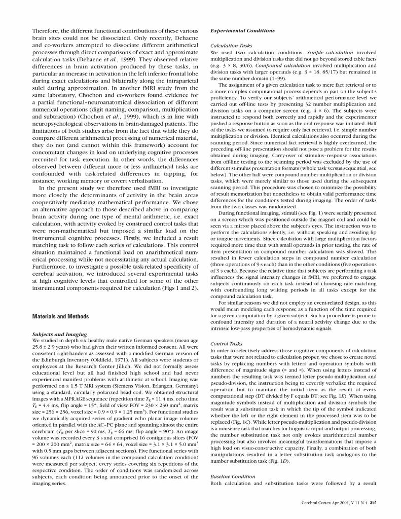
Calculation Tasks
We used two calculation conditions. Simple calculation involved
multiplication and division tasks that did not go beyond stored table facts
(e.g. 3 × 8, 30/6). Compound calculation involved multiplication and
division tasks with larger operands (e.g. 3 × 18, 85/17) but remained in
the same number domain (1–99).
The assignment of a given calculation task to mere fact retrieval or to
a more complex computational process depends in part on the subject’s
proficiency. To verify our subjects’ arithmetical performance level we
carried out off-line tests by presenting 32 number multiplication and
division tasks on a computer screen (e.g. 4 × 6). The subjects were
instructed to respond both correctly and rapidly and the experimenter
pushed a response button as soon as the oral response was initiated. Half
of the tasks we assumed to require only fact retrieval, i.e. simple number
multiplication or division. Identical calculations also occurred during the
scanning period. Since numerical fact retrieval is highly overlearned, the
preceding off-line presentation should not pose a problem for the results
obtained during imaging. Carry-over of stimulus–response associations
from off-line testing to the scanning period was excluded by the use of
different stimulus presentation formats (whole task versus sequential, see
below). The other half were compound number multiplication or division
tasks, which were merely similar to those used during the subsequent
scanning period. This procedure was chosen to minimize the possibility
of result memorization but nonetheless to obtain valid performance time
differences for the conditions tested during imaging. The order of tasks
from the two classes was randomized.
During functional imaging, stimuli (see Fig. 1) were serially presented
on a screen which was positioned outside the magnet coil and could be
seen via a mirror placed above the subject’s eyes. The instruction was to
perform the calculations silently, i.e. without speaking and avoiding lip
or tongue movements. Since calculation with large multiplication factors
required more time than with small operands in prior testing, the rate of
item presentation in compound number calculation was slowed. This
resulted in fewer calculation steps in compound number calculation
(three operations of 9 s each) than in the other conditions (five operations
of 3 s each). Because the relative time that subjects are performing a task
inf luences the signal intensity changes in fMRI, we preferred to engage
subjects continuously on each task instead of choosing rate matching
with confounding long waiting periods in all tasks except for the
compound calculation task.
For similar reasons we did not employ an event-related design, as this
would mean modeling each response as a function of the time required
for a given computation by a given subject. Such a procedure is prone to
confound intensity and duration of a neural activity change due to the
intrinsic low-pass properties of hemodynamic signals.
Control Tasks
In order to selectively address those cognitive components of calculation
tasks that were not related to calculation proper, we chose to create novel
tasks by replacing numbers with letters and operation symbols with
difference of magnitude signs (> and <). When using letters instead of
numbers the resulting task was termed letter pseudo-multiplication and
pseudo-division, the instruction being to covertly verbalize the required
operation but to maintain the initial item as the result of every
computational step (DT divided by F equals DT; see Fig. 1E). When using
magnitude symbols instead of multiplication and division symbols the
result was a substitution task in which the tip of the symbol indicated
whether the left or the right element in the processed item was to be
replaced (Fig. 1C). While letter pseudo-multiplication and pseudo-division
is a nonsense task that matches for linguistic input and output processing,
the number substitution task not only evokes anarithmetical number
processing but also involves meaningful transformations that impose a
high load on visuo-constructive capacity. Finally, a combination of both
manipulations resulted in a letter substitution task analogous to the
number substitution task (Fig. 1D).
Baseline Condition
Both calculation and substitution tasks were followed by a result
Cerebral Cortex Apr 2001, V 11 N 4 351
matching period that was considered as the baseline condition. During
this period the correct result from a series of sequentially presented
possible results was to be indicated by a button press (Fig. 1). The
position of the correct item within the series was varied across trials to
prevent predictability. This procedure removed any ambiguity between
activations related to the result matching period and activations related to
the preceding experimental task.
In addition to the brief motor activation related to pushing the button,
the result matching period maintained visual input processing and a
working memory load. Although not required for correct performance,
access to semantic representations of letters and numbers, including a
representation of magnitude in the case of numbers, most probably also
took place during this period. In short, the result matching period
controlled in a meaningful context for all aspects of the activation
conditions that did not involve the calculation or substitution procedures
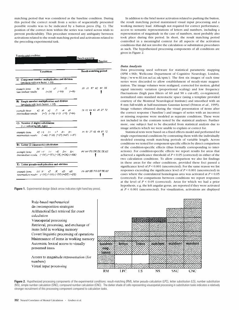
as such. The hypothesized processing components of all conditions are
given in Figure 2.
Data Analysis
Data processing used software for statistical parametric mapping
(SPM v.96b; Wellcome Department of Cognitive Neurology, London,
http://www.fil.ion.ucl.ac.uk/spm/). The first six images of each time
series were discarded to allow establishment of steady-state magnet-
ization. The image volumes were realigned, corrected for motion, global
signal intensity variation (proportional scaling) and low frequency
f luctuations (high pass filters of 60 and 90 s cut-off), co-registered,
normalized onto standard stereotactic space (using a template provided
courtesy of the Montreal Neurological Institute) and smoothed with an
8 mm full-width at half-maximum Gaussian kernel (Friston et al., 1995).
Image volumes obtained during the visual presentation of items after
the correct response (‘baseline’) and images of series with an incorrect
or missing response were modeled as separate conditions. These were
not included in the contrasts tested by the statistical analyses. Further-
more, one subject had to be discarded from statistical analysis due to
image artifacts which we were unable to explain or correct for.
Statistical tests were based on a fixed effects model and performed for
single experimental conditions by contrasting them with the individually
modeled ensuing result matching periods of variable length. Across
conditions we tested for component-specific effects by direct comparison
of the condition-specific effects (thus formally corresponding to inter-
actions). For condition-specific effects we report results for areas that
achieved a significance threshold of P < 0.05 (corrected) in either of the
two calculation conditions. To allow comparison we also list findings
in these areas for the other conditions, provided these foci passed a
significance level of P < 0.001 (uncorrected). For the same reason we list
responses exceeding the significance level of P < 0.001 (uncorrected) in
cases where the contralateral homologous area was activated at P < 0.05
(corrected). For comparisons between conditions we report responses
at the level of P < 0.05 (corrected). Areas for which we had a prior
hypothesis, e.g. the left angular gyrus, are reported if they were activated
at P < 0.001 (uncorrected). For visualization, activations are displayedFigure 1. Experimental design (black arrow indicates right hand key press).
Figure 2. Hypothesized processing components of the experimental conditions: result-matching (RM), letter pesudo-calculation (LPC), letter substitution (LS), number substitution(NS), simple number calculation (SNC), compound number calculation (CNC). The darker shade of cells representing visuospatial processing in substitution tasks indicates a relativelystronger recruitment of this processing component compared to calculation tasks.
352 Neural Correlates of Mental Calculation • Gruber et al.
at P < 0.001 (uncorrected) to enhance sensitivity in delineating spatial
extent.
Results
Off-line Testing of Performance
After removal of 17 trials with incorrect solutions and five
outliers (> mean calculation time + 3 SD) the grand mean
calculation time of the remaining 138 trials was 2.50 ± 1.20 s.
Specifically, simple calculation took 1.89 ± 0.86 s for
multiplication and 1.93 ± 0.80 s for division, whereas compound
calculation took 3.05 ± 1.21 s for multiplication and 3.03 ±
1.38 s for division. Despite the small sample size, there was a
significant effect when comparing simple with compound
number calculation (ANOVA, subjects F = 21.07, P < 0.01, items
F = 19.72, P < 0.001), simple calculations being faster for
multiplication and division. We found no significant effect of
calculation type (multiplication or division) and no interaction
between the two factors. Thus, the behavioral data give evid-
ence for additional costs in compound calculation compared
with simple calculation (presumably ref lecting additional
cognitive processes), whereas there was no such difference
between multiplication and division tasks in the subjects of this
study.
Functional Imaging
During functional imaging we could only obtain condition-
related error rates (and not reaction times) because our
experimental design avoided overt responses during calculation
as a potential source of artifacts. Analyzing these error rates we
found no significant differences between conditions (P < 0.05)
at the behavioral level, indicating that after compensation for
the additional time requirements of compound calculation our
experimental conditions were balanced for task difficulty.
Statistical comparisons of condition-specific brain activity levels
were performed in several steps. We first identified cortical
regions that were activated by each condition relative to the
respective result matching period. All five conditions showed a
similar left-dominant bilateral prefrontal, premotor and parietal
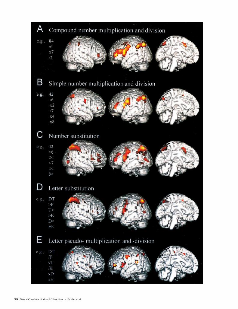
response pattern (Fig. 3 and Table 1).
In more detail, the commonly activated brain regions
comprised the prefrontal cortex lining the anterior inferior
frontal sulcus, the opercular part of the inferior frontal gyrus (BA
44), the premotor cortex (BA 6), the intraparietal sulcus and the
adjacent inferior and superior parietal cortices and the pre-
supplementary motor area, partially extending into the anterior
cingulate. The lateralization of prefrontal and parietal foci to
the left hemisphere during calculation shifted to a more sym-
metrical distribution in the substitution conditions. Letter
pseudo-calculation yielded the smallest extent of significant
activation. Our further analyses were aimed at revealing
differences between conditions. First, we contrasted simple
number calculation with letter pseudo-calculation and number
substitution. Each condition was considered with regard to the
activation it produced compared to its control period. Letter
pseudo-calculation matches number calculation for covert
linguistic processing of the presented items and operation
symbols and for maintenance of a result over a succession of
items.
The comparison between simple number calculation and
letter pseudo-calculation revealed left-sided activations at P <
0.001 (uncorrected) in dorsal parts of the inferior frontal gyrus
(BA 44/45), in the middle third of the inferior frontal sulcus, in
the ascending and descending parts of the intraparietal sulcus as
well as in the posterior cingulate cortex (BA 23/31) and the
adjacent precuneus (BA 31/7). A similar response pattern was
seen when contrasting compound number calculation with
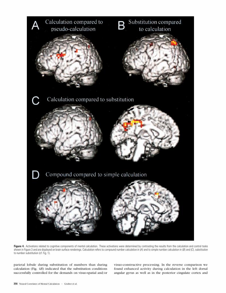
letter pseudo-calculation (Fig. 4A), with responses in the left
frontal areas and the ascending intraparietal sulcus (P < 0.05,
corrected). Given that the item presentation rate was lower in
compound calculation than in pseudo-calculation, these areas
seem to be actively engaged in the computations performed and
not merely in processing input and output.
In further comparisons we tried to differentiate activations
related to visuo-spatial symbol manipulation from activations
related to calculation proper. In number substitution the
adequate item category is maintained but the operation symbols
changed. Although < and > were explicitly chosen as in the realm
of mathematically meaningful symbols, in the substitution task
they did not indicate arithmetical operations but reordering
of symbols engaging visuo-spatial and visuo-constructive cap-
acities. Testing for greater activity during number substitution
than during number calculation, we found bilateral activation of
the superior parietal lobule (Fig. 4B), suggesting that number
substitution had an even greater demand on such capacities than
number calculation.
Comparing the parietal activation foci in Figure 4A,B, it is
apparent that the parietal activation observed across the
conditions shown in Figure 3A–D spans the superior and inferior
parietal lobules separated by the intraparietal sulcus, i.e. several
functionally distinct brain areas. The opposite comparison,
i.e. probing for greater activity in number calculation than
substitution (Fig. 4C), revealed a focus in the left dorsal angular
gyrus (P < 0.001, uncorrected). Together with the similarly
localized activation focus shown in Figure 4A, these data
underline the key role of the left angular gyrus in mental
calculation. Moreover, this comparison confirmed the finding of
strong posterior cingulate cortex activation extending into the
precuneus (P < 0.05, corrected) that had already been observed
when contrasting number calculation with letter pseudo-
calculation.
We also tested whether our experiment could identify
correlates of automatic access to a number-specific magnitude
representation in an anarithmetic context. However, we
observed no significant activity differences when directly
contrasting number with letter substitution. This indicates that
when embedded in tasks as devised here, number (as opposed to
letter) processing does not detectably engage different neuronal
structures.
In a final step of the analysis we attempted to differentiate
between the two types of calculation tasks applied, i.e. simple
and compound number calculation. When testing for greater
activity in the simple compared to the compound number
calculation task we found a response in the aforementioned
medial parietal zone covering the precuneus and posterior
cingulate cortex (P < 0.001, uncorrected). Since item presenta-
tion rate was slowed for compound number calculation, this
finding may simply ref lect a higher rate of mathematical fact
retrieval in the more rapid succession of calculations with
simple numbers. Conversely, the opposite comparison probed
for activation differences due to the more complex calculation
processes required in compound than in simple calculation.
We found left-sided activity changes at P < 0.001 (uncorrected)
in dorsal parts of the inferior frontal gyrus (BA 44/45), in
ventrolateral prefrontal cortex adjacent to the anterior inferior
frontal sulcus and in the anterior cingulate cortex (at the border
of BA 8, 24 and 32; see Fig. 4D). Interestingly, the focus in the
Cerebral Cortex Apr 2001, V 11 N 4 353
354 Neural Correlates of Mental Calculation • Gruber et al.

anterior cingulate cortex was identical to that we observed when
comparing number substitution with simple number calculation,
i.e. it was sensitive to non-arithmetical challenge.
DiscussionThe present study aimed at a better understanding of functional
contributions from the diverse activation foci detected for
mental calculation in more general comparisons (Rueckert et al.,
1996; Sakurai et al., 1996; Burbaud et al., 1999). We therefore
construed experimental control conditions that were matched
for relevant components in the set of operations required for
mental arithmetic (see Fig. 2). In a first step the different
activation conditions, including the calculation tasks, were
contrasted with result matching to reveal activation areas related
to common task components. Subsequently, direct comparisons
across conditions were made to detect significant differences
between the tasks.
The common activations across arithmetical tasks and non-
arithmetical control tasks cover a distributed cortical network
that is in accordance with results from previous studies cited
above and comprises mostly left-sided prefrontal, premotor and
parietal regions (Table 1 and Fig. 3). These brain regions are well
known to contribute to linguistic and visuo-spatial processing
and to related aspects of working memory function, i.e. the
phonological loop and the visuo-spatial sketchpad [see for
example Baddeley, Paulesu and Swartz (Baddeley, 1992; Paulesu
et al., 1993; Swartz et al., 1995)]. Hence, these results support
the notion that most of the cortical areas involved in mathemat-
ical tasks represent neural substrates not only for calculation
proper but also for other, related cognitive operations.
Correspondingly, it has been suggested that most calculation
disturbances subsequent to brain lesions arise secondary to
one or several of three so-called instrumental problems: spatial
deficit, visuo-constructive impairment and aphasia (Collignon et
al., 1977).
Besides the processes already controlled for in the baseline
condition of result matching, letter pseudo-calculation shared
inner speech and lexical processing of operation symbols with
the other conditions. It was devised as an intermediate level
control task to identify those activations in the other four
conditions that were related to execution of a meaningful
operation, i.e. active processing of item representations held
in working memory (see Fig. 2). Comparing calculation with
pseudo-calculation, increased activity was found to be left-sided
in the middle third and dorsal posterior parts of the inferior
frontal gyrus and medial and inferior lateral parietal regions
(Fig. 4A). Similar frontal activation patterns were seen when
substitution tasks were compared with letter pseudo-calculation.
In sum, additional activation in the left inferior frontal cortex
was induced by all tasks involving meaningful operations on
working memory representations of letters or numbers. The
known involvement of the dorsal inferior frontal gyrus in
phonological [for an extensive review see Indefrey and Levelt
(Indefrey and Levelt, 2000)], syntactic (Caplan et al., 1998,
1999) and semantic (Poldrack et al., 1998, Friederici et al., 2000)
processing of language suggests that the operations required
by the tasks also evoked linguistic processes. Activations in
the middle third of the left inferior frontal sulcus, on the other
hand, were within the variation range of Brodmann areas 9 and
46, as previously reported (Rajkowska and Goldman-Rakic,
1995). Considering that both areas are known regions of
working memory functions [for reviews see Ungerleider and
Goldman-Rakic (Ungerleider, 1995; Goldman-Rakic, 1996] and
that, in contrast to pseudo-calculation, the meaningful tasks
required frequent exchanges and manipulations of items to be
held in working memory, it seems plausible to assume that the
observed activations were due to additional working memory
recruitment.
In the substitution tasks the operations to be performed
preferentially relied on visuo-spatial processing resources (i.e.
replacing the left or right digit), whereas the calculation tasks
required access to number meaning and magnitude repres-
entation, arithmetical processing of the memorized items and
retrieval of arithmetical facts. We further assessed activations
related to these different operations through direct comparisons
of the respective tasks. The greater activity in the superior
Table 1Brain regions commonly activated across conditions (compared to result matching) as determined by statistical parametric mapping (see Materials and Methods)
Area Compound no. calculation Simple no. calculation No. substitution Letter substitution Letter pseudo-calculation
Anterior inferior frontal sulcusLeft –48,44,8; 6.92b –44,44,12; 4.58b –44,44,8; 5.86b –44,48,8; 3.75a –56,44,0; 5.55b
Right 48,40,12; 4.31a 52,40,8; 3.37a 48,44,4; 5.82b 48,40,4; 3.97a nsPremotor cortex
Left –48,–8,40; 6.62b –48,0,32; 6.28b –48,0,36; 6.04b –48,4,32; 4.31a –48,–8,40; 5.30b
Right ns 44,–8,32; 4.85b 52,–8,36; 3.55a 52,–12,48; 3.71a 52,–8,40; 3.60a
Inferior frontal gyrus (opercular part)Left –52,8,20; 5.78b –52,8,16; 4.76b –52,8,16; 7.27b –56,8,12; 6.16b –64,12,12; 5.87b
Right ns ns ns ns nsIntraparietal sulcus and adjacent parietal lobules
Left –28,–68,48; 7.02b –36,–68,48; 5.80b –32,–64,52; 7.81b –24,–68,52; 7.50b –20,–72,48; 3.85a
Right 12,–76,52; 4.40a 24,–64,40; 5.72b 24,–68,52; 7.74b 24,–68,52; 7.49b 32,–60,60; 4.00a
Presupplementary motor area –4,16,48; 3.97a –4,8,56; 3.60a –4,8,52; 5.46b –4,8,52; 3.98a –12,12,52; 4.08a
The values given are the spatial coordinates of activation maxima and the Z scores obtained in these areas in the various conditions (cf. Fig. 1).aP < 0.001, uncorrected; bP < 0.05, corrected; ns, not significant.
Figure 3. Activations during mental calculation and related control tasks. The results from statistical parametric mapping in five subjects were rendered onto the lateral and medialsurfaces of a standard anatomical (T1-weighted) reference brain template. The activations show brain areas more active during experimental conditions A–E (cf. Fig. 1) than duringensuing result matching. For spatial coordinates see Table 1.
Cerebral Cortex Apr 2001, V 11 N 4 355
parietal lobule during substitution of numbers than during
calculation (Fig. 4B) indicated that the substitution conditions
successfully controlled for the demands on visuo-spatial and/or
visuo-constructive processing. In the reverse comparison we
found enhanced activity during calculation in the left dorsal
angular gyrus as well as in the posterior cingulate cortex and
Figure 4. Activations related to cognitive components of mental calculation. These activations were determined by contrasting the results from the calculation and control tasksshown in Figure 3 and are displayed on brain surface renderings. Calculation refers to compound number calculation in (A) and to simple number calculation in (B) and (C), substitutionto number substitution (cf. Fig. 1).
356 Neural Correlates of Mental Calculation • Gruber et al.

precuneus (Fig. 4C). Ever since the publications of Peritz and
Henschen (Peritz, 1918; Henschen, 1919) there have been
attempts to ascertain a possible role of the (left) angular gyrus as
a specific calculation center. Converging evidence from neuro-
psychology, electrophysiology and functional neuroimaging
points to an involvement of inferior parietal regions in math-
ematical tasks, although their exact functional contributions
remain ill defined. In three patients with left parietal ischemic
infarctions overlapping along the intraparietal sulcus calculation
disturbances were apparent, especially in more complex
arithmetical problems, and were ascribed to a disruption of
working memory (Takayama et al., 1994). A working memory
deficit was also proposed in a more recent case report of a
parietal lesion including the angular gyrus, where number
transcoding but not calculation was disturbed (Markowitsch
et al., 1999). In support of this view, certain components of
event-related potentials (ERP) that were recorded at parietal sites
during mental arithmetic have been associated with task diffi-
culty and were thought to ref lect the load imposed on working
memory (Pauli et al., 1996). A different functional description
was given for a patient with a left parietal intra-cerebral
hematoma (Warrington, 1982), attributing his acalculia to faulty
access to semantic entries of arithmetical facts. Correspondingly,
a recent study using cortical electrostimulation in the left
parietal lobe demonstrated a disruption of arithmetical fact
retrieval (Whalen et al., 1997). Finally, Dehaene and Cohen
assessed two patients with a double dissociation of lesion sites
and behavioral effects (Dehaene and Cohen, 1997). They con-
cluded that the inferior parietal lobe might contain an abstract
semantic representation of numbers that is necessary to guide
arithmetical fact retrieval and to perform calculations (Dehaene
et al., 1998). A different interpretation of the cause of acalculia
was reached in a recent very detailed case report of pure
Gerstmann syndrome subsequent to a lesion of left parietal
white matter (Mayer et al., 1999). Following the concept of
a ‘Grundstörung’, the common cognitive denominator of the
disturbed functions was hypothesized to be an impairment in
mental manipulation of images. Together, these studies indicate
that several operational levels of the processes required for
mental calculation may depend on functional integrity of lateral
inferior parietal cortices and the underlying white matter.
The most recent neuroimaging studies on number processing
and mental calculation have also examined determinants of
parietal activation in greater detail. ERP and fMRI data suggest
that the intraparietal sulci are preferentially involved in approxi-
mation and the angular gyri in exact calculation (Dehaene et
al., 1999). While Rickard et al. found activations restricted to
the intraparietal sulci alone (Rickard et al., 2000), a study by
Chochon et al. supports the assumption that both the intra-
parietal sulci and the inferior parietal lobules, i.e. dorsal angular
and/or supramarginal gyri, subserve the execution of number
processing tasks (Chochon et al., 1999).
Several of our findings further the understanding of the role(s)
that different parietal brain areas play in the processing of
numbers and in mental calculation. First of all, we failed to
corroborate the hypothesis of Dehaene’s triple code theory
that the bilateral intraparietal sulcus corresponds to a specific
semantic representation of numerical quantities (Dehaene et
al., 1998). In contrast, this brain region was non-specifically
activated under all experimental conditions when compared
with result matching, irrespective of the material (numbers
or letters) and the actual nature of the task performed. This
suggests that the intraparietal sulcus participates in processes
that are instrumental but not specific to mental calculation.
On the other hand, our results extend the previous findings of
(left) dorsal angular gyrus activation during number processing
by showing that there is more activation in this region when
the brain processes numbers in an arithmetical context than in a
different cognitive task. This indicates that the angular gyrus
is sensitive to the arithmetical nature of the cognitive operation
performed and supports the assumption of a role of the angular
gyrus in excact calculation (Dehaene et al., 1999). The most
recent, albeit indirect, evidence for the role of the angular gyrus
comes from another fMRI study that, when comparing activation
patterns in perfect and imperfect calculation performers, only
found differences in the left angular gyrus, with less activation in
perfect performers, and interpreted this finding in the context
of proficiency (Menon et al., 2000).
Another important novel finding in our study relates to the
functional contribution of the medial parietal structures
activated during number calculation, i.e. the posterior cingulate
cortex and the precuneus. Both neuropsychological investi-
gations and functional neuroimaging studies have established
the importance of these areas in memory and, in particular,
retrieval functions. In semantically cued word retrieval tasks the
precuneus has been shown to be sensitive to item imageability
(Fletcher et al., 1996). On the other hand, a more recent study by
Krause and co-workers indicates that the precuneus may be
involved in episodic associative memory retrieval independent
of presentation modality and imagery content of the presented
material (Krause et al., 1999). Our results provide experimental
support for a role of this area in arithmetic fact retrieval because
the number calculation and substitution tasks differed with
respect to the necessity of this process. This finding does not
exclude the involvement of additional brain areas during
arithmetical fact retrieval in exact calculation tasks. Along these
lines, our results are in accordance with the notion that exact
calculation involves structures subserving language functions
(Dehaene et al., 1999) (see Fig. 3B and 4A). However, activations
in left inferior frontal areas which presumably underlie these
functions were canceled out when compared with tasks without
calculation but with similar demands on language functions
(Fig. 4C). Not surprisingly, the recruitment of language func-
tions, even in number processing contexts, is not restricted to
arithmetic fact retrieval. This view receives additional support
from data presented by Chochon and co-workers (Chochon et
al., 1999).
In sum, our findings provide evidence that the angular gyrus
and the medial parietal areas play essential roles in a functional
circuit ensuring mathematical performance. They support a
role of the angular gyrus in exact calculation by showing that
this structure is particularly sensitive to number processing in an
arithmetical context. They furthermore suggest a role of medial
parietal structures in the retrieval of arithmetical facts. Although
these two processes inevitably interact during calculation, the
possibility of a selective impairment has been shown in a patient
who, subsequent to operation on a left parietal tumor, presented
acalculia, agraphia, finger agnosia, right–left disorientation and
apraxia but was found to display preserved arithmetical fact
retrieval (Delazer and Benke, 1997).
A well-known behavioral feature of mental calculation that
was also observed in our subjects is the so-called problem size
effect, meaning that responses are slower on problems with
larger operands (Campbell, 1987; Siegler, 1988). More difficult
calculations require planned sequential usage of stored math-
Cerebral Cortex Apr 2001, V 11 N 4 357

ematical rules. These processes take additional time compared
to direct retrieval of arithmetical facts from memory. In other
words, whenever direct retrieval of simple table facts is in-
sufficient to solve an arithmetical problem, it will be necessary
to decompose the problem, by the use of mathematical rules,
into sub-steps for which single table facts can be successfully
retrieved. For example, 6 × 12 may be solved through trans-
formation, decomposition and recomposition in the following
way: 6 × 12 = 6 × (10 + 2) = 6 × 10 + 6 × 2 = 60 + 12 = 72.
When comparing the respective activations in our study,
compound calculation elicited higher activity in dorsal parts of
the left inferior frontal gyrus, in the anterior inferior frontal
sulcus and in the anterior cingulate cortex (see Fig. 4D).
The anterior cingulate focus was also activated in the number
substitution task when compared with simple number calcu-
lation and can therefore not be specifically related to calculation.
Its activity might indicate factors such as attentional effort or
task difficulty in monitoring result generation. Activations in
the inferior frontal cortex were also detected in more basic
comparisons (cf. Figs 4A and 3A–E, respectively) and attributed
to linguistic processing (BA 44/45) as well as to the on-line
maintenance of information in the presence of additional,
possibly interfering, cognitive processes (anterior inferior
frontal sulcus). Activity in these regions once again increased
during compound calculation as compared with simple calcu-
lation, suggesting a further involvement of language and working
memory functions during the use of rule-based decomposition
and recomposition strategies.
In conclusion, this study has confirmed that an extended
bilateral prefrontal–premotor–parietal network subserves
mental calculation. However, to a large extent these cortical
areas do not seem to be exclusively involved in arithmetical
procedures, but also in other cognitive contexts relying on
similar instrumental components, such as working memory,
processing symbolic information, mental image transformations
and inner speech. In contrast, we have demonstrated specific
functional contributions of lateral (angular gyrus) and medial
(posterior cingulate and precuneus) parietal cortices to the
processing of numerical representations during exact calcula-
tion, including arithmetical fact retrieval. Finally, our findings
suggest that the use of decomposition and recomposition
strategies in more complex calculation problems does not rely
on neuronal resources specific to mental arithmetic, but recruits
left inferior frontal areas subserving language and working
memory functions.
NotesWe would like to thank the Research Centre Jülich for providing the
facilities that made this work possible and especially Dr Stefan Posse, Dr
Jon Shah and Maisa Grosse-Ruyken for help in data acquisition. A.K. is
supported by the Volkswagen-Stiftung.
Address correspondence to Dr Oliver Gruber, Max-Planck-Institute of
Cognitive Neuroscience, PO Box 500 355, D-04303 Leipzig, Germany.
ReferencesBaddeley A (1992) Working memory. Science 255:556–559.
Boller F, Grafman J (1985) Acalculia. In: Handbook of clinical neurology
(Vinken PJ, Bruyn GW, Klawans HL, eds), pp. 473–481. Amsterdam:
North Holland.
Burbaud P, Degreze P, Lafon P, Franconi JM, Bouligand B, Bioulac B
et al. (1995) Lateralization of prefrontal activation during internal
mental calculation: a functional magnetic resonance imaging study.
J Neurophysiol 74:2194–2200.
Burbaud P, Camus O, Guehl D, Bioulac B, Caille JM, Allard M (1999) A
functional magnetic resonance imaging study of mental subtraction in
human subjects. Neurosci Lett 273:195–199.
Campbell JID (1987) Network interference and mental multiplication.
J Exp Psychol Learn Mem Cogn 13:109–123.
Caplan D, Alpert N, Waters G (1998) Effects of syntactic structure and
propositional number on patterns of regional cerebral blood f low.
J Cogn Neurosci 10:541–552.
Caplan D, Alpert N, Waters G (1999) PET studies of syntactic processing
with auditory sentence presentation. Neuroimage 9:343–354.
Chochon F, Cohen L, van de Moortele PF, Dehaene S (1999) Differential
contributions of the left and right inferior parietal lobules to number
processing. J Cogn Neurosci 11:617–630.
Collignon R, Leclerq C, Mahy J (1977) Étude de la sémiologie des troubles
du calcul observés au cours des lesions corticales. Acta Neurol Belg
77:257–275.
Dahmen W, Hartje W, Büssing A, Sturm W (1982) Disorders of calculation
in aphasic patients—spatial and verbal components. Neuropsychologia
20:145–153.
Dehaene S (1992) Varieties of numerical abilities. Cognition 44:1–42.
Dehaene S, Cohen L (1997) Cerebral pathways for calculation: double
dissociation between rote verbal and quantitative knowledge of
arithmetic. Cortex 33:219–250.
Dehaene S, Tzourio N, Frak V, Raynaud L, Cohen L, Mehler J et al. (1996)
Cerebral activations during number multiplication and comparison: a
PET study. Neuropsychologia 34:1097–1106.
Dehaene S, Dehaene-Lambertz G, Cohen L (1998) Abstract representa-
tions of numbers in the animal and human brain. Trends Neurosci
21:355–361.
Dehaene S, Spelke E, Pinel P, Stanescu R, Tsivkin S (1999) Sources
of mathematical thinking: behavioral and brain-imaging evidence.
Science 284:970–974.
Delazer M, Benke T (1997) Arithmetic facts without meaning. Cortex
33:697–710.
Fletcher PC, Shallice T, Frith CD, Frackowiak RSJ, Dolan RJ (1996) Brain
activity during memory retrieval—the inf luence of imagery and
semantic cueing. Brain 119:1587–1596.
Friederici AD, Opitz B, von Cramon DY (2000) Segregating semantic
and syntactic aspects of processing in the human brain: an fMRI
investigation of different word types. Cereb Cortex 10:698–705.
Friston KJ, Holmes AP, Worsley KJ, Poline JP, Frith CD, Frackowiak RSJ
(1995) Statistical parametric maps in functional imaging: a general
linear approach. Hum Brain Mapp 2:189–210.
Goldman-Rakic PS (1996) The prefrontal landscape: implications of
functional architecture for understanding human mentation and the
central executive. Phil Trans R Soc Lond Ser B Biol Sci 351:1445–1453.
Grafman J, Passafiume D, Faglioni P, Boller F (1982) Calculation
disturbances in adults with focal hemispheric damage. Cortex
18:37–50.
Henschen SE (Schaller WF trans.) (1919, reprinted 1925) Clinical and
anatomical contributions on brain pathology. Arch Neurol Psychiat
13:226–249.
Indefrey P, Levelt WJM (2000) The neural correlates of language
production. In: The new cognitive neurosciences, 2nd edn (Gazzaniga
M, ed.), pp. 845–865. Cambridge, MA: MIT Press.
Krause BJ, Schmidt D, Mottaghy FM, Taylor J, Halsband U, Herzog H et al.
(1999) Episodic retrieval activates the precuneus irrespective of the
imagery content of word pair associates: a PET study. Brain 122:
255–263.
Levin HS, Goldstein FC, Spiers PA. (1993) Acalculia. In: Clinical
neuropsychology (Heilman KM and Valenstein E, eds), pp. 91–122.
New York: Oxford University Press.
Markowitsch HJ, Kalbe E, Kessler J, von Stockhausen HM, Ghaemi M,
Heiss WD (1999) Short-term memory deficit after focal parietal
damage. J Clin Exp Neuropsychol 21:784–797.
Mayer E, Martory MD, Pegna AJ, Landis T, Delavelle J, Annoni JM (1999) A
pure case of Gerstmann syndrome with a subangular lesion. Brain
122:1107–1120.
McCloskey M, Caramazza A, Basili A (1985) Cognitive mechanisms in
number processing and calculation: evidence from dyscalculia. Brain
Cogn 4:171–196.
Menon V, Rivera SM, White CD, Eliez S, Glover GH, Reiss AL (2000)
Functional optimization of arithmetic processing in perfect
performers. Cogn Brain Res 9:343–345.
Oldfield RC (1971) The assessment and analysis of handedness: the
Edinburgh Inventory. Neuropsychologia 9:97–113.
358 Neural Correlates of Mental Calculation • Gruber et al.

Paulesu E, Frith CD, Frackowiak RSJ (1993) The neural correlates of the
verbal component of working memory. Nature 362:342–345.
Pauli P, Lutzenberger W, Birbaumer N, Rickard TC, Bourne LEJ (1996)
Neurophysiological correlates of mental arithmetic. Psychophysiology
33:522–529.
Peritz G (1918) Zur Pathopsychologie des Rechnens. Dtsch Z
Nervenheilkd 61:234–340.
Pesenti M, Thioux M, Seron X, De Volder A (2000) Neuroanatomical
substrates of arabic number processing, numerical comparison, and
simple addition: a PET study. J Cogn Neurosci 12:461–479.
Poldrack RA, Wagner AD, Prull MW, Desmond JE, Glover GH, Gabrieli JD
(1999) Functional specialization for semantic and phonological
processing in the left inferior prefrontal cortex. Neuroimage 10:
15–35.
Rajkowska G, Goldman-Rakic PS (1995) Cytoarchitectonic definition of
prefrontal areas in the normal human cortex: II. Variability in locations
of areas 9 and 46 and relationship to the Talairach coordinate system.
Cereb Cortex 5:323–337.
Rickard TC, Romero SG, Basso G, Wharton C, Flitman S, Grafman J (2000)
The calculating brain: an fMRI study. Neuropsychologia 38:325–335.
Roland PE, Friberg L (1985) Localization of cortical areas activated by
thinking. J Neurophysiol 5:1219–1243.
Rossor MN, Warrington EK, Cipolotti L (1995) The isolation of calculation
skills. J Neurol 242:78–81.
Rueckert L, Lange N, Partiot A, Appollonio I, Litvan I, Le Bihan D et al.
(1996) Visualizing cortical activation during mental calculation with
functional MRI. Neuroimage 3:97–103.
Sakurai Y, Momose T, Iwata M, Sasaki Y, Kanazawa I (1996) Activation of
prefrontal and posterior superior temporal areas in visual calculation.
J Neurol Sci 139:89–94.
Siegler RS (1988) Strategy choice procedures and the development of
multiplication skill. J Exp Psychol Gen 117:258–275.
Swartz BE, Halgren E, Fuster JM, Simpkins F, Gee M, Mandelkern M (1995)
Cortical metabolic activation in humans during a visual memory task.
Cereb Cortex 5:205–214.
Takayama Y, Sugishita M, Akiguchi I, Kimura J (1994) Isolated acalculia
due to left parietal lesion. Arch Neurol 51:286–291.
Ungerleider LG (1995) Functional brain imaging studies of cortical
mechanisms for memory. Science 270:769–775.
Warrington EK (1982) The fractionation of arithmetical skills: a single
case study. Q J Exp Psychol 34A:31–51.
Whalen J, McCloskey M, Lesser RP, Gordon B (1997) Localizing arithmetic
processes in the brain: evidence from a transient deficit during
cortical stimulation. J Cogn Neurosci 9:409–417.
Cerebral Cortex Apr 2001, V 11 N 4 359