A diagnostic and surgical approach to masses of the buccal ...
Upload
khangminh22Category
view
0download
0
DISSERTATION ON
“DIAGNOSTIC APPROACH AND MANAGEMENT
STRATEGIES OF INVASIVE FUNGAL SINUSITIS”
Dissertation submitted in partial fulfillment
of the regulations for the award of the
DEGREE OF M.S.DEGREE BRANCH-IV
OTORHINOLARYNGOLOGY
OF
THE TAMILNADU DR. M.G.R. MEDICAL UNIVERSITY
UPGRADED INSTITUTE OF OTORHINOLARYNGOLOGY,
MADRAS MEDICAL COLLEGE, CHENNAI.
APRIL 2017
CERTIFICATE
This is to certify that this dissertation “A Study On
Diagnostic Approach and management strategies of Invasive fungal
sinusitis ” submitted by Dr.S.SUJAYKUMAR, appearing for M.S
ENT Branch IV Degree examination in April 2017 is a bonafide record of
work done by him under our guidance and supervision in partial fulfillment
of the regulations of the TamilnaduDr.M.G.R Medical University, Chennai.
I forward this to the TamilnaduDr.M.G.R Medical University, Chennai,
Tamilnadu, India.
GUIDE
PROF. DR.M.K.RAJASEKAR MS DLO Director and Professor
Upgraded Institute Of Otorhinolaryngology
Madras Medical College
Rajiv Gandhi Govt, General Hospital, Chennai -600003
PROF. DR.M.K.RAJASEKAR,
MS., DLO., Director & Professor,
Upgraded Institute of
Otorhinolaryngology
Madras Medical College
Rajiv Gandhi Govt. General
Hospital, Chennai -600003
Dr.M.K.MURALITHARAN,
M.S.,M.ch Dean,
Madras Medical College,
Chennai.
DECLARATION
I solemnly declare that the dissertation entitled “A Study on
Diagnostic approach and management strategies of Invasive fungal
sinusitis ” is done by me at Madras Medical College, Chennai-3 during
November 2014 to September 2016 under the guidance and supervision of
Prof DR.M.K.RAJASEKAR M.S, DLO., to be submitted to The Tamil
Nadu Dr.M.G.R Medial University towards the partial fulfillment of
requirements for the award of M.S DEGREE in
OTORHINOLARYNGOLOGY, BRANCH-IV.
DR.S.SUJAYKUMAR
Post Graduate, M.S ENT,
MMC & RGGGH,
Chennai – 600003
Place: Chennai
Date:

4
ACKNOWLEDGEMENT
I would like to express my sincere gratitude to
Prof.Dr.MURALITHARAN, MS., MCH., NEUROSURGERY The
dean, madras medical college, for having permitted me to undertake this
study.
I thank my Director, Upgraded institute of otorhinolaryngology, my
Chief coordinator Prof.Dr.M K.RAJASEKAR, MS., DLO., who was
instrumental in initiating new ideas and continues to support our endeavors.
I sincerely thank my guide Prof Dr.M K RAJASEKAR, MS.,
DLO., Upgraded institute of otorhinolaryngology whose passion for
teaching and interest in patient care with active academics has been a
great source of inspiration to all the post graduates including me.
I express my sincere gratitude to Prof.Dr.MUTHUKUMAR,
M.S. D.L.O, DNB Professor, Upgraded Institute of Otorhinolaryngology,
for his valuable support
I express my sincere gratitude to
Prof.Dr.G.SANKARANARAYANAN M.S., D.L.O., DNB., Professor,
Upgraded Institute of Otorhinolaryngology, for his valuable support.
I express my sincere gratitude to Prof.Dr.N.SURESHKUMAR
M.S.D.L.O., Professor, Upgraded Institute of Otorhinolaryngology, for
his valuable support.
5
I express my sincere thanks to THE SECRETARY AND
CHAIRMAN, INSTITUTIONAL ETHICAL COMMITTEE,
Government General Hospital, Madras Medical College.
I express my sincere thanks to all the assistant professors, for
their thoughtful guidance throughout the work.
My special thanks to my assistant professors Dr.MOHAMED
SIDDIQUE M.S, DLO, Dr.VIKRAM M.S, DLO, Dr.NANMULLAI
M.S who supported me throughout my studies.
I would like to thank my Seniors, Colleagues, Juniors who were of
great help throughout the project.
I thank My Patients without whose will this would not have
been a successful effort. I also thank My Family and the Almighty for
giving me the opportunity and strength to complete this.
6
ABBREVIATIONS
AIFS : Acute invasive fungal sinusitis
CIFS : Chronic invasive fungal sinusitis
CGFS : Chronic granulomatous fungal sinusitis
DNE : Diagnostic Nasal Endoscopy
PNS : Paranasal Sinuses
HPE : Histopathological Examination
DCLD : Decompensated Liver Disease
DM : Diabetic Mellitus
7
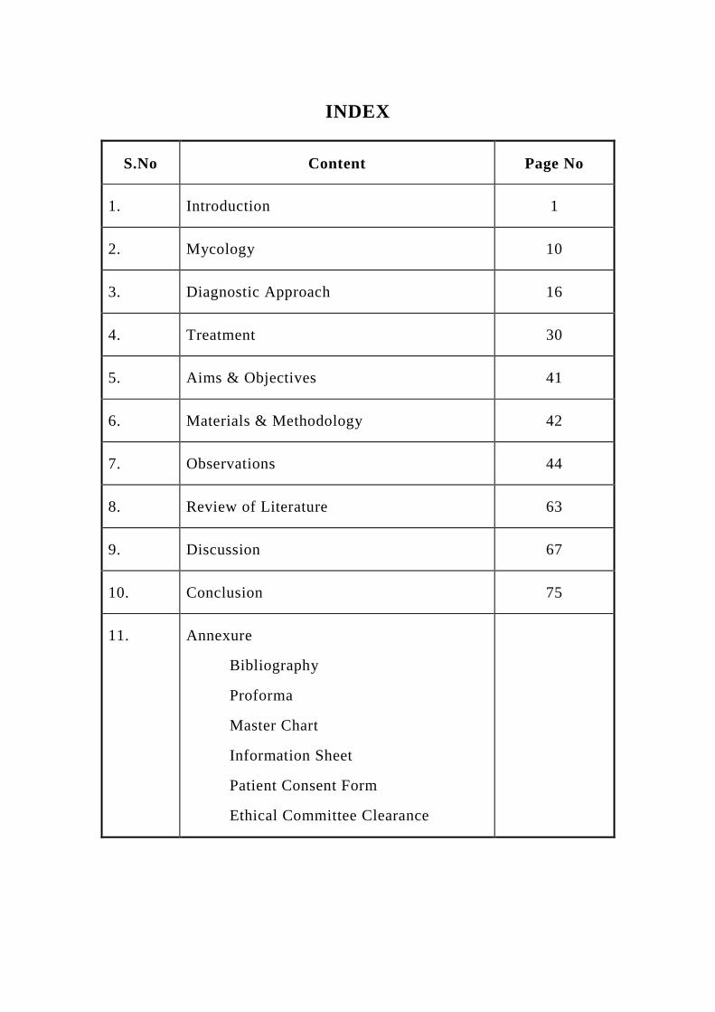
INDEX
S.No Content Page No
1. Introduction 1
2. Mycology 10
3. Diagnostic Approach 16
4. Treatment 30
5. Aims & Objectives 41
6. Materials & Methodology 42
7. Observations 44
8. Review of Literature 63
9. Discussion 67
10. Conclusion 75
11. Annexure
Bibliography
Proforma
Master Chart
Information Sheet
Patient Consent Form
Ethical Committee Clearance
1
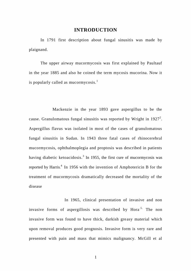
INTRODUCTION
In 1791 first description about fungal sinusitis was made by
plaignand.
The upper airway mucormycosis was first explained by Paultauf
in the year 1885 and also he coined the term mycosis mucorina. Now it
is popularly called as mucormycosis.1
Mackenzie in the year 1893 gave aspergillus to be the
cause. Granulomatous fungal sinusitis was reported by Wright in 19272.
Aspergillus flavus was isolated in most of the cases of granulomatous
fungal sinusitis in Sudan. In 1943 three fatal cases of rhinocerebral
mucormycosis, ophthalmoplegia and proptosis was described in patients
having diabetic ketoacidosis.3 In 1955, the first cure of mucormycosis was
reported by Harris.4 In 1956 with the invention of Amphotericin B for the
treatment of mucormycosis dramatically decreased the mortality of the
disease
In 1965, clinical presentation of invasive and non
invasive forms of aspergillosis was described by Hora.5.
The non
invasive form was found to have thick, darkish greasy material which
upon removal produces good prognosis. Invasive form is very rare and
presented with pain and mass that mimics malignancy. McGill et al
2
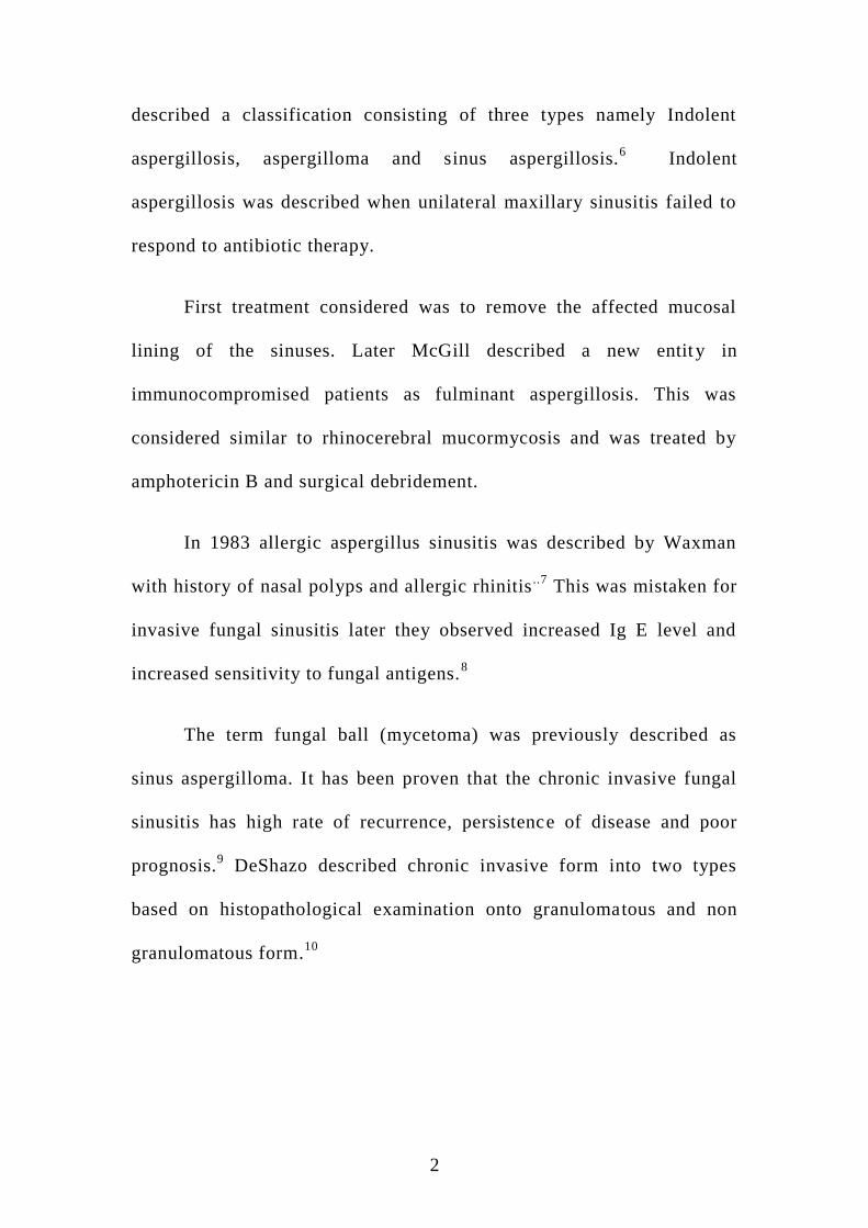
described a classification consisting of three types namely Indolent
aspergillosis, aspergilloma and sinus aspergillosis.6
Indolent
aspergillosis was described when unilateral maxillary sinusitis failed to
respond to antibiotic therapy.
First treatment considered was to remove the affected mucosal
lining of the sinuses. Later McGill described a new entit y in
immunocompromised patients as fulminant aspergillosis. This was
considered similar to rhinocerebral mucormycosis and was treated by
amphotericin B and surgical debridement.
In 1983 allergic aspergillus sinusitis was described by Waxman
with history of nasal polyps and allergic rhinitis..7
This was mistaken for
invasive fungal sinusitis later they observed increased Ig E level and
increased sensitivity to fungal antigens.8
The term fungal ball (mycetoma) was previously described as
sinus aspergilloma. It has been proven that the chronic invasive fungal
sinusitis has high rate of recurrence, persistence of disease and poor
prognosis.9 DeShazo described chronic invasive form into two types
based on histopathological examination onto granulomatous and non
granulomatous form.10
3
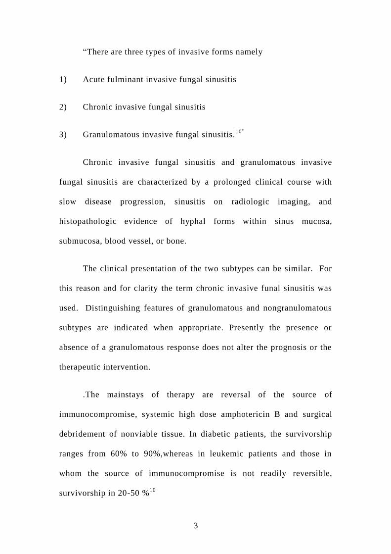
“There are three types of invasive forms namely
1) Acute fulminant invasive fungal sinusitis
2) Chronic invasive fungal sinusitis
3) Granulomatous invasive fungal sinusitis.10”
Chronic invasive fungal sinusitis and granulomatous invasive
fungal sinusitis are characterized by a prolonged clinical course with
slow disease progression, sinusitis on radiologic imaging, and
histopathologic evidence of hyphal forms within sinus mucosa,
submucosa, blood vessel, or bone.
The clinical presentation of the two subtypes can be similar. For
this reason and for clarity the term chronic invasive funal sinusitis was
used. Distinguishing features of granulomatous and nongranulomatous
subtypes are indicated when appropriate. Presently the presence or
absence of a granulomatous response does not alter the prognosis or the
therapeutic intervention.
.The mainstays of therapy are reversal of the source of
immunocompromise, systemic high dose amphotericin B and surgical
debridement of nonviable tissue. In diabetic patients, the survivorship
ranges from 60% to 90%,whereas in leukemic patients and those in
whom the source of immunocompromise is not readily reversible,
survivorship in 20-50 %10
4
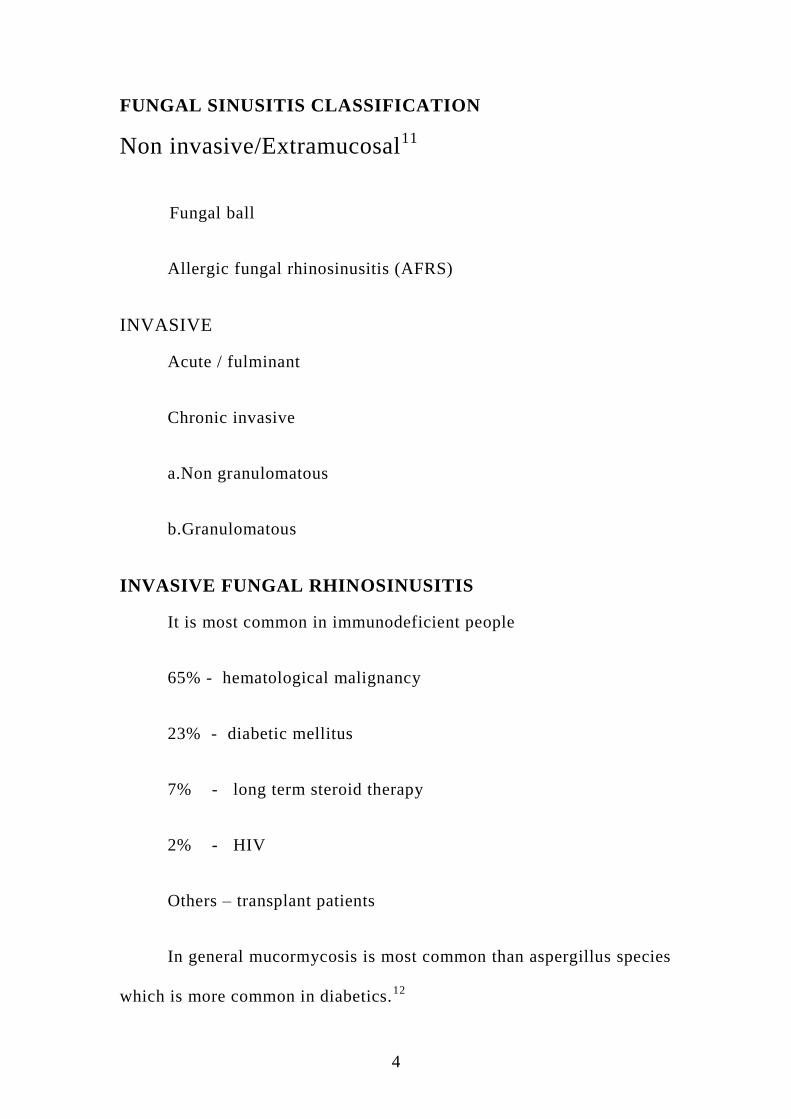
FUNGAL SINUSITIS CLASSIFICATION
Non invasive/Extramucosal11
Fungal ball
Allergic fungal rhinosinusitis (AFRS)
INVASIVE
Acute / fulminant
Chronic invasive
a.Non granulomatous
b.Granulomatous
INVASIVE FUNGAL RHINOSINUSITIS
It is most common in immunodeficient people
65% - hematological malignancy
23% - diabetic mellitus
7% - long term steroid therapy
2% - HIV
Others – transplant patients
In general mucormycosis is most common than aspergillus species
which is more common in diabetics.12
5
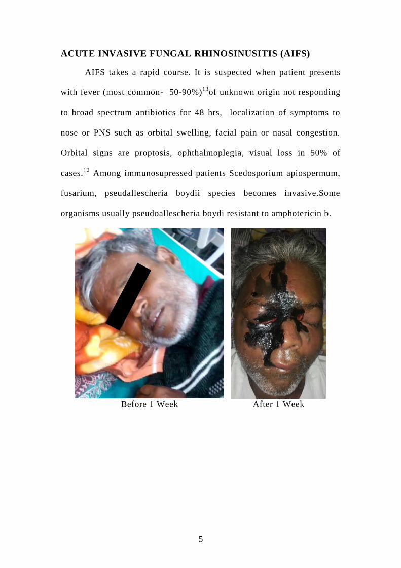
ACUTE INVASIVE FUNGAL RHINOSINUSITIS (AIFS)
AIFS takes a rapid course. It is suspected when patient presents
with fever (most common- 50-90%)13
of unknown origin not responding
to broad spectrum antibiotics for 48 hrs, localization of symptoms to
nose or PNS such as orbital swelling, facial pain or nasal congestion.
Orbital signs are proptosis, ophthalmoplegia, visual loss in 50% of
cases.12
Among immunosupressed patients Scedosporium apiospermum,
fusarium, pseudallescheria boydii species becomes invasive.Some
organisms usually pseudoallescheria boydi resistant to amphotericin b.
Before 1 Week After 1 Week

6
CHRONIC INVASIVE FUNGAL SINUSITIS (CIFS):
It can occur both in immunocompromised and competent people
and slowly progressive with prolonged course.12
CIFS is associated with
high rate of recurrence and persistent of disease. CIFS is divided into
two types.
Granulomatous form
Non granulomatous form
Clinical presentation of these two types may be similar.
Granulomatous form usually occurs in immunocompetant people and
non granulomatous in immunosuppressed. The presence or absence of
granulomatous response does not alter the progress or therapeutic
intervention.
Symptoms related to invasive disease may take months or years to
develop involving orbit or skull base.
Invasion into the orbit from PNS - proptosis which is most
common presentation of granulomatous disease.
Invasion of maxillary floor – palatal erosions
Erosion of cribriform plate - chronic headache, seizures,
decreased mental status or facial neurological deficits.

7
Extension to sphenoid sinus – orbital apex syndrome or cavernous
sinus syndrome.
Extension into pterygopalatine fossa – cranial nerve deficits.
Other complications – mycotic aneurysm, internal carotid artery
rupture, cavernous sinus thrombosis.
In chronic invasive fungal sinusitis, aspergillus were identified as
most common organism. Others like Mucor, alternaria, curvularia,
candida, bipolaris, dreschleria, sporothrix, schenchi, pseudoallescharia
boydii can also be involved.
“Chakrabarti study showed that among patients with aspergillus
causing invasive disease, 29% with granulomatous type had cutaneous
type 4 hypersensitivity to aspergillus antigen compared to non
granulomatous type in which none showed type 4 reactions.”
Deshazo has emphasized that all cases of non granulomatous type
occurred in diabetics.
Granuloma is formed by presence of indigestible organisms and
cell mediated immunity against inciting agent.
8
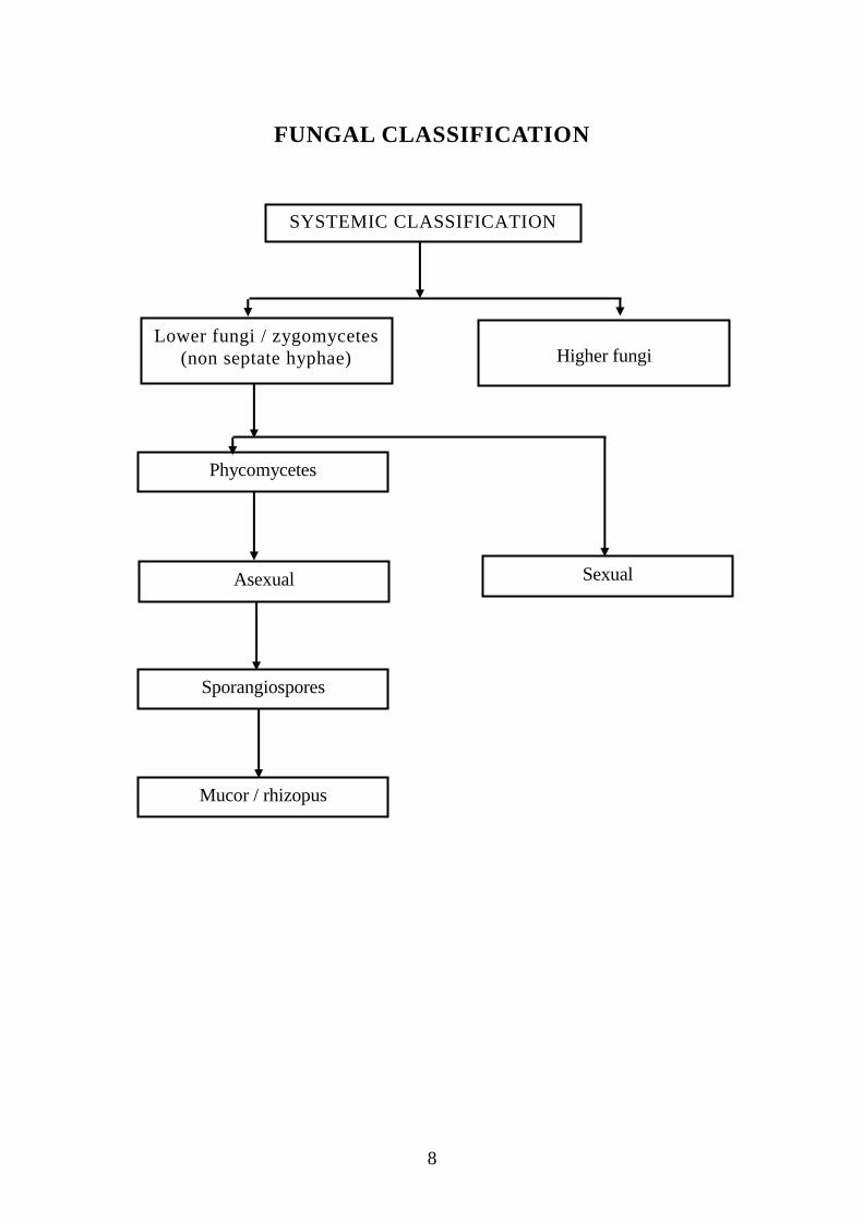
FUNGAL CLASSIFICATION
SYSTEMIC CLASSIFICATION
Lower fungi / zygomycetes
(non septate hyphae) Higher fungi
Phycomycetes
Asexual
Sporangiospores
Mucor / rhizopus
Sexual
9
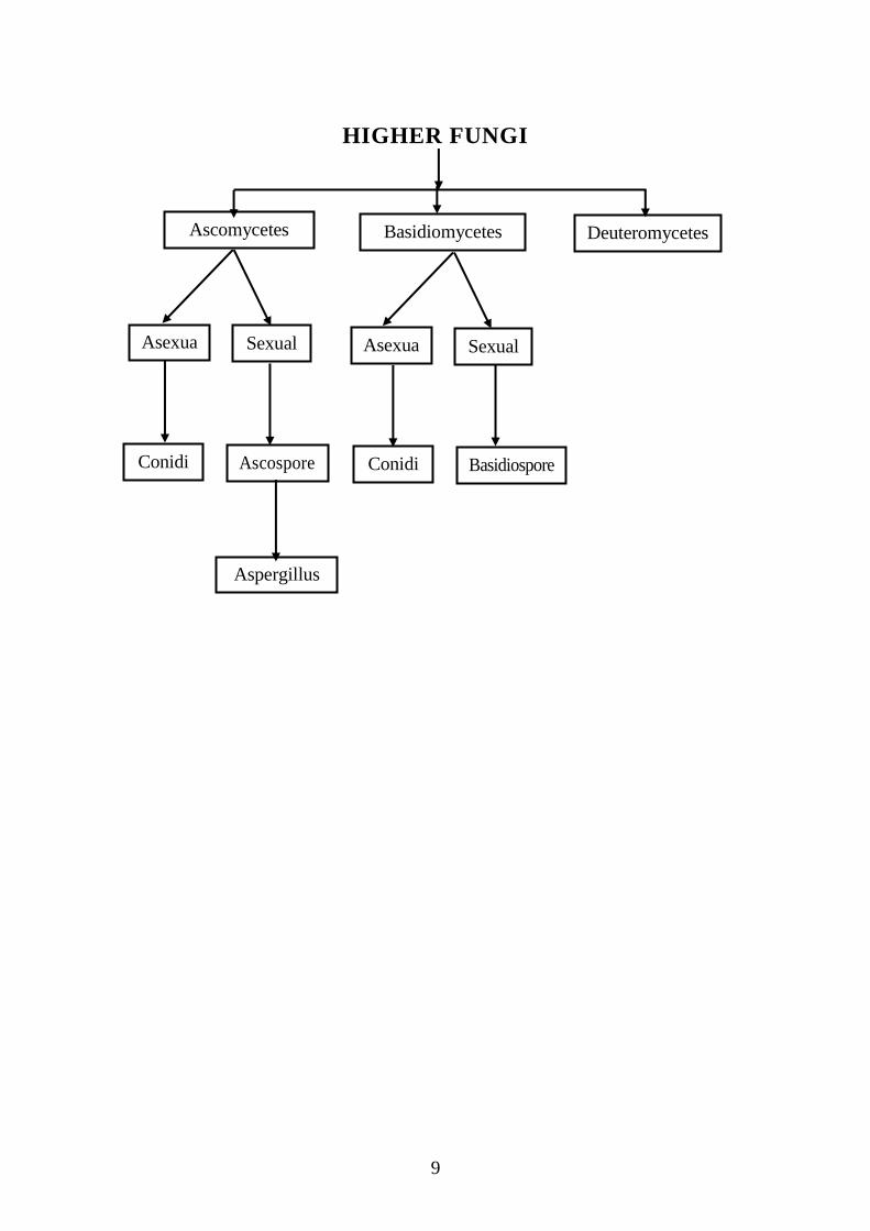
HIGHER FUNGI
Ascomycetes Basidiomycetes Deuteromycetes
Asexua
l
Sexual Asexua
l
Sexual
Conidi
a
Ascospore
s
Conidi
a
Basidiospore
s
Aspergillus
10
MYCOLOGY
MUCORMYCOSIS
“Mucormycosis is a disease caused by mucorales like Rhizopus,
rhizomucor, mucor, absidia, cunninghamella species etc.., most common
and virulent species is Rhizopus oryzae”12
.
Mucor causes vascular invasion and obliteration that leads to
ischemic necrosis.
Iron is essential for rhizopus growth. Since iron is strongly bound
to transferrin, normal human serum can inhibit the growth of Rhizopus
but in diabetics and DKA due to increased free iron and low pH
promotes its growth.12
Increased susceptibility in hemodialysis patients
is due to ability of the mucorales to extract iron from desferroxamine.
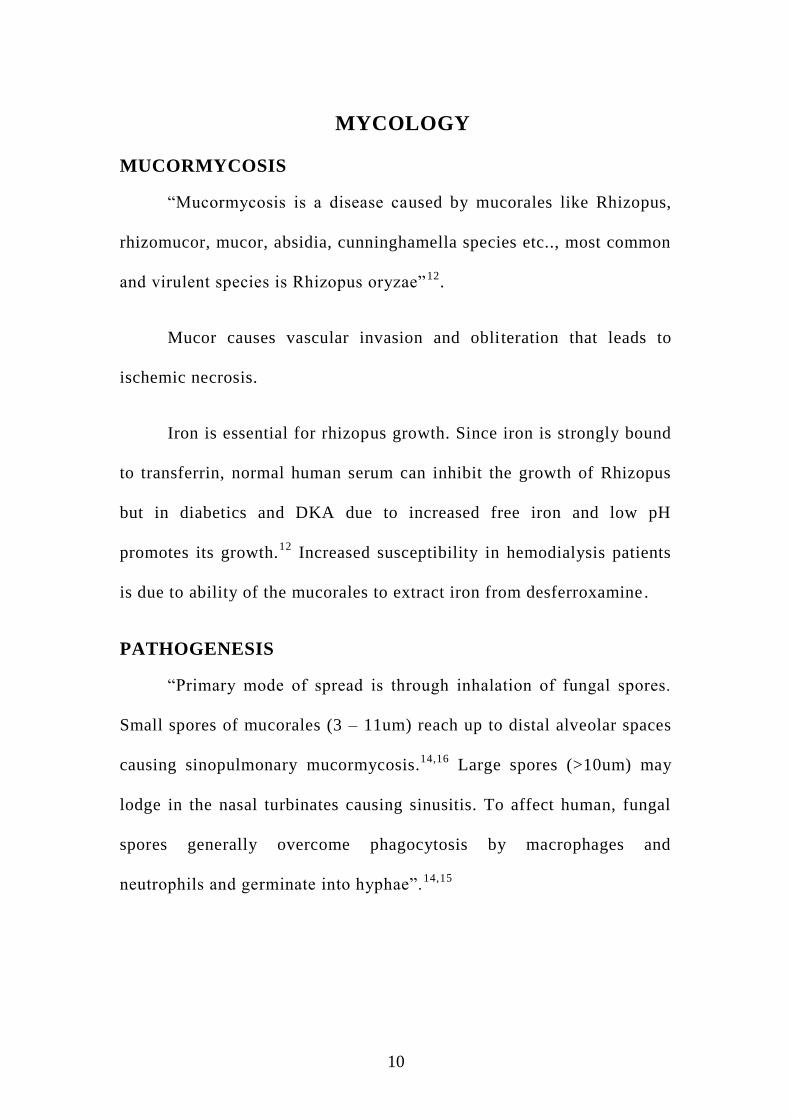
PATHOGENESIS
“Primary mode of spread is through inhalation of fungal spores.
Small spores of mucorales (3 – 11um) reach up to distal alveolar spaces
causing sinopulmonary mucormycosis.14,16
Large spores (>10um) may
lodge in the nasal turbinates causing sinusitis. To affect human, fungal
spores generally overcome phagocytosis by macrophages and
neutrophils and germinate into hyphae”.14,15
11
In chronic steroid users impairment of macrophage migration,
ingestion, phagolysosome fusion, chemotaxis occurs causing loss of
fungicidal activity against mucorales.
Recently it is found that sporangiospores are able to adhere to
subendothelial matrix proteins and invade intact endothelial barriers.
Rhizopus secretory component can be toxic to endothelial cells.
Therefore both spores and mycotoxins produced indirectly by
endosymbiotic bacteria influence the virulence of molds causing
mucormycosis5.
12
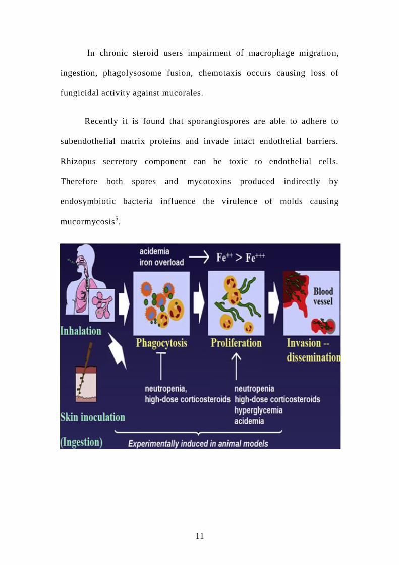
DIFFERENCE BETWEEN MUCOR AND RHIZOPUS
1) Rhizopus has rhizoid and is absent in mucor.
2) Sporangiospores of mucor arises randomly from along the aerial
mycelium whereas sporangiospores of rhizopus arises from
rhizoid.17,18
13
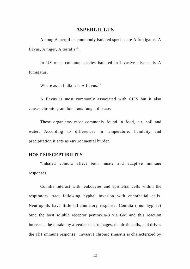
ASPERGILLUS
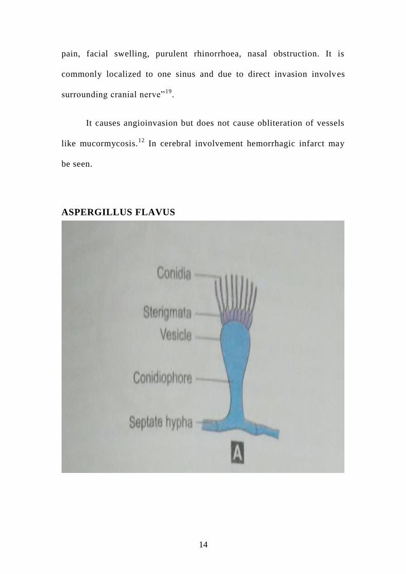
Among Aspergillus commonly isolated species are A fumigatus, A
flavus, A niger, A terralis19
.
In US most common species isolated in invasive disease is A
fumigatus.
Where as in India it is A flavus.12
A flavus is most commonly associated with CIFS but it also
causes chronic granulomatous fungal disease.
These organisms most commonly found in food, air, soil and
water. According to differences in temperature, humidity and
precipitation it acts as environmental burden.
HOST SUSCEPTIBILITY
“Inhaled conidia affect both innate and adaptive immune
responses.
Conidia interact with leukocytes and epithelial cells within the
respiratory tract following hyphal invasion with endothelial cells.
Neutrophils have little inflammatory response. Conidia ( not hyphae)
bind the host soluble receptor pentraxin-3 via GM and this reaction
increases the uptake by alveolar macrophages, dendritic cells, and drives
the Th1 immune response. Invasive chronic sinusitis is characterized by
14
pain, facial swelling, purulent rhinorrhoea, nasal obstruction. It is
commonly localized to one sinus and due to direct invasion involves
surrounding cranial nerve”19
.
It causes angioinvasion but does not cause obliteration of vessels
like mucormycosis.12
In cerebral involvement hemorrhagic infarct may
be seen.
ASPERGILLUS FLAVUS
15
ASPERGILLUS FUMIGATUS
16
DIAGNOSTIC APPROACHES:
DIAGNOSTIC NASAL ENDOSCOPY (DNE)
DNE should be done on all immunocompromised patients with
fever of unknown origin for more than 48 hours.
FINDINGS
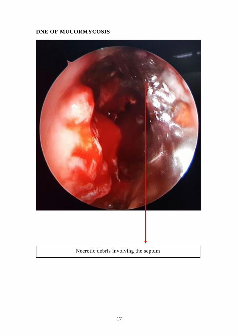
Most common and consistent findings are nasal mucosal
ulceration, discoloration and granulation.
1) Mucosal abnormalities are
a. Middle turbinate 67%
b. Septum 24%
c. Palate 19%
d. Inferior turbinate 10%
2) Decreased sensation, decreased mucosal bleeding
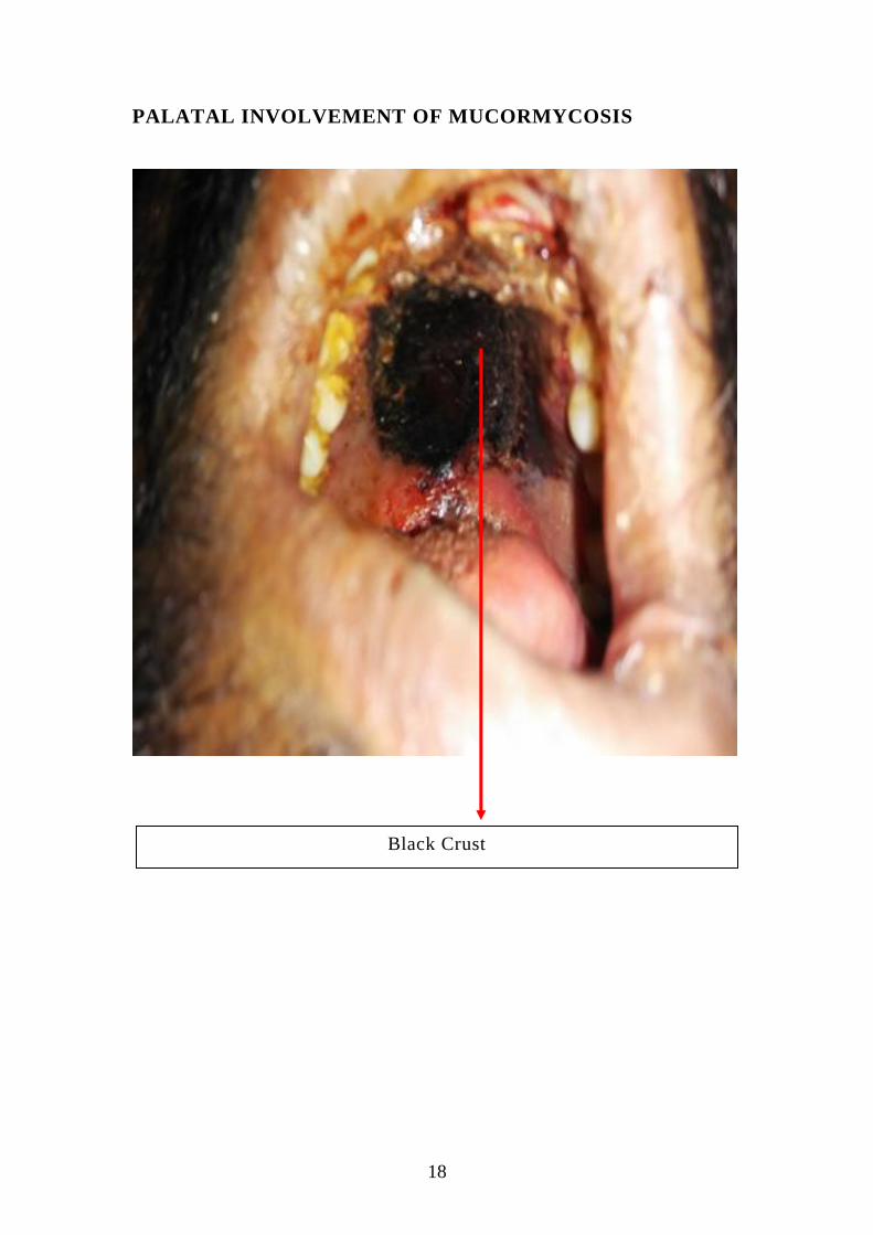
3) Black necrotic debris in nasal cavity or palate
Frank necrosis may give false negative results.MT biopsy results
100% specificity, 75% sensitivity in AIFS.13
17
DNE OF MUCORMYCOSIS
Necrotic debris involving the septum
18
PALATAL INVOLVEMENT OF MUCORMYCOSIS
Black Crust
19
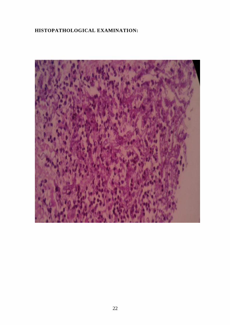
HISTOPATHOLOGICAL EXAMINATION (HPE)
Histopathologic evaluation of tissue biopsies is required to
confirm the diagnosis of invasive fungal rhinosinusitis.
“ Fungal disease is determined to be invasive if it meets the
following criteria on histopathologic examination
1) hyphal forms within the submucosa with or without angiocentric
Invasion
2) Tissue necrosis with minimal host inflammatory cell infiltration”.
Ideally, tissue should be sent for frozen and permanent sections.
Frozen section allows for a timely diagnosis, whereas permanent section
with Gomori-methenamine silver stain confirms the diagnosis and
provides important morphologic information that may be helpful in
determining the fungal species
MUCORMYCOSIS
“Definitive diagnosis of mucormycosis requires evidence of
fungal invasion. Tissue Hyphae of the Mucorales are broad (3–25 um),
thin-walled, mostly aseptate, with nondichotomous, irregular branching,
occasionally at right angles20
and sometime shows twisted and
compressed hyphae, which may be mistaken for septae, similar to
Aspergillus”.
20
Perineural invasion, which is found in 90% of tissues that contain
nerves, can be a useful diagnostic clue.
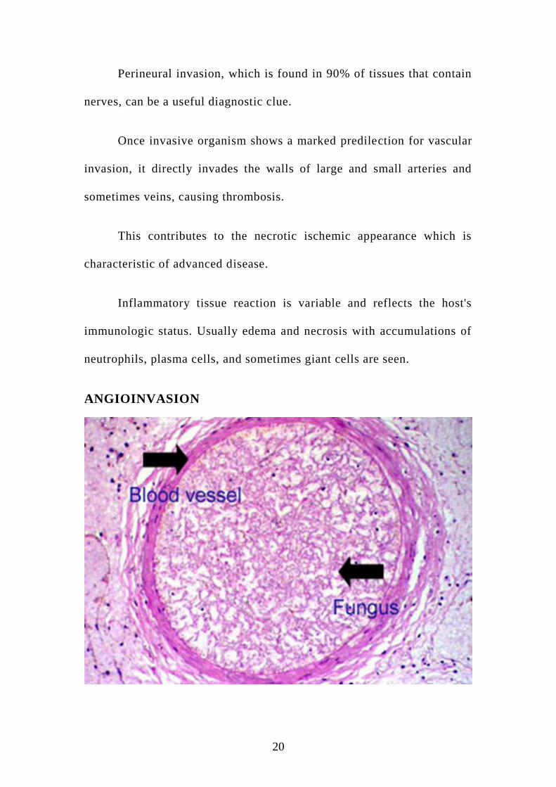
Once invasive organism shows a marked predilection for vascular
invasion, it directly invades the walls of large and small arteries and
sometimes veins, causing thrombosis.
This contributes to the necrotic ischemic appearance which is
characteristic of advanced disease.
Inflammatory tissue reaction is variable and reflects the host's
immunologic status. Usually edema and necrosis with accumulations of
neutrophils, plasma cells, and sometimes giant cells are seen.
ANGIOINVASION
21
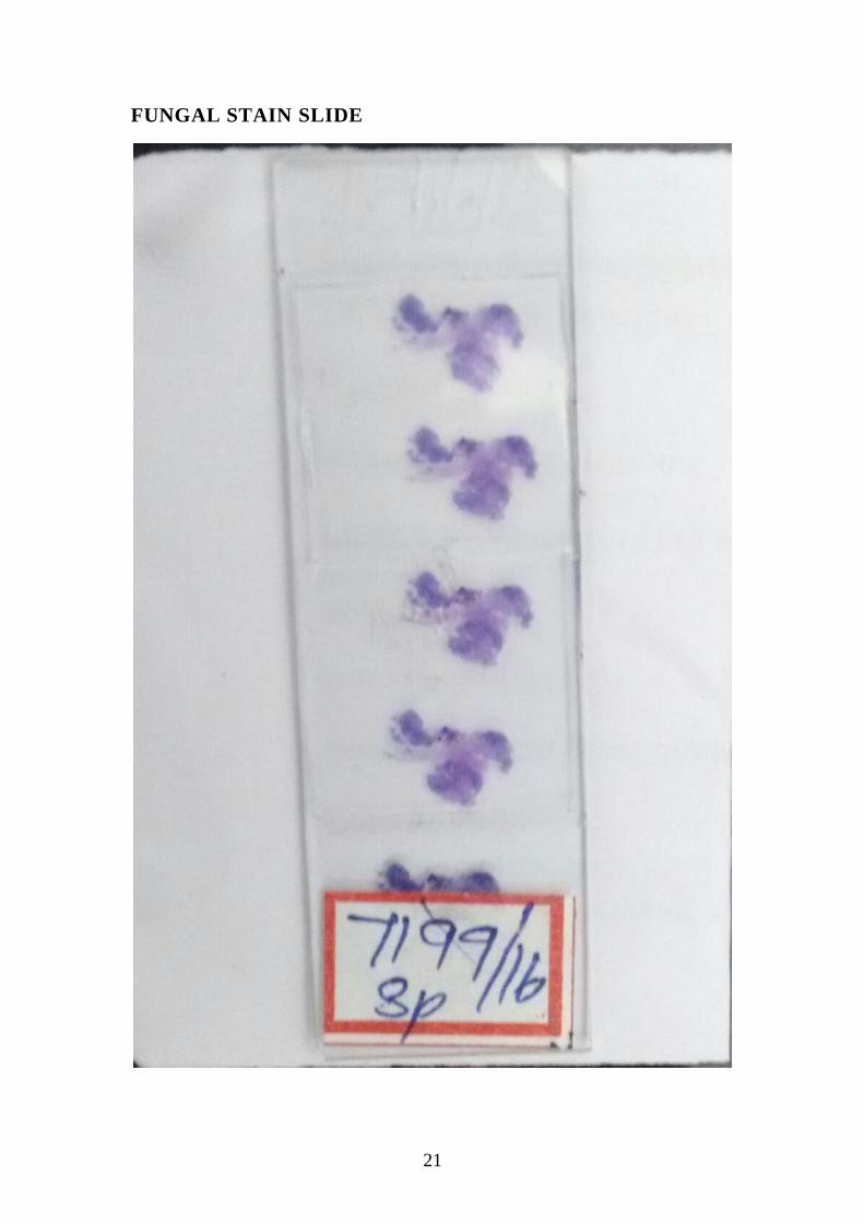
FUNGAL STAIN SLIDE
22
HISTOPATHOLOGICAL EXAMINATION:
23
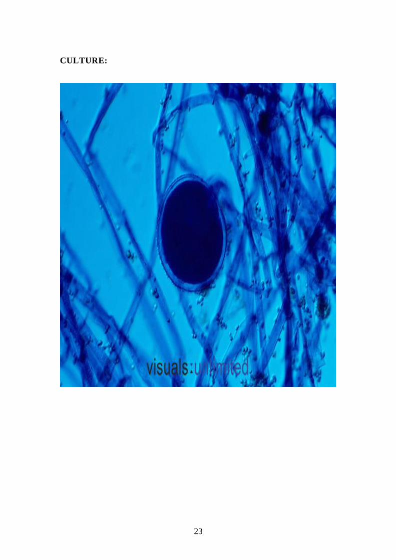
CULTURE:
24
ASPERGILLUS
It is described that gross appearance of granulomatous chronic
invasive fungal rhinosinusitis as firm, hard, rubbery, fibrous, grayish
white masses with an irregular surface.
Microscopically it is described that Aspergillus hyphae are
narrower, regular, frequently septated, and branch at 45°.
"Periarterial inflammation without direct involvement of fungal
elements and no true vascular invasion were noted.
It is classified into three variants
ii) proliferative (granulomatous pseudotubercles in a fibrous tissue
stroma),
iii) Exudative-necrotizing (with prominent foci of necrosis),
iv) Mixed form”.
Granulomatous chronic invasive fungal rhinosinusitis as
granulomas composed of eosinophilic material surrounded by fungus,
giant cells, and palisading nuclei, variable numbers of lymphocytes, and
plasma cells.20
Nongranulomatous chronic invasive fungal rhinosinusitis was
characterized by tissue necrosis with little inflammatory infiltrate and
dense hyphal accumulation resembling a fungus ball (mycetoma). The

25
fungi in this form may breach mucosal barriers to invade blood vessels
or simply cause an arteritis without vascular invasion, although both
granulomatous and nongranulomatous may result in tissue necrosis.
ASPERGILLUS VS MUCOR
CULTURE:
Sample for Culture can be taken from
Necrotic debris
Slough covering the nasal cavity
Crust
Purulent secretions in sinuses
Nasal swab for fungal culture can be insufficient

26
Alternatively aspirate from the irrigation of maxillary sinus with
small amount of saline can be used for fungal stain by KOH mount.
RADIOGRAPHY
CT PNS is most commonly used imaging to evaluate suspected
patients of invasive fungal rhinosinusitis.
Fine 2mm cut scan in both axial and coronal view is required.
“Clue to diagnosis are focal or diffuse areas of hyper attenuation
within a sinus fungal colonization, thick mucin plugs, infection and
AFS. It is important to note that both chronic invasive fungal
rhinosinusitis and AFS can cause bone erosion or expansion, suggesting
a potentially invasive process. It is noted that soft tissue infiltration of
periantral fat planes around the maxillary sinus provides early evidence
of invasive fungal rhinosinusitis in the appropriate Clinical setting”13
ADVANTAGES
1) Its ability to define bony architecture makes it as imaging of
choice for invasive fungal sinusitis.
2) Confirm the presence of sinusitis and type of sinus involved.
3) Bony architecture variations can be noted.
4) May show features suggestive of invasive fungal disease
27
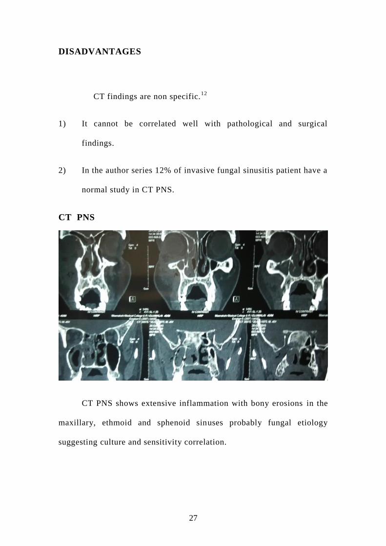
DISADVANTAGES
CT findings are non specific.12
1) It cannot be correlated well with pathological and surgical
findings.
2) In the author series 12% of invasive fungal sinusitis patient have a
normal study in CT PNS.
CT PNS
CT PNS shows extensive inflammation with bony erosions in the
maxillary, ethmoid and sphenoid sinuses probably fungal etiology
suggesting culture and sensitivity correlation.
28
MAGNETIC RESONANCE IMAGING (MRI) OF BRAIN
MR imaging is useful for assessing dural involvement and
intradural extension of disease."
ADVANTAGES
1) It is superior to CT to see the extent of intracranial and intra
orbital invasion like orbital apex syndrome, seizures, stroke.
Mortality rate is high in patient with intra cranial extension .
2) MR imaging scan can prevent unnecessary procedures in which patients
are unlikely to undergo surgery.
29
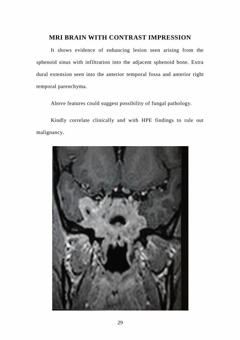
MRI BRAIN WITH CONTRAST IMPRESSION
It shows evidence of enhancing lesion seen arising from the
sphenoid sinus with infiltration into the adjacent sphenoid bone. Extra
dural extension seen into the anterior temporal fossa and anterior right
temporal parenchyma.
Above features could suggest possibility of fungal pathology.
Kindly correlate clinically and with HPE findings to rule out
malignancy.
30
TREATMENT
The mainstay of treatment of invasive fungal rhinosinusitis
continues to be a combination of antifungal antibiotics, aggressive
surgical debridement and treating the underlying cause of
immunocompromised state.
Several patients demonstrated good results for surgery in the
treatment of invasive fungal rhinosinusitis. The surgical approach to
invasive fungal rhinosinusitis has changed over the years secondary to
the advancement in endoscopic sinus surgery. Extensive surgical
resections in the form of radical maxillectomy and craniofacial
resection, are not practiced now because of more use of endoscopic
sinus debridement. Radical resections to remove disease outside the
sinonasal cavity rarely achieve negative margins or improve long-term
survival.
SURGICAL DEBRIDEMENT
Surgical debridement comprises several important goals
1) Progression of the disease is being slowed down that allows time
for bone marrow recovery
2) Fungal load is reduced, which decreases the burden on recovering
neutrophils
31
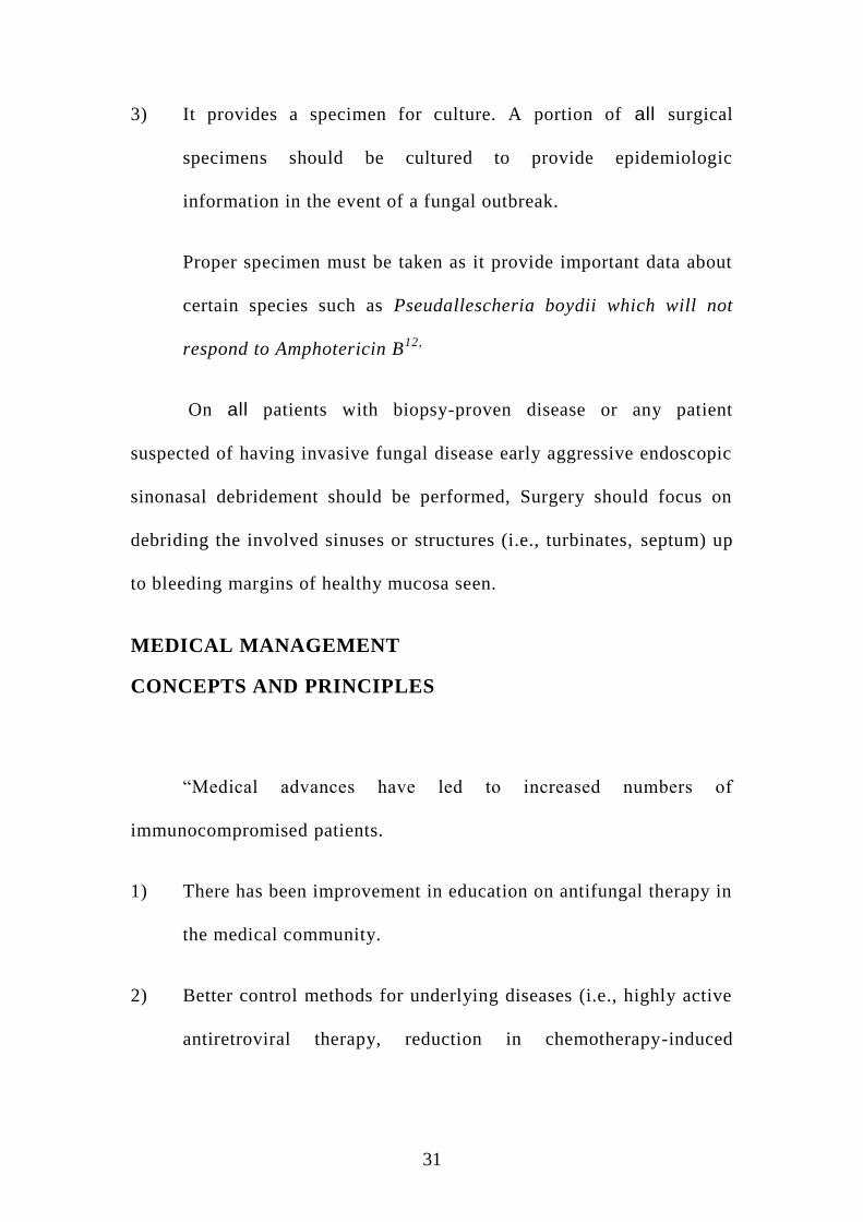
3) It provides a specimen for culture. A portion of all surgical
specimens should be cultured to provide epidemiologic
information in the event of a fungal outbreak.
Proper specimen must be taken as it provide important data about
certain species such as Pseudallescheria boydii which will not
respond to Amphotericin B12,
On all patients with biopsy-proven disease or any patient
suspected of having invasive fungal disease early aggressive endoscopic
sinonasal debridement should be performed, Surgery should focus on
debriding the involved sinuses or structures (i.e., turbinates, septum) up
to bleeding margins of healthy mucosa seen.
MEDICAL MANAGEMENT
CONCEPTS AND PRINCIPLES
“Medical advances have led to increased numbers of
immunocompromised patients.
1) There has been improvement in education on antifungal therapy in
the medical community.
2) Better control methods for underlying diseases (i.e., highly active
antiretroviral therapy, reduction in chemotherapy-induced
32
neutropenia, more options for anti rejection therapies) have been
developed.
3) Identification of specific risk factors or groups continues to be
identified.
4) Standardized antifungal susceptibility testing for yeasts is now
available for clinical decision
5) Safe triazoles (fluconazole and itraconazole)21
have demonstrated
a positive impact for antifungal prophylaxis, empiric, and
therapeutic strategies.
6) Improvements in the formulations of amphotericin B have
produced a less toxic product.21
7) Some important pivotal clinical studies in mycoses management
has been completed.
8) There continues to be enthusiasm in the pharmaceutical indus try
to identify new antifungal targets and drugs”.
ANTIFUNGAL TREATMENT PRINCIPLES
“Correct identification of the fungus is essential.
Use of standard, published antifungal regimens depends on fungus
identified and clinical syndrome.
33
Clinician should consider initial therapy as an induction phase
with optimization in both dose and antifungal drug, which gives
maximum fungicidal activity at the site of infection; consider
combination therapy in certain cases.
Control of the underlying medical or immunosuppressive
conditions is mandatory.
Clinician must pay particular attention to drug interactions,
pharmacokinetics, and resulting toxicities; this may require
measurement of drug levels in certain circumstances.
After apparent stabilization of clinical symptoms and signs of
infection with treatment, consideration of a consolidating drug
regimen in dose or drug to complete a defined course of therapy is
required.
Follow-up for relapse/reinfection after treatment should be at least
6 months to a year depending on fungus and type of infection.”
34
PHARMACOLOGY
AMPHOTERICIN B
High-dose Amphotericin B (greater than 1.25 mg/kg per day) is an
important adjunct in the treatment of invasive fungal rhinosinusitis.
A full course of Amphotericin B involves a total dose of 2 grams
or greater."
MECHANISM OF ACTION
The antifungal activity of amphotericin B is caused by its ability
to bind preferentially to ergosterol, a major component of the fungal cell
membrane.
Cell membrane permeability is increased following attachment of
this lipophilic structure to the fungal cell wall, with leakage of
intracellular components and ultimately cell death.21,22
Unfortunately, amphotericin B also binds to a lesser degree to
cholesterol in mammalian cell membranes, which probably accounts for
its toxic effects on human cells. Furthermore, amphotericin B and its
interaction with host cells also can have a positive effect by activation
of macrophages through an oxidation-dependent process.
35
DOSAGE
Since Amphotericin B produces hypersensitivity react ions, a test
dose (l mg in 50 mL of 5% dextrose over 20 minutes) to be given
before the administration of the first full dose of amphotericinB.
During this time, vital signs are monitored for any potential
reactions to this agent.
The dose of amphotericin B is not modified for patients with renal
insufficiency. Alternative regimens in such patients, however, have
included the use of alternate daily dosing (i.e., every other day dosing of
twice the usual daily target dose).
COMPLICATIONS
Infusion related reactions: Infusion-related reactions are acute in
onset and extremely common in individuals receiving amphotericin B.
Symptoms are characterized most commonly by fever, chills, rigors,
headache, nausea, malaise, and generalized aches.22
Nephrotoxicity: Nephrotoxicity may occur in 80% of patients
receiving amphotericin B therapy and is characterised by azotemia,
electrolyte wasting (K+and Mg2+) and decrease in urinary concentrating
ability. The mechanism by which this occurs is not well -defined. Renal
tubular acidosis has also been associated with amphotericin B therapy.
36
Although renal dysfunctions has been said to be reversible even
after high cumulative doses, persistent renal dysfunction was
documented among them. Particularly patients receiving a total dose
greater than 4 to 5 g of amphotericin B deoxycholate may have
permanent renal impairment.
STRATEGY TO AVOID NEPHROTOXICITY:
The approaches to minimize the risk of nephrotoxicity include
1) “Sodium supplementation to maintain intravascular volume and
inhibit the tubuloglomerular feedback system. Sodium
supplementation can be administered by way of a normal saline
bolus, 500 to 1000 mL in adult patients administered before or
following the amphotericin infusion."
2) Another strategy is avoiding the use of concomitant nephrotoxic
agents.”
Following nephrotoxicity they may develop Anemia
(Normochromic, normocytic anemia) with decrease in hemoglobin
of up to 35% from baseline has been documented routinely
following extended therapy with amphotericin B.
37
The mechanism is direct suppression of erythropoietin production
that may occur in patients with deteriorating renal function. Hemoglobin
concentrations usually return to normal within months after therapy is
discontinued.
LIPID BASED FORMULATIONS OF AMPHOTERICIN B:
Now a days, lipid-based formulations of amphotericin B has been
widely used. Amphotericin B lipid complex (ABLC) amphotericin B
cholesteryl sulfate complex (ABCD) and liposomal amphotericin B use
a variety of lipid carriers.
The pharmacokinetics of lipid-based formulations of amphotericin
B is different from that of amphotericin B deoxycholate.
These lipid-based formulations are preferentially delivered into
reticuloendotheliaI tissues, such as the liver and spleen and lesser extent
to lungs.21,22
The use of lipid-based amphotericin B may be a good alternative
for patients who are unable to tolerate the nephrotoxicity associated with
amphotericin B deoxycholate or other serious drug interactions with
agents such as cyclosporine or tacrolimus. These preparations are
probably the drugs of choice for rhinocerebral zygomycosis, that
requires administration of high doses of polyenes after surgical
debridement. Its reduced toxicity and the other advantages says the

38
significant expense of these agents in the final outcome of most the
disease.
AZOLES: IMIDAZOLES AND TRIAZOLES
Although amphotericin B remains the gold standard for most
severe life threatening systemic fungal infections, imidazoles
(clotrimazole, ketoconazole, miconazole) and triazoles (fluconazole and
itraconazole) second generation triazoles (voriconazole,posaconazole)
gives antifungal activity against many fungal pathogens without the
serious nephrotoxic effects observed with amphotericin B administration
and have been shown to be effective in treatment of systemic mycoses.
MECHANISM OF ACTION
“The azole antifungals (which includes the imidazoles and the
triazoles) acts mainly by inhibiting the cytochrome P-450 dependent
enzyme lanosterol 14demethylase, which is necessary for the conversion
of lanosterol to ergosterol.22
Ergosterol is a vital component of the
cellular membrane of fungi and disruptions in the biosynthesis of
ergosterol cause significant damage to the cell membrane by increasing
its permeability and ultimately causing cell lysis and cell death.
The antifungal activities of presently used azoles, generally are
considered fungistatic and achieve clinical concentrations when tested in
vitro. voriconazole more potent against invasive aspergillosis .22
39
SIDE EFFECTS
Triazoles are well tolerated in most patients.
The most commonly documented side effects are
Gastrointestinal upset, including symptoms such as nausea,
abdominal pain, vomiting, and diarrhea.
rash and headache
Mild elevations in liver function tests have been reported in
approximately 1% to 7% of patients
Alopecia has been reported
Doses of itraconazole of 600 mg/d and higher have a relatively
high incidence of an aldosterone-like effect with hypertension,
hypokalemia, and peripheral extremity edema.
Peripheral edema of lower legs and feet also can be seen in lower
limb
Visual abnormalities in voriconazole.22

40
TREATMENT OF UNDERLYING DISORDERS OF
IMMUNOCOMPROMISED STATE:
Insulin for diabetic patients and control of glycemic index.
Immunomodulators for leukemia and neutropenia patients.
Monitoring of CD 4 count for HIV patients and antiretroviral
therapy
Avoid long term steroids.
41
AIMS AND OBJECTIVES
1) To assess modes of presentation and complications.
2) To study the appropriate diagnostic approach for early detection
3) To study the various management plans and their efficacy
42
MATERIALS AND METHODOLOGY
STUDY PLACE
Rajiv Gandhi Government General Hospital, Chennai – 600003.
COLLABORATING DEPARTMENT
Upgraded Institute of Otorhinolaryngology
STUDY DESIGN
Retrospective and Prospective study
STUDY PERIOD
NOVEMBER 2014 to SEPTEMBER 2016
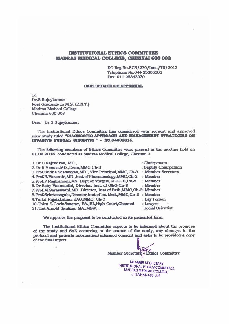
ETHICAL CLEARANCE
Applied for Institutional clearance
INCLUSION CRITERIAS
1) All patients who presents with clinical features suggestive of invasive
fungal sinusitis and was subsequently diagnosed as the same
2) Invasive fungal sinusitis with intracranial complications
EXCLUSION CRITERIA
Age below 20 yrs
INVESTIGATION
1) Diagnostic nasal endoscopy and biopsy
2) Fungal culture
3) CT- PNS
4) MRI
43
DATA COLLECTION
Clinical
BENEFIT TO THE COMMUNITY
1) Early detection of invasive fungal sinusitis
2) To reduce morbidity and mortality
Conflict of interest : nil
Financial support : nil
This is a prospective and retrospective study conducted in our
institution from November 2014 to September 2016. All patients who
presents with clinical features suggestive of invasive fungal sinusitis
included in the study. After clinical examination including routine blood
investigations, diagnostic nasal endoscopy biopsy with HPE and culture
followed to proceed with imagings like CT PNS and MRI brain was
planned if patient presented with complications. Treatment given was
debridement and antifungal agents and improving the
immunosuppressive state .
44
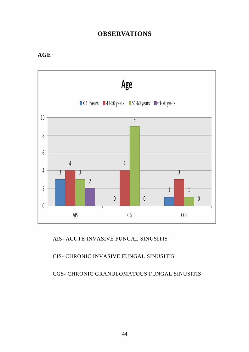
OBSERVATIONS
AGE
AIS- ACUTE INVASIVE FUNGAL SINUSITIS
CIS- CHRONIC INVASIVE FUNGAL SINUSITIS
CGS- CHRONIC GRANULOMATOUS FUNGAL SINUSITIS
45
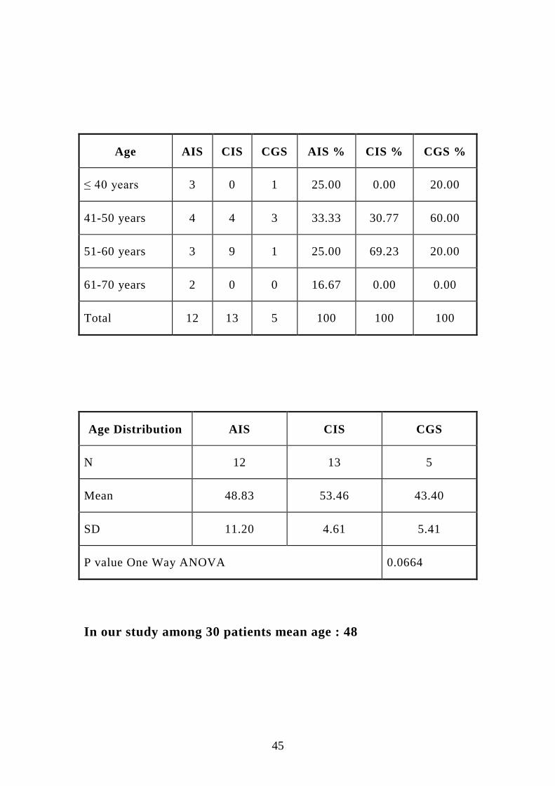
Age AIS CIS CGS AIS % CIS % CGS %
≤ 40 years 3 0 1 25.00 0.00 20.00
41-50 years 4 4 3 33.33 30.77 60.00
51-60 years 3 9 1 25.00 69.23 20.00
61-70 years 2 0 0 16.67 0.00 0.00
Total 12 13 5 100 100 100
Age Distribution AIS CIS CGS
N 12 13 5
Mean 48.83 53.46 43.40
SD 11.20 4.61 5.41
P value One Way ANOVA 0.0664
In our study among 30 patients mean age : 48
46
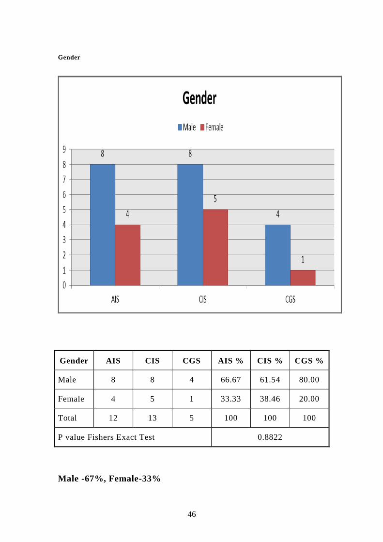
Gender
Gender AIS CIS CGS AIS % CIS % CGS %
Male 8 8 4 66.67 61.54 80.00
Female 4 5 1 33.33 38.46 20.00
Total 12 13 5 100 100 100
P value Fishers Exact Test 0.8822
Male -67%, Female-33%
47
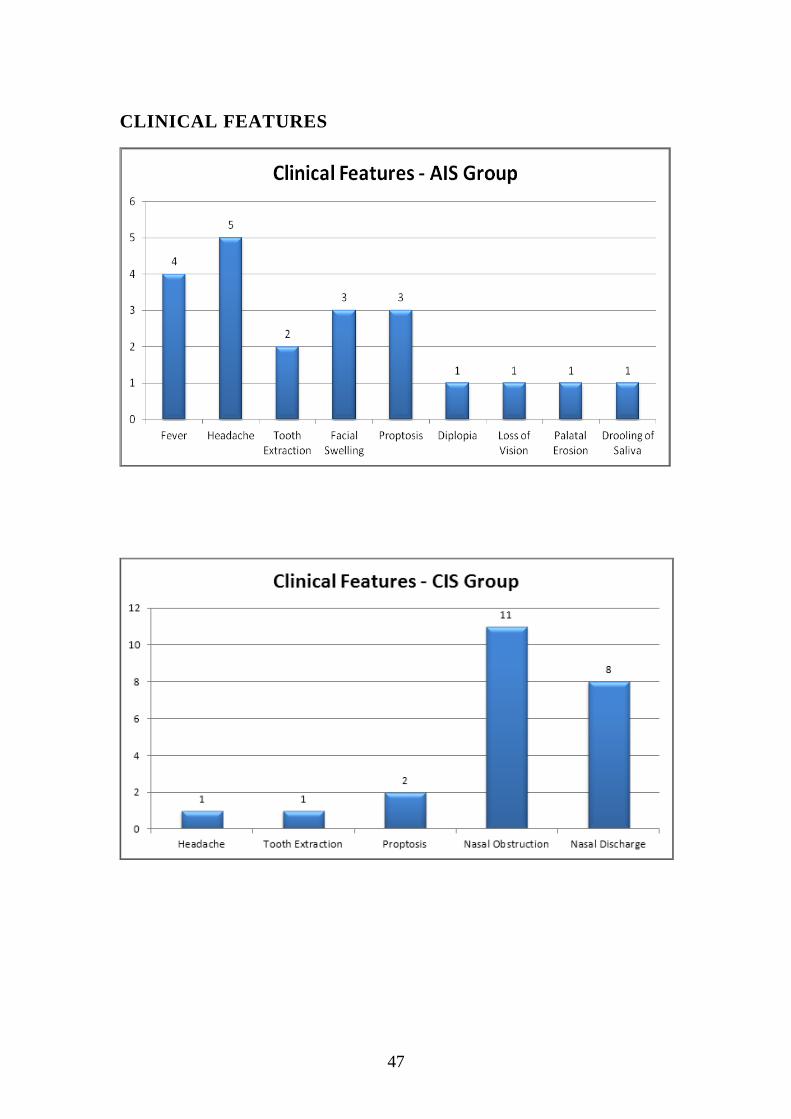
CLINICAL FEATURES
48
49
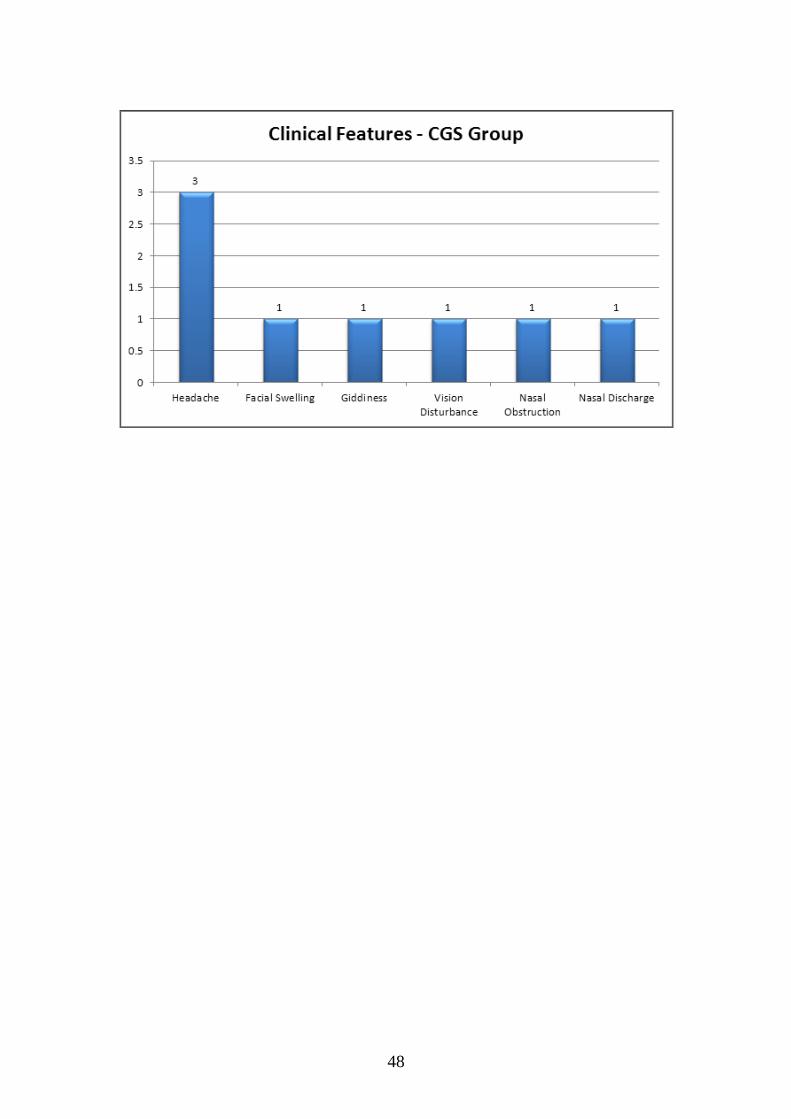
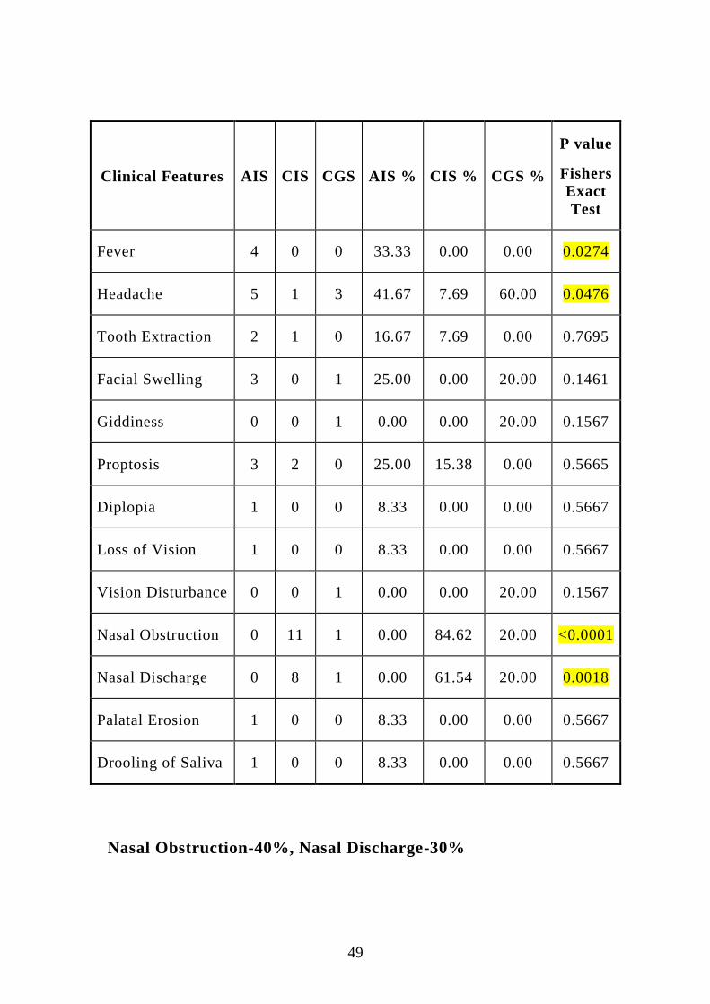
Clinical Features AIS CIS CGS AIS % CIS % CGS %
P value
Fishers
Exact
Test
Fever 4 0 0 33.33 0.00 0.00 0.0274
Headache 5 1 3 41.67 7.69 60.00 0.0476
Tooth Extraction 2 1 0 16.67 7.69 0.00 0.7695
Facial Swelling 3 0 1 25.00 0.00 20.00 0.1461
Giddiness 0 0 1 0.00 0.00 20.00 0.1567
Proptosis 3 2 0 25.00 15.38 0.00 0.5665
Diplopia 1 0 0 8.33 0.00 0.00 0.5667
Loss of Vision 1 0 0 8.33 0.00 0.00 0.5667
Vision Disturbance 0 0 1 0.00 0.00 20.00 0.1567
Nasal Obstruction 0 11 1 0.00 84.62 20.00 <0.0001
Nasal Discharge 0 8 1 0.00 61.54 20.00 0.0018
Palatal Erosion 1 0 0 8.33 0.00 0.00 0.5667
Drooling of Saliva 1 0 0 8.33 0.00 0.00 0.5667
Nasal Obstruction-40%, Nasal Discharge-30%
50
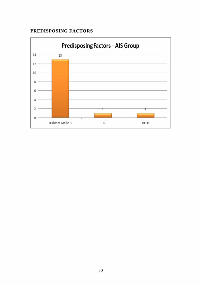
PREDISPOSING FACTORS

51

52
Predisposing
Factors AIS CIS CGS AIS % CIS % CGS %
P value
Fishers
Exact
Test
Diabetes Mellitus 13 12 1 108.33 92.31 20.00 0.0004
ABPA 0 0 1 0.00 0.00 20.00 0.1567
TB 1 0 0 8.33 0.00 0.00 0.5667
DCLD 1 1 0 8.33 7.69 0.00 >0.9999
Post renal
transplantation
on immune
suppressant
0 1 0 0.00 7.69 0.00 >0.9999
Nil 0 0 3 0.00 0.00 60.00 0.0028
DM-87% (Among these 8% were chronic steroid users)
53
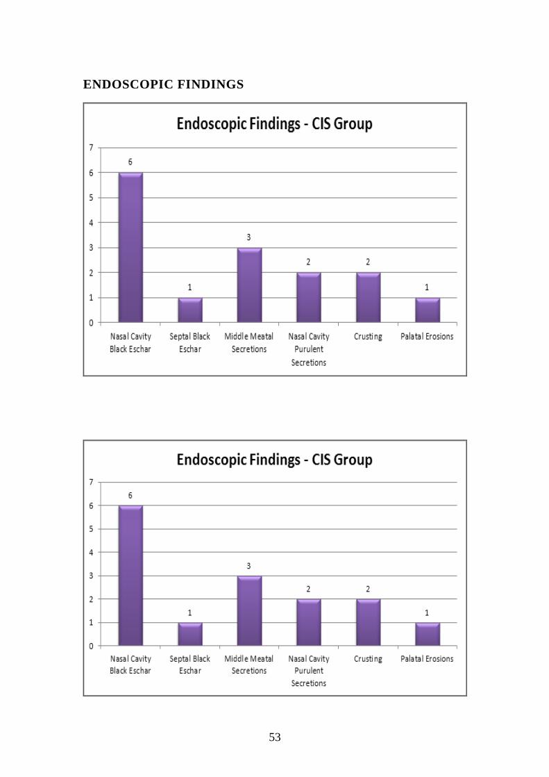
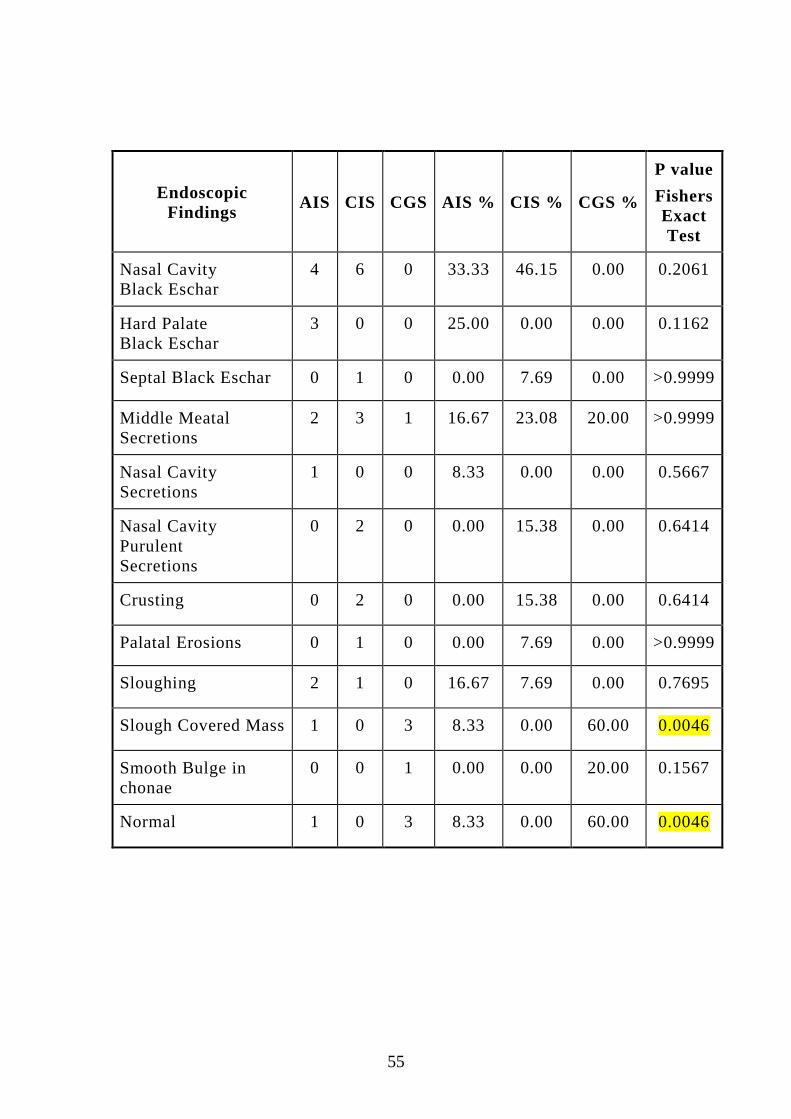
ENDOSCOPIC FINDINGS
54
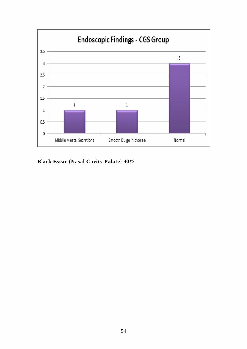
Black Escar (Nasal Cavity Palate) 40%
55
Endoscopic
Findings AIS CIS CGS AIS % CIS % CGS %
P value
Fishers
Exact
Test
Nasal Cavity
Black Eschar
4 6 0 33.33 46.15 0.00 0.2061
Hard Palate
Black Eschar
3 0 0 25.00 0.00 0.00 0.1162
Septal Black Eschar 0 1 0 0.00 7.69 0.00 >0.9999
Middle Meatal
Secretions
2 3 1 16.67 23.08 20.00 >0.9999
Nasal Cavity
Secretions
1 0 0 8.33 0.00 0.00 0.5667
Nasal Cavity
Purulent
Secretions
0 2 0 0.00 15.38 0.00 0.6414
Crusting 0 2 0 0.00 15.38 0.00 0.6414
Palatal Erosions 0 1 0 0.00 7.69 0.00 >0.9999
Sloughing 2 1 0 16.67 7.69 0.00 0.7695
Slough Covered Mass 1 0 3 8.33 0.00 60.00 0.0046
Smooth Bulge in
chonae
0 0 1 0.00 0.00 20.00 0.1567
Normal 1 0 3 8.33 0.00 60.00 0.0046
56
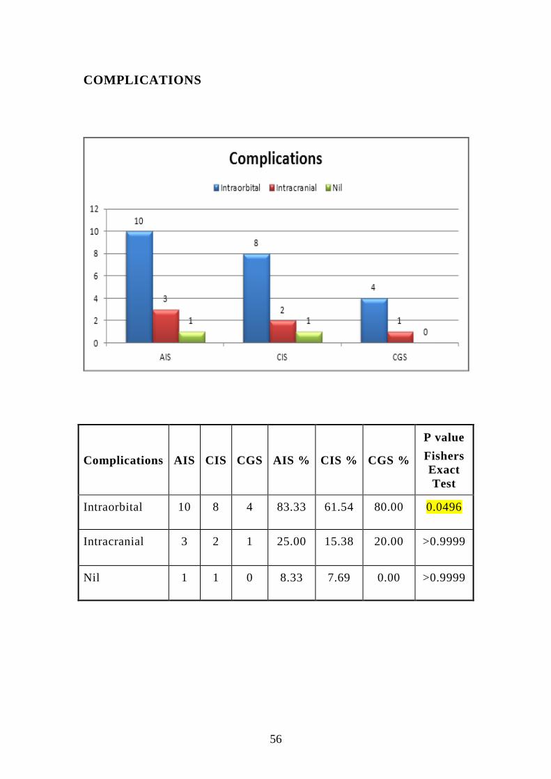
COMPLICATIONS
Complications AIS CIS CGS AIS % CIS % CGS %
P value
Fishers
Exact
Test
Intraorbital 10 8 4 83.33 61.54 80.00 0.0496
Intracranial 3 2 1 25.00 15.38 20.00 >0.9999
Nil 1 1 0 8.33 7.69 0.00 >0.9999

57
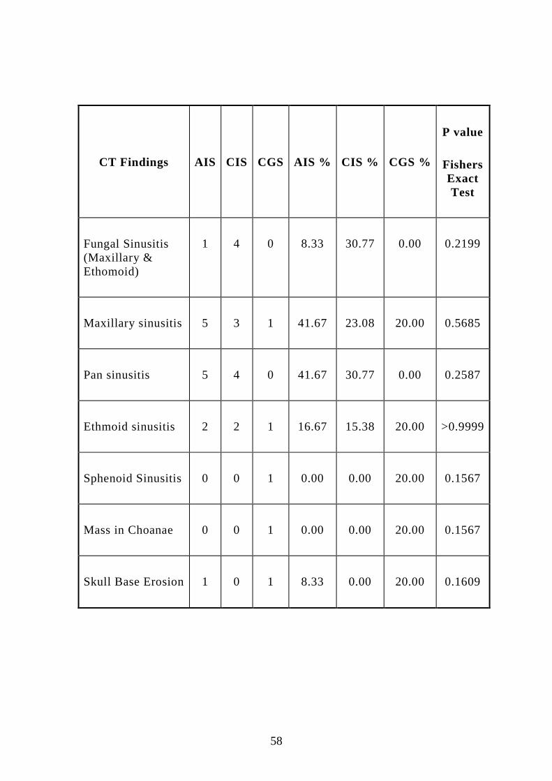
CT FINDINGS
58
CT Findings AIS CIS CGS AIS % CIS % CGS %
P value
Fishers
Exact
Test
Fungal Sinusitis
(Maxillary &
Ethomoid)
1 4 0 8.33 30.77 0.00 0.2199
Maxillary sinusitis 5 3 1 41.67 23.08 20.00 0.5685
Pan sinusitis 5 4 0 41.67 30.77 0.00 0.2587
Ethmoid sinusitis 2 2 1 16.67 15.38 20.00 >0.9999
Sphenoid Sinusitis 0 0 1 0.00 0.00 20.00 0.1567
Mass in Choanae 0 0 1 0.00 0.00 20.00 0.1567
Skull Base Erosion 1 0 1 8.33 0.00 20.00 0.1609

59
MRI FINDINGS
MRI Findings AIS CIS CGS AIS % CIS % CGS %
P
value
Fishers
Exact
Test
Intraorbital 4 5 1 33.33 38.46 20.00 0.8822
Intracranial 3 2 1 25.00 15.38 20.00 0.5574
CST 0 2 0 0.00 15.38 0.00 0.6653
Nil 3 6 3 25.00 46.15 60.00 0.3284
Intraorbital-33%, Intracranial-20%
60
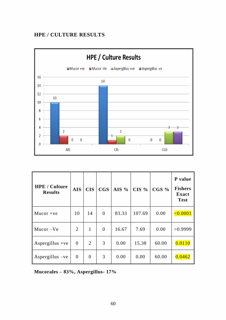
HPE / CULTURE RESULTS
HPE / Culture
Results AIS CIS CGS AIS % CIS % CGS %
P value
Fishers
Exact
Test
Mucor +ve 10 14 0 83.33 107.69 0.00 <0.0001
Mucor –Ve 2 1 0 16.67 7.69 0.00 >0.9999
Aspergillus +ve 0 2 3 0.00 15.38 60.00 0.0110
Aspergillus –ve 0 0 3 0.00 0.00 60.00 0.0462
Mucorales – 83%, Aspergillus- 17%
61
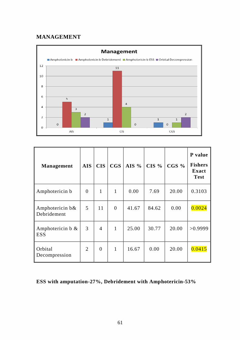
MANAGEMENT
Management AIS CIS CGS AIS % CIS % CGS %
P value
Fishers
Exact
Test
Amphotericin b 0 1 1 0.00 7.69 20.00 0.3103
Amphotericin b&
Debridement
5 11 0 41.67 84.62 0.00 0.0024
Amphotericin b &
ESS
3 4 1 25.00 30.77 20.00 >0.9999
Orbital
Decompression
2 0 1 16.67 0.00 20.00 0.0415
ESS with amputation-27%, Debridement with Amphotericin-53%
62
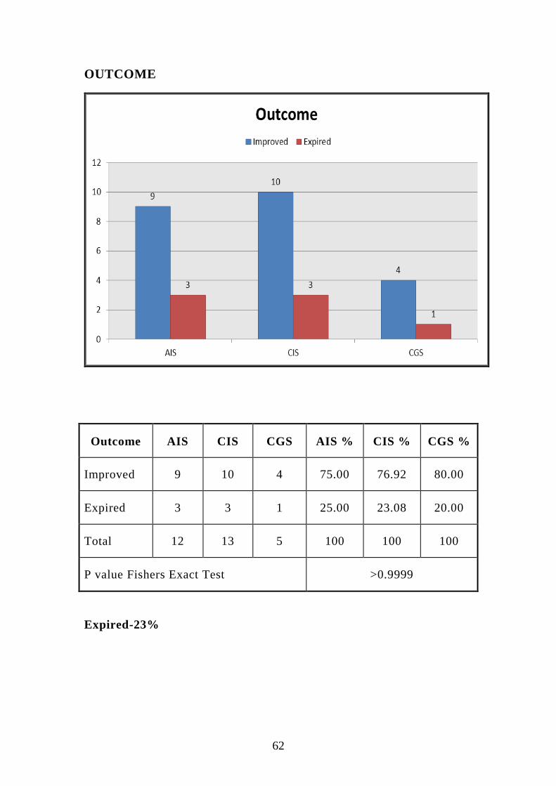
OUTCOME
Outcome AIS CIS CGS AIS % CIS % CGS %
Improved 9 10 4 75.00 76.92 80.00
Expired 3 3 1 25.00 23.08 20.00
Total 12 13 5 100 100 100
P value Fishers Exact Test >0.9999
Expired-23%
63
REVIEW OF LITERATURE
Bhansali Postgrad Med J 2004;80:670–674. doi:
10.1136/pgmj.2003.016030.23
A cohort of 23 men and 12 women with a mean (SD) age of 47.3
(14.4) years (range 18–70 years) was studied. Five patients had type 1
diabetes mellitus, 29 had type 2 diabetes mellitus, and one had
secondary diabetes. Nine patients had ROCM as the first clinical
manifestation of diabetes. The mean (SD) blood glucose at presentation
was 20.6 (8.3) mmol/l (range 10.0 to 53.3 mmol/l) and 17 patients had
ketosis/ketoacidosis. Ophthalmic symptoms and signs were pronounced:
external ophthalmoplegia (89%), proptosis (83%), visual loss (80%),
chemosis (74%), and eye lid gangrene (14%). Non-ophthalmic
manifestations included sinusitis (100%), nasal discharge/ulceration
(74%), palatal necrosis (29%), cerebral lobe involvement (20%), and
hemiparesis (17%). Computed tomography/magnetic resonance imaging
showed involvement of paranasal sinuses in all patients with ethmoid
(86%) and maxillary (80%) sinuses being most frequently involved.
Orbital involvement was observed in 80% of patients with cavernous
sinus thrombosis in 11%, and internal carotid occlusion and
hydrocephalus in 3% each. All were treated with amphotericin B (3–3.5
g) and 26 (74%) patients underwent appropriate surgery. Twenty one
patients (68%) survived with a mean (SD) follow up of 39.6 (34.1)
64
months (range 10 months to 11 years). Factors related to poor survival
included delay in diagnosis and treatment facial or eye lid gangrene,
hemiplegia, (cerebral invasion by mucorales) treatment with
amphotericin B alon.
Yohai RA, Bullock JD, Aziz AA, et al. Survival factors in rhino-
orbital-cerebral mucormycosis: major review. Surv Ophthalmol
1994;39:3–22.24
Yohai et al reviewed 145 case reports of ROCM, 60% of them had
diabetes, and analysed their ophthalmic and nonophthalmic signs and
symptoms occurring at any time during the course of disease.
particularly with regard to facial swelling (46% v 30%), facial
parasthesias (34% v 20%), nasal ulceration or necrosis (74% v 48%),
palatal necrosis (29% v 32%), and infranuclear facial palsy (46% v
22%).
Cavernous sinus thrombosis usually results from spread of
infection from the orbit and appears as a filling defect within the
enhancing sinus or as a lateral convexity, was evident in 11% which was
comparable with others.
Ferry AP, Abedi S. Diagnosis and management of rhino-
orbitocerebral mucormycosis (phycomycosis): a report of 16 personally
observed cases.25

65
Ophthalmology 1983;90:1096–104. Among 13 cases, 81% of them
had diabetes, visual loss. 80% reported black eschar of skin, nasal
mucosa, or palate in only 19% of their patients
Otolaryngology–Head and Neck Surgery (2010) 143, 614-62026
“Overall survival rate of the patients in the open surgery group (4
of 7; 57.1%) was similar to that of the endoscopically treated group (9
of 19; 47.3%). Thirteen patients (50%) died of complications related to
the underlying disease (9 of 13; 69.2%) and AIFRS (4 of 13; 30.7%).
AIFRS-specific survival rate is 76.5 percent; 90 percent (9 of 10) and
57.1 percent (4 of 7) for endoscopic and open surgery groups,
respectively. Four patients who died had pathological diagnosis of
mucormycosis (P _ 0.52)”.
Seyda Karadeniz Ugurlu*, et al (Turk J Ophthalmol 2015; 45:
169-174)27
“Among four patients (1 female, 3 male; age range, 55-77 years)
all had diabetes mellitus and two also had chronic renal failure. All
patients exhibited proptosis, sinusitis, and dark-colored lesions on the
nasopharynx and/or hard palate; three patients had ipsilateral peripheral
facial paralysis. Visual loss with no light perception occurred in 2 cases
with severe orbital involvement and in 2 cases with limited orbital
involvement. Histopathological examination of the hard palate,
nasopharynx or sinus biopsy revealed typical Mucor hyphae.”
66
Systemic liposomal amphotericin B was initiated in all patients.
The patients with limited ocular involvement received amphotericin B
both intravenously and by local irrigation; both patients had complete
recovery. The other two patients underwent orbital exenteration; one
patient died after declining systemic treatment postoperatively. Rapid
diagnosis and treatment are important for the survival of rhino-orbital
mucormycosis patients. With orbital involvement, surgical debridement
and systemic and local treatment with antifungal agents may help avoid
mutilating surgery like exenteration.
World Neurosurgery, Intracranial fungal granuloma ;Arvind
dubey,MD, 2005.
22 year retrospective review of 40 patientsshows predominant
symptom headache (83%), vomiting (65%). Location of PNS (40)%
Frontal (25%), Anterior cranial foosa (20%), sellar/parasellar (15%).
HPE Shows aspergilloma (63%) mucormycosis (18%). Microbiology
analysis shows 60% Positive28
.
67
DISCUSSION
Among 30 cases in our study of invasive fungal sinusitis the
detected species were Mucorales and Aspergillus. 25 cases of
Mucormycosis and 5 cases of invasive aspergillosis were noted in our
study. Both can be causative factor for acute and chronic invasive
(granulomatous and non granulomatous) fungal sinusitis that have high
mortality and morbidity in immunocompromised patients. Acute cases
were presented with facial swelling with pain, fever followed by tooth
extraction in 2 cases. In chronic cases patient mostly presented wi th
nasal obstruction, nasal discharge, headache. Among 26 diabetic patients
87% (2 cases were chronic steroid users-8%), 6 cases (23%) were
freshly diagnosed in our institution. Even at the time of presentation
they can present with intra orbital, intracranial complications with poor
prognosis.
Only in chronic granulomatous form association of
immunosuppressive states (like diabetes mellitus) is limited in our
study. Among five patients with CGFS 1 case (20%) presented with DM,
1 case (20%) presented with ABPA (Allergic broncho pulmonary
Aspergillosis ) other 3 cases with no predisposing factors. All CGFS
cases diagnosed as Aspergillus species (100%-usually Aspergillus
flavus). After sphenoidotomy and orbital decompression 1 case with
POL negative improved to POL positive.
68
Evaluation of patients presenting with Black eschar in nasal
cavity and hard palate, with resistant fever, DKA, features suggestive of
fungal sinusitis with or without intraorbital or intracranial complicated
patients should be made early. Black eschar in middle turbinate, septum,
palate accounts 40% of cases that clinches diagnosis of Mucormycosis.
The medical treatment with Amphotericin B can be empirically started
before diagnostic biopsy and culture. Weekly debridement should be
done to remove the necrotic debris and to reduce the fungal load for the
patients.
In Kasapoglu et al study among 26 patients with 17 males and 9
females, 65% (17 cases) showed positive for mucormycosis and 35%
(9cases) showed aspergillous.26
In our study among 30 patients 20 males
(67%) and 10 females (33%), 87% are diabetic. The reported species
are 83% are mucor and 17% are aspergillus.
In his study 19 cases underwent endoscopic surgery and 7 cases
surgical debridement was done.26
His study also shows that 61.5% of
mucormycosis and 38.5% of aspergillus cases were expired.
69
In retrospective study of Bhansali et al among 35 patients 75%
(23cases) are males and 25% (12 cases) females. Among which 9
patients newly diagnosed as diabetic mellitus and 17 cases wi th diabetic
ketoacidosis.23
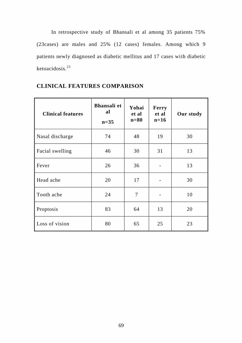
CLINICAL FEATURES COMPARISON
Clinical features
Bhansali et
al
n=35
Yohai
et al
n=80
Ferry
et al
n=16
Our study
Nasal discharge 74 48 19 30
Facial swelling 46 30 31 13
Fever 26 36 - 13
Head ache 20 17 - 30
Tooth ache 24 7 - 10
Proptosis 83 64 13 20
Loss of vision 80 65 25 23
70
Comparison of clinical features of our study with others showed
74% of nasal discharge in Bhansali et al and 30% by us.
46% of facial swelling had been observed in Bhansali23
et al and
13% by us.
Fever had been observed in 36% of cases in Yohai et al and 13%
in our study.24
30% cases showed headache in our study and 20% by Bhansali et al.
Ophthalmological findings like proptosis and loss of vision had
been reported highest of 83% and 80% in Bhansali respectively. Our
study showed 20 % proptosis and 23% of loss of vision.
Tooth ache of 24% had been reperted by Bhansali et al and 10%
by us.
The DNE finding in one patient in our study showed mass in the
choanal region. Biopsy taken from the mass showed granulomatous
lesion and when done with special stain (PAS) showed Aspergillus
species to our surprise. Due to intracranial complications with cranial
nerve palsy patient expired.In arvind dubey study shows predominant
organism was aspergillus was revealed in HPE as 63%, mucor 18%.28
71
CT FINDINGS
CT FINDINGS AIS CIS CGS PERCENTAGE
F/S/O Fungal sinusitis
(Maxillary & Ethomoid)
1 4 0 17%
Maxillary sinusitis 5 3 1 30%
Pan sinusitis 5 3 0 30%
Ethmoidal sinusitis 2 2 1 17%
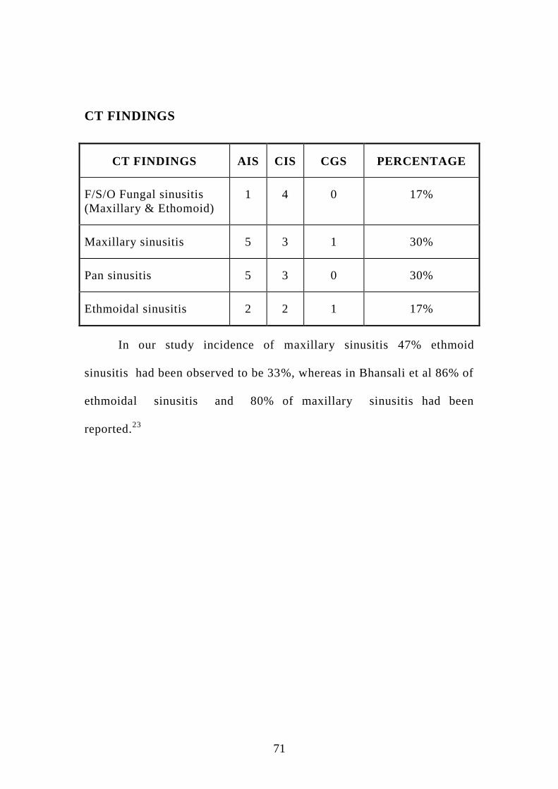
In our study incidence of maxillary sinusitis 47% ethmoid
sinusitis had been observed to be 33%, whereas in Bhansali et al 86% of
ethmoidal sinusitis and 80% of maxillary sinusitis had been
reported.23

72
COMPLICATIONS
In our study 73% cases developed intra orbital, 20% had intra
cranial, 7% had cavernous sinus thrombosis as complications. Bhansal et
al reported 80% of intra orbital and 11% of cavernous sinus thrombosis
complications. In our study among 6 intracranial complicated cases,
after debridement 2 cases ( 33%) improved well by debridement.
In our study for 2 cases flap cover was given for the defect in
forehead and medial canthal region and graft uptake was successful.
Among 30 cases 40% ended with no morbidty and all improved
well at 6 months of follow up. Among 6 intracranialy complicated
patients 2 cases were directly involved without intraorbital involvement.
CULTURE:
The culture results of our study showed 88% cases to be
mucormycosis out of 25 cases and 40% as aspergillus out of 5 cases
whereas kasapoglu et al reported 47% of mucormycosis out of 17 cases
and 89% aspergillus out of 9 cases.26
In spite of reduced viability of mucorales hyphae, the causative
agents of mucormycosis have been isolated highest by culture in our
institution due to accurate procedure followed in mycology department.
73
Out of 26 patients in Kasapoglu et al study 73% underwent
endoscopic sinus surgery and 27% were done with surgical
debridement.26
In our study of 30 cases 27% underwent endoscopic
sinus surgery and for 53% surgical debridement were done.
Among 30 cases one case of mucormycosis presented after post
renal transplant and on immunosupressent therapy (mycophenolate
mofetil) for past 5 years following which diabetic mellitus was detected.
Patient improved well after debridement, antifungal therapy and flap
cover over medial canthus region.
Among 13 cases of expired patients in Kasapoglu study 61% were
affected with mucormycosis and 39% with aspergillus.26
In our study of
30 cases 7 were expired. 86% found to have mucormycosis and 14%
with aspergillus. Among 7 expired cases 2 had decompensated liver
disease which was major cause of mortality than invasive fungal
sinusitis.
After introduction of Amphotericin B survival rate had been
improved up to 60%.27
It is said in Karadeniz et al study that Inspite of 4 cases with
orbital involvement close monitoring, medical therapy, debridement
gave good response in 2 cases.27
74
In our study many presented with orbital involvement who
showed good results with close monitoring, medical therapy and
debridement.Outcome of our study among 30 patients 7 cases were
expired (23%).
75
CONCLUSION
In conclusion, Diabetes mellitus is the most common
immunocompromised status to predispose to invasive fungal sinusitis .In
AIFS cases are presented with intraorbital complications than chronic
cases. AIFRS requires immediate medical and surgical treatment for faster
recovery and also to prevent intra cranial and intra orbital complications.
Chronic cases more commonly presented with nasal
obstruction,discharge.Subtypes of chronic form differentiated only by
HPE.Intracranial complications can occur without orbital involvement.
The combination of anti fungal therapy, surgical debridement and
improving immunosupressed state were proven to be highly efficacious
in the management of invasive fungal sinusitis. In these patients who
already have a poor health condition endoscopic approach is better which has
less trauma.
Combination of otorhinolaryngologist, radiologist, physician,
microbiologist, diabetologist, pathologist, ophthalmologist, mycologist plays a
major role for successful outcome.
8
BIBLIOGRAPHY
1) Paltauf A: Mycosis mucorina: Ein Beitrag zur Kenntniss der
menschlichen Fadenpilzerkrankungen.Virchows Arch 102:543-
564, 1885
2) Wright RE:Two cases of granuloma invading orbit due to
aspergillosis Br JOphthalmol 11:545-549,1927
3) Gregory]E, Golden A, Haymaker W: Mucormycosis of the central
nervous system: Report of three cases. Bulletin of the Johns
Hopkins Hospital 73:405-415, 1943
4) Harris JJ: Mucormycosis: Report of a case. Pediatrics 16:857-867,
1955
5) Hora JF: Primary aspergillosis of the paranasal sinuses and
associated areas. Laryngoscope 75:768-773, 1965
6) McGil11J, Simpson G, Healy GB: Fulminant aspergillosis of the
nose and paranasal sinuses. A new clinical entity. Laryngnoscope
90:748-754, 1980.
7) Waxman JE,Spector JG, Sale SR,et al: Allergic aspergillus
sinusitis: Concepts in diagnosis and treatment of a new clinical
entity. Laryngoscope 97:261-266, 1987
9
8) Rinaldi MG, Phillips P, Schwartz JG, et al: Human curvularia
infections: Report of five cases and review of the literature. Diagn
Microbiol Infect Dis 6:27-39, 1987
9) Milroy CM, Blanshard JD, Lucas S, et al: Aspergillosis of the
nose and paranasal sinuses. J Clin PathoI42:123-127, 1989
10) DeShazo RD, O'Brien M, Chapin K, et al: A new classification
and diagnostic criteria for invasive fungal sinusitis. Arch
Otolaryngol Head Neck Surg 123:1181-1188, 1997
11) David Kennedy, Diseases of the sinuses diagnosis and
management, 2001 edition, chapter 15:179-196
12) Cummings otolaryngology head and neck surgery 5th
& 6th
edition, vol 1, chapter 47,48
13) Ballenger’s manual of otorhinolaryngology- head and neck
surgery, 7th
edition
14) Ibrahim AS, Spellberg B, Walsh TJ et al. Pathogenesis of
mucormycosis, Clinical infectious disease 2012; 54 (supp 1): 516-
22
15) Dimitros Farmakiotis MDa, Dimitros P.Kontoyiannis MD SED
b –
mucormycosis- Infectious disease clinics of North America
10
16) Kontoyiannis DP, Lewis RE, Invasive zygomycosis- an update on
pathogenesis ; Clinical manifestations & management, Infectious
disease clinics of North America 2006; 20(3):581-607
17) Ananthanarayan & Panicker- Textbook of microbiology-9th
edition
18) Surinder kumar- Textbook of microbiology- 1st
edition,2012
19) Cadena, Jose, George R, Thompson and Thomas F. Patterson-
Invasive Aspergillosis: Current strategies for diagnosis and
management, Infectious disease clinics of North America 30.1
(2016):125-142
20) Robbins and Cotron- Pathologic basis of disease-9th
edition
21) KD Tripathi- Essentials of Medical Pharmacology-7th
edition
22) Katzung, Antony J.Trevor- Basic and clinical Pharmacology- 13th
edition
23) A Bhansali , S Bhadada, A Sharma, V Suresh, A Gupta, P Singh,
A Chakarbarti, R J Dash Postgrad Med J 2004;80:670–674. doi:
10.1136/pgmj.2003.016030
24) Yohai RA, Bullock JD, Aziz AA, et al. Survival factors in rhino-
orbital-cerebral mucormycosis: major review. Surv Ophthalmol
1994;39:3–22.
11
25) Ferry AP, Abedi S. Diagnosis and management of rhino-
orbitocerebral mucormycosis (phycomycosis): Ophthalmology
1983;90:1096–104
26) Fikret Kasapoglu, MD, Hakan Coskun, MD, Omer Afsin Ozmen,
MD, Halis Akalin, MD, and Beyza Ener, MD, Bursa,
Turkey,Otolaryngology–Head and Neck Surgery (2010) 143, 614-
620
27) Seyda Karadeniz Ugurlu, et al (Turk J Ophthalmol 2015; 45: 169-
174)
28) Arvind dubey, MD, Ravish V.Patwardhan,MD,Sammana sampth,
MD, Vani Santosh, MD, Sastri Kollri, MD, Anil Nanda, MD,
FACS; World Neurosurgery, Intracranial fungal granuloma-
63:254-260, 2005.
12
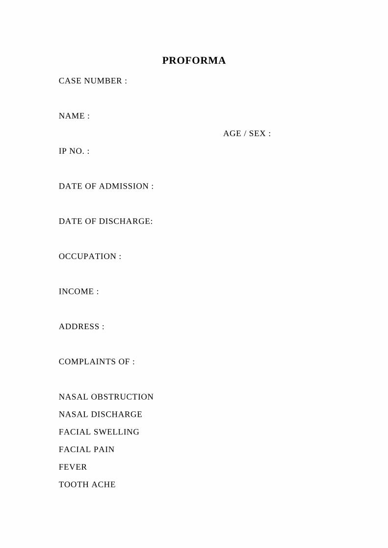
PROFORMA
CASE NUMBER :
NAME :
AGE / SEX :
IP NO. :
DATE OF ADMISSION :
DATE OF DISCHARGE:
OCCUPATION :
INCOME :
ADDRESS :
COMPLAINTS OF :
NASAL OBSTRUCTION
NASAL DISCHARGE
FACIAL SWELLING
FACIAL PAIN
FEVER
TOOTH ACHE
13
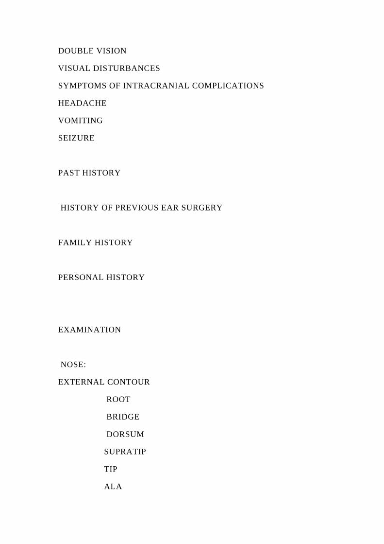
DOUBLE VISION
VISUAL DISTURBANCES
SYMPTOMS OF INTRACRANIAL COMPLICATIONS
HEADACHE
VOMITING
SEIZURE
PAST HISTORY
HISTORY OF PREVIOUS EAR SURGERY
FAMILY HISTORY
PERSONAL HISTORY
EXAMINATION
NOSE:
EXTERNAL CONTOUR
ROOT
BRIDGE
DORSUM
SUPRATIP
TIP
ALA
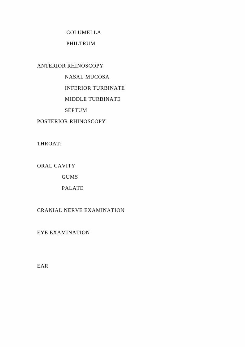
14
COLUMELLA
PHILTRUM
ANTERIOR RHINOSCOPY
NASAL MUCOSA
INFERIOR TURBINATE
MIDDLE TURBINATE
SEPTUM
POSTERIOR RHINOSCOPY
THROAT:
ORAL CAVITY
GUMS
PALATE
CRANIAL NERVE EXAMINATION
EYE EXAMINATION
EAR
15
DIAGNOSIS
PLAN
INVESTIGATIONS
COMPLETE HEMOGRAM
RENAL FUNCTION TESTS
CHEST X RAY
SEROLOGICAL TESTS
ECG
DIAGNOSTIC NASAL ENDOSCOPY
BIOPSY AND CULTURE
CT PNS
MRI BRAIN

16

17

MuhŒ¢á jftšjhŸ
br‹id uhé› fhªâ muR bghJ kU¤Jtkid¡F tU« _¡F¥gFâ
k‰W« mU»š cŸs gFâfS¡F gut¡Toa óŠir fhsh‹ ghâ¡f¥g£l
nehahËfis f©l¿í« Kiw k‰W« Ợir Kiwfis m¿ªJbfhŸs
nk‰bfhŸS« MŒî.
ïªj MuhŒ¢áÆš ghâ¡f¥g£l nehahËfis f©l¿ªJ mj‰nf‰g
mWit Ợir k‰W« kUªJfis mˤJ mªj Ợir KiwfË‹
j‹ikia¥ g‰¿ MuhŒtJ.
Ú§fŸ ïªj MuhŒ¢áÆš g§nf‰f eh§fŸ ÉU«ò»nwh«.
ïªj MuhŒ¢áÆ‹ Koîfis mšyJ fU¤J¡fis btËÆL«
nghnjh mšyJ MŒÉ‹ nghnjh j§fsJ bgaiunah mšyJ
milahs§fisnah btËÆlkh£nlh« v‹gijí« bjÇɤJ¡bfhŸ»nw«.
ïªj MŒÉš g§nf‰gJ j§fSila ÉU¥g¤â‹ ngÇšjh‹
ïU¡»wJ. nkY« Ú§fŸ vªneuK« ïªj MŒÉÈUªJ ã‹th§fyh«
v‹gijí« bjÇɤJ¡ bfhŸ»nwh«.
ïªj MuhŒ¢áÆ‹ Koîfis MuhŒ¢áÆ‹nghJ mšyJ MŒÉ‹
KoÉ‹ nghJ j§fS¡F m¿É¥ngh« v‹gijí« bjÇɤJ¡ bfhŸ»nwh«.
MŒthsÇ‹ ifbah¥g«MŒthsÇ‹ ifbah¥g«MŒthsÇ‹ ifbah¥g«MŒthsÇ‹ ifbah¥g« g§nf‰ghs® ifbah¥g«g§nf‰ghs® ifbah¥g«g§nf‰ghs® ifbah¥g«g§nf‰ghs® ifbah¥g«
njâ
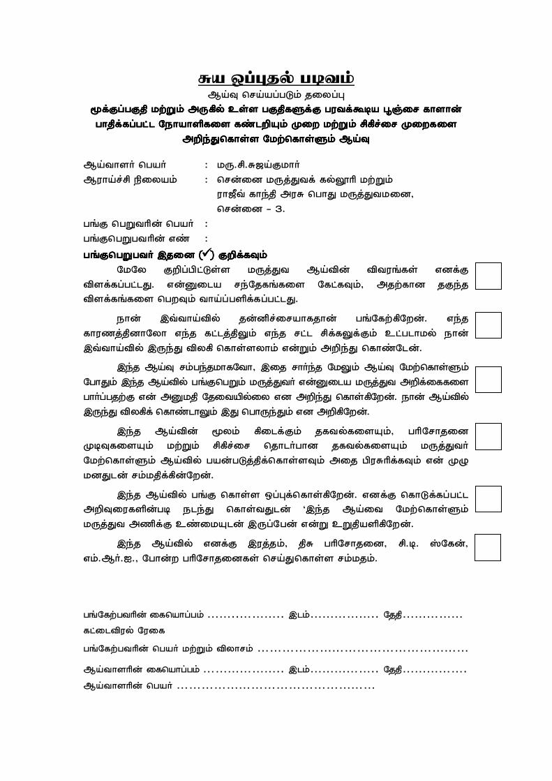
Ra x¥òjš got«
MŒî brŒa¥gL« jiy¥ò
_¡F¥gFâ k‰W« mU»š cŸs gFâfS¡F gut¡Toa óŠir fhsh‹ _¡F¥gFâ k‰W« mU»š cŸs gFâfS¡F gut¡Toa óŠir fhsh‹ _¡F¥gFâ k‰W« mU»š cŸs gFâfS¡F gut¡Toa óŠir fhsh‹ _¡F¥gFâ k‰W« mU»š cŸs gFâfS¡F gut¡Toa óŠir fhsh‹
ghâ¡f¥g£l nehahËfis f©l¿í« Kiw k‰W« Ợir Kiwfis ghâ¡f¥g£l nehahËfis f©l¿í« Kiw k‰W« Ợir Kiwfis ghâ¡f¥g£l nehahËfis f©l¿í« Kiw k‰W« Ợir Kiwfis ghâ¡f¥g£l nehahËfis f©l¿í« Kiw k‰W« Ợir Kiwfis
m¿ªJbfhŸs nk‰bfhŸS« MŒîm¿ªJbfhŸs nk‰bfhŸS« MŒîm¿ªJbfhŸs nk‰bfhŸS« MŒîm¿ªJbfhŸs nk‰bfhŸS« MŒî
MŒths® bga® : kU.á.R#ŒFkh®
MuhŒ¢á Ãiya« : br‹id kU¤Jt¡ fšÿÇ k‰W«
uhé› fhªâ muR bghJ kU¤Jtkid,
br‹id - 3.
g§F bgWtÇ‹ bga® :
g§FbgWgtÇ‹ v© :
g§FbgWgt® ïjid (g§FbgWgt® ïjid (g§FbgWgt® ïjid (g§FbgWgt® ïjid (���� ) F¿¡fî«) F¿¡fî«) F¿¡fî«) F¿¡fî«
nkny F¿¥ã£LŸs kU¤Jt MŒÉ‹ Étu§fŸ vd¡F
És¡f¥g£lJ. v‹Dila rªnjf§fis nf£fî«, mj‰fhd jFªj
És¡f§fis bgwî« thŒ¥gË¡f¥g£lJ.
eh‹ ï›thŒÉš j‹Å¢irahfjh‹ g§nf‰»nw‹. vªj
fhuz¤âdhnyh vªj f£l¤âY« vªj r£l á¡fY¡F« c£glhkš eh‹
ï›thŒÉš ïUªJ Éy» bfhŸsyh« v‹W« m¿ªJ bfh©nl‹.
ïªj MŒî r«gªjkhfnth, ïij rh®ªj nkY« MŒî nk‰bfhŸS«
nghJ« ïªj MŒÉš g§FbgW« kU¤Jt® v‹Dila kU¤Jt m¿¡iffis
gh®¥gj‰F v‹ mDkâ njitÆšiy vd m¿ªJ bfhŸ»nw‹. eh‹ MŒÉš
ïUªJ Éy»¡ bfh©lhY« ïJ bghUªJ« vd m¿»nw‹.
ïªj MŒÉ‹ _y« »il¡F« jftšfisí«, gÇnrhjid
Koîfisí« k‰W« Ợir bjhl®ghd jftšfisí« kU¤Jt®
nk‰bfhŸS« MŒÉš ga‹gL¤â¡bfhŸsî« mij ãuRÇ¡fî« v‹ KG
kdJl‹ r«kâ¡»‹nw‹.
ïªj MŒÉš g§F bfhŸs x¥ò¡bfhŸ»nw‹. vd¡F bfhL¡f¥g£l
m¿îiufË‹go elªJ bfhŸtJl‹ `ïªj MŒit nk‰bfhŸS«
kU¤Jt m¡F c©ikíl‹ ïU¥ng‹ v‹W cWâaË»nw‹.
ïªj MŒÉš vd¡F ïu¤j«, âR gÇnrhjid, á.o. °nf‹,
v«.M®.I., ngh‹w gÇnrhjidfŸ brŒJbfhŸs r«kj«.
g§nf‰gtÇ‹ ifbah¥g« ……..……….. ïl«…………….. njâ……………
f£ilÉuš nuif
g§nf‰gtÇ‹ bga® k‰W« Éyhr« ……………………………………………
MŒthsÇ‹ ifbah¥g« ……………….. ïl«…………….. njâ…………….
MŒthsÇ‹ bga® …………………………………………
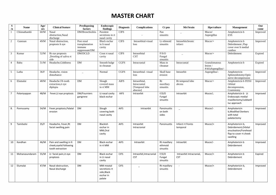
MASTER CHART
S
/n Name
Age/
sex Clinical feature
Predisposing
factors
Endoscopic
findings Diagnosis Complications Ct pns Mri brain Hpe/culture Management
Out
come
1 Chinnathambi 60/M Nasal
obstruction,Nasal discharge
DM/Bronchiolitis Purulent
secretions in rt nasal cavity
CIFS
-
Pan
sinusitis
_ Mucor/
Aspergillus
Amphotericin b
ESS
Improved
2 Ganesan 49/M Nasal obstruction,
proptosis lt eye
Post renal
transplantation immune
suppressant/DM
Black eschar
in lt nasal cavity
CIFS Intraorbital-visual
loss
Lt ethmoid
sinusitis
Intraorbitchronic
infarct
Mucor/+ Amphotericin b
Debridement ,Flap cover over lt medial
canthus
Improved
3 Kumar 52/M Rt eye proptosis
,Drooling of saliva rt side
DM/DCLD Crust rt nasal
cavity
CIFS Intraorbital
CST
F/S/O
Fungal sinusitis
_ Mucor/+ Debridement Expired
4 Babu 51/M Headache,Giddiness DM Smooth bulge
in choanae
CGFS Intracranial Mass in
choanae
Intracranial Granulomatous
lesion / PAS)Aspergillus/-
Amphotericin b Expired
5 Latha 36/F Headache, vision
disturbance
_ Normal CGFS Intraorbital- visual
loss
Skull base
erosion
Intraorbit Aspergillus/- Amphotericin
Sphenoidotomy/Optic
nerve decompression
Improved
6 Elumalai 48/M Headache f/b tooth
extraction,rt eye
diplopia
DM Slough
covered mass
in rt MM
AIFS Intraorbital,
Intracranial
[Temporal lobe abscess]
Rt
maxillary
sinusitis
Rt temporal lobe
abcess
Mucor/+ Amphotericin b FESS/
Orbital
decompression, Craniotomy
Improved
7 Palaniyappan 48/M Fever,Lt eye proptosis DM/Fourniers gangrene
Lt nasal cavity black eschar
AIFS Intraorbit F/S/O Fungal sinusitis
Intraorbit Mucor/+ Amphotericin b Lt Endoscopic medial maxillectomy/caldwell luc approach
Improved
8. Ponnusamy 54/M Fever,proptosis,Palatal erosion
DM Slough covering both nasal cavity
AIFS intraorbit Pansinusitis on both sides
_
Mucor/- Amphotericin b,Modified Denkers with total palatectomy
Improved
9 Tamilselvi 35/F Headache, Fever,Rt facial swelling,pain
DM Blackish eschar in MM,Oral cavity
AIFS Intraorbit Intracranial
Pansinusitis Infarct rt fronto temporal
Mucor/+ Amphotericin b, Debridement,Orbital enucleation/Forehead flap to cover rt cheek swelling
Improved
10 Kandhan 46/M Pain and swelling in lt cheek,eyelid following tooth extraction
DM Black eschar in rt MM
AIFS Intraorbit Rt maxillary ethmoid sinusitis
Intraorbit Mucor/- Amphotericin b Debridement
Improved
11 Mohanasundaram 55/M Lt facial pain,Lt eye proptosis
DM Black eschar in Lt nasal cavity
CIFS Intraorbit,Intracranial CST
F/S/O Fungal sinusitis
Intraorbit Intracranial, CST
Mucor/+ Amphotericin b Debridement
Expired
12 Elumalai 47/M Nasal obstruction, Nasal discharge
DM MM meatal secretions rt side,Black eschar in palate
CIFS _ Rt maxillary sinusitis
- Mucor/+ Amphotericin b, Debridement
Improved
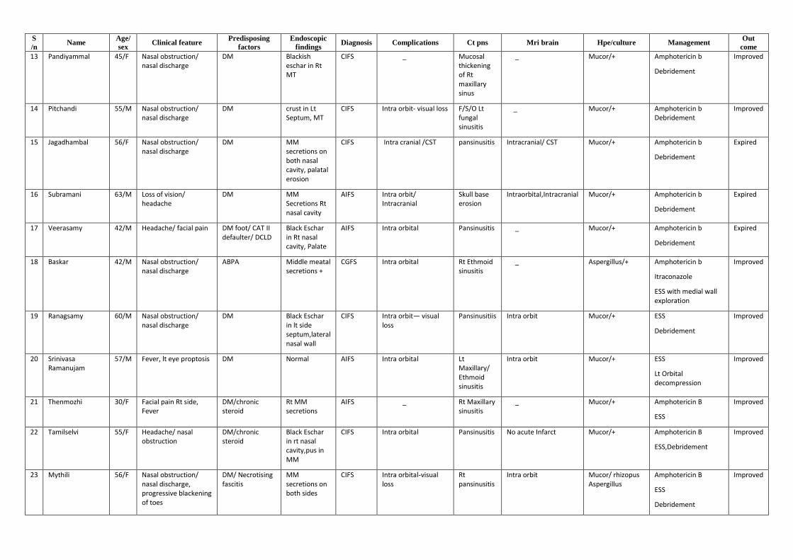
S
/n Name
Age/
sex Clinical feature
Predisposing
factors
Endoscopic
findings Diagnosis Complications Ct pns Mri brain Hpe/culture Management
Out
come
13 Pandiyammal 45/F Nasal obstruction/ nasal discharge
DM Blackish eschar in Rt MT
CIFS _ Mucosal thickening of Rt maxillary sinus
_ Mucor/+ Amphotericin b
Debridement
Improved
14 Pitchandi 55/M Nasal obstruction/ nasal discharge
DM crust in Lt Septum, MT
CIFS Intra orbit- visual loss F/S/O Lt fungal sinusitis
_ Mucor/+ Amphotericin b Debridement
Improved
15 Jagadhambal 56/F Nasal obstruction/ nasal discharge
DM MM secretions on both nasal cavity, palatal erosion
CIFS Intra cranial /CST pansinusitis Intracranial/ CST Mucor/+ Amphotericin b
Debridement
Expired
16 Subramani 63/M Loss of vision/ headache
DM MM Secretions Rt nasal cavity
AIFS Intra orbit/ Intracranial
Skull base erosion
Intraorbital,Intracranial Mucor/+ Amphotericin b
Debridement
Expired
17 Veerasamy 42/M Headache/ facial pain DM foot/ CAT II defaulter/ DCLD
Black Eschar in Rt nasal cavity, Palate
AIFS Intra orbital Pansinusitis _ Mucor/+ Amphotericin b
Debridement
Expired
18 Baskar 42/M Nasal obstruction/ nasal discharge
ABPA Middle meatal secretions +
CGFS Intra orbital Rt Ethmoid sinusitis
_ Aspergillus/+ Amphotericin b
Itraconazole
ESS with medial wall exploration
Improved
19 Ranagsamy 60/M Nasal obstruction/ nasal discharge
DM Black Eschar in lt side septum,lateral nasal wall
CIFS Intra orbit— visual loss
Pansinusitiis Intra orbit Mucor/+ ESS
Debridement
Improved
20 Srinivasa Ramanujam
57/M Fever, lt eye proptosis DM Normal AIFS Intra orbital Lt Maxillary/ Ethmoid sinusitis
Intra orbit Mucor/+ ESS
Lt Orbital decompression
Improved
21 Thenmozhi 30/F Facial pain Rt side, Fever
DM/chronic steroid
Rt MM secretions
AIFS _ Rt Maxillary sinusitis
_ Mucor/+ Amphotericin B
ESS
Improved
22 Tamilselvi 55/F Headache/ nasal obstruction
DM/chronic steroid
Black Eschar in rt nasal cavity,pus in MM
CIFS Intra orbital Pansinusitis No acute Infarct Mucor/+ Amphotericin B
ESS,Debridement
Improved
23 Mythili 56/F Nasal obstruction/ nasal discharge, progressive blackening of toes
DM/ Necrotising fascitis
MM secretions on both sides
CIFS Intra orbital-visual loss
Rt pansinusitis
Intra orbit Mucor/ rhizopus Aspergillus
Amphotericin B
ESS
Debridement
Improved
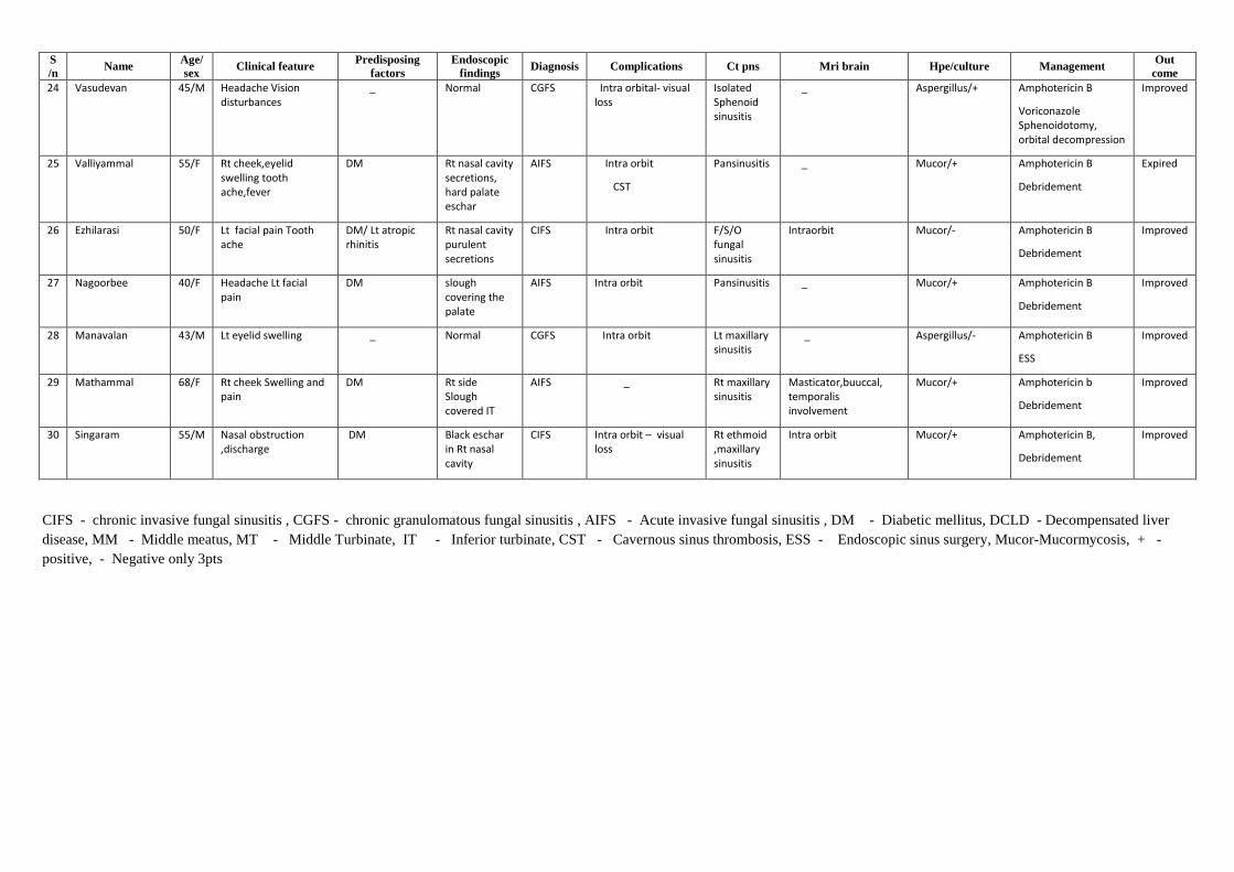
S
/n Name
Age/
sex Clinical feature
Predisposing
factors
Endoscopic
findings Diagnosis Complications Ct pns Mri brain Hpe/culture Management
Out
come
24 Vasudevan 45/M Headache Vision disturbances
_ Normal CGFS Intra orbital- visual loss
Isolated Sphenoid sinusitis
_ Aspergillus/+ Amphotericin B
Voriconazole Sphenoidotomy, orbital decompression
Improved
25 Valliyammal 55/F Rt cheek,eyelid swelling tooth ache,fever
DM Rt nasal cavity secretions, hard palate eschar
AIFS Intra orbit
CST
Pansinusitis _ Mucor/+ Amphotericin B
Debridement
Expired
26 Ezhilarasi 50/F Lt facial pain Tooth ache
DM/ Lt atropic rhinitis
Rt nasal cavity purulent secretions
CIFS Intra orbit F/S/O fungal sinusitis
Intraorbit Mucor/- Amphotericin B
Debridement
Improved
27 Nagoorbee 40/F Headache Lt facial pain
DM slough covering the palate
AIFS Intra orbit Pansinusitis _ Mucor/+ Amphotericin B
Debridement
Improved
28 Manavalan 43/M Lt eyelid swelling _ Normal CGFS Intra orbit Lt maxillary sinusitis
_ Aspergillus/- Amphotericin B
ESS
Improved
29 Mathammal 68/F Rt cheek Swelling and pain
DM Rt side Slough covered IT
AIFS _ Rt maxillary sinusitis
Masticator,buuccal, temporalis involvement
Mucor/+ Amphotericin b
Debridement
Improved
30 Singaram 55/M Nasal obstruction ,discharge
DM Black eschar in Rt nasal cavity
CIFS Intra orbit – visual loss
Rt ethmoid ,maxillary sinusitis
Intra orbit Mucor/+ Amphotericin B,
Debridement
Improved
CIFS - chronic invasive fungal sinusitis , CGFS - chronic granulomatous fungal sinusitis , AIFS - Acute invasive fungal sinusitis , DM - Diabetic mellitus, DCLD - Decompensated liver
disease, MM - Middle meatus, MT - Middle Turbinate, IT - Inferior turbinate, CST - Cavernous sinus thrombosis, ESS - Endoscopic sinus surgery, Mucor-Mucormycosis, + -
positive, - Negative only 3pts
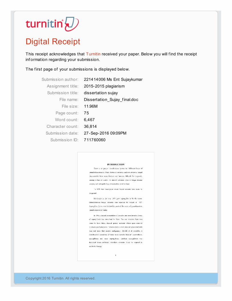
Submission author:Assignment t it le:Submission tit le:
File name:File size:
Page count:Word count:
Character count:Submission date:
Submission ID:
Digital ReceiptThis receipt acknowledges that Turnit in received your paper. Below you will f ind the receiptinf ormation regarding your submission.
The f irst page of your submissions is displayed below.
221414006 Ms Ent Sujaykumar2015-2015 plagiarismdissertation sujayDissertation_Sujay_f inal.doc11.96M756,46736,81427-Sep-2016 09:09PM711760060
Copyright 2016 Turnitin. All rights reserved.
Copyright © 2022 FDOKUMEN