Diffusion tensor imaging in moderate-to-severe pediatric traumatic brain injury: changes within an...
13
Diffusion tensor imaging in moderate-to-severe pediatric traumatic brain injury: changes within an 18 month post-injury interval Elisabeth A. Wilde & Kareem W. Ayoub & Erin D. Bigler & Zili D. Chu & Jill V. Hunter & Trevor C. Wu & Stephen R. McCauley & Harvey S. Levin Published online: 8 March 2012 # Springer Science+Business Media, LLC 2012 Abstract Traumatic brain injury (TBI) is a leading cause of death and disability in children, yet little is known regarding the pattern of TBI-related microstructural change and its impact on subsequent development. Diffusion tensor imaging (DTI) was used to examine between-group differences at two time points (planned intervals of 3 months and 18 months post-injury) and within-group longitudinal change in a group of children and adolescents aged 7–17 years with moderate- to-severe TBI (n 0 20) and a comparison group of children with orthopedic injury (OI) (n 0 21). In the 3- and 18-month cross-sectional analyses, tract-based spatial statistics (TBSS) generally revealed decreased fractional anisotropy (FA) and increased apparent diffusion coefficient (ADC) in the TBI group in regions of frontal, temporal, parietal, and occipital white matter as well as several deep subcortical structures, though areas of FA decrease were more prominent at the 3- month assessment, and areas of ADC increase were more prominent at the 18 month assessment, particularly in the frontal regions. In terms of the within-group changes over time, the OI group demonstrated primarily diffuse increases in FA over time, consistent with previous findings of DTI- measured white matter developmental change. The TBI group demonstrated primarily regions of FA decrease and ADC increase over time, consistent with presumed continued de- generative change, though regions of ADC decrease were also appreciated. These results suggest that TBI-related micro- structural changes are dynamic in children and continue until at least 18 months post-injury. Understanding the course of these changes in DTI metrics may be important in TBI for facilitating advances in management and intervention. Keywords Traumatic brain injury . Diffusion tensor imaging . Tract-based spatial statistics . Longitudinal . Children Introduction Pediatric traumatic brain injury (TBI) is the most common cause of death and disability among children. Nearly half a Brain Imaging and Behavior (2012) 6:404–416 DOI 10.1007/s11682-012-9150-y Elisabeth A. Wilde and Kareem W. Ayoub contributed equally to this work. E. A. Wilde (*) : K. W. Ayoub : T. C. Wu : S. R. McCauley : H. S. Levin Physical Medicine and Rehabilitation Alliance, Baylor College of Medicine and the University of Texas-Houston Medical School, Houston, TX, USA e-mail: [email protected] E. A. Wilde : Z. D. Chu : J. V. Hunter Department of Radiology, Baylor College of Medicine, Houston, TX, USA E. A. Wilde : S. R. McCauley : H. S. Levin Department of Neurology, Baylor College of Medicine, Houston, TX, USA K. W. Ayoub Department of Bioengineering, Rice University, Houston, TX, USA E. D. Bigler : T. C. Wu Department of Psychology, Brigham Young University, Provo, UT, USA E. D. Bigler Department of Neuroscience, Brigham Young University, Provo, UT, USA E. D. Bigler Department of Psychiatry and the Utah Brain Institute, University of Utah, Salt Lake City, UT, USA Z. D. Chu : J. V. Hunter Department of Pediatric Radiology, Texas Children’ s Hospital, Houston, TX, USA
-
Upload
independent -
Category
Documents
-
view
2 -
download
0
Transcript of Diffusion tensor imaging in moderate-to-severe pediatric traumatic brain injury: changes within an...
Diffusion tensor imaging in moderate-to-severepediatric traumatic brain injury: changeswithin an 18 month post-injury interval
Elisabeth A. Wilde & Kareem W. Ayoub &
Erin D. Bigler & Zili D. Chu & Jill V. Hunter &
Trevor C. Wu & Stephen R. McCauley & Harvey S. Levin
Published online: 8 March 2012# Springer Science+Business Media, LLC 2012
Abstract Traumatic brain injury (TBI) is a leading cause ofdeath and disability in children, yet little is known regardingthe pattern of TBI-related microstructural change and itsimpact on subsequent development. Diffusion tensor imaging
(DTI) was used to examine between-group differences at twotime points (planned intervals of 3 months and 18 monthspost-injury) and within-group longitudinal change in a groupof children and adolescents aged 7–17 years with moderate-to-severe TBI (n020) and a comparison group of childrenwith orthopedic injury (OI) (n021). In the 3- and 18-monthcross-sectional analyses, tract-based spatial statistics (TBSS)generally revealed decreased fractional anisotropy (FA) andincreased apparent diffusion coefficient (ADC) in the TBIgroup in regions of frontal, temporal, parietal, and occipitalwhite matter as well as several deep subcortical structures,though areas of FA decrease were more prominent at the 3-month assessment, and areas of ADC increase were moreprominent at the 18 month assessment, particularly in thefrontal regions. In terms of the within-group changes overtime, the OI group demonstrated primarily diffuse increases inFA over time, consistent with previous findings of DTI-measured white matter developmental change. The TBI groupdemonstrated primarily regions of FA decrease and ADCincrease over time, consistent with presumed continued de-generative change, though regions of ADC decrease were alsoappreciated. These results suggest that TBI-related micro-structural changes are dynamic in children and continue untilat least 18 months post-injury. Understanding the course ofthese changes in DTI metrics may be important in TBI forfacilitating advances in management and intervention.
Keywords Traumatic brain injury . Diffusion tensorimaging . Tract-based spatial statistics . Longitudinal .
Children
Introduction
Pediatric traumatic brain injury (TBI) is the most commoncause of death and disability among children. Nearly half a
Brain Imaging and Behavior (2012) 6:404–416DOI 10.1007/s11682-012-9150-y
Elisabeth A.Wilde and KareemW.Ayoub contributed equally to this work.
E. A. Wilde (*) :K. W. Ayoub : T. C. Wu : S. R. McCauley :H. S. LevinPhysical Medicine and Rehabilitation Alliance, Baylor College ofMedicine and the University of Texas-Houston Medical School,Houston, TX, USAe-mail: [email protected]
E. A. Wilde : Z. D. Chu : J. V. HunterDepartment of Radiology, Baylor College of Medicine,Houston, TX, USA
E. A. Wilde : S. R. McCauley :H. S. LevinDepartment of Neurology, Baylor College of Medicine,Houston, TX, USA
K. W. AyoubDepartment of Bioengineering, Rice University,Houston, TX, USA
E. D. Bigler : T. C. WuDepartment of Psychology,Brigham Young University,Provo, UT, USA
E. D. BiglerDepartment of Neuroscience,Brigham Young University,Provo, UT, USA
E. D. BiglerDepartment of Psychiatry and the Utah Brain Institute,University of Utah,Salt Lake City, UT, USA
Z. D. Chu : J. V. HunterDepartment of Pediatric Radiology, Texas Children’s Hospital,Houston, TX, USA
million children aged 0–14 years in the United States visitthe emergency department each year for TBI-related injuries,with about 25% of these injuries considered to be moderateto severe (Faul et al. 2010). Despite the prevalence ofpediatric TBI, information is limited regarding the courseof recovery within brain parenchyma following headinjury in the developing brain.
Neuroimaging has been considered an important tool inbetter understanding the sequelae of injury, as it allows in vivoexamination of the consequences of injury. However, studiesusing standard imaging techniques such as computed tomog-raphy (CT) and conventional magnetic resonance imaging(MRI) sequences to perform lesion analysis are typicallylimited to selecting specific regions of interest for analysis(Gale and Prigatano 2010; Schonberger et al. 2009) that onlypartially address the relation between more diffuse injury tobrain parenchyma and outcome, with some studies also dem-onstrating the limits of simply measuring the size or locationof focal injury and later outcome (Chastain et al. 2009; Salorioet al. 2005).
Studies utilizing more advanced neuroimaging techniquessuch as diffusion tensor imaging (DTI) of white matter havemore consistently reported stronger relations with outcomevariables, suggesting that more diffuse injury to white matter,likely via detection of traumatic axonal injury, may be a betterpredictor than focal injury of outcome in global assessment offunctioning (Kinnunen et al. 2010; Marquez de la Plata et al.2010; Warner et al. 2010a; b). DTI has been used to probe theintegrity of white matter in specific brain areas through com-mon DTI-derived metrics such as fractional anisotropy (FA)and apparent diffusion coefficient (ADC; also referred to asmean diffusivity or MD) (Huisman et al. 2004; Alexander etal. 2007). FA is derived from the tendency of water moleculesto move preferentially in parallel (rather than perpendicular)to barriers to free diffusion such as fibers, axons, or othersupport cells. A high degree of anisotropic diffusion has beenshown to be related to homogeneity in fiber orientation,increased fiber density or axonal diameter, and the ratio ofintracellular/extracellular space. ADC represents the averagediffusivity of free water movement within tissue, which alsoenables inferences regarding the microstructure in tissue; gen-erally higher average diffusivity is indicative of decreasedfiber density, axonal diameter, decreased myelinationand/or increased extracellular space.
The validity of DTI metrics in the study of TBI-relatedpathology has already been established through significantcorrelation of FA and ADC (or mean diffusivity) values aswell as other DTI-related metrics with injury indicators suchas the Glasgow Coma Scale (GCS) and outcome measuresin adult patients with head trauma (Kumar et al. 2009;Benson et al. 2007; Kraus et al. 2007; Huisman et al. 2004;Sidaros et al. 2008; Perlbarg et al. 2009). Additionally, this hasbeen established in limited studies involving children with
TBI using FA (Wozniak et al. 2007; Yuan et al. 2007) as wellas both FA and ADC, or other DTI metrics (Levin et al. 2008;Ewing-Cobbs et al. 2008; Yuan et al. 2007; Wozniak et al.2007; Wilde et al. 2006). FA reductions, ADC increases,and/or changes in other DTI-derived metrics following TBIin children and adolescents include, but are not necessarilylimited to, the corpus callosum, uncinate fasciculus, cingulumbundle, corticospinal tract, posterior limb of the internalcapsule, and the external capsule (Arfanakis et al. 2002;Huisman et al. 2004; Inglese et al. 2005; Wilde et al.2006; Ewing-Cobbs et al. 2008). However, only a fewstudies have utilized whole brain techniques in childrento uncover changes occurring throughout the brain on avoxel by voxel basis (Yuan et al. 2007). Additionally,despite consistent evidence of widespread degenerativechanges detected with DTI in adult and child patientswith moderate to severe TBI in the chronic post-injury period,relatively few longitudinal studies have been undertaken,meaning that the nature, degree, and course of degenerativeTBI-related changes in the developing brain remain poorlyunderstood. The purpose of the current study is to examinedifferences in white matter using tract-based spatial statistics(TBSS), a method of global brain analysis through whitematter skeletonization, to examine injury-related differencesin children and adolescents with TBI in comparison to a groupof orthopedically-injured (OI) children at both 3 and18 months post-injury, and within-group changes longitudi-nally. Because FA and ADC may be differentially sensitive tofactors presumed to underlie white matter integrity followingTBI (e.g., demyelination or dysmyelination, neural degenera-tion, etc.) and because previous TBI studies have reportedvariation in the degree of group differences observed betweenthese metrics as well as differences in the magnitude ofrelation with outcome measures, we examined both DTImetrics. We hypothesized that the TBI group would demon-strate multiple regions of significantly lower FA and increasedADC at both assessments, reflecting decreased fiber integrity,in relation to the OI group, that would be particularly apparentin the frontal and temporal regions as these are highlyvulnerable to TBI-related injury. Next, we wished toexplore changes within each group between the 3- and18-month assessments to determine the locations anddegree of change evident in this recovery interval. Wehypothesized that within the OI group, regions of relativeincrease in FA and decrease in ADC would emerge by18 months, particularly in frontal and temporal regions,presumed to reflect developmental myelination in thesebrain areas in children and adolescents within this age range(see Lenroot & Giedd, 2006). In the TBI group, wehypothesized that we would see evidence for continueddegenerative change in these regions manifest by decreasedFA and increased ADC by 18months secondary to TBI-relateddisruption of normal development.
Brain Imaging and Behavior (2012) 6:404–416 405

Methods and materials
Participants
This research protocol was approved by the InstitutionalReview Board of the Baylor College of Medicine and otheraffiliated institutions involved in this project. Informed con-sent was obtained from each participant or his/her legalguardian prior to enrollment in the study. The cohort ofparticipants included in this study was selected from a largersample of participants engaged in a longitudinal examina-tion of the cognitive and imaging effects of pediatric braininjury (see also Levin et al. 2011; McCauley et al. 2011; Oniet al. 2010; Wilde et al. 2010; Wu et al. 2010). In the currentreport, where the objective was to examine the effects ofTBI on brain tissue over time as assessed by DTI, we includ-ed only those participants who had useable DTI data at both 3and 18 months post-injury time points. Although the actualmean post-injury interval for the initial assessment was closerto 4 months than 3 months, the study design included aplanned assessment at 3 months post-injury, and we refer tothe initial assessment as the “3-month” assessment.
The TBI group was composed of 20 participants (11males and 9 females) between the ages of 8.2 and17.5 years (mean013.6.±2.9) who sustained either com-plicated mild or moderate-to-severe TBI. The severity ofthe injury was determined by the lowest postresuscitationGlasgow Coma Scale (GCS) scores (Teasdale and Jennett1974). Severe TBI was defined as a GCS score of 3–8,and moderate TBI as GCS score of 9–12. A complicatedmild TBI was defined as a GCS score of 13–15, but withacute (within 24 h post-injury) trauma-related CT findings(e.g., contusion, hematoma, etc.). Participants with complicat-ed mild TBI were included in our “moderate to severe” patientcohort because the presence of cerebral lesions on CT hasbeen demonstrated to be predictive of poor cognitive outcomeat 12 months post-injury, and more similar to the outcome ofindividuals who experience moderate TBI than those whoexperience uncomplicated mild TBI (Levin et al. 2009). Giv-en these classifications, there were 13 severe, 4 moderate and3 complicated mild cases. The mean modified Injury SeverityScale (m-ISS; not including the head region) score for indi-viduals with TBI was 22.6±11.6 (range 12.0–50.0). All par-ticipants in the TBI group had at least one focal lesion; themost common site for focal lesion was in the frontal lobes,followed by the temporal lobes and parietal lobes. Lesionvolume for lesions in the frontal lobes ranged from 0.06 to23.02 cubic centimeters. Table 1 lists focal lesion location foreach participant used in this analysis.
Participants in the OI group included 15 males and 6females, and had a mean age of 12.1±2.5 years. The meanm-ISS score for individuals with OI was 6.0±2.5 (range4.0–9.0).
The selection of participants with OI was designed toequate the groups by controlling for risk factors that predis-pose individuals to accidents including pre-existing behavior-al problems, family variables, and subtle learning disabilities(Stancin et al. 1998). The OI group included participants whowere hospitalized overnight for bone fractures of the upper orlower extremities. Mechanisms of injury for this group includ-ed predominantly sports and play-related falls. A detaileddevelopmental questionnaire was administered to legal guard-ians to confirm the absence of significant previous headtrauma in the OI children and was cross-referenced withmedical records and/or physician report of relevant historyand, when available, clinical imaging results that includenegative CT scans for the OI group.
All participants in both the TBI and OI groups had nopre-injury history of neurologic or psychiatric disorders andwere recruited from consecutive admissions to emergencyrooms at Level-1 trauma centers in Houston, Dallas, andMiami. In both groups, participants were excluded ifthere was any evidence of previous head injury, childabuse, pre-existing neurologic disorders (e.g. epilepsy),diagnosed learning disabilities, pre-existing severe psychiatricdisorders (e.g. schizophrenia), prematurity or low birthweight (birth weight <2,500 g and gestation <37 weeks),penetrating injury, hypoxia (PO2 <96 mmHg) or hypotension(systolic blood pressure 2 standard deviations below the
Table 1 Lesion locations for each participant with traumatic brain injury
Participant Lesion location
1 parietal
2 frontal, temporal, basal ganglia
3 frontal
4 frontal, temporal
5 frontal, parietal, cerebellar
6 frontal, temporal
7 frontal, temporal, parietal, corpus callosum, cerebellar
8 frontal, parietal
9 frontal, temporal, parietal, corpus callosum, basal ganglia
10 frontal, temporal, parietal,
11 frontal
12 frontal
13 frontal, temporal, parietal
14 frontal
15 frontal, temporal, parietal, corpus callosum
16 frontal, temporal, parietal, corpus callosum, basal ganglia,cerebellar
17 frontal, temporal, parietal, corpus callosum, basal ganglia
18 frontal, temporal, parietal
19 frontal, corpus callosum
20 frontal, temporal
406 Brain Imaging and Behavior (2012) 6:404–416
mean for the age group). Detailed information for demo-graphic and injury characteristics for both groups appearsin Table 2.
Imaging protocols
All participants underwent MRI without sedation on Philips1.5 Tesla Intera or Achieva scanners (Philips, Cleveland, OH)at Texas Children’s Hospital in Houston, the Rogers MRICenter or University of Texas Southwestern Medical Centerin Dallas, or the Miami Children’s Hospital in Miami usingcomparable platforms and software. Regular quality assurancetesting was performed on all three scanners including Amer-ican College of Radiology (ACR) phantom testing, and allscanners were consistently noted to be within a range consid-ered acceptable throughout the course of the study. Addition-ally, similar ranges of values for Weisskoff stabilitymeasurements (Weisskoff 1996) (minimum 1/SNR index,peak-to-peak and RMS stability) taken on the day of scanindicated stability of scanners over time. Participants fromboth the OI and TBI groups were scanned at each site (Dallas:5 TBI, 12 OI; Houston: 6 TBI, 7 OI; Miami: 9 TBI, 2 OI), andno systematic differences were detected in the quantitativeDTI values derived from each site for the OI participants.
DTI parameters included an axial single-shot spin-echoecho-planar imaging sequence with 15 diffusion-encodingdirections; 256-mm field of view (FOV); acquisition voxelsize, 2.69×2.69×2.7 mm3; repetition time (TR), 6318.0 ms;echo time (TE), 51 ms, sensitivity encoding (SENSE)reduction factor of 2; 2 B factors with 0 s/mm2, low B,and 860 s/mm2, high B), with 2 acquisitions to averagethe signal of the two DTI scans in order to ensure better
signal-to-noise ratio. Acquisitions consisted of 55 slices.A SENSE 8-channel head coil was used.
Image processing
All pre-processing was completed with the FunctionalMRI of the Brain (FMRIB) diffusion toolbox (FDT)software included in the FMRIB Software Library(FSL; http://www.fmrib.ox.ac.uk/fsl) (see also Smith etal. 2006; Smith et al. 2007 for additional detail). Headmotion and eddy current artifact were corrected via alinear, affine registration algorithm, and DTI metrics (e.g.FA and ADC) were performed using FDT’s automatedcalculation. FSL’s brain extraction tool (BET) was usedto remove all non-brain voxels from the zero-diffusionweighted image of each tensor of interest (i.e., fractionalanisotropy (FA) and apparent diffusion coefficient (ADC)(Smith, 2002).
Tract-based spatial statistics analysis
All subjects' FA data were then aligned into a commonspace using the nonlinear registration tool FNIRT, whichuses a b-spline representation of the registration warp field.The target image used in the registrations was chosen tobe the most “typical” subject in the study in order togenerate a study-specific FA target image. This targetimage was then affine-aligned into MNI152 standardspace. All participants’ images were then transformed intostandard 1×1×1 mm3 MNI152 space by combining the non-linear transform to the target FA image with the affine trans-form from that target to MNI152 space. This resulted in astandard-space version of each subject’s FA image, which
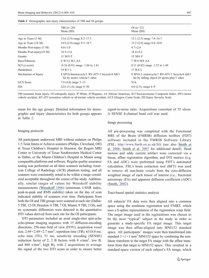
Table 2 Demographic and injury characteristics of TBI and OI groups
TBI (n020) OI (n021)Mean (SD) Mean (SD)
Age in Years (3 M) 13.6 (2.9) range 8.2–17.5 12.1 (2.5) range 7.4–16.7
Age in Years (18 M) 14.8 (2.9) range 9.3–18.7 13.2 (2.6) range 8.8–18.0
Months Post-injury (3 M) 4.0±1.0 4.7±2.6
Months Post-injury(18 M) 18.5±3.6 18.4±4.2
Gender 11 M/9 F 15 M/6 F
Race/Ethnicity 6 W/12 H/2 AA 7 W/6 H/8 AA
SCI (z-score) -0.16 (0.91) range -1.86 to 1.41 0.11 (0.82) range -1.52 to 1.48
Handedness 19 R/1 L 17 R/4 L
Mechanism of Injury 8 MVA/4motorcycle/1 RV-ATV/1 bicycle/4 fall/1hit by motor vehicle/1 other
0 MVA/1 motorcycle/1 RV-ATV/1 bicycle/6 fall/1hit by falling object/10 sports-play/1 other
GCS Score 7.9 (4.0) range 3–15 N/A
ISS 22.6 (11.6); range 9–50 6.0 (2.5); range 4–9
TBI traumatic brain injury, OI orthopedic injury, W White, H Hispanic, AA African American, SCI Socioeconomic Composite Index, MVA motorvehicle accident, RV-ATV recreation vehicle or all-terrain vehicle accident, GCS Glasgow Coma Scale, ISS Injury Severity Scale
Brain Imaging and Behavior (2012) 6:404–416 407
was then merged into a single FA 4D image containingall participant data. For ADC images, the FA transforma-tion matrices were utilized to achieve the same nonlinearregistration. All subjects’ ADC data were then merged into asingle 4D image volume. A general linear model was used toanalyze group differences (cross-sectional and longitudinal)using a voxel-wise analysis utilizing permutation-based test-ing to analyze differences in FA and ADC. Threshold freecluster enhancement was used as a correction tool for multiplecomparisons.
Other statistical analyses
Demographic statistics between the TBI and OI groupwere compared using Chi-square testing (for gender,race/ethnicity, high-versus low-velocity mechanism of injury)or Fisher’s exact testing (for handedness and mechanismof injury) for categorical variables. A Student t-test analysiswas performed for: 1) age at injury at both 3- and 18-monthassessments, 2) post-injury interval for both 3- and 18-monthassessments, and 3) Socioeconomic Composite Index(SCI) z-score (Yeates et al. 1997). The threshold forstatistical significance was set at p<0.05.
Results
Demographic results
The two groups did not differ significantly for post-injuryinterval for either 3- or 18-month assessment, handedness,gender, or socioeconomic status as measured by SCI z-score.The TBI and OI groups differed significantly in themechanism of injury with TBI patients sustaining a majorityof motor vehicle accidents, and OI participants sustaining amajority of sports- and play-related injuries; when categorizedas high- versus low-speed injuries, the groups significantlydiffered (χ²(1)015.7, p00. 0001). As expected, m-ISS scoresalso significantly differed between groups (t(18.43)0-5.92),p0<0.0001), with the TBI group having a higher mean
m-ISS score. Groups tended to differ for age at injury atboth the 3-month assessment (t(39)0-1.85, p00.07) andthe 18-month (t(39)0-1.89, p00.07) assessments, with the TBIgroup being slightly older than the OI group. Groups alsodiffered in race/ethnicity (χ²(2)05.7, p00.06), with the TBIgroup having a higher proportion of Hispanic participants,and the OI group having a higher proportion of African-American participants. For a detailed list of means anddemographic and injury characteristics, please see Table 2.
TBSS results
All results are bilateral unless otherwise specified.
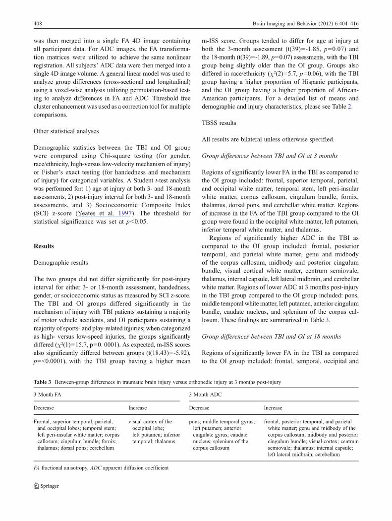
Group differences between TBI and OI at 3 months
Regions of significantly lower FA in the TBI as compared tothe OI group included: frontal, superior temporal, parietal,and occipital white matter, temporal stem, left peri-insularwhite matter, corpus callosum, cingulum bundle, fornix,thalamus, dorsal pons, and cerebellar white matter. Regionsof increase in the FA of the TBI group compared to the OIgroup were found in the occipital white matter, left putamen,inferior temporal white matter, and thalamus.
Regions of significantly higher ADC in the TBI ascompared to the OI group included: frontal, posteriortemporal, and parietal white matter, genu and midbodyof the corpus callosum, midbody and posterior cingulumbundle, visual cortical white matter, centrum semiovale,thalamus, internal capsule, left lateral midbrain, and cerebellarwhite matter. Regions of lower ADC at 3 months post-injuryin the TBI group compared to the OI group included: pons,middle temporal white matter, left putamen, anterior cingulumbundle, caudate nucleus, and splenium of the corpus cal-losum. These findings are summarized in Table 3.
Group differences between TBI and OI at 18 months
Regions of significantly lower FA in the TBI as comparedto the OI group included: frontal, temporal, occipital and
Table 3 Between-group differences in traumatic brain injury versus orthopedic injury at 3 months post-injury
3 Month FA 3 Month ADC
Decrease Increase Decrease Increase
Frontal, superior temporal, parietal,and occipital lobes; temporal stem;left peri-insular white matter; corpuscallosum; cingulum bundle; fornix;thalamus; dorsal pons; cerebellum
visual cortex of theoccipital lobe;left putamen; inferiortemporal; thalamus
pons; middle temporal gyrus;left putamen; anteriorcingulate gyrus; caudatenucleus; splenium of thecorpus callosum
frontal, posterior temporal, and parietalwhite matter; genu and midbody of thecorpus callosum; midbody and posteriorcingulum bundle; visual cortex; centrumsemiovale; thalamus; internal capsule;left lateral midbrain; cerebellum
FA fractional anisotropy, ADC apparent diffusion coefficient
408 Brain Imaging and Behavior (2012) 6:404–416
parietal white matter, corpus callosum, cingulum bundle,anterior commissure, right thalamus, fornix, and cerebellarwhite matter. Regions of significantly higher FA in the TBIgroup as compared to the OI group include: pons, midbrain,visual cortical white matter, left thalamus, and parietal whitematter (not overlapping with the decreases in FA in the samelobe of the brain).
Regions of significantly higher ADC in the TBI ascompared to the OI group included: frontal, right temporal, andparietal white matter, genu, midbody, and splenium of thecorpus callosum, thalamus, midbrain and right cerebellar whitematter. The TBI group showed significantly lower regions ofADC at 18 months post-injury in the pons, left temporal,caudate nucleus, and parietal white matter. As with the3-month assessment data, corresponding brain structures thatexhibited group differences in FA and ADC were not neces-sarily overlapping within the same general region; for example,differences in ADC in the temporal lobes were located anteriorto differences in FA. Findings are summarized in Table 4.
Longitudinal analyses: TBI group
Regions of significantly increased FA were seen from 3 to18 months post-injury in the TBI group in the putamen,posterior temporal lobe, and thalamus. Regions of signifi-cantly decreased FA included frontal, anterior and centraltemporal, occipital and parietal white matter, genu andsplenium of the corpus callosum, cerebellum, brainstem,and cingulum bundle.
Regions of significantly decreased ADC included frontaland parietal white matter, pons, posterior temporal, thalamus,splenium and body of the corpus callosum, and cerebellarwhite matter. Regions of significantly increased ADC couldbe seen over time in the parahippocampal and anteriortemporal white matter, genu of the corpus callosum, andparietal white matter. Findings are summarized in Table 5.
Longitudinal analysis: OI group
The OI group showed significant increases in FA betweenthe two time points in the cerebellum, temporal, frontal,occipital, and parietal lobe white matter, midbrain, fornix,thalamus, insula, putamen, and whole corpus callosum.Verylimited regions of FA decrease between 3 to 18 monthspost-injury were seen in the temporal stem and scatteredthroughout the subcortical parietal white matter.
ADCwas significantly increased in the OI group in regionsof the cerebellar white matter, thalamus, and caudate.Both increases and decreases in ADC were found in thetemporal, frontal, occipital, and parietal lobe white matter.Regions with significantly decreased ADC included anteriorand posterior temporal white matter, temporal stem, genu andsplenium of the corpus callosum. Findings are summarizedin Table 6.
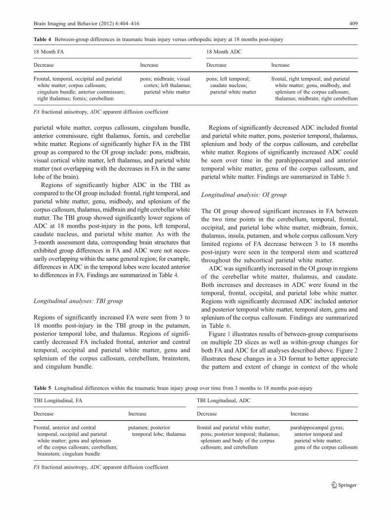
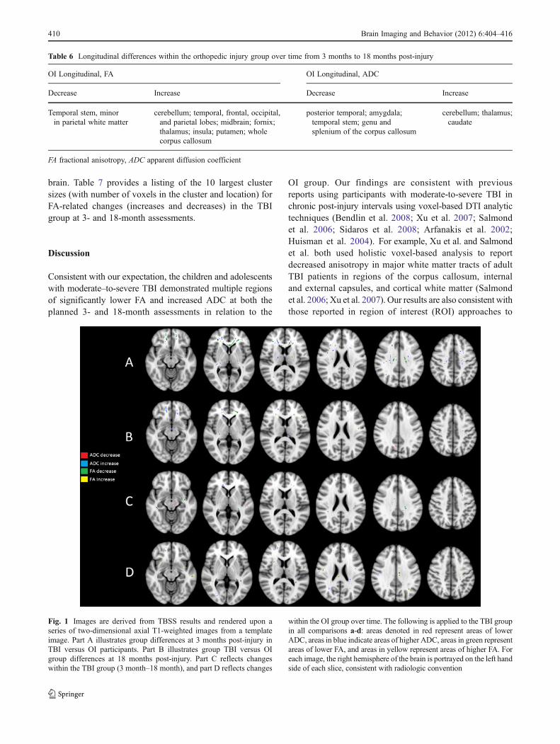
Figure 1 illustrates results of between-group comparisonson multiple 2D slices as well as within-group changes forboth FA and ADC for all analyses described above. Figure 2illustrates these changes in a 3D format to better appreciatethe pattern and extent of change in context of the whole
Table 4 Between-group differences in traumatic brain injury versus orthopedic injury at 18 months post-injury
18 Month FA 18 Month ADC
Decrease Increase Decrease Increase
Frontal, temporal, occipital and parietalwhite matter; corpus callosum;cingulum bundle; anterior commissure;right thalamus; fornix; cerebellum
pons; midbrain; visualcortex; left thalamus;parietal white matter
pons; left temporal;caudate nucleus;parietal white matter
frontal, right temporal, and parietalwhite matter; genu, midbody, andsplenium of the corpus callosum;thalamus; midbrain; right cerebellum
FA fractional anisotropy, ADC apparent diffusion coefficient
Table 5 Longitudinal differences within the traumatic brain injury group over time from 3 months to 18 months post-injury
TBI Longitudinal, FA TBI Longitudinal, ADC
Decrease Increase Decrease Increase
Frontal, anterior and centraltemporal, occipital and parietalwhite matter; genu and spleniumof the corpus callosum; cerebellum;brainstem; cingulum bundle
putamen; posteriortemporal lobe; thalamus
frontal and parietal white matter;pons; posterior temporal; thalamus;splenium and body of the corpuscallosum; and cerebellum
parahippocampal gyrus;anterior temporal andparietal white matter;genu of the corpus callosum
FA fractional anisotropy, ADC apparent diffusion coefficient
Brain Imaging and Behavior (2012) 6:404–416 409
brain. Table 7 provides a listing of the 10 largest clustersizes (with number of voxels in the cluster and location) forFA-related changes (increases and decreases) in the TBIgroup at 3- and 18-month assessments.
Discussion
Consistent with our expectation, the children and adolescentswith moderate–to-severe TBI demonstrated multiple regionsof significantly lower FA and increased ADC at both theplanned 3- and 18-month assessments in relation to the
OI group. Our findings are consistent with previousreports using participants with moderate-to-severe TBI inchronic post-injury intervals using voxel-based DTI analytictechniques (Bendlin et al. 2008; Xu et al. 2007; Salmondet al. 2006; Sidaros et al. 2008; Arfanakis et al. 2002;Huisman et al. 2004). For example, Xu et al. and Salmondet al. both used holistic voxel-based analysis to reportdecreased anisotropy in major white matter tracts of adultTBI patients in regions of the corpus callosum, internaland external capsules, and cortical white matter (Salmondet al. 2006; Xu et al. 2007). Our results are also consistent withthose reported in region of interest (ROI) approaches to
Table 6 Longitudinal differences within the orthopedic injury group over time from 3 months to 18 months post-injury
OI Longitudinal, FA OI Longitudinal, ADC
Decrease Increase Decrease Increase
Temporal stem, minorin parietal white matter
cerebellum; temporal, frontal, occipital,and parietal lobes; midbrain; fornix;thalamus; insula; putamen; wholecorpus callosum
posterior temporal; amygdala;temporal stem; genu andsplenium of the corpus callosum
cerebellum; thalamus;caudate
FA fractional anisotropy, ADC apparent diffusion coefficient
Fig. 1 Images are derived from TBSS results and rendered upon aseries of two-dimensional axial T1-weighted images from a templateimage. Part A illustrates group differences at 3 months post-injury inTBI versus OI participants. Part B illustrates group TBI versus OIgroup differences at 18 months post-injury. Part C reflects changeswithin the TBI group (3 month–18 month), and part D reflects changes
within the OI group over time. The following is applied to the TBI groupin all comparisons a-d: areas denoted in red represent areas of lowerADC, areas in blue indicate areas of higher ADC, areas in green representareas of lower FA, and areas in yellow represent areas of higher FA. Foreach image, the right hemisphere of the brain is portrayed on the left handside of each slice, consistent with radiologic convention
410 Brain Imaging and Behavior (2012) 6:404–416
DTI analyses which found decreased FA in the corpuscallosum, posterior limb of the internal capsule, centrumsemiovale, and the cerebral peduncles in adults with TBIas compared to OIs (Sidaros et al. 2008). Finally, thesefindings are consistent with changes in both adults andchildren following TBI using other advanced imagingmodalities such as volumetric analysis of white matter,suggesting that TBI affects virtually all major regions ofthe brain (Bendlin et al. 2008; Bigler et al. 2010).
We had hypothesized that TBI-related DTI changeswould be most apparent in the frontal and temporalregions of the brain. Acknowledging that the exact areasof identified injury for a cohort in group analyses ofmoderate-to-severe TBI will differ by study due to thelocation, extent, severity, and mechanism of focal injuryin the individuals within that particular cohort, our studyindicated prominent involvement of frontal regions in ourpediatric TBI group. DTI-related changes such as thosedemonstrated in the frontal lobes on the between-groupanalyses at 3 and 18 months are expected given theknown vulnerability of this region to mechanical forces
during TBI and the prevelance of resulting focal lesions,as evidenced in our sample. However, there was evidencefor continued deleterious change as measured bydecreases in FA and increases in ADC in this regionover time in the within-group analysis of the TBI groupas illustrated in Figs. 1 and 2. It is possible that injurysustained during childhood and adolescence, a periodwhere frontal white matter is still maturing structurallyand physiologically, may alter the course of normal develop-ment in this important region presumed to be involved inexecutive functioning, attention and cognitive and behavioralcontrol (Levin and Hanten 2005; Liston et al. 2006;Wozniak et al. 2007).
Next, we had anticipated that within the OI group,regions of relative increase in FA and decrease in ADCwould emerge by 18 months post-injury, particularly infrontal and temporal regions, presumably reflecting develop-mental myelination in these brain areas in children andadolescents within this age range. Indeed, increased FAwas found in several regions over time in the OI group.Generally, studies related to white matter development
Fig. 2 Part a illustrates group differences at 3 months post-injury inTBI versus OI participants derived from TBSS and rendered upon aseries of three-dimensional T1-weighted images from a templateimage as this allows a more global perspective of changes occurringthroughout the brain. Part b illustrates group TBI versus OI groupdifferences at 18 months post-injury. Part c reflects changes within
the TBI group (18 month–3 month), and part d reflects changeswithin the OI group over time. The following is applied to the TBIgroup in all comparisons (a-d): areas denoted in red represent areasof lower ADC, areas in blue indicate areas of higher ADC, areas ingreen represent areas of lower FA, and areas in yellow representareas of higher FA
Table 7 Table of the ten largestclusters of significant FAfindings in the TBI group ateach assessment
FA fractional anisotropy,TBI traumatic brain injury,3 M 3 months post-injury,18 M 18 months post-injury
Cluster index 3 M decrease 3 M increase 18 M decrease 18 M increase
Voxels Z Voxels Z Voxels Z Voxels Z
1 1898 6.05 18 3.67 358 4.45 77 4.07
2 1662 5.90 18 3.25 222 4.40 67 5.19
3 926 4.85 17 3.55 211 4.55 62 4.20
4 576 5.27 17 3.28 190 4.64 55 4.57
5 368 4.13 16 3.77 168 5.77 55 3.20
6 361 4.31 15 3.45 130 4.25 45 4.24
7 280 3.87 13 3.04 109 4.45 41 4.73
8 280 4.61 12 3.30 74 4.21 40 3.91
9 241 3.87 10 3.84 59 4.74 39 3.16
10 187 4.71 10 2.69 52 3.47 33 4.87
Brain Imaging and Behavior (2012) 6:404–416 411
utilizing DTI have demonstrated age-related increases inFA with concomitant decreases in ADC, most likelyreflecting the ongoing process of myelination, thoughsynaptogenesis and synaptic pruning continue throughoutchildhood and may also contribute to changes in thesemetrics. Various white matter structures have beenreported to show changes in DTI-derived metrics instudies of normal development, consistent with severalof the regions that were demonstrated in the currentstudy (Ashtari et al. 2007; Barnea-Goraly et al. 2005;Bonekamp et al. 2007; Clayden et al. 2011; Eluvathingalet al. 2007; Giorgio et al. 2010; Lebel et al. 2008;Muetzel et al. 2008; Schmithorst et al. 2002). We notethat FA increases were not as restricted to the frontal andtemporal white matter as we predicted, but were locateddiffusely throughout the brain. Additionally, we note thatthe within-group changes predominantly manifested asincreased FA, with fewer than expected concomitantchanges in decreased ADC. The increases in FA werenotably absent in the TBI longitudinal analysis, poten-tially indicating alteration in the timing or the course ofthe expected developmental trajectory in participants withTBI. Additional research will need to be conducted tomore precisely determine the mechanism underlyingthese white matter changes and the course these changesfollow.
Finally, in the within-group analysis of the TBI group, weanticipated finding evidence for continued degenerativechange in brain regions, as manifest by decreased FA andincreased ADC over time, particularly in the frontal andtemporal lobes. We note that few longitudinal studies usingDTI in moderate-to-severe TBI have been reported. Bend-lin et al. reported that adult patients with moderate-to-severe TBI exhibited decreased FA and increased meandiffusivity (MD) over time in several major fiber bundlesincluding the corpus callosum, cingulum bundle, and theinferior fronto-occipital fasciculus (Bendlin et al. 2008).Similarly, Sidaros and colleagues reported that adultpatients with TBI evidenced decreased FA and increasedADC over time in the splenium of the corpus callosum andcerebral peduncle (Sidaros et al. 2008). Kumar et al.reported persistent decreases in FA at 6 months post-injury in the genu of the corpus callosum and the anteriorand posterior internal capsule as well as additional areas ofFA decrease in other aspects of the corpus callosum inparticipants with TBI as compared to controls (Kumar etal. 2009). In the only other study reporting longitudinalchanges in a pediatric cohort of moderate-to-severe TBI,Wu and colleagues restricted analysis to tractography inthe corpus callosum and found ADC increases in thecorpus callosum in TBI and OI groups at 18 monthsversus 3 months post-injury (Wu et al. 2010). Our findingswere generally consistent with these previous reports.
However, it is important to note that at both time points,and longitudinally within the TBI group, both increases anddecreases in the FA and ADC were observed. Generally, theexpected degenerative changes (i.e., lower FA and higherADC in the TBI group) were predominant in the TBI groupin between-group comparisons at both assessments, butwere interspersed with additional relatively small regionsof change in the opposite direction (e.g., higher FA incertain regions in the TBI group). Interestingly, in some ofthe studies referenced above, each described evidence ofdeleterious change, but also findings suggestive of modestdegrees of apparent recovery when the later time point wascompared to the earlier one, as was observed in our currentstudy. For example, Sidaros and colleagues found increasedFA in the adult TBI group compared to the OI group in thecentrum semiovale and internal capsule (Sidaros et al. 2008;Sidaros et al. 2009). Bendlin et al. reported decreased MD inthe adult TBI group over time in regions including theinternal capsule and portions of the occipital white matter(Bendlin et al. 2008). Finally, Wu and colleagues alsodescribed an increase in FA over time in the pediatricTBI group (Wu et al. 2010). The presence of apparentnon-degenerative additional changes in our study is con-sistent with previous studies despite differences in anal-ysis techniques and methodology. Wu et al. utilized atractography-based approach to DTI analysis of pediatricpatients with TBI and Sidaros et al. analyzed adult TBIpatients with a smaller post-injury time interval andusing an ROI-based approach (Sidaros et al. 2008; Wuet al. 2010). In our study, particularly in non-frontalregions, we note that group differences for FA decreaseand ADC increase in the cross-sectional analyses generallyappear more to be diminished in size and distribution at18 months as compared to 3 months, presumably indicatingsome seeming amelioration of the initial deleterious changes.In fact, there are regions of ADC decrease in parietal andoccipital areas as well as in the midbrain (see Fig. 1).Admittedly, these changes are less visible than the degenera-tive changes, but highlight the complex and dynamic aspect ofthe changes subsequent to TBI in the immature brain.
Although there were some small areas of commonalitybetween DTI-related group differences in FA and ADC, theextent and location of differences in these metrics did notnecessarily overlap at either time point, suggesting that eachmetric reveals somewhat unique information about themicrostructural environment. Although several publishedstudies have included two or more DTI metrics (e.g., FA,MD, ADC, axial diffusivity or AD, or radial diffusivity orRD), the relation of these metrics to each other and also tooutcome or cognition in different states of pathology is notfully understood, and few researchers have addressed thisdiscrepancy. The variation in which DTI metrics areutilized, as well as the time at which they are measured,
412 Brain Imaging and Behavior (2012) 6:404–416
may partially account for reported differences in therelation of DTI findings to cognitive and clinical outcome.For example, Perlbarg and colleagues (2009) reported thatADC in the cerebral peduncles, inferior longitudinal fas-ciculus, posterior limb of internal capsule and posteriorcorpus callosum measured at 5–53 days did not significantlydiffer between groups of TBI participants with favorableversus unfavorable outcome though FA did differ betweengroups at this post-injury interval (Perlbarg et al. 2009).Moreover, in this study, FAwas related to one-year outcome,though ADC was not related to outcome and did notsignificantly differ between groups. Similarly, Sidaros etal. (2008) found no ADC changes at 5–11 weeks despitehaving seen widespread white matter differences in FA,and limited differences in ADC were seen at 12 monthspost-injury (i.e., only in the posterior corpus callosum)despite multiple regions of change in FA at this post-injuryinterval (Sidaros et al. 2008). Salmond et al. (2006) reported aslight difference in regions of change between DTI metrics.Whereas anisotropy decreases were apparent in deep whitematter and spread throughout the cortical white matter,diffusivity increases primarily manifested in the TBIgroup in the cerebellum, insula, cingulate, and somecortical white matter (Salmond et al. 2006). Clearly thesemetrics may demonstrate sensitivity to microstructuralchanges within brain tissue that may follow differingtime courses, vary by region due to site-specific structur-al organization, and are influenced by different forms ofpathology. A better understanding of metric-specificchanges in different regions of the brain will be important inelucidating and monitoring underlying pathological pro-cesses in vivo and how these contribute to brain functionand recovery following TBI.
Unlike ROI approaches to DTI analysis, TBSS is anautomated technique that allows for analysis of potentialregions of significance throughout the brain, rather thanlimiting analysis to specific hypotheses and predefinedROI. TBSS provides important information regarding theoverall pattern of microstructural change. The use of acombination of linear and non-linear registration combinedwith projection onto a skeletal tract of the pooled subjectssuch that only the white matter tracts common to all subjectsin the study are analyzed, minimizes the former concernregarding registration. DTI in general has known limitationsrelating to the accuracy of FA and ADC in regions with agreat deal of crossing fibers, and newer techniques thatcan better resolve this issue could be applied in the future(e.g., diffusion spectrum imaging).
This study represents the first prospective study utilizingTBSS DTI analysis in a cohort of children and adolescentswith moderate to severe TBI at two fixed time points.The relatively large sample size compared to previous studiesand inclusion of a comparison cohort of demographically-
similar children with orthopedic injury represent additionalstrengths of the study. Finally, our study included comparisonof two DTI parameters (i.e., FA and ADC) to examine micro-structural changes occurring at these time intervals.
Limitations of the study include heterogeneity in injuryseverity as well as the location, extent and nature of focalpathology. An additional limitation involves the potentialinfluence of age in the group comparisons. While the groupsdid not technically differ in terms of mean age, it should benoted that the difference was marginally significant, with themean age of TBI group being slightly higher than the OIgroup. In the current report, we included all subjects withcomplete imaging data of sufficient quality for both 3 and18 month imaging occasions (i.e., no missing imaging data)that were between the ages of 8–17 years. Since develop-mental changes were of interest, we elected not to controlage in the analyses as this would potentially eliminate someof the expected age-related change we were attempting toexamine in the OI group. However, we do acknowledge thatthe marginal differences between groups in terms of age orthe precise distribution of age within each group could alsocontribute to our findings. Ideally, future studies wouldexamine the impact of TBI in a true longitudinal designwith stratification by age. Additionally, we note thatrace/ethnicity differed between our TBI and OI groups.Our recruitment strategy was to enroll every eligbleparticipant from a prospective consecutive sample, regardlessof the specifics of the demographic data. The inclusion of arelatively high number of “minority” participants is reflectiveof the samples we recruited from, where Houston and Mi-ami in particular have higher numbers of Hispanic andAfrican American subjects. We know of no particulardata that reveals a systematic bias in DTI data due to race/ethnicity per se, and more important estimates of socio-economic status, such as the Social Composite Index werecomparable between groups.
Future research is necessary to investigate the injurymechanisms underlying the different metrics in DTI-relatedchanges in TBI, as understanding of the dynamic post-injury changes following TBI remains incomplete. Wealso acknowledge that the pattern of DTI-related changesand their persistence over time may differ for patientswith mild TBI (see Mayer et al., 2010). Additionally, thepresent study is limited to comparison of white matterand subcortical gray matter structures that also containwhite matter, such as the thalamus and the basal ganglia;future analysis of cortical gray matter changes in diffusivitymay also be of importance. Future studies that incorporatemulti-modality imaging in pediatric TBI should also beperformed. This study was intentionally limited to structuralimaging-related changes, but studies exploring the complexrelation of these changes to other medical injury variables,outcome and cognition are underway.
Brain Imaging and Behavior (2012) 6:404–416 413
Conclusions
In this study we report that TBI-related changes continueuntil at least 18 months post-injury as indicated by significantchanges in FA and ADC. These results are generally consis-tent with other studies revealing decreased FA and increasedADC following TBI at different chronic post-injury intervalsin both children and adults. Children and adolescents withTBI failed to demonstrate the expected pattern of FA increasesthat was observed in the OI group, suggesting that TBI maydisrupt the timing or course of development subsequent toTBI. We observed that DTI-related changes between FA andADC metrics did not overlap perfectly; they were affected indifferent regions, and the extent and location of their changealso varied with time between 3 and 18 months post-injury.A better understanding of the long-term dynamic changesoccurring within the immature brain following TBI mayincrease our understanding of neuroplasticity and continuingdegenerative change, which in turn, may facilitate advancesin management and intervention. Additionally, our studyreaffirms the need to examine multiple DTI metrics andto further investigate the relation of these metrics to eachother. Future analyses will include additional examinationof the relation of imaging changes to cognitive andfunctional outcome as well as multi-modal imaging analysesof pediatric TBI.
Acknowledgements This work was supported by the National Insti-tute Neurological Disorders and Stroke grant R01-NS21889 (“Neuro-behavioral outcome of head injury in children,” Levin, PI). We alsoacknowledge the generous contribution of Mission Connect of theTIRR Foundation. We gratefully acknowledge the contribution ofAna C. Vasquez, Deleene Menefee, PhD., Summer Lane, Lori Cook,Sandra B. Chapman, PhD., and Gillian Hotz, PhD. in data collection,and Joshua Cooper and Alyssa P. Ibarra in manuscript preparation. Wethank the participants and their families for their participation in thisresearch. None of the authors have any financial or other relationship(s)that could be construed as a conflict of interest with respect to the contentof this manuscript. The content is solely the responsibility of the authorsand does not necessarily represent the official views of the NationalInstitutes of Health.
References
Alexander, A. L., Lee, J. E., Lazar, M., & Field, A. S. (2007). Diffusiontensor imaging of the brain. Neurotherapeutics, 4(3), 316–329.doi:10.1016/j.nurt.2007.05.011.
Arfanakis, K., Haughton, V. M., Carew, J. D., Rogers, B. P., Dempsey,R. J., & Meyerand, M. E. (2002). Diffusion tensor MR imaging indiffuse axonal injury. AJNR. American Journal of Neuroradiology,23(5), 794–802.
Ashtari, M., Cervellione, K. L., Hasan, K. M., Wu, J., McIlree, C.,Kester, H., et al. (2007). White matter development during lateadolescence in healthy males: A cross-sectional diffusion tensorimaging study. NeuroImage, 35(2), 501–510.
Barnea-Goraly, N., Menon, V., Eckert, M., Tamm, L., Bammer, R.,Karchemskiy, A., et al. (2005). White matter development during
childhood and adolescence: A cross-sectional diffusion tensorimaging study. Cereb Cortex, 15(12), 1848–1854.
Bendlin, B. B., Ries, M. L., Lazar, M., Alexander, A. L., Dempsey, R.J., Rowley, H. A., et al. (2008). Longitudinal changes in patientswith traumatic brain injury assessed with diffusion-tensor andvolumetric imaging. NeuroImage, 42(2), 503–514. doi:10.1016/j.neuroimage.2008.04.254.
Benson, R. R., Meda, S. A., Vasudevan, S., Kou, Z., Govindarajan,K. A., Hanks, R. A., et al. (2007). Global white matteranalysis of diffusion tensor images is predictive of injuryseverity in traumatic brain injury. Journal of Neurotrauma,24(3), 446–459.
Bigler, E. D., Abildskov, T. J., Wilde, E. A., McCauley, S. R., Li, X.,Merkley, T. L., et al. (2010). Diffuse damage in pediatric traumaticbrain injury: a comparison of automated versus operator-controlled quantification methods. NeuroImage, 50(3), 1017–1026.doi:10.1016/j.neuroimage.2010.01.003.
Bonekamp, D., Nagae, L.M., Degaonkar,M.,Matson,M., Abdalla,W.M.,Barker, P. B., et al. (2007). Diffusion tensor imaging in children andadolescents: Reproducibility, hemispheric, and age-related differen-ces. NeuroImage, 34(2), 733–742.
Chastain, C. A., Oyoyo, U. E., Zipperman, M., Joo, E., Ashwal, S.,Shutter, L. A., et al. (2009). Predicting outcomes of traumaticbrain injury by imaging modality and injury distribution.Journal of Neurotrauma, 26(8), 1183–1196. doi:10.1089/neu.2008.0650.
Clayden, J. D., Jentschke, S., Munoz,M., Cooper, J.M., Chadwick,M. J.,Banks, T., et al. (2011). Normative development of white mattertracts: Similarities and differences in relation to age, gender, andintelligence. Cereb Cortex, In press.
Eluvathingal, T. J., Hasan, K. M., Kramer, L., Fletcher, J. M., &Ewing-Cobbs, L. (2007). Quantitative diffusion tensor tractogra-phy of association and projection fibers in normally developingchildren and adolescents. Cereb Cortex, 17(12), 2760–2768.
Ewing-Cobbs, L., Prasad, M. R., Swank, P., Kramer, L., Cox, C. S., Jr.,Fletcher, J. M., et al. (2008). Arrested development and disruptedcallosal microstructure following pediatric traumatic brain injury:relation to neurobehavioral outcomes. NeuroImage, 42(4), 1305–1315. doi:10.1016/j.neuroimage.2008.06.031.
Faul, M., Xu, L., Wald, M., & al., e. (2010). Traumatic Brain Injury inthe United States: Emergency Department Visits, Hospitaliza-tions, and Deaths, 2002-2006. (pp. 1-74): US Department ofHealth and Human Services, Centers for Disease Control andPrevention.
Gale, S. D., & Prigatano, G. P. (2010). Deep white matter volume lossand social reintegration after traumatic brain injury in children.The Journal of Head Trauma Rehabilitation, 25(1), 15–22.doi:10.1097/HTR.0b013e3181c39960.
Giorgio, A., Watkins, K. E., Chadwick, M., James, S., Winmill, L.,Douaud, G., et al. (2010). Longitudinal changes in grey and whitematter during adolescence. NeuroImage, 49(1), 94–103.
Huisman, T. A., Schwamm, L. H., Schaefer, P. W., Koroshetz, W. J.,Shetty-Alva, N., Ozsunar, Y., et al. (2004). Diffusion tensorimaging as potential biomarker of white matter injury in diffuseaxonal injury. AJNR. American Journal of Neuroradiology, 25(3),370–376.
Inglese, M., Makani, S., Johnson, G., Cohen, B. A., Silver, J. A.,Gonen, O., et al. (2005). Diffuse axonal injury in mild traumaticbrain injury: a diffusion tensor imaging study. Journal ofNeurosurgery, 103(2), 298–303.
Kinnunen, K. M., Greenwood, R., Powell, J. H., Leech, R., Hawkins,P. C., Bonnelle, V., et al. (2010). White matter damage andcognitive impairment after traumatic brain injury. Brain.doi:10.1093/brain/awq347.
Kraus, M. F., Susmaras, T., Caughlin, B. P., Walker, C. J., Sweeney, J.A., & Little, D. M. (2007). White matter integrity and cognition in
414 Brain Imaging and Behavior (2012) 6:404–416

chronic traumatic brain injury: a diffusion tensor imaging study.Brain, 130(Pt 10), 2508–2519.
Kumar, R., Gupta, R. K., Husain, M., Chaudhry, C., Srivastava, A.,Saksena, S., et al. (2009). Comparative evaluation of corpuscallosum DTI metrics in acute mild and moderate traumatic braininjury: its correlation with neuropsychometric tests. Brain Injury,23(7), 675–685. doi:10.1080/02699050903014915.
Lebel, C., Walker, L., Leemans, A., Phillips, L., & Beaulieu, C. (2008).Microstructural maturation of the human brain from childhood toadulthood. NeuroImage, 40(3), 1044–1055.
Lenroot, R. K., Giedd, J. N. (2006). Brain development in children andadolescents: Insights from anatomical magnetic resonance imag-ing. Neuroscience & Biobehavioral Reviews, 30(6):718–29.doi:10.1016/j.neubiorev.2006.06.001
Levin, H. S., & Hanten, G. (2005). Executive functions after traumaticbrain injury in children. Pediatric Neurology, 33(2), 79–93.doi:10.1016/j.pediatrneurol.2005.02.002.
Levin, H. S., Wilde, E. A., Chu, Z., Yallampalli, R., Hanten, G. R., Li,X., et al. (2008). Diffusion tensor imaging in relation to cognitiveand functional outcome of traumatic brain injury in children.The Journal of Head Trauma Rehabilitation, 23(4), 197–208.doi:10.1097/01.HTR.0000327252.54128.7c.
Levin, H. S., Hanten, G., & Li, X. (2009). The relation of cognitivecontrol to social outcome after paediatric TBI: Implicationsfor intervention. Developmental Neurorehabilitation, 12(5),320–329. doi:10.3109/17518420903087673.
Levin, H. S., Wilde, E. A., Hanten, G., Li, X., Chu, Z. D., Vasquez, A.C., et al. (2011). Mental state attributions and diffusion tensorimaging after traumatic brain injury in children. DevelopmentalNeuropsychology, 36(3), 273–287.
Liston, C., Watts, R., Tottenham, N., Davidson, M. C., Niogi, S.,Ulug, A. M., et al. (2006). Frontostriatal microstructure mod-ulates efficient recruitment of cognitive control.Cerebral Cortex, 16(4), 553–560. doi:10.1093/cercor/bhj003.
Marquez de la Plata, C. D., Yang, F. G., Wang, J. Y., Krishnan, K.,Bakhadirov, K., Paliotta, C., et al. (2010). Diffusion tensor imag-ing biomarkers for traumatic axonal injury: analysis of threeanalytic methods. Journal of International NeuropsychologicalSociety, 17(1), 24–35. doi:10.1017/S1355617710001189.
Mayer, A.R., Ling, J., Mannell, M.V., Gasparovic, C., Phillips, J.P.,Doezema, D., Reichard, R., and Yeo,R.A. (2010). A prospectivediffusion tensor imaging study in mild traumatic brain injury.Neurology 74, 643–650.
McCauley, S. R., Wilde, E. A., Bigler, E. D., Chu, Z., Yallampalli, R.,Oni, M. B., et al. (2011). Diffusion tensor imaging of incentiveeffects in prospective memory after pediatric traumatic braininjury. Journal of Neurotrauma, 28(4), 503–516.
Muetzel, R. L., Collins, P. F., Mueller, B. A. M., Schissel, A., Lim, K. O,& Luciana, M. (2008). The development of corpus callosum micro-structure and associations with bimanual task performance inhealthy adolescents. NeuroImage, 39(4), 1918–1925.
Oni, M. B., Wilde, E. A., Bigler, E. D., McCauley, S. R., Wu, T. C.,Yallampalli, R., et al. (2010). Diffusion tensor imaging analysis offrontal lobes in pediatric traumatic brain injury. Journal of ChildNeurology, 25(8), 976–984.
Perlbarg, V., Puybasset, L., Tollard, E., Lehericy, S., Benali, H., &Galanaud, D. (2009). Relation between brain lesion location andclinical outcome in patients with severe traumatic brain injury: Adiffusion tensor imaging study using voxel-based approaches.Human Brain Mapping, 30(12), 3924–3933.
Salmond, C. H., Menon, D. K., Chatfield, D. A., Williams, G. B., Pena,A., Sahakian, B. J., et al. (2006). Diffusion tensor imaging inchronic head injury survivors: correlations with learning andmemory indices. NeuroImage, 29(1), 117–124. doi:10.1016/j.neuroimage.2005.07.012.
Salorio, C. F., Slomine, B. S., Grados, M. A., Vasa, R. A., Christensen,J. R., & Gerring, J. P. (2005). Neuroanatomic correlates ofCVLT-C performance following pediatric traumatic brain injury.Journal of International Neuropsychological Society, 11(6),686–696. doi:10.1017/S1355617705050885.
Schmithorst, V. J., Wilke, M., Dardzinski, B. J., & Holland, S. K.(2002). Correlation of white matter diffusivity and anisotropywith age during childhood and adolescence: A cross-sectionaldiffusion-tensor MR imaging study. Radiology, 222(1), 212–218.
Schonberger, M., Ponsford, J., Reutens, D., Beare, R., & O'Sullivan, R.(2009). The Relationship between age, injury severity, and MRIfindings after traumatic brain injury. Journal of Neurotrauma, 26(12), 2157–2167. doi:10.1089/neu.2009.0939.
Sidaros, A., Engberg, A. W., Sidaros, K., Liptrot, M. G., Herning, M.,Petersen, P., et al. (2008). Diffusion tensor imaging duringrecovery from severe traumatic brain injury and relation toclinical outcome: a longitudinal study. Brain, 131(Pt 2), 559–572.doi:10.1093/brain/awm294.
Sidaros, A., Skimminge, A., Liptrot, M. G., Sidaros, K., Engberg, A.W., Herning, M., et al. (2009). Long-term global and regionalbrain volume changes following severe traumatic brain injury:a longitudinal study with clinical correlates. NeuroImage, 44(1), 1–8. doi:10.1016/j.neuroimage.2008.08.030.
Smith, S. M. (2002). Fast robust automated brain extraction. HumanBrain Mapping, 17(3):143–155.
Smith, S. M., Jenkinson, M., Johansen-Berg, H., Rueckert, D.,Nichols, T. E., Mackay, C. E., et al. (2006). Tract-basedspatial statistics: voxelwise analysis of multi-subject diffusiondata. NeuroImage, 31(4), 1487–1505.
Smith, S. M., Johansen-Berg, H., Jenkinson,M., Rueckert, D., Nichols, T.E., Miller, K. L., et al. (2007). Acquisition and voxelwise analysis ofmulti-subject diffusion data with tract-based spatial statistics. NatProtoc, 2(3), 499-503.
Stancin, T., Taylor, H. G., Thompson, G. H., Wade, S., Drotar, D., &Yeates, K. O. (1998). Acute psychosocial impact of pediatricorthopedic trauma with and without accompanying brain injuries.The Journal of Trauma, 45(6), 1031–1038.
Teasdale, G., & Jennett, B. (1974). Assessment of coma and impairedconsciousness. A pratical scale. Lancet, 13(7872), 81–84.
Warner, M. A., Marquez de la Plata, C., Spence, J., Wang, J. Y., Harper,C., Moore, C., et al. (2010). Assessing spatial relationships betweenaxonal integrity, regional brain volumes, and neuropsychologicaloutcomes after traumatic axonal injury. Journal of Neurotrauma, 27(12), 2121–2130. doi:10.1089/neu.2010.1429.
Warner, M. A., Youn, T. S., Davis, T., Chandra, A., Marquez de laPlata, C., Moore, C., et al. (2010). Regionally selective atrophyafter traumatic axonal injury. Archives of Neurology, 67(11),1336–1344. doi:10.1001/archneurol.2010.149.
Weisskoff, R. M. (1996). Simple measurement of scanner stability forfunctional NMR imaging of activation in the brain. MagneticResonance in Medicine, 36(4), 643–645.
Wilde, E. A., Chu, Z., Bigler, E. D., Hunter, J. V., Fearing, M. A.,Hanten, G., et al. (2006). Diffusion tensor imaging in the corpuscallosum in children after moderate to severe traumatic braininjury. Journal of Neurotrauma, 23(10), 1412–1426.
Wilde, E. A., Ramos, M. A., Yallampalli, R., Bigler, E. D., McCauley,S. R., Chu, Z., et al. (2010). Diffusion tensor imaging of thecingulum bundle in children after traumatic brain injury.Developmental Neuropsychology, 35(3), 333–351.
Wozniak, J. R., Krach, L., Ward, E., Mueller, B. A., Muetzel, R.,Schnoebelen, S., et al. (2007). Neurocognitive and neuroimagingcorrelates of pediatric traumatic brain injury: a diffusion tensorimaging (DTI) study. Archives of Clinical Neuropsychology, 22(5), 555–568. doi:10.1016/j.acn.2007.03.004.
Brain Imaging and Behavior (2012) 6:404–416 415

Wu, T. C., Wilde, E. A., Bigler, E. D., Li, X., Merkley, T. L., Yallampalli,R., et al. (2010). Longitudinal Changes in the Corpus Callosumfollowing Pediatric Traumatic Brain Injury. DevelopmentalNeuroscience. doi:10.1159/000317058.
Xu, J., Rasmussen, I. A., Lagopoulos, J., & Haberg, A. (2007).Diffuse axonal injury in severe traumatic brain injury visu-alized using high-resolution diffusion tensor imaging. Jour-nal of Neurotrauma, 24 (5) , 753–765. doi:10.1089/neu.2006.0208.
Yeates, K. O., Taylor, H. G., Drotar, D., Wade, S. L., Klein, S., Stancin,T., et al. (1997). Preinjury family environment as a determinant ofrecovery from traumatic brain injuries in school-age children.Journal of International Neuropsychological Society, 3(6), 617–630.
Yuan, W., Holland, S. K., Schmithorst, V. J., Walz, N. C., Cecil, K. M.,Jones, B. V., et al. (2007). Diffusion tensor MR imaging revealspersistent white matter alteration after traumatic brain injury ex-perienced during early childhood. AJNR. American Journal ofNeuroradiology, 28(10), 1919–1925.
416 Brain Imaging and Behavior (2012) 6:404–416