Enhancement of Neurogenesis and Memory by a Neurotrophic Peptide in Mild to Moderate Traumatic Brain...
52
Neurosurgery Enhancement of Neurogenesis and Memory by a Neurotrophic Peptide in Mild to Moderate Traumatic Brain Injury --Manuscript Draft-- Manuscript Number: Article Type: Research - Animal Section/Category: Neurotrauma Corresponding Author: Muhammad Omar Chohan, MD University of New Mexico Albuquerque, NM UNITED STATES Order of Authors: Muhammad Omar Chohan, MD Gloria Statom, MS Syed Faraz Kazim, MD Olga Bragina, BS Narjes Baazaoui, MS Edwin Nemoto, PhD Khalid Iqbal, PhD Howard Yonas, MD Manuscript Region of Origin: UNITED STATES Abstract: Background: Traumatic Brain Injury (TBI) is a risk factor for Alzheimer disease (AD), a neurocognitive disorder with similar cellular abnormalities. We recently discovered a small molecule (Peptide 6) corresponding to an active region of human ciliary neurotrophic factor, with neurogenic and neurotrophic properties in mouse models of AD and Down syndrome. Objective: This study describes hippocampal abnormalities in a mouse model of mild to moderate TBI and their reversal by Peptide 6. Methods: TBI was induced in adult C57Bl6 mice using controlled cortical impact (CCI) with 1.5 mm of cortical penetration. The animals were treated with 50 nmol/animal/day of Peptide 6 or saline for 30 days. Dentate gyrus (DG) neurogenesis, dendritic and synaptic density and AD biomarkers were quantitatively analyzed and behavioral tests were performed. Results: Ipsilateral neuronal loss in CA1 and parietal cortex, and elevation of Alzheimer-type hyperphosphorylated tau and A-beta were seen in TBI-mice. When compared to saline, Peptide 6 treatment increased number of newborn neurons, but not uncommitted progenitors, in DG by 80%. Peptide 6 treatment also reversed TBI- induced dendritic and synaptic density loss while increasing activity in tri-synaptic hippocampal circuitry, ultimately leading to improvement in memory recall on behavioral testing. Conclusion: Long-term treatment with Peptide 6 enhances the pool of newborn neurons in DG, prevents neuronal loss in CA1 and parietal cortex, preserves dendritic and synaptic architecture in the hippocampus and improves performance on a hippocampus- Powered by Editorial Manager® and ProduXion Manager® from Aries Systems Corporation
Transcript of Enhancement of Neurogenesis and Memory by a Neurotrophic Peptide in Mild to Moderate Traumatic Brain...
Neurosurgery
Enhancement of Neurogenesis and Memory by a Neurotrophic Peptide in Mild toModerate Traumatic Brain Injury
--Manuscript Draft--
Manuscript Number:
Article Type: Research - Animal
Section/Category: Neurotrauma
Corresponding Author: Muhammad Omar Chohan, MDUniversity of New MexicoAlbuquerque, NM UNITED STATES
Order of Authors: Muhammad Omar Chohan, MD
Gloria Statom, MS
Syed Faraz Kazim, MD
Olga Bragina, BS
Narjes Baazaoui, MS
Edwin Nemoto, PhD
Khalid Iqbal, PhD
Howard Yonas, MD
Manuscript Region of Origin: UNITED STATES
Abstract: Background:
Traumatic Brain Injury (TBI) is a risk factor for Alzheimer disease (AD), aneurocognitive disorder with similar cellular abnormalities. We recently discovered asmall molecule (Peptide 6) corresponding to an active region of human ciliaryneurotrophic factor, with neurogenic and neurotrophic properties in mouse models ofAD and Down syndrome.Objective:This study describes hippocampal abnormalities in a mouse model of mild to moderateTBI and their reversal by Peptide 6.
Methods:
TBI was induced in adult C57Bl6 mice using controlled cortical impact (CCI) with 1.5mm of cortical penetration. The animals were treated with 50 nmol/animal/day ofPeptide 6 or saline for 30 days. Dentate gyrus (DG) neurogenesis, dendritic andsynaptic density and AD biomarkers were quantitatively analyzed and behavioral testswere performed.
Results:
Ipsilateral neuronal loss in CA1 and parietal cortex, and elevation of Alzheimer-typehyperphosphorylated tau and A-beta were seen in TBI-mice. When compared tosaline, Peptide 6 treatment increased number of newborn neurons, but notuncommitted progenitors, in DG by 80%. Peptide 6 treatment also reversed TBI-induced dendritic and synaptic density loss while increasing activity in tri-synaptichippocampal circuitry, ultimately leading to improvement in memory recall onbehavioral testing.
Conclusion:
Long-term treatment with Peptide 6 enhances the pool of newborn neurons in DG,prevents neuronal loss in CA1 and parietal cortex, preserves dendritic and synapticarchitecture in the hippocampus and improves performance on a hippocampus-
Powered by Editorial Manager® and ProduXion Manager® from Aries Systems Corporation

dependent memory task in TBI mice. These findings necessitate further inquiry intotherapeutic potential of small molecules based on neurotrophic factors.
Suggested Reviewers: Michal Novak, PhDInstitute of Neuroimmunology, 84510 Bratislava, Slovak [email protected]
Claudia S Robertson, MDBaylor College of Medicine, One Baylor Plaza Houston, TX [email protected]
David Hovda, PhDProfessor, David Geffen School of Medicine at UCLA, 10833 Le Conte Avenue18-228Semel, Box 957039, Los Angeles, CA [email protected]
Jesus Avila, PhDCentro de Biología Molecular Severo Ochoa, Universidad Autónoma de Madrid,Madrid, [email protected]
Vivian Tabar, MDMemorial Sloan-Kettering Cancer [email protected]
Opposed Reviewers:
Additional Information:
Question Response
Significance of the Work:
Please include a brief statementsummarizing the significance of the workand in particular how it differs from andadvances existing literature.
Traumatic brain injury (TBI) is a risk factor for Alzheimer Disease (AD), aneurocognitive disorder with similar cellular and molecular abnormalities in thehippocampus. At the cellular level, it creates a situation of failed repair response due tolack of neurotrophic milieu. We recently reported the discovery of a small molecule(Peptide 6) based on human Ciliary Neurotrophic Factor that was both neurogenic andneurotrophic in animal models of AD and Down syndrome. In this study, we describethe effect of this molecule in a mouse model of mild to moderate TBI that displayedimpaired hippocampal neurogenesis, significant dendritic and synaptic loss andelevation of AD-type biomarkers. We report that peripheral administration of Peptide 6not only enhanced neurogenesis but also countered cellular, synaptic and dendriticloss in the hippocampus and improved memory on behavioral testing. The fact that thismolecule is neurogenic, is given peripherally and non-invasively and is blood-brainbarrier permeable, demonstrates its potential in hippocampal regeneration in the post-traumatic brain. Our data further emphasizes the role of neurotrophic factorsupplementation in TBI and makes a strong case for neurotrophic factor-based noveltherapies for the injured hippocampus.
Compliance with Research ReportingGuidelines:Neurosurgery endorses several reportingguidelines and requires authors to submittheir research articles in accordance withthe appropriate guideline statement(s)and checklist(s). Completed applicablechecklists and flow diagrams must beincluded with submissions.
Research articles that must be submittedaccording to the appropriate reportingguideline(s) include, but are not limited to:randomized trials, systematic reviews,meta-analyses of interventions, meta-analyses of observational studies,diagnostic accuracy studies, andobservational epidemiological studies (eg,case series, cohort, case-control, andcross-sectional studies). Consult the
Not Applicable - Submission Does Not Report Research That Requires Adherence toReporting Guideline(s)
Powered by Editorial Manager® and ProduXion Manager® from Aries Systems Corporation
EQUATOR Network, which maintains auseful, up-to-date list of guidelines as theyare published, with links to articles andchecklists: http://www.equator-network.org.
Please confirm below that information isreported according to the relevantreporting guideline(s) and any requiredmaterials are included with thesubmission:
Statistical Analysis:
For manuscripts that report statistics, theEditor requires that the authors provideevidence of statistical consultation orexpertise.
If your article includes statistics, has theinformation reported been evaluated byan expert?
Yes
IRB/Ethics Approval:
Please indicate if your study has receivedinstitutional review board/ethics approval.If yes, these materials are readilyavailable should the Editor request them.
Yes
Powered by Editorial Manager® and ProduXion Manager® from Aries Systems Corporation

Nelson M. Oyesiku, MD, PhD, FACS
Editor-in-Chief
Neurosurgery
Dear Sir:
We are submitting our manuscript entitled: “Enhancement of Neurogenesis and Memory by a
Neurotrophic Peptide in Mild to Moderate Traumatic Brain Injury” by Chohan et. al to be
considered for publication in Neurosurgery.
Traumatic brain injury (TBI) is a risk factor for Alzheimer Disease (AD) with common cellular
and molecular abnormalities in the hippocampus. At the cellular level, it presents a situation of
failed repair response due to lack of neurotrophic milieu. We recently reported the discovery of a
small molecule (Peptide 6) based on human Ciliary Neurotrophic Factor that was both
neurogenic and neurotrophic in animal models of AD and Down syndrome. In this study, we
describe the effect of this molecule in a mouse model of mild to moderate TBI that displayed
impaired hippocampal neurogenesis, significant dendritic and synaptic loss and elevation of AD-
type biomarkers. We report that peripheral administration of Peptide 6 not only enhanced
neurogenesis but also countered cellular, synaptic and dendritic loss in the hippocampus and
improved memory on behavioral testing. The fact that this molecule is neurogenic and is given
peripherally and non-invasively, demonstrates its potential in hippocampal regeneration in the
post-traumatic brain. Our data further emphasizes the role of neurotrophic factor
supplementation in TBI and makes a strong case for neurotrophic factor-based novel therapies
for the injured hippocampus.
All authors have read and concur with the contents of the manuscript. The manuscript has not
been previously published in whole or in part or submitted elsewhere for review except in a
conference forum as disclosed in the manuscript in the “Title Page”.
We certify that we have each made a substantial contribution so as to qualify for authorship. We
report no financial or other potential conflicts of interest.
These studies were conducted under protocol # 100954 of the Institutional Animal Care and Use
Committee of the University of New Mexico.
Thanking you in anticipation for your kind consideration
Muhammad O. Chohan, M.D.
Department of Neurosurgery
University of New Mexico Hospitals
Cover Letter

Enhancement of Neurogenesis and Memory by a Neurotrophic Peptide in Mild to
Moderate Traumatic Brain Injury
*Muhammad Omar Chohan MD1, Gloria Statom MS
1; Syed Faraz Kazim MD
2,3, Olga
Bragina BS1, Narjes Baazaoui MS
2, Edwin Nemoto PhD
1, Khalid Iqbal PhD
2, Howard
Yonas MD1
Affiliations: 1Department of Neurosurgery, University of New Mexico Hospital,
Albuquerque, New Mexico
2Department of Neurochemistry, Inge Grundke-Iqbal Floor,
New York State Institute for Basic Research in Developmental Disabilities,
Staten Island, NY, USA
3Neural and Behavioral Science Graduate Program, State University of New York
(SUNY) Downstate Medical Center, 450 Clarkson Avenue, Brooklyn, NY, USA.
Corresponding author:
Muhammad Omar Chohan, MD
Department of Neurosurgery,
MSC 10-5615, 1 University of New Mexico
Albuquerque, NM 87131
Tel: 505-272-24280
Fax: 505-272-6091
Email: [email protected]
Title Page
Conflict of Interest: The authors report no personal or institutional financial interest in
drugs, materials, or devices described in this submission
Acknowledgements: We wish to thank Julie Blanchard, PhD for her assistance in
planning and analysis of behavioral studies
Note: This study was presented, in part, at the 63rd
annual meeting of Congress of
Neurological Surgeons, October 19-23, 2013, San Francisco, CA
Dedicated to Dr. Inge Grundke-Iqbal who directed the original studies on which this
work is based, and who passed away on September 22, 2012, before the completion of
this study.

1
Enhancement of Neurogenesis and Memory by a Neurotrophic Peptide in Mild to
Moderate Traumatic Brain Injury
Manuscript

2
ABSTRACT
Background:
Traumatic Brain Injury (TBI) is a risk factor for Alzheimer disease (AD), a
neurocognitive disorder with similar cellular abnormalities. We recently discovered a
small molecule (Peptide 6) corresponding to an active region of human ciliary
neurotrophic factor, with neurogenic and neurotrophic properties in mouse models of AD
and Down syndrome.
Objective:
This study describes hippocampal abnormalities in a mouse model of mild to
moderate TBI and their reversal by Peptide 6.
Methods:
TBI was induced in adult C57Bl6 mice using controlled cortical impact (CCI)
with 1.5 mm of cortical penetration. The animals were treated with 50 nmol/animal/day
of Peptide 6 or saline for 30 days. Dentate gyrus (DG) neurogenesis, dendritic and
synaptic density and AD biomarkers were quantitatively analyzed and behavioral tests
were performed.
Results:
Ipsilateral neuronal loss in CA1 and parietal cortex, and elevation of Alzheimer-
type hyperphosphorylated tau and A-beta were seen in TBI-mice. When compared to
saline, Peptide 6 treatment increased number of newborn neurons, but not uncommitted
progenitors, in DG by 80%. Peptide 6 treatment also reversed TBI-induced dendritic and
synaptic density loss while increasing activity in tri-synaptic hippocampal circuitry,
ultimately leading to improvement in memory recall on behavioral testing.
3
Conclusion:
Long-term treatment with Peptide 6 enhances the pool of newborn neurons in DG,
prevents neuronal loss in CA1 and parietal cortex, preserves dendritic and synaptic
architecture in the hippocampus and improves performance on a hippocampus-dependent
memory task in TBI mice. These findings necessitate further inquiry into therapeutic
potential of small molecules based on neurotrophic factors.
Keywords: Ciliary Neurotrophic Factor; Dentate Gyrus; Memory; Neurogenesis; Neural
Stem Cells; Therapeutics; Traumatic brain injury
Short Title: Neurotrophic Peptide treatment in TBI

4
INTRODUCTION:
Traumatic Brain Injury (TBI) is the leading cause of death and disability in children and
adults from ages 1 to 441 with an annual incidence of 1.7 million in the US, causing
enormous economic burden1-3
and no proven therapy to improve long-term outcomes4, 5
.
Owing to the inherent vulnerability of hippocampus to trauma, cognitive impairment is
widely accepted as the most devastating deficit in long-term survivors of TBI. In fact,
hippocampal neuronal loss accompanies >80% of fatal TBI6 and apoptotic events in the
hippocampus can be observed up to 12 months following TBI7. Observations from
experimental models of TBI indicate enhanced stem cell proliferation in the hippocampal
dentate gyrus (DG) following TBI and is widely considered an endogenous repair
mechanism to counter cognitive decline after brain trauma8, 9
. The biological drive behind
increased stem cell proliferation in the hippocampus is based, in part, on neurotrophic
factor dynamics within the local microenvironment10, 11
. Consequently, increasing adult
hippocampal neurogenesis and stimulating neuronal plasticity pharmacologically is
considered a very useful strategy towards inhibiting cognitive decline following TBI12-15
.
While administration of neurotrophic factors has generated much excitement in the
literature 16, 17
, adverse effects and difficult pharmacokinetics have limited the clinical
usefulness of this approach 18
. We previously reported the development of an 11-mer
peptide based on a biologically active region of human Ciliary Neurotrophic Factor
(CNTF)19
. This peptide, Peptide 6, was shown to have significant neurogenic and
neurotrophic effects in the DG of normal adult C57BL6 mice19
, as well as transgenic
mouse models of Down syndrome and Alzheimer disease (AD), disorders with
documented abnormal hippocampal neurogenesis and cognitive impairment20, 21
. Given
the parallels between chronic hippocampal degeneration of Alzheimer-type and acute
hippocampal disruption in trauma, particularly neurotrophic factors imbalance22-26
, we
hypothesized a similar neurogenic effect of Peptide 6 in mouse model of TBI. Here, we
report that chronic administration of Peptide 6 to a controlled cortical impact (CCI)-
mouse model of mild-to-moderate TBI resulted in increased neuronal differentiation of
progenitors in the DG. Furthermore, 30-day administration of Peptide 6 ameliorated TBI-
induced decrease in dendritic and synaptic density in DG and CA1 regions of the
5
hippocampus. There was also significant increase in the immunoreactivity of immediate-
early gene zif268 in the CA1 region, indicating increased neuronal activity and activation
of the traditional excitatory tri-synaptic pathway in the hippocampus. These findings
were translated into measurable improvement in memory on behavioral testing. This
study necessitates further inquiry into therapeutic potential of novel small molecules
based on neurotrophic factors.

6
METHODS:
All materials and methods including TBI induction, treatment and behavioral paradigms,
animal sacrifice, tissue processing, stereology and statistical methodology are detailed in
the “Supplementary Material and Methods”. These studies were conducted under
protocol # 100954 of the Institutional Animal Care and Use Committee of the University
of New Mexico.
7
RESULTS:
Anatomical extent of brain injury in TBI-mice:
Imaging studies performed 6 weeks after induction of TBI revealed extent of structural
damage to the brain. Although the depth of brain deformation was kept at 1.5mm, T2W
MRI showed involvement beyond the contusion, including hippocampus and its
subregions: DG, CA1 and CA3 regions (Figure 1C-D). Nissl staining revealed a smaller
and shrunk ipsilateral hippocampus compared to contralateral side (Figures 1E and 5A).
There was obvious shrinkage of ipsilateral parietal somatosensory cortex on Nissl
staining (Figures 1E and 5C). Although MRI T2 signal abnormalities extended medially
into the secondary, and possibly, primary motor area, there was no evidence of gross
motor abnormality on swimming speeds between the injured and sham groups (Figure
6B).
Proliferation of Neural Progenitors in TBI-mice treated with Peptide 6:
There was no significant change in the number of BrdU immunoreactive (BrdU-IR) cells
in the granule cell layer (GCL) of DG on chronic administration of Peptide 6 following
trauma (Figure 2). Although stereological analysis of the number of progenitors in sub-
regions of the DG revealed an increase in the number of BrdU-IR cells in the subgranular
zone (SGZ) in Peptide 6 treated animals when compared to the saline control group
(Figure 2C and Table 1), this did not reach statistical significance (mean ± SEM: 3303 ±
765 control, 4850 ± 568 Peptide 6, p=0.11, Student’s t-test).
Similarly, stereological counts did not reveal any significant increase of BrdU-IR in the
inner GCL (iGCL) of TBI-mice treated with Peptide 6 when compared to saline (3197 ±
380 versus 2100± 595, p=0.11, Student’s t-test) or outer GCL (oGCL, 1203 ± 220 versus
1588 ± 224, p=0.25, Student’s t-test; Figure 2 C and E, Table 1). Together, these data
suggest that Peptide 6 did not increase neural progenitor cell proliferation in the GCL of
DG.
8
Neuronal differentiation of progenitor cells in the dentate gyrus:
More than half of newly born progenitors die before maturation (a process that takes at
least three weeks27
); net neurogenesis in the DG is, therefore, determined by the number
of progenitors that survive as mature neurons28
. In order to determine whether Peptide 6
induced differentiation of DG progenitors into mature neurons, we counted the number of
BrdU-IR cells expressing the mature neuronal marker NeuN in the GCL of the DG, i.e.
Brdu-NeuN-IR cells after 30 days of injury (Figure 2 and Table 2). We found an 80%
increase in BrdU-NeuN-IR cells in Peptide 6 treated TBI-mice when compared with the
saline control group (mean ± SEM: 1901 ± 265 versus 1057 ± 217, p= 0.036, Student’s t-
test; Figure 2D and Table 2). Sub region analysis of GCL again revealed focal and
specific increase in treatment induced newborn neurons in the iGCL (1225 ±169 versus
648± 176, p=0.03, Student’s t-test) but not in oGCL(658± 124 versus 409 ± 64, p=0.13,
Students’ t-test; Figure 2D and F, Table 2).
Together, this suggests that Peptide 6 caused a surge in the number of newborn mature
neurons without increasing the number of progenitors. Whether this was due to better
survival of new neurons or an increase in neuronal fate commitment of progenitors (or
both) is unclear from these data.
Aberrant migration or “ectopic birth” of progenitors in the dentate gyrus:
A relatively small number of newborn cells (20-25%) are found in the oGCL (or
migrating towards the molecular layer), while the rest remain in iGCL29
. A significant
increase in number of progenitors in the oGCL is considered abnormal and has been
implicated in abnormal connectivity30
such as that seen in Schizophrenia31
. We therefore,
counted the number of BrdU-IR in the iGCL and oGCL separately and calculated an
“ectopic birth index” (EBI=oGCL/GCL) as described previously (Figure 2 E-F)19, 30
.
EBI analysis revealed that 40% of newborn cells in saline treated group were found in the
oGCL compared with 32% in Peptide 6 treated animals (Figure 2E and Table 1).
Although this did not reach statistical significance (p=0.08, Student’s t-test), it does

9
suggest that an abnormally high proportion of new born cells are found in the oGCL in
response to TBI, and that perhaps there is evidence, albeit weak, to suggest that Peptide 6
treatment corrects this aberrant migration or “ectopic birth”. A similar, yet non-
significant, trend was also seen in newborn mature neurons (43% in saline treated versus
34% in Peptide 6 treated, p=0.15, Student’s t-test; Figure 2F and Table 2).
TBI-induced reduction in hippocampal dendritic and synaptic density and its recovery by
Peptide 6:
We further investigated the effect of TBI on dendritic network and synapses in injured
mice by measuring expression of MAP2 (a dendritic marker) and synaptophysin (a
synaptic marker). Quantification of fluorescence intensity in DG (including molecular
layer, granule cell layer, and hilus), CA3 and CA1 sub-regions revealed site and side-
specific decreases in post -TBI mice (Figure 3). MAP2 expression was significantly
decreased in all three hippocampal regions ipsilateral to the side of injury compared to
the contralateral side (Bonferroni’s post-hoc test, p<0.001for DG, CA3, and CA1; Figure
3B). Peptide 6 treatment increased MAP2 staining in the ipsilateral DG, CA3, and CA1
regions (an increase of 28.2%, 8.1%, and 21.1% respectively compared to ipsilateral
saline treated TBI hippocampus). However, this difference was only statistically
significant in DG region (Bonferroni’s post-hoc test, p>0.05, Student’s t-test, p=0.04;
Figure 3B, left panel) and a trend was evident in CA1 region (Bonferroni’s post-hoc test,
p>0.05, Student’s t-test, p=0.06). Moreover, in Peptide 6 treated TBI mice, MAP2
intensity in DG did not differ significantly between ipsilateral and contralateral sides
(Figure 3B left panel). In the CA1 region, MAP2 expression was significantly higher on
contralateral side in Peptide 6 treated mice than the saline treated group (Figure 3B, right
panel, Bonferroni’s post-hoc test, p<0.001).
TBI-induced reduction in hippocampal synaptic density and its recovery by Peptide 6:
Synaptophysin expression was also significantly decreased in ipsilateral DG
(Bonferroni’s post-hoc test, p>0.05, Student’s t-test, p=0.02) but not in CA3 and CA1
10
regions compared to the contralateral side in saline treated TBI mice (Figure 4 A-B).
Similar to MAP2 data, synaptophysin expression was also increased by 27.2%, 23.5%,
and 40% in DG, CA3, and CA1 regions, respectively, on the ipsilateral side in Peptide 6
treated mice compared to controls; the differences were either statistically significant or a
strong trend was evident (Figure 4B; DG, Bonferroni’s post-hoc test, p>0.05, Student’s t-
test, p=0.037; CA3, Bonferroni’s post-hoc test, p>0.05, Student’s t-test, p=0.09; CA1,
Bonferroni’s post-hoc test, p<0.05) Together, these findings suggest a “rescue effect” of
Peptide 6 on TBI-induced decrease in the level of MAP2 and synaptophysin in various
regions of the hippocampus.
Neuronal loss in ipsilateral hippocampus and parietal cortex:
We next sought to determine neuronal loss in hippocampus and parietal cortex at 6 weeks
post injury (Figure 5). Quantitative cell counts of Nissl stained neurons revealed an 18%
neuronal loss in the ipsilateral CA1 region of hippocampus in saline treated animals
compared to contralateral side (83 ± 5.7 versus 98 ± 7.8, Bonferroni’s post-hoc test,
p<0.05, Figure 5A). Interestingly, in Peptide 6 treated TBI-animals, this neuronal loss
was significantly reduced in the ipsilateral CA1 region when compared with saline
treated animals (100.5 ± 8.3 versus 83 ± 5.7, Bonferroni’s post-hoc test, p<0.05, Figure
5A-B)
There was a 37% loss of neurons in the ipsilateral parietal cortex, site of primary impact
of TBI, compared to contralateral side in saline treated animals (94 ±1.2 versus 129 ± 5.7,
Bonferroni’s post-hoc test, p<0.05, Figure 5C-D). Similar to CA1 region, in Peptide 6
treated TBI-animals, neuronal loss was significantly stunted in the ipsilateral cortex when
compared to saline treated mice (94 ±1.2 versus 126 ± 12, Bonferroni’s post-hoc test,
p>0.05, Student’s t-test, p=0.03, Figure 5D).
Immediate-early gene expression in hippocampal circuitry:
Changes in expression of immediate early genes (IEGs) such as FBJ osteosarcoma gene
(c-fos), activity regulated cytoskeletal –associated protein (Arc) and early growth
response 1 (Egr1 or Zif268) have been shown to be strongly coupled to neuronal activity
associated with learning and memory27, 32
and their expression in new born cells is

11
detectable by 3-4 weeks33, 34
. We therefore, measured zif268-IR in hippocampus of
control and treated TBI-mice that were sacrificed within 3 hours of behavioral testing
(Figure 5E-F). Although DG and CA3 regions did not reveal any differences, we found
an increase of over 129% in zif268-IR in CA1 region of TBI-mice treated with Peptide 6
when compared with controls, indicating increased activity in the traditional tri-synaptic
pathway of hippocampal circuitry (Fig. 5F; Student’s t-test, p<0.001).
Elevation of Alzheimer-related biomarkers in TBI-mice:
Mild-to-moderate TBI is a risk factor for later development of AD35-39
. In order to assess
if TBI-mice displayed molecular abnormalities of AD, we measured the relative
immunofluorescence of hyperphosphorylated tau and Aβ, two key hallmarks of AD.
Quantitative immunofluorescence of PHF1 staining (against tau phosphorylated at serine
396 and 404) revealed significant elevation of hyperphosphorylated tau in ipsilateral CA1
region in both saline and Peptide 6 treated TBI mice (15% and 30% increase respectively,
Bonferroni’s post-hoc test, p<0.05, Supplementary Figure 1). Similarly, staining with
4G8 (against Aβ and APP) revealed significant elevations in ipsilateral parietal cortex
and CA1 regions (14% and 22% increase, respectively, Bonferroni’s post-hoc test,
p<0.05) compared to contralateral regions in saline treated TBI mice (Supplementary
Figure 1). There was no significant effect of Peptide 6 treatment on these AD-related
biomarkers (Bonferroni’s post-hoc test, p>0.05).
Behavioral impairment in TBI-mice and the effect of Peptide 6 in cognitive recovery:
We evaluated the effect of Peptide 6 on cognition using a spatial reference memory test
in the Morris water-maze (MWM). This spatial reference memory task assesses
hippocampus-dependent reference memory in rodents, requiring that mice use a spatial
navigational strategy to find a fixed submerged escape platform. For this purpose, we
tested mice before injury and treatment (saline or Peptide 6) to establish baseline
performances. The same mice were then injured and treated (saline or Peptide 6) and
underwent another set of testing on the MWM (Figure 6A). This allowed us to study the
effect of Peptide 6 treatment on TBI mice. All groups learnt equally well under both

12
testing conditions. Pre and post TBI testing revealed no differences in escape latencies
during training phase of the MWM, i.e. in both instances, escape latencies decreased
from day 1-day 4 of training (p>0.05, repeated measures ANOVA, Figure 6C).
During probe trial, there was significant improvement in latency to target, percent time
and percent distance travelled in the target quadrant in Peptide 6 treated animals when
compared to saline treated TBI mice (p<0.01, one-way ANOVA, post hoc Tukey, Figure
6D-F).
13
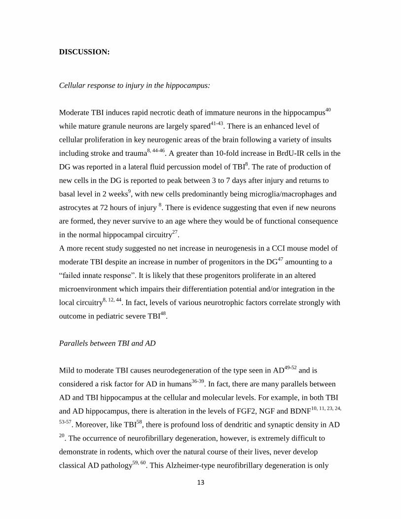
DISCUSSION:
Cellular response to injury in the hippocampus:
Moderate TBI induces rapid necrotic death of immature neurons in the hippocampus40
while mature granule neurons are largely spared41-43
. There is an enhanced level of
cellular proliferation in key neurogenic areas of the brain following a variety of insults
including stroke and trauma8, 44-46
. A greater than 10-fold increase in BrdU-IR cells in the
DG was reported in a lateral fluid percussion model of TBI8. The rate of production of
new cells in the DG is reported to peak between 3 to 7 days after injury and returns to
basal level in 2 weeks9, with new cells predominantly being microglia/macrophages and
astrocytes at 72 hours of injury 8. There is evidence suggesting that even if new neurons
are formed, they never survive to an age where they would be of functional consequence
in the normal hippocampal circuitry27
.
A more recent study suggested no net increase in neurogenesis in a CCI mouse model of
moderate TBI despite an increase in number of progenitors in the DG47
amounting to a
“failed innate response”. It is likely that these progenitors proliferate in an altered
microenvironment which impairs their differentiation potential and/or integration in the
local circuitry8, 12, 44
. In fact, levels of various neurotrophic factors correlate strongly with
outcome in pediatric severe TBI48
.
Parallels between TBI and AD
Mild to moderate TBI causes neurodegeneration of the type seen in AD49-52
and is
considered a risk factor for AD in humans36-39
. In fact, there are many parallels between
AD and TBI hippocampus at the cellular and molecular levels. For example, in both TBI
and AD hippocampus, there is alteration in the levels of FGF2, NGF and BDNF10, 11, 23, 24,
53-57. Moreover, like TBI
58, there is profound loss of dendritic and synaptic density in AD
20. The occurrence of neurofibrillary degeneration, however, is extremely difficult to
demonstrate in rodents, which over the natural course of their lives, never develop
classical AD pathology59, 60
. This Alzheimer-type neurofibrillary degeneration is only

14
seen in transgenic mice overexpressing mutated forms of human tau and or APP and at
much older ages60-65
. The findings of elevated Aβ and hyperphosphorylated tau in
ipsilateral CA1 regions of TBI mice in the current study are therefore, both interesting
and remarkable.
Effect of TBI on hippocampal dendritic and synaptic density:
It is reported that within 3 hours of CCI induced injury in rodents, there is profound loss
of MAP2 immunofluorescence in the apical dendrites of pyramidal neurons in the
ipsilateral cortex that extends beyond the area of contusion58, 66
. It is interesting to note
that despite neuronal cell death in the hippocampus following TBI, the degree of
cognitive impairment does not match with the amount of cell death. Even though, mature
granule neurons are largely spared41-43
, there is dramatic drop in the density of dendritic
spines and synapses in the surviving mature hippocampal neurons in moderate TBI after
72 hours of injury43
. Our data confirms significant decreases in MAP2 staining
throughout the hippocampus, including DG, CA1 and CA3 regions and similar decreases
in synaptophysin levels in DG at 30 days following injury. This suggests that these
changes persist in a chronic fashion and are likely to be involved in long-term cognitive
deficits.
Behavioral performance of TBI mice:
We evaluated the effect of Peptide 6 on cognition using a spatial reference memory test
in the MWM, the most frequently utilized protocol to study hippocampus dependent
spatial learning and memory in rodents67
. The hippocampus processes information about
the relationships among distal environmental cues into a spatial map where spatial
coordinates of the submerged platform are encoded67
. The hippocampus is also crucial
for memory storage, consolidation and restitution of the spatial information 68
. This test
was especially appropriate because TBI did not cause impairment in swim speeds in these
mice. Pre injury training on the MWM showed no group differences in learning (Figure
6C), establishing baseline uniformity in spatial encoding. This was maintained in the post
TBI training phase with no differences between saline and Peptide 6 treated mice.
15
However on probe trial, we found a significant improvement in measures of memory
retention in Peptide 6 treated animals when compared with saline treatment.
CA1 susceptibility in TBI:
Of the various hippocampal sub regions, CA1 is most vulnerable to hypoxic and ischemic
insults 69, 70
when compared to DG and CA371-73
. In this study, we saw elevations of
Alzheimer-type biomarkers only in CA1 region. Although we found significant decrease
in synaptic and dendritic densities in all regions of the hippocampus, neuron cell loss was
only significantly seen in CA1 region. This was not only prevented with Peptide 6
treatment, its effect on MAP2 and synaptophysin IR was most profound in the CA1
region. Consequently, there was also an upregulation of immediate-early gene expression
in CA1 region of the hippocampus suggesting that mature CA1 neurons were actively
participating in hippocampal dependent spatial paradigms. In MWM, activation of CA1
and CA3 regions is temporally and functionally distinct during different phases of the test
74 75
. For example, when CA3 is experimentally blocked, direct activation of CA1 place
cells might be sufficient for retrieval of spatial information during probe test75
. It is
therefore, not surprising that in the current study, TBI-mice did not display learning
deficits but were impaired in recall.
Neurotrophic factor replacement in TBI and challenges in clinical utility:
Supplementing the hippocampus with neurotrophic factors that drive stem cells
towards differentiation and sustain local microenvironment is a powerful concept in
neuropharma48
. Several studies have reported enhancement of neurogenesis in TBI
models through neurotrophic factor supplementation such as S100B, erthyropoietin,
EGF and FGF and superior performance in memory tasks12-15
. While the
administration of neurotrophic factors has generated much excitement in the
literature, invasive mode of delivery, adverse side effects and difficult
pharmacokinetics have very much limited the clinical usefulness of this approach18
.
16
Peptide 6 and its potential clinical applications:
The ciliary neurotrophic factor (CNTF) is a major determining factor for neurogenesis,
both in hippocampus and subventricular zone29, 76
. Like other neurotrophins18
, the
therapeutic potential of exogenous CNTF is eclipsed by its short half-life when
administered peripherally, requiring invasive mode of administration with unpredictable
pharmacokinetics77
. Now in a mild-to-moderate TBI mouse model, we show that Peptide
6 enhances differentiation of newly born progenitors in the dentate gyrus 30 days after
injury. We believe that this neurogenic effect of Peptide 6 is due to its partial inhibition
of leukemia inhibitory factor (LIF) activity by directly binding to the D1 cap region of
LIF receptor19
. The fact that there was no statistical increase in the number of progenitors
suggests a specific effect of Peptide 6 in promoting neuronal maturation and survival that
is not seen naturally after TBI9, 47
. Importantly, the newborn mature neurons remain
within the physiological confines of the GCL and display no ectopic migration or birth, a
phenomenon linked with epilepsy and schizophrenia30, 31
. We saw a robust correction of
dendritic and synaptic markers in key regions of the injured hippocampus suggesting that
Peptide 6 provides a neurotrophic milieu that might help sustain local microenvironment
after mild-to-moderate brain injury.

17
CONCLUSIONS
Here, we demonstrate enhancement of neurogenesis by a small molecule in a mouse
model of mild to moderate TBI. We further demonstrate that chronic administration of
this molecule resulted in recovery of dendritic density and synaptic loss and activation of
the traditional tri-synaptic memory pathway in the hippocampus of injured mice. This
also resulted in improvement in memory on behavioral testing. The fact that this
molecule is neurogenic and is given peripherally and non-invasively, demonstrates its
potential in hippocampal regeneration in the post-traumatic brain. Our data further
emphasizes the role of neurotrophic factor supplementation in TBI and makes a strong
case for neurotrophic factor-based novel therapies for the injured hippocampus.

18
REFERENCES
1. Faul M XL, Wald MM, Coronado VG. Traumatic brain injury in the United
States: emergency department visits, hospitalizations, and deaths. Atlanta
(GA)2010.
2. Langlois JA, Rutland-Brown W, Wald MM. The epidemiology and impact of
traumatic brain injury: a brief overview. The Journal of head trauma
rehabilitation. Sep-Oct 2006;21(5):375-378.
3. Finkelstein E CP, Miller T The Incidence and Economic Burden of Injuries in the
United States. New York (NY): Oxford University Press; 2006.
4. Jain KK. Neuroprotection in traumatic brain injury. Drug discovery today. Dec
2008;13(23-24):1082-1089.
5. Margulies S, Hicks R. Combination therapies for traumatic brain injury:
prospective considerations. Journal of neurotrauma. Jun 2009;26(6):925-939.
6. Kotapka MJ, Graham DI, Adams JH, Gennarelli TA. Hippocampal pathology in
fatal human head injury without high intracranial pressure. Journal of
neurotrauma. Jun 1994;11(3):317-324.
7. Williams S, Raghupathi R, MacKinnon MA, McIntosh TK, Saatman KE, Graham
DI. In situ DNA fragmentation occurs in white matter up to 12 months after head
injury in man. Acta neuropathologica. Dec 2001;102(6):581-590.
8. Chirumamilla S, Sun D, Bullock MR, Colello RJ. Traumatic brain injury induced
cell proliferation in the adult mammalian central nervous system. Journal of
neurotrauma. Jun 2002;19(6):693-703.
9. Dash PK, Mach SA, Moore AN. Enhanced neurogenesis in the rodent
hippocampus following traumatic brain injury. Journal of neuroscience research.
Feb 15 2001;63(4):313-319.
10. Yoshimura S, Takagi Y, Harada J, et al. FGF-2 regulation of neurogenesis in
adult hippocampus after brain injury. Proceedings of the National Academy of
Sciences of the United States of America. May 8 2001;98(10):5874-5879.

19
11. Yoshimura S, Teramoto T, Whalen MJ, et al. FGF-2 regulates neurogenesis and
degeneration in the dentate gyrus after traumatic brain injury in mice. The Journal
of clinical investigation. Oct 2003;112(8):1202-1210.
12. Sun D, Bullock MR, McGinn MJ, et al. Basic fibroblast growth factor-enhanced
neurogenesis contributes to cognitive recovery in rats following traumatic brain
injury. Experimental neurology. Mar 2009;216(1):56-65.
13. Kleindienst A, Harvey HB, Rice AC, et al. Intraventricular infusion of the
neurotrophic protein S100B improves cognitive recovery after fluid percussion
injury in the rat. Journal of neurotrauma. May 2004;21(5):541-547.
14. Kleindienst A, McGinn MJ, Harvey HB, Colello RJ, Hamm RJ, Bullock MR.
Enhanced hippocampal neurogenesis by intraventricular S100B infusion is
associated with improved cognitive recovery after traumatic brain injury. Journal
of neurotrauma. Jun 2005;22(6):645-655.
15. Lu D, Mahmood A, Qu C, Goussev A, Schallert T, Chopp M. Erythropoietin
enhances neurogenesis and restores spatial memory in rats after traumatic brain
injury. Journal of neurotrauma. Sep 2005;22(9):1011-1017.
16. Miller RG, Moore DH, Jackson CE. Western ALS Study Group. Amyotrophic
lateral sclerosis and other motor neuron disorders : official publication of the
World Federation of Neurology, Research Group on Motor Neuron Diseases. Sep
2004;5 Suppl 1:121-124.
17. Emerich DF, Thanos CG. Intracompartmental delivery of CNTF as therapy for
Huntington's disease and retinitis pigmentosa. Curr Gene Ther. Feb
2006;6(1):147-159.
18. Price RD, Milne SA, Sharkey J, Matsuoka N. Advances in small molecules
promoting neurotrophic function. Pharmacology & therapeutics. Aug
2007;115(2):292-306.
19. Chohan MO, Li B, Blanchard J, et al. Enhancement of dentate gyrus
neurogenesis, dendritic and synaptic plasticity and memory by a neurotrophic
peptide. Neurobiology of aging. Aug 2011;32(8):1420-1434.
20. Blanchard J, Wanka L, Tung YC, et al. Pharmacologic reversal of neurogenic and
neuroplastic abnormalities and cognitive impairments without affecting Abeta and

20
tau pathologies in 3xTg-AD mice. Acta neuropathologica. Nov 2010;120(5):605-
621.
21. Blanchard J, Bolognin S, Chohan MO, Rabe A, Iqbal K, Grundke-Iqbal I. Rescue
of synaptic failure and alleviation of learning and memory impairments in a
trisomic mouse model of down syndrome. J Neuropathol Exp Neurol. Dec
2011;70(12):1070-1079.
22. Hicks RR, Numan S, Dhillon HS, Prasad MR, Seroogy KB. Alterations in BDNF
and NT-3 mRNAs in rat hippocampus after experimental brain trauma. Brain
research. Molecular brain research. Sep 1997;48(2):401-406.
23. Oyesiku NM, Evans CO, Houston S, et al. Regional changes in the expression of
neurotrophic factors and their receptors following acute traumatic brain injury in
the adult rat brain. Brain research. Jul 3 1999;833(2):161-172.
24. Yang K, Perez-Polo JR, Mu XS, et al. Increased expression of brain-derived
neurotrophic factor but not neurotrophin-3 mRNA in rat brain after cortical
impact injury. Journal of neuroscience research. Apr 15 1996;44(2):157-164.
25. Tatebayashi Y, Lee MH, Li L, Iqbal K, Grundke-Iqbal I. The dentate gyrus
neurogenesis: a therapeutic target for Alzheimer's disease. Acta neuropathologica.
Mar 2003;105(3):225-232.
26. Chen H, Tung YC, Li B, Iqbal K, Grundke-Iqbal I. Trophic factors counteract
elevated FGF-2-induced inhibition of adult neurogenesis. Neurobiology of aging.
Aug 2007;28(8):1148-1162.
27. Deng W, Aimone JB, Gage FH. New neurons and new memories: how does adult
hippocampal neurogenesis affect learning and memory? Nature reviews.
Neuroscience. May 2010;11(5):339-350.
28. Kempermann G, Gast D, Kronenberg G, Yamaguchi M, Gage FH. Early
determination and long-term persistence of adult-generated new neurons in the
hippocampus of mice. Development. Jan 2003;130(2):391-399.
29. Emsley JG, Hagg T. Endogenous and exogenous ciliary neurotrophic factor
enhances forebrain neurogenesis in adult mice. Experimental neurology. Oct
2003;183(2):298-310.

21
30. Donovan MH, Yazdani U, Norris RD, Games D, German DC, Eisch AJ.
Decreased adult hippocampal neurogenesis in the PDAPP mouse model of
Alzheimer's disease. The Journal of comparative neurology. Mar 1
2006;495(1):70-83.
31. Duan X, Chang JH, Ge S, et al. Disrupted-In-Schizophrenia 1 regulates
integration of newly generated neurons in the adult brain. Cell. Sep 21
2007;130(6):1146-1158.
32. Guzowski JF, Timlin JA, Roysam B, McNaughton BL, Worley PF, Barnes CA.
Mapping behaviorally relevant neural circuits with immediate-early gene
expression. Current opinion in neurobiology. Oct 2005;15(5):599-606.
33. Snyder JS, Choe JS, Clifford MA, et al. Adult-born hippocampal neurons are
more numerous, faster maturing, and more involved in behavior in rats than in
mice. The Journal of neuroscience : the official journal of the Society for
Neuroscience. Nov 18 2009;29(46):14484-14495.
34. Jessberger S, Kempermann G. Adult-born hippocampal neurons mature into
activity-dependent responsiveness. The European journal of neuroscience. Nov
2003;18(10):2707-2712.
35. Omalu BI, DeKosky ST, Hamilton RL, et al. Chronic traumatic encephalopathy in
a national football league player: part II. Neurosurgery. Nov 2006;59(5):1086-
1092; discussion 1092-1083.
36. Lye TC, Shores EA. Traumatic brain injury as a risk factor for Alzheimer's
disease: a review. Neuropsychology review. Jun 2000;10(2):115-129.
37. Jiang T, Yu JT, Tian Y, Tan L. Epidemiology and etiology of Alzheimer's
disease: from genetic to non-genetic factors. Current Alzheimer research. Oct
2013;10(8):852-867.
38. Jellinger KA. Traumatic brain injury as a risk factor for Alzheimer's disease. J
Neurol Neurosurg Psychiatry. Mar 2004;75(3):511-512.
39. Sivanandam TM, Thakur MK. Traumatic brain injury: a risk factor for
Alzheimer's disease. Neuroscience and biobehavioral reviews. May
2012;36(5):1376-1381.

22
40. Zhou H, Chen L, Gao X, Luo B, Chen J. Moderate traumatic brain injury triggers
rapid necrotic death of immature neurons in the hippocampus. Journal of
neuropathology and experimental neurology. Apr 2012;71(4):348-359.
41. Saatman KE, Feeko KJ, Pape RL, Raghupathi R. Differential behavioral and
histopathological responses to graded cortical impact injury in mice. Journal of
neurotrauma. Aug 2006;23(8):1241-1253.
42. Rola R, Mizumatsu S, Otsuka S, et al. Alterations in hippocampal neurogenesis
following traumatic brain injury in mice. Experimental neurology. Nov
2006;202(1):189-199.
43. Gao X, Deng P, Xu ZC, Chen J. Moderate traumatic brain injury causes acute
dendritic and synaptic degeneration in the hippocampal dentate gyrus. PloS one.
2011;6(9):e24566.
44. Liu F, You Y, Li X, et al. Brain injury does not alter the intrinsic differentiation
potential of adult neuroblasts. The Journal of neuroscience : the official journal of
the Society for Neuroscience. Apr 22 2009;29(16):5075-5087.
45. Gao X, Enikolopov G, Chen J. Moderate traumatic brain injury promotes
proliferation of quiescent neural progenitors in the adult hippocampus.
Experimental neurology. Oct 2009;219(2):516-523.
46. Hill-Felberg SJ, McIntosh TK, Oliver DL, Raghupathi R, Barbarese E.
Concurrent loss and proliferation of astrocytes following lateral fluid percussion
brain injury in the adult rat. Journal of neuroscience research. Jul 15
1999;57(2):271-279.
47. Gao X, Chen J. Moderate traumatic brain injury promotes neural precursor
proliferation without increasing neurogenesis in the adult hippocampus.
Experimental neurology. Jan 2013;239:38-48.
48. Chiaretti A, Piastra M, Polidori G, et al. Correlation between neurotrophic factor
expression and outcome of children with severe traumatic brain injury. Intensive
care medicine. Aug 2003;29(8):1329-1338.
49. Omalu BI, DeKosky ST, Minster RL, Kamboh MI, Hamilton RL, Wecht CH.
Chronic traumatic encephalopathy in a National Football League player.
Neurosurgery. Jul 2005;57(1):128-134; discussion 128-134.

23
50. Small GW, Kepe V, Siddarth P, et al. PET scanning of brain tau in retired
national football league players: preliminary findings. The American journal of
geriatric psychiatry : official journal of the American Association for Geriatric
Psychiatry. Feb 2013;21(2):138-144.
51. Fakhran S, Yaeger K, Alhilali L. Symptomatic white matter changes in mild
traumatic brain injury resemble pathologic features of early Alzheimer dementia.
Radiology. Oct 2013;269(1):249-257.
52. Chauhan NB. Chronic neurodegenerative consequences of traumatic brain injury.
Restorative neurology and neuroscience. Jan 7 2014.
53. Stopa EG, Gonzalez AM, Chorsky R, et al. Basic fibroblast growth factor in
Alzheimer's disease. Biochem Biophys Res Commun. Sep 14 1990;171(2):690-
696.
54. Hock C, Heese K, Hulette C, Rosenberg C, Otten U. Region-specific neurotrophin
imbalances in Alzheimer disease: decreased levels of brain-derived neurotrophic
factor and increased levels of nerve growth factor in hippocampus and cortical
areas. Archives of neurology. Jun 2000;57(6):846-851.
55. Fahnestock M, Michalski B, Xu B, Coughlin MD. The precursor pro-nerve
growth factor is the predominant form of nerve growth factor in brain and is
increased in Alzheimer's disease. Molecular and cellular neurosciences. Aug
2001;18(2):210-220.
56. Hicks RR, Baldwin SA, Scheff SW. Serum extravasation and cytoskeletal
alterations following traumatic brain injury in rats. Comparison of lateral fluid
percussion and cortical impact models. Molecular and chemical neuropathology /
sponsored by the International Society for Neurochemistry and the World
Federation of Neurology and research groups on neurochemistry and
cerebrospinal fluid. Sep-Dec 1997;32(1-3):1-16.
57. Truettner J, Schmidt-Kastner R, Busto R, et al. Expression of brain-derived
neurotrophic factor, nerve growth factor, and heat shock protein HSP70 following
fluid percussion brain injury in rats. Journal of neurotrauma. Jun 1999;16(6):471-
486.

24
58. Posmantur RM, Kampfl A, Taft WC, et al. Diminished microtubule-associated
protein 2 (MAP2) immunoreactivity following cortical impact brain injury.
Journal of neurotrauma. Mar 1996;13(3):125-137.
59. Chohan MO, Haque N, Alonso A, et al. Hyperphosphorylation-induced self
assembly of murine tau: a comparison with human tau. J Neural Transm. Aug
2005;112(8):1035-1047.
60. Dodart JC, Mathis C, Bales KR, Paul SM. Does my mouse have Alzheimer's
disease? Genes, brain, and behavior. Aug 2002;1(3):142-155.
61. Masliah E, Sisk A, Mallory M, Mucke L, Schenk D, Games D. Comparison of
neurodegenerative pathology in transgenic mice overexpressing V717F beta-
amyloid precursor protein and Alzheimer's disease. The Journal of neuroscience :
the official journal of the Society for Neuroscience. Sep 15 1996;16(18):5795-
5811.
62. Tatebayashi Y, Miyasaka T, Chui DH, et al. Tau filament formation and
associative memory deficit in aged mice expressing mutant (R406W) human tau.
Proceedings of the National Academy of Sciences of the United States of America.
Oct 15 2002;99(21):13896-13901.
63. Lewis J, McGowan E, Rockwood J, et al. Neurofibrillary tangles, amyotrophy and
progressive motor disturbance in mice expressing mutant (P301L) tau protein.
Nature genetics. Aug 2000;25(4):402-405.
64. Hsiao K, Chapman P, Nilsen S, et al. Correlative memory deficits, Abeta
elevation, and amyloid plaques in transgenic mice. Science. Oct 4
1996;274(5284):99-102.
65. Oddo S, Caccamo A, Shepherd JD, et al. Triple-transgenic model of Alzheimer's
disease with plaques and tangles: intracellular Abeta and synaptic dysfunction.
Neuron. Jul 31 2003;39(3):409-421.
66. Chen LJ, Wang YJ, Tseng GF. Compression alters kinase and phosphatase
activity and tau and MAP2 phosphorylation transiently while inducing the fast
adaptive dendritic remodeling of underlying cortical neurons. Journal of
neurotrauma. Sep 2010;27(9):1657-1669.

25
67. Morris RG, Garrud P, Rawlins JN, O'Keefe J. Place navigation impaired in rats
with hippocampal lesions. Nature. Jun 24 1982;297(5868):681-683.
68. Riedel G, Micheau J, Lam AG, et al. Reversible neural inactivation reveals
hippocampal participation in several memory processes. Nature neuroscience. Oct
1999;2(10):898-905.
69. Schmidt-Kastner R, Freund TF. Selective vulnerability of the hippocampus in
brain ischemia. Neuroscience. 1991;40(3):599-636.
70. Aitken PG, Schiff SJ. Selective neuronal vulnerability to hypoxia in vitro.
Neuroscience letters. Jun 6 1986;67(1):92-96.
71. Crepel V, Krnjevic K, Ben-Ari Y. Developmental and regional differences in the
vulnerability of rat hippocampal slices to lack of glucose. Neuroscience.
1992;47(3):579-587.
72. Kass IS, Lipton P. Calcium and long-term transmission damage following anoxia
in dentate gyrus and CA1 regions of the rat hippocampal slice. The Journal of
physiology. Sep 1986;378:313-334.
73. Kreisman NR, Soliman S, Gozal D. Regional differences in hypoxic
depolarization and swelling in hippocampal slices. Journal of neurophysiology.
Feb 2000;83(2):1031-1038.
74. Laeremans A, Sabanov V, Ahmed T, et al. Distinct and simultaneously active
plasticity mechanisms in mouse hippocampus during different phases of Morris
water maze training. Brain structure & function. Feb 23 2014.
75. Florian C, Roullet P. Hippocampal CA3-region is crucial for acquisition and
memory consolidation in Morris water maze task in mice. Behavioural brain
research. Oct 5 2004;154(2):365-374.
76. Yang P, Arnold SA, Habas A, Hetman M, Hagg T. Ciliary neurotrophic factor
mediates dopamine D2 receptor-induced CNS neurogenesis in adult mice. The
Journal of neuroscience : the official journal of the Society for Neuroscience. Feb
27 2008;28(9):2231-2241.
77. Chen ZY, Cao L, Wang LM, et al. Development of neurotrophic molecules for
treatment of neurodegeneration. Current protein & peptide science. Sep
2001;2(3):261-276.

26
27
FIGURE LEGENDS
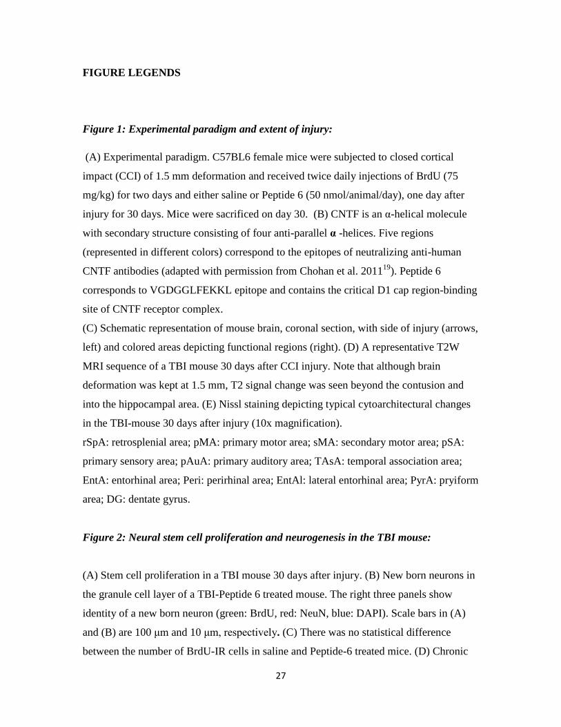
Figure 1: Experimental paradigm and extent of injury:
(A) Experimental paradigm. C57BL6 female mice were subjected to closed cortical
impact (CCI) of 1.5 mm deformation and received twice daily injections of BrdU (75
mg/kg) for two days and either saline or Peptide 6 (50 nmol/animal/day), one day after
injury for 30 days. Mice were sacrificed on day 30. (B) CNTF is an α-helical molecule
with secondary structure consisting of four anti-parallel α -helices. Five regions
(represented in different colors) correspond to the epitopes of neutralizing anti-human
CNTF antibodies (adapted with permission from Chohan et al. 201119
). Peptide 6
corresponds to VGDGGLFEKKL epitope and contains the critical D1 cap region-binding
site of CNTF receptor complex.
(C) Schematic representation of mouse brain, coronal section, with side of injury (arrows,
left) and colored areas depicting functional regions (right). (D) A representative T2W
MRI sequence of a TBI mouse 30 days after CCI injury. Note that although brain
deformation was kept at 1.5 mm, T2 signal change was seen beyond the contusion and
into the hippocampal area. (E) Nissl staining depicting typical cytoarchitectural changes
in the TBI-mouse 30 days after injury (10x magnification).
rSpA: retrosplenial area; pMA: primary motor area; sMA: secondary motor area; pSA:
primary sensory area; pAuA: primary auditory area; TAsA: temporal association area;
EntA: entorhinal area; Peri: perirhinal area; EntAl: lateral entorhinal area; PyrA: pryiform
area; DG: dentate gyrus.
Figure 2: Neural stem cell proliferation and neurogenesis in the TBI mouse:
(A) Stem cell proliferation in a TBI mouse 30 days after injury. (B) New born neurons in
the granule cell layer of a TBI-Peptide 6 treated mouse. The right three panels show
identity of a new born neuron (green: BrdU, red: NeuN, blue: DAPI). Scale bars in (A)
and (B) are 100 μm and 10 μm, respectively. (C) There was no statistical difference
between the number of BrdU-IR cells in saline and Peptide-6 treated mice. (D) Chronic

28
treatment with Peptide 6 significantly increased the number of newborn neurons (BrdU-
NeuN-IR cells) in the dentate gyrus of TBI mice. (E & F) “Ectopic birth index”
calculation suggested that up to 40% of newborn cells and 43% of newborn neurons were
located beyond the iGCL of DG in TBI-saline mice suggesting “ectopic birth” and/or
aberrant migration. Although not statistically significant, this number was lower in
Peptide 6 treated group (32% and 34% respectively).
BrdU (5’bromodeoxyuridine), BrdU-IR (BrdU immunoreactive), oGCL (outer granule
cell layer), iGCL (inner granule cell layer), DG (dentate gyrus). Statistical analysis done
using Student’s t-test with p value <0.05 (*)
“Ectopic birth index” calculations were performed using the formula (oGCL/GCL)
according to Donovan et al., 200630
.
Figure 3: Loss of dendritic density in TBI hippocampus and its recovery by Peptide 6
(A) Photomicrographs illustrating MAP2 immunoreactivity in different areas of the
hippocampus in saline and Peptide 6 treated TBI-mice. (b) There was a significant loss of
dendritic density in the ipsilateral DG, CA3 and CA1 subregions of TBI-mice
hippocampus compared to contralateral regions. (B, left panel) Chronic treatment with
Peptide 6 increased MAP2 staining by 28% in DG ipsilateral to the injury as compared to
saline treated mice. A similar, but non-significant, trend was also observed in CA3 (B,
middle panel) and CA1 (C, right panel) regions in Peptide 6 treated mice. Scale bar = 100
μm.
MAP2 (microtubule associated protein 2), TBI-P6 (TBI-Peptide 6 treated group)
* (p<0.05), **(p<0.01), ***(p<0.001), one way ANOVA, Bonferroni’s post-hoc test or
Student’s t-test.
Figure 4: Synaptic loss in TBI hippocampus and its recovery by Peptide 6

29
(A) Photomicrographs illustrating synaptophysin immunoreactivity in different areas of
the hippocampus in saline and Peptide 6 treated mice. (B) Compared to contralateral side,
there was significant decrease in synaptophysin immunoreactivity in ipsilateral DG but
not CA3 or CA1 regions of TBI-mice hippocampus. There was a 27%, 23% and 40%
increase in synaptic density in DG (B, left panel), CA3 (B, middle panel) and CA1 (B,
right panel) regions on the ipsilateral side in Peptide 6 treated TBI-mice as compared to
saline treated animals. Scale bar = 100 μm.
DG (dentate gyrus), TBI-P6 (TBI-Peptide 6 treated group)
* (p<0.05), one way ANOVA, Bonferroni’s post-hoc test, or Student’s t-test.
Figure 5: Cortical neuronal loss and Immediate-early gene expression in hippocampus
(A-D) Photomicrographs demonstrating Nissl staining in the CA1 region (A) and parietal
cortex (C). There was significant loss of neurons in the ipsilateral CA1 region (B) and
parietal cortex (D) in saline treated TBI mice compared to contralateral side. Chronic
treatment with Peptide 6 prevented this loss. (E) Photomicrographs illustrating
immediate-early gene expression (zif 268) in the CA1 region of saline and Peptide 6
treated TBI-mice, 30 days after injury and within 3 hours of performance in a
hippocampus-dependent memory task. There was an almost 130% increase in the levels
of zif268 in CA1 region of Peptide 6 treated TBI-mice as compared to saline treated
animals in both ipsilateral and contralateral sides (F). Scale bar = 50 μm in (A and C) and
100 μm in (E)
zif268 (early growth response 1 gene), TBI-P6 (TBI-Peptide 6 treated group)
***(p<0.001), Student’s t-test
Figure 6: Behavioral testing on Morris Water Maze:
(A) Behavioral testing paradigm on Morris Water Maze (MWM). After 3 weeks of
acclimatization, all animals underwent 4 day training on the MWM to establish baseline
learning. The mice were retrained on day 26 after TBI and treatment (saline or Peptide 6)

30
with the probe trial on day 30. (B) There was no difference in pre or post TBI swim
speeds between the various groups. (C) All animals showed equivalent learning on
MWM as evidenced by decreasing escape latencies from day 1 to day 4 (repeated
measures ANOVA). Additionally, there was no effect of Peptide 6 on learning in TBI
animals. (D) On probe trial, Peptide 6 treatment caused significant improvement in
latency to target, (E) percent time spent in target quadrant and (F) percent distance
covered in target quadrant.
TBI-P6 (TBI-Peptide 6 treated group), D1 (day 1)
** (p<0.01) one-way ANOVA, post hoc Tukey
Supplementary Figure 1:
Elevation of Alzheimer-type biomarkers in TBI mice. (A) Immunohistochemical staining
of hippocampus and parietal cortex revealed elevation of hyperphosphorylated tau and
Aβ, two key hallmarks of Alzheimer disease in ipsilateral versus contralateral regions.
Red: PHF1 staining against phosphor-tau at serine 396/404 (Scale bar = 100 μm); Green:
4G8 staining against APP and Aβ (Scale bar = 50 μm)
*(p<0.05), **(p<0.01), ***(p<0.001), one way ANOVA, Bonferroni’s post hoc test
Table 1
Stereological counts (mean ± SEM) of newborn progenitors (BrdU-IR) in various layers
of the dentate gyrus.
Table 2
Stereological counts (mean ± SEM) of newborn neurons (BrdU-NeuN-IR cells) in
various layers of the dentate gyrus.

Figure 1Click here to download high resolution image
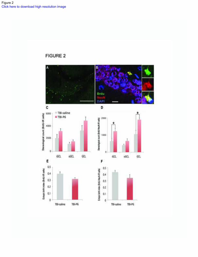
Figure 2Click here to download high resolution image

Figure 3Click here to download high resolution image

Figure 4Click here to download high resolution image
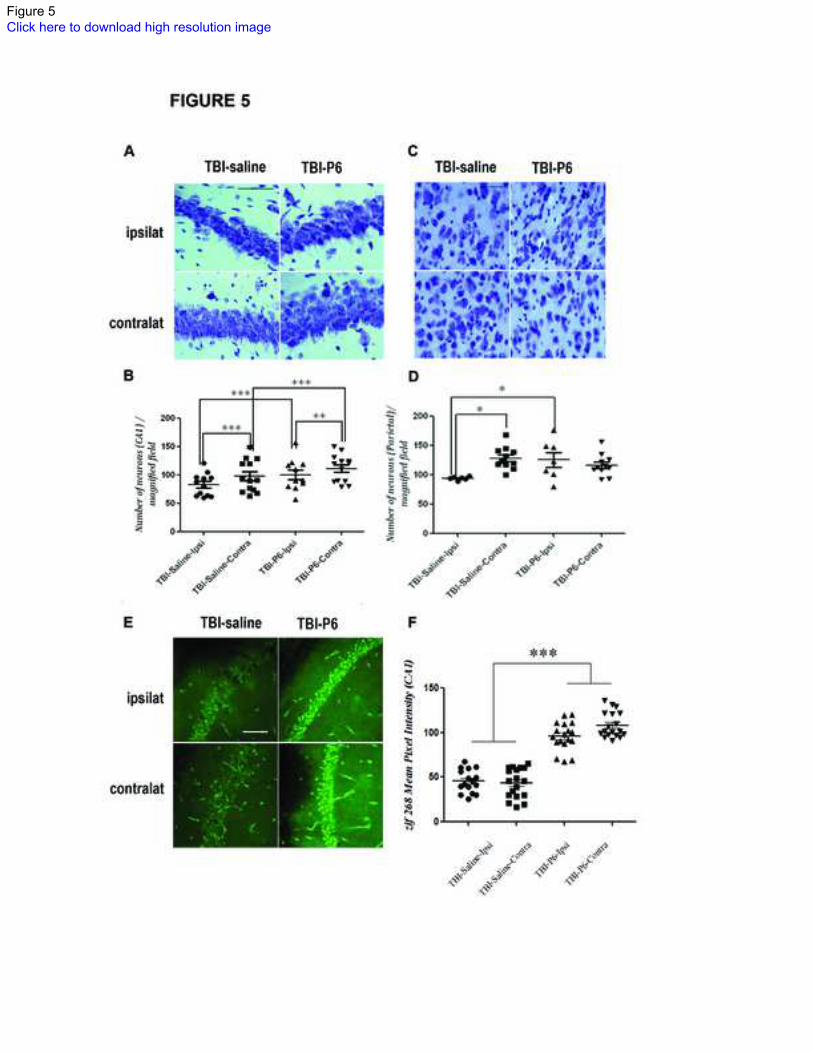
Figure 5Click here to download high resolution image

Figure 6Click here to download high resolution image
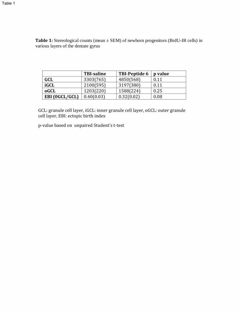
Table 1: Stereological counts (mean ± SEM) of newborn progenitors (BrdU-IR cells) in
various layers of the dentate gyrus
TBI-saline TBI-Peptide 6 p value GCL 3303(765) 4850(568) 0.11 iGCL 2100(595) 3197(380) 0.11 oGCL 1203(220) 1588(224) 0.25 EBI (0GCL/GCL) 0.40(0.03) 0.32(0.02) 0.08
GCL: granule cell layer, iGCL: inner granule cell layer, oGCL: outer granule cell layer, EBI: ectopic birth index
p-value based on unpaired Student’s t-test
Table 1

Table 2: Stereological counts (mean ± SEM) of newborn neurons (BrdU-NeuN-IR cells)
in various layers of the dentate gyrus
TBI-saline TBI-Peptide 6 p value GCL 1057(217) 1901(265) 0.03* iGCL 648(176) 1225(169) 0.03* oGCL 409(64) 658(124) 0.13 EBI(0GCL/GCL) 0.43(0.03) 0.34(0.05) 0.15
GCL: granule cell layer, iGCL: inner granule cell layer, oGCL: outer granule cell layer, EBI: ectopic birth index
p-value based on unpaired Student’s t-test (*p<0.05)
Table 2

Supplemental Figure 1Click here to download high resolution image

METHODS:
Generation of Peptide 6:
Based on our previous study1, and epitope mapping of neutralizing antibodies to human
CNTF (Pepscan, Lelystad, The Netherlands), we developed an 11-mer peptide, Peptide 6,
which is both neurogenic and neurotrophic2 (Figure 1b).
Antibodies:
The following primary antibodies were used for immunohistochemistry (IHC): anti-BrdU
(1:400; Accurate, Westbury, NY), anti-NeuN (1:500; Chemicon, Temecula, CA), SMI52
(anti-MAP2; 1:1000, Cavance, CA), anti-synaptophysin (1:200; Millipore, Temecula,
CA), Egr-1 (anti-zif 268; 1:100, Santa Cruz Biotechnology, Santa Cruz, CA), 4G8 (anti-
Aβ; 1:200; in-house) and PHF1 (monoclonal antibody to phosphorylated tau at serine
396 and serine 404; 1:200; a gift from Dr. Peter Davies, Albert Einstein College of
Medicine). The following secondary antibodies were used: Alexa 488 conjugated goat
anti-mouse IgG and Alexa 594 conjugated goat anti-rabbit or anti-rat IgG (Molecular
Probes, Carlsbad, CA, USA). Nuclear staining was obtained using DAPI (Sigma-Aldrich,
St. Louis, MO).
Animals and Housing
All in vivo studies were performed on 3-4-month-old female mice of C57BL6 strain
(Jackson Laboratories, Bar Harbor, ME). All animals were group-housed (3 animals per
Supplemental MethodsClick here to download Supplemental Content (for online-only posting): Supplementary Materials and Methods.docx

cage) with a 12:12 light: dark cycle, with free access to food and water and were given a
period of acclimatization before any studies were performed. The studies were conducted
under protocol # 100954 of the Institutional Animal Care and Use Committee of the
University of New Mexico.
Induction of TBI in mice:
Induction of TBI in mice was based on the CCI model as described previously3. Briefly,
mice were anesthetized with isoflurane (5% induction, 2% maintenance with 70% nitrous
oxide and 30% oxygen) and placed in a stereotaxic frame. After a midline skin incision, a
5 mm left lateral craniotomy was made using a motorized drill. CCI was induced with an
impact device using a 3 mm diameter metal tip with a velocity of 5 m/sec to a penetration
depth of 1.5 mm below the dura. The impact was centered at 2.7 mm lateral to midline
and 3.0 mm anterior to lambda. The craniotomy site was sealed with Loctite Gel glue
(Henkel Corp., Rocky Hill, CT) and the scalp was sutured. The mice were given
antibiotic prophylaxis with 500 units/gm of IM Bicillin (Pfizer, NY) peri-operatively and
0.01 mg/kg twice daily of buprenorphine (Reckitt Benckiser Pharmaceuticals, N
Chesterfield, VA) for three days for comfort.
Administration of BrdU and Peptide 6
To study neurogenesis, animals were given intraperitoneal (i.p.) injections of Peptide 6
(50 nmol/animal/day) or saline (control) for 30 days. Mice in either treatment group

received twice-daily injections of BrdU (75 mg/kg, Sigma-Aldrich, St. Louis, MO) for
two days beginning one-day post trauma (Figure 1a).
Tissue processing and immunohistochemistry
Tissue processing and IHC was performed as described previously2. Briefly, mice were
anesthetized with pentobarbital (125 mg/kg) and transcardially perfused with 0.1M
phosphate buffered saline (PBS) followed by 4% paraformaldehyde (PFA). The brains
were then kept in 4% PFA solution for 24 hours and stored in 30% sucrose solution at
4°C till further use. Coronal sections (35 μm) were obtained on a freezing sliding
microtome and stored in glycol anti-freeze solution (ethylene glycol, glycerol and 0.1M
PBS in 3:3:4 ratio) at -22°C until further use. IHC was performed on free-floating
sections. Every 5th
brain section was chosen for cell counts and every 10th
section for
immunoreactivity quantification using uniform random sampling.
Stereology & confocal microscopy
Neurogenesis was assessed in the DG by counting the number of BrdU-immunoreactive
(BrdU-IR) and BrdU-NeuN-IR cells in various layers of the DG. The granule cell layer
(GCL) was further subdivided into an inner and out half (iGCL and oGCL) as described
previously4. To assess cell proliferation, Brdu-IR cells were counted in every 5
th section
using a 40x oil objective of a Nikon 90i fluorescent microscope equipped with a Nikon
C1 3-laser confocal system. Employing principles of unbiased stereology, the optical
fractionator method was used to estimate cell counts for the DG5. For each brain, at least
100 cells were counted based on coefficient of error determinations. To assess neuronal
maturation, BrdU-NeuN-IR cells were counted using 100x oil objective in every 10th
section. To ensure objectivity, z-stacks were collected for each double IR cell and
analyzed later by generating maximum projection and 3D constructs. A cell was counted
only when it showed double IR on 3D reconstructed images.
For MAP2, synaptophysin, zif268, PHF1 and 4G8-IR, the entire area of GCL, CA3 and
CA1 was outlined on every 10th
section at 20x magnification and maximum projection
images were created based on confocal z-stacks using Nikon 90i fluorescent microscope
equipped with Nikon C1 three-laser confocal system and a Nikon DS U1 digital camera.
A mean pixel intensity (MPI) was recorded for each anatomical area using Image-Pro
Plus 5.0 software (Media Cybernetics, Silver Spring, MD).
MRI acquisition:
MRI was performed on a 4.7 Tesla dedicated research MR scanner (Bruker Biospin,
Ettlingen, Germany), equipped with a small-bore linear radiofrequency
coil (72 mm). All data processing was performed using in-house developed software
written in 64-bit MATLAB (Mathworks, Natick, MA, USA) running on a UNIX
machine. Initial localizer images were acquired followed by T2-weighted (T2WI) and
diffusion-weighted images.

Behavioral Testing:
Behavioral testing included spatial reference memory test in the Morris Water Maze
(MWM).
Spatial reference memory task: The test used is an adaptation of the procedure previously
described by Morris [5]. The procedure was performed in a 180 cm diameter circular
tank. The pool was filled with water (21°C±1) made opaque by adding non-toxic white
paint (RichArt, Northvale, NJ). The maze had visual cues on all sides for spatial
orientation. A digital camera with 40X optical zoom was mounted ~6 feet above the top
of the maze to capture video. The escape platform was transparent measuring 4x4 inches
and submerged ~ 1 cm below the water line. Ceiling mounted, fluorescent bulbs provided
room lighting. All animals were first acclimated to the behavioral testing room before
training was started. The test was performed over two phases on 2 groups of mice (11-12
mice each). The first phase was done on uninjured and untreated animals to establish
baselines. Second phase was done 26 days after TBI and while treatment (saline or
Peptide 6) was in its final week (Figure 6A). All mice were trained for 4 days and given
4 trials per animal. During each trial, the mice were released randomly from the N, S, E
or W side of the pool. They were given 60 seconds per trial to find the submerged
platform. At the end of each trial the animals were left on the platform for 20 s then dried
and returned to their home cage until the next trial. A test for retention (i.e. a probe trial,
PT) was given 24 h after the last day of training. During the PT mice were allowed to
swim in the tank without the escape platform for 60 seconds. The measures of learning
were the time and the distance swum to reach the escape platform. For PT, the tank was
divided into 4 imaginary quadrants and a small zone where the escape platform had been
(i.e. target quadrant). The measures of memory on the PT were percent of time spent and
percent of distance covered in the target quadrant. On day 27 after TBI induction, the
mice were retrained as above and a probe trial performed on day 30 coinciding with the
final day of treatment. All analyses were performed on ANY-MAZE software (Stoelting
Co., Wood Dale, IL).

Statistics:
Data are presented as mean with standard error of mean (SEM). Kolmogorov-Smirnov
test was used to determine normality of the data. For stereological studies an unpaired
Student’s t-test was used. For immunohistochemical analysis involving multiple groups,
ANOVA with post hoc Bonferroni’s test was used. Further intergroup comparisons
involving the effect of TBI and/or treatment were performed using unpaired Student’s t-
test. For behavioral studies, either one-way or repeated measures ANOVA tests were
used with post hoc Tukey or Bonferroni tests where appropriate. Statistical outliers were
detected using Grubbs test for normally distributed data. Differences with p < 0.05 were
considered significant.
REFERENCES
1. Chen H, Tung YC, Li B, Iqbal K, Grundke-Iqbal I. Trophic factors counteract
elevated FGF-2-induced inhibition of adult neurogenesis. Neurobiology of aging.
Aug 2007;28(8):1148-1162.
2. Chohan MO, Li B, Blanchard J, et al. Enhancement of dentate gyrus
neurogenesis, dendritic and synaptic plasticity and memory by a neurotrophic
peptide. Neurobiology of aging. Aug 2011;32(8):1420-1434.
3. Exo JL, Shellington DK, Bayir H, et al. Resuscitation of traumatic brain injury
and hemorrhagic shock with polynitroxylated albumin, hextend, hypertonic
saline, and lactated Ringer's: Effects on acute hemodynamics, survival, and
neuronal death in mice. Journal of neurotrauma. Dec 2009;26(12):2403-2408.
4. Chohan MO, Li B, Blanchard J, et al. Enhancement of dentate gyrus
neurogenesis, dendritic and synaptic plasticity and memory by a neurotrophic
peptide. Neurobiology of aging. Aug 2011;32(8):1420-1434.
5. West MJ, Slomianka L, Gundersen HJ. Unbiased stereological estimation of the
total number of neurons in thesubdivisions of the rat hippocampus using the
optical fractionator. The Anatomical record. Dec 1991;231(4):482-497.