Calcineurin inhibitors dampen humoral immunity by acting directly on naive B cells

Effects of high-intensity aerobic interval training versus moderate
exercise on hemodynamic, metabolic, and neuro-humoral
abnormalities of young normotensive women at high familial risk
for hypertension (Pre-print version)
Emmanuel G. Ciolac, PhD;1,2,3 Edimar A. Bocchi, MD, PhD;1 Luiz A. Bortolotto, MD, PhD;1
Vitor O. Carvalho, PhD;1,3 Julia M. D. Greve, MD, PhD;2 Guilherme V. Guimarães, PhD1,3
1. Heart Institute do Hospital das Clínicas da Faculdade de Medicina da Universidade de São Paulo, São Paulo,
Brazil.
2. Institute of Orthopedics and Traumatology do Hospital das Clínicas da Faculdade de Medicina da
Universidade de São Paulo, Laboratory of Kinesiology, São Paulo, Brazil.
3. Centro de Práticas Esportivas da Universidade de São Paulo, Laboratory of Physical Activity and Health, São
Paulo, Brazil.
Publishers’ version at Hypertension Research (2010) 33, 836–843; published online 7 May
2010; http://dx.doi.org/10.1038/hr.2010.72
Please cite this article as: Ciolac EG, et al. Effects of high-intensity aerobic interval training
versus moderate exercise on hemodynamic, metabolic, and neuro-humoral abnormalities of
young normotensive women at high familial risk for hypertension. Hypertension Research
2010; 33: 836–843.
Correspondence to: Dr. Emmanuel G Ciolac, Instituto de Ortopedia e Traumatologia do
HCFMUSP, Laboratório de Estudos do Movimento, Rua Dr. Ovídio Pires de Campos, 333,
Segundo Andar, Ala C, Sao Paulo 05403-010, Brazil.
E-mail: [email protected]

Ciolac EG, et al. – Exercise and Familial Hypertension (Pre-print version)
Publishers’ version at Hypertension Research (2010) 33: 836–843; http://dx.doi.org/10.1038/hr.2010.72
ABSTRACT
Background: Exercise training has an important role in the prevention and treatment of
hypertension, but its effects on the early metabolic and hemodynamic abnormalities observed
in normotensive subjects offspring of hypertensive parents (FH+) has not been studied. We
compared high-intensity interval (AIT) and moderate-intensity continuous exercise training
(CMT) with regard hemodynamic, metabolic and hormonal variables in FH+.
Methods and Results: Forty-four healthy women FH+ (25.0±4.4 years), randomized to
control (ConFH+) or to a 3 times-a-week equal volume AIT (80-90% of VO2MAX) or CMT
(50-60% of VO2MAX), and 15 healthy women with normotensive parents (ConFH−; 25.3±3.1
years) had their hemodynamic, metabolic and hormonal variables analyzed before and after
16 weeks of follow-up. Ambulatorial blood pressure (ABP), glucose and cholesterol levels
were similar among all groups, but FH+ groups displayed higher insulin, insulin sensitivity,
carotid-femoral pulse wave velocity (PWV), norepinephrine and endothelin-1 (ET-1), and
lower nitrite/nitrate (NOx) levels than ConFH−. AIT and CMT were equally effective in
improving ABP (P<0.05), insulin and insulin sensitivity (P<0.001); however, AIT was
superior in improving cardiorespiratory fitness (15% versus 8%; P<0.05), PWV (P<0.01), and
BP, norepinephrine, ET-1 and NOx response to exercise (P<0.05).
Conclusion: Exercise intensity was an important factor for improving cardiorespiratory
fitness and reversing hemodynamic, metabolic and hormonal alterations involved in the
pathophysiology of hypertension. These findings may have important implications for
exercise training programs for the prevention of an inherited hypertensive disorder.
Key words: exercise; hypertension; endothelial function; arterial stiffness; nervous system,
sympathetic; prevention
Ciolac EG, et al. – Exercise and Familial Hypertension (Pre-print version)
Publishers’ version at Hypertension Research (2010) 33: 836–843; http://dx.doi.org/10.1038/hr.2010.72
1
INTRODUCTION
Essential arterial hypertension (HPT) is the most common risk factor for cardiovascular
morbidity and mortality, and is associated with substantial health care expenditure.1,2
Consequently, primary prevention in individuals at high risk for hypertension has been
emphasized.3 Among the population at high risk for hypertension, those who have
hypertensive parents (FH+) are worthy of special attention. It is well established that positive
family history of hypertension is a strong risk factor for future HPT in nonhypertensive
offspring, independently of other risk factors.4 Moreover, hemodynamic, metabolic and
hormonal abnormalities, as well as concentrations of biomarkers that may play a key role in
the development of hypertension are increased in nonhypertensive offspring of parents with
HPT.5-7
Higher levels of physical activity8,9 and cardiorespiratory fitness9 has shown to reduce the risk
of HPT for healthy normotensive persons. Acute10,11 and chronic12-14 exercise can also reduce
resting blood pressure (BP) in hypertensive adults. Moreover, exercise training has shown to
improve several factors involved in the pathophysiology of HPT, including sympathetic
activity,15 endothelial function,16 pulse wave velocity (PWV),14 and insulin sensitivity.12,15,16
However, relatively little attention has been given to the physiologic adaptations following
exercise training in FH+. A small number of studies are suggestive of exercise-induced
reduction in the cardiovascular response to stress17,18 and sympathetic activity19,20 in FH+, but
because of the cross-sectional nature of these studies it is not possible to infer direct causality.
Moderate-intensity continuous exercise training (CMT) has been traditionally recommended
for healthy21 and hypertensive subjects.22 However, the optimal exercise frequency, intensity,
time, and type for HPT treatment and prevention need to be more precisely defined to
optimize benefits.22 Several studies have shown that high-intensity aerobic interval training
(AIT) is superior to CMT for improving cardiorespiratory fitness in healthy subjects23 and in

Ciolac EG, et al. – Exercise and Familial Hypertension (Pre-print version)
Publishers’ version at Hypertension Research (2010) 33: 836–843; http://dx.doi.org/10.1038/hr.2010.72
2
patients with cardiovascular disease.16,24 Moreover, AIT has shown to be more effective for
reducing PWV of HPT patients,14 as well as for improving endothelial function, insulin
sensitivity, and blood glucose and HDL-cholesterol in subjects with metabolic syndrome.16
However, longitudinal studies analyzing the effects of AIT, or even CMT, on the
hemodynamic, metabolic and hormonal profile of FH+ subjects is still lacking. The aim of the
present study was therefore to assess the effects of AIT versus CMT on hemodynamic,
metabolic and hormonal status of young normotensive women FH+. We hypothesized that
AIT is superior to CMT for improving these variables in FH+.
Ciolac EG, et al. – Exercise and Familial Hypertension (Pre-print version)
Publishers’ version at Hypertension Research (2010) 33: 836–843; http://dx.doi.org/10.1038/hr.2010.72
3
METHODS
Population and Study Design
We studied 44 healthy young college women FH+, randomically assigned to AIT (n = 16),
CMT (n = 16) or control group (ConFH+; n = 12). Fifteen healthy young college women with
normotensive parents formed the control group with negative family history of HPT
(ConFH−). All women were 20-30 years, had blood pressure (BP) below 130/80 mmHg
(measurements on 2 different occasions on triplicate at 2-minute intervals), and had regular
menstrual cycle confirmed by questionnaire. Positive family history of hypertension was
defined as treatment for essential hypertension for at least 2 years confirmed by records of the
parents’ physicians. Negative family history of hypertension was defined as the absence of
any evidence for HPT (blood pressure below 130/85 mmHg) or history of cardiovascular
disease in both parents, confirmed by a questionnaire sent to the parents, by records of the
parents’ physicians, and by measure of parents’ BP (measurements on 2 different occasions in
triplicate at 2-minute intervals). Exclusion criteria included use of medication or oral
contraceptive, presence of any kind of disease (based on history, medical examination, and
exercise stress testing), and smoking. Pregnancy or lactation, and involvement in regular
physical activity or exercise program during the previous 12 month were also exclusion
criteria. After a thorough explanation of the study design and protocol, written informed
consent was obtained from each subject before participation as approved by the ethics
committee at our institution. Subjects’ characteristics at inclusion are summarized at Table 1.
Following a 12 h overnight fast, subjects had their weight, height and waist circumference,
and waist-to-hip ratio determined. After being seated for at least 5 minutes, venous blood was
taken for analyses of insulin, glucose and lipids. Between 2 and 3 p.m. of the day after the
first visit, subjects had their BP and pulse rate determined by 3 measurements with 2-minute
intervals and after a 15-minute seated rest. Carotid-femoral PWV was then measured, and a
Ciolac EG, et al. – Exercise and Familial Hypertension (Pre-print version)
Publishers’ version at Hypertension Research (2010) 33: 836–843; http://dx.doi.org/10.1038/hr.2010.72
4
twenty-four-hour ambulatory BP (ABP) monitoring initiated at the end of this visit. Two days
after the second visit (between 8 to 11 a.m.), a maximal graded exercise test (GXT) with
blood sampling for norepinephrine, endothelin-1 (ET-1) and nitrite/nitrate (NOx) analyses
was performed. All measurements were performed in a quiet air-conditioned (21-24oC) room,
during the follicular phase of the volunteers’ menstrual cycle, before and after 16 weeks of
follow-up.
Ambulatory Blood Pressure Monitoring
We used a Spacelab 9207 device (Spacelabs Medical Inc., Redmond, WA, USA) to evaluate
24-h ABP. BP was recorded at 15-minute intervals during the day and 20-minute intervals at
night. The averages of the 24-hour, daytime, and nighttime systolic and diastolic BP were
analyzed.
Pulse Wave Velocity
Volunteers were asked to refrain from strenuous physical activities and caffeine or alcohol
content beverages for 24 hours, and having a light meal up to 3 hours prior to the test.
Carotid-femoral PWV was analyzed with a noninvasive automatic device (Complior; Colson;
Garges les Gonesses, France) by an experienced observer blinded to the subjects’ group.
Briefly, common carotid artery and femoral artery pressure waveforms were recorded
noninvasively using a pressure-sensitive transducer (TY-306-Fukuda; Fukuda; Tokyo, Japan).
The distance between the recording sites (D) was measured in a straight line with a flexible
meter, and PWV was automatically calculated as PWV = D/t, where (t) means pulse transit
time.25 Measurements were repeated over 10 different cardiac cycles, and the mean was used
for the final analysis.

Ciolac EG, et al. – Exercise and Familial Hypertension (Pre-print version)
Publishers’ version at Hypertension Research (2010) 33: 836–843; http://dx.doi.org/10.1038/hr.2010.72
5
Exercise Testing
Volunteers were asked to refrain from strenuous physical activities and caffeine or alcohol
content beverages for 24 hours prior to the GXT. Because plasma NOx levels can be
influenced by diet,26 volunteers were also asked to follow a low nitrite/nitrate diet for 5 days
prior to the GXT. Subjects’ last meals were ingested at least 4 hours before the start of the
GXT. Testing was carried at morning (between 9:00 and 12:00 a.m.) and after one hour of
rest in the supine position. A 20-gauge elastic tube was inserted into the antecubital vein
before the beginning of resting period. Blood sampling was performed at the end of resting
period (resting), immediately after GXT (exercise), and 10 minutes after cessation of GXT
(recovery) to evaluate plasma norepinephrine, ET-1 and NOx levels. GXT was performed on
a programmable treadmill (TMX425 Stress Treadmill; TarckMaster; Newton, KS, USA) in a
temperature-controlled room (21-23ºC) using a ramp protocol until exhaustion, and with
monitoring of cardiac rhythm (CardioSoft 6.5; GE Medical Systems IT; Milwaukee, WI,
USA), BP (Tango Stress BP; SunTech Medical Inc.; Morrisville, NC, USA) as previously
described [7]. Ventilation (VE), oxygen uptake (VO2), and carbon dioxide output (VCO2)
were measured breath-by-breath by a computerized system (Vmax Encore29; SensorMedics
Corp.; Yorba Linda, CA, USA). The respiratory exchange ratios (RER) were recorded as the
averaged samples obtained during each stage of the protocol. The highest VO2 uptake level
was considered the maximal value (VO2MAX). Anaerobic threshold (AT) was determined by
the V-slope method, and respiratory compensation point (RCP) was determined as the point at
which a rapid rise in VE/VCO2 and a fall in partial pressure of CO2 were observed.27 AT and
RCP were identified by two experienced observers, blinded for the subjects’ group, and were
used for exercise prescription. When there was divergence between the two, a third observer
was consulted in order to reach a consensus.

Ciolac EG, et al. – Exercise and Familial Hypertension (Pre-print version)
Publishers’ version at Hypertension Research (2010) 33: 836–843; http://dx.doi.org/10.1038/hr.2010.72
6
Blood Analyses
All blood samples were immediately placed on ice, centrifuged at 2000 rpm for 15 min at
4oC, and plasma and serum were stored at −80o C until assayed. Serum glucose, total
cholesterol and fractions, and triglycerides were analyzed by standard methods using a
Dimension RXL Max automatic analyzer (Dade Behring; Newark, DE, USA). Serum
insulin and ET-1 were measured by radioimmunoassay as previously described.7 Plasma NOx
levels was measured by spectrophotometry using a commercially available kit (Colorimetric
Nitric Oxide Assay Kit; Calbiochem; USA), according to the method described by Green et
al.28 Norepinephrine was extracted from plasma by aluminum oxide, and measured by high-
performance liquid chromatography. To estimate insulin sensitivity, the homeostasis
assessment model (HOMA) was used.
Exercise Training Protocol
Both exercise groups performed a 3 times-a-week endurance training program
(walking/running on a treadmill) for 16 weeks, under supervision of an exercise specialist. All
subjects were instructed not to add any leisure exercise during the study period. Exercise
session consisted of 5 min of warm-up, 40 min of endurance exercise (AIT or CMT) and 15
min of calisthenics. Endurance exercise intensity was determined according to the workload
reached during the GXT and was prescribed to promote the same cardiovascular workload for
both AIT and CMT. AIT consisted of 2 min walking at the AT (50-60% of VO2MAX)
alternating with 1 min of walking/running at CRP (80-90% of VO2MAX) for 40 min. CMT
consisted of 40 min walking at 60-70% of VO2MAX, representing the same total training load
as the AIT. All subjects exercised using a heart rate monitoring device during every training
session, to ensure that the subjects were training on their corresponding heart rate relative to

Ciolac EG, et al. – Exercise and Familial Hypertension (Pre-print version)
Publishers’ version at Hypertension Research (2010) 33: 836–843; http://dx.doi.org/10.1038/hr.2010.72
7
VO2MAX. The speed of the treadmill was continually adjusted along as training adaptations
occurred, to ensure that all training sessions were carried out at the desired heart rate
throughout the 16-week training period. An exercise training compliance of 70% was set as
criteria for completing the study.
Statistical Analyses
All data are reported as means ± standard deviation. The statistical program SPSS 12.0 for
Windows (SPSS Inc., Chicago, IL, USA) was used to perform the statistical analysis. The
Kolmogorov–Smirnov test was applied to ensure a Gaussian distribution of the results. One-
way ANOVA was used to analyze differences in the subjects’ characteristics at baseline. Inter
and intra-group comparisons of the variables were made by two-way ANOVA (group vs.
time) with repeated measurements. Bonferroni post-hoc analysis was used to determine
significant data that was indicated by one-way or two-way ANOVA. The unpaired t test was
used to compare the post-exercise cardiorespiratory fitness (VO2MAX) improvements. The
significance level was set at P < 0.05.
To obtain an estimate of the effect size we might expect for the variables in our sample, we
relied on the results of exercise training studies similar to ours.16,23,24 Considering that the
results of those studies produced a 5.5 to 35% increase in VO2MAX, with the lower increases
being promoted by continuous moderated exercise (5.5 to 16%), we estimated that an overall
sample of 12 subjects for each group would be required to provide a power of 85% to detect a
VO2MAX change of 10% with a two-sided alpha of < 0.05.
Ciolac EG, et al. – Exercise and Familial Hypertension (Pre-print version)
Publishers’ version at Hypertension Research (2010) 33: 836–843; http://dx.doi.org/10.1038/hr.2010.72
8
RESULTS
Subjects Characteristics and Exercise Compliance. Age, weight, BMI, waist
circumference, waist-to-hip ratio, and office BP were similar between groups at baseline
(Table 1). During the experimental period, 13 volunteers were unable to complete the study: 7
were unable to complete the exercise program for personal reasons (4 AIT group), 3 did not
have the minimal exercise training compliance of 70% (1 AIT group), and 3 were unable to
perform follow-up tests for personal reasons (ConFH−). There were no significant differences
for the baseline characteristics between the subjects that complete and do not complete the
study (data not shown).
After the follow-up, weight, BMI, waist circumference, waist-to-hip ratio, and office BP did
not change significantly in all groups. Exercise training compliance was also not different
between AIT (83.2 ± 6.3%) and CMT group (83.7 ± 3.9%).
Lipids, Glucose and Insulin Assays. Total-cholesterol and fractions, triglycerides, and
glucose levels were similar between groups at baseline, and did not change significantly after
16 weeks of follow-up. ConFH− group displayed lower insulin and higher insulin sensitivity
(HOMA) levels than AIT, CMT and ConFH+ group at baseline (P < 0.01). However, both
AIT and CMT group improved insulin and insulin sensitivity levels after the follow-up (P <
0.001). With the improvement, AIT group (but not CMT group) post follow-up insulin and
insulin sensitivity levels were similar to ConFH− group, and better than ConFH+ group (P <
0.05). Insulin and insulin sensitivity did not change significantly in ConFH− and ConFH+
group after the follow-up (Table 2).
Pulse Wave Velocity. PWV were lower in ConFH− than in AIT, CMT, and ConFH+ group
at baseline (P < 0.001). After the follow-up, AIT group reduced PWV (P < 0.01) to levels

Ciolac EG, et al. – Exercise and Familial Hypertension (Pre-print version)
Publishers’ version at Hypertension Research (2010) 33: 836–843; http://dx.doi.org/10.1038/hr.2010.72
9
similar to the observed in ConFH− group, and lower to the observed in ConFH+ group. CMT
group also displayed lower PWV levels after the follow-up, but the reduction was not
significant (P = 0.06). PWV did not change significantly in ConFH− and ConFH+ group after
the follow-up (Table 3). There were not significant difference among all four groups in the
BP and heart rate measured before the PWV assessment, before and after the experimental
protocol.
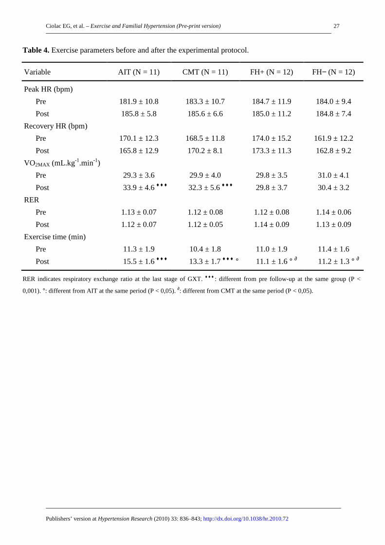
Cardiorespiratory Fitness and BP Response to Exercise. At baseline, cardiovascular and
ventilatory variables were similar among all four groups (Table 4). AIT and CMT group
increased VO2MAX after the follow-up, but the increase in AIT group was greater than in CMT
group (AIT = 15.8±6.3%; CMT = 8.0±6.1%; P<0.05). Heart rate did not change significantly
for all groups after the follow-up (Table 4). However, both AIT and CMT group improved BP
response to exercise after the follow-up. Recovery systolic and diastolic BP, and resting and
exercise diastolic BP were reduced in AIT group after the follow-up (P < 0.01). In the CMT
group, only recovery systolic BP and exercise diastolic BP were reduced after the follow-up
(P < 0.05). With the reduction, exercise diastolic BP were lower in AIT group than in
ConFH+ (P < 0.05) after the follow-up (Figure 1).
Norepinephrine, Endothelin-1 and NOx. At baseline, ConFH− displayed lower levels of
norepinephrine (resting, exercise and recovery) and ET-1 (resting and exercise), and higher
levels of NOx (resting, exercise and recovery) than the observed in the three FH+ groups (P <
0.05). On the other hand, AIT reduced resting, exercise and recovery norepinephrine (P <
0.05), reduced resting and exercise ET-1 (P < 0.05), and increased resting, exercise and
recovery NOx (P < 0.01). In the CMT group, only resting and exercise norepinephrine,
resting ET-1, and exercise and recovery NOx were improved after the follow-up (P < 0.05).

Ciolac EG, et al. – Exercise and Familial Hypertension (Pre-print version)
Publishers’ version at Hypertension Research (2010) 33: 836–843; http://dx.doi.org/10.1038/hr.2010.72
10
With these improvements, the AIT group (but not CMT group) displayed lower
norepinephrine and ET-1 levels, and higher NOx levels than ConFH+ after the follow-up (P <
0.05). Norepinephrine, ET-1 and NOx levels did not change significantly in the ConFH− and
ConFH+ after the follow-up (Figure 2).
Ambulatory Blood Pressure. 24-h, daytime and nighttime ABP were not significant
different among all four groups at baseline. Both AIT and CMT were beneficial to the ABP,
reducing systolic and diastolic 24-h and nighttime BP after the follow-up (P < 0.05). ABP
levels did not change significantly in ConFH− and ConFH+ group after the follow-up (Table
3).

Ciolac EG, et al. – Exercise and Familial Hypertension (Pre-print version)
Publishers’ version at Hypertension Research (2010) 33: 836–843; http://dx.doi.org/10.1038/hr.2010.72
11
DISCUSSION
The primary finding of the present study is that exercise intensity was an important factor for
improving cardiorespiratory fitness and restoring the hemodynamic, metabolic and hormonal
abnormalities of nonhypertensive women offspring of HPT parents, including PWV, BP
response to exercise, and norepinephrine, ET-1 and NOx levels. To the best of our
knowledge, this is a pioneering study in evaluation of the effects of exercise intensity in FH+
women.
Lipids, Glucose and Insulin. The existence of early abnormalities of glucose and insulin
metabolism has been shown among healthy FH+ subjects.7,29 In our study, the three FH+
groups displayed higher insulin levels and insulin sensitivity than ConFH−. On the other
hand, both AIT and CMT reduced insulin levels and insulin sensitivity to levels similar to
those of ConFH−. The mechanism involved on these reductions was not investigated.
However, the improvement of insulin metabolism by exercise training has shown to be in
function of a greater signaling and insulin action in muscle,16,30 resulting from a decreased
intracellular accumulation of triglycerides and increased fat acid oxidation [16,31]. In contrast
to our results, high intensity interval exercise has shown to be more effective than moderate
intensity continuous for insulin and insulin sensitivity.16 In this study, only interval exercise
improved insulin levels and insulin sensitivity in middle-aged and older subjects with
metabolic syndrome, after 16 weeks of follow-up. Thus, it is possible that in subjects with
greater insulin and metabolic abnormalities, only the high intensity promoted by interval
exercise may improve insulin action, while moderate intensity exercise may be sufficient for
healthy women.
Exercise training has also shown to increase HDL-cholesterol, and reduce triglycerides and
LDL-cholesterol. Results from the HERITAGE study,31 including 675 normolipidemic
subjects (376 women), showed that women increased HDL-cholesterol by 1.4 mg/dL (3%),

Ciolac EG, et al. – Exercise and Familial Hypertension (Pre-print version)
Publishers’ version at Hypertension Research (2010) 33: 836–843; http://dx.doi.org/10.1038/hr.2010.72
12
and reduced triglycerides and LDL-cholesterol by 0.6 mg/dL(0.6%) and 4.4 mg/dL (4%),
respectively, after a 5 month of exercise training. According to this, our study showed that
AIT increased HDL-cholesterol by 2.3 mg/dL (3.8%), and reduced triglycerides and LDL-
cholesterol by 0.7 mg/dL (0.9%) and 12.3 mg/dL (11.7%), respectively, while CMT increased
HDL-cholesterol by 3.6 mg/dL (6.3%), and reduced LDL-cholesterol (but not triglycerides)
by 6.0 mg/dL (5.6%). However, these improvements were not statistically significant.
Pulse Wave Velocity. It is well known that there is a progressive increase in arterial stiffness
with aging,33,34 with greater increases being observed among HPT subjects.34 Moreover,
healthy FH+ subjects has shown increased arterial stiffness levels before any increase in
BP.6,7 According to this, the three FH+ group of our study displayed higher PWV levels than
ConFH−. On the other hand, regular aerobic exercise has shown to attenuate age associated
arterial stiffness, as well as to reduce arterial stiffness when it is already established.35
However, moderate-intensity exercise has failed in improving arterial stiffness of isolated
systolic HPT subjects.36 A recent study from our group has shown that AIT, but not CMT,
reduced arterial stiffness of HPT women after 16 weeks of follow-up.14 In accordance, the
present study showed a significant reduction of the PWV after 16 weeks of AIT (P < 0.01),
while only a tendency tower reduction was observed after 16 weeks of CMT (P = 0.06).
These apparently conflicting results among the studies may be explained by the exercise
intensity and the subjects’ health status. The mechanisms involved in the age related arterial
stiffness increase include degradation of elastic matrix, endothelial dysfunction, hypertrophy
and hyperplasia of smooth muscle cells, and increase of collagen content, which appears to be
accelerated in the presence of HPT.37 Thus, exercise training programs of higher intensity (i.e.
interval exercise), may be more effective for the reduction of arterial stiffness of the
populations that have some alteration on this variable. Improvements in the endothelial
function and sympathetic activity for the arterial smooth muscle cells are possible

Ciolac EG, et al. – Exercise and Familial Hypertension (Pre-print version)
Publishers’ version at Hypertension Research (2010) 33: 836–843; http://dx.doi.org/10.1038/hr.2010.72
13
mechanisms involved in the arterial stiffness reduction induced by exercise training.37
Although both exercise training programs improved norepinephrine levels, only AIT
improved plasma NOx, a biomarker of endothelial function.38 Thus, it seems reasonable to
suggest that AIT was more effective for reducing PWV by the improvement of endothelial
function, as well as by an indirect reduction of sympathetic tonus induced by an increase of
the nitric oxide inhibitory effect.39
Cardiorespiratory Fitness and BP Response to Exercise. AIT increased VO2MAX to a
higher degree than CMT. This is in line with previous studies that have showed greater
efficiency of high-intensity interval training over moderate-intensity exercise in improving
cardiorespitory fitness of healthy subjects,23 as well as in coronary artery disease,24 heart
failure,40 metabolic syndrome16 and intermittent claudication patients.41 The superiority of
high-intensity interval training for improving cardiorespiratory fitness has important clinical
implication, since there is a strong association between low cardiorespiratory fitness and HPT
incidence.9 Moreover, of all established risk factors, low cardiorespiratory fitness appears to
be the strongest predictor of mortality.42 Increase of central O2 delivery (cardiac output) and
peripheral O2 uptake (arteriovenous oxygen difference) contribute to training-induced
improves of cardiorespiratory fitness.40 Because most evidence suggests that stroke volume is
the main factor that limits cardiorespiratory fitness, the interval training design, characterized
by short work periods at higher intensities alternated with low-intensity exercise periods,
promote a greater challenge for the pumping ability of the heart than what would be possible
lower exercise intensities.16
Besides cardiorespiratory fitness, BP response to exercise has important implication for HPT
prognostic, as supported by the association between exaggerated BP response to exercise and
incidence of future hypertension.43 Greater BP response to exercise among normotensive
women offspring of hypertensive parents when compared to normotensive with normotensive
Ciolac EG, et al. – Exercise and Familial Hypertension (Pre-print version)
Publishers’ version at Hypertension Research (2010) 33: 836–843; http://dx.doi.org/10.1038/hr.2010.72
14
parents has also been shown.7 In the present study, the three groups with positive family
history of HPT displayed a 2-17 mmHg greater (but not statistically significant) baseline BP
response to exercise when compared to the ConFH− group. On the other hand, AIT also
displayed greater effects on the BP response to exercise. Lower levels of resting diastolic BP,
exercise diastolic BP, and recovery systolic and diastolic BP were observed after the 16
weeks of AIT. With these improvements, AIT group displayed lower exercise diastolic BP
when compared to the ConFH+ group. In the CMT group, only exercise diastolic BP and
recovery systolic BP was reduced in CMT. The greater improvements in the norepinephrine,
ET-1 and NOx response to exercise observed after AIT are possible explanation for the
differences in BP response to exercise between groups.
Norepinephrine, Endothelin-1 and NOx. Although the precise mechanism is not completely
understood, increased activation of sympathetic nervous system and impaired endothelial
vasodilator function probably play a key role in the pathogenesis of essential hypertension.7 A
recent study from our group has shown that young normotensive women with positive family
history of HPT displays increased norepinephrine and ET-1 levels, as well as lower NOx
levels, both at resting and in response to exercise.7 Moreover, women with two HPT parents
displayed higher norepinephrine and ET-1 levels, as well as higher NOx levels, when
compared to women with only one HPT parent. The increased norepinephrine and ET-1
response to exercise, and the lower NOx response exercise observed in the three groups with
positive family history of HPT, are in line with our previous study. On the other hand,
relatively little attention has been given to the importance of physiological adaptations
following exercise in FH+. Few studies using cross-sectional design, with aerobically trained
and untrained FH+ and FH−, have examine if physical fitness modifies the disturbances in
sympathetic nervous system and endothelial function associated with FH+. Although negative
finds has been shown,44 the studies have generally suggested that there is an exercise-induced

Ciolac EG, et al. – Exercise and Familial Hypertension (Pre-print version)
Publishers’ version at Hypertension Research (2010) 33: 836–843; http://dx.doi.org/10.1038/hr.2010.72
15
reduction in the sympathetic nervous system and ET-1 levels in FH+.19,20,45 However, because
of the cross-sectional nature of the studies it is not possible to infer direct causality. The
present study confirm the important role of exercise training in reverting the sympathetic
nervous system and endothelial function alteration associated with FH+, that has been
suggested by the cross-sectional studies. We demonstrated that both AIT and CMT improve
norepinephrine, ET-1 and NOx response to exercise, with better results being found after AIT.
After the experimental protocol, AIT group displayed reduced resting, exercise and recovery
norepinephrine, reduced resting and exercise ET-1, and increased resting, exercise and
recovery NOx. In the CMT group, only resting and exercise norepinephrine, resting ET-1, and
exercise and recovery NOx were improved after the follow-up. With these results, only AIT
group displayed lower norepinephrine and ET-1 levels, and higher NOx levels than ConFH+
after the follow-up. The reason for these differences in ET-1 and NOx response to exercise
between groups is not fully understood, but it seems reasonable to suggest that the low- and
high-intensity training exercise programs affect shear stress in the arterial wall differently
during exercise training and that this may yield differences in molecular responses.
Ambulatory Blood Pressure. A recent meta-analysis has shown mean reductions of 0.3/0.1
mmHg and 2.5/3.1 mmHg for systolic/diastolic daytime and nighttime ABP of normotensive
subjects, respectively.13 The data are in line with results of present study that showed no
significant reductions of 0.8/0.8 mmHg and 1.9/1.0 mmHg for systolic/diastolic daytime
ABP, and significant reductions of 3.9/3.5 mmHg and 3.5/3.0 mmHg for systolic/diastolic
nighttime ABP in the AIT and CMT group, respectively. In accordance with previous studies
analyzing post-exercise effects on the ABP of HPT patients,10,14 the present study did not
shown any difference in the ABP reduction between high-intensity interval training and
moderate-intensity continuous exercise.

Ciolac EG, et al. – Exercise and Familial Hypertension (Pre-print version)
Publishers’ version at Hypertension Research (2010) 33: 836–843; http://dx.doi.org/10.1038/hr.2010.72
16
Study Limitations. Sympathetic activity was assessed indirectly by measuring plasma
norepinephrine. However, it is one of the preferred tests for analyzing acute effects of stress
tests and give similar results that the observed with microneurography.46 The marker NOx is a
metabolite of nitric oxide and may not be the best measure of nitric oxide
production/availability, because of the effects that diet may be on its measurement. However,
the women were oriented to follow a low nitrite/nitrate diet for the 5 days preceding the GXT,
which has shown to minimize the effects of diet on NOx measurement.26 Moreover, plasma
NOx following GXT are related to endothelial function.38
Clinical Implications. HPT is an important risk factor associated with all-cause and
cardiovascular mortality, as well as a greater life expectance without cardiovascular
disease.2,47 Moreover, BP is strong and linearly associated with coronary heart disease and
stroke2 that are the most common cause of mortality among women.48 Preventing or delaying
the onset of HPT may therefore have large impact on public health, mainly in individuals at
high risk for HPT.49 On the other hand, the present study showed for the first time that young
normotensive FH+ women may reverse hemodynamic, metabolic and hormonal alterations
that are involved in the pathophysiology of HPT with participation in exercise program, with
greater improvements being observed after AIT. Besides the superiority of AIT for improving
arterial stiffness, and BP, ET-1 and NOx response to exercise, the greater exercise-induced
increase in cardiorespiratory fitness observed after AIT has also important implications
because of all established risk factors, low cardiorespiratory fitness appears to be the strongest
predictor of mortality.42
In summary, the results of present study showed that, in young normotensive women
offspring of hypertensive patients, high-intensity interval training was superior to moderate-
intensity exercise in reversing hemodynamic, metabolic and hormonal alterations that are
involved in the pathophysiology of HPT. These findings may have important implications in

Ciolac EG, et al. – Exercise and Familial Hypertension (Pre-print version)
Publishers’ version at Hypertension Research (2010) 33: 836–843; http://dx.doi.org/10.1038/hr.2010.72
17
the design of exercise programs for prevention of an inherited hypertensive disorder.
Although multicenter prospective studies using high-intensity interval training for preventing
HPT in women with higher familial risk are needed to advance our conclusions, the results of
present study suggest that high-intensity interval training programs may yield more favorable
results than programs with moderate-intensity.
ACKNOWLEDGEMENT
This work was supported by the Fundação de Amparo à Pesquisa do Estado de São Paulo
(FAPESP # 2004/00568-8). Emmanuel G Ciolac has been supported by the Sociedade de
Cardiologia do Estado de São Paulo, and Dr. Guilherme V Guimarães has been supported by
the Conselho Nacional de Pesquisa (CNPq # 304733/2008-3).
DISCLOSURES
No conflicts to declare.

Ciolac EG, et al. – Exercise and Familial Hypertension (Pre-print version)
Publishers’ version at Hypertension Research (2010) 33: 836–843; http://dx.doi.org/10.1038/hr.2010.72
18
REFERENCES
1. Lawes CM, Vander Hoorn S, Rodgers A. Global burden of blood pressure-related disease,
2001. Lancet 2008; 371: 1513-1518.
2. Chobanian AV, Bakris GL, Black HR, Cushiman WC, Green LA, Izzo JI, Jones DW,
Materson BW, Oparil S, Wright JTJr, Rochella EJ. The Seventh Report of the Joint
National Committee on Prevention, Detection, Evaluation, and Treatment of High Blood
Pressure. Hypertension 2003; 42:1206-1252.
3. Whelton PK, He J, Appel LJ, Cutler JA, Havas S, Kotchen TA, Roccella EJ, Stout R,
Vallbona C, Winston MC, Karimbakas J. Primary prevention of hypertension: clinical and
public health advisory from The National High Blood Pressure Education Program. JAMA
2002; 288:1882-1888.
4. Wang NY, Young JH, Meoni LA, Ford DE, Erlinger TP, Klag MJ. Blood pressure change
and risk of hypertension associated with parental hypertension: The Johns Hopkins
Precursors Study. Arch Intern Med 2008; 168: 643-648.
5. Lieb W, Pencina MJ, Wang TJ, Larson MG, Lanier KJ, Benjamin EJ, Levy D, Tofler GH,
Meigs JB, Newton-Cheh C, Vasan RS. Association of parental hypertension with
concentrations of select biomarkers in nonhypertensive offspring. Hypertension 2008; 52:
381-386.
6. Kucerová J, Filipovsky J, Staessen JA, Cwynar M, Wojciechowska W, Stolarz K,
Kuznetsova T, Gasowski J, Dolejsová M, Grodzicki T, Kawecka-Jaszcz K, Fagard R.
Arterial characteristics in normotensive offspring of parents with or without a history of
hypertension. Am J Hypertens 2006; 19: 264-269.
7. Ciolac EG, Bocchi EA, Bortolotto LA, Carvalho VO, Greve JM, Guimarães GV.
Hemodynamic, metabolic, and neuro-humoral abormalities in young normotensive
women at high familial risk for hypertension. J Hum Hypertens 2010; 24: 814-822.

Ciolac EG, et al. – Exercise and Familial Hypertension (Pre-print version)
Publishers’ version at Hypertension Research (2010) 33: 836–843; http://dx.doi.org/10.1038/hr.2010.72
19
8. Hayashi T, Tsumura K, Suematsu C, Okada K, Fujii S, Endo G. Walking to work and the
risk for hypertension in men: the Osaka Health Survey. Ann Intern Med 1999; 131:21-26.
9. Chase NL, Sui X, Lee D, Blair S. The association of cardiorespiratory fitness and physical
activity with incidence of hypertension in men. Am J Hypertens 2009; 22:417-424.
10. Ciolac EG, Guimarães GV, D’Ávila VM, Bortolotto LA, Doria EL, Bocchi EA. Acute
effects of continuous or interval aerobic exercise on 24-h ambulatory blood pressure of
long-term treated hypertensive. Int J Cardiol 2009; 133; 381-387.
11. Ciolac EG, Guimarães GV, D’Ávila VM, Dória E, Bocchi EA. Acute aerobic exercise
reduces 24-h ambulatory blood pressure levels in long-term-treated hypertensive patients.
Clinics 2008. 63: 753-758.
12. Ciolac EG, Guimarães GV. Physical exercise and metabolic syndrome. Rev Bras Med
Esporte 2004; 10: 325-330.
13. Pescatello LS, Kulikowich JM. The aftereffects of dynamic exercise on ambulatory blood
pressure. Med Sci Sports Exerc 2001; 33: 1855-1861.
14. Guimarães GV, Ciolac EG, Carvalho VO, D’Ávila VM, Bortolotto LA, Bocchi EA.
Effects of continuous versus interval exercise training on the blood pressure and arterial
stiffness in treated hypertensive subjects: a randomized controlled study. Hypert Res
2010; 33: 627-632.
15. Cornelissen VA, Fagard RH. Effects of endurance training on blood pressure, blood
pressure-regulating mechanisms, and cardiovascular risk factors. Hypertension 2005; 46:
667-675.
16. Tjønna AE, Lee SJ, Rognmo Ø, Stølen TO, Bye A, Haram PM, Loennechen JP, Al-Share
QY, Skogvoll E, Slørdahl SA, Kemi OJ, Najjar SM, Wisløff U. Aerobic interval training
versus continuous moderate exercise as a treatment for the metabolic syndrome. A pilot
study. Circulation 2008; 118: 346-354.

Ciolac EG, et al. – Exercise and Familial Hypertension (Pre-print version)
Publishers’ version at Hypertension Research (2010) 33: 836–843; http://dx.doi.org/10.1038/hr.2010.72
20
17. Holmes DS, Cappo BM. Prophylactic effect of aerobic fitness on cardiovascular arousal
among individuals with a family history of hypertension. J Psychosom Res 1987; 31: 601-
605.
18. Jackson EM, Dishman RK. Hemodynamic responses to stress among black women:
fitness and parental hypertension. Med Sci Sports Exerc 2002; 34: 1097-1104.
19. Buckworth J, Dishman RK, Cureton KJ. Autonomic responses of women with parental
hypertension: Effects of physical activity and fitness. Hypertension 1994; 24: 576-584.
20. Lénárd Z, Studinger P, Mersich B, Pavlik G, Kollai M. Cardiovagal autonomic function in
sedentary and trained offspring of hypertensive parents. J Physiol 2005; 565: 1031-1038.
21. Haskell WL, Lee IM, Pate RR, Powell KE, Blair SN, Franklin BA, Macera CA, Heath
GW, Thompson PD, Bauman A. Physical activity and public health. Updated
recommendation for adults from the American College of Sports Medicine and the
American Heart Association. Circulation 2007; 116: 1081-1093.
22. American College of Sports Medicine. ACSM Stand Position on Exercise and
Hypertension. Med Sci Sporsts Exerc 2004; 36: 533-553.
23. Helgerud J, Hoydal K, Wang E, Karlsen T, Berg P, Bjerkaas M, Simonsen T, Helgesen C,
Hjorth N, Bach R, Hoff J. Aerobic high intensity intervals improve VO2max more than
moderate training. Med Sci Sports Exerc 2007; 39: 665-671.
24. Rognmo O, Hetland E, Helgerud J, Hoff J, Slordahl SA. High intensity aerobic interval
exercise is superior to moderate intensity exercise for increasing aerobic capacity in
patients with coronary artery disease. Eur J Cardiovasc Prev Rehabil 2004; 11: 216-222.
25. Asmar R, Benetos A, Topouchian J, Laurent P, Pannier B, Brisac AM, Target R, Levy BI.
Assessment of arterial distensibility by automatic pulse wave velocity measurement:
validation and clinical application studies. Hypertension 1995; 26:485-490.
Ciolac EG, et al. – Exercise and Familial Hypertension (Pre-print version)
Publishers’ version at Hypertension Research (2010) 33: 836–843; http://dx.doi.org/10.1038/hr.2010.72
21
26. Wang J, Brown MA, Tam SH. Effects of diet on measurement of nitric oxide metabolites.
Clin Exp Pharm Physiol 1997; 24: 418-420.
27. Wasserman K, Hansen JE, Sue D, Whipp BJ, Casaburi R. Principles of exercise testing
and interpretation. 4th ed, Philadelphia, Lippincott Williams & Wilkins, 2004; p. 32-35.
28. Hevel J, Marletta MA. Nitric-oxide synthase assays. Methods in Enzymol 1994; 233: 250-
258.
29. Goldstein IB, Shapiro D, Weiss RE. How family history and risk factors for hypertension
relate to ambulatory blood pressure in healthy adults. J Hypertens 2008; 26: 276-283.
30. Koval JA, Maezono K, Patti ME, Pendergrass M, DeFronzo RA, Mandarino LJ. Effects of
exercise and insulin on insulin signaling proteins in human skeletal muscle. Med Sci
Sports Exerc 1999; 31: 998-1004.
31. Bonen A, Dohm GL, van Loon LJ. Lipid metabolism, exercise and insulin action. Essays
Biochem 2006;42: 47-59.
32. Leon AS, Rice T, Mandel S, Després JP, Bergeron J, Gagnon J, Rao DC, Skinner JS,
Wilmore JH, Bouchard C. Blood lipid response to 20 weeks of supervised exercise in a
large biracial population: the HERITAGE Family Study. Metabolism 2000; 49: 513-520.
33. Vaitkevicious PV, Fleg JL, Engel JH, O’Connor FC, Wright JG, Lakatta LE, Yin FCP,
Lakatta EG. Effects of age and aerobic capacity on arterial stiffness in healthy adults.
Circulation 1993; 88: 1456-1462.
34. Benetos A, Adamopoulos C, Bureau JM, Temmar M, Labat C, Bean K, Thomas F;
Pannier B, Asmar R, Zureik M, Safar M, Guize L. Determinants of accelerated
progression of arterial stiffness in normotensive subjects and in treated hypertensive
subjects over a 6-Year period. Circulation 2002; 105: 1202-1207.
35. Tanaka H, Dinenno FA, Monahan KD, Clevenger CM, DeSouza CA, Seals DR. Aging,
Habitual exercise, and dynamic arterial compliance. Circulation 2000; 102: 1270-1275.

Ciolac EG, et al. – Exercise and Familial Hypertension (Pre-print version)
Publishers’ version at Hypertension Research (2010) 33: 836–843; http://dx.doi.org/10.1038/hr.2010.72
22
36. Ferrier KE, Waddell TK, Gatzka CD, Cameron JD, Dart AM, Kingwell BA. Aerobic
exercise training does not modify large-artery compliance in isolated systolic
hypertension. Hypertension 2001; 38: 222-226.
37. Zieman SJ, Melenovsky V, Kass DA. Mechanism, pathophysiology, and therapy of
arterial stiffness. Artherioscler Thromb Vasc Biol 2005; 25: 932-943.
38. Allen JD, Cobb FR, Kraus WE, Gow AJ. Total nitrogen oxide following exercise testing
reflects endothelial function and discriminates health status. Free Radic Biol Med 2006;
41: 740-747.
39. Zanzinger J. Role of nitric oxide in the neural control of cardiovascular function.
Cardiovasc Res 1999; 43: 639-649.
40. Wisloff U, Stoylen A, Loennechen JP, Bruvold M, Rognmo O, Haram PM, Tjønna AE,
Helgerud J, Slørdahl SA, Lee SJ, Videm V, Bye A, Smith GL, Najjar SM, Ellingsen O,
Skjærpe T. Superior cardiovascular effect of aerobic interval training versus moderate
continuous training in heart failure patients: a randomized study. Circulation 2007; 115:
3086-3094.
41. Slordahl SA, Wang E, Hoff J, Kemi OJ, Amundsen BH, Helgerud J. Effective training for
patients with intermittent claudication. Scand Cardiovasc J 2005; 39: 244-249.
42. Myers J, Prakash M, Froelicher V, Do D, Partington S, Atwood JE. Exercise capacity and
mortality among men referred for exercise testing. N Engl J Med 2002; 346: 793-801.
43. Sharabi Y, Ben-Cnaan R, Hanin A, Martonovitch G, Grossman E. The significance of
hypertensive response to exercise as a predictor of hypertension and cardiovascular
disease. J Hum Hyperten 2001; 15: 353-356.
44. Hamer M, Boutcher YN, Boutcher SH. Cardiovascular and renal responses to mental
challenge in highly and moderately active males with family history of hypertension. J
Hum Hypert 2002; 16: 319-326.
Ciolac EG, et al. – Exercise and Familial Hypertension (Pre-print version)
Publishers’ version at Hypertension Research (2010) 33: 836–843; http://dx.doi.org/10.1038/hr.2010.72
23
45. Tanzilli G, Barillà F, Pannitteri G, Greco C, Comito C, Schiariti M, Papalia U, Mangieri
E. Exercise training counteracts the abnormal release of plasma endothelin-1 in normal
subjects at risk for hypertension. Ital Heart J 2003; 4: 107-112.
46. Grassi G, Seravalle G, Bolla G, Quarti-Trevano F, Dell'Oro R, Arenare F, Mancia G.
Heart rate as a sympathetic marker during acute adrenergic challenge. J Hypertens 2008;
26: 70-75.
47. Franco OH, Peeters A, Bonneux L, Laet C. Blood pressure in adulthood and life
expectancy with cardiovascular disease in men and women: Life course analysis.
Hypertension 2005; 46:1-7.
48. Barrios V, Escobar C, Echarri R, Matalí A. Gender and Blood Pressure Control.
Hypertension 2008; 51: e58.
49. Mosca L, Appel LJ, Benjamin EJ, Berra K, Chandra-Strobos N, Fabunmi RP, Grady D,
Haan CK, Hayes SN, Judelson DR, Keenan NL, McBride P, Oparil S, Ouyang P, Oz MC,
Mendelsohn ME, Pasternak RC, Pinn VW, Robertson RM, Schenck-Gustafsson K, Sila
CA, Smith SCJr, Sopko G, Taylor AL, Walsh BW, Wenger NK, Williams CL. Evidence-
based guidelines for cardiovascular disease prevention in women. Circulation 2004; 109:
672-693.

Ciolac EG, et al. – Exercise and Familial Hypertension (Pre-print version)
Publishers’ version at Hypertension Research (2010) 33: 836–843; http://dx.doi.org/10.1038/hr.2010.72
24
TABLES
Table 1. Subjects characteristics.
Variable AIT (N = 16) CMT (N = 16) FH+ (N = 12) FH− (N = 15)
History of HPT
Father
Mother
Both
2
9
5
5
3
8
4
4
4
- - - - -
- - - - -
- - - - -
Age (years) 24.4 ± 3.8 26.6 ± 4.9 25.3 ± 3.7 25.3 ± 3.1
Weight (kg) 62.1 ± 12.5 63.5 ± 12.6 61.4 ± 11.0 62.3 ± 8.9
BMI (kg/m2) 23.5 ± 4.8 24.3 ± 4.6 23.8 ± 3.9 23.5 ± 2.4
Waist circumference (cm) 80.6 ± 10.8 81.4 ± 11.2 79.6 ± 12.0 79.5 ± 8.8
Waist-to-hip ratio 0.85 ± 0.04 0.86 ± 0.04 0.84 ± 0.07 0.84 ± 0.04
Office BP (mmHg)
Systolic
Diastolic
106.1 ± 9.9
65.1 ± 9.5
105.3 ± 9.3
64.9 ± 6.8
105.9 ± 8.3
62.3 ± 8.0
104.0 ± 9.5
62.7 ± 10.5

Ciolac EG, et al. – Exercise and Familial Hypertension (Pre-print version)
Publishers’ version at Hypertension Research (2010) 33: 836–843; http://dx.doi.org/10.1038/hr.2010.72
25
Table 2. Blood parameters before and after the experimental protocol.
**: different from AIT, CMT and FH+ at the same period (P < 0.01). #: different from FH+ at the same period (P < 0.05). ♦♦♦:
different from pre follow-up at the same group (P < 0.001).
Variable AIT (N = 11) CMT (N = 11) FH+ (N = 12) FH− (N = 12)
Glucose (mg/dL)
Pre
Post
83.3 ± 7.3
83.8 ± 4.3
84.3 ± 4.6
85.3 ± 6.2
85.3 ± 6.2
81.0 ± 8.7
84.2 ± 4.4
84.3 ± 4.2
Insulin (µUI/mL)
Pre
Post
7.9 ± 3.2
5.1 ± 2.9♦♦♦#
9.0 ± 3.3
6.5 ± 3.0 ♦♦♦
7.5 ± 3.0
7.2 ± 2.9
4.9 ± 2.4 **
5.3 ± 2.0 #
Insulin sensitivity (HOMA)
Pre
Post
1.53 ± 0.93
1.06 ± 0.88♦♦♦#
1.88 ± 0.73
1.37 ± 0.67♦♦♦
1.57 ± 0.67
1.55 ± 0.63
1.05 ± 0.43 **
1.10 ± 0.44 #
Total Cholesterol (mg/dL)
Pre
Post
HDL-colesterol (mg/dL)
Pre
Post
LDL-colesterol (mg/dL)
Pre
Post
179.6 ± 27.2
170.6 ± 23.1
60.5 ± 16.9
62.8 ± 16.0
104.8 ± 21.6
92.5 ± 15.5
177.6 ± 35.8
174.8 ± 31.0
56.6 ± 7.7
60.2 ± 9.5
107.3 ± 32.2
101.3 ± 25.4
177.8 ± 31.0
181.4 ± 41.2
64.2 ± 19.7
66.7 ± 19.4
106.3 ± 25.4
99.7 ± 25.8
161.4 ± 31.2
162.8 ± 26.5
66.7 ± 19.4
62.9 ± 8.6
99.7 ± 25.8
96.8 ± 20.6
Triglycerides (mg/dL)
Pre
Post
73.8 ± 26.8
73.1 ± 27.9
68.3 ± 20.5
70.3 ± 26.7
70.3 ± 26.7
75.3 ± 33.4
69.8 ± 26.5
71.8 ± 26.2

Ciolac EG, et al. – Exercise and Familial Hypertension (Pre-print version)
Publishers’ version at Hypertension Research (2010) 33: 836–843; http://dx.doi.org/10.1038/hr.2010.72
26
Table 3. Hemodynamic parameters before and after the experimental protocol.
***: different from AIT, CMT and FH+ at the same period (P < 0.001). #: different from FH+ at the same period (P < 0.05).
Different from pre follow-up at the same group (♦: P < 0.05; ♦♦: P < 0.01).
Variable AIT (N = 16) CMT (N = 16) FH+ (N = 12) FH− (N = 15)
PWV (m/s)
Pre
Post
7.47 ± 0.48
7.06 ± 0.42 ♦♦ #
7.55 ± 0.73
7.23 ± 0.65
7.38 ± 0.67
7.53 ± 0.59
6.93 ± 0.18 ***
6.97 ± 0.31 #
24-hour BP (mmHg)
Systolic
Pre
Post
Diastolic
Pre
Post
Daytime BP (mmHg)
Systolic
Pre
Post
Diastolic
Pre
Post
Nighttime BP (mmHg)
Systolic
Pre
Post
Diastolic
Pre
Post
113.3 ± 6.4
111.3 ± 5.8 ♦
71.1 ± 3.4
69.1 ± 3.8 ♦
117.3 ± 6.9
116.5 ± 6.4
75.6 ± 3.7
74.4 ± 4.8
103.6 ± 5.6
99.7 ± 5.5 ♦♦
60.8 ± 4.4
57.3 ± 3.8 ♦
112.6 ± 5.7
109.9 ± 6.7 ♦
69.4 ± 5.0
67.3 ± 3.2 ♦
116.8 ± 6.6
114.9 ± 7.1
73.9 ± 5.5
72.9 ± 4.0
102.8 ± 5.7
99.3 ± 6.0 ♦
59.5 ± 5.1
56.5 ± 3.4 ♦
110.5 ± 4.3
109.3 ± 5.8
68.3 ± 5.2
67.6 ± 5.6
115.3 ± 5.3
114.1 ± 6.8
73.3 ± 6.5
73.1 ± 6.6
102.3 ± 4.5
101.3 ± 4.7
59.3 ± 3.4
58.4 ± 3.7
110.8 ± 7.5
111.0 ± 6.2
68.9 ± 4.9
69.1 ± 3.9
114.3 ± 7.5
115.0 ± 5.5
73.3 ± 5.0
73.7 ± 3.4
102.4 ± 6.1
101.8 ± 7.4
59.5 ± 4.6
58.8 ± 5.4
Ciolac EG, et al. – Exercise and Familial Hypertension (Pre-print version)
Publishers’ version at Hypertension Research (2010) 33: 836–843; http://dx.doi.org/10.1038/hr.2010.72
27
Table 4. Exercise parameters before and after the experimental protocol.
RER indicates respiratory exchange ratio at the last stage of GXT. ♦♦♦: different from pre follow-up at the same group (P <
0,001). °: different from AIT at the same period (P < 0,05). ∂: different from CMT at the same period (P < 0,05).
Variable AIT (N = 11) CMT (N = 11) FH+ (N = 12) FH− (N = 12)
Peak HR (bpm)
Pre
Post
181.9 ± 10.8
185.8 ± 5.8
183.3 ± 10.7
185.6 ± 6.6
184.7 ± 11.9
185.0 ± 11.2
184.0 ± 9.4
184.8 ± 7.4
Recovery HR (bpm)
Pre
Post
170.1 ± 12.3
165.8 ± 12.9
168.5 ± 11.8
170.2 ± 8.1
174.0 ± 15.2
173.3 ± 11.3
161.9 ± 12.2
162.8 ± 9.2
VO2MAX (mL.kg-1.min-1)
Pre
Post
29.3 ± 3.6
33.9 ± 4.6 ♦♦♦
29.9 ± 4.0
32.3 ± 5.6 ♦♦♦
29.8 ± 3.5
29.8 ± 3.7
31.0 ± 4.1
30.4 ± 3.2
RER
Pre
Post
1.13 ± 0.07
1.12 ± 0.07
1.12 ± 0.08
1.12 ± 0.05
1.12 ± 0.08
1.14 ± 0.09
1.14 ± 0.06
1.13 ± 0.09
Exercise time (min)
Pre
Post
11.3 ± 1.9
15.5 ± 1.6 ♦♦♦
10.4 ± 1.8
13.3 ± 1.7 ♦♦♦ °
11.0 ± 1.9
11.1 ± 1.6 ° ∂
11.4 ± 1.6
11.2 ± 1.3 ° ∂

Ciolac EG, et al. – Exercise and Familial Hypertension (Pre-print version)
Publishers’ version at Hypertension Research (2010) 33: 836–843; http://dx.doi.org/10.1038/hr.2010.72
28
FIGURE LEGEND
Figure 1. Blood pressure response to exercise test before and after the experimental protocol.
A: AIT. B: CMT. C: ConFH+. D: ConFH−. #: different from ConFH+ at the same period (p <
0,05). Different from post follow-up at the same group (♦: P <0,05; ♦♦: P < 0,01; ♦♦♦: P <
0,001).
Figure 2. Norepinephrine (A), endothelin-1 (B), and NOx (C) before and after the
experimental protocol. 1: resting; 2: exercise; 3: recovery. *: different from AIT, CMT and
ConFH+ at pre follow-up (p < 0,05); #: different from ConFH+ post follow-up (p < 0,05);
Different from post follow-up at the same group (♦: P <0,05; ♦♦: P < 0,01).

Pre Post
Resting Exercise Recovery
A
50
80
110
140
170
♦ ♦
Blo
od P
ress
ure
(mm
Hg)
♦ ♦ ♦ ♦ ♦ ♦ ♦
#Resting Exercise Recovery
B ♦
50
80
110
140
170
Blood P
ressure (mm
Hg)
♦ ♦
Resting Exercise Recovery
Blo
od P
ress
ure
(mm
Hg)
C
50
80
110
140
170
Resting Exercise Recovery
D
Blood P
ressure (mm
Hg)
50
80
110
140
170
Figure 1

Pre Post
#
End
othe
lin-1
(pg
/mL)
*♦ #
3
4
5
6
♦
AIT CMT ConFH+
ConFH−
#♦ # * #
3
4
5
6
AIT CMT ConFH+
ConFH−
3
4
5
6
###
AIT CMT ConFH+
ConFH−
B-3B-2B-1
0
100
200
300
Nor
epin
ephr
ine
(pg/
mL)
* #
♦
♦ #
AIT CMT ConFH+
ConFH−
0
500
1000
1500
2000
2500
♦
♦ # *#
AIT CMT ConFH+
ConFH−
0
200
400
600
* #♦ #
AIT CMT ConFH+
ConFH−
A-2A-1 A-3
* #
♦♦ #
NO
x (µ
M/m
L)
0
15
30
45
AIT CMT ConFH+
ConFH−
#
0
20
40
60 * ♦♦ #
♦
AIT CMT ConFH+
ConFH−
0
20
40
60
* #♦♦ #
♦
AIT CMT ConFH+
ConFH−
C-3C-2C-1
Figure 2
Copyright © 2022 FDOKUMEN