Correction: Sirtinol Treatment Reduces Inflammation in Human Dermal Microvascular Endothelial Cells
Upload
transparencylsCategory
view
6download
0
Acute Humoral Xenograft Rejection: Destruction of theMicrovascular Capillary Endothelium inPig-to-Nonhuman Primate Renal Grafts
Akira Shimizu, Shane M. Meehan, Tomasz Kozlowski, Tomasz Sablinski,Francesco L. Ierino, David K.C. Cooper, David H. Sachs, and Robert B. Colvin
Department of Pathology (AS, SMM, RBC), Transplantation Biology Research Center (TK, TS, FLI, DKCC, DHS),
Massachusetts General Hospital/Harvard Medical School, Boston, Massachusetts
SUMMARY: The major cause of xenograft loss beyond hyperacute rejection is a form of injury, traditionally termed delayedxenograft rejection (DXR), whose pathogenesis is unknown. Here we analyze the immunologic and morphologic features of DXRthat develops in pig kidney xenografts transplanted into nonhuman primates. Kidneys from miniature swine were transplantedinto cynomolgus monkeys (n 5 14) or baboons (n 5 11) that received regimens aimed to induce mixed chimerism and tolerance.No kidney was rejected hyperacutely. Morphologic and immunohistochemical studies were performed on serial biopsies, and aneffort was made to quantify the pathologic features seen. The early phase of DXR (Days 0–12) was characterized by focaldeposition of IgM, IgG, C3, and scanty neutrophil and macrophage infiltrates. The first abnormality recognized was glomerularand peritubular capillary endothelial cell death as defined by in situ DNA nick-end labeling (TUNEL). Damaged endothelial cellsunderwent apoptosis and, later, frank necrosis. The progressive phase developed around Day 6 and was characterized byprogressive deposition of IgM, IgG, C3, and prominent infiltration of cytotoxic T cells and macrophages, with a small number ofNK cells. Thrombotic microangiopathy developed in the glomeruli and peritubular capillaries with TUNEL1 endothelial cells,platelet aggregation, and destruction of the capillary network. Only rare damaged arterial endothelial cells and tubular epithelialcells were observed, with rare endothelialitis and tubulitis. In the advanced phase of DXR, interstitial hemorrhage and infarctionoccurred. During the development of DXR, the number of TUNEL1 cells increased, and this correlated with progressivedeposition of antibody. The degree of platelet aggregation correlated with the number of TUNEL1 damaged endothelial cells. Weconclude that peritubular and glomerular capillary endothelia are the primary targets of renal DXR rather than tubular epithelialcells or arterial endothelium and that the earliest detectable change is endothelial cell death. DXR was characterized byprogressive destruction of the microvasculature (glomeruli and peritubular capillaries) and formation of fibrin-platelet thrombi.Both cytotoxic cells and antibodies potentially mediate the endothelial damage in DXR; however, in this model, DXR is largelyhumorally mediated and is better termed “acute humoral xenograft rejection.” (Lab Invest 2000, 80:815–830).
Animal donors are a potential solution to the criticalshortage of organs for transplantation in humans.
Pigs, including major histocompatibility complex (MHC)-inbred miniature swine, are considered the likely sourcefor clinical xenotransplantation (Cooper et al, 1991; Sa-chs, 1994), facilitated by modification of the donor bygenetic engineering. However, normal porcine organstransplanted into primates are aggressively rejected bythe recipient’s immune system. Hyperacute rejection(HAR) occurs within minutes to hours after transplanta-tion and is mediated by natural anti-Gal-1–3-Gal (Gal)antibody and complement activation (Daniels and Platt,1997; Good et al, 1992). HAR can be prevented by
strategies such as depletion (or neutralization) of anti-Galantibody, depletion or inhibition of complement, or theuse of organs from pigs transgenic for human comple-ment regulatory proteins (Lambrigts et al, 1998). How-ever, even when HAR is avoided, the xenograft under-goes a delayed form of rejection, referred to as delayedxenograft rejection (DXR), which destroys the graft overa period of days and is now viewed as the majorimmunologic barrier to successful xenotransplantation(Bach et al, 1996; Lambrigts et al, 1998; Lawson andPlatt, 1996). The immunologic basis (antibody, T cells,NK cells, and their target) of DXR is still uncertain.
At our center, a major effort is being made to induceimmunological tolerance to pig tissues in nonhumanprimates (cynomolgus monkeys and baboons) (Sachsand Sablinski, 1995; Sykes et al, 1994). HAR is over-come by depletion of natural antibody (Kozlowski et al,1998; Sablinski et al, 1995, 1997). Our developing pro-tocol, designed to promote tolerance, has been de-scribed in detail previously (Kozlowski et al, 1999; Sab-linski et al, 1995, 1997). It includes depletion of anti-pigantibody, transient ablation of the recipient’s cellularimmunity, short courses of cyclosporine (CyA) and 15-
Received December 23, 1999.Supported by grant from the National Institutes of Health PO1-HL 18646and a grant from BioTransplant, Inc.Current affiliation for SMM is Department of Pathology, University ofChicago, Chicago, Illinois.Address reprint requests to: Dr. R. B. Colvin, Department of Pathology,Massachusetts General Hospital, 55 Fruit Street (WRN 225), Boston, MA02114. Fax: 617-726-7533; [email protected]
0023-6837/00/8006-815$03.00/0LABORATORY INVESTIGATION Vol. 80, No. 6, p. 815, 2000Copyright © 2000 by The United States and Canadian Academy of Pathology, Inc. Printed in U.S.A.
Laboratory Investigation • June 2000 • Volume 80 • Number 6 815

Table 1. Summary of Survival of Porcine Renal Xenografts
Animal
Induction therapyWBI (days,
SL-sublethal,L-lethal)
Removal ofnatural antibody
(days)
Pharmacologicimmunosuppressive
therapy
PorcineBMTx or
SLA Class IIgene
transduction
Kidneygraft
survival(days)
Post-transplant day ofkidney biopsy
(grading of rejection)
Group AM1393 8 8(P-A)M1294 None day 0 CYA, DSG None 7 7(P-A)M1994 0 0(E)M3294 1 1(E)Group BM1094 WBI (day-6, SL),
ATG15 15(E)
M1194 WBI (day-6, SL),ATG
0 0(E)
M1594 WBI (day-6, SL),ATG
12 12(A)
M2094 WBI (day-6, SL),TI, ATG
2 2(E)
M4094 WBI (day-6, SL),TI, ATG
0 0(E)
M3394 WBI (day-6, SL),TI, ATG
day 0 CYA, DSG BMTx 10 10(P)
M3694 WBI (day-6, SL),TI, ATG
10 10(E)
M6593 WBI (day-6, SL),TI, ATG
9 6(P), 8(P), 9(P)
M1195 WBI (day-6, SL),TI, ATG
7 7(A)
M0894 WBI (day-3, SL),TI, ATG
12 12(E)
B94-107 WBI (day-6, SL),TI, ATG
9 0(N), 5(E), 8(E), 9(P)
B55-126(1) WBI (day-6, SL),TI, ATG
11 0(N), 6(P), 7(P),11(A)
Group CB75-37 WBI (day-22, SL),
TI, ATGday -6, 0 CYA, DSG, MMF 6 0(N), 2(E), 6(P)
B55-125 WBI (day-22, SL),TI, ATG
day -6, 0 CYA, DSG, MMF BMTx 3 0(N), 3(E)
B55-126(2) WBI (day-20, SL),TI, ATG
day -14, 0 CYA, DSG 14 0(N), 2(E), 5(E), 9(P),14(A)
B94-105 WBI (day-351,SL), TI, ATG
day -3, -2, 0, 4 CYA, BQR 6 0(N), 6(A)
Group DB75-13 WBI (day-104,
SL), TI, ATGday -7, -5, -2, 0 CYA, DSG, MMF,
sCR1SLA 8 0(N), 8(P)
B75-18 WBI (day-111, L) day -7, -5, -2, 0 CYA, DSG, MMF,sCR1
SLA 12 0(N), 12(A)
B74-34 WBI (day-111, L) day -7, -5, -2, 0 CYA, DSG, MMF,sCR1
Neo 13 0(N), 13(A)
B116-26 WBI (day-111, L) day -7, -5, -2, 0 CYA, DSG, MMF,sCR1
SLA 7 0(N), 7(P)
B17-60 WBI (day-111, L) day -7, -5, -2, 0 CYA, DSG, MMF, CVF Neo 6 0(N), 6(P)
Animal: M, cynomolgus monkey; B, baboon.Induction therapy: WBI, whole body irradiation (sublethal: 300cGy, lethal: 900cGy); TI, thymic irradiation (700cGy at day -1); ATG, anti-thymocyte globulin (50
mg/kg/day; days -3, -2, -1).Removal of natural antibodies: Hemoperfusion through a pig liver or an aGal immunoaffinity column, or apheresis and plasma perfusion through an aGal
immunoaffinity column.Pharmacologic immunosuppressive therapy: CYA, cyclosporine A 15 mg/kg/day (day 0 to day 28); BQR, brequinqr 5 mg/kg/day (day -2 to day 11); MMF,
mycophenolate mofetil 40 mg/kg/day (day 0 to day 14); DSG, 15-deoxyspergualin 6 mg/kg/day (day 0 to day 14); sCR1, soluble complement receptor-1, 12.5 or 25mg/kg/day (day 0 to day 8); CVF, cobra venom factor 0.20–0.25 mg/kg/day (day -1 to day 15).
Porcine bone marrow transplantation (BMTx), or SLA Class II gene (SLA) or neomycin-resistance gene (Neo) transduction: Porcine BMTx-donor pig cells infusionintravenously; SLA or Neo transduction-retrovirus mediated SLA class II DR gene (SLA) or neomycin-resistance gene (Neo) transfer into autologous bone marrowwith reinfusion into recipient baboon.
Histological staging of delayed xenograft rejection: N, no rejection; E, early phase; P, progressive phase; A, advanced phase.
Shimizu et al
816 Laboratory Investigation • June 2000 • Volume 80 • Number 6

deoxyspergualin (DSG), and donor-specific bone mar-row infusion.
With this protocol, however, DXR develops with lossof graft function. The present study examines themorphological characteristics of DXR in this model,using serial biopsies from pig renal xenografts. Wefocused on antibody, cellular phenotype, platelet accu-mulation, and endothelial cell damage in DXR and con-clude that the earliest feature is apoptosis and necrosisof the endothelium of the microvasculature (renal glo-meruli and peritubular capillaries). Subsequent destruc-tion of the microvascular capillary network with plateletaggregation is associated with graft loss.
Results
Development of Delayed Xenograft Rejection
Of the 25 kidney xenotransplants performed, noneunderwent HAR, but all were lost to delayed xenograftrejection (DXR) (Table 1). Table 1 summarizes the
survival of the porcine renal xenografts. Sequentialcreatinine levels indicated that graft dysfunction typi-cally developed between Days 6 and 14 (Fig. 1). Threeanimals had relatively good graft function when deathoccurred from systemic complications, which havebeen discussed elsewhere (Sablinski et al, 1997; Koz-lowski et al, 1999).
The rejection process could be divided into threephases based on the extent of antibody deposition,cellular infiltration, and other morphological alter-ations. The first (early) phase of DXR was character-ized by various degrees of antibody deposition inglomeruli and small vessels, an absence of cellularinfiltration, and focal segmental glomerular “solidifica-tion” with endothelial swelling and loss of patentcapillaries (Fig. 2A). Peritubular capillaries occasion-ally showed apoptotic nuclear changes. Glomerularand peritubular capillary endothelial nuclei weresometimes positive with the TdT–mediated dUTP–biotin nick-end labeling technique (TUNEL1; see be-low). No morphologic changes were evident in thesmall arteries. In the second (progressive) phase, bothantibody deposition and cellular infiltration could bedocumented. Glomerular and peritubular capillary al-terations progressed, with thrombi and focal interstitialhemorrhage (Fig. 2B). No changes were seen in thearteries. The third (advanced) phase was character-ized by necrotizing arteritis and glomerulitis with mul-tiple thrombi, and extensive interstitial hemorrhageand infarction (Fig. 2C). This sequence occurred in allof the xenografts, but with differing rates of progres-sion (Table 1). To clarify the morphological character-istics of DXR and to help determine the mechanismsof injury, further studies of morphological markerswere quantified in the early, progressive, and ad-vanced phases, independent of the day on which thesample was taken.
Deposition of Antibody and Complement
Immunofluorescent analysis of the porcine renal xeno-grafts showed a progressive deposition of IgM, IgG,
Figure 1.Serum creatinine levels in cynomolgus monkeys and baboons with life-supporting porcine renal xenografts (n 5 12, includes some of all groups).Recipients in which the native kidneys were not excised have been excluded.
Figure 2.Development of DXR seen in a porcine kidney transplanted into B55–126 (2) on Day 5 (the early phase) (A), Day 9 (the progressive phase) (B), and Day 14 (theadvanced phase) (C). A to C: hematoxylin and eosin (H&E) stain, 3250.
Mechanisms of Acute Humoral Xenograft Rejection
Laboratory Investigation • June 2000 • Volume 80 • Number 6 817

C3, and fibrin (Fig. 3, A to C). Immunohistochemistryfor IgM (Fig. 3) and semiquantitative analysis (Fig. 4)revealed that no IgM was deposited immediately afterreperfusion of the kidney. However, trace deposits ofIgM began to accumulate in the glomeruli during thefirst day and gradually extended into the peritubularcapillaries and arteries by Day 5. Thereafter, progres-sive deposition of IgM was observed in these threestructures. Deposition of IgG was similar. There was amore rapid and extensive IgM deposition in the kid-neys of experimental Groups 1, 3, and 4 when com-pared with Group 2 (where the induction regimenimmediately preceded kidney transplantation) (Fig. 4).Little or no C3 deposits were detected in the kidneysof the animals that received soluble complementreceptor-1 (sCR1) or cobra venom factor (CVF).
Characterization of the Cellular Infiltrate
The early phase had few leukocytes, ie, polymorpho-nuclear leukocytes (PMNs) and macrophages, in theperitubular capillaries and glomeruli (Fig. 5A), butthereafter, a progressive mononuclear cells (MNC)infiltrate developed in the interstitium. Immunoperox-idase staining showed the cells expressed CD3,CD68, and/or GMP-17 (Fig. 5, B to D). Double staining
for CD3 and GMP-17 cells showed that many GMP-171 cytotoxic cells were of T cell origin (Fig. 5E).Moreover, in all xenografts in the progressive phase,abundant infiltrating CD31, CD41 and CD81 cells,and GMP-171 MNCs were seen (Fig. 5, F to I). Theextent of CD41 cell infiltration was similar to or moreprominent than that of CD81 cells (Fig. 5, G and H). Arelatively small number of N421 NK cells was identi-fied (Fig. 5J).
Quantitative analyses of GMP-171 PMN, GMP-171MNC, and CD681 macrophage infiltration at variousperiods after transplantation are shown in Figures 6and 7. Progressive infiltration of PMNs, cytotoxicMNCs, and macrophages was seen, especially in theperitubular interstitium and glomeruli (from Day 6) inthe progressive and advanced phases of rejection.The infiltration of GMP-171 cytotoxic MNCs in thekidneys of Experimental Groups 1, 3, and 4 (whennearly complete recovery of WBC and CD21 cellcounts in the peripheral blood occurred after inductionregimen [Kozlowski et al, 1999]) was more prominentthan that in the Group 2 kidneys (where the inductionregimen was carried out immediately before kidneytransplantation) (Fig. 6). The numbers of PMNs andmacrophages did not differ significantly among the
Figure 3.Antibody and complement deposition in a renal xenograft, B55–126 (2). Immunofluorescent studies (Day 9) demonstrate moderate deposition of IgM (A), IgG (B),and C3 (C) in the glomeruli, and weak focal deposition in the peritubular capillaries (3300). Immunoperoxidase staining for IgM at 1 hour (D), on Day 5 (E), andon Day 9 (F) shows progressive deposition in the glomeruli, peritubular capillaries, and arteries (3250).
Shimizu et al
818 Laboratory Investigation • June 2000 • Volume 80 • Number 6

Figure 4.Semiquantitative analysis of IgM staining in peritubular capillaries (A, D), glomeruli (B, E) and arteries (C, F). A to C, Progressive IgM deposition was observed fromDay 5 and was more extensive in the Groups 1 (M), 3 (‚) and 4 (E) recipients than in those of Group 2 (F). D to F, Progressive IgM deposition in peritubularcapillaries and glomeruli was seen from the early phase through the advanced phase (N, no rejection; E, early phase; P, progressive phase; and A, advanced phase).In the arteries, only minimal deposition of IgM occurred until the advanced phase. E in D to F represents the mean value at each stage.
Figure 5.The cellular infiltrates in DXR. A, In the early phase, few CD681 macrophages (arrows) are seen in peritubular capillaries and glomeruli (3250). In the progressivephase, prominent CD31 T cells (B), GMP-171 cytotoxic MNCs (C), and CD681 macrophages (D) are seen in the interstitium (3250). E, Double staining for CD3(brown) and GMP-17 (blue) on Day 7 in B55–126 (1) shows that many GMP-171 cytotoxic cells are of CD31 T cell origin (arrows). A few GMP-171 mononuclearcells that do not stain for CD3 may be of NK cell origin (�) (3800). In the progressive phase, prominent CD31 cells (F), CD41 cells (G), CD81 cells (H), andGMP-171 cytotoxic MNCs (I) are seen. In addition, a relatively small number of N421 NK cells (J) are observed (arrows). F to J: 3500.
Mechanisms of Acute Humoral Xenograft Rejection
Laboratory Investigation • June 2000 • Volume 80 • Number 6 819

groups. Although there were small numbers of cyto-toxic MNCs and macrophages infiltrating the arteries,a PMN infiltrate developed during the progressive andadvanced phases of rejection (Fig. 7), and was asso-ciated with necrotizing arteritis. Only rare cytotoxicMNCs were seen infiltrating the tubules, and tubulitiswas uncommon. However, PMN and macrophageinfiltrates were observed in the tubules from the pro-gressive to the advanced phases (Fig. 7) and wereassociated with extensive tubular necrosis.
Endothelial Cell Injury, Platelet Aggregation, and RenalCapillary Destruction
Graft injury was examined morphologically by electronmicroscopy and by the TUNEL method, which candetect damaged cells in the process of undergoingeither apoptosis or necrosis (Mundle, 1995). Fromultrastructural observations, both apoptotic and ne-crotic endothelial cells were seen in the peritubularand glomerular capillaries (Fig. 8). In the early phase,the first morphological abnormality recognized usingthe TUNEL method was glomerular and peritubularcapillary endothelial labeling (Fig. 9A). From the pro-gressive to the advanced phases, TUNEL1 cells wereprominent (Fig. 9, B and C).
The TUNEL1 damaged cells were quantitated atvarious times (Fig. 10). In the peritubular capillaries
Figure 6.The number of GMP-171 PMNs (A–D), GMP-171 MNCs (E to H), and CD681 macrophages (I to L) infiltrating the peritubular interstitium (A, E, I), glomeruli (B,F, J), arteries (C, G, K), and tubules (D, H, L). In the peritubular interstitium and glomeruli, progressive GMP-171 PMN, GMP-171 MNC, and CD681 macrophageinfiltration develops from Day 6. Although the numbers of PMNs and macrophages do not differ between each group, the frequency of GMP-171 MNCs in the porcinekidneys in Groups 1 (M), 3 (‚), and 4 (E) is higher than in those of Group 2 (F).
Figure 7.The numbers of GMP-171 PMN (Œ), GMP-171 MNC (F), and CD681macrophages (f) infiltrating the peritubular interstitium (A), glomeruli (B),arteries (C), and tubules (D) in the various phases of DXR (N, no rejection; E,the early phase; P, the progressive phase; and A, the advanced phase).Cytotoxic MNC, PMN, and macrophage infiltrate develops in the progressiveand advanced phases in the interstitium and glomeruli, whereas minimalinfiltration is seen in the arteries and tubules. In the advanced phase, a PMNinfiltrate develops in arteries with necrotizing arteritis, and a PMN andmacrophage infiltrate develops in tubules with tubular necrosis. ‚, E, and M
represent the mean values in each cells.
Shimizu et al
820 Laboratory Investigation • June 2000 • Volume 80 • Number 6

and glomeruli, TUNEL1 cells were seen in the earlyphase and increased in number through the advancedphase. TUNEL1 cells were rarely observed in arteriesand tubules, except in the advanced phase. Withregards to the pathogenesis of endothelial cell injury inDXR, a significant correlation between the degree ofIgM deposition and the number of TUNEL1 damagedcells was seen in the peritubular capillaries, glomeruli,and arteries (Fig. 10, I to K). In contrast, there was nocorrelation between the numbers of GMP-171 cyto-toxic MNCs and TUNEL1 cells in the peritubularcapillaries and arteries (Fig. 10, L and N), althoughthere was mild correlation in the glomeruli (Fig. 10M).
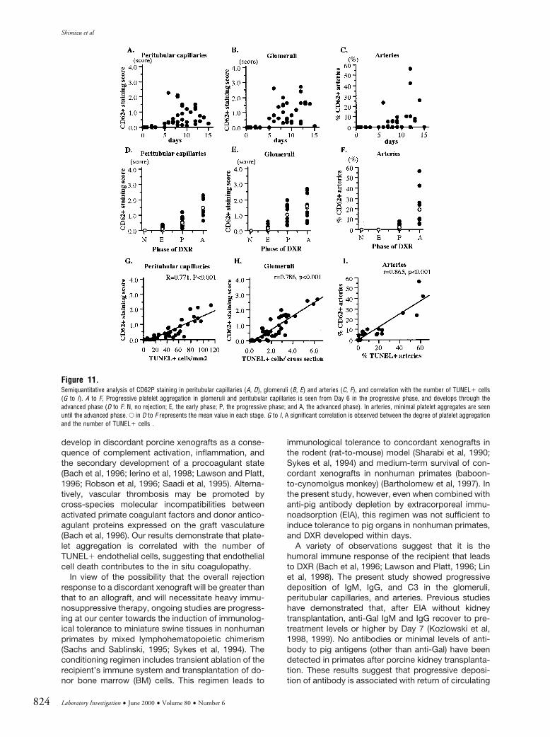
Immunohistochemistry for CD62P and semiquanti-tative analysis of these samples showed progressivedeposition of platelets in the xenografts (Fig. 9, D to F,and Fig. 11). Platelet aggregation was recognized inglomeruli in the early phase and gradually increased inthe glomeruli and peritubular capillaries during theprogressive and advanced phases. In the advancedphase, it was also seen in small arteries. A significantcorrelation was observed between the number ofTUNEL1 cells and the extent of platelet aggregation inthe peritubular capillaries, glomeruli, and arteries (Fig.11, G to I).
The status of the renal vasculature was examined byGS-IB4 immunohistochemistry for Gal expressed onpig endothelial cells. Along with the development ofglomerular injury, GS-IB4 reactivity was lost from theglomerular capillary network, indicating the destruc-tion of glomerular vasculature (Fig. 12B). These glo-merular alterations were accompanied by TUNEL1endothelial cells, multiple thrombi, marked depositionof IgM, and GMP-171 MNCs (Fig. 12, A to D). Theperitubular capillaries, identified by GS-IB4 staining,were absent around tubules with TUNEL1 endothelialcells. There was deposition of IgM and accumulationof GMP-171 cytotoxic MNCs (Fig. 12, E to H). How-ever, only rare endothelialitis and damaged endo-thelial cells were observed in the arteries, except inadvanced DXR, in which the arteries were charac-terized by fibrinoid necrosis or endarteritis withTUNEL1 cells, thrombi, IgM deposition, as well asGMP-171 PMN infiltration (Fig. 13, A to D). Onlyrare tubulitis was observed. Grouped TUNEL1 tu-bular cells were found in damaged tubules (Fig.13E), suggesting that tubular necrosis had occurredin the advanced phase. In the peritubular capillariesaround necrotic tubules, many GMP-171 PMNswere seen (Fig. 13F), but neither GMP-171 cyto-
Figure 8.Endothelial cell apoptosis and necrosis in glomeruli and peritubular capillaries. Loss of endothelial cells is seen in glomerular capillaries with an apoptotic cell (*)(Day 5/B94–107, 31900) (A) or necrotic cell debris (*) (Day 8/B75–13, 33400) (B). Glomerular mesangial and epithelial cells are almost intact, indicating that thecapillary endothelium is the primary target in the glomeruli. Loss of endothelial cells with an apoptotic cell (*) (Day 5/B94–107, 34500) (C) or hydropic necrosisof endothelial cells (*) (Day 6/B17–60, 33400) (D) are seen in peritubular capillaries.
Mechanisms of Acute Humoral Xenograft Rejection
Laboratory Investigation • June 2000 • Volume 80 • Number 6 821

toxic MNC infiltration nor IgM deposition was ob-served in these sites.
Discussion
The present study provides evidence that microvas-cular endothelial cell injury, characterized by endothe-lial cell apoptosis and necrosis, occurs from the earlyphase of DXR and increases during the later phases. Itis associated with progressive deposition of antibody.Our results indicate that the primary target in DXR isthe microvascular endothelium, rather than the arterialendothelium and tubular epithelial cells. Graft lossoccurs in the advanced phase with widespread de-struction of the capillary network and platelet accu-mulation.
In xenotransplantation, both antibody and cellularimmunity are believed to be major factors in thepathogenesis of DXR (Bach et al, 1996; Lawson andPlatt, 1996). However, the nature of the graft injury thatoccurs in DXR has not yet been well-characterized,although there is accumulating evidence that endo-thelial cell activation occurs (Bach et al, 1996; Han-cock, 1997; Lawson and Platt, 1996; Palmetshofer etal, 1998). We observed by electron microscopy bothendothelial cell apoptosis and necrosis. The TUNEL
method can detect damaged cells in the process ofboth apoptosis and necrosis in tissue sections (Gav-rieli et al, 1992; Mundle, 1995). This assay, therefore,has the advantage of allowing the detection of dam-aged cells induced by cell- and antibody-mediatedimmune injury. Indeed, prominent TUNEL1 cells wereseen in thrombotic microangiopathic lesions in glo-meruli and peritubular capillaries. More importantly,the first abnormality recognized in the porcine renalxenografts was endothelial labeling with TUNEL in themicrovasculature, before DXR could be diagnosedmorphologically. Our results demonstrate that theTUNEL assay is a sensitive method of assessing graftcell injury in both humoral- and cell-mediated rejectionand may have a practical value in the diagnosis ofearly xenograft rejection.
In experimental or human renal allograft rejection,numerous TUNEL1 damaged cells are seen in tu-bules, glomeruli, peritubular capillaries, and small ar-teries, together with tubulitis, glomerulitis, and endo-thelialitis, associated with a T cell infiltrate (Meehan etal, 1997; Palmetshofer et al, 1998; Shimizu et al,1997a; 1997b). In contrast to renal allograft rejection,only rare TUNEL1 arterial endothelial cells and mini-mal TUNEL1 tubular cells were observed in DXR,
Figure 9.Progressively increasing TUNEL1 damaged cells (A to C) and CD62P1 platelet aggregation (D to F) in the early (A, D), progressive (B, E) and advanced (C, F) phases.A to C: TUNEL method, 3250. D to F: CD62P stain, 3400. TUNEL1 peritubular and glomerular endothelial cells and glomerular platelet accumulation are presentbefore a prominent infiltrate appears (A, D).
Shimizu et al
822 Laboratory Investigation • June 2000 • Volume 80 • Number 6

indicating that these cells are spared by DXR. Incontrast, peritubular and glomerular capillary endo-thelia are primary targets of renal DXR. Indeed, GMP-171 cytotoxic MNCs infiltrated the peritubular capil-laries, glomeruli, and interstitium, whereas there wereonly minimal infiltrates in the tubules and arteries, evenin the progressive and advanced phases. The progres-sive deposition of antibody also occurred in the peri-tubular capillaries and glomeruli from the early phase.
In the kidney, endothelial cell injury can lead tothrombotic microangiopathy (Ballermann, 1998;Colvin, 1996). In the present study, endothelial celldeath was associated with platelet aggregation as wellas destruction of the vascular capillary network of theperitubular capillaries and glomeruli. Morphologicalthrombotic microangiopathy developed, with multipleplatelet-fibrin thrombi. It has been reported that intra-vascular fibrin deposition and platelet activation can
Figure 10.The number of TUNEL1 cells in the peritubular capillaries (A, E), glomeruli (B, F), arteries (C, G), and tubules (D, H), and correlation with graft IgM deposition (Ito K) or GMP-171 graft infiltrating cytotoxic MNCs (L to N). A to H, TUNEL1 cells in the peritubular capillaries and glomeruli are seen from Day 2 in the early phase,and are prominent in the progressive and advanced phases (E to H: N, no rejection, E, the early phase; P, the progressive phase; and A, the advanced phase). RareTUNEL1 cells are observed in arteries and tubules, except in advanced DXR. E in E to H represents the mean value in each stage. The number of TUNEL1 cellsand the extent of IgM deposition correlate significantly by linear regression (I to K); however, only weak correlation is seen between the numbers of TUNEL1 cellsand GMP-171 MNCs (L to N).
Mechanisms of Acute Humoral Xenograft Rejection
Laboratory Investigation • June 2000 • Volume 80 • Number 6 823
develop in discordant porcine xenografts as a conse-quence of complement activation, inflammation, andthe secondary development of a procoagulant state(Bach et al, 1996; Ierino et al, 1998; Lawson and Platt,1996; Robson et al, 1996; Saadi et al, 1995). Alterna-tively, vascular thrombosis may be promoted bycross-species molecular incompatibilities betweenactivated primate coagulant factors and donor antico-agulant proteins expressed on the graft vasculature(Bach et al, 1996). Our results demonstrate that plate-let aggregation is correlated with the number ofTUNEL1 endothelial cells, suggesting that endothelialcell death contributes to the in situ coagulopathy.
In view of the possibility that the overall rejectionresponse to a discordant xenograft will be greater thanthat to an allograft, and will necessitate heavy immu-nosuppressive therapy, ongoing studies are progress-ing at our center towards the induction of immunolog-ical tolerance to miniature swine tissues in nonhumanprimates by mixed lymphohematopoietic chimerism(Sachs and Sablinski, 1995; Sykes et al, 1994). Theconditioning regimen includes transient ablation of therecipient’s immune system and transplantation of do-nor bone marrow (BM) cells. This regimen leads to
immunological tolerance to concordant xenografts inthe rodent (rat-to-mouse) model (Sharabi et al, 1990;Sykes et al, 1994) and medium-term survival of con-cordant xenografts in nonhuman primates (baboon-to-cynomolgus monkey) (Bartholomew et al, 1997). Inthe present study, however, even when combined withanti-pig antibody depletion by extracorporeal immu-noadsorption (EIA), this regimen was not sufficient toinduce tolerance to pig organs in nonhuman primates,and DXR developed within days.
A variety of observations suggest that it is thehumoral immune response of the recipient that leadsto DXR (Bach et al, 1996; Lawson and Platt, 1996; Linet al, 1998). The present study showed progressivedeposition of IgM, IgG, and C3 in the glomeruli,peritubular capillaries, and arteries. Previous studieshave demonstrated that, after EIA without kidneytransplantation, anti-Gal IgM and IgG recover to pre-treatment levels or higher by Day 7 (Kozlowski et al,1998, 1999). No antibodies or minimal levels of anti-body to pig antigens (other than anti-Gal) have beendetected in primates after porcine kidney transplanta-tion. These results suggest that progressive deposi-tion of antibody is associated with return of circulating
Figure 11.Semiquantitative analysis of CD62P staining in peritubular capillaries (A, D), glomeruli (B, E) and arteries (C, F), and correlation with the number of TUNEL1 cells(G to I). A to F, Progressive platelet aggregation in glomeruli and peritubular capillaries is seen from Day 6 in the progressive phase, and develops through theadvanced phase (D to F: N, no rejection; E, the early phase; P, the progressive phase; and A, the advanced phase). In arteries, minimal platelet aggregates are seenuntil the advanced phase. E in D to F represents the mean value in each stage. G to I, A significant correlation is observed between the degree of platelet aggregationand the number of TUNEL1 cells .
Shimizu et al
824 Laboratory Investigation • June 2000 • Volume 80 • Number 6

anti-Gal antibody. Significant correlation between theprogressive deposition of IgM and the development ofendothelial cell injury was observed in the presentstudy. In the Group 4 animals, which received treat-ment with sCR-1 or CVF, DXR occurred despite min-imal deposition of complement, suggesting that anti-body deposition led to DXR with or withoutcomplement activation, although more typical andsevere DXR is associated with the presence of com-plement activation. We therefore conclude that anti-bodies play an important role in the pathogenesis ofDXR and suggest that DXR might be successfullyprevented or treated by therapies aimed at the hu-moral immune response to porcine antigens.
However, cellular elements also appear to play asignificant role in the development of DXR (Blakely etal, 1994; Candinas et al, 1996; Kobayashi et al, 1997;Leventhal et al, 1993). Some studies conclude thatinfiltrating cells consist primarily of macrophages andNK cells in rodents (Blakely et al, 1994; Candinas et al,1996) and are T cell-independent, in view of the lownumber of T cells in the infiltrates in the xenograftedorgans (Candinas et al, 1996). Others have also ar-gued a role for macrophages and NK cells (Blakely et
al, 1994; Candinas et al, 1996; Fryer et al, 1997;Kobayashi et al, 1997; Seebach and Waneck, 1997).
In the present study, the cellular response wascharacterized by a massive infiltration of CD31 Tcells, together with a similarly intense macrophageinfiltrate, and relatively small numbers of N421 NKcells. Many CD31 GMP-171 cytotoxic T cells werepresent in the infiltrate. Recent observations concern-ing human anti-pig cellular xenoreactivity support ourfindings (Auchincloss, 1994; Yamada et al, 1996).These studies demonstrated a human anti-pig, cell-mediated cytotoxic response, involving CD81 cells incell-mediated cytotoxicity, as well as CD41 cells asimportant mediators of macrophage, T cell, and B cellactivation. Our results demonstrate that similar fre-quencies of CD41 and CD81 T cells infiltrated thegraft in DXR, although CD81 T cells predominate inallograft rejection (Colvin, 1995). This suggests thatthe Class II-restricted, T cell-mediated reactions havea role in DXR.
In summary, in our evolving regimen designed topromote tolerance in the pig-to-primate renal xeno-graft model, HAR can be overcome by anti-pigantibody depletion. However, with the return of
Figure 12.The development of thrombotic microangiopathy in renal glomeruli (A to D) and peritubular capillaries (E to H). The TUNEL stain shows the prominent TUNEL1 cells(arrows) in glomerular (A) and peritubular (E) capillaries (3800); � in (A) indicates the thrombus. GS-IB4 stain shows loss of capillary lumina in the glomeruli (B)or around tubules (F), indicating destruction of the renal capillary network (3800); (arrow) indicates the remaining GS-IB41 capillaries. IgM deposition is seen inglomerli (capillaries and mesangium) (C) and peritubular capillaries (G) with damaged endothelial cells (arrow) (IgM stain, 3800). Infiltrating GMP-171 cells inglomerular capillaries (*) (D) or peritubular capillaries (arrows) (H) are in contact with apoptotic bodies (�) (GMP-17 stain, D: 31000, H: 3800).
Mechanisms of Acute Humoral Xenograft Rejection
Laboratory Investigation • June 2000 • Volume 80 • Number 6 825

natural anti-Gal antibody to the recipient’s circula-tion, DXR develops, resulting in graft failure. Whilethe cytotoxic cell infiltrate might add to the severityof DXR, antibody would appear to play an importantrole in its pathogenesis in this model. Morphologi-cally, DXR is characterized by the development ofthrombotic microangiopathy, with endothelial cellapoptosis and necrosis, followed by destruction ofthe microvasculature and platelet aggregation.These features are quite similar to acute humoralallograft rejection in humans (Collins et al, 1999;Trpkov et al, 1996). We therefore prefer the term“acute humoral xenograft rejection” rather thanDXR.
Materials and Methods
Animals and Surgical Procedures
Cynomolgus monkeys (Macaca fasicularis; CharlesRiver Laboratories, Wilmington, Massachusetts),weighing 5 to 8 kg, or baboons (Papio anubis;Biological Resources, Houston, Texas), weighing 8to 15 kg, were used as recipients. Bone marrow and
kidney donors were from the Massachusetts Gen-eral Hospital (MGH) herd of MHC-inbred miniatureswine (Charles River Laboratories). One miniatureswine also provided a liver for antibody adsorptionfrom the recipient by hemoperfusion.
The surgical techniques of harvesting donor or-gans, recipient splenectomy, and kidney transplan-tation, as well as details of supportive care, havebeen described previously (Ierino et al, 1998; Koz-lowski et al, 1999; Sablinski et al, 1995, 1997). Openkidney biopsies were taken on several posttrans-plant days and at the time when the graft wasrejected or the animal died. Graft function wasmonitored by plasma creatinine (Cr) and blood ureanitrogen (BUN) levels. All experiments were con-ducted according to the NIH Guide for the Care andUse of Laboratory Animals and were approved bythe MGH Subcommittee on Research Animal Care.
Pharmacologic Immunosuppressive Therapy
Pharmacologic immunosuppressive therapy (PI) is de-tailed in Table 1.
Figure 13.Arterial endothelial cell injury (A to D) and tubular epithelial cell necrosis (E, F) in the advanced phase. Loss of endothelial cells in a small artery (arrow) is recognizedby GS-IB4 stain (A) (3500), and with TUNEL1 cells (arrows) (B) (TUNEL method, 3800); arrowhead in A indicates remaining endothelial cells in small arteries.Endarteritis is observed with IgM deposition (IgM stain, 3500) (C) and GMP-171 PMNs (D) (GMP-17 stain, 3800). Grouped TUNEL1 tubular cells are seen indamaged tubules suggesting tubular epithelial cell necrosis (TUNEL method, 3500) (E), and many GMP-171 PMNs are found in peritubular capillaries with tubularnecrosis, (GMP-17 stain, 3500) (F).
Shimizu et al
826 Laboratory Investigation • June 2000 • Volume 80 • Number 6

Removal of Anti-Pig Antibody
Antibody depletion from the nonhuman primate wasachieved by extracorporeal immunoadsorption (EIA)by either (a) hemoperfusion through a pig liver or animmunoaffinity column of Gal trisaccharide type 6(Alberta Research Council, Edmonton, Canada) for 60minutes, or (b) apheresis using a COBE-Spectrapheresis system (Blood Component Technology, Inc.,Lakewood, Colorado), with plasma perfusion throughan Gal column (three plasma volumes). Details ofthese procedures have been described previously byKozlowski et al (1998).
Experimental Groups and Tolerance Induction Regimens
Four groups of animals were studied (Table 1), distin-guished by the type of induction regimens they re-ceived. Group 1 (n 5 4) underwent EIA on Day 0 andreceived CyA and DSG, but did not undergo a toler-ance induction regimen.
Group 2 (n 5 12) underwent a nonmyeloablativetolerance induction regimen pretransplant, followedby EIA, porcine bone marrow (BM) infusion, andkidney transplantation on Day 0. The nonmyeloabla-tive regimen consisted of (a) whole body irradiation(WBI) (300cGy) (Day 26 or Day 23), (b) thymic irradi-ation (TI) (700cGy) (Day 21), (c) splenectomy (Day 26or Day 0), (d) antithymocyte globulin (ATG) (ATGAM,Upjohn Company, Kalamazoo, Michigan) given intra-venously at 50 mg/kg/day for three consecutive days(Days 23, 22, and 21), and (e) porcine BM transplan-tation on Day 0. Details of this regimen have beendescribed previously (Kozlowski et al, 1999; Sablinskiet al, 1995, 1997). EIA was performed on Day 0,immediately before BM infusion and kidney transplan-tation; CyA and DSG were administered until graftrejection.
Group 3 (n 5 4) underwent the nonmyeloablativetolerance induction regimen (described above) morethan 20 days before kidney transplantation. The whiteblood cell (WBC) count (including CD21 T and NKcells) in the peripheral blood decreased after inductionregimens, but recovered close to the pre-inductionlevel by Days 20 to 27 (Kozlowski et al, 1999). PorcineBM infusion and kidney transplantation were per-formed on Day 0 and were combined with EIA, CyA,and DSG.
Group 4 (n 5 5) received autologous BM, which wastransduced with either a retroviral vector containingthe swine leukocyte antigen (SLA) Class II DR gene orthe neomycin-resistance gene. The transduced BMwas infused four months before porcine kidney trans-plantation was performed using a donor matched forthe Class II gene. Before the infusion of the autologousBM, one baboon (B77–13) underwent the nonmyeloa-blative induction regimen (as in Group B). The otherfour baboons received myeloablative WBI (900cGy).This latter protocol did not include TI or ATG but didinclude a course of sCR1 (n 5 4) or CVF (n 5 1)administered until the kidney was rejected. Engraft-ment of the BM occurred on Days 18 to 21. Approxi-
mately four months later, splenectomy and porcinekidney transplantation was performed on Day 0, com-bined with EIA, CyA, and DSG. Details of this regimenhave been described previously (Ierino et al, 1998).
Histological and Immunohistochemical Examination
For light microscopic examination, tissue was fixed in10%-buffered formalin and embedded in paraffin.Sections were examined after hematoxylin and eosin(H&E) and periodic acid-Schiff (PAS) staining.
For electron microscopic studies, tissue was fixed in2.5% glutaraldehyde in phosphate buffer (pH 7.4) andpostfixed with 1% osmium tetroxide, dehydrated, andembedded in Epon 812. Ultrathin sections werestained with lead citrate and examined with a Phillips301 electron microscope.
For immunofluorescence studies for the detectionof deposition of immunoglobulin, complement, andfibrin, frozen tissue sections were stained by the directimmunofluorescent technique (Leventhal et al, 1993;Lin et al, 1998), using fluorescein isothiocyanate(FITC)-conjugated rabbit polyclonal antibody to hu-man IgG, IgM, C3, and fibrinogen (all from DAKO,Carpinteria, California). Rabbit anti-human albumin(DAKO) was used as control. In order to examine thecorrelation between graft injury and antibody deposi-tion, immunohistochemistry for detection of IgM andIgG was also performed in paraffin-embedded tissuesections, using the standard avidin-biotin-horseradish-peroxidase complex (ABC) technique reported else-where (Meehan et al, 1997). After being deparaffinizedand incubated with trypsin for 12 minutes at 37 °C,sections were incubated with rabbit anti-human IgG(A090, DAKO) or IgM (A091, DAKO) followed by abiotin-labeled goat anti-rabbit IgG (Vector, Burlingame,California), ABC (Vector), and hydrogen peroxide (H2O2)containing 3,39-diaminobendizine (DAB) (Research Ge-netics, Hansville, Alabama). No difference in immuno-pathological findings was observed between the immu-nofluorescence and immunoperoxidase techniques,although the relatively extensive deposition of IgG andIgM seen in immunoperoxidase staining may be associ-ated with the differing sensitivities of the two methods.
Tissue for immunohistochemistry staining to identifythe phenotype of infiltrating cells was examined by theABC technique in frozen sections, using antibodiesagainst human CD3 (rabbit polyclonal anti-humanCD3, DAKO), CD8 (DK25, DAKO, or Leu 2a; BectonDickinson, San Jose, California), CD4 (Leu 3a, BectonDickinson), CD68 (KP-1, DAKO), and GMP-17 (TIA-1;Coulter Immunology, Hialeah, Florida), and anti-rhesus monkey NK cell antibody (N42) (Gengozian etal, 1992). To examine the correlation between graftinjury and cell infiltrate, CD31, GMP-171, or CD681cells were stained by the ABC technique in paraffin-embedded tissue sections. Double immunostainingwith CD3 and GMP-17 for the identification of cyto-toxic T cells was performed in formalin-fixed paraffinsections by using a two-color staining technique(Meehan et al, 1997). First, the sections were stainedwith a mouse monoclonal antibody to GMP-17 and
Mechanisms of Acute Humoral Xenograft Rejection
Laboratory Investigation • June 2000 • Volume 80 • Number 6 827

incubated with alkaline phosphatase-labeled anti-mouse IgG (OEM concept, Grafelfing, Germany) with ablue reaction product (Alkaline Phosphatase Sub-strate Kit III, Vector). Sections were then stained witha rabbit polyclonal antibody to CD3, HRP-labeledanti-rabbit IgG (OEM concept), hydrogen peroxide(H2O2) containing 3,39-diaminobendizine (DAB) (Re-search Genetics) (brown reaction product). The ABCtechnique, using biotin-labeled Griffonia (Bandeiraea)simplicifolia Isolectin B4 (GS-IB4, Vector), was em-ployed to detect Gal expressed on pig vascular endo-thelial cells (Oriol et al, 1993).
Aggregation of platelets was identified by the ex-pression of CD62P, which was stained by the ABCmethod in frozen sections, using anti-human CD62P(Becton Dickinson). Although activated endothelialcells also express CD62P, this antibody does notcross-react with pig tissues or cells (data not shown).The expression of CD62P in the renal xenografts,therefore, indicated the aggregation of cynomolgusmonkey or baboon platelets.
In histological sections, damaged nuclear DNA as-sociated with apoptosis was labeled by the terminaldeoxynucleotidyl transferase (TdT)-mediated dUTP-biotin nick-end labeling (TUNEL) method (Gavrieli et al,1992). After being deparaffinized and incubated withproteinase K (100 g/ml) for 15 minutes, sections wererinsed in TdT buffer and incubated with TdT 1:25 andbiotinylated-dUTP 1:20 in TdT buffer for 60 minutes at37 °C. The biotinylated nuclei were detected withavidin-peroxidase and H2O2 containing DAB. Controlsfor immunohistochemistry or the TUNEL method in-cluded substitution of the primary antibody with anirrelevant murine monoclonal antibody or omission ofthe dUTP or TdT, respectively.
Quantification of Histological Findings
Morphometric studies were performed to determinethe number of infiltrating GMP-171 polymorphonu-clear leukocytes (PMNs), GMP-171 mononuclearcells (MNCs) and CD681 cells, and frequency ofTUNEL1 damaged cells in the peritubular capillariesor the peritubular interstitium (both intracapillary andperitubular interstitial compartments combined), glo-meruli, tubules, and arteries. The results were calcu-lated as the average number of GMP-171 PMNs,GMP-171 MNCs, CD681 cells or TUNEL1 cells pergrid area in the tubules, peritubular capillaries orperitubular interstitium, or glomerular or arterial cross-sections.
For the evaluation of IgM deposition or CD62P1platelet aggregation, each glomerulus or each gridarea was graded semiquantitatively using a gradedsystem reported by others (Zachem et al, 1997; Shi-hab et al, 1995): Grade 0, no localized increase instaining; Grade 1, little increase in staining (depositionin 2 or 3 glomerular or peritubular capillaries); Grade 2,moderate increase in staining (involving ,25% of theglomerular or peritubular capillaries); Grade 3, intenseincrease in staining (involving ,50% of capillaries);and Grade 4, maximal increase in staining (.50%
capillary involvement). Deposition of IgM or CD62Pdeposition in arteries was determined by the percent-age of arteries affected.
More than 40 fields of renal cortex (at 4003, usingan optical grid area of 0.0625 mm2), more than 40glomerular cross-sections, and all arterial cross-sections in all fields of the renal cortex were counted ineach kidney sample without prior knowledge of theclinical or histologic findings. Counts were expressedas the numbers of positive cells per mm2, as thenumber of positive cells per glomerular or arterialcross-section, or as the percentage of the arteriesinvolved. Since the pattern of graft injury was similarwhether the recipient was cynomolgus monkey orbaboon, the results have been combined. Resultswere expressed as the mean, and correlation betweendata was assessed by Pearson’s method.
Acknowledgements
The authors wish to thank Ms. Patricia Della Pelle forexpert technical assistance and our many colleaguesat both the Massachusetts General Hospital and BioTransplant, Inc., who have contributed towards thestudies reported in this paper.
ReferencesAuchincloss HJ (1994). Cell-mediated xenoresponses:Strong or weak? Clin Transplant 8:155–159.
Bach FH, Winkler H, Ferran C, Hancock WW, and Robson SC(1996). Delayed xenograft rejection. Immunol Today 17:379–385.
Ballermann BJ (1998). Endothelial cell activation. Kidney Int53:1810–1826.
Bartholomew A, Cosimi AB, Sachs DH, Bilin M, Boskovic S,Colvin R, Hong H, Johnson M, Kimikawa M, Leguern A,Meehan S, Sablinski T, Wee SL, and Powelson J (1997). Astudy of tolerance in a concordant xenograft model. Trans-plant Proc 29:923–924.
Blakely ML, Van der Werf WJ, Berndt MC, Dalmasso AP,Bach FH, and Hancock WW (1994). Activation of intragraftendothelial and mononuclear cells during delayed xenograftrejection. Transplantation 58:1059–1066.
Candinas D, Belliveau S, Koyamada N, Miyatake T, Hechen-leitner P, Mark W, Bach FH, and Hancock WW (1996). T cellindependence of macrophage and natural killer cell infiltra-tion, cytokine production, and endothelial activation duringdelayed xenograft rejection. Transplantation 62:1920–1927.
Collins AB, Schneeberger EE, Pascual MA, Saidman SL,Williams WW, Tolkoff-Rubin N, Cosimi AB, and Colvin RB(1999). Complement activation in acute humoral renal allo-graft rejection: Diagnostic significance of C4d deposits inperitubular capillaries. J Am Soc Nephrol 10:2208–2214.
Colvin RB (1995). General features of the allograft response.In: Colvin RB, Bhan AK, and McCluskey RT, editors. Diag-nostic immunopathology, 2nd ed. New York: Raven, 311–328.
Colvin RB (1996). The renal allograft biopsy. Kidney Int50:1069–1082.
Shimizu et al
828 Laboratory Investigation • June 2000 • Volume 80 • Number 6

Cooper DKC, Ye Y, Rolf LL, and Zuhdi N (1991). The pig aspotential organ donor for man. In: Cooper DKC, Kemp E,Reemtsma K, and White DJG, editors. Xenotransplantation.Heidelberg: Springer, 481–500.
Daniels LJ and Platt JL (1997). Hyperacute xenograft rejec-tion as an immunologic barrier to xenotransplantation. Kid-ney Int Suppl 58:S28–S35.
Fryer JP, Chen S, Johnson E, Simone P, Sun LH, Goswitz JJ,and Matas AJ (1997). The role of monocytes and macro-phages in delayed xenograft rejection. Xenotransplantation4:40–48.
Gavrieli Y, Sherman Y, and Ben-Sasson SA (1992). Identifi-cation of programmed cell death in situ via specific labelingof nuclear DNA fragmentation. J Cell Biol 119:493–501.
Gengozian N, Good RA, and Fu SM (1992). Development andcharacterization of monoclonal antibodies to lymphocyte cellsurface antigens of the rhesus monkey. Transplantation53:1306–1312.
Good AH, Cooper DKC, Malcolm AJ, Ippolito RM, Koren E,Neethling FA, Ye Y, Zuhdi N, and Lamontagen LR (1992).Identification of carbohydrate structures that bind humanantiporcine antibodies: Implications for discordant xenograft-ing in humans. Transplant Proc 24:559–562.
Hancock WW (1997). Beyond hyperacute rejection: Strate-gies for development of pig-primate xenotransplantation.Kidney Int Suppl 58:S36–S40.
Ierino FL, Kozlowski T, Siegel JB, Shimizu A, Colvin RB,Banerjee PT, Cooper DKC, Cosimi AB, Bach FH, Sachs DH,and Robson SC (1998). Disseminated intravascular coagula-tion in association with the delayed rejection of pig-to-baboon renal xenografts. Transplantation 66:1439–1450.
Kobayashi T, Taniguchi S, Neethling FA, Rose AG, HancockWW, Ye Y, Niekrasz M, Kosanke S, Wright LJ, White DJ, andCooper DKC (1997). Delayed xenograft rejection of pig-to-baboon cardiac transplants after cobra venom factor ther-apy. Transplantation 64:1255–1261.
Kozlowski T, Ierino FL, Lambrigts D, Foley A, Andrews D,Awwad M, Monroy R, Cosimi AB, Cooper DKC, and SachsDH (1998). Depletion of anti-Gal1–3Gal antibody in baboonsby specific aGal immunoaffinity columns. Xenotransplanta-tion 5:122–131.
Kozlowski T, Shimizu A, Lambrigts D, Yamada K, FuchimotoY, Glaser R, Monroy R, Xu Y, Awwad M, Colvin RB, CosimiAB, Robson SC, Fishman J, Spitzer TR, Cooper DKC, andSachs DH (1999). Porcine kidney and heart transplantation inbaboons undergoing a tolerance induction regimen andantibody adsorption. Transplantation 67:18–30.
Lambrigts D, Sachs DH, and Cooper DKC (1998). Discordantorgan xenotransplantation in primates, World experience andcurrent status: Transplantation 66:547–561.
Lawson JH and Platt JL (1996). Molecular barriers to xeno-transplantation. Transplantation 62:303–310.
Leventhal JR, Matas AJ, Sun LH, Reif S, Bolman RM 3rd,Dalmasso AP, and Platt JL (1993). The immunopathology ofcardiac xenograft rejection in the guinea pig-to-rat model.Transplantation 56:1–8.
Lin SS, Weidner BC, Byrne GW, Diamond LE, Lawson JH,Hoopes CW, Daniels LJ, Daggett CW, Parker W, Harland RC,Davis RD, Bollinger RR, Logan JS, and Platt JL (1998). Therole of antibodies in acute vascular rejection of pig-to-baboon cardiac transplants. J Clin Invest 101:1745–1756.
Meehan SM, McCluskey RT, Pascual M, Preffer FI, AndersonP, Schlossman ST, and Colvin RB (1997). Cytotoxicity andapoptosis in human renal allografts: Identification, distribu-tion, and quantitation of cells with a cytotoxic granule proteinGMP-17 (TIA-1) and cells with fragmented nuclear DNA. LabInvest 76:639–649.
Mundle SD (1995). The two in situ techniques do not differ-entiate between apoptosis and necrosis but rather revealdistinct patterns of DNA fragmentation in apoptosis. LabInvest 72:611–612.
Oriol R, Ye Y, Koren E, and Cooper DKC (1993). Carbohy-drate antigens of pig tissues reacting with human naturalantibodies as potential targets for hyperacute vascular rejec-tion in pig-to-man organ xenotransplantation. Transplanta-tion 56:1433–1442.
Palmetshofer A, Galili U, Dalmasso AP, Robson SC, andBach FH (1998). Galactosyl epitope-mediated activation ofporcine aortic endothelial cells: Type II activation. Transplan-tation 65:971–978.
Robson SC, Siegel JB, Lesnikoski BA, Kopp C, Candinas UR,and Bach FH (1996). Aggregation of human platelets inducedby porcine endothelial cells is dependent upon both activa-tion of complement and thrombin generation. Xenotransplan-tation 3:24–34.
Saadi S, Holzknecht RA, Patte CP, Stern DM, and Platt JL(1995). Complement-mediated regulation of tissue factoractivity in endothelium. J Exp Med 182:1807–1814.
Sablinski T, Gianello PR, Bailin M, Bergen KS, Emery DW,Fishman JA, Foley A, Hatch T, Hawley RJ, Kozlowski T, LorfT, Meehan S, Monroy R, Powelson JA, Colvin RB, Cosimi AB,and Sachs DH (1997). Pig to monkey bone marrow andkidney xenotransplantation. Surgery 121:381–391.
Sablinski T, Latinne D, Gianello P, Bailin M, Bergen K, ColvinRB, Foley A, Hong HZ, Lorf T, Meehan S, Monroy R,Powelson JA, Sykes M, Tanaka M, Cosimi AB, and Sachs DH(1995). Xenotransplantation of pig kidneys to nonhumanprimates: I. Development of the model. Xenotransplantation2:264–270.
Sachs DH (1994). The pig as a potential xenograft donor. VetImmunol Immunopathol 43:185–191.
Sachs DH and Sablinski T (1995). Tolerance across discor-dant xenogeneic barriers. Xenotransplantation 2:234–239.
Seebach JD and Waneck GL (1997). Natural killer cells inxenotransplantation. Xenotransplantation 4:201–211.
Sharabi Y, Aksentijevich I, Sundt TM 3rd, Sachs DH, andSykes M (1990). Specific tolerance induction across a xeno-geneic barrier: Production of mixed rat/mouse lymphohema-topoietic chimeras using a nonlethal preparative regimen. JExp Med 172:195–202.
Shihab FS, Yamamoto T, Nast CC, Cohen AH, Nobel NA,Gold LI, and Border WA (1995). Transforming growth factor-band matrix protein expression in acute and chronic rejectionof human renal allografts. Am J Soc Nephrol 6:286–294.
Shimizu A, Yamada K, Meehan SM, Sachs DH, and ColvinRB. (1997a). Intragraft cellular events associated with toler-ance in pig allografts: The acceptance reaction. TransplantProc 29:1155.
Shimizu A, Yamada K, Sachs DH, and Colvin RB. (1997b).Progressive to chronic rejection vs tolerance in pig renalallografts (Abstract). J Am Soc Nephrol 8:667.
Mechanisms of Acute Humoral Xenograft Rejection
Laboratory Investigation • June 2000 • Volume 80 • Number 6 829

Sykes M, Lee LA, and Sachs DH (1994). Xenograft tolerance.Immunol Rev 141:245–276.
Trpkov K, Campbell P, Pazderka F, Cockfield S, Solez K, andHalloran PF (1996). Pathologic features of acute renal allo-graft rejection associated with donor-specific antibody.Transplantation 61:1586–1592.
Yamada K, Seebach J, DerSimonian H, and Sachs DH(1996). Human anti-pig T-cell mediated cytotoxity. Xeno-transplantation 3:179–187.
Zachem CR, Alpers CE, Way W, Shankland SJ, Couser WG,and Johnson RJ (1997). A role for P-selectin in neutrophil andplatelet infiltration in immune complex glomerulonephritis.J Am Soc Nephrol 8:1838–1844.
Shimizu et al
830 Laboratory Investigation • June 2000 • Volume 80 • Number 6
Copyright © 2022 FDOKUMEN