Molecular Genetics of Mosquito Resistance to Malaria Parasites
Upload
independentCategory
view
1download
0
A Quantitative Analysis of the MicrovascularSequestration of Malaria Parasites in theHuman Brain
Kamolrat Silamut,* Nguyen H. Phu,†
Christopher Whitty,‡ Gareth D.H. Turner,‡§
Karina Louwrier,§ Nguyen T.H. Mai,†
Julie A. Simpson,*‡ Tran T. Hien,† andNicholas J. White*‡
From the Faculty of Tropical Medicine,* Mahidol University,
Bangkok, Thailand; the Center for Tropical Diseases,† Cho Quan
Hospital, Ho Chi Minh City, Viet Nam; The Oxford-Wellcome
Centre for Tropical Diseases,‡ Nuffield Department of Clinical
Medicine, University of Oxford, and the Department of Clinical
Biochemistry and Cellular Science,§ University of Oxford,
United Kingdom
Microvascular sequestration was assessed in thebrains of 50 Thai and Vietnamese patients who diedfrom severe malaria (Plasmodium falciparum , 49; P.vivax , 1). Malaria parasites were sequestered in 46cases; in 3 intravascular malaria pigment but no par-asites were evident; and in the P. vivax case there wasno sequestration. Cerebrovascular endothelial ex-pression of the putative cytoadherence receptorsICAM-1, VCAM-1, E-selectin, and chondroitin sulfateand also HLA class II was increased. The median(range) ratio of cerebral to peripheral blood para-sitemia was 40 (1.8 to 1500). Within the same braindifferent vessels had discrete but different popula-tions of parasites, indicating that the adhesion char-acteristics of cerebrovascular endothelium changeasynchronously during malaria and also that signifi-cant recirculation of parasitized erythrocytes follow-ing sequestration is unlikely. The median (range) ra-tio of schizonts to trophozoites (0.15:1; 0.0 to 11.7)was significantly lower than predicted from the par-asite life cycle (P < 0.001). Antimalarial treatmentarrests development at the trophozoite stages whichremain sequestered in the brain. There were signifi-cantly more ring form parasites (age < 26 hours) inthe cerebral microvasculature (median range: 19%;0–90%) than expected from free mixing of these cellsin the systemic circulation (median range ring para-sitemia: 1.8%; 0–36.2%). All developmental stages ofP. falciparum are sequestered in the brain in severemalaria. (Am J Pathol 1999, 155:395–410)
Severe falciparum malaria remains one of the most im-portant causes of death in the tropics. Cerebral malaria isthe major lethal manifestation of this infection. The se-questration of red blood cells containing mature forms ofPlasmodium falciparum in the cerebral microvasculature isconsidered to be the essential underlying pathologicalprocess, although how this leads to coma and deathremains unresolved.1–4 Sequestration results from theadherence of parasitized red blood cells to vascular en-dothelium.5 In vitro studies suggest that infected redblood cells begin to cytoadhere at the late ring or earlytrophozoite stage of parasite development.6 Studies invivo and histopathological observations in fatal P. falcipa-rum malaria and also in the lethal sequestering animalmalarias suggest that the red cells containing more ma-ture parasites are sequestered in the brain and otherorgans from the middle of the parasite life cycle untilschizont rupture.7–16 The next generation of motiledaughter merozoites is then released and invades morered cells to continue the blood stage infection. In mostpatients with falciparum malaria relatively few P. falcipa-rum trophozoites and schizonts are seen in the peripheralblood, while these more mature parasites are abundant inthe capillaries and venules of vital organs (particularly thebrain).10 The kinetic processes involved and the micro-vascular distribution of sequestration have not been char-acterized previously. Post-mortem studies have givenconflicting impressions on the predominant stage of par-asite development encountered in the brain. MacPhersonet al10 found that both trophozoites and schizonts pre-dominated, whereas Lemercier et al17 reported almostexclusively schizonts. Others have reported that allstages of parasite development are represented.7,8,11
The aims of this study were to describe the microvascu-lature pattern and distribution of parasite developmentalstages in the brain in fatal cases of severe falciparummalaria.
Supported by The Wellcome Trust of Great Britain through the Wellcome-Mahidol University- Oxford Tropical Medicine Research Programme inThailand, and the Wellcome Trust Clinical Research Unit, Center forTropical Diseases in Vietnam.
Accepted for publication April 11, 1999.
Address reprint requests to Prof. Nicholas J. White, Faculty of TropicalMedicine, 420/6 Rajvithi Road, Bangkok 10400, Thailand. E-mail:[email protected].
American Journal of Pathology, Vol. 155, No. 2, August 1999
Copyright © American Society for Investigative Pathology
395

Methods
These studies were conducted at the Center for TropicalDiseases, Cho Quan Hospital, Ho Chi Minh City, VietNam, and Paholpolpayuhasena Hospital, Kanchanaburi,Thailand. All patients were admitted with severe falcipa-rum malaria and were treated with either intravenousquinine dihydrochloride (20 mg salt/kg over 4 hours,followed by 10 mg/kg every 8 hours), intramuscular qui-nine (same dose regimen), or intramuscular artemether(4 mg/kg followed by 2 mg/kg every 8 hours). All diedfrom direct complications of their illnesses. Most of thepatients in Vietnam were included in a prospective dou-ble-blind comparison of artemether and quinine whichhas been reported elsewhere.18 The pathological as-sessments in these patients were made before unblind-ing the study.
Procedures
Post-mortem sampling was performed only after fully in-formed permission was granted from attendant relatives.Where a full autopsy was performed (in Vietnam only),these samples were taken after removal of the brain. Insix cases samples were examined from three sites: me-dulla, cerebellum and cortex. When an autopsy was notperformed samples were taken using a Vim-Silvermanneedle inserted through the superior orbital foramen orthe foramen magnum. For this study the specimens werenot sectioned, but smeared by placing the brain speci-men between two glass microscope slides, pressingthese together, and making thin smears.19 This methodpreserves long fragments of capillaries and venules.Brain biopsies were performed within 2 hours of death,and autopsies within 6 hours. Slides were fixed in abso-lute methanol and stained by the reverse Field’s meth-od.20 Malaria parasites and pigment were easily identi-fied. Intravascular parasites were counted and staged.Peripheral blood slides were prepared either from bloodtaken post-mortem (by ventricular puncture) or, if an an-te-mortem blood smear had been taken within 4 hoursbefore death, this was used.
Morphological Assessment of Parasite Stage ofDevelopment
In Vitro Study
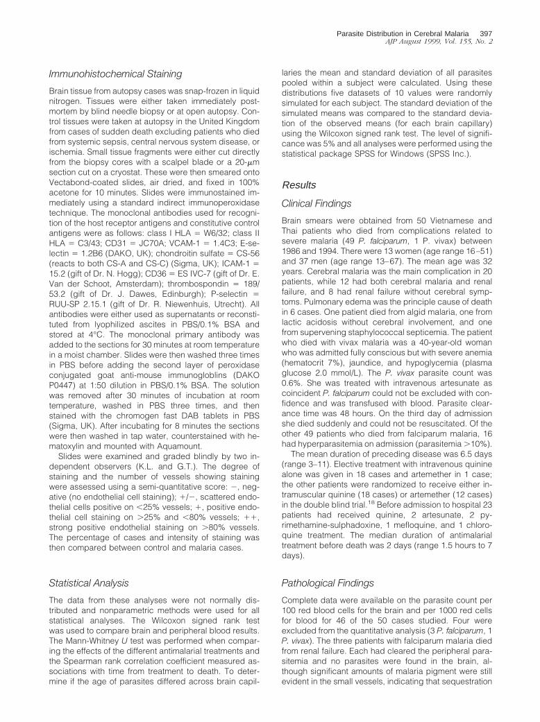
In order to validate the subsequent histopathologicalassessment of parasite stage of development, in vitrocultures two of different isolates of P. falciparum weresynchronized carefully by repeated exposure to sorbitoluntil a 1-hour time window was obtained. Smears werethen taken throughout one asexual life cycle (every 2hours until 42 hours, then every 1 hour). The parasitemorphology at precise times before the midpoint ofschizogony in vitro (see Figure 1) formed the basis of theassessment of parasite development stages in the cere-bral vessels. The effects of IC99 concentrations of quinineand artemether on parasite morphology were assessed
from in vitro cultures of parasite isolates with predeter-mined antimalarial susceptibility.
In Vivo Studies
Within the cerebral vessels the red cells and the ma-laria parasites were compressed within the confines ofthe packed sequestered capillaries or venules. The di-ameter of a capillary is smaller than that of an erythrocyte,and red cells have to bend and deform in their passagethrough the microvasculature. The separation betweenred cells in brain smears was often difficult to determine,but the individual parasite nuclei and cytoplasm andmalaria pigment were readily identified. In order to as-sess the effects of red cell compression within narrowvessels on parasite morphology, simulation experimentswere conducted in the rat. The midpoint capillary diam-eters in the rat are smaller than those in humans, and sodistortion should have been greater in the experimentsthan in the clinical samples. Large volume in vitro P.falciparum cultures (5 ml of blood at 15% hematocrit and10–30% parasitemia) were prepared and infused via a26-gauge catheter inserted into the aortic arch of anes-thetized (6% chloral hydrate given by intraperitoneal in-jection) male Wistar rats. The inferior vena cava anddescending aorta were clamped, and an equal volume ofblood removed as that infused. The objective was to fillthe cerebral vessels with human blood from the parasiteculture. As the infusion was finished, cardiac arrest wasinduced. The brain was removed and smears from graymatter prepared in exactly the same way as the post-mortem slides were prepared in the clinical study. P.falciparum infected red cells at different stages of devel-opment in the cerebral microvasculature of the rat inbrain smears were compared with those in heart bloodsmears.
Parasite Staging and Counting
In the brain smear from each case of fatal falciparummalaria a minimum of 5 capillaries were selected ran-domly, the parasites counted, and at least 10 examinedfor stage of development. A minimum of 20 sequentialred cells in each capillary were examined. Discrete sec-tions of capillaries or venules containing less than 20 redcells were not assessed. Eight stages of parasite devel-opment (approximate parasite age) were recorded: tiny(0–6 hours) small (6–16 hours), and large rings (16–26hours); early (26–30 hours), middle (30–34 hours), andlate trophozoites (34–38 hours) and schizonts with up to(38–44 hours) or more than five nuclei (44–48 hours)(Figure 1). A total of 100 parasites were staged. Thedistribution of malaria pigment in each vessel was alsonoted, and any white cells or gametocytes were re-corded. Thin film peripheral blood smears on admissionand before death were examined in the same way forparasite count and stage of development assessment.Each brain and blood smear was examined indepen-dently by two people, and the smears re-read if there wassignificant disagreement.
396 Silamut et alAJP August 1999, Vol. 155, No. 2
Immunohistochemical Staining
Brain tissue from autopsy cases was snap-frozen in liquidnitrogen. Tissues were either taken immediately post-mortem by blind needle biopsy or at open autopsy. Con-trol tissues were taken at autopsy in the United Kingdomfrom cases of sudden death excluding patients who diedfrom systemic sepsis, central nervous system disease, orischemia. Small tissue fragments were either cut directlyfrom the biopsy cores with a scalpel blade or a 20-mmsection cut on a cryostat. These were then smeared ontoVectabond-coated slides, air dried, and fixed in 100%acetone for 10 minutes. Slides were immunostained im-mediately using a standard indirect immunoperoxidasetechnique. The monoclonal antibodies used for recogni-tion of the host receptor antigens and constitutive controlantigens were as follows: class I HLA 5 W6/32; class IIHLA 5 C3/43; CD31 5 JC70A; VCAM-1 5 1.4C3; E-se-lectin 5 1.2B6 (DAKO, UK); chondroitin sulfate 5 CS-56(reacts to both CS-A and CS-C) (Sigma, UK); ICAM-1 515.2 (gift of Dr. N. Hogg); CD36 5 ES IVC-7 (gift of Dr. E.Van der Schoot, Amsterdam); thrombospondin 5 189/53.2 (gift of Dr. J. Dawes, Edinburgh); P-selectin 5RUU-SP 2.15.1 (gift of Dr. R. Niewenhuis, Utrecht). Allantibodies were either used as supernatants or reconsti-tuted from lyophilized ascites in PBS/0.1% BSA andstored at 4°C. The monoclonal primary antibody wasadded to the sections for 30 minutes at room temperaturein a moist chamber. Slides were then washed three timesin PBS before adding the second layer of peroxidaseconjugated goat anti-mouse immunogloblins (DAKOP0447) at 1:50 dilution in PBS/0.1% BSA. The solutionwas removed after 30 minutes of incubation at roomtemperature, washed in PBS three times, and thenstained with the chromogen fast DAB tablets in PBS(Sigma, UK). After incubating for 8 minutes the sectionswere then washed in tap water, counterstained with he-matoxylin and mounted with Aquamount.
Slides were examined and graded blindly by two in-dependent observers (K.L. and G.T.). The degree ofstaining and the number of vessels showing stainingwere assessed using a semi-quantitative score: 2, neg-ative (no endothelial cell staining); 1/2, scattered endo-thelial cells positive on ,25% vessels; 1, positive endo-thelial cell staining on .25% and ,80% vessels; 11,strong positive endothelial staining on .80% vessels.The percentage of cases and intensity of staining wasthen compared between control and malaria cases.
Statistical Analysis
The data from these analyses were not normally dis-tributed and nonparametric methods were used for allstatistical analyses. The Wilcoxon signed rank testwas used to compare brain and peripheral blood results.The Mann-Whitney U test was performed when compar-ing the effects of the different antimalarial treatments andthe Spearman rank correlation coefficient measured as-sociations with time from treatment to death. To deter-mine if the age of parasites differed across brain capil-
laries the mean and standard deviation of all parasitespooled within a subject were calculated. Using thesedistributions five datasets of 10 values were randomlysimulated for each subject. The standard deviation of thesimulated means was compared to the standard devia-tion of the observed means (for each brain capillary)using the Wilcoxon signed rank test. The level of signifi-cance was 5% and all analyses were performed using thestatistical package SPSS for Windows (SPSS Inc.).
Results
Clinical Findings
Brain smears were obtained from 50 Vietnamese andThai patients who died from complications related tosevere malaria (49 P. falciparum, 1 P. vivax) between1986 and 1994. There were 13 women (age range 16–51)and 37 men (age range 13–67). The mean age was 32years. Cerebral malaria was the main complication in 20patients, while 12 had both cerebral malaria and renalfailure, and 8 had renal failure without cerebral symp-toms. Pulmonary edema was the principle cause of deathin 6 cases. One patient died from algid malaria, one fromlactic acidosis without cerebral involvement, and onefrom supervening staphylococcal septicemia. The patientwho died with vivax malaria was a 40-year-old womanwho was admitted fully conscious but with severe anemia(hematocrit 7%), jaundice, and hypoglycemia (plasmaglucose 2.0 mmol/L). The P. vivax parasite count was0.6%. She was treated with intravenous artesunate ascoincident P. falciparum could not be excluded with con-fidence and was transfused with blood. Parasite clear-ance time was 48 hours. On the third day of admissionshe died suddenly and could not be resuscitated. Of theother 49 patients who died from falciparum malaria, 16had hyperparasitemia on admission (parasitemia .10%).
The mean duration of preceding disease was 6.5 days(range 3–11). Elective treatment with intravenous quininealone was given in 18 cases and artemether in 1 case;the other patients were randomized to receive either in-tramuscular quinine (18 cases) or artemether (12 cases)in the double blind trial.18 Before admission to hospital 23patients had received quinine, 2 artesunate, 2 py-rimethamine-sulphadoxine, 1 mefloquine, and 1 chloro-quine treatment. The median duration of antimalarialtreatment before death was 2 days (range 1.5 hours to 7days).
Pathological Findings
Complete data were available on the parasite count per100 red blood cells for the brain and per 1000 red cellsfor blood for 46 of the 50 cases studied. Four wereexcluded from the quantitative analysis (3 P. falciparum, 1P. vivax). The three patients with falciparum malaria diedfrom renal failure. Each had cleared the peripheral para-sitemia and no parasites were found in the brain, al-though significant amounts of malaria pigment were stillevident in the small vessels, indicating that sequestration
Parasite Distribution in Cerebral Malaria 397AJP August 1999, Vol. 155, No. 2
had occurred at some time. No parasites and no pigmentwere seen in the brain vessels of the patient who diedfrom vivax malaria.
Sequestration
For the 46 patients with residual malaria parasites, theparasite count in the brain median (range) 5 66.5%(1–99) was significantly higher than in the peripheralblood median (range) 5 1.4% (,0.1–36.6) (p , 0.001).This confirms significant sequestration in the cerebralmicrovasculature. To investigate whether sequestrationin the brain was associated with the duration of or type ofantimalarial drug treatment an index reflecting the pro-portion of sequestered erythrocytes at the time of deathwas calculated.
Sequestration index (SI) 5 10 3 (number of parasitized
cells in brain/100 red cells) 4 ~number of parasitized!
cells in peripheral blood just before death/1000 red cells)
For patients where the peripheral blood parasite countwas extremely low, the conservative value of 0.05% waschosen in order to calculate SI. The overall median (IQR,range) sequestration index was 40 (9.9–273.8 : 1.8–1500). The median (IQR, range) ratio of the proportion ofparasitized red cells in the brain to the admission para-sitemia was 15.3 (3.8–41.1: 0.53–1360). There was asignificant negative association between time from treat-ment to death and SI (Spearman correlation coefficient 520.53, p , 0.001). The median (range) sequestrationindex for patients treated with quinine was similar to those
who received artemether (41.1 (2.3–1360) versus 39.4(1.8–1500), p 5 0.91). The median (IQR, range) SI wasnot significantly different between those patients admit-ted with cerebral malaria (n 5 28), and those who wereconscious on admission (n 5 18); 40 (9.4–299; 1.8–1500) versus 38.5 (9.9–280, 2.3–720), p 5 0.75. Elevenpatients with no clinical history or signs of cerebral ma-laria had high parasite counts in the brain (more than50% parasitemia), and in seven of these, over 75% of redcells seen were parasitized, indicating that significantcerebral sequestration had occurred (sequestration in-dex range 3.9–42.2).
Stage of Parasite Development
Assessment in Vitro
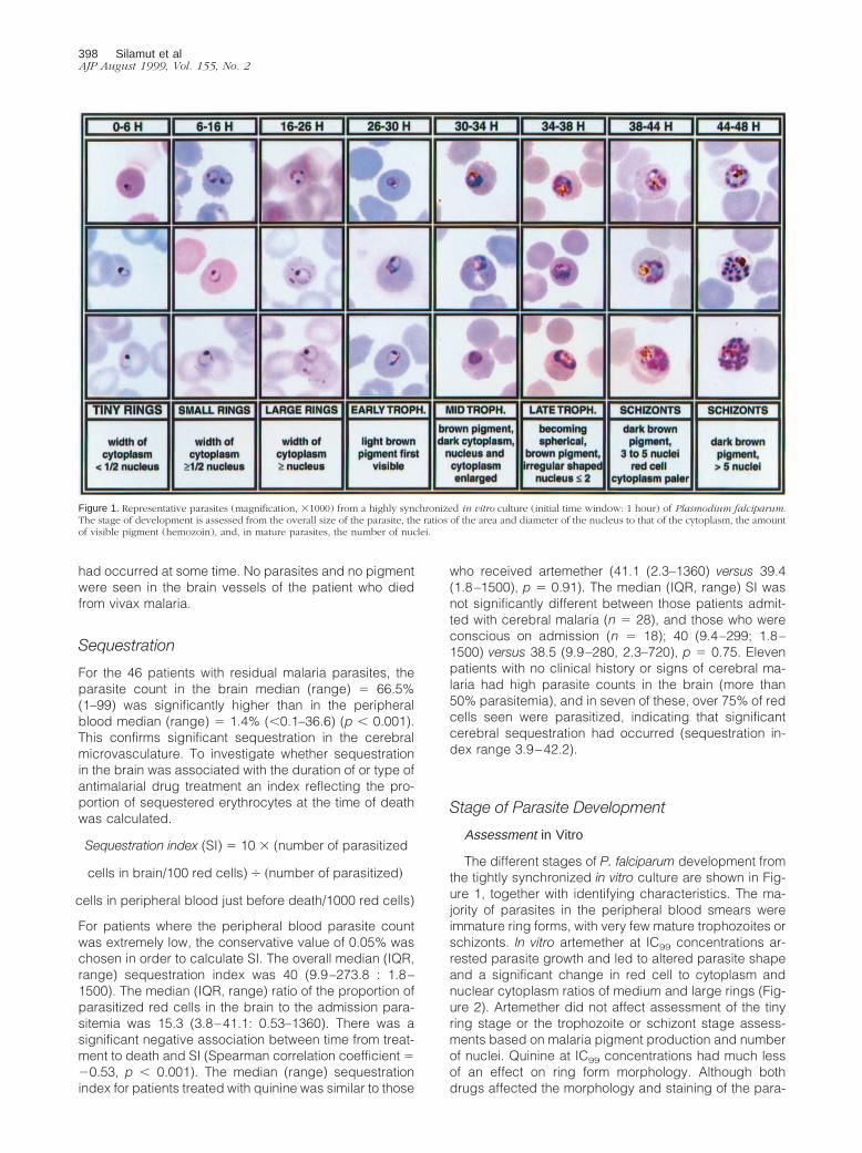
The different stages of P. falciparum development fromthe tightly synchronized in vitro culture are shown in Fig-ure 1, together with identifying characteristics. The ma-jority of parasites in the peripheral blood smears wereimmature ring forms, with very few mature trophozoites orschizonts. In vitro artemether at IC99 concentrations ar-rested parasite growth and led to altered parasite shapeand a significant change in red cell to cytoplasm andnuclear cytoplasm ratios of medium and large rings (Fig-ure 2). Artemether did not affect assessment of the tinyring stage or the trophozoite or schizont stage assess-ments based on malaria pigment production and numberof nuclei. Quinine at IC99 concentrations had much lessof an effect on ring form morphology. Although bothdrugs affected the morphology and staining of the para-
Figure 1. Representative parasites (magnification, 31000) from a highly synchronized in vitro culture (initial time window: 1 hour) of Plasmodium falciparum.The stage of development is assessed from the overall size of the parasite, the ratios of the area and diameter of the nucleus to that of the cytoplasm, the amountof visible pigment (hemozoin), and, in mature parasites, the number of nuclei.
398 Silamut et alAJP August 1999, Vol. 155, No. 2
site cytoplasm, neither drug affected the overall distribu-tion assessment or caused disappearance of pigment.
Simulation Experiments
In the simulation experiments in 10 rats, the malariaparasites, particularly the younger forms, appearedsmaller in the cerebral vessels than in the peripheral(heart) blood. We investigated whether the following re-lationships were preserved: (a) the relative size of theparasite to the size of the parasitized red cell; (b) theamount of parasite cytoplasm in relation to the nucleus;(c) the amount and distribution of malaria pigment (he-mozoin); (d) the number of parasite nuclei.
The size of the parasite in relation to the size of the redcell (a) was distorted and could not be assessed reliably,largely because of the difficulty in identifying reliably thered cell borders. The parasite cytoplasmic dimensions(b) were significantly smaller in the brain vessels, andthere was compression of the food vacuole. For example,for parasites at the young ring stage in smears fromculture the mean cytoplasmic diameter was 0.67 (0.14)compared with 0.54 (0.08) in the rat brain vessels (p 50.002). However, the ratio of the thickness of the cyto-plasm on the opposite side of the food vacuole in relationto the diameter of the nucleus still allowed a stage as-sessment, and this was not significantly different to that inthe heart blood smears; eg small rings 1.86 (0.32) in
culture versus 1.94 (0.33) in the brain (p 5 0.48). Theother relationships were preserved; in particular intra-and extraparasitic malaria pigment (c) was easily identi-fied and quantitated. There was no significant differencebetween the overall stage distributions assessed in therat heart blood and rat cerebral vessel smears (p 5 0.26).These data suggested that stage assessments derivedfrom the in vitro experiments could be made in the cere-bral vessels in the clinical samples.
Morphological Criteria
The following criteria were used in the assessment ofparasites in the brains of the fatal cases. Red cells wereidentified by their pink (hemoglobin) coloration and, ifpossible, discrete borders. It was not always possible todistinguish the red cell borders. Malaria parasites wereidentified from their characteristic staining and intravas-cular location. Ring stages were identified by their char-acteristic shape, mature trophozoites by the presence ofassociated malaria pigment and schizonts by the pres-ence of more than two nuclei. Criteria for distinguishingbetween merozoites in an intact schizont, a rupturedschizont, and ring forms in newly invaded erythrocyteswere established in brain smears from five cases whereall were evident. The mean (SD) distance between adja-cent merozoites in formed schizonts, choosing the fivemost separated merozoites, was 0.33 (0.23) (N 5 139),
Figure 2. Early trophozoites in in vitro culture before exposure (A and D) and then 12 (B and E) and 24 hours (C and F) after exposure to IC99 concentrationsof artemether (top row: A–C) and quinine (bottom row: D–F).
Parasite Distribution in Cerebral Malaria 399AJP August 1999, Vol. 155, No. 2
compared to 0.77 (0.65) mm (N 5 127) between mero-zoites liberated from a ruptured schizont, and 3.29 (2.06)mm (N 5 191) between the parasite nuclei in adjacentring infected erythrocytes. Thus circular collections ofmerozoites with an average intermerozoite separation of,0.5 mm were considered intact schizonts, irregular col-lections of merozoites with an average separation be-tween 0.5 and 1.25 mm were considered ruptured schi-zonts, and parasite nuclei separated by .1.25 mm,apparently surrounded by a red cell, were considered asnewly invaded erythrocytes.
Clinical Study
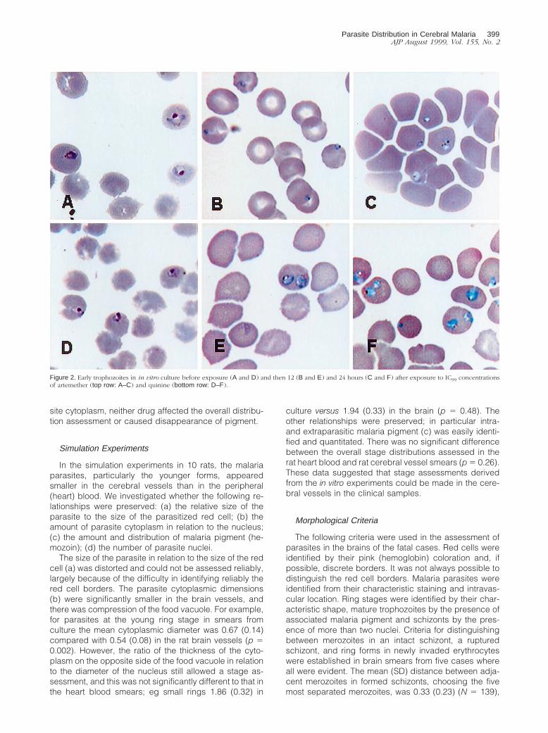
Although all stages of malaria parasite developmentwere seen in these fatal cases, the most frequent parasiteform found in the brain was the late trophozoite stage(Figure 3, Table 1). Using the midpoint of the modal stageto represent the parasite age (in hours) for each patient,the brain vessels were found to have parasites that had amodal age approximately 21 hours older than the periph-eral blood: median (range) 5 32 (3–46) compared with11 (3–36) hours (P , 0.001). The age of parasites in boththe brain and the blood was not significantly differentbetween those treated with quinine (N 5 25) and arte-mether; (N 5 5) median (range) for the brain was 32
(3–46) versus 36 (3–46) hours respectively and for theblood was 11 (3–36) versus 21 (3–32) hours, respectively.Peripheral parasitemias in the other patients were too lowfor accurate assessment of stage distributions. The ageof parasites in the brain was correlated negatively withthe time from starting treatment to death (Spearman cor-relation coefficient 5 20.42, p 5 0.004). There was norelationship between the peripheral blood stage and du-
Figure 3. Brain vessels from four different cases of fatal falciparum malaria showing accumulations of different parasite stages. A, late trophozoites, B, schizontswith abundant pigment; C, mid-stage trophozoites; D; ring forms containing no intraerythrocytic pigment.
Table 1. Distribution of the Modal Developmental Stage ofP. falciparum Parasites in the Peripheral Blood andBrain in Fatal Falciparum Malaria
Blood*(n 5 30)
N (%)
Brain*(n 5 45)
N (%)
Tiny rings (0–6 h) 8 (26%) 5 (11%)Small rings (6–16 h) 12 (40%) 3 (7%)Large rings (16–26 h) 6 (20%) 4 (9%)Early trophozoites (26–30 h) 0 4 (9%)Middle trophozoites (30–34 h) 3 (10%) 8 (18%)Late trophozoites (34–38 h) 1 (3%) 18 (40%)Schizonts
5 nuclei (38–44 h) 0 0.5 nuclei (44–48 h) 0 3 (7%)
*Only for those patients where at least 20 parasites were staged.
400 Silamut et alAJP August 1999, Vol. 155, No. 2
ration of treatment (Spearman correlation coefficient 50.11, p 5 0.56).
Mature Trophozoites and Schizonts
If it is assumed that there is no reason why death shouldbe associated with a particular stage of parasite devel-opment, then the distribution of mature stages in thecerebral vessels should be random between patients. Toassess this the trophozoite and schizont counts werenormalized, by converting each to the number per hour,to adjust for the difference in their respective time spans.Schizonts were often seen (Figure 4) but were signifi-cantly under-represented relative to trophozoites in thecerebral microvasculature. The median ratio of schizontsto trophozoites was 0.15 (range, 0.0 to 11.7) and thisdiffered significantly from the expected value of 1 (p ,0.001). Examining multiple sites from six brains demon-strated no evidence that this was due to a sampling error.Those brains which had large numbers of schizonts hadthem in every site sampled, and those that had none inone site had none in others. Thus there were no obviousdifferences between sites from the same brain.
Ring Form Parasites
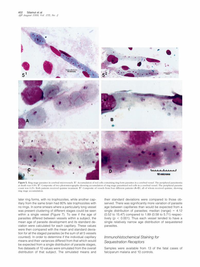
It is believed that parasites ,26 hours old circulate freelyin the bloodstream, whereas parasites .26 hours old arelargely stuck in the capillaries and venules. In order to
see whether the proportion of young parasites present inthe brain was the same as that which would be expectedfrom the numbers present in circulating blood, the pro-portion of red cells containing parasites ,26 hours oldwas calculated (for the brain red cells containing para-sites .26 hours old were excluded from the denomina-tor). The brain was found to have a considerably higherproportion of red cells containing young parasites com-pared to peripheral blood: median (range) 5 19.0% (0–90%) versus 1.8% (0–36.2%) (p 5 0.001). All stages ofparasites in the first half of the asexual life cycle wereover represented (ie this preponderance was not con-fined to the more mature ring form parasites) (Figure 5A–C). The proportion of all parasites ,26 hours old thatwere very young rings (aged 0–6 hours) was similar inthe brain and peripheral blood; median (range); 21.3%(0–100%) compared with 28.6% (0–94.4%), respec-tively, (p 5 0.22). In four of the 50 cases, more than 70%of the parasites in the brain were ring forms.
Microvascular Distribution of Parasite Stages
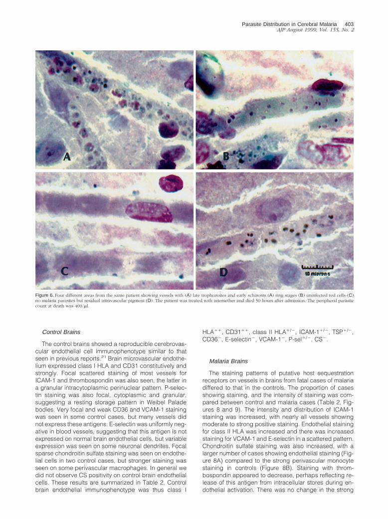
Individual capillaries within each brain showed very widevariations in the numbers of parasitized red blood cells(PRBC). In several brains some capillaries contained onlyunparasitized red cells, whilst others in the same sectionhad 100% (all 20/20) of red cells parasitized. Stage ofdevelopment also varied considerably (Figure 6). Forexample, one capillary might have 60% of parasites in the
Figure 4. Brain vessels from four different cases of fatal falciparum malaria showing sequestration of intact schizonts (A and B), and free merozoites after schizontrupture (C and D). The intraparasitic malaria pigment has stained black in A and B, and the free pigment in C is colored brown-black.
Parasite Distribution in Cerebral Malaria 401AJP August 1999, Vol. 155, No. 2
later ring forms, with no trophozoites, while another cap-illary from the same brain had 80% late trophozoites withno rings. In some smears where a particularly long vesselwas present clustering of different stages could be seenwithin a single vessel (Figure 7). To see if the age ofparasites differed between vessels within a subject, themean age of parasite development and its standard de-viation were calculated for each capillary. These valueswere then compared with the mean and standard devia-tion for all the staged parasites (ie the sum of all 5 vesselscounted). In order to determine if the individual capillarymeans and their variances differed from that which wouldbe expected from a single distribution of parasite stages,five datasets of 10 values were simulated from the overalldistribution of that subject. The simulated means and
their standard deviations were compared to those ob-served. There was significantly more variation of parasiteage between capillaries than would be expected from asingle distribution of parasites; median (range) 5 4.12(0.52 to 15.47) compared to 1.89 (0.58 to 5.71) respec-tively (p , 0.001). Thus each vessel tended to have asingle relatively narrow age distribution of sequesteredparasites.
Immunohistochemical Staining forSequestration Receptors
Samples were available from 13 of the fatal cases offalciparum malaria and 10 controls.
Figure 5. Ring stage parasites in cerebral microvessels. 51: Accumulation of red cells containing ring form parasites in a cerebral vessel. The peripheral parasitemiaat death was 0.9%. 52: Composite of two photomicrographs showing accumulation of ring stage parasitized red cells in a cerebral vessel. The peripheral parasitecount was 0.2%. Both patients received quinine treatment. 53: Composite of vessels from four different patients (A–D), all of whom received quinine, showingring stage accumulation.
402 Silamut et alAJP August 1999, Vol. 155, No. 2
Control Brains
The control brains showed a reproducible cerebrovas-cular endothelial cell immunophenotype similar to thatseen in previous reports.21 Brain microvascular endothe-lium expressed class I HLA and CD31 constitutively andstrongly. Focal scattered staining of most vessels forICAM-1 and thrombospondin was also seen, the latter ina granular intracytoplasmic perinuclear pattern. P-selec-tin staining was also focal, cytoplasmic and granular,suggesting a resting storage pattern in Weibel Paladebodies. Very focal and weak CD36 and VCAM-1 stainingwas seen in some control cases, but many vessels didnot express these antigens. E-selectin was uniformly neg-ative in blood vessels, suggesting that this antigen is notexpressed on normal brain endothelial cells, but variableexpression was seen on some neuronal dendrites. Focalsparse chondroitin sulfate staining was seen on endothe-lial cells in two control cases, but stronger staining wasseen on some perivascular macrophages. In general wedid not observe CS positivity on control brain endothelialcells. These results are summarized in Table 2. Controlbrain endothelial immunophenotype was thus class I
HLA11, CD3111, class II HLA1/2, ICAM-11/2, TSP1/2,CD362, E-selectin2, VCAM-12, P-sel1/2, CS2.
Malaria Brains
The staining patterns of putative host sequestrationreceptors on vessels in brains from fatal cases of malariadiffered to that in the controls. The proportion of casesshowing staining, and the intensity of staining was com-pared between control and malaria cases (Table 2, Fig-ures 8 and 9). The intensity and distribution of ICAM-1staining was increased, with nearly all vessels showingmoderate to strong positive staining. Endothelial stainingfor class II HLA was increased and there was increasedstaining for VCAM-1 and E-selectin in a scattered pattern.Chondroitin sulfate staining was also increased, with alarger number of cases showing endothelial staining (Fig-ure 8A) compared to the strong perivascular monocytestaining in controls (Figure 8B). Staining with throm-bospondin appeared to decrease, perhaps reflecting re-lease of this antigen from intracellular stores during en-dothelial activation. There was no change in the strong
Figure 6. Four different areas from the same patient showing vessels with (A) late trophozoites and early schizonts (A) ring stages (B) uninfected red cells (C)no malaria parasites but residual intravascular pigment (D). The patient was treated with artemether and died 50 hours after admission. The peripheral parasitecount at death was 400/ml.
Parasite Distribution in Cerebral Malaria 403AJP August 1999, Vol. 155, No. 2
and constitutive expression of CD31 and class I HLA(Figure 8C), and no increase in the patchy, low levels ofCD36 and P-selectin staining (Figure 8D). Thus endothe-lial cells in the brain of malaria cases show increasedexpression of ICAM-1, VCAM-1, E-selectin, CS, and classII HLA, and decreased TSP.
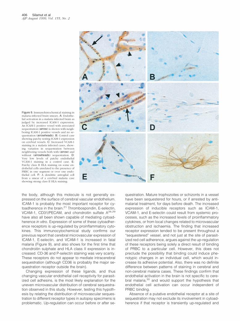
No specific changes in endothelial cell phenotypecould be related to the presence of parasites in a partic-ular vessel. CD31 staining did not appear to vary in termsof its distribution on the endothelial lumen. CD31 andICAM-1 staining was widespread, and as such werequantitatively the only markers widely distributed enoughto account for the bulk of parasitized erythrocyte seques-tration. However, areas of focal staining for other potentialreceptors such as VCAM-1 and CS were also seen.Within vessels from the same patient, heterogeneity inparasite sequestration compared to the presence of re-
ceptor staining was seen. Thus whereas generalized en-dothelial activation was present in many vessels asjudged by increased expression of ICAM-1 or VCAM-1,this did not equate with uniform parasite sequestration inall vessels (Figure 9, A–D). Accurate staging of parasitedevelopment was not possible in the immunohistochem-ical sections as parasite morphology was less well pre-served, but with rough assessments (based on size, in-traparasitic malaria pigment, and number of nuclei) thevariation in endothelial staining along a vessel did notappear to be related to the stage of development of thelocally sequestered parasites. There were no evidentdifferences between the eleven cases of cerebral malariaand the two fatal malaria cases who did not have cerebralmalaria. Both these cases also had variable increases inICAM-1, VCAM-1, and E-selectin staining. Some vesselshad patchy class II staining on some endothelial cells
Figure 7. Composite photomicrograph (magnification, 31000) of a brain smear from a fatal case of falciparum malaria. Four vessels are seen (A–D). Nearly allthe red cells in A and B are parasitized, whereas few parasites are seen in C and D. A is intensely parasitized containing alternating clusters of trophozoites andschizonts. Residual adherent pigment in the far right of the vessel indicates earlier schizont rupture. In B younger trophozoites are seen evenly spaced on the leftand schizonts on the right. The patient received artemether but died 4 hours later after the parasitemia had doubled from 14.5% to 34.9%.
Table 2. Comparison of Immunohistochemical Staining between Controls and Malaria Cases
AntigenStaining pattern in
control cases*Staining pattern in
malaria cases* Changes
CD31/PECAM 100 (1) 100 (1 to 11) No changeClass I HLA 100 (11) 100 (11) No changeClass II HLA 50 (1/2) 100 (1) Increased % cases and intensityICAM-1 90 (1/2) 100 (1 to 11) Increased intensityVCAM 50 (2 to 1/2) 69 (1/2) Increased % cases and intensityE-selectin 0 (2) 38 (2 to 1/2) Increased % cases and intensityCD36 40 (2) 38 (2) No changeThrombospondin 100 (1/2) 61 (1/2) Decreased % casesChondroitin sulfate A 20 (2) 61 (1/2) Increased % cases and intensityP-selectin 60 (1/2) 61 (1/2) No change
*Percentage of cases showing staining and average score.
404 Silamut et alAJP August 1999, Vol. 155, No. 2
(Figure 9E), but in general if a vessel showed positivestaining for a marker of endothelial activation, this waspresent throughout its length, rather than being related tothe presence of PRBC in one segment or over one endo-thelial cell. There was little evidence for lymphocyte orplatelet accumulation in vessels in any case. Scatteredmonocytes were seen in several malaria cases (CD361,class I, and II HLA1 staining), and numerous class II HLApositive microglial cells were seen (Figure 9F).
Discussion
Shortly after the discovery of the malaria parasite byLaveran, malaria researchers in Rome noticed the dis-crepancy between the number and stages of parasitedevelopment in the peripheral blood and the brains ofpatients who died from cerebral malaria.22 Only theyounger parasite stages of the potentially lethal P. falci-parum were seen in peripheral blood smears, whereasthe more mature parasites were abundant in the cerebralvessels. In contrast all stages of parasite developmentwere represented in the peripheral blood of patients withthe benign malarias. This process, known as sequestra-tion, is not uniform. The intensity of sequestration differsbetween different organs,10,12,15 and also differs be-tween the different parts of the brain.23 As noted seques-tration is not a feature of the other human malarias (P.vivax, P. malariae, P. ovale), in which all stages of parasitedevelopment may be seen in peripheral blood smears. Inthis series one patient died from vivax malaria, but therewas no evidence of cerebral sequestration in this case.The present study provides formal mathematical proof of
sequestration in lethal falciparum malaria, and shows thatsequestration of P. falciparum infected erythrocytes variesconsiderably even at the microvascular level.
It is possible that the preparation of brain smears leadsto overestimation of sequestration, because capillarieswithout cytoadherent red cells may be missed on micro-scopic examination, or may be more likely to empty dur-ing the preparation of the brain smear, than those withsequestered parasites. However, this is unlikely to be amajor confounder as capillaries and venules showing noevidence of cytoadherent parasitized erythrocytes wereoften seen, whereas others in the same section, severalmicrons away, were intensely parasitized (Figures 5 and6). Assessment of the stage of parasite development invivo is also confounded by the effects of antimalarial drugtreatment and compression within the confines of narrowvessels. However, in vitro and animal experiments indi-cated that these would not lead to large errors in stageassessment. The heterogeneity in the microvascular dis-tribution of parasitized erythrocytes in fatal malaria em-phasises the importance of the vascular endothelial re-ceptors for parasitized red cell cytoadherence indetermining the extent and distribution of sequestration.Cytoadherence is mediated by the binding of parasitederived adhesins, expressed on the surface of the para-sitized erythrocyte and also possibly modifications in thehost cell membrane, to vascular endothelial cell recep-tors.1,24 The principal parasite adhesin is PfEMP1, a highmolecular weight, antigenically variant product of the vargene superfamily.25–27 Multiple vascular receptors forcytoadherence have been identified in vitro. Quantita-tively CD36 appears to be the most important receptor in
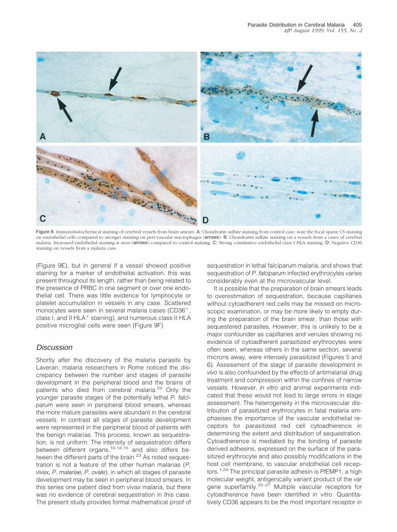
Figure 8. Immunohistochemical staining of cerebral vessels from brain smears. A: Chondroitin sulfate staining from control case: note the focal sparse CS stainingon endothelial cells compared to stronger staining on peri-vascular macrophages (arrows). B: Chondroitin sulfate staining on a vessels from a cases of cerebralmalaria. Increased endothelial staining is seen (arrows) compared to control staining. C: Strong constitutive endothelial class I HLA staining. D: Negative CD36staining on vessels from a malaria case.
Parasite Distribution in Cerebral Malaria 405AJP August 1999, Vol. 155, No. 2
the body, although this molecule is not generally ex-pressed on the surface of cerebral vascular endothelium.ICAM-1 is probably the most important receptor for cy-toadherence in the brain.21 Thrombospondin, E-selectin,VCAM-1, CD31/PECAM, and chondroitin sulfate A28,29
have also all been shown capable of mediating cytoad-herence in vitro. Expression of some of these cytoadher-ence receptors is up-regulated by proinflammatory cyto-kines. This immunocytochemical study confirms ourprevious report that cerebral microvascular expression ofICAM-1, E-selectin, and VCAM-1 is increased in fatalmalaria (Figure 9), and also shows for the first time thatchondroitin sulphate and HLA class II expression is in-creased. CD-36 and P-selectin staining was very scanty.These receptors do not appear to mediate intracerebralsequestration (although CD36 is probably the major se-questration receptor outside the brain).
Changing expression of these ligands, and thuschanging vascular endothelial cell receptivity for parasit-ized cell adhesins, is the most likely explanation for theuneven microvascular distribution of cerebral sequestra-tion observed in this study. However, testing this hypoth-esis by relating the distribution of microvascular seques-tration to different receptor types in autopsy specimens isproblematic. Up-regulation can occur before or after se-
questration. Mature trophozoites or schizonts in a vesselhave been sequestered for hours, or if arrested by anti-malarial treatment, for days before death. The increasedexpression of inducible receptors such as ICAM-1,VCAM-1, and E-selectin could result from systemic pro-cesses, such as the increased levels of proinflammatorycytokines, or from local changes related to microvascularobstruction and ischaemia. The finding that increasedreceptor expression tended to be present throughout a“sequestered” vessel, and not just at the site of parasit-ized red cell adherence, argues against the up-regulationof these receptors being solely a direct result of bindingof PRBC to a particular cell. However, this does notpreclude the possibility that binding could induce phe-notypic changes in an individual cell, which would in-crease its adhesive potential. Also, there was no definitedifference between patterns of staining in cerebral andnon-cerebral malaria cases. These findings confirm thatendothelial activation in the brain is not specific to cere-bral malaria,30 and would support the hypothesis thatendothelial cell activation can occur independent ofPRBC binding.
Absence of a putative endothelial receptor at a site ofsequestration may not exclude its involvement in cytoad-herence if that receptor is transiently up-regulated and
Figure 9. Immunohistochemical staining inmalaria-infected brain smears. A: Endothe-lial activation in a malaria infected brain asjudged by increased ICAM-1 expression.An ICAM-1 positive vessel with associatedsequestration (arrow) is shown with neigh-boring ICAM-1 positive vessels and no se-questration (arrowheads). B: Control caseshowing patchy resting ICAM-1 expressionon cerebral vessels. C: Increased VCAM-1staining in a malaria infected cases, show-ing variation in sequestration betweenneighboring vessels both with (arrow) andwithout (arrowheads) sequestration. D:Very low levels of patchy endothelialVCAM-1 staining in a control case. E:Patchy class II HLA staining on some en-dothelial cells unrelated to the presence ofPRBC in one segment or over one endo-thelial cell. F: A dendritic astroglial cellfrom a smear of a cerebral malaria caseshowing strong class II HLA staining.
406 Silamut et alAJP August 1999, Vol. 155, No. 2
turned over rapidly. Because each case reflects a singletime point during the disease process, which varies witheach patient, differences in the temporal expression ofparticular receptors could be missed. However the co-expression of markers of endothelial activation such asE-selectin and VCAM-1, which have very different timecourses of expression on endothelial cells in vitro, sug-gests that in vivo they are being subjected continuouslyto waves of activation on a more chronic time course,which does allow us to see both being expressed at thesame time.
The parasites within the cytoadherent erythrocytes ineach vessel or section of a vessel tended to be of similarage, whereas the distribution of parasite stages in thevessels overall was much broader, ie there was spatialclustering in parasite age. Presumably small blood ves-sels increase their receptivity to cytoadherence by in-creasing expression of these vascular ligands and selectfor those circulating parasitized erythrocytes newly capa-ble of adhesion. All the red cells infected by more matureparasites would already be adherent (ie not circulating).Thus as the parasites mature and begin to express redcell surface adhesins, they are filtered out by those ves-sels newly capable of accepting them. Rolling is followedby static cytoadherence.31 Blood flow is not stopped bythis process. There must be significant flow even throughvessels partially occluded by cytoadherent parasitizederythrocytes to allow this filtration to continue, althoughthe unparasitized erythrocytes presumably undergo con-siderable deformation in squeezing past the adherentand relatively nondeformable sequestered red cells.1
This is the only explanation to account for the consider-ably higher proportion of parasitized red cells in thecerebral vessels compared to the peripheral blood. Thisstudy also suggests that once parasitized red cells ad-here, they do not detach again and circulate freely. If theydid recirculate this would lead to mixing of stages, andthe parasites sequestered in each vessel should reflectthe broad stage distribution of malaria parasites in thesecond 24 hours of their development.
Heterogeneity in microvascular sequestration in thebrain may well explain the rarity of permanent neurolog-ical damage in cerebral malaria. It is likely that microvas-cular obstruction and consequent ischaemia provides astrong stimulus to vasodilation.32 Blood flow throughthose capillaries and venules which are not obstructed byparasitized cells would be expected to be maximal. Al-tered vessel wall shear stresses would also induce re-lease of the potent vasodilator nitric oxide.33 It has beensuggested that nitric oxide may cause coma in cerebralmalaria, although the source hypothesized has been cy-tokine up-regulated, inducible isoform of nitric oxide syn-thase (iNOS), rather than vascular wall endothelial iso-form (eNOS), or the neuronal isoform (nNOS).2,3,34
Although there may be a background increase in vascu-lar endothelial nitric oxide generation because of thesystemic increase in proinflammatory cytokine release,35
local factors are likely to predominate before terminalshock develops. Release of vasodilatory mediators wouldbe expected to be greatest adjacent to cytoadherent redcells. Thus delivery of oxygen and metabolic substrates
in areas with partially occluded vessels may be sufficientto prevent permanent neuronal cell death. Even appar-ently blocked capillaries may still allow passage of unin-fected red cells.
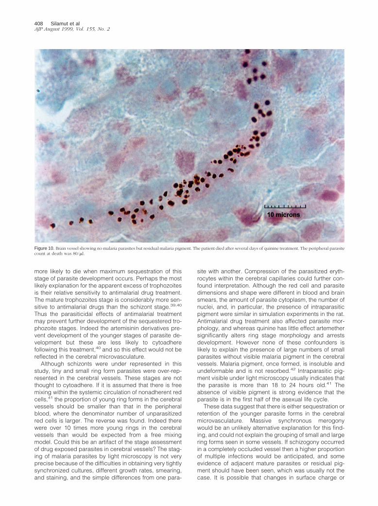
Although cerebral malaria was associated with intensesequestration within the brain, this was also seen in pa-tients who did not lose consciousness. This may reflectthe time between admission and death and does notdisprove the concept that cerebral malaria is associatedspecifically with cerebral sequestration. Other patients inthis series, particularly those who died after several daysof antimalarial treatment, had few or no residual parasitesin their cerebral vessels. This has been noted previous-ly36,37 and has led some investigators to suggest thatcerebral sequestration is not a necessary prerequisite forcerebral malaria.3 Nearly all such cases reported hadreceived many days of treatment, and in this series re-sidual pigment was evident within the cerebral microvas-culature, even if no parasites were seen (Figure 10). Thusthis relatively large study provides no support for thecontention that cerebral malaria can occur in the ab-sence of cerebral sequestration. Sequestration occurs inall patients with falciparum malaria and is a consistentpathological feature of severe malaria. If it was not thenschizonts and mature trophozoites would be seen regu-larly in peripheral blood films. The absence of parasitesfrom the brains of fatal cases indicates effective antima-larial treatment and clearance of infected erythrocytes.Such treatment cannot always reverse malaria related orsecondary pathological processes in severely ill patients.
In this investigation, compared with the number oftrophozoites recorded, we found less than the expectednumber of schizonts in the cerebral microvasculature.Previous in vitro and in vivo studies have suggested thatthe schizont or segmenter stage lasts for at least 10hours.38 Thus in a series of fatal cases, the number ofschizonts and mature trophozoites sequestered would beexpected to be approximately similar assuming a randomdistribution of parasite stages at the time of death. Schi-zonts are thought to remain sequestered until fully matureand then to burst leaving a cytoadherent red cell ghost.The number of trophozoites documented in this studywas nearly four times greater than that of the schizonts.There are several possible explanations for this finding.Schizogony may be significantly shorter than estimatedcurrently. Data from continuous in vitro culture suggestthat this is very unlikely. It is possible that schizont rupturein vivo occurs in a proportion of cases much earlier thanthought (ie before 18–24 merozoites have developed).Another explanation would be premature detachment ofcytoadherent schizonts. The surface of schizonts arethought to contain less PfEMP1 molecules than maturetrophozoites. If schizonts do detach, then they are notseen in peripheral blood films, and they do not appear toadhere again in the brain. It is possible they are clearedby the spleen, although semi-quantitative assessments ofthe parasitized cell population in the spleen indicates thatall parasite stages are found, without an unusual predom-inance of schizonts.12 Alternatively it is possible that themore metabolically active mature trophozoite stage isparticularly virulent for the host, and patients may be
Parasite Distribution in Cerebral Malaria 407AJP August 1999, Vol. 155, No. 2
more likely to die when maximum sequestration of thisstage of parasite development occurs. Perhaps the mostlikely explanation for the apparent excess of trophozoitesis their relative sensitivity to antimalarial drug treatment.The mature trophozoites stage is considerably more sen-sitive to antimalarial drugs than the schizont stage.39,40
Thus the parasiticidal effects of antimalarial treatmentmay prevent further development of the sequestered tro-phozoite stages. Indeed the artemisinin derivatives pre-vent development of the younger stages of parasite de-velopment but these are less likely to cytoadherefollowing this treatment,40 and so this effect would not bereflected in the cerebral microvasculature.
Although schizonts were under represented in thisstudy, tiny and small ring form parasites were over-rep-resented in the cerebral vessels. These stages are notthought to cytoadhere. If it is assumed that there is freemixing within the systemic circulation of nonadherent redcells,41 the proportion of young ring forms in the cerebralvessels should be smaller than that in the peripheralblood, where the denominator number of unparasitizedred cells is larger. The reverse was found. Indeed therewere over 10 times more young rings in the cerebralvessels than would be expected from a free mixingmodel. Could this be an artifact of the stage assessmentof drug exposed parasites in cerebral vessels? The stag-ing of malaria parasites by light microscopy is not veryprecise because of the difficulties in obtaining very tightlysynchronized cultures, different growth rates, smearing,and staining, and the simple differences from one para-
site with another. Compression of the parasitized eryth-rocytes within the cerebral capillaries could further con-found interpretation. Although the red cell and parasitedimensions and shape were different in blood and brainsmears, the amount of parasite cytoplasm, the number ofnuclei, and, in particular, the presence of intraparasiticpigment were similar in simulation experiments in the rat.Antimalarial drug treatment also affected parasite mor-phology, and whereas quinine has little effect artemethersignificantly alters ring stage morphology and arrestsdevelopment. However none of these confounders islikely to explain the presence of large numbers of smallparasites without visible malaria pigment in the cerebralvessels. Malaria pigment, once formed, is insoluble andundeformable and is not resorbed.42 Intraparasitic pig-ment visible under light microscopy usually indicates thatthe parasite is more than 18 to 24 hours old.41 Theabsence of visible pigment is strong evidence that theparasite is in the first half of the asexual life cycle.
These data suggest that there is either sequestration orretention of the younger parasite forms in the cerebralmicrovasculature. Massive synchronous merogonywould be an unlikely alternative explanation for this find-ing, and could not explain the grouping of small and largering forms seen in some vessels. If schizogony occurredin a completely occluded vessel then a higher proportionof multiple infections would be anticipated, and someevidence of adjacent mature parasites or residual pig-ment should have been seen, which was usually not thecase. It is possible that changes in surface charge or
Figure 10. Brain vessel showing no malaria parasites but residual malaria pigment. The patient died after several days of quinine treatment. The peripheral parasitecount at death was 80/ml.
408 Silamut et alAJP August 1999, Vol. 155, No. 2
membrane fluidity in ring stage infected red cells couldalter their normal transit through the microcirculation andlead them to accumulate, preferentially in branch vesselswhich are normal closed to flow. Whichever the mecha-nism, red cells containing young rings, hitherto thoughtincapable of sequestration, are concentrated consider-ably in the cerebral vessels. Whether this is because ofadhesion to vascular endothelium or to other parasitizedcells or simply reflects their reduced deformability is notknown, but it does suggest that peripheral parasitecounts are an even greater underestimate of the parasiteburden in severe falciparum malaria than previouslythought.
Acknowledgments
We thank the Director and staff of the Center for TropicalDiseases, Cho Quan Hospital, Ho Chi Minh City andPaholpolpayuhasena Hospital, Kanchanaburi for theirsupport for these studies. We are particularly grateful todoctors T.T.H. Chau, T.M.E. Davis, N.P.J. Day, S.Krishna, P.P. Loc, S. Pukrittayakamee, D.X.T. Sinh, W.Supanaranond, T.T.M. Trang, and D.J. Waller for helpwith clinical management.
References
1. White NJ, Ho M: The pathophysiology of malaria. Adv Parasitol 1992,31:83–173
2. Clark IA, Rockett KA, Cowden WB: Possible central role of nitric oxidein conditions clinically similar to cerebral malaria. Lancet 1992, 340:894–896
3. Clark IA, Rockett KA: The cytokine theory of cerebral malaria. Para-sitol Today 1994, 10:410–412
4. Berendt AR, Turner GDH, Newbold CI: Cerebral malaria: the seques-tration hypothesis. Parasitol Today 1994, 10:412–414
5. Udeinnya IJ, Schmidt JA, Aikawa M, Miller LM, Green I: Falciparummalaria infected erythrocytes specifically bind to cultured humanendothelial cells. Science 1981, 213:555–557
6. Miller LH: Distribution of mature trophozoites and schizonts of Plas-modium falciparum in the organs of Aotus trivirgatus, the night mon-key. Am J Trop Med Hyg 1969, 18:860–865
7. Marchiafava E: Pernicious malaria. Am J Hyg 1931, 13:1–568. Gaskell JF, Millar WL: Studies on malignant malaria in Macedonia. Q
J Med 1920, 13:381–4269. Spitz S: The pathology of acute falciparum malaria. Mil Surg 1946,
99:555–57210. MacPherson GG, Warrell MJ, White NJ, Looarreesuwan S, Warrell
DA: Human cerebral malaria—a quantitative ultrastructural analysisof parasitized erythrocyte sequestration. Am J Pathol 1985, 119:385–401
11. Oo MM, Aikawa M, Than T, Aye TM, Myint PT, Igarashi I, SchoeneWC: Human cerebral malaria: a pathological study. J NeuropatholExp Neurol 1987, 46:223–231
12. Riganti M, Pongponratn E, Tegoshi T, Looareesuwan S, PunpoowongB, Aikawa M: Human cerebral malaria in Thailand: a clinico-patho-logical correlation. Immunol Lett 1990, 25:199–205
13. Sengupta SK, Naraqi S: The brain in cerebral malaria: a pathologicalstudy of 24 fatal cases in Papua New Guinea. PNG Med J 1992,35:270–274
14. Walker O, Salako LA, Sowunmi A, Thomas JO, Sodeine O, Bondi FS:Prognostic risk factors and post mortem findings in cerebral malariain children. Trans R Soc Trop Med Hyg 1992, 86:491–493
15. Nakazawa S, Looareesuwan S, Fujioka H, Pongponratn E, Luc KD,Rabbege J, Aikawa M: A correlation between sequestered parasit-
ized erythrocytes in subcutaneous tissue and cerebral malaria. Am JTrop Med Hyg 1995, 53:544–546
16. Smith CD, Brown AE, Nakazawa S, Fujioka H, Aikawa M: Multi-organerythrocyte sequestration and ligand expression in rhesus monkeysinfected with Plasmodium coatneyi malaria. Am J Trop Med Hyg1996, 55:379–383
17. Lemercier G, Bert J, Nouhouyi M, Rey M, Collomb H: Leneuropaludisme: Aspects electroencephalographiques, neuro-pathologiques, problemes physiopathologiques. Pathol Biol 1969,17:459–472
18. Hien TT, Day NPJ, Phu NH, Mai NTH, Chau TTH, Loc PP, Sinh DX,Chuong LV, Vinh H, Waller D, Peto TEA, White NJ: A controlled trial ofartemether or quinine in Vietnamese adults with severe falciparummalaria. N Engl J Med 1996, 335:76–83
19. Raja RN: Post-mortem examination in cerebral malaria. A new simplemethod of demonstrating parasites in the capillaries of the brain.Indian Med Gazette 1922, 57:298–299
20. White NJ, Silamut K: Rapid diagnosis of malaria. Lancet 1989, 1:43521. Turner GDH, Morrison H, Jones M, Davis TME, Looaresuwan S, Buley
I, Gatter KC, Newbold CI, Pukritayakamee S, Nagachinta B, White NJ,Berendt AR: An immunohistochemical study of the pathology of fatalmalaria. Am J Pathol 1994, 145:1057–1069
22. Marchiafava E, Bignami A: On Summer-Autumnal Fever. The NewSydenham Society, London, 1894
23. Sein KK, Maeno Y, Thuc HV, Anh TK, Aikawa M: Differential seques-tration of parasitized erythrocytes in the cerebrum and cerebellum inhuman cerebral malaria. Am J Trop Med Hyg 1993, 48:504–511
24. Sherman IW, Crandall I, Smith H: Membrane proteins involved in theadherence of Plasmodium falciparum-infected erythrocytes to theendothelium. Biol Cell 1992, 74:161–178
25. Su X, Heatwole VM, Wertheimer SP, Guinet F, Herrfeldt JA, PetersonDS, Ravetch JA, Wellems TE: The large diverse gene family varencodes proteins involved in cytoadherence and antigenic variationof Plasmodium falciparum-infected erythrocytes. Cell 1995, 82:89–100
26. Baruch DI, Pasloske BL, Singh HB, Bi X, Ma XC, Feldman M, TaraschiTF, Howard RJ: Cloning the P. falciparum gene encoding PfEMP1, amalaria variant antigen and adherence receptor on the surface ofparasitized human erythrocytes. Cell 1995, 82:77–87
27. Smith JD, Chitnis CE, Craig AG, Roberts DJ, Hudson-Taylor DE,Peterson DS, Pinches R, Newbold CI, Miller LH: Switches in expres-sion of Plasmodium falciparum var genes correlate with changes inantigenic and cytoadherent phenotypes of infected erythrocytes. Cell1995, 82:101–110
28. Rogerson S, Chaiyaroj S, Ng K, Reeder J, Brown G: Chondroitinsulphate A is a cell surface receptor for Plasmodium falciparuminfected erythrocytes. J Exp Med 1995, 182:15–20
29. Robert C, Pouvelle B, Meyer P, Muanza K, Fujioka H, Aikawa M,Scherf A, Gysin J: Chondroitin-4-sulfate (proteoglycan), a receptor forPlasmodium falciparum-infected erythrocytes adherence on brainmicrovascular endothelial cells. Res Immunol 1995, 146:383–393
30. Turner GDH, Chuong LV, Mai NTH, Chau TTH, Phu NH, Bethell D,Wyllie S, Louwrier K, Fox S, Gatter KC, Day NPJ, Hien TT, White NJ,Berendt AR: Systemic endothelial activation occurs in both mild andsevere malaria: correlating dermal microvascular endothelial cellphenotype and soluble cell adhesion molecules with disease sever-ity. Am J of Pathol 1998, 152:1477–1487
31. Cooke BM, Berendt AR, Craig AG, Macgregor J, Newbold CI: Rollingand stationary cytoadhesion of red blood cells parasitized by Plas-modium falciparum: separate roles for ICAM-1, CD36 and throm-bospondin. Br J Haematol 1994, 87:162–170
32. Warrell DA, White NJ, Veall N, Looareesuwan S, Chanthavanich P,Phillips RE, Karbwang J, Pongpaew P, Krishna S: Cerebral anaerobicglycolysis and reduced cerebral oxygen transport in human cerebralmalaria. Lancet 1988, 2:534–538
33. Umans JG, Levi R: Nitric oxide in the regulation of blood flow andarterial pressure. Annu Rev Physiol 1995, 57:771–90
34. Al Yaman FM, Mokela D, Genton G, Rockett KA, Alpers MP, Clark IA:Association between serum levels of reactive nitrogen intermediatesand coma in children with cerebral malaria in Papua New Guinea.Trans R Soc Trop Med Hyg 1996, 90:270–273
35. Bhagat K, Vallance P: The local venous response to endotoxin inhumans. Circulation 1996, 94:490–497
36. Kean BH, Smith JA: Death due to Aestivo-autumnal malaria. A resume
Parasite Distribution in Cerebral Malaria 409AJP August 1999, Vol. 155, No. 2

of one hundred autopsy cases. Am J Trop Med Hyg 1944, 24:317–322
37. Boonpucknavig V, Boonpucknavig, Udomsangpetch R, Nitiyanant P:An immunofluorescence study of cerebral malaria: a correlation withhistopathology. Arch Pathol Lab Med 1990, 114:1028–1034
38. Li G-Q, Guo X-B, Jian H-X, Wang X-H: Clinical trials of artemisinin andits derivatives in the treatment of malaria in China. Trans R Soc TropMed Hyg 1994, 88:Suppl 1, 5–6
39. ter Kuile F, White NJ, Holloway P, Pasvol G, Krishna S: Plasmodiumfalciparum: in vitro studies of the pharmacodynamic properties ofdrugs used for the treatment of severe malaria. Exp Parasitol 1993,76:86–95
40. Udomsangpetch R, Pipitaporn B, Krishna S, Angus B, Pukrittayaka-mee S, Bates I, Suputtamongkol Y, Kyle DE, White NJ: Antimalarialdrugs reduce cytoadherence and rosetting of Plasmodium falcipa-rum. J Infect Dis 1996, 173:691–698
41. Silamut K, White NJ: Relation of the stage of parasite development inthe peripheral blood to prognosis in severe falciparum malaria. TransR Soc Trop Med Hyg 1993, 87:436–443
42. Taramelli D, Basilico N, De Palma AM, Sarasella M, Ferrante P,Mussoni L, Olliaro P: The effect of synthetic malaria pigment (B-haematin) on adhesion molecule expression and interleukin-6 pro-duction by human endothelial cells. Trans R Soc Trop Med Hyg 1998,92:57–62
410 Silamut et alAJP August 1999, Vol. 155, No. 2
Copyright © 2022 FDOKUMEN