Targeting the efficacy of a dendrimer-based nanotherapeutic in heterogeneous xenograft tumors in...
15
Targeting the efficacy of a dendrimer-based nanotherapeutic in heterogeneous xenograft tumors in vivo Andrzej Myc, Jolanta Kukowska-Latallo, Peter Cao, Ben Swanson, Julianna Battista, Thomas Dunham, and James R. Baker Jr Michigan Nanotechnology Institute for Medicine and Biological Sciences, University of Michigan Medical School, BSRB, Ann Arbor, Michigan, USA Abstract Our earlier studies have shown the in vitro and in vivo targeting of a generation 5 (G5) dendrimer- based multifunctional conjugate that contained folic acid (FA) as the targeting agent and methotrexate (MTX) as the chemotherapeutic drug. To clinically apply the synthesized G5-FA-MTX nanotherapeutic, it is important that the anticancer conjugate elicits cytotoxicity specifically and consistently. Toward this objective, we evaluated the large-scale synthesis of a G5-FA-MTX conjugate (Lot # 123–34) for its cytotoxic potential and specificity in vitro and in vivo. The cytotoxicity and specificity were tested by using a coculture assay in which FA receptor-expressing and nonexpressing cells (KB and SK-BR-3 cells, respectively) were cultured together and preferential killing was examined. The in-vitro data were compared with the in-vivo data obtained from a heterogeneous xenograft tumor model. The animal model of the artificial heterogeneous xenograft tumor showed that the nanotherapeutic was preferentially cytotoxic to KB cells. Keywords coculture assay; dendrimer; heterogeneous xenograft tumor model; methotrexate; neoplasm; targeted drug delivery Introduction Traditional anticancer chemotherapy uses drugs that have low functional specificity for cancer cells [1]. As a result, anticancer drugs generally have relatively small therapeutic indices, which lead to considerable nonspecific toxicity. Thus, there is a great need for anticancer drugs that specifically target cancer cells while leaving normal tissue intact. Several approaches have been explored in an attempt to achieve site-specific chemotherapy, including localized drug administration [2], antibody-mediated targeting [3], and exploitation in the vascular structure and lymphatic drainage of solid tumors through the enhanced permeation and retention effect [4]. A large range of nano-carriers and macromolecules have been evaluated for their potential to enhance the biodistribution of chemotherapeutics to solid tumors [4–8]. Recently, a large number of applications have been found for hyperbranched polymers and dendrimers in delivering drugs and genetic materials into cells [9–19]. Compared with Correspondence to Andrzej Myc, PhD, Center for Biologic Nanotechnology, Rm 9346 MSRB III, University of Michigan, Ann Arbor, MI 48109, USA, Tel: + 1 734 647 0052; fax: + 1 734 936 2990; [email protected]. Andrzej Myc and Jolanta Kukowska-Latallo contributed equally to the study NIH Public Access Author Manuscript Anticancer Drugs. Author manuscript; available in PMC 2010 February 10. Published in final edited form as: Anticancer Drugs. 2010 February ; 21(2): 186. doi:10.1097/CAD.0b013e328334560f. NIH-PA Author Manuscript NIH-PA Author Manuscript NIH-PA Author Manuscript
-
Upload
independent -
Category
Documents
-
view
1 -
download
0
Transcript of Targeting the efficacy of a dendrimer-based nanotherapeutic in heterogeneous xenograft tumors in...

Targeting the efficacy of a dendrimer-based nanotherapeutic inheterogeneous xenograft tumors in vivo
Andrzej Myc, Jolanta Kukowska-Latallo, Peter Cao, Ben Swanson, Julianna Battista, ThomasDunham, and James R. Baker JrMichigan Nanotechnology Institute for Medicine and Biological Sciences, University of MichiganMedical School, BSRB, Ann Arbor, Michigan, USA
AbstractOur earlier studies have shown the in vitro and in vivo targeting of a generation 5 (G5) dendrimer-based multifunctional conjugate that contained folic acid (FA) as the targeting agent and methotrexate(MTX) as the chemotherapeutic drug. To clinically apply the synthesized G5-FA-MTXnanotherapeutic, it is important that the anticancer conjugate elicits cytotoxicity specifically andconsistently. Toward this objective, we evaluated the large-scale synthesis of a G5-FA-MTXconjugate (Lot # 123–34) for its cytotoxic potential and specificity in vitro and in vivo. Thecytotoxicity and specificity were tested by using a coculture assay in which FA receptor-expressingand nonexpressing cells (KB and SK-BR-3 cells, respectively) were cultured together and preferentialkilling was examined. The in-vitro data were compared with the in-vivo data obtained from aheterogeneous xenograft tumor model. The animal model of the artificial heterogeneous xenografttumor showed that the nanotherapeutic was preferentially cytotoxic to KB cells.
Keywordscoculture assay; dendrimer; heterogeneous xenograft tumor model; methotrexate; neoplasm; targeteddrug delivery
IntroductionTraditional anticancer chemotherapy uses drugs that have low functional specificity for cancercells [1]. As a result, anticancer drugs generally have relatively small therapeutic indices, whichlead to considerable nonspecific toxicity. Thus, there is a great need for anticancer drugs thatspecifically target cancer cells while leaving normal tissue intact.
Several approaches have been explored in an attempt to achieve site-specific chemotherapy,including localized drug administration [2], antibody-mediated targeting [3], and exploitationin the vascular structure and lymphatic drainage of solid tumors through the enhancedpermeation and retention effect [4]. A large range of nano-carriers and macromolecules havebeen evaluated for their potential to enhance the biodistribution of chemotherapeutics to solidtumors [4–8].
Recently, a large number of applications have been found for hyperbranched polymers anddendrimers in delivering drugs and genetic materials into cells [9–19]. Compared with
Correspondence to Andrzej Myc, PhD, Center for Biologic Nanotechnology, Rm 9346 MSRB III, University of Michigan, Ann Arbor,MI 48109, USA, Tel: + 1 734 647 0052; fax: + 1 734 936 2990; [email protected] Myc and Jolanta Kukowska-Latallo contributed equally to the study
NIH Public AccessAuthor ManuscriptAnticancer Drugs. Author manuscript; available in PMC 2010 February 10.
Published in final edited form as:Anticancer Drugs. 2010 February ; 21(2): 186. doi:10.1097/CAD.0b013e328334560f.
NIH
-PA Author Manuscript
NIH
-PA Author Manuscript
NIH
-PA Author Manuscript

conventional polymers, dendrimers have structural and functional advantages emanating fromtheir precise architecture and low polydispersity, which are synthesized in a layer-by-layermanner (called generation) around a core unit, resulting in a high level of control over size,branching points, and surface functionality [5]. The ability to tailor dendrimer properties totherapeutic needs makes them ideal carriers for small molecules such as drugs andbiomolecules. Dendrimers have nanoscale container and nano-scaffolding properties and arebio-compatible [5,20–25].
Conjugation of methotrexate (MTX) and folic acid (FA) to hydrazide-terminated dendrimersrevealed that MTX and FA can be coupled to the dendrimer with conjugation ratios of 4.7 and12.6, respectively [16]. In another study a 2.5 and 3-generation Poly(amido amine) dendrimers(PAMAM) dendrimer was used for the synthesis of an anticancer drug containing MTX. Theconjugates increased the sensitivity of drug-resistant cell lines as compared with free MTX[26]. We have recently reported dendrimer-based drug conjugates that target cancer cellsthrough the folate receptors [18,27–31], prostate-specific membrane antigen [32], Her2/neu[33], and αυβ3 integrins [34]. For the ‘targeted’ drug delivery agents to be useful for clinicalapplication, the compounds have to consistently show specific killing of the targeted cells.Therefore, measuring the effect of anti-cancer agents on the growth and survival of neoplasticcell populations in vitro is critical in the preclinical evaluation of the therapeutics [35,36].Recently, we have evaluated several lots of generation 5 (G5)-FA-MTX conjugate that targetMTX to KB cells through the folate receptor and have found them preferentially cytotoxic toKB cells in vitro [31]. In this study, we extended the evaluation of the targeted nanotherapeuticby examining its effectiveness in an artificial heterogeneous tumor (AHT) in-vivo model[37,38]. This AHT, which consists of cells from the human epithelial KB cell line, whichoverexpresses the folate receptor, and the human breast adenocarcinoma, SK-BR-3, cell lineallows to further confirm the targeting specificity and efficacy of a dendrimer-basednanotherapeutic.
Materials and methodsMaterials
G5 and 7 nonacetylated dendrimers (G5-PAMAM and G7-PAMAM) were synthesized at M-NIMBS, University of Michigan, as described earlier [28,39]. The G7-PAMAM was used asa transfecting agent in the transfection of KB and SK-BR-3 cells with green fluorescent protein(GFP) and red fluorescent protein (RFP) plasmid DNA, respectively [40]. The plasmidspDsRed1-N1 and pAcGFP1-C1, which constitutively express red and green fluorescentproteins, respectively, under control of a PCMV IE promoter, were purchased from Clontech(Mountain View, California, USA).
The G5-PAMAM partially acetylated dendrimers were used to synthesize nanotherapeuticsconjugated with FA and MTX [28,30]. The G5-PAMAM onto which four FA and 10 MTXmolecules were attached (Lot # 123–34), was manufactured by Cambrex Corporation (EastRutherford, New Jersey). Free MTX was purchased from Sigma (St. Louis, Missouri, USA).
Cell culturesThe KB, a human epithelial cancer cell line (ATCC, Manassas, Virginia, USA), overexpressesfolate receptors, especially when grown in a medium with a low concentration of FA [41]. TheKB cells were grown as a monolayer cell culture at 37°C and 5% CO2 in either folic acid-deficient or complete (with 2.27 μmol/l FA) RPMI 1640 medium supplemented with penicillin(100 U/ml), streptomycin (100 μg/ml), and 10% heat-inactivated FBS. The SK-BR-3, a humanbreast adenocarcinoma cell line (ATCC), was maintained in McCoy’s 5a medium
Myc et al. Page 2
Anticancer Drugs. Author manuscript; available in PMC 2010 February 10.
NIH
-PA Author Manuscript
NIH
-PA Author Manuscript
NIH
-PA Author Manuscript

supplemented with 10% heat-activated FBS, penicillin (100 U/ml), streptomycin (100 μg/ml),and 2 μmol/l L-glutamine.
To perform the coculture experiments, KB-GFP and SK-BR-3 RFP cells were mixed and grownto compare the growth rate of the two cell lines. The estimated doubling times for the KB-GFPand SK-BR-3-RFP cells were 24.8 ± 2.0 and 48.0 ± 7.3 h, respectively. To study the cytotoxiceffect of the G5-FA-MTX conjugate in the coculture assay, 1 × 104 KB and 4 × 10⁴ SK-BR-3were mixed and seeded per well of a 12-well plate (0.5 ml/well). Twenty-four hours later thecells were treated with increasing concentrations of the G5-FA-MTX conjugate in completeor FA-deficient RPMI medium for 48–96 h and were analyzed using flow cytometry.
Transfection of KB and SK-BR-3 cellsCell transfection was performed as described earlier [42]. Briefly, KB and SK-BR-3 cells wereplated in 6-well plates 18 h before transfection to achieve 60–70% confluency. G7-PAMAMand either pDsRed1-N1 or pAcGFP1-C1 DNA were mixed to form a dendrimer/DNA complexfor SKBR-3 or KB cells, respectively. The complex made with 1 μg of DNA and 6.5 μg ofdendrimer was mixed with water and then allowed to form for 15 min at room temperature.The cells were washed with a serum-free medium, and 30 μl of the complex (1 μg DNA perwell) was added to each well in 0.5 ml of the serum-free medium and incubated for 3 h at 37°C, 5% CO2. The medium containing the complex was then replaced with complete growthmedium. The cells remained in culture for 24 h before the selection of plasmid-transfectedcells, using G418 (200 μg/ml). Single clones of stable transfectants were established bygrowing cells in a selective medium and cloning them using the limiting dilution technique.The expression of the reporter gene was analyzed using fluorescence microscopy and flowcytometry.
Quantitative PCR analysisThe following primers for the 500 bp fragment of the FBP α gene-spanning intron sequenceswere used for PCR. The primer set used was 5′-GCATGTGAATGCAGGTGA-3′ and 5′-ACGGGCTTTCTAGGCAA-3′. The primer set used for the 500 bp fragment of thehousekeeping gene (HKG), β-actin was 5′-TGCATCCTGTCGGCAA-3′ and 5′-TACGCCTCTGgCCTA-3′. All primers were prepared by idtdna.com. Total RNA was isolatedfrom cell lines using Tri Reagent (MRC, Cincinnati, Ohio, USA). cDNA synthesis (reversetranscription) was carried out with 1 μg of total RNA in the reaction containing 5 mmol/lMgCl2, 500 μmol/l of each dNTP, 2.5 μmol/l random hexamer primers, and 2.5 U/μl of reversetranscriptase (Superscript II RT, Invitrogen, Carlsbad, California, USA). Total RNA wasreverse transcribed for 10 min at 25°C, 50 min at 42°C, and for 15 min at 70°C. After reversetranscription the samples were incubated with RNase H (0.1 U/μl) for 20 min at 37°C to removethe remaining RNA template. Quantitative PCR was performed using 0.001–0.1 μg of cDNA,0.3 μmol/l of each primer, 1.25 U of Platinum Taq (Invitrogen), 0.2 mmol/l of each dNTP, 1.5mmol/l MgCl2, 0.5X SYBR Green (Invitrogen), 0.2 mg/ml of bovine serum albumin FractionV, 150 mmol/l trehalose, and 0.2% Tween 20. PCR was carried out in a Cepheid SmartCycler(Sunnyvale, California, USA) in a total volume of 25 μl, incubated at 95°C for 2 min, followedby 45 cycles of denaturation at 95°C (15 s), annealing at 66°C (15 s) and extension at 72°C(25 s). Finally, the melt curve of the product was made at 60–95°C at 0.2°C/s. Relative foldchange (2−ΔCt) was calculated by normalizing the expression level of the FBP gene of interest(GOI) to the β-actin HKG. The expression levels of the two genes were divided as follows:
Myc et al. Page 3
Anticancer Drugs. Author manuscript; available in PMC 2010 February 10.
NIH
-PA Author Manuscript
NIH
-PA Author Manuscript
NIH
-PA Author Manuscript

Determining the 50% cytotoxic dose of methotrexate using the XTT assayThe cytotoxic dose of MTX resulting in a 50% reduction of the viable cells (CD50) wasdetermined by XTT assay for both cell lines. CD50 was calculated by the extrapolation of thecorresponding dose–response curve on a log-linear plot, using the portions of the curve thattransected the 50% response point. The CD50 (MTX) was 37 nmol/l for KB and 33 nmol/l forSK-BR-3 cells, respectively, indicating that these two cell lines are equally sensitive to thedrug.
Flow cytometry analysisAt the end of the coculture assay, the cells were harvested, resuspended in PBS containing0.1% bovine serum albumin, and acquired on a Beckman–Coulter EPICS-XL MCL flowcytometer. Collected data were analyzed using Expo32 software (Beckman-Coulter, Miami,Florida, USA). Dead cells were stained with propidium iodide dye as described earlier [43].To estimate the absolute number of cells in each coculture, equal volume samples of thecoculture were acquired and analyzed using flow cytometry.
Recipient animal and tumor modelA heterogeneous xenograft tumor model was established in NOD C.B-17 SCID mice, usingtumor cells as described earlier [37]. Briefly, 5–6-week-old FOX CHASE SCID (C.B-17/lcrCrl-scidBR) female mice were purchased from the Charles River Laboratories (Wilmington,Massachusetts, USA) and housed in a specific, pathogen-free animal facility at the Universityof Michigan Health System in accordance with the regulations of the University’s Committeeon the Use and Care of Animals as well as with federal guidelines, including the Principles ofLaboratory Animal Care. Animals were fed ad libitum with Laboratory Autoclavable RodentDiet 5010 (PMI Nutrition International, St. Louis, Missouri, USA). The food was changed toa folate-deficient diet (TestDiet, Richmond, Indiana, USA) 7 days before tumor implantation[27]. AHTs were established on both flanks of the mice using two human cell lines, each oneexpressing a different fluorescent protein. Subcutaneous injection of 106 of cells of each clone(green KB-GFP and red SK-BR-3-RFP) was performed in a volume of 0.2 ml of PBS in bothflanks of the mice. Subcutaneous tumor nodules appeared 7 days postimplantation.Subcutaneous injections of either the nanotherapeutic (G5-FA-MTX) or free MTX in 0.2 mlof saline were started 7 days after implantation. The mice were given equimolar concentrationsof MTX or the nanotherapeutic (13.7 nmoles) per subcutaneous injection. The formula chosento compute tumor volume was for a standard volume of an ellipsoid, where V=4/3;π (½ length× ½ width × ½ depth) as described elsewhere [44,45]. With the assumption that width equalsdepth and π equals 3, the formula used was V=½ × length × width2.
StatisticsAll data are expressed as a mean of values and ± SEs. Differences between the experimentalgroups and the control group were tested using the analysis of variance test.
ResultsPCR analysis of the α-FBP receptor on KB and SK-BR-2 cells
The receptor status of cell lines was confirmed by quantitative RT-PCR and revealed theexpression of folate binding protein mRNA in the KB-GFP cell clones and the KB parentalcell line and the absence of the specific message in the SK-BR-3-RFP cell clones (Table 1).
Myc et al. Page 4
Anticancer Drugs. Author manuscript; available in PMC 2010 February 10.
NIH
-PA Author Manuscript
NIH
-PA Author Manuscript
NIH
-PA Author Manuscript

In-vitro evaluation of G5-FA-methotrexate nanotherapeutic cytotoxicity against KB and SK-BR-3 cells
To evaluate the G5-FA-MTX nanotherapeutic, the coculture technique was applied asdescribed by us earlier [31]. Using the coculture assay, we could calculate the absolute numberof cells in both populations, which offers insightful information on the interaction of the testedanticancer drugs with both targeted and nontargeted (bystander) cells. The KB GFP andnonfluorescent SK-BR-3 cells were cocultured together 24 h before the treatment. Afterwardthe cells were treated with the G5-FA-MTX nanotherapeutic for 48 h. The cells were harvested,stained with propidium iodide to exclude dead cells, and analyzed using flow cytometry. Figure1 represent a dot plot of the cocultured control and treated cells. As shown in Fig. 1b, the G5-FA-MTX nanotherapeutic preferentially reduced the viability of the KB-GFP cells, comparedwith the SK-BR-3 cells, which do not express the α-FBP receptor and compared with theuntreated control culture (Fig. 1a). The preferential cytotoxic effect of the nanotherapeutic onthe KB-GFP cells cocultured with SK-BR-3 cells after 48-h treatment is shown in Fig. 1. Itwas statistically significant in a broad range of nanotherapeutic concentrations; however, inthe higher concentrations it was also cytotoxic to some extent to SK-BR-3 cells. The cytotoxiceffect was abolished when the cells were cultured in a medium containing high levels of FA(2.27 μmol/l), indicating competition for α-FBP receptors between FA and the G5-FA-MTXnano-therapeutic and the inability of the conjugate to kill KB cells in an excess of FA (Fig. 2avs. Fig. 2b). The cytotoxic effect of the nanotherapeutic was dose-dependent and time-dependent.
Evaluation of the G5-FA-methotrexate nanotherapeutic in vivoExperimental animals were injected subcutaneously in both flanks with a mixture of 106 ofKB-GFP (green fluorescent) and 106 SK-BR-3-RFP (red fluorescent) cells. Seven days afterthe heterogeneous tumor implantation, the mice were treated by subcutaneous injection ofeither the G5-FA-MTX or free MTX in 0.2 ml of saline, or saline alone. The injections weregiven twice a week for 4 weeks. The tumor size was measured on days 11 and 32 afterimplantation. Both cell clones contributed to tumor size, resulting in more frequent greenfluorescent (KB-GFP) than red fluorescent (SK-BR-3-RFP) cells (data not shown). As shownin Fig. 3, significant inhibition of tumor growth was observed on days 11 and 32 in the animalstreated with G5-FA-MTX, compared with the saline-treated animals (P < 0.04). Treatmentwith free MTX inhibited tumor growth to some extent, compared with the inhibition of tumorgrowth in the saline-treated animals. There were also fewer targeted than non-targeted cells inthe tumor post-treatment (Table 2).
DiscussionCurrent cancer chemotherapies are dose-limited by side effects resulting from the cytotoxicityof normal and tumor cells. To overcome this problem, significant efforts have been made totarget anticancer drugs exclusively to neoplastic cells [46]. This approach, however, has hadlimited success because of the absence of high-affinity targeting ligands for cancer cells. Inaddition, many targeted materials exceed the size required to escape the vasculature (< 50 nm)and interact with individual cells. Therefore, having a targeting therapeutic that is small enoughto reach tumor cells and internalize with specificity is desirable.
Dendrimer-based drug delivery molecules have several potential advantages. Dendrimers arecomparable in size to proteins and small enough (< 7.0 nm diameter) to escape the vasculatureand target tumor cells, while remaining below the renal filtration threshold. Dendrimers arenot retained in the filter organs and therefore do not need to undergo hepatic metabolism. TheG5 PAMAM dendrimer is stable, nonimmunogenic [47–49], and on average contains 110primary amines. These amines provide ample reactive sites for the conjugation of the complex
Myc et al. Page 5
Anticancer Drugs. Author manuscript; available in PMC 2010 February 10.
NIH
-PA Author Manuscript
NIH
-PA Author Manuscript
NIH
-PA Author Manuscript
drug delivery systems and multiple chemical moieties, such as radiopharmaceuticals, dyes, andcontrast agents. Dendrimers can also be chemically synthesized in large quantities, allowingfor the potential scale-up of the technology.
In our earlier report we documented for the first time tumor cell-targeted delivery and thetherapeutic effect of dendrimer–drug conjugates in vivo [27]. We showed a significantenhancement in the therapeutic index of a targeted dendrimer – drug conjugate over a freedrug. This enhancement may have been due to a decrease in nonspecific toxicity, an increasein drug effectiveness, or both. Uptake of the targeted but not the control dendrimer conjugatecan be partially inhibited in the tumor by an earlier administration of an excess of free FA[27]. This is consistent with the in-vitro binding and internalization studies of themultifunctional dendrimer conjugate [18].
Although chemical and functional analysis of the G5-FA-MTX nanotherapeutic confirmed itsstructure and biological properties, there is still a need for a simple and dependable method toevaluate the large-scale synthesized nanotherapeutics in vivo. In our earlier study, we applieda coculture assay to test the effect of the drug on targeted and non-targeted cell populationssimultaneously [31]. We found that this method better assesses preferential cytotoxicity towardtargeted cells than the single cell-type cultures used in XTT, clonogenic, and other types ofcytotoxicity assays. In this study, in addition to the coculture assay, we extended preclinicalevaluation and employed an artificial heterogeneous xenograft tumor model in NOD C.B-17SCID mice to evaluate the new generation of nanotherapeutics. The tumor model applied hereis capable of assessing the preferential toxicity of a nanotherapeutic against targeted cells (KB)in vivo. We observed tumor growth inhibition in mice treated with the nanotherapeutic,compared with mice treated with either free MTX or saline. Tumor growth inhibition afternanotherapeutic treatment, although significant, was not as dramatic as observed in our earlierstudy, in which we used a homogeneous xenograft tumor model consisting of a single targetedcell line (KB) and an intravenous delivery route [27]. In this study the AHT model consistingof fluorescent KB and SK-RB-3 cells allowed for direct examination of a relative number oftargeted and non-targeted cells in the tumor using flow cytometry. The results showed that thetargeted nanodevice preferentially killed KB-GFP cells. Indeed, despite the two-fold differencein doubling time (22.5 and 42.5 h for KB-GFP and SK-BR-3-RFP, respectively), there werefewer targeted than non-targeted cells in the tumor post treatment (Fig. 3 and Table 2). TheAHT used in this study enabled us to compare the targeting efficiency of the dendrimer–drugnanoconjugate to targeted (KB-GFP) and nontargeted (SKBR-3-RFP) cells. With the recentdevelopment of vectors expressing fluorescent proteins, which allow for the easy identificationof different cell populations, a more sophisticated heterogeneous tumor model can be designedto supply more detailed information on the interaction of the targeted nanotherapeutics withcell populations having different growth rates in vivo.
ConclusionIn this report, we used a coculture assay and an artificial heterogeneous xenograft tumor modelin NOD C.B-17 SCID mice to evaluate a new generation of nanotherapeutics both in vitro andin vivo. Data obtained from both studies showed preferential killing of targeted cells by thenanotherapeutic. These findings confirm the specificity of the G5-FA-MTX nanotherapeuticand predict its clinical applicability as a potential drug for targeted anticancer therapy.
AcknowledgmentsThis study has been funded in whole or in part with federal funds from the National Cancer Institute, National Institutesof Health, under award 1 R01 CA119409.
Myc et al. Page 6
Anticancer Drugs. Author manuscript; available in PMC 2010 February 10.
NIH
-PA Author Manuscript
NIH
-PA Author Manuscript
NIH
-PA Author Manuscript

The authors wish to thank Patricia Gold and Dr Pascale Leroueil for editorial help in preparing the manuscript forsubmission.
References1. Malhotra V, Perry MC. Classical chemotherapy: mechanisms, toxicities and the therapeutic window.
Cancer Biol Ther 2003;2 (4 Suppl 1):S2–S4. [PubMed: 14508075]2. Read TA, Thorsen F, Bjerkvig R. Localised delivery of therapeutic agents to CNS malignancies: old
and new approaches. Curr Pharm Biotechnol 2002;3:257–273. [PubMed: 12164481]3. Albrecht H, DeNardo SJ. Recombinant antibodies: from the laboratory to the clinic. Cancer Biother
Radiopharm 2006;21:285–304. [PubMed: 16999595]4. Iyer AK, Khaled G, Fang J, Maeda H. Exploiting the enhanced permeability and retention effect for
tumor targeting. Drug Discov Today 2006;11:812–818. [PubMed: 16935749]5. Gillies ER, Frechet JM. Dendrimers and dendritic polymers in drug delivery. Drug Discov Today
2005;10:35–43. [PubMed: 15676297]6. Pinto RC, Neufeld RJ, Ribeiro AJ, Veiga F. Nanoencapsulation II. Biomedical applications and current
status of peptide and protein nanoparticulate delivery systems. Nanomedicine 2006;2:53–65.[PubMed: 17292116]
7. Portney NG, Ozkan M. Nano-oncology: drug delivery, imaging, and sensing. Anal Bioanal Chem2006;384:620–630. [PubMed: 16440195]
8. Kaminskas LM, Kelly BD, McLeod VM, Boyd BJ, Krippner GY, Williams ED, Porter CJ.Pharmacokinetics and tumor disposition of PEGylated, methotrexate conjugated poly-l-lysinedendrimers. Mol Pharm 2009;6:1190–1204. [PubMed: 19453158]
9. Cloninger MJ. Biological applications of dendrimers. Curr Opin Chem Biol 2002;6:742–748.[PubMed: 12470726]
10. Frey H, Haag R. Dendritic polyglycerol: a new versatile biocompatible-material. J Biotechnol2002;90:257–267. [PubMed: 12071228]
11. Godbey WT, Wu KK, Mikos AG. Poly(ethylenimine) and its role in gene delivery. J Control Release1999;60:149–160. [PubMed: 10425321]
12. Kannan S, Kolhe P, Raykova V, Glibatec M, Kannan RM, Lieh-Lai M, Bassett D. Dynamics ofcellular entry and drug delivery by dendritic polymers into human lung epithelial carcinoma cells. JBiomater Sci Polym Ed 2004;15:311–330. [PubMed: 15147164]
13. Khandare J, Kolhe P, Pillai O, Kannan S, Lieh-Lai M, Kannan RM. Synthesis, cellular transport, andactivity of polyamidoamine dendrimer-methylprednisolone conjugates. Bioconjug Chem2005;16:330–337. [PubMed: 15769086]
14. Kolhe P, Khandare J, Pillai O, Kannan S, Lieh-Lai M, Kannan R. Hyperbranched polymer-drugconjugates with high drug payload for enhanced cellular delivery. Pharm Res 2004;21:2185–2195.[PubMed: 15648249]
15. Kolhe P, Misra E, Kannan RM, Kannan S, Lieh-Lai M. Drug complexation, in vitro release andcellular entry of dendrimers and hyperbranched polymers. Int J Pharm 2003;259:143–160. [PubMed:12787643]
16. Kono K, Liu M, Frechet JM. Design of dendritic macromolecules containing folate or methotrexateresidues. Bioconjug Chem 1999;10:1115–1121. [PubMed: 10563782]
17. Ooya T, Lee J, Park K. Effects of ethylene glycol-based graft, star-shaped, and dendritic polymerson solubilization and controlled release of paclitaxel. J Control Release 2003;93:121–127. [PubMed:14636718]
18. Thomas TP, Majoros IJ, Kotlyar A, Kukowska-Latallo JF, Bielinska A, Myc A, Baker JR Jr. Targetingand inhibition of cell growth by an engineered dendritic nanodevice. J Med Chem 2005;48:3729–3735. [PubMed: 15916424]
19. Wiwattanapatapee R, Carreno-Gomez B, Malik N, Duncan R. Anionic PAMAM dendrimers rapidlycross adult rat intestine in vitro: a potential oral delivery system? Pharm Res 2000;17:991–998.[PubMed: 11028947]
20. Cheng Y, Wang J, Rao T, He X, Xu T. Pharmaceutical applications of dendrimers: promisingnanocarriers for drug delivery. Front Biosci 2008;13:1447–1471. [PubMed: 17981642]
Myc et al. Page 7
Anticancer Drugs. Author manuscript; available in PMC 2010 February 10.
NIH
-PA Author Manuscript
NIH
-PA Author Manuscript
NIH
-PA Author Manuscript

21. Gupta U, Agashe HB, Asthana A, Jain NK. Dendrimers: novel polymeric nanoarchitectures forsolubility enhancement. Biomacromolecules 2006;7:649–658. [PubMed: 16529394]
22. Li Y, Cheng Y, Xu T. Design, synthesis and potent pharmaceutical applications of glycodendrimers:a mini review. Curr Drug Discov Technol 2007;4:246–254. [PubMed: 18045087]
23. Svenson S. Dendrimers as versatile platform in drug delivery applications. Eur J Pharm Biopharm2009;71:445–462. [PubMed: 18976707]
24. Svenson S, Chauhan AS. Dendrimers for enhanced drug solubilization. Nanomed 2008;3:679–702.25. Svenson S, Tomalia DA. Dendrimers in biomedical applications – reflections on the field. Adv Drug
Deliv Rev 2005;57:2106–2129. [PubMed: 16305813]26. Gurdag S, Khandare J, Stapels S, Matherly LH, Kannan RM. Activity of dendrimer-methotrexate
conjugates on methotrexate-sensitive and -resistant cell lines. Bioconjug Chem 2006;17:275–283.[PubMed: 16536456]
27. Kukowska-Latallo JF, Candido KA, Cao Z, Nigavekar SS, Majoros IJ, Thomas TP, et al. Nanoparticletargeting of anticancer drug improves therapeutic response in animal model of human epithelialcancer. Cancer Res 2005;65:5317–5324. [PubMed: 15958579]
28. Majoros IJ, Thomas TP, Mehta CB, Baker JR Jr. Poly(amidoamine) dendrimer-based multifunctionalengineered nanodevice for cancer therapy. J Med Chem 2005;48:5892–5899. [PubMed: 16161993]
29. Patri AK, Majoros IJ, Baker JR. Dendritic polymer macromolecular carriers for drug delivery. CurrOpin Chem Biol 2002;6:466–471. [PubMed: 12133722]
30. Quintana A, Raczka E, Piehler L, Lee I, Myc A, Majoros I, et al. Design and function of a dendrimer-based therapeutic nanodevice targeted to tumor cells through the folate receptor. Pharm Res2002;19:1310–1316. [PubMed: 12403067]
31. Myc A, Douce TB, Ahuja N, Kotlyar A, Kukowska-Latallo J, Thomas TP, Baker JR Jr. Preclinicalantitumor efficacy evaluation of dendrimer-based methotrexate conjugates. Anticancer Drugs2008;19:143–149. [PubMed: 18176110]
32. Patri AK, Myc A, Beals J, Thomas TP, Bander NH, Baker JR Jr. Synthesis and in vitro testing ofJ591 antibody-dendrimer conjugates for targeted prostate cancer therapy. Bioconju Chem2004;15:1174–1181.
33. Shukla R, Thomas TP, Peters JL, Desai AM, Kukowska-Latallo J, Patri AK, et al. HER2 specifictumor targeting with dendrimer conjugated anti-HER2 mAb. Bioconjug Chem 2006;17:1109–1115.[PubMed: 16984117]
34. Shukla R, Thomas TP, Peters J, Kotlyar A, Myc A, Baker JR Jr. Tumor angiogenic vasculaturetargeting with PAMAM dendrimer–RGD conjugates. Chem Commun 2005;46:5739–5741.
35. Bosanquet AG, Burlton AR, Bell PB, Harris AL. Ex vivo cytotoxic drug evaluation by DiSC assayto expedite identification of clinical targets: results with 8-chloro-cAMP. Br J Cancer 1997;76:511–518. [PubMed: 9275029]
36. Weinstein JN, Myers TG, O’Connor PM, Friend SH, Fornace AJ Jr, Kohn KW, et al. An information-intensive approach to the molecular pharmacology of cancer. Science 1997;275:343–349. [PubMed:8994024]
37. Leith JT, Michelson S, Faulkner LE, Bliven SF. Growth properties of artificial heterogeneous humancolon tumors. Cancer Res 1987;47:1045–1051. [PubMed: 3802089]
38. Leith JT, Faulkner LE, Bliven SF, Michelson S. Compositional stability of artificial heterogeneoustumors in vivo: use of mitomycin C as a cytotoxic probe. Cancer Res 1988;48:2669–2673. [PubMed:3129182]
39. Shi X, Majoros IJ, Patri AK, Bi X, Islam MT, Desai A, et al. Molecular heterogeneity analysis ofpoly(amidoamine) dendrimer-based mono- and multifunctional nanodevices by capillaryelectrophoresis. Analyst 2006;131:374–381. [PubMed: 16496045]
40. Bielinska AU, Chen C, Johnson J, Baker JR Jr. DNA complexing with polyamidoamine dendrimers:implications for transfection. Bioconjug Chem 1999;10:843–850. [PubMed: 10502352]
41. Antony AC, Kane MA, Portillo RM, Elwood PC, Kolhouse JF. Studies of the role of a particulatefolate-binding protein in the uptake of 5-methyltetrahydrofolate by cultured human KB cells. J BiolChem 1985;260:14911–14917. [PubMed: 4066659]
Myc et al. Page 8
Anticancer Drugs. Author manuscript; available in PMC 2010 February 10.
NIH
-PA Author Manuscript
NIH
-PA Author Manuscript
NIH
-PA Author Manuscript

42. Kukowska-Latallo JF, Bielinska AU, Johnson J, Spindler R, Tomalia DA, Baker JR Jr. Efficienttransfer of genetic material into mammalian cells using Starburst polyamidoamine dendrimers. ProcNatl Acad Sci U S A 1996;93:4897–4902. [PubMed: 8643500]
43. Jacobs DB, Pipho C. Use of propidium iodide staining and flow cytometry to measure anti-mediatedcytotoxicity: resolution of complement-sensitive and resistant target cells. J Immunol Methods1983;62:101–108. [PubMed: 6192174]
44. Euhus DM, Hudd C, LaRegina MC, Johnson FE. Tumor measurement in the nude mouse. J SurgOncol 1986;31:229–234. [PubMed: 3724177]
45. Tomayko MM, Reynolds CP. Determination of subcutaneous tumor size in athymic (nude) mice.Cancer Chemother Pharmacol 1989;24:148–154. [PubMed: 2544306]
46. Leamon CP, Reddy JA. Folate-targeted chemotherapy. Adv Drug Deliv Rev 2004;56:1127–1141.[PubMed: 15094211]
47. Eliyahu H, Barenholz Y, Domb AJ. Polymers for DNA delivery. Molecules 2005;10:34–64. [PubMed:18007276]
48. Tomalia DA, Reyna LA, Svenson S. Dendrimers as multi-purpose nanodevices for oncology drugdelivery and diagnostic imaging. Biochem Soc Trans 2007;35(Pt 1):61–67. [PubMed: 17233602]
49. Kukowska-Latallo J. Personal communication. ▪ 2009; ▪: ▪–▪.
Myc et al. Page 9
Anticancer Drugs. Author manuscript; available in PMC 2010 February 10.
NIH
-PA Author Manuscript
NIH
-PA Author Manuscript
NIH
-PA Author Manuscript

Fig. 1.Flow cytometry analysis of the coculture of KB-GFP (green fluorescent, quadrant 4) and SK-BR-3 (nonfluorescent, quadrant 3) control cells (a) and nanotherapeutic (40 nmol/l G5-FA-MTX) treated cells (b) after 72-h incubation. In the control coculture, 4606 (37.6%) KB cells(green fluorescence, quadrant 4), 7165 (58.5%) SK-BR-3 cells (nonfluorescent, quadrant 3)and 317 (2.6%) dead cells [stained with propidium iodide (PI), red fluorescence, quadrant 1]were acquired. In the treated coculture, 1782 (23.2%) KB cells (green fluorescence, quadrant4), 4446 (58.0%) SK-BR-3 cells (nonfluorescent, quadrant 3), and 1284 (16.7%) dead cells(stained with PI, red fluorescence, quadrant 1) were acquired. To estimate the absolute number
Myc et al. Page 10
Anticancer Drugs. Author manuscript; available in PMC 2010 February 10.
NIH
-PA Author Manuscript
NIH
-PA Author Manuscript
NIH
-PA Author Manuscript
of cells in each coculture, equal volume samples of the coculture were acquired and analyzedusing flow cytometry.
Myc et al. Page 11
Anticancer Drugs. Author manuscript; available in PMC 2010 February 10.
NIH
-PA Author Manuscript
NIH
-PA Author Manuscript
NIH
-PA Author Manuscript
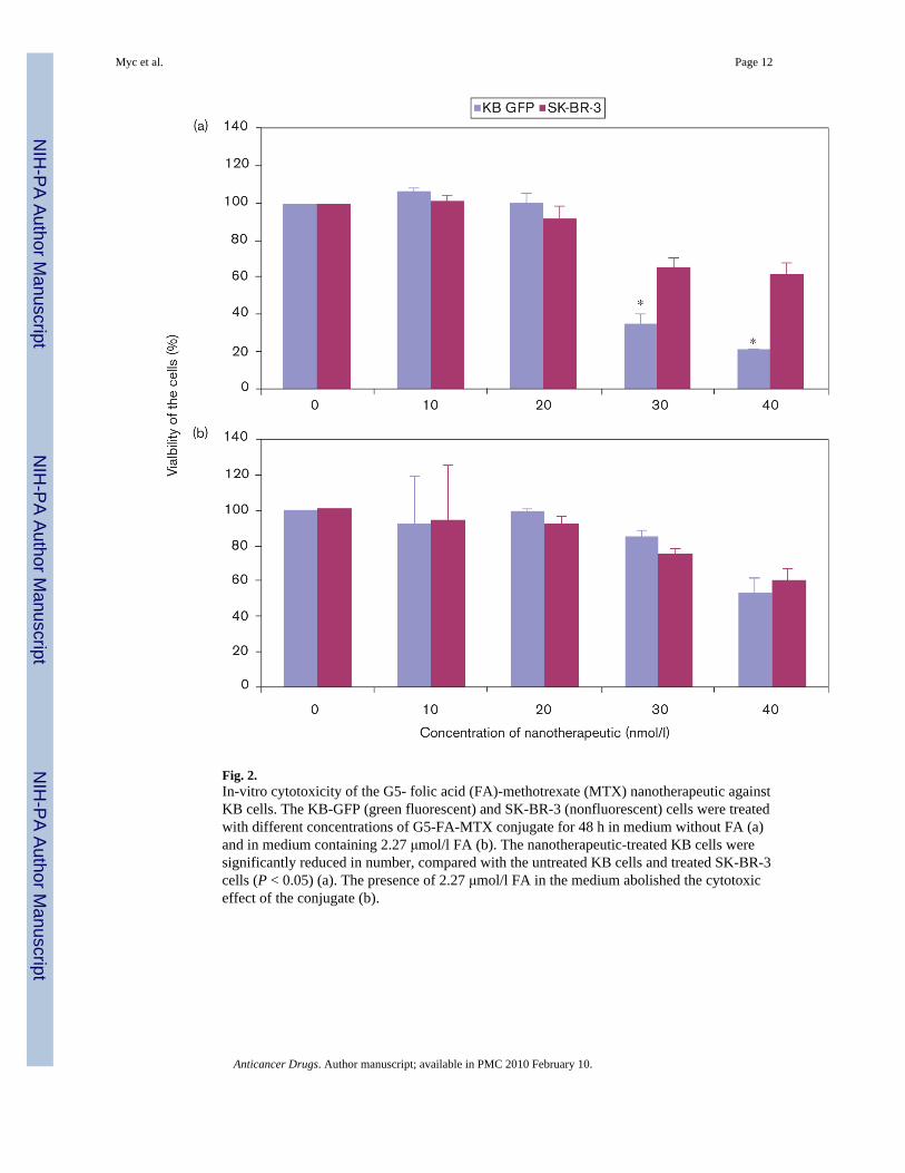
Fig. 2.In-vitro cytotoxicity of the G5- folic acid (FA)-methotrexate (MTX) nanotherapeutic againstKB cells. The KB-GFP (green fluorescent) and SK-BR-3 (nonfluorescent) cells were treatedwith different concentrations of G5-FA-MTX conjugate for 48 h in medium without FA (a)and in medium containing 2.27 μmol/l FA (b). The nanotherapeutic-treated KB cells weresignificantly reduced in number, compared with the untreated KB cells and treated SK-BR-3cells (P < 0.05) (a). The presence of 2.27 μmol/l FA in the medium abolished the cytotoxiceffect of the conjugate (b).
Myc et al. Page 12
Anticancer Drugs. Author manuscript; available in PMC 2010 February 10.
NIH
-PA Author Manuscript
NIH
-PA Author Manuscript
NIH
-PA Author Manuscript

Fig. 3.Inhibition of tumor growth in SCID mice. Mice were inoculated subcutaneously with a mixture(1:1) of KB-GFP and SK-BR-3-RFP; 106 cells of each cell line in 0.2 ml of PBS in each flank.Subcutaneous injections of equimolar concentrations (13.7 nmoles) of the nanotherapeutic(G5-folic acid (FA)-methotrexate (MTX) lot#123–34) or free MTX in 0.2 ml of saline werestarted 7 days after implantation. The mice were treated twice a week for 4 weeks. Tumorvolumes (mm3) were measured at 11 and 32 days after implantation (P < 0.04).
Myc et al. Page 13
Anticancer Drugs. Author manuscript; available in PMC 2010 February 10.
NIH
-PA Author Manuscript
NIH
-PA Author Manuscript
NIH
-PA Author Manuscript

NIH
-PA Author Manuscript
NIH
-PA Author Manuscript
NIH
-PA Author Manuscript
Myc et al. Page 14
Table 1
PCR analysis of α-FBP receptor on KB and SK-BR-3 cells
Cell lines Fold change of FOLR1 (α-FBP) versus β-actin
KB 23.6
KB GFP 63.5
SK-BR-3 RFP 0.1
GRF, green flourescent protein; RFF, red flourescent protein.
Anticancer Drugs. Author manuscript; available in PMC 2010 February 10.

NIH
-PA Author Manuscript
NIH
-PA Author Manuscript
NIH
-PA Author Manuscript
Myc et al. Page 15
Tabl
e 2
Num
ber o
f KB
(gre
en) a
nd S
K-B
R-3
(red
) cel
ls h
arve
sted
from
tum
or o
f con
trol a
nim
als a
nd a
nim
als t
reat
ed w
ith M
TX o
r nan
othe
rape
utic
a
KB
GFP
SK-B
R-3
RFP
NA
vera
geSD
Ave
rage
SD
Salin
e6
1748
377.
722
212
7.1
MTX
615
9062
6.6
320
137.
7
G5-
FA-M
TX6
1273
*29
1.9
217
44.1
MTX
, met
hotre
xate
; SD
, sta
ndar
d de
viat
ion.
a Cel
ls w
ere
rele
ased
from
tum
or a
s des
crib
ed in
Mat
eria
l and
met
hods
and
fixe
d. T
hen
10 0
00 c
ells
from
eac
h tu
mor
sam
ple
wer
e an
alyz
ed o
n flo
w c
ytom
etry
.
* P <
0.04
.
Anticancer Drugs. Author manuscript; available in PMC 2010 February 10.