Asp49 phospholipase A 2–elaidoylamide complex: a new mode of inhibition
1
2
3
4 Q1
5
67
9
1011121314
1516171819202122
2 3
43
Neurochemistry International xxx (2012) xxx–xxx
NCI 3186 No. of Pages 10, Model 5G
27 June 2012
Contents lists available at SciVerse ScienceDirect
Neurochemistry International
journal homepage: www.elsevier .com/locate /nci
Differential participation of phospholipase A2 isoforms during iron-inducedretinal toxicity. Implications for age-related macular degeneration
G. Rodríguez Diez, R.M. Uranga 1, M.V. Mateos 1, N.M. Giusto, G.A. Salvador ⇑Instituto de Investigaciones Bioquímicas de Bahía Blanca, Universidad Nacional del Sur and Consejo Nacional de Investigaciones Científicas y Técnicas, 8000 Bahía Blanca, Argentina
a r t i c l e i n f o
24252627282930313233343536
Article history:Received 13 December 2011Received in revised form 6 June 2012Accepted 14 June 2012Available online xxxx
Keywords:RetinaIronPLA2
Oxidative stressAMDCOX-2
37383940
0197-0186/$ - see front matter � 2012 Published byhttp://dx.doi.org/10.1016/j.neuint.2012.06.012
Abbreviations: [14C]DPPC, 1-[14C]palmitoyl-2phosphocholine; [14C]PAPC, 1-palmitoyl-2-[14C]aracphocholine; 4-HNE, 4-hydroxynonenal; AA, arachidmacular degeneration; ATK, arachidonoyl trifluorome50-triphosphate; BEL, bromoenol lactone; BSA, bocyclooxigenase; cPLA2, cytosolic phospholipase A2;N,N0-1,2-ethandiylbis[N-(carboxymethyl)glycine] disolular signal-regulated kinases; HEPES, 4-(2-hydroxyetfonic acid; HRP, horseradish peroxidase; iPLA2, calciumA2; MAPK, mitogen-activated protein kinases; MEKkinase kinase; MTT, 3-(4,5-dimethylthiazol-2-yl)-2mide; NBIA, neuronal brain iron accumulation; PAL, pbuffer saline; PLA2, phospholipase A2; PMSF, phenylretinal degeneration; ROS, reactive oxygen species; SSDS–PAGE, sodium dodecyl sulfate–polyacrylamidthiobarbituric acid; TBARS, thiobarbituric acid reactdiamino-2,3-dicyano-1,4-bis[2-aminophenylthio]buta⇑ Corresponding author. Address: Instituto de Inv
Bahía Blanca, Centro Científico y Tecnológico CONICEidad Nacional del Sur, Edificio E1, Camino La CarrindaArgentina. Tel.: +54 291 4861201; fax: +54 291 4861
E-mail address: [email protected] (G.A. Salvad1 These authors contributed equally to this work.
Please cite this article in press as: Rodríguez DImplications for age-related macular degenerat
a b s t r a c t
Both elevated iron concentrations and the resulting oxidative stress condition are common signs in ret-inas of patients with age-related macular degeneration (AMD). The role of phospholipase A2 (PLA2) duringiron-induced retinal toxicity was investigated. To this end, isolated retinas were exposed to increasingFe2+ concentrations (25, 200 or 800 lM) or to the vehicle, and lipid peroxidation levels, mitochondrialfunction, and the activities of cytosolic PLA2 (cPLA2) and calcium-independent PLA2 (iPLA2) were studied.Incubation with Fe2+ led to a time- and concentration-dependent increase in retinal lipid peroxidationlevels whereas retinal cell viability was only affected after 60 min of oxidative injury.
A differential release of arachidonic acid (AA) and palmitic acid (PAL) catalyzed by cPLA2 and iPLA2
activities, respectively, was also observed in microsomal and cytosolic fractions obtained from retinasincubated with iron. AA release diminished as the association of cyclooxigenase-2 increased in micro-somes from retinas exposed to iron. Retinal lipid peroxidation and cell viability were also analyzed inthe presence of cPLA2 inhibitor, arachidonoyl trifluoromethyl ketone (ATK), and in the presence of iPLA2
inhibitor, bromoenol lactone (BEL). ATK decreased lipid peroxidation levels and also ERK1/2 activationwithout affecting cell viability. BEL showed the opposite effect on lipid peroxidation. Our results demon-strate that iPLA2 and cPLA2 are differentially regulated and that they selectively participate in retinal sig-naling in an experimental model resembling AMD.
� 2012 Published by Elsevier Ltd.
41
42
44
45
46
47
48
49
50
51
52
53
54
55
56
57
58
59
60
61
62
63
64
65
Elsevier Ltd.
-[14C]palmitoyl-sn-glycero-3-hidonoyl-sn-glycero-3-phos-
onic acid; AMD, age-relatedthyl ketone; ATP, adenosine-vine serum albumin; COX,
DTT, dithiothreitol; EDTA,dium salt; ERK1/2, extracel-hyl)-1-piperazine ethanesul--independent phospholipase, mitogen-activated protein,5-diphenyltetrazolium bro-almitic acid; PBS, phosphatemethylsulfonyl fluoride; RD,DS, sodium dodecyl sulfate;
e gel electrophoresis; TBA,ive substances; U0126, 1,4-diene.estigaciones Bioquímicas deT Bahía Blanca and Univers-nga km 7, 8000 Bahía Blanca,200.or).
iez, G., et al. Differential particion. Neurochem. Int. (2012), ht
1. Introduction
Metallo-neurobiology has undergone a significant evolution inthe last 20 years. Still, although there is much experimental evi-dence on various aspects of the involvement of iron in several neu-rodegenerative diseases, the role of this metal in the centralnervous system and, particularly, in neurodegenerative processeshas not been fully elucidated to date (Dunaief, 2006).
Iron is necessary for normal retinal cellular function. Still, ironoverload has been found to be associated with retinal degenerativedisorders such as ocular siderosis, intraocular hemorrhage, and thehereditary diseases aceruloplasminemia and pantothenate kinase-associated neurodegeneration (Dunaief et al., 2005; Dunaief, 2006;Hadziahmetovic et al., 2008; Hahn et al., 2004; He et al., 2007;Wong et al., 2007). Furthermore, it is well known that reactive oxy-gen species (ROS) may contribute to the pathogenesis of age-related macular degeneration (AMD) and that they can beproduced in the Fenton reaction catalyzed by ferric (Fe3+) and fer-rous (Fe2+) ions (Lukinova et al., 2009). Previous post mortem re-search has found that iron concentrations are higher in AMDretinas than in non-affected retinas (Blasiak et al., 2011). In addi-tion, the age-related increase of iron levels in the macula, the up-regulation of transferrin in AMD, the development of AMD-like
ipation of phospholipase A2 isoforms during iron-induced retinal toxicity.tp://dx.doi.org/10.1016/j.neuint.2012.06.012
66
67
68
69
70
71
72
73
74
75
76
77
78
79
80
81
82
83
84
85
86
87
88
89
90
91
92
93
94
95
96
97
98
99
100
101
102
103
104
105
106
107
108
109
110
111
112
113
114
115
116
117
118
119
120
121
122
123
124
125
126
127
128
129
130
131
132
133
134
135
136
137
138
139
140
141
142
143
144
145
146
147
148
149
150
151
152
153
154
155
156
157
158
159
160
161
162
163
164
165
166
167
168
169
170
171
172
173
174
175
176
177
178
179
180
181
182
183
184
185
186
187
188
189
2 G. Rodríguez Diez et al. / Neurochemistry International xxx (2012) xxx–xxx
NCI 3186 No. of Pages 10, Model 5G
27 June 2012
syndromes in ceruloplasmin- and hephaestin-deficient mice, andthe association between polymorphism of iron homeostasis genesand AMD support the leading role of this metal in this pathology(Blasiak et al., 2011; Garcia-Castineiras, 2010; Hadziahmetovicet al., 2008; Wong et al., 2007). Another disease also characterizedby excessive iron accumulation is hemochromatosis. Most patientswith hereditary hemochromatosis have a mutation in the histo-compatibility leukocyte antigen class I-like protein involved in ironhomeostasis (HFE) gene product (Feder et al., 1996). This proteinforms, in general, a stable complex with the transferrin receptor,lowering its affinity for transferrin (Feder et al., 1998). Patientswith mutations in the HFE gene evidence elevated transferrin bind-ing to transferrin receptors, ending in, in turn, a higher iron uptakeinto tissues. This genetic disorder is also associated with retinalabnormalities, including, in some cases, retinal pigment epitheliumatrophy or angioid streaks. Furthermore, a study conducted in twopostmortem patients also showed drusen formation, the clinicalhallmark of AMD (Dunaief, 2006).
On the other hand, several neurodegenerative disorders withneuronal brain iron accumulation (NBIA) have been associatedwith defective phospholipase A2 (PLA2) signaling. Mutations inPLA2G6 gene, which encodes a calcium-independent group VIPLA2, have been reported in NBIA (Morgan et al., 2006). Moreover,the up-regulation of secretory phospholipase A2 IIA and its partic-ipation in neuronal apoptosis have been reported during cerebralischemia (Adibhatla and Hatcher, 2010; Yagami et al., 2002). PLA2sbelong to a superfamily of enzymes that hydrolyze the sn-2 fattyacids of membrane phospholipids. These proteins are known toplay multiple roles related to the maintenance of membranephospholipid homeostasis and production of a variety of lipidmediators (Burke and Dennis, 2009b). There are more than 20 dif-ferent types of PLA2s and, in spite of their common function inhydrolyzing phospholipid fatty acids, they are diversely encodedby a number of genes and are regulated by different mechanisms.The most recent classification involves the following main groups:(i) low molecular secretory PLA2 (sPLA2) which includes groups I–III, V and IX–XIV; (ii) high molecular calcium-dependent cytosolicPLA2 (cPLA2) which includes groups IVA–IVF; (iii) high molecularcalcium-independent PLA2 (iPLA2) which includes groups VIA-1,VIA-2 and VIB–VIF; and (iv) platelet-activating factor acetylhydro-lase which includes groups VIIA–VIIB and VIIIA–VIIIB (Burke andDennis, 2009a; Sun et al., 2010).
Although the physiological role of these PLA2s in the regulationof neuronal cell function has not yet been fully elucidated, there isincreasing evidence about their involvement in receptor signalingand transcriptional pathways that link oxidative events to inflam-matory responses underlying many neurodegenerative diseases(Sun et al., 2007). Previous research also revealed the importantrole of cPLA2, sPLA2 and iPLA2 in modulating neuronal excitatoryfunctions, in the inflammatory responses, and, as stated above, inchildhood neurological disorders associated with NBIA, respec-tively (Farooqui et al., 2006; Morgan et al., 2006; Moses et al.,2006; Svensson et al., 2005). Furthermore, the up-regulation ofmRNA of various subgroups of sPLA2 during light-induced retinaldegeneration (RD) has been reported particularly in the retina(Tanito et al., 2008; Yang et al., 2008). Moreover, the increase inarachidonic acid (AA) release, one of the main products of PLA2 ac-tion, has been suggested to be involved in the pathogenesis of RD(Kashiwagi et al., 2000; Wang and Kolko, 2010). iPLA2 expressionhas also been involved in the proliferation of retinal pigment epi-thelium cells during RD associated with AMD (Kolko et al., 2009).Evidence presented here shows a correlation between iron, RDand PLA2-derived signaling. However, the specific role of PLA2s inRD still remains unknown. Thus, taking into account this back-ground, the main goal of the present work was to characterizePLA2 activities and their regulation in an experimental model of
Please cite this article in press as: Rodríguez Diez, G., et al. Differential particImplications for age-related macular degeneration. Neurochem. Int. (2012), ht
RD. To this end, isolated bovine retinas were exposed to increasingFe2+ concentrations, and cellular viability, lipid peroxidation levelsand cPLA2 and iPLA2 activities were analyzed. The role of PLA2 iso-forms in retinal damage and the involvement of mitogen-activatedprotein kinase (MAPK) ERK1/2 in PLA2 regulation were alsostudied.
2. Materials and methods
2.1. Materials
1-Palmitoyl-2-[14C]arachidonoyl-sn-glycero-3-phosphocholine(38.0 mCi/mmol, [14C]PAPC) and 1-[14C]palmitoyl-2-[14C]palmi-toyl-sn-glycero-3-phosphocholine (111.0 mCi/mmol, [14C]DPPC)were obtained from New England Nuclear-Dupont (Boston, MA,USA). PLA2 inhibitors [arachidonoyl trifluoromethyl ketone (ATK)and bromoenol lactone (BEL)], mitogen-activated protein kinasekinase (MEK) 1/2 inhibitor [1,4-diamino-2,3-dicyano-1,4-bis[2-aminophenylthio]butadiene (U0126)], Triton X-100, 3-[4,5-dimethylthiazol-2-yl]-2,5-diphenyltetrazolium bromide (MTT),and thiobarbituric acid (TBA) were obtained from Sigma–Aldrich(St. Louis, MO, USA). All other chemicals were of the highest purityavailable. Mouse polyclonal IgG2a, anti-phospho-Tyr204-extracel-lular signal-regulated kinases (ERK) 1/2, rabbit polyclonal anti-phosphotyrosine (PY20), rabbit polyclonal anti-ERK2, rabbit poly-clonal anti-phospho-Thr180/Tyr182-p38, rabbit polyclonal anti-b-tubulin, rabbit polyclonal anti-calnexin, polyclonal horseradishperoxidase (HRP)-conjugated goat anti-rabbit IgG, and polyclonalHRP-conjugated goat anti-mouse IgG were purchased from SantaCruz Biotechnology, Inc. (Santa Cruz, CA, USA). Rabbit polyclonalanti-cyclooxygenase (COX)-2 was purchased from Cayman Chemi-cal (Ann Arbor, MI, USA). Rabbit anti-phospho-Ser505-cPLA2 waspurchased from Cell Signaling Technology (Boston, MA, USA).
2.2. Experimental treatments
Fresh bovine eyes were obtained from a local abattoir andplaced and stored in crushed ice. Retinas were dissected on ice(4 �C) under normal lighting conditions and washed with salinesolution. Entire retinas were preincubated at 37 �C for 30 min witheither inhibitors (50 lM ATK, 25 lM BEL or 10 lM U0126) or thevehicle, and they were subsequently exposed for 5 or 60 min toeither FeSO4 (25, 200 or 800 lM) or its vehicle as previously de-scribed (Uranga et al., 2007). Entire retinas were incubated underan O2:CO2 (95:5, vol/vol) atmosphere with gentle agitation for allexperiments. All incubations were performed in Locke’s buffer[154 mM NaCl, 5.6 mM KCl, 3.6 mM NaHCO3, 1 mM MgCl2,2.3 mM CaCl2, 5 mg/ml glucose, 5 mM 4-(2-hydroxyethyl)-1-piper-azine ethanesulfonic acid (HEPES), pH 7.2] unless stated otherwise.After incubation, retinas were washed in Locke’s buffer to be fur-ther used for experimental procedures.
2.3. Isolation of subcellular fractions
Subcellular fractions were obtained as previously describedwith slight modifications (Salvador and Giusto, 2006). Briefly,homogenates (20% wt/vol) from the dissected retinas to be usedfor subcellular fractionation were prepared in a medium contain-ing 0.32 M sucrose, 1 mM N,N0-1,2-ethandiylbis[N-(carboxy-methyl)glycine] disodium salt (EDTA), 1 mM dithiothreitol (DTT),2 mg/ml leupeptin, 1 mg/ml aprotinin, 1 mg/ml pepstatin,0.1 mM phenylmethylsulfonyl fluoride (PMSF), and 10 mM HEPES(pH 7.4). Homogenates to be used for MTT reduction, thiobarbitu-ric acid reactive substances (TBARS) and Western blot assays, andradical scavenging measurement were prepared in Locke’s buffer.
ipation of phospholipase A2 isoforms during iron-induced retinal toxicity.tp://dx.doi.org/10.1016/j.neuint.2012.06.012
190
191
192
193
194
195
196
197
198
199
200
201
202
203
204
205
206
207
208
209
210
211
212
213
214
215
216
217
218
219
220
221
222
223
224
225
226
227
228
229
230
231
232
233
234
235
236
237
238
239
240
241
242
243
244
245
246
247
248
249
250
251
252
253
254
255
256
257
258
259
260
G. Rodríguez Diez et al. / Neurochemistry International xxx (2012) xxx–xxx 3
NCI 3186 No. of Pages 10, Model 5G
27 June 2012
Independently of the buffer used, retinas were homogenized by 10strokes with a Thomas tissue homogenizer. Homogenates werecentrifuged at 1800�g for 7.5 min at 4 �C using a JA-21 rotor in aBeckman J2-21 centrifuge. The pellet (corresponding to crude nu-clear fraction and cellular debris) was discarded and the superna-tant was retained and centrifuged at 14,000�g for 20 min at 4 �C.The mitochondrial pellet was stored at�80 �C, and the supernatantwas centrifuged at 85,500�g for 1 h at 4 �C using a SW28.1 rotor ina Beckman Optima LK-90 ultracentrifuge. The final pellet was con-sidered as microsomal fraction and the supernatant as cytosolicfraction. Total homogenates and microsomal and cytosolic frac-tions were used for the experiments detailed below. Protein con-tent of retinal subcellular fractions was determined followingLowry (Lowry et al., 1951).
261
262
263
264
265
266
267
268
269
270
271
272
273
274
275
276
277
278
279
280
281
282
283
284
2.4. PLA2 activity assays
To determine cPLA2 activity, sn-2 fatty acid hydrolysis wasdetermined using lipid vesicles containing [14C]PAPC and cold 1-palmitoyl-2-oleoyl-sn-glycero-3-phosphocholine to yield60,000 dpm (0.100 mM) per assay in a buffer containing 0.025%Triton X-100, 0.16 mM CaCl2, 0.2 mg/ml bovine serum albumin(BSA), 4 mM DTT, 100 mM HEPES, pH 7.5 (Svensson et al., 2005).In order to analyze the involvement of phospholipase A1/lysophos-pholipase A2 activity in AA generation, lysophospholipid formationfrom [14C]PAPC was measured under the same experimental condi-tions as those used for cPLA2 activity. These assays were performedeither in the presence or in the absence of 2 mM PMSF (as lyso-phospholipase A2 inhibitor) (Pete et al., 1996).
To determine iPLA2 activity, sn-2 fatty acid hydrolysis wasdetermined using lipid vesicles containing [14C]DPPC and coldDPPC to yield 60,000 dpm (0.100 mM) per assay in a buffer con-taining 0.025% Triton X-100, 10 mM EDTA, 2 mM adenosine-50-tri-phosphate (ATP), 100 mM HEPES, pH 7.5 (Svensson et al., 2005).Seventy-five microlitres of the corresponding lipid vesicles wereadded to 75 ll of treated retinal subcellular fractions (150 lg ofprotein, final volume 150 ll).
All reactions were carried out at 37 �C for 20 min, with gentleagitation, and stopped by the addition of 5 ml of chloroform/meth-anol (2:1, vol/vol).
285
286
287
288
289
290
291
292
293
294
295
296
297
298
299
300
301
302
303
304
305
306
2.5. Extraction and isolation of lipids
Lipids were extracted according to Folch et al. (1957) the lipidextract was subsequently washed with 0.2 volumes of 0.05% CaCl2
and the lowest phase was obtained after centrifugation at 900�gfor 5 min. Neutral lipids monoacylglycerol, diacylglycerol, and freefatty acids were then separated by one-dimensional thin-layerchromatography using silica gel G plates (Merck) in a mobile phaseconsisting of hexane/diethyl ether/acetic acid (50:50:2.6, vol/vol).DPPC and PAPC as well as the remaining retinal phospholipidswere retained at the spotting site. Lipids were visualized by expo-sure of the plate to iodine vapors. The spots corresponding to freefatty acids were scraped off the plate and quantified by liquidscintillation.
Lysophospholipids were separated from phospholipids andneutral lipids by one-dimensional thin-layer chromatographyusing silica gel H plates (Merck) in a mobile phase consisting ofchloroform/methanol/ammonia (65:25:5, vol/vol). Lipids werevisualized by exposure of the plate to iodine vapors. The spots cor-responding to lysophospholipids and phospholipids were scrapedoff the plate and quantified by liquid scintillation.
Please cite this article in press as: Rodríguez Diez, G., et al. Differential particImplications for age-related macular degeneration. Neurochem. Int. (2012), ht
2.6. MTT reduction assay
To determine retinal reducing activity, the extent of MTT reduc-tion to insoluble intracellular formazan crystals was measured.This reduction depends on the activity of intracellular dehydrogen-ases and is independent of the changes in the plasma membraneintegrity. MTT reduction was measured in total homogenates ob-tained from entire retinas exposed to either 25, 200 and 800 lMFeSO4 or to the vehicle, and in the presence or absence of the cor-responding inhibitors. The method used in the present study hasbeen previously described (Uranga et al., 2007). Briefly, MTT wasdissolved in phosphate buffer saline (PBS) at a concentration of5 mg/ml. MTT solution was mixed with total homogenate (1:10;MTT:homogenate, vol/vol) and allowed to incubate for 2 h at37 �C with gentle agitation. At the end of incubation with MTT,300 ll of solubilization buffer [20% sodium dodecyl sulfate (SDS),pH 4.7] were added and mixed thoroughly to dissolve formazancrystals. The extent of MTT reduction was then measured spectro-photometrically at 570 nm with the absorbance at 650 nm sub-tracted to account for cellular debris. Results were expressed asfinal optical density values.
2.7. COX-2 activity assays
To determine COX-2 activity, the generation of prostaglandins Fand E (PGF2 and PGE2) was measured by using lipid vesicles con-taining [14C]arachidonic acid to yield 60,000 dpm (0.100 mM) perassay in a buffer containing 0.025% Triton X-100, 0.16 mM CaCl2,0.2 mg/ml BSA, 4 mM DTT, 100 mM HEPES, pH 7.5. Reactions werecarried out at 37 �C for 20 min, with gentle agitation, and stoppedby the addition of 5 ml of chloroform/methanol (2:1, vol/vol). Pros-taglandins were then separated as described by Franchi et al.(2000), by one-dimensional thin-layer chromatography using silicagel G plates (Merck) in a mobile phase consisting of bencene/diox-ane/acetic acid (60:30:3, vol/vol) and purified standards of PGE2
and PGF2. PGE2 and PGF2 were visualized by exposure of the plateto iodine vapors. The corresponding spots were scraped off theplate and quantified by liquid scintillation.
2.8. Lipid peroxidation assay
Lipid peroxidation was measured using the TBA assay as previ-ously described (Mateos et al., 2008). Briefly, after incubation inthe presence of either Fe2+ or the vehicle, and in the presence or ab-sence of the corresponding inhibitors, 1 ml of 30% TCA was addedto 0.5 ml of total homogenate (1.5 mg protein/ml). Then, 0.1 ml of5 N HCl and 1 ml of 0.75% TBA were added. Tubes were capped, themixtures were heated at 100 �C for 15 min in a boiling water bathand samples were centrifuged at 1000�g for 10 min. TBARS weremeasured in the supernatant at 535 nm. Results were expressedas units of absorbance at 535 nm per mg of protein [Abs535 nm (arbitrary units)/mg protein].
2.9. SDS–PAGE and Western blot assays
Samples from total homogenates and microsomal and cytosolicfractions were denatured with Laemmli sample buffer at 100 �C for5 min (Laemmli, 1970). Equivalent amounts of proteins were sep-arated by SDS–polyacrylamide gel electrophoresis (SDS–PAGE) on10% polyacrylamide gels and then transferred to a polyvinylidenefluoride membrane (Millipore, Bedford, MA) using a Mini Trans-Blot cell electroblotter (BIO-RAD Life Science Group, California)for 2 h. Membranes were blocked with 5% nonfat dry milk in TTBSbuffer [20 mM Tris–HCl, pH 7.5, 100 mM NaCl, and 0.1% (wt/vol)Tween 20] for 1 h at room temperature. Membranes were thenincubated with the following primary antibodies: (i) anti-COX-2
ipation of phospholipase A2 isoforms during iron-induced retinal toxicity.tp://dx.doi.org/10.1016/j.neuint.2012.06.012
307
308
309
310
311
312
313
314
315
316
317
318
319
320
321
322
323
324
325
326
327
328
329
330
331
332
333
334
335
336
337
338
339
340
341
342
343
344
345
346
347
348
349
350
351
352
353
354
355
356
357
358
359
360
361
362
363
364
365
366
367
368
369
370
371
372
373
4 G. Rodríguez Diez et al. / Neurochemistry International xxx (2012) xxx–xxx
NCI 3186 No. of Pages 10, Model 5G
27 June 2012
(1:1000) or anti-phospho-p38 (1:1000) overnight at 4 �C, or (ii)anti-phospho-ERK1/2 (1:1000), anti-b-tubulin (1:1000), anti-ERK2 (1:2000) and anti-phosphotyrosine PY20 (1:750) for 2 h atroom temperature. After incubation membranes were, washedthree times with TTBS, and then exposed to the appropriate HRP-conjugated secondary antibody (anti-rabbit or anti-mouse) for1 h at room temperature. Membranes were again washed threetimes with TTBS. Immunoreactive bands were detected by en-hanced chemiluminescence (ECL; Amersham Biosciences, USA)using standard X-ray film (ECL, Amersham Biosciences, USA).Immunoreactive bands were quantified using image analysis soft-ware (Image J, a freely available application in the public domainfor image analysis and processing, developed and maintained byWayne Rasband at the Research Services Branch, National Instituteof Mental Health).
2.10. Statistical analysis
Statistical analysis was performed using one-way ANOVA testto compare means and followed by Fisher’s least significant differ-ence (LSD) test. p-Values lower than 0.05 were considered statisti-cally significant.
3. Results
3.1. Determination of cellular damage during iron-induced retinaltoxicity
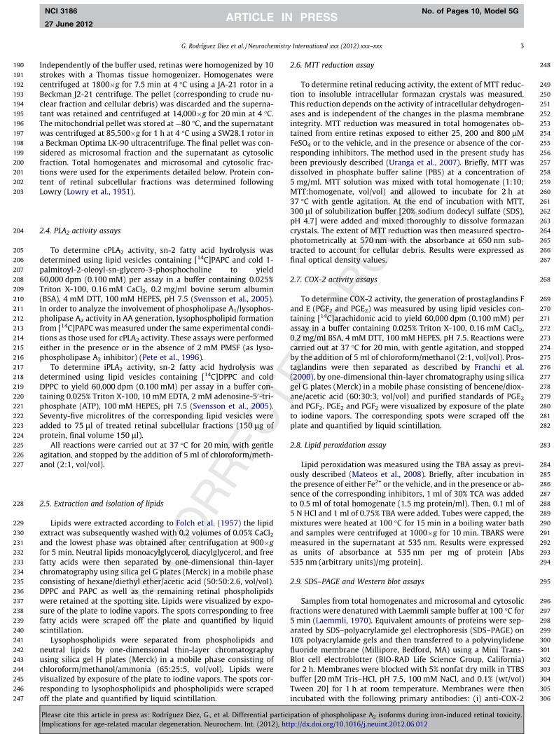
Our first goal was the characterization of iron-induced retinaldamage by determining lipid peroxidation levels and cell viability.To this end, entire isolated retinas were incubated in the presenceof variable concentrations of FeSO4 (25, 200 and 800 lM) for differ-ent periods of time (5 and 60 min). Controls were also assessed byreplacing Fe2+ by an equal volume of water (vehicle). The genera-tion of malondialdehyde, a marker of lipid peroxidation, was ana-lyzed by measuring TBARS. As shown in Fig. 1A and B, TBARSgeneration was found to be increased in a Fe2+ concentration-and time-dependent manner. The increase in lipid peroxidation
Fig. 1. Characterization of Fe2+-induced damage in the retina. (A and B) Lipid peroxidation5 min (A) and 60 min (B). (C and D) Measurement of MTT reduction after the same incubcontrol and represent the mean ± SD of at least three independent experiments. ⁄Significtest followed by LSD test).
Please cite this article in press as: Rodríguez Diez, G., et al. Differential particImplications for age-related macular degeneration. Neurochem. Int. (2012), ht
products could be immediately observed. After a 5 min exposureto the metal, TBARS increased by 0.6, 2.2 and 3.6 times with re-spect to controls at 25, 200 and 800 lM Fe2+, respectively(Fig. 1A). Maximal malondialdehyde generation was observed after60 min of Fe2+ exposure (Fig. 1B). Under these experimental condi-tions, TBARS levels were 1.8, 6.5, and 9 times higher than controlsat 25, 200 and 800 lM Fe2+, respectively.
Retinal cell viability was analyzed by measuring MTT reductionunder the same above-mentioned experimental conditions. After5 min of incubation with 25, 200 and 800 lM Fe2+ it showed nochanges (Fig. 1C) while it was observed to be affected at 200 and800 lM Fe2+ after 60 min of incubation (Fig. 1D).
3.2. MAPK activation and tyrosine phosphorylation are triggeredduring iron-induced retinal toxicity
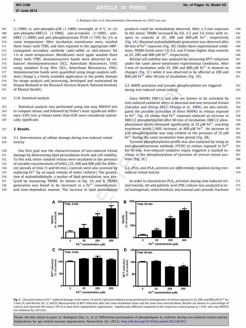
Since MAPKs ERK1/2 and p38 are known to be activated byiron-induced oxidative stress in neuronal and non-neuronal tissues(Salvador and Oteiza, 2011; Uranga et al., 2009), we also investi-gated the possible activation of these MAPKs in retinas exposedto Fe2+. Fig. 2A shows that Fe2+ exposure induced an increase inERK1/2 phosphorylation after 60 min of incubation. ERK1/2 phos-phorylation levels increased significantly at 25 lM Fe2+, reachingmaximum levels (160% increase) at 800 lM Fe2+. An increase inp38 phosphorylation was only evident in the presence of 25 lMFe2+ during the same incubation time period (Fig. 2B).
Tyrosine phosphorylation profile was also analyzed by using ananti-phosphotyrosine antibody (PY20) in retinas exposed to Fe2+
for 60 min. Iron-induced oxidative injury triggered a marked in-crease in the phosphorylation of tyrosines of several retinal pro-teins (Fig. 2C).
3.3. cPLA2 and iPLA2 activities are differentially regulated during iron-induced retinal toxicity
In order to characterize PLA2 activities during iron-induced ret-inal toxicity, AA and palmitic acid (PAL) release was analyzed in to-tal homogenate, mitochondrial, microsomal and cytosolic fractions
assay performed in homogenates of retinas exposed to 25, 200, and 800 lM Fe2+ foration times and the same iron concentrations. Results are shown as a percentage ofantly different compared to the respective control group (p < 0.05, one-way ANOVA
ipation of phospholipase A2 isoforms during iron-induced retinal toxicity.tp://dx.doi.org/10.1016/j.neuint.2012.06.012
374
375
376
377
378
379
380
381
382
383
384
385
386
387
388
389
390
391
392
393
394
395
396
397
398
399
400
401
402
403
404
405
406
407
408
409
410
411
412
413
414
415
416
417
418
419
420
421
422
423
424
425
426
427
428
429
430
431
432
433
Fig. 2. Effect of Fe2+ exposure on MAPK and tyrosine phosphorylation. (A) Western blot analysis of ERK1/2 phosphorylation performed in homogenates of retinas (30 lgprotein per lane) exposed to 25, 200, and 800 lM Fe2+ for 60 min. (B) p38 Phosphorylation assessed under the same experimental conditions as those described in (A). (C)Tyrosine phosphorylation pattern analyzed by Western blot under the same conditions as those described in (A and B). One representative Western blot image (for PY20) ofthree different experiments is shown. Bands of proteins were quantified using scanning densitometry, and phospho-ERK1/2 levels were normalized to ERK2 levels andphospho-p38 levels were normalized to b-tubulin levels. Results are shown as a percentage of the corresponding control conditions and represent the mean ± SD of at leastthree independent experiments. ⁄Significantly different compared to the respective control group (p < 0.05, one-way ANOVA test followed by LSD test).
G. Rodríguez Diez et al. / Neurochemistry International xxx (2012) xxx–xxx 5
NCI 3186 No. of Pages 10, Model 5G
27 June 2012
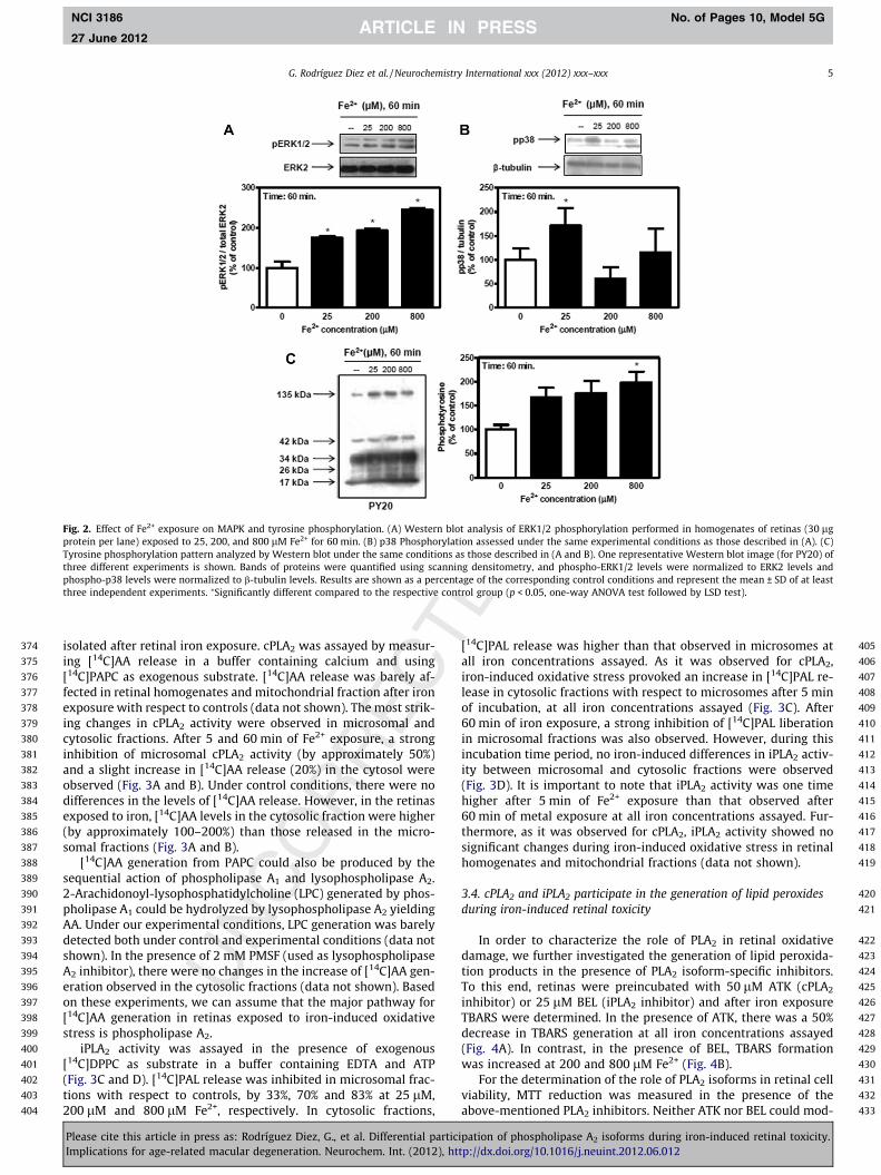
isolated after retinal iron exposure. cPLA2 was assayed by measur-ing [14C]AA release in a buffer containing calcium and using[14C]PAPC as exogenous substrate. [14C]AA release was barely af-fected in retinal homogenates and mitochondrial fraction after ironexposure with respect to controls (data not shown). The most strik-ing changes in cPLA2 activity were observed in microsomal andcytosolic fractions. After 5 and 60 min of Fe2+ exposure, a stronginhibition of microsomal cPLA2 activity (by approximately 50%)and a slight increase in [14C]AA release (20%) in the cytosol wereobserved (Fig. 3A and B). Under control conditions, there were nodifferences in the levels of [14C]AA release. However, in the retinasexposed to iron, [14C]AA levels in the cytosolic fraction were higher(by approximately 100–200%) than those released in the micro-somal fractions (Fig. 3A and B).
[14C]AA generation from PAPC could also be produced by thesequential action of phospholipase A1 and lysophospholipase A2.2-Arachidonoyl-lysophosphatidylcholine (LPC) generated by phos-pholipase A1 could be hydrolyzed by lysophospholipase A2 yieldingAA. Under our experimental conditions, LPC generation was barelydetected both under control and experimental conditions (data notshown). In the presence of 2 mM PMSF (used as lysophospholipaseA2 inhibitor), there were no changes in the increase of [14C]AA gen-eration observed in the cytosolic fractions (data not shown). Basedon these experiments, we can assume that the major pathway for[14C]AA generation in retinas exposed to iron-induced oxidativestress is phospholipase A2.
iPLA2 activity was assayed in the presence of exogenous[14C]DPPC as substrate in a buffer containing EDTA and ATP(Fig. 3C and D). [14C]PAL release was inhibited in microsomal frac-tions with respect to controls, by 33%, 70% and 83% at 25 lM,200 lM and 800 lM Fe2+, respectively. In cytosolic fractions,
Please cite this article in press as: Rodríguez Diez, G., et al. Differential particImplications for age-related macular degeneration. Neurochem. Int. (2012), ht
[14C]PAL release was higher than that observed in microsomes atall iron concentrations assayed. As it was observed for cPLA2,iron-induced oxidative stress provoked an increase in [14C]PAL re-lease in cytosolic fractions with respect to microsomes after 5 minof incubation, at all iron concentrations assayed (Fig. 3C). After60 min of iron exposure, a strong inhibition of [14C]PAL liberationin microsomal fractions was also observed. However, during thisincubation time period, no iron-induced differences in iPLA2 activ-ity between microsomal and cytosolic fractions were observed(Fig. 3D). It is important to note that iPLA2 activity was one timehigher after 5 min of Fe2+ exposure than that observed after60 min of metal exposure at all iron concentrations assayed. Fur-thermore, as it was observed for cPLA2, iPLA2 activity showed nosignificant changes during iron-induced oxidative stress in retinalhomogenates and mitochondrial fractions (data not shown).
3.4. cPLA2 and iPLA2 participate in the generation of lipid peroxidesduring iron-induced retinal toxicity
In order to characterize the role of PLA2 in retinal oxidativedamage, we further investigated the generation of lipid peroxida-tion products in the presence of PLA2 isoform-specific inhibitors.To this end, retinas were preincubated with 50 lM ATK (cPLA2
inhibitor) or 25 lM BEL (iPLA2 inhibitor) and after iron exposureTBARS were determined. In the presence of ATK, there was a 50%decrease in TBARS generation at all iron concentrations assayed(Fig. 4A). In contrast, in the presence of BEL, TBARS formationwas increased at 200 and 800 lM Fe2+ (Fig. 4B).
For the determination of the role of PLA2 isoforms in retinal cellviability, MTT reduction was measured in the presence of theabove-mentioned PLA2 inhibitors. Neither ATK nor BEL could mod-
ipation of phospholipase A2 isoforms during iron-induced retinal toxicity.tp://dx.doi.org/10.1016/j.neuint.2012.06.012
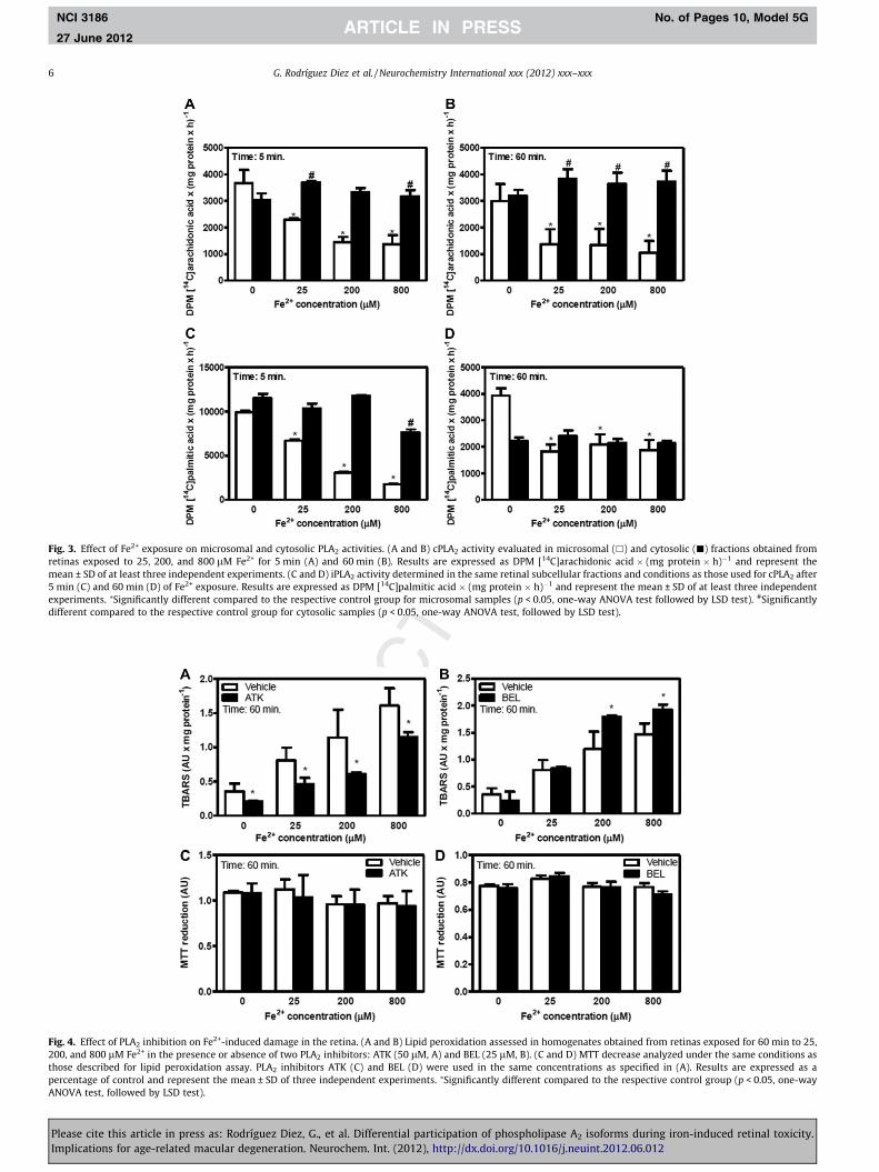
Fig. 3. Effect of Fe2+ exposure on microsomal and cytosolic PLA2 activities. (A and B) cPLA2 activity evaluated in microsomal (h) and cytosolic (j) fractions obtained fromretinas exposed to 25, 200, and 800 lM Fe2+ for 5 min (A) and 60 min (B). Results are expressed as DPM [14C]arachidonic acid � (mg protein � h)�1 and represent themean ± SD of at least three independent experiments. (C and D) iPLA2 activity determined in the same retinal subcellular fractions and conditions as those used for cPLA2 after5 min (C) and 60 min (D) of Fe2+ exposure. Results are expressed as DPM [14C]palmitic acid � (mg protein � h)�1 and represent the mean ± SD of at least three independentexperiments. ⁄Significantly different compared to the respective control group for microsomal samples (p < 0.05, one-way ANOVA test followed by LSD test). #Significantlydifferent compared to the respective control group for cytosolic samples (p < 0.05, one-way ANOVA test, followed by LSD test).
Fig. 4. Effect of PLA2 inhibition on Fe2+-induced damage in the retina. (A and B) Lipid peroxidation assessed in homogenates obtained from retinas exposed for 60 min to 25,200, and 800 lM Fe2+ in the presence or absence of two PLA2 inhibitors: ATK (50 lM, A) and BEL (25 lM, B). (C and D) MTT decrease analyzed under the same conditions asthose described for lipid peroxidation assay. PLA2 inhibitors ATK (C) and BEL (D) were used in the same concentrations as specified in (A). Results are expressed as apercentage of control and represent the mean ± SD of three independent experiments. ⁄Significantly different compared to the respective control group (p < 0.05, one-wayANOVA test, followed by LSD test).
6 G. Rodríguez Diez et al. / Neurochemistry International xxx (2012) xxx–xxx
NCI 3186 No. of Pages 10, Model 5G
27 June 2012
Please cite this article in press as: Rodríguez Diez, G., et al. Differential participation of phospholipase A2 isoforms during iron-induced retinal toxicity.Implications for age-related macular degeneration. Neurochem. Int. (2012), http://dx.doi.org/10.1016/j.neuint.2012.06.012

434
435
436
437
438
439
440
441
442
443
444
445
446
447
448
449
450
451
452
453
454
455
456
457
458
459
460
461
462
463
464
465
466
467
468
469
470
471
472
473
474
475
476
477
478
479
480
481
Fig. 5. Crosstalk between PLA2 and ERK1/2 under Fe2+-induced oxidative stress. (A) Western blot analysis of cPLA2 phosphorylation in microsomal and cytosolic fractionsobtained from retinas (30 lg protein per lane) exposed to 25, 200, and 800 lM Fe2+ for 60 min. Calnexin was used as loading control. One representative Western blot imageof three different experiments is shown. Western blot analysis of ERK1/2 phosphorylation in homogenates of retinas (30 lg protein per lane) preincubated with either U0126(10 lM, B) or ATK (50 lM, D) and then exposed to 25, 200, and 800 lM Fe2+ for 60 min. One representative Western blot image of three different experiments is shown. Bandsof proteins were quantified using scanning densitometry, and phospho-ERK1/2 levels were normalized to total ERK2 levels. Results are shown as a percentage of thecorresponding control condition and represent the mean ± SD of at least three independent experiments. ⁄Significantly different compared to the respective control group(p < 0.05, one-way ANOVA test followed by LSD test). (C) cPLA2 activity analyzed in microsomal and cytosolic fractions obtained from retinas preincubated with 10 lM U0126or the vehicle, and then exposed to 200 lM Fe2+ for 60 min and in the presence or absence of U0126. Results are expressed as DPM [14C]arachidonic acid � (mg protein � h)�1
and represent the mean ± SD of at least three independent experiments. ⁄Significantly different compared to the respective control group (p < 0.05, one-way ANOVA test,followed by LSD test).
G. Rodríguez Diez et al. / Neurochemistry International xxx (2012) xxx–xxx 7
NCI 3186 No. of Pages 10, Model 5G
27 June 2012
ify retinal cell viability under all experimental conditions assayed(Fig. 4C and D).
3.5. Cross-talk between cPLA2 and ERK1/2 during iron-induced retinaltoxicity
One of the most common mechanisms of cPLA2 activation is itsphosphorylation by ERK1/2 (Chakraborti, 2003; Ghosh et al., 2006;Leslie, 1997; Mariggio et al., 2006; Nicotra et al., 2005; Zhu et al.,2001). cPLA2 phosphorylation in Ser 505 was inhibited in themicrosomal fractions from retinas exposed to iron. In contrast,cPLA2 phosphorylation was increased in the cytosolic fractions ob-tained from retinas exposed to iron (Fig. 5A). In order to establish acorrelation between the activation of ERK1/2 and the differentialrelease of AA during iron-induced oxidative stress, cPLA2 activitywas analyzed in retinas preincubated with MAPK inhibitorU0126. In the presence of U0126, ERK1/2 phosphorylation wasinhibited at all iron concentrations assayed (Fig. 5B). Under theseexperimental conditions, the inhibition of AA release in micro-somal fractions and the increase of AA release in cytosolic fractionsprovoked by iron exposure (200 lM) were completely abolished(Fig. 5C).
We also checked the state of ERK1/2 phosphorylation in thepresence of cPLA2 inhibitor. ATK was found to inhibit the increasein ERK1/2 phosphorylation previously observed at 25 and 200 lMFe2+ (Fig. 5D). At 800 lM Fe2+, ERK1/2 activation underwent nochanges in the presence of ATK.
Please cite this article in press as: Rodríguez Diez, G., et al. Differential particImplications for age-related macular degeneration. Neurochem. Int. (2012), ht
3.6. COX-2 expression and activity increase during iron-inducedretinal toxicity
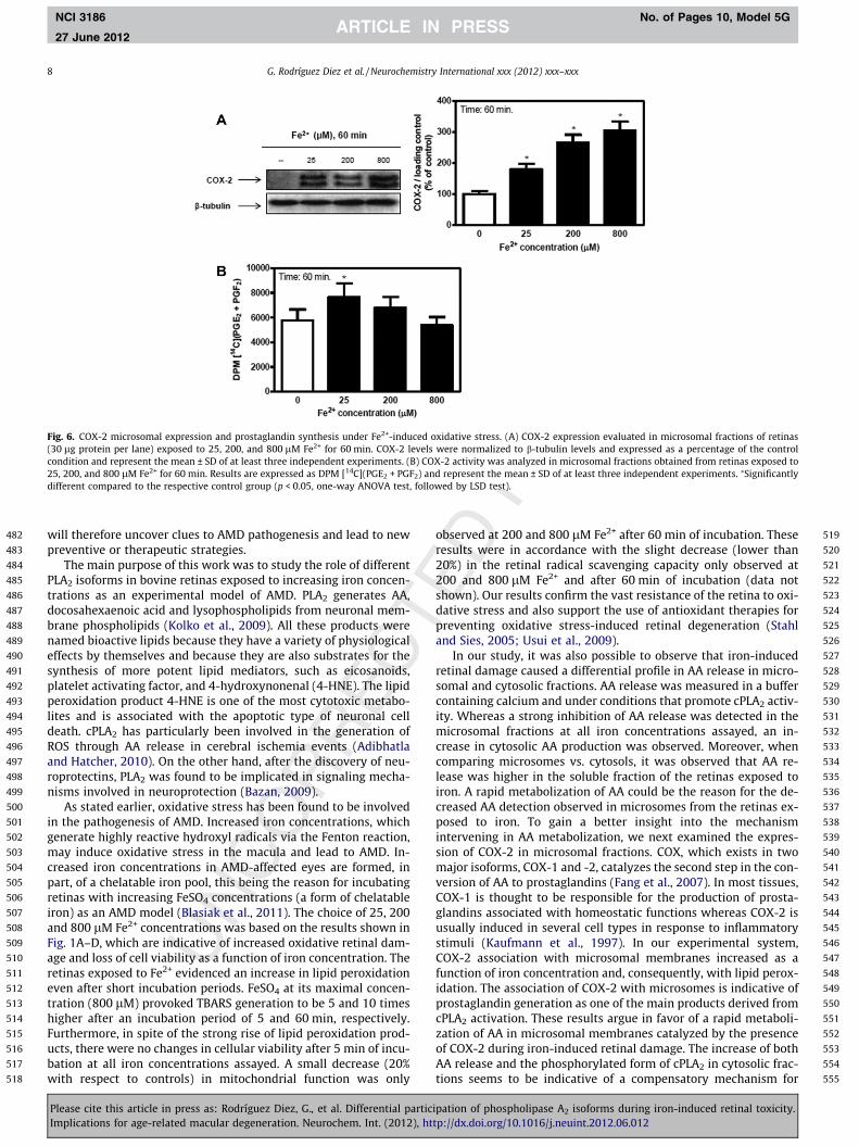
COX-2 and cPLA2 are involved in eicosanoid production andthey also seem to participate in several pathological processes suchas the progression of several types of cancer and oxidative stressevents (Linkous et al., 2010). We therefore further studied if iron-induced retinal toxicity could affect COX-2 expression. COX-2 asso-ciation with microsomal membranes augmented as a function ofiron concentration, this increase being 80%, 170% and 210% higherthan that in controls (Fig. 6A). The generation of PGF2 and PGE2, themain products of COX-2 activity, was increased in retinas exposedto 25 lM Fe2+ (Fig. 6B).
4. Discussion
Retinal damage is a major concern in relation to a number ofocular diseases leading to blindness. AMD, one of these devastatingconditions, is characterized by the deterioration of the maculawhich severely impairs central vision. Based on observationsshowing iron overload in retinas of AMD patients compared tohealthy subjects, excessive retinal iron content and the resultingoxidative stress condition have been proposed to contribute to thispathology (Dunaief, 2006). A poorly studied issue is the connectionbetween iron-induced oxidative stress, retinal degeneration andthe associated signaling events. Further studies in this direction
ipation of phospholipase A2 isoforms during iron-induced retinal toxicity.tp://dx.doi.org/10.1016/j.neuint.2012.06.012
482
483
484
485
486
487
488
489
490
491
492
493
494
495
496
497
498
499
500
501
502
503
504
505
506
507
508
509
510
511
512
513
514
515
516
517
518
519
520
521
522
523
524
525
526
527
528
529
530
531
532
533
534
535
536
537
538
539
540
541
542
543
544
545
546
547
548
549
550
551
552
553
554
555
Fig. 6. COX-2 microsomal expression and prostaglandin synthesis under Fe2+-induced oxidative stress. (A) COX-2 expression evaluated in microsomal fractions of retinas(30 lg protein per lane) exposed to 25, 200, and 800 lM Fe2+ for 60 min. COX-2 levels were normalized to b-tubulin levels and expressed as a percentage of the controlcondition and represent the mean ± SD of at least three independent experiments. (B) COX-2 activity was analyzed in microsomal fractions obtained from retinas exposed to25, 200, and 800 lM Fe2+ for 60 min. Results are expressed as DPM [14C](PGE2 + PGF2) and represent the mean ± SD of at least three independent experiments. ⁄Significantlydifferent compared to the respective control group (p < 0.05, one-way ANOVA test, followed by LSD test).
8 G. Rodríguez Diez et al. / Neurochemistry International xxx (2012) xxx–xxx
NCI 3186 No. of Pages 10, Model 5G
27 June 2012
will therefore uncover clues to AMD pathogenesis and lead to newpreventive or therapeutic strategies.
The main purpose of this work was to study the role of differentPLA2 isoforms in bovine retinas exposed to increasing iron concen-trations as an experimental model of AMD. PLA2 generates AA,docosahexaenoic acid and lysophospholipids from neuronal mem-brane phospholipids (Kolko et al., 2009). All these products werenamed bioactive lipids because they have a variety of physiologicaleffects by themselves and because they are also substrates for thesynthesis of more potent lipid mediators, such as eicosanoids,platelet activating factor, and 4-hydroxynonenal (4-HNE). The lipidperoxidation product 4-HNE is one of the most cytotoxic metabo-lites and is associated with the apoptotic type of neuronal celldeath. cPLA2 has particularly been involved in the generation ofROS through AA release in cerebral ischemia events (Adibhatlaand Hatcher, 2010). On the other hand, after the discovery of neu-roprotectins, PLA2 was found to be implicated in signaling mecha-nisms involved in neuroprotection (Bazan, 2009).
As stated earlier, oxidative stress has been found to be involvedin the pathogenesis of AMD. Increased iron concentrations, whichgenerate highly reactive hydroxyl radicals via the Fenton reaction,may induce oxidative stress in the macula and lead to AMD. In-creased iron concentrations in AMD-affected eyes are formed, inpart, of a chelatable iron pool, this being the reason for incubatingretinas with increasing FeSO4 concentrations (a form of chelatableiron) as an AMD model (Blasiak et al., 2011). The choice of 25, 200and 800 lM Fe2+ concentrations was based on the results shown inFig. 1A–D, which are indicative of increased oxidative retinal dam-age and loss of cell viability as a function of iron concentration. Theretinas exposed to Fe2+ evidenced an increase in lipid peroxidationeven after short incubation periods. FeSO4 at its maximal concen-tration (800 lM) provoked TBARS generation to be 5 and 10 timeshigher after an incubation period of 5 and 60 min, respectively.Furthermore, in spite of the strong rise of lipid peroxidation prod-ucts, there were no changes in cellular viability after 5 min of incu-bation at all iron concentrations assayed. A small decrease (20%with respect to controls) in mitochondrial function was only
Please cite this article in press as: Rodríguez Diez, G., et al. Differential particImplications for age-related macular degeneration. Neurochem. Int. (2012), ht
observed at 200 and 800 lM Fe2+ after 60 min of incubation. Theseresults were in accordance with the slight decrease (lower than20%) in the retinal radical scavenging capacity only observed at200 and 800 lM Fe2+ and after 60 min of incubation (data notshown). Our results confirm the vast resistance of the retina to oxi-dative stress and also support the use of antioxidant therapies forpreventing oxidative stress-induced retinal degeneration (Stahland Sies, 2005; Usui et al., 2009).
In our study, it was also possible to observe that iron-inducedretinal damage caused a differential profile in AA release in micro-somal and cytosolic fractions. AA release was measured in a buffercontaining calcium and under conditions that promote cPLA2 activ-ity. Whereas a strong inhibition of AA release was detected in themicrosomal fractions at all iron concentrations assayed, an in-crease in cytosolic AA production was observed. Moreover, whencomparing microsomes vs. cytosols, it was observed that AA re-lease was higher in the soluble fraction of the retinas exposed toiron. A rapid metabolization of AA could be the reason for the de-creased AA detection observed in microsomes from the retinas ex-posed to iron. To gain a better insight into the mechanismintervening in AA metabolization, we next examined the expres-sion of COX-2 in microsomal fractions. COX, which exists in twomajor isoforms, COX-1 and -2, catalyzes the second step in the con-version of AA to prostaglandins (Fang et al., 2007). In most tissues,COX-1 is thought to be responsible for the production of prosta-glandins associated with homeostatic functions whereas COX-2 isusually induced in several cell types in response to inflammatorystimuli (Kaufmann et al., 1997). In our experimental system,COX-2 association with microsomal membranes increased as afunction of iron concentration and, consequently, with lipid perox-idation. The association of COX-2 with microsomes is indicative ofprostaglandin generation as one of the main products derived fromcPLA2 activation. These results argue in favor of a rapid metaboli-zation of AA in microsomal membranes catalyzed by the presenceof COX-2 during iron-induced retinal damage. The increase of bothAA release and the phosphorylated form of cPLA2 in cytosolic frac-tions seems to be indicative of a compensatory mechanism for
ipation of phospholipase A2 isoforms during iron-induced retinal toxicity.tp://dx.doi.org/10.1016/j.neuint.2012.06.012
556
557
558
559
560
561
562
563
564
565
566
567
568
569
570
571
572
573
574
575
576
577
578
579
580
581
582
583
584
585
586
587
588
589
590
591
592
593
594
595
596
597
598
599
600
601
602
603
604
605
606
607
608
609
610
611
612
613
614
615
616
617
618
619
620
621
622
623
624
625
626
627628629630631632633634635636637638639640641642643644645646647648649650651652653654655656657658659660661662663664665666667668669670671672673674675676677678679680681682683684685686
G. Rodríguez Diez et al. / Neurochemistry International xxx (2012) xxx–xxx 9
NCI 3186 No. of Pages 10, Model 5G
27 June 2012
excluding activated cPLA2 from microsomal membranes duringiron-induced retinal toxicity.
Previous research has demonstrated that iPLA2-VIA is involvedin the regulation of retinal pigment epithelium phagocytosis ofphotoreceptor outer segments and that it may be also involvedin the regulation of photoreceptor cell renewal (Kolko et al.,2007). iPLA2 activity in the proposed model was measured byquantification of PAL release. iPLA2 activity showed a similar pro-file than that observed for cPLA2 after a short time of oxidative in-jury (Kolko et al., 2009). After 60 min of iron-induced retinaldamage, PAL release showed no differences between microsomaland cytosolic fractions. However, it is important to note that after5 min of iron exposure, iPLA2 activity was higher than after 60 minof incubation with the metal. These results argue in favor of an ac-tive participation of iPLA2 activity, particularly at short incubationtime periods.
ATK and BEL were used in order to determine the role of cPLA2
and iPLA2 in iron-induced retinal lipid peroxidation. ATK was ob-served to be able to prevent iron-induced lipid peroxidation fromincreasing whereas BEL generated the opposite effect. These resultsare indicative of a differential participation of these isoforms in thelipid peroxidation process. A similar role for cPLA2 has been re-ported in relation to cerebral ischemia and many other brainpathologies (Adibhatla and Hatcher, 2010; Kishimoto et al.,2010). The inhibition of cPLA2 by Ginkgo biloba extract has beenshown to provide protection against oxidative stress-induced neu-ronal death (Zhao et al., 2011). Results obtained for iPLA2 indicate apotentially protective role against lipid peroxidation. This could bein accordance with the participation of iPLA2 in phospholipidremodeling process (Balsinde et al., 2006). Our results show thateven though PLA2 isoforms are involved in the lipid peroxidationprocess, their participation does not affect cellular viability.
ERK1/2 activation has been reported to be involved both in neu-ronal survival and cellular death. One of the multiple physiologicaltargets of ERK1/2 is cPLA2. This PLA2 isoform is activated when ser-ines 505 and 727 are ERK1/2. We demonstrate here that iron-in-duced retinal oxidative stress activates ERK1/2 and that thelocalization of AA release between cytosolic and microsomal com-partments is a mechanism regulated by ERK1/2. It could also be ob-served that iron-induced oxidative stress increases AA release inthe cytosolic fraction and cPLA2 inhibition by ATK attenuatedERK1/2 activation at 25 and 200 lM Fe2+, thus highlighting theimportance of PLA2 as a mediator of MAPK signaling during iron-induced retinal degeneration. Similar results were observed in cyc-lic stretching of rabbit proximal tubular cells (Alexander et al.,2004). These findings support the hypothesis of a bidirectional reg-ulation between cPLA2 and ERK1/2 during iron-induced retinal oxi-dative stress, which, in turn, suggests a direct relationship betweencPLA2 activity, lipid peroxidation and ERK1/2 activation.
The implications of cPLA2 participation during iron-induced RDand the way in which this enzyme could be used as a therapeutictarget against iron overload-related diseases have not yet beenfully elucidated. Nonetheless, our findings about retinal acuteexposure to iron could contribute to the understanding of themolecular mechanisms underlying more chronic processes trig-gered by iron accumulation which could be involved in the patho-genesis of AMD.
687688689690691692693694695696697698
Funding
This work was supported by Grants from the Universidad Nac-ional del Sur, the Consejo Nacional de Investigaciones Científicas yTécnicas [Grant No. PIP 11220090100687] (CONICET), the Funda-ción Florencio Fiorini and the Agencia Nacional de PromociónCientífica y Tecnológica [Grant No. PICT-2010-0936] (ANPCYT).
Please cite this article in press as: Rodríguez Diez, G., et al. Differential particImplications for age-related macular degeneration. Neurochem. Int. (2012), ht
Acknowledgements
R.M.U., M.V.M., N.M.G. and G.A.S. are research members of theCONICET. G.R.D. is a research fellow of the CONICET. We thankFrigorífico Villa Olga and Frigorífico Viñuela, especially Dr. Fran-cisco Príncipe, Dr. Daniel Boero and Mr. José Belmar for kindly pro-viding bovine eyes. We specially thank Dr. Beatriz Caputto forkindly providing the anti-calnexin antibody.
References
Adibhatla, R.M., Hatcher, J.F., 2010. Lipid oxidation and peroxidation in CNS healthand disease: from molecular mechanisms to therapeutic opportunities.Antioxid. Redox. Signal. 1, 125–169.
Alexander, L.D., Alagarsamy, S., Douglas, J.G., 2004. Cyclic stretch-induced cPLA2mediates ERK 1/2 signaling in rabbit proximal tubule cells. Kidney Int. 2, 551–563.
Balsinde, J., Perez, R., Balboa, M.A., 2006. Calcium-independent phospholipase A2and apoptosis. Biochim. Biophys. Acta 11, 1344–1350.
Bazan, N.G., 2009. Neuroprotectin D1-mediated anti-inflammatory and survivalsignaling in stroke, retinal degenerations, and Alzheimer’s disease. J. Lipid Res.,S400–S405.
Blasiak, J., Szaflik, J., Szaflik, J.P., 2011. Implications of altered iron homeostasis forage-related macular degeneration. Front. Biosci., 1551–1559.
Burke, J.E., Dennis, E.A., 2009a. Phospholipase A2 biochemistry. Cardiovasc. DrugsTher. 1, 49–59.
Burke, J.E., Dennis, E.A., 2009b. Phospholipase A2 structure/function, mechanism,and signaling. J. Lipid Res., S237–S242.
Chakraborti, S., 2003. Phospholipase A(2) isoforms: a perspective. Cell Signal. 7,637–665.
Dunaief, J.L., 2006. Iron induced oxidative damage as a potential factor in age-related macular degeneration: the Cogan lecture. Invest. Ophthalmol. Vis. Sci.11, 4660–4664.
Dunaief, J.L., Richa, C., Franks, E.P., Schultze, R.L., Aleman, T.S., Schenck, J.F.,Zimmerman, E.A., Brooks, D.G., 2005. Macular degeneration in a patient withaceruloplasminemia, a disease associated with retinal iron overload.Ophthalmology 6, 1062–1065.
Fang, I.M., Yang, C.H., Yang, C.M., Chen, M.S., 2007. Linoleic acid-induced expressionof inducible nitric oxide synthase and cyclooxygenase II via p42/44 mitogen-activated protein kinase and nuclear factor-kappaB pathway in retinal pigmentepithelial cells. Exp. Eye Res. 5, 667–677.
Farooqui, A.A., Ong, W.Y., Horrocks, L.A., 2006. Inhibitors of brain phospholipase A2activity: their neuropharmacological effects and therapeutic importance for thetreatment of neurologic disorders. Pharmacol. Rev. 3, 591–620.
Feder, J.N., Gnirke, A., Thomas, W., Tsuchihashi, Z., Ruddy, D.A., Basava, A.,Dormishian, F., Domingo Jr., R., Ellis, M.C., Fullan, A., Hinton, L.M., Jones, N.L.,Kimmel, B.E., Kronmal, G.S., Lauer, P., Lee, V.K., Loeb, D.B., Mapa, F.A.,McClelland, E., Meyer, N.C., Mintier, G.A., Moeller, N., Moore, T., Morikang, E.,Prass, C.E., Quintana, L., Starnes, S.M., Schatzman, R.C., Brunke, K.J., Drayna, D.T.,Risch, N.J., Bacon, B.R., Wolff, R.K., 1996. A novel MHC class I-like gene ismutated in patients with hereditary haemochromatosis. Nat. Genet. 4, 399–408.
Feder, J.N., Penny, D.M., Irrinki, A., Lee, V.K., Lebron, J.A., Watson, N., Tsuchihashi, Z.,Sigal, E., Bjorkman, P.J., Schatzman, R.C., 1998. The hemochromatosis geneproduct complexes with the transferrin receptor and lowers its affinity forligand binding. Proc. Natl. Acad. Sci. USA 4, 1472–1477.
Folch, J., Lees, M., Sloane Stanley, G.H., 1957. A simple method for the isolation andpurification of total lipids from animal tissues. J. Biol. Chem. 1, 497–509.
Franchi, A., Di, G.G., Farina, M., de los Santos, A.R., Marti, M.L., Gimeno, M.A., 2000.Differential action of non-steroidal antiinflammatory drugs on humangallbladder cyclooxygenase and lipoxygenase. Medicina (B Aires) 5 (Pt 1),580–586.
Garcia-Castineiras, S., 2010. Iron, the retina and the lens: a focused review. Exp. EyeRes. 6, 664–678.
Ghosh, M., Tucker, D.E., Burchett, S.A., Leslie, C.C., 2006. Properties of the Group IVphospholipase A2 family. Prog. Lipid Res. 6, 487–510.
Hadziahmetovic, M., Dentchev, T., Song, Y., Haddad, N., He, X., Hahn, P., Pratico, D.,Wen, R., Harris, Z.L., Lambris, J.D., Beard, J., Dunaief, J.L., 2008. Ceruloplasmin/hephaestin knockout mice model morphologic and molecular features of AMD.Invest. Ophthalmol. Vis. Sci. 6, 2728–2736.
Hahn, P., Qian, Y., Dentchev, T., Chen, L., Beard, J., Harris, Z.L., Dunaief, J.L., 2004.Disruption of ceruloplasmin and hephaestin in mice causes retinal ironoverload and retinal degeneration with features of age-related maculardegeneration. Proc. Natl. Acad. Sci. USA 38, 13850–13855.
He, X., Hahn, P., Iacovelli, J., Wong, R., King, C., Bhisitkul, R., Massaro-Giordano, M.,Dunaief, J.L., 2007. Iron homeostasis and toxicity in retinal degeneration. Prog.Retin. Eye Res. 6, 649–673.
Kashiwagi, T., Meyer-Rochow, V.B., Nishimura, K., Eguchi, E., 2000. Light activationof phospholipase A2 in the photoreceptor of the crayfish (Procambarus clarkii).Acta Neurobiol. Exp. (Wars) 1, 9–16.
Kaufmann, W.E., Andreasson, K.I., Isakson, P.C., Worley, P.F., 1997. Cyclooxygenasesand the central nervous system. Prostaglandins 3, 601–624.
Kishimoto, K., Li, R.C., Zhang, J., Klaus, J.A., Kibler, K.K., Dore, S., Koehler, R.C.,Sapirstein, A., 2010. Cytosolic phospholipase A2 alpha amplifies early
ipation of phospholipase A2 isoforms during iron-induced retinal toxicity.tp://dx.doi.org/10.1016/j.neuint.2012.06.012
699700701702703704705706707708709710711712713714715716717718719720721722723724725726727728729730731732733734735736737738739740741742743744
745746747748749750751752753754755756757758759760761762763764765766767768769770771772773774775776777778779780781782783784785786787788789
790
10 G. Rodríguez Diez et al. / Neurochemistry International xxx (2012) xxx–xxx
NCI 3186 No. of Pages 10, Model 5G
27 June 2012
cyclooxygenase-2 expression, oxidative stress and MAP kinase phosphorylationafter cerebral ischemia in mice. J. Neuroinflammation, 42.
Kolko, M., Wang, J., Zhan, C., Poulsen, K.A., Prause, J.U., Nissen, M.H., Heegaard, S.,Bazan, N.G., 2007. Identification of intracellular phospholipases A2 in thehuman eye: involvement in phagocytosis of photoreceptor outer segments.Invest. Ophthalmol. Vis. Sci. 3, 1401–1409.
Kolko, M., Kiilgaard, J.F., Wang, J., Poulsen, K.A., Andreasen, J.R., la, C.M., Nissen,M.H., Heegaard, S., Bazan, N.G., Prause, J.U., 2009. Calcium-independentphospholipase A2 regulates retinal pigment epithelium proliferation and maybe important in the pathogenesis of retinal diseases. Exp. Eye Res. 3, 383–391.
Laemmli, U.K., 1970. Cleavage of structural proteins during the assembly of thehead of bacteriophage T4. Nature 5259, 680–685.
Leslie, C.C., 1997. Properties and regulation of cytosolic phospholipase A2. J. Biol.Chem. 27, 16709–16712.
Linkous, A.G., Yazlovitskaya, E.M., Hallahan, D.E., 2010. Cytosolic phospholipase A2and lysophospholipids in tumor angiogenesis. J. Natl. Cancer Inst. 18, 1398–1412.
Lowry, O.H., Rosebrough, N.J., Farr, A.L., Randall, R.J., 1951. Protein measurementwith the Folin phenol reagent. J. Biol. Chem. 1, 265–275.
Lukinova, N., Iacovelli, J., Dentchev, T., Wolkow, N., Hunter, A., Amado, D., Ying, G.S.,Sparrow, J.R., Dunaief, J.L., 2009. Iron chelation protects the retinal pigmentepithelial cell line ARPE-19 against cell death triggered by diverse stimuli.Invest. Ophthalmol. Vis. Sci. 3, 1440–1447.
Mariggio, S., Bavec, A., Natale, E., Zizza, P., Salmona, M., Corda, D., Di, G.M., 2006.Galpha13 mediates activation of the cytosolic phospholipase A2alpha throughfine regulation of ERK phosphorylation. Cell Signal. 12, 2200–2208.
Mateos, M.V., Uranga, R.M., Salvador, G.A., Giusto, N.M., 2008. Activation ofphosphatidylcholine signalling during oxidative stress in synaptic endings.Neurochem. Int. 6–8, 199–206.
Morgan, N.V., Westaway, S.K., Morton, J.E., Gregory, A., Gissen, P., Sonek, S., Cangul,H., Coryell, J., Canham, N., Nardocci, N., Zorzi, G., Pasha, S., Rodriguez, D.,Desguerre, I., Mubaidin, A., Bertini, E., Trembath, R.C., Simonati, A., Schanen, C.,Johnson, C.A., Levinson, B., Woods, C.G., Wilmot, B., Kramer, P., Gitschier, J.,Maher, E.R., Hayflick, S.J., 2006. PLA2G6, encoding a phospholipase A2, ismutated in neurodegenerative disorders with high brain iron. Nat. Genet. 7,752–754.
Moses, G.S., Jensen, M.D., Lue, L.F., Walker, D.G., Sun, A.Y., Simonyi, A., Sun, G.Y.,2006. Secretory PLA2-IIA: a new inflammatory factor for Alzheimer’s disease. J.Neuroinflammation, 28.
Nicotra, A., Lupo, G., Giurdanella, G., Anfuso, C.D., Ragusa, N., Tirolo, C., Marchetti, B.,Alberghina, M., 2005. MAPKs mediate the activation of cytosolic phospholipaseA2 by amyloid beta (25–35) peptide in bovine retina pericytes. Biochim.Biophys. Acta 2–3, 172–186.
Pete, M.J., Wu, D.W., Exton, J.H., 1996. Subcellular fractions of bovine brain degradephosphatidylcholine by sequential deacylation of the sn-1 and sn-2 positions.Biochim. Biophys. Acta 3, 325–332.
Please cite this article in press as: Rodríguez Diez, G., et al. Differential particImplications for age-related macular degeneration. Neurochem. Int. (2012), ht
Salvador, G.A., Giusto, N.M., 2006. Phospholipase D from photoreceptor rod outersegments is a downstream effector of RhoA: evidence of a light-dependentmechanism. Exp. Eye Res. 1, 202–211.
Salvador, G.A., Oteiza, P.I., 2011. Iron overload triggers redox-sensitive signals inhuman IMR-32 neuroblastoma cells. Neurotoxicology 1, 75–82.
Stahl, W., Sies, H., 2005. Bioactivity and protective effects of natural carotenoids.Biochim. Biophys. Acta 2, 101–107.
Sun, G.Y., Horrocks, L.A., Farooqui, A.A., 2007. The roles of NADPH oxidase andphospholipases A2 in oxidative and inflammatory responses inneurodegenerative diseases. J. Neurochem. 1, 1–16.
Sun, G.Y., Shelat, P.B., Jensen, M.B., He, Y., Sun, A.Y., Simonyi, A., 2010.Phospholipases A2 and inflammatory responses in the central nervoussystem. Neuromolecular. Med. 2, 133–148.
Svensson, C.I., Lucas, K.K., Hua, X.Y., Powell, H.C., Dennis, E.A., Yaksh, T.L., 2005.Spinal phospholipase A2 in inflammatory hyperalgesia: role of the small,secretory phospholipase A2. Neuroscience 2, 543–553.
Tanito, M., Kaidzu, S., Ohira, A., Anderson, R.E., 2008. Topography of retinal damagein light-exposed albino rats. Exp. Eye Res. 3, 292–295.
Uranga, R.M., Mateos, M.V., Giusto, N.M., Salvador, G.A., 2007. Activation ofphosphoinositide-3 kinase/Akt pathway by FeSO4 in rat cerebral cortexsynaptic endings. J. Neurosci. Res. 13, 2924–2932.
Uranga, R.M., Giusto, N.M., Salvador, G.A., 2009. Iron-induced oxidative injurydifferentially regulates PI3K/Akt/GSK3beta pathway in synaptic endings fromadult and aged rats. Toxicol. Sci. 2, 331–344.
Usui, S., Komeima, K., Lee, S.Y., Jo, Y.J., Ueno, S., Rogers, B.S., Wu, Z., Shen, J., Lu, L.,Oveson, B.C., Rabinovitch, P.S., Campochiaro, P.A., 2009. Increased expression ofcatalase and superoxide dismutase 2 reduces cone cell death in retinitispigmentosa. Mol. Ther. 5, 778–786.
Wang, J., Kolko, M., 2010. Phospholipases A2 in ocular homeostasis and diseases.Biochimie 6, 611–619.
Wong, R.W., Richa, D.C., Hahn, P., Green, W.R., Dunaief, J.L., 2007. Iron toxicity as apotential factor in AMD. Retina 8, 997–1003.
Yagami, T., Ueda, K., Asakura, K., Hata, S., Kuroda, T., Sakaeda, T., Takasu, N., Tanaka,K., Gemba, T., Hori, Y., 2002. Human group IIA secretory phospholipase A2induces neuronal cell death via apoptosis. Mol. Pharmacol. 1, 114–126.
Yang, L.P., Wu, L.M., Guo, X.J., Li, Y., Tso, M.O., 2008. Endoplasmic reticulum stress isactivated in light-induced retinal degeneration. J. Neurosci. Res. 4, 910–919.
Zhao, Z., Liu, N., Huang, J., Lu, P.H., Xu, X.M., 2011. Inhibition of cPLA2 activation byGinkgo biloba extract protects spinal cord neurons from glutamateexcitotoxicity and oxidative stress-induced cell death. J. Neurochem. 6, 1057–1065.
Zhu, X., Sano, H., Kim, K.P., Sano, A., Boetticher, E., Munoz, N.M., Cho, W., Leff, A.R.,2001. Role of mitogen-activated protein kinase-mediated cytosolicphospholipase A2 activation in arachidonic acid metabolism in humaneosinophils. J. Immunol. 1, 461–468.
ipation of phospholipase A2 isoforms during iron-induced retinal toxicity.tp://dx.doi.org/10.1016/j.neuint.2012.06.012
Copyright © 2022 FDOKUMEN