The Age-Related Macular Degeneration (AMD) - MDPI
14
Citation: Cho, Y.-K.; Lee, S.-M.; Kang, Y.-J.; Kang, Y.-M.; Jeon, I.-C.; Park, D.-H. The Age-Related Macular Degeneration (AMD)-Preventing Mechanism of Natural Products. Processes 2022, 10, 678. https:// doi.org/10.3390/pr10040678 Academic Editor: Bonglee Kim Received: 2 March 2022 Accepted: 29 March 2022 Published: 30 March 2022 Publisher’s Note: MDPI stays neutral with regard to jurisdictional claims in published maps and institutional affil- iations. Copyright: © 2022 by the authors. Licensee MDPI, Basel, Switzerland. This article is an open access article distributed under the terms and conditions of the Creative Commons Attribution (CC BY) license (https:// creativecommons.org/licenses/by/ 4.0/). processes Review The Age-Related Macular Degeneration (AMD)-Preventing Mechanism of Natural Products Yeon-Kyoung Cho 1,† , Seung-Min Lee 2,† , Yeong-Ji Kang 1 , Yeong-Mo Kang 1 , In-Chul Jeon 1, * and Dae-Hun Park 3, * 1 College of Health and Welfare, Dongshin University, Naju 58245, Korea; [email protected] (Y.-K.C.); [email protected] (Y.-J.K.); [email protected] (Y.-M.K.) 2 School of Veterinary Medicine, Kangwon National University, Chuncheon 24341, Korea; [email protected] 3 Oriental Medicine, Dongshin University, Naju 58245, Korea * Correspondence: [email protected] (I.-C.J.); [email protected] (D.-H.P.); Tel.: +82-10-4372-0001 (I.-C.J.); +82-10-9930-5494 (D.-H.P.) † These authors contributed equally to this work. Abstract: Age-related macular degeneration (AMD) is related to central visual loss in elderly people and, based on the increment in the percentage of the aging population, the number of people suffering from AMD could increase. AMD is initiated by retinal pigment epithelium (RPE) cell death, finally leading to neovascularization in the macula lutea. AMD is an uncurable disease, but the symptom can be suppressed. The current therapy of AMD can be classified into four types: device-based treatment, anti-inflammatory drug treatment, anti-vascular endothelial growth factor treatment, and natural product treatment. All these therapies have adverse effects, however early AMD therapy used with products has several advantages, as it can prevent RPE cell apoptosis in safe doses. Cell death (apoptosis) is caused by various factors, such as oxidative stress, inflammation, carbonyl stress, and a deficiency in essential components for cells, and RPE cell death is related to oxidative stress, inflammation, and carbonyl stress. Some natural products have anti-oxidative effects, anti- inflammation effects, and/or anti-carbonylation effects. The AMD preventive mechanism of natural products varies, with some natural products activating one or more anti-apoptotic pathways, such as the Nrf2/HO-1 anti-oxidative pathway, the anti-inflammasome pathway, and the anti-carbonyl pathway. As AMD drug candidates from natural products effectively inhibit RPE cell death, they have the potential to be developed as drugs for preventing early (dry) AMD. Keywords: age-related macular degeneration (AMD); natural products; preventing mechanism; dry AMD 1. Introduction 1.1. Definition and Classification of Age-Related Macular Degeneration (AMD) There are several blindness causes/diseases, such as uncorrected refractive errors, cataracts, age-related macular degeneration, glaucoma, diabetic retinopathy, corneal opacity, and trachoma, and many cases of vision loss is strongly related to aging [1]. At least 2.2 billion people suffer from visual impairment, and half of them, 1 billion people, have vision problems at a moderate level or worse [2]. Age-related macular degeneration (AMD) is one of the causes of blindness that are related to aging; its representative symptom is the loss of central vision [3], and it is caused by retinal pigment epithelial cell death (apoptosis) [4]. AMD can be classified into three categories: normal, geographic atrophy (dry) and exudative (wet) [4]; the dry AMD is divided into early and intermediate, and the exudate AMD can be called advanced [5]. The classification of AMD is based on atrophy occurrence, angiogenesis, and the size/location of drusens [6]. In this review, we discuss the advantages of natural products as treatment methods for geographic atrophy Processes 2022, 10, 678. https://doi.org/10.3390/pr10040678 https://www.mdpi.com/journal/processes
-
Upload
khangminh22 -
Category
Documents
-
view
0 -
download
0
Transcript of The Age-Related Macular Degeneration (AMD) - MDPI

�����������������
Citation: Cho, Y.-K.; Lee, S.-M.; Kang,
Y.-J.; Kang, Y.-M.; Jeon, I.-C.; Park,
D.-H. The Age-Related Macular
Degeneration (AMD)-Preventing
Mechanism of Natural Products.
Processes 2022, 10, 678. https://
doi.org/10.3390/pr10040678
Academic Editor: Bonglee Kim
Received: 2 March 2022
Accepted: 29 March 2022
Published: 30 March 2022
Publisher’s Note: MDPI stays neutral
with regard to jurisdictional claims in
published maps and institutional affil-
iations.
Copyright: © 2022 by the authors.
Licensee MDPI, Basel, Switzerland.
This article is an open access article
distributed under the terms and
conditions of the Creative Commons
Attribution (CC BY) license (https://
creativecommons.org/licenses/by/
4.0/).
processes
Review
The Age-Related Macular Degeneration (AMD)-PreventingMechanism of Natural ProductsYeon-Kyoung Cho 1,†, Seung-Min Lee 2,†, Yeong-Ji Kang 1, Yeong-Mo Kang 1, In-Chul Jeon 1,*and Dae-Hun Park 3,*
1 College of Health and Welfare, Dongshin University, Naju 58245, Korea; [email protected] (Y.-K.C.);[email protected] (Y.-J.K.); [email protected] (Y.-M.K.)
2 School of Veterinary Medicine, Kangwon National University, Chuncheon 24341, Korea; [email protected] Oriental Medicine, Dongshin University, Naju 58245, Korea* Correspondence: [email protected] (I.-C.J.); [email protected] (D.-H.P.); Tel.: +82-10-4372-0001 (I.-C.J.);
+82-10-9930-5494 (D.-H.P.)† These authors contributed equally to this work.
Abstract: Age-related macular degeneration (AMD) is related to central visual loss in elderly peopleand, based on the increment in the percentage of the aging population, the number of people sufferingfrom AMD could increase. AMD is initiated by retinal pigment epithelium (RPE) cell death, finallyleading to neovascularization in the macula lutea. AMD is an uncurable disease, but the symptomcan be suppressed. The current therapy of AMD can be classified into four types: device-basedtreatment, anti-inflammatory drug treatment, anti-vascular endothelial growth factor treatment, andnatural product treatment. All these therapies have adverse effects, however early AMD therapyused with products has several advantages, as it can prevent RPE cell apoptosis in safe doses. Celldeath (apoptosis) is caused by various factors, such as oxidative stress, inflammation, carbonylstress, and a deficiency in essential components for cells, and RPE cell death is related to oxidativestress, inflammation, and carbonyl stress. Some natural products have anti-oxidative effects, anti-inflammation effects, and/or anti-carbonylation effects. The AMD preventive mechanism of naturalproducts varies, with some natural products activating one or more anti-apoptotic pathways, suchas the Nrf2/HO-1 anti-oxidative pathway, the anti-inflammasome pathway, and the anti-carbonylpathway. As AMD drug candidates from natural products effectively inhibit RPE cell death, theyhave the potential to be developed as drugs for preventing early (dry) AMD.
Keywords: age-related macular degeneration (AMD); natural products; preventing mechanism;dry AMD
1. Introduction1.1. Definition and Classification of Age-Related Macular Degeneration (AMD)
There are several blindness causes/diseases, such as uncorrected refractive errors,cataracts, age-related macular degeneration, glaucoma, diabetic retinopathy, corneal opacity,and trachoma, and many cases of vision loss is strongly related to aging [1]. At least2.2 billion people suffer from visual impairment, and half of them, 1 billion people, havevision problems at a moderate level or worse [2]. Age-related macular degeneration (AMD)is one of the causes of blindness that are related to aging; its representative symptomis the loss of central vision [3], and it is caused by retinal pigment epithelial cell death(apoptosis) [4]. AMD can be classified into three categories: normal, geographic atrophy(dry) and exudative (wet) [4]; the dry AMD is divided into early and intermediate, andthe exudate AMD can be called advanced [5]. The classification of AMD is based onatrophy occurrence, angiogenesis, and the size/location of drusens [6]. In this review, wediscuss the advantages of natural products as treatment methods for geographic atrophy
Processes 2022, 10, 678. https://doi.org/10.3390/pr10040678 https://www.mdpi.com/journal/processes

Processes 2022, 10, 678 2 of 14
(early/dry) AMD and their therapeutic mechanisms, which are related to anti-oxidative,anti-inflammatory, and anti-carbonyl effects.
1.2. Statistics of AMD
AMD is the fourth most common cause of vision loss worldwide [7]. Worldwide, in2019, the population aged 65 years was 703 million (9 percent), a number that is expected todouble to 1.5 billion in 2050 (16 percent) [8]. In 2020, there were 196 million AMD patients,but in 2040, this population is forecast to grow to 288 million [9], as the prevalence of AMDin the population aged 80 years or over is 66% and the severity of AMD occurrence shouldincrease [10]. Furthermore, as the percentage of elderly people rapidly increases, the risk ofAMD occurrence might be thought of as more severe.
1.3. Problems of Current Treatments
The current treatment for AMD can be classified into four categories, device-basedtreatment, anti-inflammatory drug treatment, anti-vascular endothelial growth factor (anti-VEGF) treatment, and nutritional treatment [11], and there are several adverse effectsin device-based treatment, anti-inflammatory drug treatment, and anti-VEGF treatment(Table 1).
Table 1. Current treatments for age-related macular degeneration and the adverse effects of eachtreatment.
Treatment Principles of Treatment Adverse Effects References
Device-basedtreatment
(1) To eliminate the drusen(2) To inhibit the choroidalvascularization
Macular edema,retinovascular disease
[12][13]
Anti-inflammatorydrug treatment
(1) To block the inflammatorypathway(2) To inactivate cyclo-oxygenaseor/and lipoxygenase
Hypertension, insulinresistance, insomnia,skin thinning, gastric
ulceration
[14]
Anti-vascularendothelial growthfactor (anti-VEGF)
treatment
(1) To prohibit VEGF activation(2) To bind to the VEGF receptor(3) To block the tyrosine kinasepathway
Bleeding andinfection risk by
intravitreal injection[15]
2. Pathogenesis of Age-Related Macular Degeneration (AMD)2.1. The Basic Structure of the Eye
In order to keep clear, visible sight, the macula has four component parts, including aphotoreceptor, retinal pigment epithelium (RPE), Bruch’s membrane, and choriocapillaris;the photoreceptor converts light into a signal, it can firmly fix on the RPE, and the Bruch’smembrane supplies blood and exchanges gases (O2 and CO2) between the choriocapillarisand the RPE to support the photoreceptor’s maintenance [4]. RPE death (apoptosis)increases AMD severity, as it makes it difficult to supply nutrition and to exchange gases.
2.2. The Pathogenic Factors of AMD
The important pathogenic factors of AMD are bis-retinoid N-retinyl-N-retinylideneethanolamine (A2E), especially A2E-epoxides [16] and blue light (in the wavelength rangeof 480 ± 20 nm) [17]. A2E is a hydrophobic quaternary amine form of lipofuscin [18],which consists of aging pigments and lipofuscins that are distributed in many regions ofbio-organisms, such as neurons, the heart, and the retinal pigment epithelium (RPE) [19].A2E is a very unstable pigment, which easily changes to its oxidized form (A2E-epoxide)and, during A2E oxidization, many reactive oxygen species (ROSs) are produced [20]. Bluelight can induce rapid A2E oxidization and then cause irreversible injury to the visionsystem [21]. A2E is easily cleaved by light, and then photocleaved A2E accumulates tomake drusen [22].

Processes 2022, 10, 678 3 of 14
2.3. The Relation of Age-Related Macular Degeneration (AMD) and Retinal Pigment EpithelialCell Death (Apoptosis)
RPE is one of the very useful structures for the visible system, as it not only maintainsthe photoreceptor’s normal status through supplying blood and exchanging gases (O2 andCO2) from the choriocapillaris [4], but also preserves the immune function via defendingagainst various harmful stimulators, such as reactive oxygen species (ROS), carbonylatedmetabolites, and inflammatory factors [23]. It has high levels of superoxide dismutase(SOD) [24–26] and catalase [27] for chelating ROS. As polyunsaturated fatty acids (PUFAs)are plentiful in the RPE membrane, it is very vulnerable to reactive oxygen species [28] and,due to aging, the balance between the level of the ROS and that of anti-oxidants can easilybe disrupted [29]. Many ROS, such as hydrogen peroxide (H2O2), superoxide anion radical(O2−), hydroxyl radical (OH·), and nitric oxide (NO), are toxic to the cytoplasmic membrane,
as they can initiate oxidization, which destroys the membrane structure [20,29]; however, inthe case of NO, which is one of the most harmful ROS, the relation between AMD occurrenceand NO’s function is unclear, as some groups reported that the level of serum NO washigher in AMD patients than in normal people [30], but other groups presented the oppositeresults [31]. When the ROS destroy the RPE layer, many ophthalmological problems, suchas AMD and blindness, can occur. A2E can be changed to A2E-epoxide by blue light, and,at the same time, many ROS, such as H2O2, O2
−, and OH·, can be synthesized and startthe oxidization of RPEs, thus destroying them [16,20,32,33]. Although drusen is one of thehallmarks for discriminating AMD [34], there are many debates regarding drusen formation.However, it is related to both RPE cell death and ROS synthesis via A2E oxidization [35].
Accumulated drusen not only induces central region blindness, but also stimulatesinflammation in RPE [36], and the exacerbation of an inflammatory response is a trigger ofAMD occurrence [35]. Although inflammation is one of the homeostasis responses of bio-organisms and is an important immune defense mechanism against foreign bodies [37], andinflammation is necessary to effectively control abnormal situations, when an exacerbatedinflammatory situation occurs, the inflammatory cells can stimulate ROS synthesis andcan cause excessive oxidative stress on the inflammatory regions [38]. In order to maintainthe immune balance, there is a network among immune cells that can modulate cytokines,including pro-inflammatory ones (interleukin (IL)-1β, IL-6, IL-13, and tumor necrosis factor(TNF)-α) [39] and anti-inflammatory ones (IL-10 and tumor growth factor (TGF)-1) [40]. Theoxidative stress is closely connected with inflammation, and, in particular, A2E stimulatesRPE cell inflammation by increasing the level of IL-1β via the NLRP3 inflammasome [34].
Carbonyl stress means that, without the oxidative stress during the Maillard reaction,carbonylated products, such as malondialdehyde (MDA), 4-hydroxynonenal (4-HNE), and4-hydroxyhexanal (4-HHE), are synthesized from some compounds, such as lipids, protein,and DNA, via carbonylation [41]. PUFAs in the membrane layer in RPE cells consist of50% docosahexaenoic acid (DHA), 10% oleic acid (OA), and 8% arachidonic acid (AA) [42],carbonylated products are derived from PUFAs in RPE cells [43], and many diseases arerelated to their functions [42]. The chemical structures of 4-HNE and 4-HHE are similar,and both of them have cytotoxicity based on similar pathogenic pathways, such as blockingsome enzymes’ functions and stimulating cell cycle arrest and cell death, and, as theycan be derived from PUFAs in RPE cell membranes, they are important biomarkers forevaluating AMD [43,44]. For example, the level of serum MDA in patients who suffer fromwet-type AMD is higher [45], and the levels of MDA and 4-HNE in the retina are higher innormal mice than in intravitreal paraquat-injected mice used as AMD models [44].
From the toxicological point of view, although each type of stress can induce RPE cellsto be damaged by itself, all of them participate via their toxicological network in AMDoccurrence via RPE cell apoptosis (Figure 1).

Processes 2022, 10, 678 4 of 14
Figure 1. Age-related macular degeneration (AMD) occurrence caused by oxidative stress, carbonylstress, and inflammation. A2E-epoxide and reactive oxygen species (ROS) are synthesized by A2Eand the blue light reaction. The synthesized ROS attack the cell membrane of the retinal pigmentepithelium (RPE) and destroy it via oxidative stress. The gathered ROS recruit the immune cellsand secrete immune mediators, such as IL-1β, IL-6, IL-13, TNF-α, and IFN-γ (inflammation). Theattack of ROS on the cell membrane in RPE initiates the carbonylation of (1) the cell membrane’s lipidlayer and (2) DNA, and carbonylated products, such as malondialdehyde (MDA), 4-hydroxynonenal(4-HNE), and 4-hydroxyhexanal (4-HHE), disrupting the cell membrane and DNA (carbonyl stress).The correlation of these stresses induces RPE cell death (apoptosis). ©: carbohydrate.
3. Three Stresses and the Mechanism of Natural Products to Prevent Retinal PigmentEpithelial Cell Death (Apoptosis)3.1. Anti-Oxidative Stress and Natural Products3.1.1. Nuclear Factor Erythroid-Derived-2-like (Nrf2)/Heme Oxygenase-1(HO-1)-Antioxidant Responsive Element (ARE) System
The nuclear factor erythroid-derived-2-like (Nrf2)/heme oxygenase-1 (HO-1)-antioxidantresponsive element (ARE) system is one of the representative antioxidant systems, and itis an effective defensive pathway in RPE against oxidative stress [46]. Nrf2 binds to theKelch domain of Keap1 [47], but when oxidative stress occurs, this combination can be dis-sociated, inducing the transcription of several genes protecting against hazards, producinganti-cancer, anti-oxidative, anti-inflammatory, and detoxifying effects. [48–51]. HO-1, whichis transcribed by Nrf2, has several biological effects, such as oxidative stress prevention,apoptosis regulation, and inflammation modulation [52]. Within natural products, thereare many Nrf2/HO-2 modulators that can regulate various diseases, such as Alzheimer’sdisease [53], cerebral ischemia [54,55], chronic obstructive pulmonary disease [56], dia-betes [57,58], Parkinson’s disease [59,60], and stroke [61]. Recently, there has been anincreasing study of natural product-derived controllers on the Nrf2/HO-1-ARE systemfor preventing AMD occurrence/severity (Figure 2). The chemical formula of canolol is4-vinyl-2,6-dimethoxyphenol, which is isolated from canola oil and has Nrf2/HO-1-relatedanti-oxidative effects in RPE cell apoptosis via the ERK pathway [62]; genipin is a glycosidicligand that is included in Mast and Eucommia ulmoides, regulates Nrf2/HO-1 signalingfor suppressing oxidative stress, and finally inhibits RPE cell death through the regula-tion of B-cell lymphoma-2 (Bcl-2), Bcl-2-associated X protein (BAX), and capsase-3 [63];hesperetin, which can be isolated from Citrus aurantium L’s peel, increases Nrf2/HO-1-ARE’s activation and upregulates levels of both SOD and glutathione [64]; narigenin,4′,5,7-trihydroxyflavanone is a component that is plentiful in grapefruit and increases the ac-tivations of Nrf2 and HO-1, which can decrease intracellular ROS [65]; and salvianolic acidA is the water extract of Saliva miltiorrhiza, which stimulates Nrf2/HO-2 activation and hasprotective effects against RPE cell death through phosphoinositide 3-kinase (PI3K)/AMP-

Processes 2022, 10, 678 5 of 14
activated protein kinase (AKT)/mammalian target of rapamycin complex 1 (mTORC1)signaling [66].
Figure 2. The anti-oxidative mechanism of natural products through the Nrf2/HO-1-ARE system.Nrf2 and the Keap 1 complex are dissociated by ERK/JNK/p38MAPK signals, and Nrf2 entersthe nucleus in order to function as an HO-1 transcription factor. This ARE system is initiated byseveral natural products, such as canolol, genipin, Eucommia ulmoides, hesperetin, narigenin, andSaliva miltiorrhiza. Nrf2, nuclear factor erythroid 2-related factor 2; HO-1, heme oxygenase-1; ARE,antioxidant response element; Keap 1, Kelch-like ECH-associated protein 1; ERK, extracellular signal-regulated kinase; JNK, c-Jun N-terminal kinase; and p38MAPK, p38 mitogen-activated proteinkinase.
3.1.2. Other Anti-Oxidative Stress Pathways
There are many natural anti-oxidants that can alleviate AMD occurrence or decreaseAMD severity, and there are many natural products related to controlling AMD, suchas carotenoids, including β-carotene; vitamin E, including tocopherols and tocotrienols;vitamin C, which is known as ascorbic acid and ascorbate; and selenium [67,68]. An ethanolextract of Arctium lappa L. leaves contains large amounts of polyphenols and flavonoidsand prevents RPE cell death via down-regulating intracellular ROS levels [69]. Delpinidinis one of the anthocyanidins, which are a class of polyphenols in fruits and vegetablesand consist of six components, including cyanidin, delphinidin, malvidin, pleargonidin,peonidin, and petunidin [70], with anti-apoptotic effects on H2O2-damaged RPE cellsvia the modulation of the Bcl family [71]. Glabridin is one of the isoflavonoids, whichoriginates from Glycyrrhiza glabra L. root and prevents NaIO3-induced RPE cell death viathe extracellular signal-regulated kinase (ERK) 1/2 and p38 mitogen-activated proteinkinase (MAPK) pathway [72].
3.2. Anti-Inflammatory Effects of Natural Products3.2.1. Inflammasome and Inflammation
Inflammasomes are multiprotein oligomers in the cytoplasm and are produced duringinflammatory responses [73]. They are usually observed in the epithelial barrier tissues,which act as the first line of bio-organisms’ defense; when foreign bodies enter the host,pattern recognition receptors (PRRs) in the host’s epithelial tissues respond to microbe-derived pathogen-associated molecular patterns (PAMPs) or danger-associated molecularpatterns (DAMPs), which are generated by the host cells. Recently, the study of inflamma-some and natural products has increased (Figure 3) [74]. The recognition of the entranceof PAMPs or DAPs is initiated by PRRs’ binding, including Toll-like receptors (TLRs),retinoic acid-inducible gene (RIG)-I-like receptors (RLRs), NOD-like receptors (NLRs), andC-type lectin receptors (CLRs) [75]. As a result of inflammasomes’ activation, monocytesare recruited around the foreigners’ entrance site, changed to macrophages or dendriticcells, and secrete cytokines and chemokines, such as IL-1β, TNF-α, IL-6, and IL-12 (se-creted by M1 macrophages) or IL-4 and IL-13 (secreted by M2 macrophages) [76–79].

Processes 2022, 10, 678 6 of 14
Baicalin is an isolated flavonoid from Scutellaria baicalensis Georgi that suppresses the NLR3inflammasome-induced pyroptosis of RPE cells [80]; Lycium barbarum polysaccharidesinhibits pyroptosis, which is induced by an NLRP3/caspase-1/membrane N-terminalcleavage product of GSDMD (GSDMD-N) [81]; puerarin is an isolated constituent from thePueraria montana root that suppresses NLRP3 inflammasome activation [82]; grape seedproanthocyanidin extract was found to alleviate RPE cell senescence, which was causedthrough the nicotinamide phosphoribosyltransferase (NAMP)/SIRTI/NLRP3 pathway [83];and cyanidin-3-glucoside (C3G), which is plentiful in purple-colored fruits/vegetables,down-regulates NLRP3 inflammasome activation [84].
Figure 3. The inflammasome synthesis/activation and the anti-inflammasome effect of naturalproducts. (1) PRRs in the cell membrane bind to PAMPs or DAMPs. (2) After the recognition ofPRRs, NF-κB p65/p55 and IκB can be separated from each other and NF-κB p65 enters the nucleusin order to function as a transcription factor for both NLRP3 and pro-IL-1β (3). (4) A2E producesphagosomes and they release cathepsin B. (5) ROS are generated and stimulate the complex formationof NLRP3, active IL-1β, and cathepsin B to activate caspase-1. (6) Active caspase-1 binds to IL-18 toproduce the inflammasome. Natural products, such as baicalin, Lycium barbarum, puerarin, grapeseed, and cyanidin-3-glucoside, can block inflammasome formation to ameliorate inflammationin PRE cells. PRRs, pattern recognition receptors; PAMPs, microbe-derived pathogen-associatedmolecular patterns; DAMPs, danger-associated molecular patterns; NLRP3, NLR family pyrin domaincontaining 3; and A2E, N-retinylidene-N-retinyl-ethanolamine.
3.2.2. Other Anti-Inflammatory Pathways
Curcumin [1,7-bis-(4-hydroxy-3-methoxyphenyl)-hepta-1,6-diene-3,5-dione], which isone of the major constituents in Curcuma longa, has several biological effects, includinganti-allergy, anti-oxidative, and anti-inflammatory effects; in particular, it suppresses bcl-2family-related RPE cell death through the extracellular signal-regulated kinase (ERK)/NF-κB-COX-2 signaled anti-inflammation pathway [85]. Nepetin, which is called eupafolin or6-methoxyluteolin, a flavonoid isolated from several herbs, such as Eupatorium ballotaefolium,Rosmarinus officinalis, and Clerodendrum petasites [86–88], decreases inflammation-relatedcytokines, such as IL-6 and IL-8, via the NF-κB/MAPK (ERK1/2, JNK and p38MAPK)pathway [89]. Resveratrol [3,4′,5 trihydroxy-stilbene], which is one of the components ingrapes, can suppress the levels of inflammatory cytokines, such as IL-1β, IL-6, TNF-α,and TGF-β, via the NF-κB pathway in RPE cells [90]. Saffron is one of the componentsof Crocus sativus L.’s stigma and has anti-inflammatory effects [91] through modulatingboth inflammation-related cytokines such as proinflammatory cytokines (IL-1β, IL-17,IFN-γ, and TNF-α) and anti-inflammatory cytokines (IL-4 and IL-10) [92]. Wogonin [5,7-

Processes 2022, 10, 678 7 of 14
dihydroxy-8-methoxyflavone], which is isolated from Scutellaria baicalensis Georgi root,down-regulates LPS-induced inflammatory cytokines, such as IL-1β, IL-6, IL-8, and TNF-α,through the TLR4-NF-κB/COX-2 signal [93].
3.3. Anti-Carbonyl Stress Effects and Natural Products
Allicin is an organosulfur compound isolated from Allium sativum L. It has severalbiological effects, such as antimicrobial effects, immune-regulation, and anti-cancer ef-fects [94]; in an H2O2-induced AMD in vitro model, it decreased the level of MDA [95].Flavonoid-rich fractions from blueberries suppressed active carbonyl compounds, such asMDA and 4-HHE, and the lipid oxidative compound lipid hydroperoxide (LOOH) [42].Freeze-dried grapes, which contain resveratrol, flavans, flavonols, anthocyanins, and sim-ple phenolics, effectively prevented 4-HNE in RPE cells and then blocked blindness causedby RPE actin’s damage [96]. Quercetin and C3G, which are plentiful in various fruits andvegetables, prevented RPE cell damage through lipid oxidation and carbonyl stress via4-HNE [97]. Tomatoes have many constituents, including carotenoids, such as lycopene,phytoene, phytofluene, β-carotene, γ-carotene, δ-carotene, and lutein, and polyphenols,such as naringenin chalcone, rutin, quercetin, and chlorogenic acid [98]; tomato extractcontains high levels of β-carotene, lycopene, and traces of lutein, which decreased proteincarbonyls in H2O2-treated RPE cells by 30% [99]. Based on a human study, a nutritionalsupplement containing 408 mg of vitamin C, 241 mg of vitamin E, 30 mg of zinc, and 9 mgof lutein once a day for 3 months effectively reduced serum MDA levels [100].
4. Discussion
AMD is related to aging and the percentage of AMD patients is higher in older people.In 2020, the United Nations reported that, in 2019, the number of people aged 65 yearsor more was 703 million worldwide, and in 2050, this number could almost double toreach 1.5 million [8]. In 2020, 196 million people suffered from AMD, and in 2040, thisnumber is expected to rise to 288 million [9]. Depending on aging, the severity and theoccurrence of AMD might increase, and although there are several treatment methods,such as device-based therapy, anti-inflammatory drug therapy, and anti-VEGF treatment,because AMD is an incurable disease, the best treatment is to prevent its occurrence. Thecurrent incidence rate of AMD in young and juvenile people has been rapidly increasing asthe usage time of smart devices increases [101]; in order to reduce morbidity, smart devices’usage time should be reduced [102].
AMD is initiated by RPE cell death and, as it is difficult to supply blood to photore-ceptors, angiogenesis finally occurs from the choroid vessel to photoreceptors [103]. RPEcell death is related to oxidative stress, inflammatory stress, and carbonyl stress [23], andalthough each type of stress is harmful to cells, all three stresses influence RPE cell death asall occur simultaneously. It is difficult to completely cure AMD, and thus its prevention isthe best therapeutic strategy. In this study, the relation between AMD and the suppressionof the three types of stress by natural products was reviewed. These natural products in-clude anti-oxidants, including Nrf2/HO-1 stimulators, namely, canolol, genipin, hesperetin,narigenin, salvianolic acid, carotenoids, Arctium lappa L. leaves, delpinidin, polyphenols,and glabridin [62–72]; anti-inflammatory agents, such as baicalin, Lycium barbarum polysac-charides, puerarin, grape seed proanthocyanidin, C3G, curcumin, nepetin, resveratrol,saffron, and wogonin [80–87,90,91,93]; and anti-carbonyl stress suppressors, such as allicin,blueberries, grapes, quercetin, tomatoes, and nutritional supplements containing vitamin C,vitamin E, zinc, and lutein [42,82,94,96,98–100]. However, there are several AMD-preventedmechanisms of natural products excluding anti-oxidation, anti-inflammation, and anti-carbonylation. Docosa-hexaenoic acid, which is a major compound in omega-3 fatty acid,inhibited RPE cell death through the down-regulation of pro-apoptotic factors, such asBax and Bad, and the up-regulation of anti-apoptotic factors, such as Bcl-2 and Bcl-xL [33].Dietary lutein, which originates from fruits and vegetables, such as kiwi fruit, grapes,orange juice, maize, spinach, and zucchini [104], significantly decreased AMD incidence in

Processes 2022, 10, 678 8 of 14
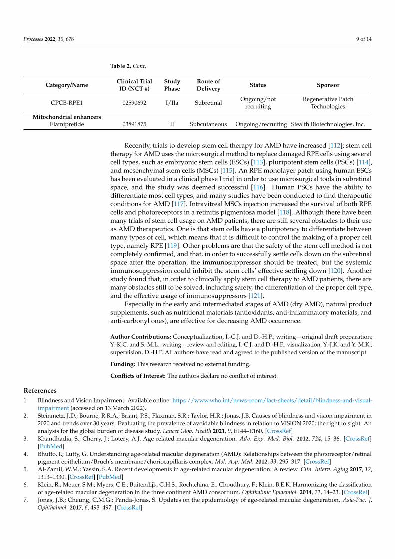
humans [105,106], and the anti-AMD mechanism can be supposed to inhibit ROS-inducedRPE cell damage via the 25C cycle and cyclin B1-related G2/M phase arrest [107]. Tauro-cholic acid, which is a yellowish crystalline bile acid and is called cholaic acid, cholytaurine,or acidum cholatauricum [108], inhibited both VEGF-induced tube formation and the migra-tion of choroidal endothelial cells (RF/6A cells) in a study of anti-angiogenesis effects [109].Vaccinium uliginosum L. fractions, which contain a large amount of polyphenol, preventedblue-light-induced ARPE 19 cell damage [110]. In 2022, de Guimaraes et al. [111] reportedthat drug candidates for early (dry) AMD treatment were undergoing a clinical study(Table 2). Based on the therapeutic target, they can be classified as antioxidants, reducers oftoxic byproducts, visual cycle modulators, anti-inflammatory and complement inhibitiondrugs, neuroprotection, gene therapy, cell-based therapies, mitochondrial enhancers, andnanosecond laser therapy.
Table 2. Ongoing clinical trials for early (dry) AMD drugs.
Category/Name Clinical TrialID (NCT #)
StudyPhase
Route ofDelivery Status Sponsor
AntioxidativeAge-related eye disease study
(AREDS) 00000145 III Oral Completed National Eye Institute
AREDS2 00345176 III Oral Completed National Eye Institute
OT-551 00306488 II Topical Completed National Institutes of HealthClinical Center
Reduction in toxicbyproductsGSK933776 01342926 II IV Completed GlaxoSmithKline
RN6G 01577381 II IV Terminated Pfizer
Visual cycle modulatorsACU-4429 01802866 IIb/III Oral Completed Kutoba Vision, Inc.Fenretinide 00429936 II Oral Completed Sirion Therapeutics, Inc.
C20-D3-vitamin A (ALK-001) 03845582 III Oral Ongoing/recruiting Aleus Pharmaceuticals, Inc.
Anti-inflammatory andcomplement inhibition
Eculizumab 00935883 II IV Completed Philip J. Rosenfeld, MDLampalizumab 02247531 III Intravitreal Terminated Hoffman-La Roche
Sirolimus (rapamycin) 00766649 I/II Subconjunctival Completed National Eye InstituteAvacincaptad pegol (Zimura) 02686658 II/III Intravitreal Completed IVERIC bio, Inc.
Pegcetacoplan (APL-2) 03525613 III Intravitreal Completed/notrecruiting Apellis Pharmaceuticals, Inc.
Tedisolumab (LFG316) 01527500 II Intravitreal Completed Novartis Pharmaceuticals, Inc.Risuteganib 03626636 I Intravitreal Completed Allegro Ophthalmics
NeuroprotectionCiliary nerve trophic factor 00447954 II Intravitreal Completed National Eye Institute
Brimonidine tartrate 02087085 IIB Intravitreal Terminated Allergan
Gene therapyAAVCAGsCD59 03144999 I Intravitreal Completed Hemera Biosciences
GT005 03846193 I/II Subretinal Ongoing/recruiting Gyroscope Therapeutics
Cell-based therapies
Palucorcel (CNTO-2476) 01226628 I/II Subretinal Completed Janssen Research &Development, LLC
MA09-hRPE 01344993 I/II Subretinal Completed Astelas Institute forRegenerative Medicine
Processes 2022, 10, 678 9 of 14
Table 2. Cont.
Category/Name Clinical TrialID (NCT #)
StudyPhase
Route ofDelivery Status Sponsor
CPCB-RPE1 02590692 I/IIa Subretinal Ongoing/notrecruiting
Regenerative PatchTechnologies
Mitochondrial enhancersElamipretide 03891875 II Subcutaneous Ongoing/recruiting Stealth Biotechnologies, Inc.
Recently, trials to develop stem cell therapy for AMD have increased [112]; stem celltherapy for AMD uses the microsurgical method to replace damaged RPE cells using severalcell types, such as embryonic stem cells (ESCs) [113], pluripotent stem cells (PSCs) [114],and mesenchymal stem cells (MSCs) [115]. An RPE monolayer patch using human ESCshas been evaluated in a clinical phase I trial in order to use microsurgical tools in subretinalspace, and the study was deemed successful [116]. Human PSCs have the ability todifferentiate most cell types, and many studies have been conducted to find therapeuticconditions for AMD [117]. Intravitreal MSCs injection increased the survival of both RPEcells and photoreceptors in a retinitis pigmentosa model [118]. Although there have beenmany trials of stem cell usage on AMD patients, there are still several obstacles to their useas AMD therapeutics. One is that stem cells have a pluripotency to differentiate betweenmany types of cell, which means that it is difficult to control the making of a proper celltype, namely RPE [119]. Other problems are that the safety of the stem cell method is notcompletely confirmed, and that, in order to successfully settle cells down on the subretinalspace after the operation, the immunosuppressor should be treated, but the systemicimmunosuppression could inhibit the stem cells’ effective settling down [120]. Anotherstudy found that, in order to clinically apply stem cell therapy to AMD patients, there aremany obstacles still to be solved, including safety, the differentiation of the proper cell type,and the effective usage of immunosuppressors [121].
Especially in the early and intermediated stages of AMD (dry AMD), natural productsupplements, such as nutritional materials (antioxidants, anti-inflammatory materials, andanti-carbonyl ones), are effective for decreasing AMD occurrence.
Author Contributions: Conceptualization, I.-C.J. and D.-H.P.; writing—original draft preparation;Y.-K.C. and S.-M.L.; writing—review and editing, I.-C.J. and D.-H.P.; visualization, Y.-J.K. and Y.-M.K.;supervision, D.-H.P. All authors have read and agreed to the published version of the manuscript.
Funding: This research received no external funding.
Conflicts of Interest: The authors declare no conflict of interest.
References1. Blindness and Vision Impairment. Available online: https://www.who.int/news-room/fact-sheets/detail/blindness-and-visual-
impairment (accessed on 13 March 2022).2. Steinmetz, J.D.; Bourne, R.R.A.; Briant, P.S.; Flaxman, S.R.; Taylor, H.R.; Jonas, J.B. Causes of blindness and vision impairment in
2020 and trends over 30 years: Evaluating the prevalence of avoidable blindness in relation to VISION 2020; the right to sight: Ananalysis for the global burden of disease study. Lancet Glob. Health 2021, 9, E144–E160. [CrossRef]
3. Khandhadia, S.; Cherry, J.; Lotery, A.J. Age-related macular degeneration. Adv. Exp. Med. Biol. 2012, 724, 15–36. [CrossRef][PubMed]
4. Bhutto, I.; Lutty, G. Understanding age-related macular degeneration (AMD): Relationships between the photoreceptor/retinalpigment epithelium/Bruch’s membrane/choriocapillaris complex. Mol. Asp. Med. 2012, 33, 295–317. [CrossRef]
5. Al-Zamil, W.M.; Yassin, S.A. Recent developments in age-related macular degeneration: A review. Clin. Interv. Aging 2017, 12,1313–1330. [CrossRef] [PubMed]
6. Klein, R.; Meuer, S.M.; Myers, C.E.; Buitendijk, G.H.S.; Rochtchina, E.; Choudhury, F.; Klein, B.E.K. Harmonizing the classificationof age-related macular degeneration in the three continent AMD consortium. Ophthalmic Epidemiol. 2014, 21, 14–23. [CrossRef]
7. Jonas, J.B.; Cheung, C.M.G.; Panda-Jonas, S. Updates on the epidemiology of age-related macular degeneration. Asia-Pac. J.Ophthalmol. 2017, 6, 493–497. [CrossRef]

Processes 2022, 10, 678 10 of 14
8. World Population Ageing 2019. Available online: Chrome-extension://efaidnbmnnnibpcajpcglclefindmkaj/viewer.html?pdfurl=https%3A%2F%2Fwww.un.org%2Fen%2Fdevelopment%2Fdesa%2Fpopulation%2Fpublications%2Fpdf%2Fageing%2FWorldPopulationAgeing2019-Highlights.pdf&clen=5752163&chunk=true (accessed on 6 February 2022).
9. Wong, W.L.; Su, X.; Li, X.; Cheung, C.M.G.; Klein, R.; Cheng, C.-Y.; Wong, T.Y. Global prevalence of age-related maculardegeneration and disease burden projection for 2020 and 2040: A systemic review and meta-analysis. Lancet Glob. Health 2014, 2,e106–e116. [CrossRef]
10. Klein, R.; Klein, B.E.K. The prevalence of age-related eye diseases and visual impairment in aging: Current estimates. Investig.Opthalmology Vis. Sci. 2013, 54, ORSF5–ORSF13. [CrossRef] [PubMed]
11. Cho, Y.K.; Park, D.H.; Jeon, I.C. Medication trends for age-related macular degeneration. Int. J. Mol. Sci. 2021, 22, 11837.[CrossRef] [PubMed]
12. Mainster, M.A.; Reichal, E. Transpupillary thermotherapy for age-related macular degeneration: Long-pulse photocoagulation,apoptosis, and heat shock proteins. Ophthalmic Surg. Lasers 2000, 31, 359–373. [CrossRef] [PubMed]
13. Bresnick, G.H. Diabetic maculopathy. Acritical review highlighting diffuse macular edema. Ophthalmology 1983, 90, 1301–1317.[CrossRef]
14. Kapugi, M.; Cunningham, K. Corticosteroids. Orthop. Nurs. 2019, 38, 336–339. [CrossRef] [PubMed]15. Falavarjani, K.G.; Nguyen, Q.D. Adverse events and complications associated with intravitreal injection of antio-VEGF agents: A
review of literature. Eye 2013, 27, 787–794. [CrossRef] [PubMed]16. Sparrow, J.R.; Vollmer-Snarr, H.R.; Zhou, J.; Jang, Y.P.; Jockusch, S.; Itagaki, Y.; Nakanishi, K. A2E-epoxides damage DNA in
retinal pigment epithelial cells. J. Bio. Chem. 2003, 278, 18207–18213. [CrossRef] [PubMed]17. Ham, W.T.; Fuffolo, J.J.; Mueller, H.A.; Clarke, A.M.; Moon, M.E. Histologic analysis of photochemical lesions produced in rhesus
retina by short-wave-length light. Investig. Ophthalmol. Vis. Sci. 1978, 17, 1029–1035.18. Eldred, G.E. Lipofuscin fluorophore inhibits lysosomal protein degradation and may cause early stages of macular degeneration.
Gerontology 1995, 41, 15–28. [CrossRef]19. Crough, R.K.; Koutalos, Y.; Kono, M.; Schey, K.; Ablonczy, Z. A2E and lipofuscin. Prog. Mol. Biol. Transl. Sci. 2015, 134, 449–463.
[CrossRef]20. Yin, D. Biochemical basis of lipofuscin, ceroid, and age pigment-like fluorophores. Free Radic. Biol. Med. 1996, 21, 871–888.
[CrossRef]21. Ouyang, X.; Yang, J.; Hong, Z.; Wu, Y.; Xie, Y.; Wang, G. Mechanisms of blue light-induced eye hazard and protective measures: A
review. Biomed. Pharmacother. 2020, 130, 110577. [CrossRef] [PubMed]22. Wu, Y.; Yanase, E.; Feng, X.; Siegel, M.M.; Sparrow, J.R. Structural characterization of bisretinoid A2E photocleavage products and
implications for age-related macular degeneration. Proc. Natl. Acad. Sci. USA 2010, 107, 7275–7280. [CrossRef] [PubMed]23. Kauppinen, A.; Niskanen, H.; Suuronen, T.; Kinnunen, K.; Salminene, A.; Kaarniranta, K. Oxidative stress activates NLRP3
inflammasomes in ARPE-19 cells-implications for age-related macular degeneration (AMD). Immunol. Lett. 2012, 147, 29–33.[CrossRef]
24. Newsome, D.A.; Dobard, E.P.; Liles, M.R.; Oliver, P.D. Human retinal pigment epithelium contains two distinct species ofsuperoxide dismutase. Investig. Ophthalmol. Vis. Sci. 1990, 31, 2508–2513.
25. Imamura, Y.; Noda, S.; Hashizume, K.; Shinoda, K.; Yamaguchi, M.; Uchiyama, S.; Shimizu, T.; Mizushima, Y.; Shirasawa, T.;Tsubota, K. Drusen, choroidal neovascularization, and retinal pigment epithelium dysfunction in SOD1-deficient mice: A modelof age-related macular degeneration. Proc. Natl. Acad. Sci. USA 2006, 103, 11282–11287. [CrossRef] [PubMed]
26. Justilien, V.; Pang, J.J.; Renganathan, K.; Zhan, X.; Crabb, J.W.; Kim, S.R.; Sparrow, J.R.; Hauswirth, W.W.; Lewin, A.S. SOD2knockdwon mouse model of early AMD. Investig. Opthalmol. Vis. Sci. 2007, 48, 4407–4420. [CrossRef]
27. Tate, D.J., Jr.; Miceli, M.V.; Newsome, D.A. Phagocytosis and H2O2 induce catalase and metallothionein gene expression inhuman retinal pigment epithelial cells. Investig. Opthalmol. Vis. Sci. 1995, 36, 1271–1279.
28. Futterman, S.; Andrews, J.S. The fatty acid composition of human retinal vitamin A ester and the lipids of human retinal tissue.Investig. Ophthalmol. 1964, 3, 441–444.
29. Khandhadia, S.; Lotery, A. Oxidation and age-related macular degeneration: Insights from molecular biology. Expert Rev. Mol.Med. 2010, 12, e34. [CrossRef]
30. Evereklioglu, C.; Er, H.; Doganay, S.; Cekmen, M.; Turkoz, Y.; Otlu, B.; Ozerol, E. Nitric oxide and lipid peroxidation are increasedand associated with decreased antioxidant enzyme activities in patients with age-related macular degeneration. Doc. Ophthalmol.2003, 106, 129–136. [CrossRef]
31. Totan, Y.; Cekic, O.; Borazan, M.; Uz, E.; Sogut, S.; Akyol, O. Plasma malondialdehyde and nitric oxide levels in age-relatedmacular degeneration. Br. J. Ophthalmol. 2001, 85, 1426–1428. [CrossRef]
32. Jarrett, S.G.; Boulton, M.E. Consequences of oxidative stress in age-related macular degeneration. Mol. Asp. Med. 2012, 33,399–417. [CrossRef]
33. Bazan, N.G. Survival signaling in retinal pigment epithelial cells in response to oxidative stress: Significance in retinal degenera-tions. Adv. Exp. Med. Biol. 2006, 572, 531–540. [CrossRef]
34. Anderson, O.A.; Finkelstein, A.; Shima, D.T. A2E induces IL-1β production in retinal pigment epithelial cells via the NLRP3inflammasome. PLoS ONE 2013, 8, e67263. [CrossRef]

Processes 2022, 10, 678 11 of 14
35. Augustin, A.J.; Kirchhof, J. Inflammation and the pathogenesis of age-related macular degeneration. Expert Opin. Ther. Targets2009, 13, 641–651. [CrossRef] [PubMed]
36. Anderson, D.H.; Mullins, R.F.; Hageman, G.S.; Johnson, L.V. A role for local inflammation in the formation of drusen in the agingeye. Am. J. Ophthalmol. 2002, 134, 411–431. [CrossRef]
37. Lee, S.Y.; Cho, S.S.; Li, Y.C.; Bae, C.S.; Park, K.M.; Park, D.H. Anti-inflammatory effect of Curcuma longa and Allium hookerico-treatment via NF-κB and COX-2 pathways. Sci. Rep. 2020, 10, 5718. [CrossRef]
38. Collin, T. Acute and Chronic Inflammation in Robbins Pathological Basis of Disease; Cotran, R.S., Kumar, V., Collins, T., Eds.; SaundersWB: Philadelphia, PA, USA, 1999; pp. 50–88.
39. Zhang, J.M.; An, J. Cytokines, inflammation and pain. Int. Anesthesiol. Clin. 2007, 45, 27–37. [CrossRef] [PubMed]40. Musolino, C.; Allegra, A.; Innao, V.; Allegra, A.G.; Pioggia, G.; Gangemi, S. Inflammatory and anti-inflammatory equilibrium,
proliferative and antiproliferative balance: The role of cytokines in multiple myeloma. Mediat. Inflamm. 2017, 2017, 1852517.[CrossRef]
41. Degenhardt, T.P.; Brinkmann-Frye, E.; Thorpe, S.R.; Baynes, J.W. Role of carbonyl stress in aging and age-related disease. In TheMaillard Reaction in Foods and Medicine; O’Brien, J., Nursten, H.E., Crabbe, M.J.C., Ames, J.M., Eds.; Woodhead Publishing Seriesin Food Science; Technology and Nutrition: Cambridge, UK, 2005; pp. 3–10.
42. Liu, Y.; Zhang, D.; Hu, J.; Liu, G.; Chen, J.; Sun, L.; Jiang, Z.; Zhang, X.; Chen, Q.; Ji, B. Visible light-induced lipid peroxidation ofunsaturated fatty acids in the retina and the inhibitory effects of blueberry polyphenols. J. Agric. Food Chem. 2015, 63, 9295–9305.[CrossRef] [PubMed]
43. Long, E.K.; Picklo Sr., M.J. Trans-4-hydroxy-2-hexenal, a product of n-3 fatty acid peroxidation: Make some room HNE. Free Radic.Biol. Med. 2010, 49, 1–8. [CrossRef]
44. Lederman, M.; Hagbi-Levi, S.; Grunin, M.; Obolensky, A.; Berenshtein, E.; Bannin, E.; Chevion, M.; Chowers, I. Degenerationmodulates retinal response to transient exogenous oxidative injury. PLoS ONE 2014, 9, e87751. [CrossRef]
45. Zor, R.K.; Ersan, S.; Kucuk, E.; Yildirim, G.; Sari, I. Serum malondialdehyde, monocyte chemoattractant protein-1, and vitamin Clevels in wet type age-related macular degeneration patients. Ther. Adv. Ophthalmol. 2020, 12, 1–8. [CrossRef]
46. Lambros, M.L.; Plafker, S.M. Oxidative stress and the Nrf2 anti-oxidant transcription factor in age-related macular degeneration.Adv. Exp. Med. Biol. 2016, 854, 67–72. [CrossRef] [PubMed]
47. Itoh, K.; Wakabayashi, N.; Katoh, Y.; Ishii, T.; Igarashi, K.; Engel, J.D.; Yamamoto, M. Keap1 represses nuclear activation ofantioxidant responsive elements by Nrf2 through binding to the amino-terminal Neh2 domain. Genes Dev. 1999, 13, 76–86.[CrossRef] [PubMed]
48. Kensler, T.W.; Wakabayashi, N.; Biswal, S. Cell survival responses to environmental stresses via the Keap 1-Nrf2-ARE pathway.Annu. Rev. Pharmacol. Toxicol. 2007, 47, 89–116. [CrossRef] [PubMed]
49. Mitsuishi, Y.; Motohashi, H.; Yamamoto, M. The Keap1-Nrf2 system in cancer: Stress response and anabolic metabolism. Front.Oncol. 2012, 2, 200. [CrossRef] [PubMed]
50. Satoh, T.; Lipton, S. Recent advances in understanding Nrf2 as a druggable target: Development of pro-electrophilic andnon-covalent Nrf2 activators to overcome systemic side effects of electrophilic drugs like dimethyl fumarate. F1000Research 2017,6, 2138. [CrossRef] [PubMed]
51. Silva, M.D.F.; Proccoli, L.; Morroni, F.; Sita, G.; Seghetti, F.; Viegas, C., Jr.; Tarozzi, A. The Keap1/Nrf2-ARE pathway as apharmacological target for chalcones. Molecules 2018, 23, 1803. [CrossRef] [PubMed]
52. Loboda, A.; Damulewicz, M.; Pyza, E.; Jozkowicz, A.; Dulak, J. Role of Nrf2/HO-1 system in development, oxidative stressresponse and diseases: An evolutionarily conserved mechanism. Cell. Mol. Life Sci. 2016, 73, 3221–3247. [CrossRef] [PubMed]
53. Zhao, X.; Zou, Y.; Xu, H.; Fan, L.; Guo, H.; Li, X.; Li, G.; Zhang, X.; Dong, M. Gastrodin protect primary cultured rat hippocampalneurons against amyloid-beta peptide-induced neurotoxicity via ERK1/2-Nrf2 pathway. Brain Res. 2012, 1482, 13–21. [CrossRef]
54. Cao, X.; Xiao, H.; Zhang, Y.; Zou, L.; Chu, Y.; Chu, X. 1,5-dicaffeoylquinic acid-mediated glutathione synthesis through activationof Nrf2 protects against 0GD/reperfusion-induced oxidative stress in astrocytees. Brain Res. 2010, 1347, 142–148. [CrossRef][PubMed]
55. Son, T.G.; Camandola, S.; Arumugam, T.V.; Cutler, R.G.; Telljohann, R.S.; Mughal, M.R.; Moore, T.A.; Luo, W.; Yu, Q.S.; Johnson,D.A.; et al. Plumbagin, a novel Nrf2/ARE activator, protects against cerebra ischemia. J. Neurochem. 2010, 112, 1316–1326.[CrossRef]
56. Guan, S.P.; Tee, W.; Ng, D.S.; Chan, T.K.; Peh, H.Y.; Ho, W.E.; Cheng, C.; Mak, J.C.; Wong, W.S.F. Andrographolide protectsagainst cigarette smoke-induce doxidative lung injury via augmentation of Nrf2 activity. Br. J. Pharmacol. 2013, 168, 1707–1718.[CrossRef] [PubMed]
57. Lee, B.W.; Chun, S.W.; Kim, S.H.; Lee, Y.; Kang, E.S.; Cha, B.S.; Lee, H.C. Lithospermic acid B protects β-cells from cytokine-induced apoptosis by alleviating apoptotic pathways and activating anti-apoptotic pathways of Nrf2-HO-1 and Sirt1. Toxicol.Appl. Pharmacol. 2011, 252, 47–54. [CrossRef] [PubMed]
58. Lee, B.H.; Hsu, W.H.; Huang, T.; Chang, Y.Y.; Hsu, Y.W.; Pan, T.M. Effects of monascin on anti-inflammation mediated by Nrf2activation in advanced glycation end product-treated THP-1 monocytes and methylglyoxal-treated wistart rats. J. Agric. FoodChem. 2013, 61, 1288–1298. [CrossRef] [PubMed]

Processes 2022, 10, 678 12 of 14
59. Zhang, Z.; Cui, W.; Li, G.; Yuan, S.; Xu, D.; Hoi, M.P.M.; Lin, Z.; Dou, J.; Han, Y.; Lee, S.M.Y. Baicalein protects against 6-OHDA-induced neurotoxicity through activation of Keap1/Nrf2/HO-1 and involving PKCα and PI3K/AKT signaling pathways. J.Agric. Food Chem. 2012, 60, 8171–8182. [CrossRef] [PubMed]
60. Kim, S.S.; Lim, J.; Bang, Y.; Gal, J.; Lee, S.U.; Cho, Y.C.; Yoon, G.; Kang, B.Y.; Cheon, S.H.; Choi, H.J. Licochalcone E acti-vates Nrf2/antioxidant response element signaling pathway in both neuronal and microglial cells: Therapeutic relevance toneurodegenerative disease. J. Nutr. Biochem. 2012, 23, 1314–1323. [CrossRef] [PubMed]
61. Shah, Z.A.; Li, R.C.; Ahmad, A.S.; Kensler, T.W.; Yamamoto, M.; Biswal, S.; Dore, S. The flavanol (1)-epicatechin prevents strokedamage through the Nrf2/HO1 pathway. Cereb. Blood Flow Metab. 2020, 30, 1951–1961. [CrossRef]
62. Dong, X.; Li, Z.; Wang, W.; Zhang, W.; Liu, S.; Zhang, X.; Fang, J.; Maeda, H.; Matsukura, M. Protective effect of canolol fromoxidative stress-induced cell damage in ARPE-19 cells via an ERK mediated antioxidative pathway. Mol. Vis. 2011, 17, 2040–2048.[PubMed]
63. Zhao, H.; Wang, R.; Ye, M.; Zhang, L. Genipin protects against H2O2-induced oxidative damage in retinal pigment epithelial cellsby promoting Nrf2 signaling. Int. J. Mol. Med. 2019, 43, 936–944. [CrossRef] [PubMed]
64. Zhu, C.; Dong, Y.; Liu, H.; Ren, H.; Cui, Z. Hesperetin protects against H2O2-triggered oxidative damage via upregulation of theKeap-1-Nrf2/HO-1 signal pathway in ARPE-19 cells. Biomed. Pharmacother. 2016, 88, 124–133. [CrossRef]
65. Chen, W.; Ye, Y.; Wu, Z.; Lin, J.; Wang, Y.; Ding, Q.; Yang, X.; Yang, W.; Lin, B.; Lin, B. Temporary upregulation of Nrf2 bynaringenin alleviates oxidative damage in the retina and ARPE-19 cells. Oxidative Med. Cell. Longev. 2021, 2021, 4053276.[CrossRef]
66. Zhang, H.; Liu, Y.Y.; Jiang, Q.; Li, K.R.; Zhao, Y.X.; Cao, C.; Yao, J. Salvianolic acid A protects RPE cells against oxidative stressthrough activation of Nrf2/HO-1 signaling. Free Radic. Biol. Med. 2014, 69, 219–228. [CrossRef] [PubMed]
67. Age-Related Eye Disease Study Research Group. A randomized, placebo-controlled, clinical trial of high-dose supplementationwith vitamins C and E, beta carotene, and zinc for age-related macular degeneration and vision loss: AREDS report no. 8. Arch.Ophthalmol. 2001, 119, 1417–1436. [CrossRef]
68. Mayne, S.T. Antioxidant nutrients and chronic disease: Use of biomarkers of exposure and oxidative stress status in epidemiologicresearch. J. Nutr. 2003, 133, 933S–940S. [CrossRef] [PubMed]
69. Kim, D.H.; Choi, Y.R.; Shim, J.; Choi, Y.S.; Kim, Y.T.; Kim, M.K.; Kim, M.J. Suppressive effect of Arctium lappa L. leaves on retinaldamage against A2E-induced ARPE-19 cells and mice. Molecules 2020, 25, 1737. [CrossRef] [PubMed]
70. Kong, J.M.; Chia, L.S.; Goh, N.K.; Chia, T.F.; Brouillard, R. Analysis and biological activities of anthocyanins. Phytochemistry 2003,64, 923–933. [CrossRef]
71. Ni, T.; Yang, W.; Xing, Y. Protective effects of delphinidin against H2O2-induced oxidative injuries in human retinal pigmentepithelial cells. BioSci. Rep. 2019, 39, BSR20190689. [CrossRef]
72. Aung, K.H.; Liu, H.; Ke, Z.; Jiang, S.; Huang, J. Glabridin attenuates the retinal degeneration induced by sodium lodate in vitroand in vivo. Front. Pharmacol. 2020, 11, 566699. [CrossRef] [PubMed]
73. Broz, P.; Dixit, V.M. Inflammasomes: Mechanism of assembly, regulation and signaling. Nat. Rev. Immunol. 2016, 16, 407–420.[CrossRef]
74. Winsor, N.; Krustev, C.; Bruce, J.; Philpott, D.J.; Girardin, S.E. Canonical and noncanonical inflammasomes in intestinal epithelialcells. Cell. Microbiol. 2019, 21, e13079. [CrossRef]
75. Takeuchi, O.; Akira, S. Pattern recognition receptors and inflammation. Cell 2010, 140, 805–820. [CrossRef] [PubMed]76. Shi, Y.; Zheng, W.; Rock, K.L. Cell injury releases endogenous adjuvants that stimulate cytotoxic T cell responses. Proc. Natl. Acad.
Sci. USA 2000, 97, 14590–14595. [CrossRef] [PubMed]77. Shi, Y.; Rock, K.L. Cell death releases endogenous adjuvants that selectively enhance immune surveillance of particulate antigens.
Eur. J. Immunol. 2002, 32, 155–162. [CrossRef]78. Mosser, D.M. The many faces of macrophage activation. J. Leukoc. Biol. 2003, 73, 209–212. [CrossRef] [PubMed]79. Gordon, S.; Martinez, F.O. Alternative activation of macrophages: Mechanism and functions. Immunity 2020, 32, 593–604.
[CrossRef] [PubMed]80. Sun, H.J.; Jin, X.M.; Xu, J.; Xiao, Q. Baicalin alleviates age-related macular degeneration via miR-223/NLRP3-regulated pyroptosis.
Pharmacology 2019, 105, 28–38. [CrossRef]81. Yang, M.; So, K.F.; Lo, A.C.Y.; Lam, W.C. The effect of Lycium barbarum polysaccharides on pyroptosis-associated amyloid β1-40
oligomers-induced adult retinal pigment epithelium 19 cell damage. Int. J. Mol. Sci. 2020, 21, 4658. [CrossRef]82. Wang, K.; Zhu, X.; Zhang, K.; Yao, Y.; Zhuang, M.; Tan, C.; Zhou, F.; Zhu, L. Puerarin inhibits amyloid β-induced NLRP3
inflammasome activation in retinal pigment epithelial cells via suppressing ROS-dependent oxidative and endoplasmic reticulumstresses. Exp. Cell Res. 2017, 357, 335–340. [CrossRef] [PubMed]
83. Wan, W.; Zhu, W.; Wu, Y.; Long, Y.; Liu, H.; Wan, W.; Wan, G.; Yu, J. Grape seed proanthocyanidin extract moderated retinalpigment epithelium cellular senescence through NAMPT/SIRTI/NLRP3 pathway. J. Inflamm. Res. 2021, 14, 3129–3143. [CrossRef]
84. Jin, X.; Wang, C.; Wu, W.; Liu, T.; Ji, B.; Zhou, F. Cyanidin-3-glucoside alleviates 4-hydroxyhexenal-induced NLRP3 inflammasomeactivation via JNK-c-Jun/AP-1 pathway in human retinal pigment epithelial cells. J. Immunol. Res. 2018, 2018, 5604610. [CrossRef]
85. Franzone, F.; Nebbioso, M.; Pergolizzi, T.; Attanasio, G.; Musacchio, A.; Greco, A.; Limoli, P.G.; Artico, M.; Spandidos, D.A.;Taurone, S.; et al. Anti-inflammatory role of curcumin in retinal disorders (Review). Exp. Ther. Med. 2021, 22, 790. [CrossRef]

Processes 2022, 10, 678 13 of 14
86. Militao, G.C.; Albuquerque, M.R.; Pessoa, O.D.; Pessoa, C.; Moraes, M.E.; de Moraes, M.O.; Costa-Lotufo, L.V. Cytotoxic activityof nepetin, a flavonoid from Eupatorium ballotaefolium HBK. Die Pharm. 2004, 59, 965–966. [CrossRef]
87. Bai, N.; He, K.; Roller, M.; Lai, C.S.; Shao, X.; Pan, M.H.; Ho, C.T. Flavonoids and phenolic compounds from Rosmarinus officinalis.J. Agric. Food Chem. 2010, 58, 5363–5367. [CrossRef]
88. Thitilertdecha, P.; Rowan, M.G.; Guy, R.H. Topical formulation and dermal delivery of active phenolic compounds in the Thaimedicinal plant-Clerodendrum petasites S. Moore. Int. J. Pharm. 2015, 478, 39–45. [CrossRef] [PubMed]
89. Chen, X.; Han, R.; Hao, P.; Wang, L.; Liu, M.; Jin, M.; Kong, D.; Li, X. Nepetin inhibits IL-1β induced inflammation via NF-κB andMAPKs signaling pathways in ARPE-19 cells. Biomed. Pharmacother. 2018, 101, 87–93. [CrossRef] [PubMed]
90. Hsu, Y.A.; Chen, C.S.; Wang, Y.C.; Lin, E.S.; Chang, C.Y.; Chen, J.J.Y.; Wu, M.Y.; Lin, H.J.; Wan, L. Anti-inflammatory effects ofresveratrol on human retinal pigment cells and a myopia animal model. Curr. Issues Mol. Biol. 2021, 43, 716–727. [CrossRef][PubMed]
91. Heitmar, R.; Brwon, J.; Kyrou, I. Saffron (Crocus sativus L.) in ocular diseases: A narrative review of the existing evidence fromclinical studies. Nutrients 2019, 11, 649. [CrossRef] [PubMed]
92. Fernandez-Albarral, J.A.; Martinez-Lopez, M.A.; Marco, E.M.; de Hoz, R.; Martin-Sanchez, B.; Felipe, D.S.; Salobrar-Garcia, E.;Lopez-Cuenca, I.; Pinazo-Duran, M.D.; Salazar, J.J.; et al. Is saffron able to prevent the dysregulation of retinal cytokines inducedby ocular hypertension in mice? J. Clin. Med. 2021, 10, 4801. [CrossRef] [PubMed]
93. Chen, C.; Guo, D.; Lu, G. Wogonin protects human retinal pigment epithelium cells from LPS-induced barrier dysfunction andinflammatory responses by regulating the TLR4/NF-κB signaling pathway. Mol. Med. Rep. 2017, 15, 2289–2295. [CrossRef][PubMed]
94. Borlinghaus, J.; Albrecht, F.; Gruhlke, M.C.; Nwachukwu, I.D.; Slusarenko, A.J. Allicin: Chemistry and biological properties.Molecules 2014, 19, 12591–12618. [CrossRef] [PubMed]
95. Tu, G.; Zhang, Y.F.; Wei, W.; Li, L.; Zhang, Y.; Yang, J.; Xing, Y. Allicin attenuates H2O2-induced cytotoxicity in retinal pigmentedepithelial cells by regulating the levels of reactive oxygen species. Mol. Med. Rep. 2016, 13, 2320–2326. [CrossRef] [PubMed]
96. Yu, C.C.; Nandrot, E.F.; Dun, Y.; Finnemann, S.C. Dietary antioxidants prevent age-related retinal pigment epithelium actindamage and blindness in mice lacking ανβ5 integrin. Free Radic. Biol. Med. 2012, 52, 660–670. [CrossRef] [PubMed]
97. Wang, Y.; Kim, H.J.; Sparrow, J.R. Quercetin and cyanidin-3-glucoside protect against photooxidation and photodegradation ofA2E in retinal pigment epithelial cells. Exp. Eye Res. 2017, 160, 45–55. [CrossRef] [PubMed]
98. Marti, R.; Rosello, S.; Cebolla-Cornejo, J. Tomato as a source of carotenoids and polyphenols targeted to cancer prevention.Cancers 2016, 8, 58. [CrossRef]
99. Chichili, G.R.; Nohr, D.; Frank, J.; Flaccus, A.; Fraser, P.D.; Enfissi, E.M.A.; Biesalski, H.K. Protective effects of tomato extract withelevated β-carotene levels on oxidative stress in ARPE-19 cells. Br. J. Nutr. 2006, 96, 643–649. [CrossRef] [PubMed]
100. Matsuura, T.; Takayama, K.; Kaneko, H.; Ye, F.; Fukukita, H.; Tsunekawa, T.; Kataoka, K.; Hwang, S.J.; Nagasaka, Y.; Ito, Y.;et al. Nutritional supplementation inhibits the increase in serum malondialdehyde in patients with wet age-related maculardegeneration. Oxidative Med. Cell. Longev. 2017, 2017, 9548767. [CrossRef]
101. Khurana, R.N.; Hoang, C.; Khanani, A.M.; Steklov, N.; Singerman, L.J. A smart mobile application to monitor viual function indiabetic retinopathy and age-related macular degeneration: The clear study. Am. J. Ophthalmol. 2021, 227, 222–230. [CrossRef][PubMed]
102. Oeverhaus, M.; Hirche, H.; Esser, J.; Eckstein, A.; Schaperdoth-Gerlings, B. Evaluation of the medical treatment situation of thevisually impaired: Significant differences between young and old. Ophthalmologe 2019, 116, 164–171. [CrossRef] [PubMed]
103. Bressler, S.B. Introduction: Understanding the role of angiogenesis and antiangiogenic agents in age-related macular degeneration.Ophthalmologe 2009, 116, S1–S7. [CrossRef] [PubMed]
104. Sommerburg, O.; Keunen, J.E.; Bird, A.C.; vanKuijk, F.J. Fruits and vegetables that are sources for lutein and zeaxanthin: Themacular pigment in human eyes. Br. J. Ophthalmol. 1998, 82, 907–910. [CrossRef]
105. The Age-Related Eye Disease Study 2 (AREDS2) Research Group. Secondary analysis of the effects of lutein/zeaxanthin onage-related macular degeneration progression. JAMA Ophthalmol 2014, 132, 142–149. [CrossRef]
106. Tan, J.S.L.; Wang, J.J.; Flood, V.; Rochtchina, E.; Smith, W.; Mitchell, P. Dietary antioxidants and the long-term incidence ofage-related macular degeneration. The blue mountains eye study. Ophthalmologe 2008, 115, 334–341. [CrossRef] [PubMed]
107. Liu, H.; Liu, W.; Zhou, X.; Long, C.; Kuang, X.; Hu, J.; Tang, Y.; Liu, L.; He, J.; Huang, Z.; et al. Protective effect of lutein onARPE-19 cells upon H2O2-induced G2/M arrest. Mol. Med. Rep. 2017, 16, 2069–2074. [CrossRef] [PubMed]
108. Sawkat, A.M. Cellular regulation of hepatic bile acid transport in health and cholestasis. Hepatology 2004, 39, 581–590. [CrossRef]109. Warden, C.; Barnett, J.M.; Brantley, M.A., Jr. Taurocholic acid inhibits features of age-related macular degeneration in vitro. Exp.
Eye Res. 2020, 193, 107974. [CrossRef] [PubMed]110. Lee, B.L.; Kang, J.H.; Kim, H.M.; Jeong, S.H.; Jang, D.S.; Jang, Y.P.; Choung, S.Y. Polyphenol-enriched Vaccinium uliginosum L.
fractions reduce retinal damage induced by blue light in A2E-laden ARPE19 cell cultures and mice. Nutr. Res. 2016, 36, 1402–1414.[CrossRef] [PubMed]
111. De Guimaraes, T.A.C.; Varela, M.D.; Georgiou, M.; Michaelides, M. Treatments for dry age-related macular degeneration:Therapeutic avenues, clinical trials and future directions. Br. J. Ophthalmol. 2022, 106, 297–304. [CrossRef] [PubMed]

Processes 2022, 10, 678 14 of 14
112. Nazari, H.; Zhang, L.; Zhu, D.; Chader, G.J.; Falabella, P.; Stefanini, F.; Rowland, T.; Clegg, D.O.; Kashani, A.H.; Hinton, D.R.; et al.Stem cell based therapies for age-related macular degeneration: The promises and the challenges. Prog. Retin. Eye Res. 2015, 48,1–39. [CrossRef] [PubMed]
113. Lund, R.D.; Wang, S.; Klimanskaya, I.; Holmes, T.; Ramos-Kelsey, R.; Lu, B.; Girman, S.; Bischoff, N.; Sauve, Y.; Lanza, R. Humanembryonic stem cell-derived cells rescue visual function in dystrophic RCS rats. Cloning Stem Cells 2006, 8, 189–199. [CrossRef]
114. Takahashi, K.; Yamanaka, S. Induction of pluripotent stem cells from mouse embryonic and adult fibroblast cultures by definedfactors. Cell 2006, 126, 663–676. [CrossRef]
115. Otani, A.; Dorrell, M.I.; Kinder, K.; Moreno, S.K.; Nusinowitz, S.; Banin, E.; Heckenlively, J.; Friedlander, M. Rescue of retinaldegeneration by intravitreally injected adult bone marrow-derived lineage-negative hematopoietic stem cells. J. Clin. Investig.2004, 114, 765–774. [CrossRef]
116. Da Cruz, L.; Fynes, K.; Georgiadis, O.; Kerby, J.; Luo, Y.H.; Ahmado, A.; Vernon, A.; Daniels, J.T.; Nommiste, B.; Hasan, S.M.; et al.Phase 1 clinical study of an embryonic stem cell-derived retinal pigment epithelium patch in age-related macular degeneration.Nat. Biotechnol. 2018, 36, 328–337. [CrossRef]
117. Morizur, L.; Herardot, E.; Monville, C.; M’Barek, K.B. Human pluripotent stem cells: A toolbox to understand and treat retinaldegeneration. Mol. Cell. Neurosci. 2020, 107, 103523. [CrossRef] [PubMed]
118. Arnhold, S.; Absenger, Y.; Klein, H.; Addicks, K.; Schraermeyer, U. Transplantation of bone marrow-derived mesenchymal stemcells rescue photoreceptor cells in the dystrophic retina of the rhodopsin knockout mouse. Graefes Arch. Clin. Exp. Ophthalmol.2007, 245, 414–422. [CrossRef]
119. Mu, Y.; Zhao, M.; Su, G. Stem cell-based therapies for age-related macular degeneration: Current status and prospects. Int. J. Clin.Exp. Med. 2014, 7, 3843–3852. [PubMed]
120. Idelson, M.; Alper, R.; Obolensky, A.; Yachimovich-Cohen, N.; Rachmilewitz, J.; Ejzenberg, A.; Beider, E.; Banin, E.; Reubinoff, B.Immunological properties of human embryonic stem cell-derived retinal pigment epithelial cells. Stem Cell Rep. 2018, 11, 681–695.[CrossRef] [PubMed]
121. O’Neill, H.C.; Limnios, I.J.; Barnett, N.L. Advancing a stem cell therapy for age-related macular degeneration. Curr. Stem Cell ResTher. 2020, 15, 89–97. [CrossRef]


![nato stanag 4285 ed*]*amd*2 89 - TORFone](https://static.fdokumen.com/doc/165x107/631c6ed2b8a98572c10ce6b9/nato-stanag-4285-edamd2-89-torfone.jpg)