Asp49 phospholipase A 2–elaidoylamide complex: a new mode of inhibition
8
Asp49 phospholipase A 2 –elaidoylamide complex: a new mode of inhibition q Dessislava N. Georgieva, a,d Wojciech Rypniewski, a,b Azat Gabdoulkhakov, c Nicolay Genov, d and Christian Betzel a, * a Zentrum f€ ur Experimentelle Medizin, Institut f€ ur Biochemie und Molekularbiologie I, Universit€ atsklinikum Hamburg-Eppendorf, c/o DESY, Notkestrasse 85, Geb. 22a, 22603 Hamburg, Germany b Institute of Bioorganic Chemistry, Polish Academy of Sciences, Noskowskiego 12/14, 61-704 Poznan, Poland c Institute of Protein Research RAS, Pushchino, Moscow Region 142290, Russia d Institute of Organic Chemistry, Bulgarian Academy of Sciences, Sofia 1113, Bulgaria Received 22 April 2004 Available online 7 June 2004 Abstract The inhibition of phospholipase A 2 s (PLA 2 s) is of pharmacological and therapeutic interest because these enzymes are involved in several inflammatory diseases. Elaidoylamide is a powerful inhibitor of a neurotoxic PLA 2 from the Vipera ammodytes merid- ionalis venom. The X-ray structure of the enzyme–inhibitor complex reveals a new mode of Asp49 PLA 2 inhibition by a fatty acid hydrocarbon chain. The structure contains two identical homodimers in the asymmetric unit. In each dimer one subunit is rotated by 180° with respect to the other and the two molecules are oriented head-to-tail. One molecule of elaidoylamide is bound simultaneously to the substrate binding sites of two associated neurotoxic phospholipase A 2 molecules. The inhibitor binds symmetrically to the hydrophobic channels of the two monomers. The structure can be used to design anti-inflammatory drugs. Ó 2004 Elsevier Inc. All rights reserved. Keywords: Phospholipase A 2 ; Neurotoxin; Elaidoylamide; X-ray structure Phospholipase A 2 s (phosphatide sn-2 acylhydrolases, PLA 2 s; EC 3.1.1.4) are a rapidly growing superfamily of enzymes which hydrolyze stereospecifically the sn-2 ester bond of natural phospholipids. They catalyze reactions at lipid-aqueous interfaces. PLA 2 s play an important role in a number of biological processes such as phospholipid metabolism and remodelling, mediator production, homeostasis of cellular membranes, host defense, and signal transduction [1]. The liberated ly- sophospholipids are important in cell signalling, mem- brane perturbation, and can induce tissue damage [2,3]. One of the reaction products is arachidonic acid which is a precursor of eicosanoids, potent mediators of inflam- mation and signal transduction. In this way, PLA 2 is involved in chronic inflammatory diseases such as rheumatoid arthritis, asthma, platelet aggregation, acute hypersensitivity and pancreatitis, sepsis and septic shock, respiratory distress syndrome, etc. [4–8]. Snake venom PLA 2 s exert additionally a wide variety of pharmacological activities: neurotoxicity, cardiotoxicity, myotoxicity, hypotensive, antiplatelet, hemolytic, hem- orrhagic, edema-inducing, coagulant or anticoagulant effects [9]. For this reason it is of pharmacological and medical interest to develop specific inhibitors for PLA 2 . The venomous snake Vipera ammodytes meridionalis is of public health significance in Europe. Envenomation by this viper causes very often rapid death due to the effect of the venom on the neuromuscular junctions, which prevents the binding of acetylcholine to its re- ceptor. The major toxic factor has been identified as a basic PLA 2 associated with an acidic non-toxic PLA 2 - like protein in a neurotoxic complex [10,11]. The acidic component modulates the enzymatic and pharmaco- logical activities of the toxic subunit [11]. q Abbreviations: PLA 2 , phospholipase A 2 . * Corresponding author. Fax: +49-40-89984747. E-mail address: [email protected] (C. Betzel). 0006-291X/$ - see front matter Ó 2004 Elsevier Inc. All rights reserved. doi:10.1016/j.bbrc.2004.05.106 Biochemical and Biophysical Research Communications 319 (2004) 1314–1321 BBRC www.elsevier.com/locate/ybbrc
-
Upload
independent -
Category
Documents
-
view
3 -
download
0
Transcript of Asp49 phospholipase A 2–elaidoylamide complex: a new mode of inhibition

Biochemical and Biophysical Research Communications 319 (2004) 1314–1321
BBRCwww.elsevier.com/locate/ybbrc
Asp49 phospholipase A2–elaidoylamide complex:a new mode of inhibitionq
Dessislava N. Georgieva,a,d Wojciech Rypniewski,a,b Azat Gabdoulkhakov,c
Nicolay Genov,d and Christian Betzela,*
a Zentrum f€ur Experimentelle Medizin, Institut f€ur Biochemie und Molekularbiologie I, Universit€atsklinikum Hamburg-Eppendorf,
c/o DESY, Notkestrasse 85, Geb. 22a, 22603 Hamburg, Germanyb Institute of Bioorganic Chemistry, Polish Academy of Sciences, Noskowskiego 12/14, 61-704 Poznan, Poland
c Institute of Protein Research RAS, Pushchino, Moscow Region 142290, Russiad Institute of Organic Chemistry, Bulgarian Academy of Sciences, Sofia 1113, Bulgaria
Received 22 April 2004
Available online 7 June 2004
Abstract
The inhibition of phospholipase A2s (PLA2s) is of pharmacological and therapeutic interest because these enzymes are involved
in several inflammatory diseases. Elaidoylamide is a powerful inhibitor of a neurotoxic PLA2 from the Vipera ammodytes merid-
ionalis venom. The X-ray structure of the enzyme–inhibitor complex reveals a new mode of Asp49 PLA2 inhibition by a fatty acid
hydrocarbon chain. The structure contains two identical homodimers in the asymmetric unit. In each dimer one subunit is rotated
by 180� with respect to the other and the two molecules are oriented head-to-tail. One molecule of elaidoylamide is bound
simultaneously to the substrate binding sites of two associated neurotoxic phospholipase A2 molecules. The inhibitor binds
symmetrically to the hydrophobic channels of the two monomers. The structure can be used to design anti-inflammatory drugs.
� 2004 Elsevier Inc. All rights reserved.
Keywords: Phospholipase A2; Neurotoxin; Elaidoylamide; X-ray structure
Phospholipase A2s (phosphatide sn-2 acylhydrolases,
PLA2s; EC 3.1.1.4) are a rapidly growing superfamily of
enzymes which hydrolyze stereospecifically the sn-2 ester
bond of natural phospholipids. They catalyze reactions
at lipid-aqueous interfaces. PLA2s play an important
role in a number of biological processes such as
phospholipid metabolism and remodelling, mediator
production, homeostasis of cellular membranes, hostdefense, and signal transduction [1]. The liberated ly-
sophospholipids are important in cell signalling, mem-
brane perturbation, and can induce tissue damage [2,3].
One of the reaction products is arachidonic acid which is
a precursor of eicosanoids, potent mediators of inflam-
mation and signal transduction. In this way, PLA2 is
involved in chronic inflammatory diseases such as
qAbbreviations: PLA2, phospholipase A2.* Corresponding author. Fax: +49-40-89984747.
E-mail address: [email protected] (C. Betzel).
0006-291X/$ - see front matter � 2004 Elsevier Inc. All rights reserved.
doi:10.1016/j.bbrc.2004.05.106
rheumatoid arthritis, asthma, platelet aggregation, acute
hypersensitivity and pancreatitis, sepsis and septic
shock, respiratory distress syndrome, etc. [4–8]. Snake
venom PLA2s exert additionally a wide variety of
pharmacological activities: neurotoxicity, cardiotoxicity,
myotoxicity, hypotensive, antiplatelet, hemolytic, hem-
orrhagic, edema-inducing, coagulant or anticoagulant
effects [9]. For this reason it is of pharmacological andmedical interest to develop specific inhibitors for PLA2.
The venomous snake Vipera ammodytes meridionalis
is of public health significance in Europe. Envenomation
by this viper causes very often rapid death due to the
effect of the venom on the neuromuscular junctions,
which prevents the binding of acetylcholine to its re-
ceptor. The major toxic factor has been identified as a
basic PLA2 associated with an acidic non-toxic PLA2-like protein in a neurotoxic complex [10,11]. The acidic
component modulates the enzymatic and pharmaco-
logical activities of the toxic subunit [11].

D.N. Georgieva et al. / Biochemical and Biophysical Research Communications 319 (2004) 1314–1321 1315
Here, we report the crystal structure of PLA2 isolatedfrom the neurotoxic complex of the V. a. meridionalis
venom and complexed to elaidoylamide (the amide of
trans-9-octadecenoic acid). Fatty acids are important
components of the biological membranes where they are
bound to phospholipids and cholesterol esters or exist as
free compounds. Fatty acid derivatives modulate the
lipid membrane properties which, on the other side,
regulate membrane protein functions such as enzymeactivity, protein–membrane interactions, etc. [12]. We
have found that elaidoylamide is a powerful inhibitor of
the V. a. meridionalis venom PLA2 [13]. The present
structure reveals a new mode of PLA2 inhibition: the
hydrocarbon chain of one molecule of fatty acid is
bound simultaneously to the substrate binding hydro-
phobic channels of two PLA2 molecules. To our
knowledge, this is the first crystal structure of Asp49PLA2 inhibited by an added fatty acid.
Materials and methods
Crude venom was collected from V. a. meridionalis inhabiting
southeastern Europe. The neurotoxin Vipoxin was isolated as de-
scribed in [14]. The isolation of His48 PLA2 was performed according
to the procedure given in [15]. Elaidoylamide is a generous gift of Prof.
M. Jain from the University of Delaware, USA. The crystallization
conditions are described in [13]. Diffraction data were collected from a
flash-frozen crystal at 100K with a synchrotron radiation (k ¼ 0:802�A)
at the consortium’s beamline X13 at HASYLAB/DESY—Hamburg.
The images were processed using the DENZO (SCALEPACK) suite
program [16]. The initial phase problem was solved by the molecular
replacement techniques using the program AMoRe [17] and using our
coordinates of Vipoxin refined to 1.4�A resolution (PDB code: 1jlt).
The refinement was performed by molecular-dynamic techniques using
the program CNS [18]. The CCP4 program suite [19] and the program
TURBO-FRODO [20] were used for calculations and model building,
respectively. The final model has been analyzed using the program
PROCHECK [21].
Atomic coordinates. Coordinates of the refined model of the PLA2–
elaidoylamide complex have been deposited in the Protein Data Bank
with the entry code 1RGB.
Fig. 1. Three-dimensional structure of the complex between the in-
hibitor elaidoylamide and the dimer of V. a. meridionalis neurotoxic
phospholipase A2, ribbon representation. Molecule A is colored in red,
molecule B—in green, and the hydrocarbon chain of the inhibitor—in
grey. The monomers are related by a non-crystallographic 2-fold
symmetry. (For interpretation of the references to color in this figure
legend, the reader is referred to the web version of this paper.)
Results and discussion
Overall structure
The crystal of elaidoylamide–PLA2 complex be-
longed to the space group P212121 with unit cell di-
mensions of a ¼ 46:6�A, b ¼ 82:7�A, c ¼ 199:5�A. The
structure contains two identical homodimers in the
asymmetric unit. One of the dimers is rotated by ap-
proximately 90� with respect to the other. In the dimerAB the molecular dyad is approximately parallel to the
C-axis and in the dimer CD the molecular dyad is
approximately parallel to the B-axis. In each dimer one
molecule of the inhibitor is bound simultaneously to
both protomers which are related by a non-crystallo-
graphic 2-fold symmetry. The shape of the homodimer,
which is composed of two covalently identical subunits,is similar to that of oblate ellipsoid. The relative ori-
entation of the partners can be described as head-to-
tail (Fig. 1). Data-collection and refinement parameters
are summarized in Table 1.
The “catalytic network” formed by His48, Tyr52,
Tyr73, and Asp99, as well as secondary structure ele-
ments characteristic for the Group II PLA2s, is con-
served also in the enzyme isolated from the V. a.
meridionalis venom. In the present structure the e-NH2
of Lys69 of one subunit forms a salt bridge with the
carboxylic group of Asp49 and vice versa which stabi-
lizes the local conformation. The structure is dominated
by two long and antiparallel a-helices (residues 40–54
and 89–109), a shorter N-terminal a-helix (residues
2–14) and the so-called “b-wing,” a section of antipar-
allel b-sheet (residues 74–85) which extends outwardfrom the globule. The structure is stabilized by the di-
sulfide bonds Cys27–Cys126, Cys29–Cys45, Cys44–
Cys105, Cys51–Cys98, Cys61–Cys91, Cys96–Cys84, and
Cys133–Cys50. The mutual orientation of the two long
a-helices is fixed by two S–S bridges, Cys44–Cys105 and
Cys51–Cys98.
The interfacial region between the two subunits. Stabil-
ization of the dimer
The homodimeric complex is stabilized through ionic,
hydrophobic, and hydrogen-bond interactions summa-

able 2
ntermolecular contacts stabilizing the complex between the potent
hibitor elaidoylamide and the dimer of the V. a. meridionalis phos-
holipase A2
PLA2
molecule A
Distance
(�A)
Elaidoy-
lamide
Distance
(�A)
PLA2
molecule B
His48 ND1 3.0 N1
Gly30 N 3.0 O2
His48 CE1 4.2 C2
Phe5 CE2 4.4 C2
Gly30 CA 3.7 C3
C4 4.4 Trp31 CZ2
C5 3.7 Trp31 CE2
C6 4.0 Trp31 CD1
Leu2 CD1 4.0 C7 4.2 Trp31 CD1
C8 3.5 Leu2 CD1
Lys69 NZ 3.7 C9
Trp31 CE2 4.5 C9
Trp31 CZ2 3.7 C10
Trp31 CZ2 3.7 C11
C11 3.4 Gly30 CA
Trp31 CH2 3.4 C12
C13 4.1 Cys29 CA
C14 3.6 Phe5 CE2
C14 3.8 Phe106 CZ
C15 3.5 Cys45 CB
C15 4.0 Phe5 CZ
C16 3.4 Phe5 CZ
C16 3.7 Cys45 CA
C16 3.8 His48 CB
C16 3.5 Phe106 CZ
C17 3.2 Phe106 CZ
C18 3.6 Phe106 CG
C18 4.0 Ala102 CB
C18 3.4 Cys44 CB
Table 1
Data-collection and refinement statistics
Space group P21211Unit-cell parameters (�A)
a 46.57
b 82.68
c 119.47
Crystal volume per Dalton (VM �A3 Da�1) 2.13
Data collection and refinement
Wavelength used (�A) 0.802
Resolution range (�A) 20.0–3.3
Total number of reflections used 69.923
No. of unique reflections 7251
Average I=r 13 (3)
Rmergea 0.117 (0.573)
Completeness (%) 99.1 (98.6)
Refinement statistics
Rvalue=Rfree 0.234/0.286
No. of amino acids 488
Modelled water molecules 0
Average atomic B factor (�A2) 34
R.m.s. deviations from ideal values
Bond distances (�A) 0.009
Bond angle (�) 1.6
Ramachandran plot: non-Gly residues in (%)
Most favored regions 64.7
Additionally allowed regions 31.7
Generously allowed regions 3.6
Number of diffraction images 127
Values in parentheses are for the high resolution bin.aRmerge ¼
PjI � hIij
PI, where hIi is the average intensity and the
summation is done for all reflections.
1316 D.N. Georgieva et al. / Biochemical and Biophysical Research Communications 319 (2004) 1314–1321
rized in Table 2. The hydrophobic substrate binding
channels are occupied by the hydrocarbon chain of the
inhibitor. The interface between the monomers includes
mainly non-polar side chains. The bulky indole groupsof Trp31 and Trp20 from both subunits are involved in
hydrophobic interactions which play an important role
for the stabilization of the complex. The salt bridge
between the carboxylic O atoms of Asp49 from one
subunit and Lys69 NZ, from the other, contributes
significantly to the structural integrity of the bound
polypeptide chains. We have observed the same inter-
action also in the cases of heterodimeric PLA2 com-plexes [22,23]. Finally, the long hydrocarbon chain of
elaidoylamide, which occupies the substrate-binding
channels of the monomers and participates in a number
of interactions with non-polar groups, serves as a hy-
drophobic anchor stabilizing the dimer (Fig. 1). The
inhibitor additionally supports the mutual orientation of
the two monomers.
Binding of elaidoylamide to the dimer of Asp49 PLA2
Fig. 2 shows Fo )Fc omit electron density of elai-
doylamide bound simultaneously to the substrate binding
T
I
in
p
channels leading to the catalytic sites of the two PLA2
molecules associated in a dimer. In this way the inhibitor
blocks the access of the substrate to the enzyme active site.
This is a new type of inhibition of Asp49 PLA2 by added
fatty acid. Dynamic light scattering measurements con-
firmed that the V. a. meridionalis PLA2 molecule, at the
concentration used for the crystallization experiments,
forms stable dimers in solution [24]. The dimers are cat-
alytically and pharmacologically active. The structureshows that the binding of elaidoylamide is stabilized by
twohydrogen bonds between the inhibitor’s amide group,
on one side, and the ND1 atom of the catalytic site His48,
and the main chain nitrogen atom of Gly30, on the other.
The length of these bonds was calculated to be 3.0�A(Table 2). Theoretical ab initio fitting of an amide ligand
(formamide) at the PLA2 active site allowed one to pre-
dict the existence of a hydrogen bond between ND1 ofHis48 and the nitrogen atom of the amide [25]. The
“mouths” of the two hydrophobic channels are arranged
in a manner suitable for simultaneous binding of a fatty
acid chain. The inhibitor binds to the monomers in
symmetrical manner. The hydrocarbon chain of elai-
doylamide participates in a number of hydrophobic in-
teractions with residues from the substrate binding
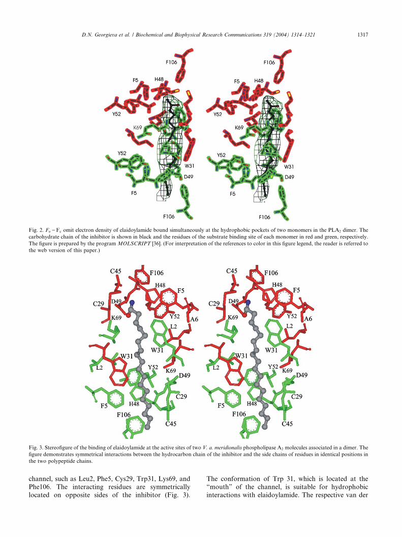
Fig. 3. Stereofigure of the binding of elaidoylamide at the active sites of two V. a. meridionalis phospholipase A2 molecules associated in a dimer. The
figure demonstrates symmetrical interactions between the hydrocarbon chain of the inhibitor and the side chains of residues in identical positions in
the two polypeptide chains.
Fig. 2. Fo )Fc omit electron density of elaidoylamide bound simultaneously at the hydrophobic pockets of two monomers in the PLA2 dimer. The
carbohydrate chain of the inhibitor is shown in black and the residues of the substrate binding site of each monomer in red and green, respectively.
The figure is prepared by the program MOLSCRIPT [36]. (For interpretation of the references to color in this figure legend, the reader is referred to
the web version of this paper.)
D.N. Georgieva et al. / Biochemical and Biophysical Research Communications 319 (2004) 1314–1321 1317
channel, such as Leu2, Phe5, Cys29, Trp31, Lys69, and
Phe106. The interacting residues are symmetrically
located on opposite sides of the inhibitor (Fig. 3).
The conformation of Trp 31, which is located at the
“mouth” of the channel, is suitable for hydrophobic
interactions with elaidoylamide. The respective van der
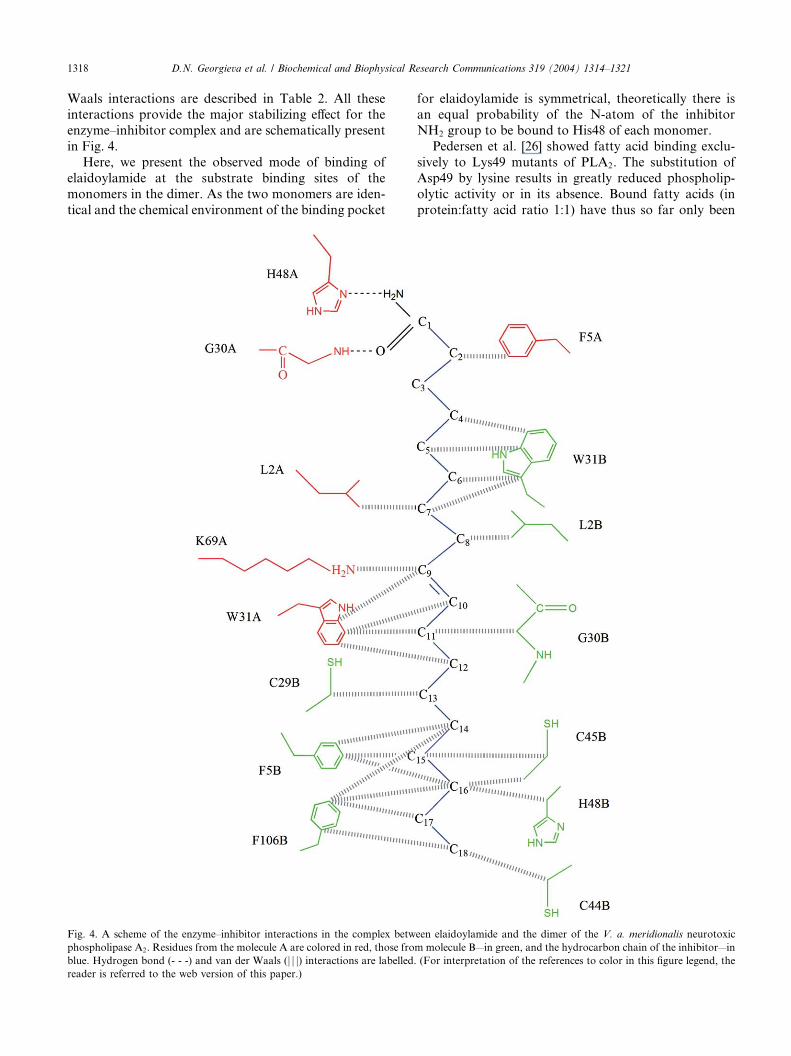
1318 D.N. Georgieva et al. / Biochemical and Biophysical Research Communications 319 (2004) 1314–1321
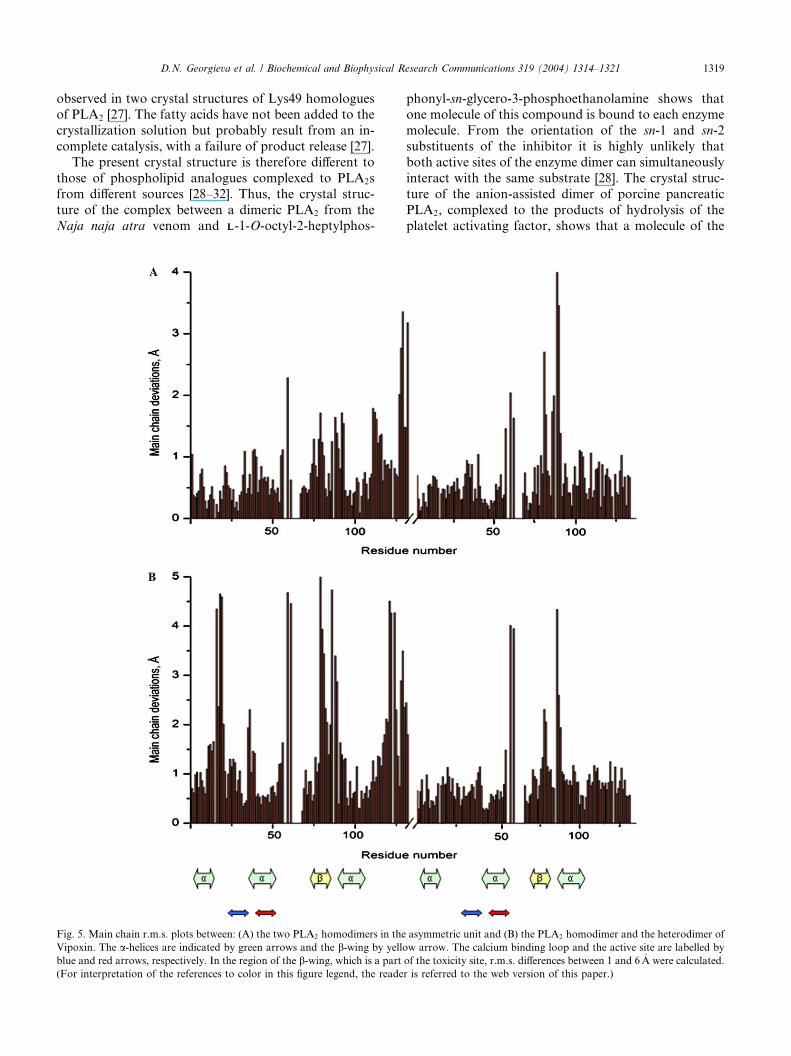
Waals interactions are described in Table 2. All theseinteractions provide the major stabilizing effect for the
enzyme–inhibitor complex and are schematically present
in Fig. 4.
Here, we present the observed mode of binding of
elaidoylamide at the substrate binding sites of the
monomers in the dimer. As the two monomers are iden-
tical and the chemical environment of the binding pocket
Fig. 4. A scheme of the enzyme–inhibitor interactions in the complex betw
phospholipase A2. Residues from the molecule A are colored in red, those fro
blue. Hydrogen bond (- - -) and van der Waals (j j j) interactions are labelled.reader is referred to the web version of this paper.)
for elaidoylamide is symmetrical, theoretically there isan equal probability of the N-atom of the inhibitor
NH2 group to be bound to His48 of each monomer.
Pedersen et al. [26] showed fatty acid binding exclu-
sively to Lys49 mutants of PLA2. The substitution of
Asp49 by lysine results in greatly reduced phospholip-
olytic activity or in its absence. Bound fatty acids (in
protein:fatty acid ratio 1:1) have thus so far only been
een elaidoylamide and the dimer of the V. a. meridionalis neurotoxic
m molecule B—in green, and the hydrocarbon chain of the inhibitor—in
(For interpretation of the references to color in this figure legend, the
D.N. Georgieva et al. / Biochemical and Biophysical Research Communications 319 (2004) 1314–1321 1319
observed in two crystal structures of Lys49 homologuesof PLA2 [27]. The fatty acids have not been added to the
crystallization solution but probably result from an in-
complete catalysis, with a failure of product release [27].
The present crystal structure is therefore different to
those of phospholipid analogues complexed to PLA2s
from different sources [28–32]. Thus, the crystal struc-
ture of the complex between a dimeric PLA2 from the
Naja naja atra venom and LL-1-O-octyl-2-heptylphos-
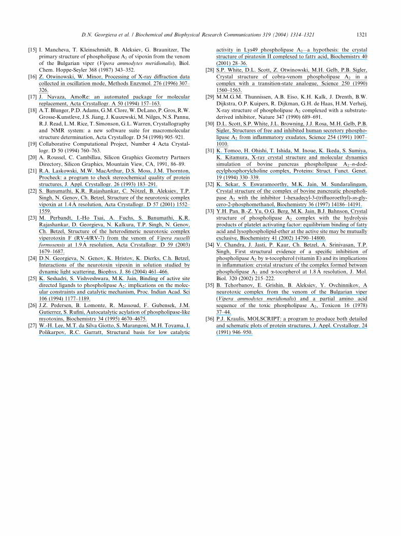
Fig. 5. Main chain r.m.s. plots between: (A) the two PLA2 homodimers in the
Vipoxin. The a-helices are indicated by green arrows and the b-wing by yello
blue and red arrows, respectively. In the region of the b-wing, which is a part
(For interpretation of the references to color in this figure legend, the reade
phonyl-sn-glycero-3-phosphoethanolamine shows thatone molecule of this compound is bound to each enzyme
molecule. From the orientation of the sn-1 and sn-2
substituents of the inhibitor it is highly unlikely that
both active sites of the enzyme dimer can simultaneously
interact with the same substrate [28]. The crystal struc-
ture of the anion-assisted dimer of porcine pancreatic
PLA2, complexed to the products of hydrolysis of the
platelet activating factor, shows that a molecule of the
asymmetric unit and (B) the PLA2 homodimer and the heterodimer of
w arrow. The calcium binding loop and the active site are labelled by
of the toxicity site, r.m.s. differences between 1 and 6�A were calculated.
r is referred to the web version of this paper.)

1320 D.N. Georgieva et al. / Biochemical and Biophysical Research Communications 319 (2004) 1314–1321
product acetate is bound to subunit A and the otherproduct, 1-octadecyl-sn-glycero-3-phosphocholine, to
subunit B [33]. In the complex between a-tocopheroland dimeric PLA2 from the Vipera russelli venom one
molecule of vitamin E is bound specifically to one of the
subunits [34].
Comparison of the neurotoxic homodimeric PLA2 struc-
ture with that of the heterodimeric toxin Vipoxin
The V. a. meridionalis neurotoxic complex Vipoxin
binds to the postsynaptic membranes of the neuromus-
cular junctions preventing the binding of acetylcholine
to its receptor [35]. In this way it blocks the neuro-muscular transmission of skeletal muscles and exerts its
lethal effect. The separation of the toxic PLA2 from the
chaperone subunit changes the target of the physiolog-
ical attack from the post- to presynaptic membranes
affecting the neurotransmitter release. To correlate the
change of the pharmacological activity with possible
structural changes in the separated neurotoxin, we have
compared the toxic dimeric PLA2 structure with that ofVipoxin using the PDB code 1jlt. The overall confor-
mation of the two homodimers in the asymmetric unit is
highly similar, with an average r.m.s. displacement of
0.9�A for the main chains (Fig. 5A). Comparison of each
of the two PLA2 homodimers with the hetorodimer
Vipoxin showed an average main chain r.m.s. of 1.4�A(Fig. 5B). However, more differences were observed in
the region of the b-wing (residues 76–81), which is a partof the toxicity site, with r.m.s. deviations of 1–5�A. This
suggests that the change of the pharmacological activity
is connected with conformational changes in the toxic
PLA2.
Conclusions
The present investigations reveal a new mode of
PLA2 inhibition by an amide of fatty acid. Probably, the
proposed inhibitor will be tolerant to living organisms.
Additional studies in this direction will be performed.
The design of specific PLA2 inhibitors is of great phar-
macological interest and requires a detailed knowledge
of the mode of binding of the inhibitor to the enzyme
active site. The three-dimensional structure of the com-plex elaidoylamide–PLA2 can be used to design anti-in-
flammatory drugs. The simple hydrocarbon chain of the
inhibitor can be in principle well adjusted in the very ho-
mologous (conserved) substrate binding channels leading
to the catalytic sites of Group I/II/III PLA2s. In this re-
spect the inhibitor can have more universal applications.
The change of the target of the physiological attack
of the separated from Vipoxin neurotoxic PLA2 fromthe post- to presynaptic membranes of the neuromus-
cular junctions is connected with a conformational
change of its polypeptide chain in the region of thetoxicity site.
Acknowledgments
The authors thank the Deutsche Forschungsgemeinschaft for fi-
nancial support by the Projects 436 BUL 113/115/01 and BE 1443/9-1.
D. N. Georgieva thanks the Alexander von Humboldt Foundation,
Bonn, Germany, for providing a Research Fellowship, IV-BUL/
1073481 STP.
References
[1] E. Valentin, G. Lambeau, Increasing molecular diversity of
secreted phospholipases A2 and their receptors and binding
proteins, Biochim. Biophys. Acta 1488 (2000) 59–70.
[2] D.A. Six, E.A. Dennis, The expanding superfamily of phospho-
lipase A2 enzymes: classification and characterization, Biochim.
Biophys. Acta 1488 (2000) 1–19.
[3] J. Balsinde, M.A. Balboa, E.A. Dennis, Antisense inhibition of
group VI Ca2þ-independent phospholipase A2 blocks phospho-
lipid fatty acid remodelling in murine P388D1 macrophages, J.
Biol. Chem. 272 (1997) 29317–29321.
[4] P. Vadas, W. Pruzanski, Role of secretory phospholipase A2 in the
pathobiology of disease, Lab. Invest. 4 (1986) 391–404.
[5] P. Vadas, W. Pruzanski, E. Stefanski, Extracellular phospholipase
A2: causative agent in circulatory collapse of septic shock?, Agents
Act. 24 (1988) 320–325.
[6] A. Aarsman, F. Neys, H. van den Helm, F. Kuypers, H. van den
Bosch, Sera of patients suffering from inflammatory diseases
contain group IIA but not group V phospholipase A2, Biochim.
Biophys. Acta 1502 (2000) 257–263.
[7] T.J. Nevalainen, Serum phospholipase A2 in inflammatory
diseases, Clin. Chem. 39 (1993) 2453–2459.
[8] D.K. Kim, T. Fukuda, B.T. Thomson, B. Cockrill, C. Halles, J.V.
Bonventre, Bronchoalveolar lavage fluid phospholipase A2 activ-
ities are increased in human adult respiratory distress syndrome,
Am. J. Physiol. 269 (1995) 109–118.
[9] M.Z. Huang, P. Gopalakrishnakone, M.C.M. Chung, R.M. Kini,
Complete amino acid sequence of an acidic cardiotoxic phospho-
lipase A2 from the venom of Ophiophagus hannah (king cobra): a
novel cobra venom enzyme with pancreatic loop, Arch. Biochem.
Biophys. 338 (1997) 150–156.
[10] B. Aleksiev, R. Shipolini, Weitere untersuchungen zur fraktionie-
rung und reiningung der toxischen proteine aus dem gift der
bulgarischen viper (Vipera ammodytes meridionalis), Hoppe-Sey-
ler-Z. Physiol. Chem. 352 (1971) 1183–1187.
[11] B. Aleksiev, B. Chorbanov, Action on phosphatidylcholine of the
toxic phospholipase A2 from the venom of Bulgarian viper (Vipera
ammodytes meridionalis), Toxicon 14 (1976) 477–484.
[12] S.S. Funari, F. Barcelo, P.V. Escriba, Effects of oleic acid and its
congeners, elaidic and stearic acids, on the structural properties of
phosphatidylethanolamine membranes, J. Lipid Res. 44 (2003)
567–575.
[13] D.N. Georgieva, W. Rypniewski, M. Perbandt, M. Jain, N.
Genov, Ch. Betzel, Crystallization and preliminary X-ray diffrac-
tion studies of a toxic phospholipase A2 from the venom of Vipera
ammodytes meridionalis complexed to a synthetic inhibitor,
Biochim. Biophys. Acta 1650 (2003) 1–3.
[14] B. Tchorbanov, B. Aleksiev, A simple procedure for the isolation
of vipoxin—a neurotoxin with weak phospholipase activity from
the venom of the Bulgarian viper (Vipera ammodytes meridionalis),
J. Appl. Biochem. 3 (1981) 558–561.
D.N. Georgieva et al. / Biochemical and Biophysical Research Communications 319 (2004) 1314–1321 1321
[15] I. Mancheva, T. Kleinschmidt, B. Aleksiev, G. Braunitzer, The
primary structure of phospholipase A2 of vipoxin from the venom
of the Bulgarian viper (Vipera ammodytes meridionalis), Biol.
Chem. Hoppe-Seyler 368 (1987) 343–352.
[16] Z. Otwinowski, W. Minor, Processing of X-ray diffraction data
collected in oscillation mode, Methods Enzymol. 276 (1996) 307–
326.
[17] J. Navaza, AmoRe: an automated package for molecular
replacement, Acta Crystallogr. A 50 (1994) 157–163.
[18] A.T.Blunger, P.D.Adams,G.M.Clore,W.DeLano, P.Gros,R.W.
Grosse-Kunstleve, J.S. Jiang, J. Kuszewski,M.Nilges, N.S. Pannu,
R.J. Read, L.M. Rice, T. Simonson, G.L.Warren, Crystallography
and NMR system: a new software suite for macromolecular
structure determination, Acta Crystallogr. D 54 (1998) 905–921.
[19] Collaborative Computational Project, Number 4 Acta Crystal-
logr. D 50 (1994) 760–763.
[20] A. Roussel, C. Cambillau, Silicon Graphics Geometry Partners
Directory, Silicon Graphics, Mountain View, CA, 1991, 86–89.
[21] R.A. Laskowski, M.W. MacArthur, D.S. Moss, J.M. Thornton,
Procheck: a program to check stereochemical quality of protein
structures, J. Appl. Crystallogr. 26 (1993) 183–291.
[22] S. Banumathi, K.R. Rajashankar, C. N€otzel, B. Aleksiev, T.P.
Singh, N. Genov, Ch. Betzel, Structure of the neurotoxic complex
vipoxin at 1.4�A resolution, Acta Crystallogr. D 57 (2001) 1552–
1559.
[23] M. Perbandt, I.-Ho Tsai, A. Fuchs, S. Banumathi, K.R.
Rajashankar, D. Georgieva, N. Kalkura, T.P. Singh, N. Genov,
Ch. Betzel, Structure of the heterodimeric neurotoxic complex
viperotoxin F (RV-4/RV-7) from the venom of Vipera russelli
formosensis at 1.9�A resolution, Acta Crystallogr. D 59 (2003)
1679–1687.
[24] D.N. Georgieva, N. Genov, K. Hristov, K. Dierks, C.h. Betzel,
Interactions of the neurotoxin vipoxin in solution studied by
dynamic light scattering, Biophys. J. 86 (2004) 461–466.
[25] K. Seshadri, S. Vishveshwara, M.K. Jain, Binding of active site
directed ligands to phospholipase A2: implications on the molec-
ular constraints and catalytic mechanism, Proc. Indian Acad. Sci
106 (1994) 1177–1189.
[26] J.Z. Pedersen, B. Lomonte, R. Massoud, F. Gubensek, J.M.
Gutierrez, S. Rufini, Autocatalytic acylation of phospholipase-like
myotoxins, Biochemistry 34 (1995) 4670–4675.
[27] W.-H. Lee, M.T. da Silva Giotto, S. Marangoni, M.H. Toyama, I.
Polikarpov, R.C. Garratt, Structural basis for low catalytic
activity in Lys49 phospholipase A2—a hypothesis: the crystal
structure of piratoxin II complexed to fatty acid, Biochemistry 40
(2001) 28–36.
[28] S.P. White, D.L. Scott, Z. Otwinowski, M.H. Gelb, P.B. Sigler,
Crystal structure of cobra-venom phospholipase A2 in a
complex with a transition-state analogue, Science 250 (1990)
1560–1563.
[29] M.M.G.M. Thunnissen, A.B. Eiso, K.H. Kalk, J. Drenth, B.W.
Dijkstra, O.P. Kuipers, R. Dijkman, G.H. de Haas, H.M. Verheij,
X-ray structure of phospholipase A2 complexed with a substrate-
derived inhibitor, Nature 347 (1990) 689–691.
[30] D.L. Scott, S.P. White, J.L. Browning, J.J. Rosa, M.H. Gelb, P.B.
Sigler, Structures of free and inhibited human secretory phospho-
lipase A2 from inflammatory exudates, Science 254 (1991) 1007–
1010.
[31] K. Tomoo, H. Ohishi, T. Ishida, M. Inoue, K. Ikeda, S. Sumiya,
K. Kitamura, X-ray crystal structure and molecular dynamics
simulation of bovine pancreas phospholipase A2–n-dod-
ecylphosphorylcholine complex, Proteins: Struct. Funct. Genet.
19 (1994) 330–339.
[32] K. Sekar, S. Eswaramoorthy, M.K. Jain, M. Sundaralingam,
Crystal structure of the complex of bovine pancreatic phospholi-
pase A2 with the inhibitor 1-hexadecyl-3-(trifluoroethyl)-sn-gly-
cero-2-phosphomethanol, Biochemistry 36 (1997) 14186–14191.
[33] Y.H. Pan, B.-Z. Yu, O.G. Berg, M.K. Jain, B.J. Bahnson, Crystal
structure of phospholipase A2 complex with the hydrolysis
products of platelet activating factor: equilibrium binding of fatty
acid and lysophospholipid-ether at the active site may be mutually
exclusive, Biochemistry 41 (2002) 14790–14800.
[34] V. Chandra, J. Jasti, P. Kaur, Ch. Betzel, A. Srinivasan, T.P.
Singh, First structural evidence of a specific inhibition of
phospholipase A2 by a-tocopherol (vitamin E) and its implications
in inflammation: crystal structure of the complex formed between
phospholipase A2 and a-tocopherol at 1.8�A resolution, J. Mol.
Biol. 320 (2002) 215–222.
[35] B. Tchorbanov, E. Grishin, B. Aleksiev, Y. Ovchinnikov, A
neurotoxic complex from the venom of the Bulgarian viper
(Vipera ammodytes meridionalis) and a partial amino acid
sequence of the toxic phospholipase A2, Toxicon 16 (1978)
37–44.
[36] P.J. Kraulis, MOLSCRIPT: a program to produce both detailed
and schematic plots of protein structures, J. Appl. Crystallogr. 24
(1991) 946–950.


![kkt_6_dan_7_pemupukan_2014 [Compatibility Mode]](https://static.fdokumen.com/doc/165x107/6322b43c28c445989105e2db/kkt6dan7pemupukan2014-compatibility-mode.jpg)

![Ruth Knight presentation.ppt [Compatibility Mode]](https://static.fdokumen.com/doc/165x107/631d5d013ba403638902baaf/ruth-knight-presentationppt-compatibility-mode.jpg)
![Dr T Malakoutian.ppt [Compatibility Mode]](https://static.fdokumen.com/doc/165x107/63364bfed2b7284203084459/dr-t-malakoutianppt-compatibility-mode.jpg)