Diagnosis of dengue infection using a modified gold electrode with hybrid organic–inorganic...
7
Diagnosis of dengue infection using a modified gold electrode with hybrid organic–inorganic nanocomposite and Bauhinia monandra lectin Cesar A.S. Andrade a,⇑ , Maria D.L. Oliveira b , Celso P. de Melo c , Luana C.B.B. Coelho b , Maria T.S. Correia b , Maurício L. Nogueira d , Pankaj R. Singh e , Xiangqun Zeng e a Centro Acadêmico de Vitória, UFPE, 55608-680, Vitória de Santo Antão, PE, Brazil b Departamento de Bioquímica, UFPE, 50670-901 Recife, PE, Brazil c Departamento de Física, UFPE, 50670-901 Recife, PE, Brazil d Departamento de Doenças Infecciosas e Parasitárias, FAMERP, 15090-000 São Jose do Rio Preto, SP, Brazil e Department of Chemistry, Oakland University, 2200 Squirrel Road, Rochester, MI 48309, USA article info Article history: Received 30 May 2011 Accepted 2 July 2011 Available online 3 August 2011 Keywords: Impedance spectroscopy Cyclic voltammetry Dengue Lectin Bauhinia monandra abstract A sensitive and selective biosensor for dengue serotyping was successfully developed. The biosensor uses a novel gold nanoparticles-polyaniline hybrid composite (AuNpPANI) for the immobilization of Bauhinia monandra lectin (BmoLL). The nanocomposite was applied to a bare gold electrode surface by chemical adsorption, and BmoLL was subsequently electrostatically adsorbed to the nanocomposite-modified sur- face. Atomic force microscopy (AFM), cyclic voltammetry (CV) and electrochemical impedance (EI) tech- niques were applied to evaluate the immobilization of BmoLL on AuNpPANI. The AFM images for AuNpPANI-BmoLL-DEN systems indicate a homogenous, compact and dense film of the conjugate. In the EI analyses, an obvious difference of the electron transfer resistance between the AuNpPANI-modified electrode and the bare gold electrode was observed. Among three dengue serotypes studied, dengue sero- type 2 (DEN2) has higher values for R CT , and lower values for both n and Q. These are indications of a lar- ger blocking effect and smaller capacitive dispersion, resulting from the higher agglutination of glycoproteins from the DEN2 sera. The selective BmoLL recognition for various dengue serotypes may be attributed to different patterns of glycoproteins in the sera produced by the glycoprotein immunore- sponse from patients infected by the dengue virus. Ó 2011 Elsevier Inc. All rights reserved. 1. Introduction Dengue viral diagnostics are typically performed by bioassays such as the growth of the virus in a cell culture from a specimen taken from the patient, detection of virus-specific antibodies in the blood, detection of virus antigens, detection of virus nucleic acids, examination of virus particles by electron microscopy and hemagglutination assay. However, these methods have been either too complex, time-consuming, lacking the necessary sensitivity and specificity or too expensive to be widely deployed. Thus, diag- nostic methods that can identify the disease reliably and rapidly, and subsequently treat dengue virus infection at an early stage are urgently needed [1]. The dengue virus nonstructural glycoprotein, NS1, may present with different molecular mass, depending on the glycosilation. Some data suggest conflicting results for the use of this protein as a potential dengue hemorrhagic fever (DHF) predictor [2]. On the other hand, the level of some cytokines and the soluble vascular cell adhesion molecule 1 (VCAM-1) in blood samples may indicate a pattern for dengue fever (DF) and DHF recognition [3]. VCAM-1 is expressed on the surfaces of endothelial cells following stimulation with endotoxin and cytokines such as IL-1, TNF, IL-4, and IL-13 [3], as well as in dengue infections. Recent studies [3] have shown that the VCAM-1, a highly conserved glyco- protein produced in both membrane-associated and secreted forms and abundant in the serum of patients in the early stages of the immunoresponse of dengue virus infection may be an appro- priate marker of acute and hemorrhagic dengue virus infection. Lectins are carbohydrate-binding proteins of nonimmune origin and are involved in numerous cellular processes according to their characteristic structure and common interaction principles [4]. Previous studies of our group demonstrated the electrochemical response of the Concanavalin A (Con A) modified electrode for evaluation of a biosensor response for serum glycoproteins from patients infected by the dengue virus. This study revealed different responses for the DHF and DF sera [5]. Thus, we hypothesize that naturally occurring lectins can be used to detect serum glycoproteins immunoresponse from patients infected by the dengue virus. In the present work, the Bauhinia monandra (BmoLL), 0021-9797/$ - see front matter Ó 2011 Elsevier Inc. All rights reserved. doi:10.1016/j.jcis.2011.07.013 ⇑ Corresponding author. Fax: +55 81 3523 3351. E-mail address: [email protected] (C.A.S. Andrade). Journal of Colloid and Interface Science 362 (2011) 517–523 Contents lists available at ScienceDirect Journal of Colloid and Interface Science www.elsevier.com/locate/jcis
Transcript of Diagnosis of dengue infection using a modified gold electrode with hybrid organic–inorganic...

Journal of Colloid and Interface Science 362 (2011) 517–523
Contents lists available at ScienceDirect
Journal of Colloid and Interface Science
www.elsevier .com/locate / jc is
Diagnosis of dengue infection using a modified gold electrode with hybridorganic–inorganic nanocomposite and Bauhinia monandra lectin
Cesar A.S. Andrade a,⇑, Maria D.L. Oliveira b, Celso P. de Melo c, Luana C.B.B. Coelho b, Maria T.S. Correia b,Maurício L. Nogueira d, Pankaj R. Singh e, Xiangqun Zeng e
a Centro Acadêmico de Vitória, UFPE, 55608-680, Vitória de Santo Antão, PE, Brazilb Departamento de Bioquímica, UFPE, 50670-901 Recife, PE, Brazilc Departamento de Física, UFPE, 50670-901 Recife, PE, Brazild Departamento de Doenças Infecciosas e Parasitárias, FAMERP, 15090-000 São Jose do Rio Preto, SP, Brazile Department of Chemistry, Oakland University, 2200 Squirrel Road, Rochester, MI 48309, USA
a r t i c l e i n f o
Article history:Received 30 May 2011Accepted 2 July 2011Available online 3 August 2011
Keywords:Impedance spectroscopyCyclic voltammetryDengueLectinBauhinia monandra
0021-9797/$ - see front matter � 2011 Elsevier Inc. Adoi:10.1016/j.jcis.2011.07.013
⇑ Corresponding author. Fax: +55 81 3523 3351.E-mail address: [email protected] (C.A.S. And
a b s t r a c t
A sensitive and selective biosensor for dengue serotyping was successfully developed. The biosensor usesa novel gold nanoparticles-polyaniline hybrid composite (AuNpPANI) for the immobilization of Bauhiniamonandra lectin (BmoLL). The nanocomposite was applied to a bare gold electrode surface by chemicaladsorption, and BmoLL was subsequently electrostatically adsorbed to the nanocomposite-modified sur-face. Atomic force microscopy (AFM), cyclic voltammetry (CV) and electrochemical impedance (EI) tech-niques were applied to evaluate the immobilization of BmoLL on AuNpPANI. The AFM images forAuNpPANI-BmoLL-DEN systems indicate a homogenous, compact and dense film of the conjugate. Inthe EI analyses, an obvious difference of the electron transfer resistance between the AuNpPANI-modifiedelectrode and the bare gold electrode was observed. Among three dengue serotypes studied, dengue sero-type 2 (DEN2) has higher values for RCT, and lower values for both n and Q. These are indications of a lar-ger blocking effect and smaller capacitive dispersion, resulting from the higher agglutination ofglycoproteins from the DEN2 sera. The selective BmoLL recognition for various dengue serotypes maybe attributed to different patterns of glycoproteins in the sera produced by the glycoprotein immunore-sponse from patients infected by the dengue virus.
� 2011 Elsevier Inc. All rights reserved.
1. Introduction
Dengue viral diagnostics are typically performed by bioassayssuch as the growth of the virus in a cell culture from a specimentaken from the patient, detection of virus-specific antibodies inthe blood, detection of virus antigens, detection of virus nucleicacids, examination of virus particles by electron microscopy andhemagglutination assay. However, these methods have been eithertoo complex, time-consuming, lacking the necessary sensitivityand specificity or too expensive to be widely deployed. Thus, diag-nostic methods that can identify the disease reliably and rapidly,and subsequently treat dengue virus infection at an early stageare urgently needed [1].
The dengue virus nonstructural glycoprotein, NS1, may presentwith different molecular mass, depending on the glycosilation.Some data suggest conflicting results for the use of this proteinas a potential dengue hemorrhagic fever (DHF) predictor [2]. Onthe other hand, the level of some cytokines and the soluble
ll rights reserved.
rade).
vascular cell adhesion molecule 1 (VCAM-1) in blood samplesmay indicate a pattern for dengue fever (DF) and DHF recognition[3]. VCAM-1 is expressed on the surfaces of endothelial cellsfollowing stimulation with endotoxin and cytokines such as IL-1,TNF, IL-4, and IL-13 [3], as well as in dengue infections. Recentstudies [3] have shown that the VCAM-1, a highly conserved glyco-protein produced in both membrane-associated and secretedforms and abundant in the serum of patients in the early stagesof the immunoresponse of dengue virus infection may be an appro-priate marker of acute and hemorrhagic dengue virus infection.
Lectins are carbohydrate-binding proteins of nonimmune originand are involved in numerous cellular processes according to theircharacteristic structure and common interaction principles [4].Previous studies of our group demonstrated the electrochemicalresponse of the Concanavalin A (Con A) modified electrode forevaluation of a biosensor response for serum glycoproteins frompatients infected by the dengue virus. This study revealed differentresponses for the DHF and DF sera [5]. Thus, we hypothesize thatnaturally occurring lectins can be used to detect serumglycoproteins immunoresponse from patients infected by thedengue virus. In the present work, the Bauhinia monandra (BmoLL),
518 C.A.S. Andrade et al. / Journal of Colloid and Interface Science 362 (2011) 517–523
a galactose-specific lectin, purified and characterized from leaves ofB. monandra Kurz obtained from the northeast region of Brazil [6–8],was shown to be able to detect the dengue virus using electrochem-ical impedance readout that is sensitive, rapid and serotype-specific.
Studies by electrochemical impedance spectroscopy (EIS) dem-onstrate that proteins do not interact significantly with a gold elec-trode surface, not even through nonspecific interactions [9]. Thus,the introduction of accessible cysteine residue by genetic engineer-ing techniques has served as a valuable method to immobilize pro-teins onto a metal electrode surface in a controlled manner [10].Genetic engineering techniques frequently include expensive re-agents and time-consuming procedures. New immobilizationschemes and advanced sensing materials are therefore highly val-ued for improving the analytical capabilities of biosensing devices.
Because of the large specific surface area of nanoparticles, excel-lent conductivity and surface charge existing in the gold nanopar-ticles-polyaniline hybrid composites (AuNpPANI), BmoLL can beeasily immobilized with high density and high activity. Polyaniline(PANI) reveals redox functions only in acid media (pH < 4) [11] afeature that limits its broad use, specifically in combination withbiomaterials. However, BmoLL is stable in this pH range and hasa negative surface charge [12].
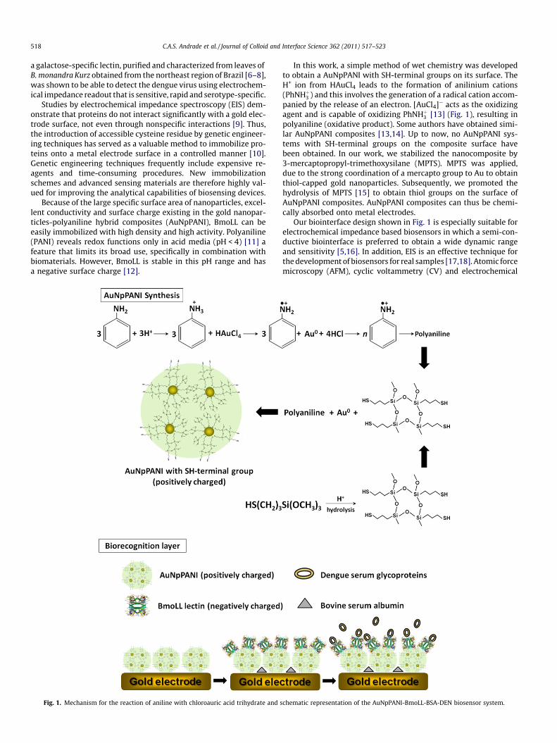
Fig. 1. Mechanism for the reaction of aniline with chloroauric acid trihydrate and s
In this work, a simple method of wet chemistry was developedto obtain a AuNpPANI with SH-terminal groups on its surface. TheH+ ion from HAuCl4 leads to the formation of anilinium cations(PhNHþ3 ) and this involves the generation of a radical cation accom-panied by the release of an electron. [AuCl4]� acts as the oxidizingagent and is capable of oxidizing PhNHþ3 [13] (Fig. 1), resulting inpolyaniline (oxidative product). Some authors have obtained simi-lar AuNpPANI composites [13,14]. Up to now, no AuNpPANI sys-tems with SH-terminal groups on the composite surface havebeen obtained. In our work, we stabilized the nanocomposite by3-mercaptopropyl-trimethoxysilane (MPTS). MPTS was applied,due to the strong coordination of a mercapto group to Au to obtainthiol-capped gold nanoparticles. Subsequently, we promoted thehydrolysis of MPTS [15] to obtain thiol groups on the surface ofAuNpPANI composites. AuNpPANI composites can thus be chemi-cally absorbed onto metal electrodes.
Our biointerface design shown in Fig. 1 is especially suitable forelectrochemical impedance based biosensors in which a semi-con-ductive biointerface is preferred to obtain a wide dynamic rangeand sensitivity [5,16]. In addition, EIS is an effective technique forthe development of biosensors for real samples [17,18]. Atomic forcemicroscopy (AFM), cyclic voltammetry (CV) and electrochemical
chematic representation of the AuNpPANI-BmoLL-BSA-DEN biosensor system.

C.A.S. Andrade et al. / Journal of Colloid and Interface Science 362 (2011) 517–523 519
impedance spectroscopy (EIS) techniques were used to investigatethe immobilization of BmoLL on AuNpPANI to evaluate its interac-tion with the patients’ sera infected by dengue serotypes 1, 2, and3 (DEN1, DEN2, and DEN3).
2. Materials and methods
2.1. Materials
BmoLL lectin was purified by one of us (Coelho, LCBB) [6] fromthe UFPE’s Laboratory of Glycoproteins. The dengue sera were ob-tained from the Laboratory of Virology/FAMERP stocks. In thiswork we used the sera of patients contaminated with dengue ser-otypes 1, 2 and 3 (three patients for each serotype) and serum con-trol (three patients). All sera were previously characterized asdengue negative (serum control) and dengue positive (serotypes1, 2 and 3) using RT-PCR [19]. Aniline was purchased from Vetec(Brasil) and only used after distillation. HAuCl4�3H2O, 3-mercapto-propyl-trimethoxysilane (MPTS), bovine serum albumin (BSA) andpotassium ferricyanide were purchased from Sigma Chemical (St.Louis, MO, USA). Potassium ferrocyanide was obtained from Merck.Phosphate buffer saline (PBS) pH 7.2 was purchased from Gibco(Carlsbad, CA, USA).
2.2. Preparation of AuNpPANI composites
Hybrid organic–inorganic composites were obtained accordingto our early work [20]. The preparation of AuNpPANI was per-formed in a round bottom glass flask containing ethanol (20 mL)and the compounds aniline (0.030 mol/L), MPTS(6.46 � 10�2 mol/L), and HAuCl4�3H2O (0.81 mmol/L), which weresubsequently added and submitted to agitation (1100 rpm) for48 h. Thus, the samples were centrifuged for 10 min at 12,000RPM (to remove large aggregates), and 500 lL of 0.1 M HCl wassubsequently added to obtain positively-charged PANI and pro-mote the hydrolysis of MPTS [15].
2.3. Electrode surface modification
The gold electrode surface was freshly polished prior to its usewith 0.05 lm a-Al2O3 paste, and rigorously rinsed with doubly dis-tilled water following each polish. Next, the electrode was im-mersed in freshly prepared Piranha solution (30% H2O2 andconcentrated H2SO4, 3:1 v/v) for 5 min. The polished electrodewas then cleaned ultrasonically sequentially in water for 5 min.After being sonicated in deionized water, the electrode was elec-trochemically pretreated by cyclic potential scanning between1.4 and �0.2 V in 0.2 M H2SO4 until a cyclic voltammogram ofclean gold electrode was obtained. BmoLL solution (0.3 mg/mL)was mixed with 300 lL of AuNpPANI composite in a beaker andkept under stirring for 10 min. The AuNpPANI-BmoLL-modifiedelectrode was obtained by dropping AuNpPANI-BmoLL solutiononto the bare gold electrode surface at 25 �C for 2 min. Finally,the AuNpPANI-BmoLL-modified electrode was rinsed with waterto remove unbound lectin and incubated in a PBS solution contain-ing 0.2% BSA for 20 min at 25 �C in order to block the remaining ac-tive sites. The AuNpPANI-BmoLL-BSA-modified electrode wasexposed to negative serum control and serum from patients in-fected by dengue serotype 1, 2 and 3 diluted in 10 mM pH 7.4PBS solution (dilutions of 1:25, 1:50, 1:75, 1:100 and 1:150) for10 min at room temperature.
2.4. Atomic force microscopy measurements
Atomic force microscopy (AFM) measurements were performedwith a commercial PicoPlus microscope (Molecular Imaging, USA).Cantilevers with a Cr–Au tip (NSC18, MikroMasch, F0 = 90 KHz,nominal spring constant = 5.5 N m�1) were used for the tappingmode AFM in air at room temperature (approximately 25 �C)[21]. Scan areas varying from 0.5 lm � 0.5 lm with a resolutionof 512 � 512 pixels were obtained. The AFM Gwyddion softwarewas used to analyze the recorded scans [22]. HOPG substratewas obtained from Advanced Ceramics Corp. (Grade ZYH, pyrolyticGraphite Monochromator). To eliminate artifacts, images were ob-tained from at least two macroscopically separated areas on eachsample.
2.5. Electrochemical measurements
Electrochemical measurements were carried out on a PARSTAT2263 (Princeton Applied Research, USA) potentiostat. The imped-ance spectra were recorded in the frequency range of 100 mHz to100 kHz. All tests were conducted on an open circuit potential,and a single modulated AC potential of 10 mV was applied. Imped-ance measurement was performed in the presence of a10 mM K4[Fe(CN)6]4�/K3[Fe(CN)6]3� (1:1) mixture as a redox probein 10 mM PBS solution containing 0.15 M NaCl [23]. CV measure-ments were performed in a conventional electrochemical cell con-taining a three-electrode setup with an Ag/AgCl (saturated KCl)reference electrode and swept the potential between �0.3 and+0.7 V with a scan rate of 80 mV s�1. A platinum wire and a mod-ified gold disk (d = 2 mm) were used as auxiliary and working elec-trodes, respectively. All measurements were performed intriplicate.
3. Results and discussion
3.1. AFM characterization
Fig. 2 demonstrates AFM images of the AuNpPANI before (a) andafter (b) BmoLL interaction, respectively. From Fig. 2a well-definedAuNpPANI particles are clearly seen on the surface and are wellisolated from each other, owing to the electrostatic repulsion.The AuNpPANI particles formed were in the range of �45 nm andheight of 16 nm, resulting in a large surface area and easier attach-ment of BmoLL molecules. After BmoLL interaction, the diameter ofthe AuNpPANI slightly increased, indicating that BmoLL moleculeswere immobilized on the surface of AuNpPANI.
The AFM images for AuNpPANI-BmoLL-DEN systems indicate ahomogenous, compact and dense film of the conjugate. Thus,BmoLL acts as a binding protein to the sensing glycoprotein, whichanchors very well to the AuNpPANI sensor surface. Regardless ofthe dengue serotype, on adsorbing glycoproteins onto the AuNpPA-NI-BmoLL sensor layer, the AuNpPANI-BmoLL was covered by afine structured film with a thickness ranging from 2 to 15 nm(Fig. 2c–e). However, DEN3 (Fig. 2e) and negative control (Fig. 2f)presented the weakest adhesion to AuNpPANI-BmoLL, with lowervalues for height than DEN1 (Fig. 2a) and DEN2 (Fig. 2b).
3.2. Electrochemical characteristics of AuNpPANI-BmoLL-DEN system
The stepwise modification on the gold electrode is accompaniedby a decrease in the amperometric response and increase in thepeak-to-peak separation between the cathodic and anodic wavesof the redox probe. This is consistent with the hindering of the elec-tron-transfer barriers introduced on assembly of these layers. Inparticular, after interactions with DEN2 an obvious disappearance

Fig. 2. An AFM topographic image of the HOPG surface modified with AuNpPANI (a), AuNpPANI-BmoLL (b), AuNpPANI-BmoLL-DEN1 (c), AuNpPANI-BmoLL-DEN2 (d),AuNpPANI-BmoLL-DEN3 (e) and AuNpPANI-BmoLL-DENnegative (f). The dilution used of dengue serotypes was 1:50. All scan areas were varied from 0.5 lm � 0.5 lm with aresolution of 512 � 512 pixels.
520 C.A.S. Andrade et al. / Journal of Colloid and Interface Science 362 (2011) 517–523
of the anodic and cathodic peaks was obtained, probably through aspecific interaction between lectin-glycoproteins present in testedserum. The reason is that the lectin-glycoprotein complex acts asthe inert electron and mass-transfer blocking layer and hindersthe diffusion of ferricyanide toward the electron surface [16].
3.3. Characterization of surface assembly and recognition ofglycoproteins from dengue serotypes by EIS
Faradaic impedance spectra were observed during the stepwisemodification process (Fig. 3). The bare gold electrode reveals a verysmall semicircle domain, indicating a very low electron-transferresistance RCT. After the electrode was modified with AuNpPANIcomposite, the RCT was increased, due to the deposition of nano-particles on the electrode surface with SH-terminal groups, whichacts as an electrostatic barrier that restricts the ability of the redoxprobe to access the layer and retards the electron transfer kineticsbetween the redox probe and the electrode. BmoLL lectin mole-cules electrostatically combined with AuNpPANI again increasedthe RCT, indicating that the AuNpPANI-BmoLL system obstructedthe electron transfer of the electrochemical probe. To blockremaining sites, the AuNpPANI-BmoLL-BSA system was obtainedand a new increase in the RCT was observed (Fig. 3).
Fig. 3. Nyquist plots for the stepwise modification process: bare gold electrode (j),AuNpPANI (s), AuNpPANI-BmoLL (�) and AuNpPANI-BmoLL-BSA (�). Supportingelectrolyte 10 mM K4[Fe(CN)6]/K3[Fe(CN)6] 1:1 containing 0.15 M NaCl in PBSsolution pH 7.4.
Further optimized experimental factors include the dilution ofdengue serum. Five different concentrations of dengue serotypes(1:25, 1:50, 1:75, 1:100 and 1:150) were tested (Fig. 4). We ob-served higher values for the dielectric response for DEN2 at differ-ent concentrations. However, significant differences were obtainedfor DEN2, suggesting a specific interaction between BmoLL lectin-glycoproteins at the modified electrode surface. The AuNpPANI-BmoLL system has a higher sensitivity for the detection of dengueglycoproteins, and the detection limit of dengue glycoproteins inserum reaches a dilution as low as 1:150. However, the low dengueserum dilution (1:25) might create a blockage of the AuNpPANI-BmoLL-modified electrode.
The impedance data were fitted with a Boukamp non-linearleast square fitting program [24], using the modified Randlesequivalent circuit (inset of Fig. 5), which includes the solutionresistance (RX), electron transfer resistance (RCT), constant phaseelement (Q) and Warburg impedance element (W). After the circuitdescription code has been specified and the starting values havebeen entered, the frequency range of the data is displayed. Weightfactors were obtained based on the Nyquist graphical data, hencethe frequency range selected for analysis by the NLLS-fit was100 mHz to 100 kHz, while maintaining the parameters fixed inthe fit procedure. We selected the value of 400 O for RO, and the va-lue of 2.4 � 103 O for RCT. The Q value was 10�6 F and the value of nwas equal to 0.5, and finally the fit procedure was performed andfor the Warburg element we adopted the value of 10�4. All analy-ses are used with high confidence data to start with a fast analyt-ical fit procedure (lambda is then set to zero).
Ideally, W and RX represented the bulk properties of the electro-lyte solution and diffusion features of the redox probe in solutionand are thus not affected by modifications of the electrode surface.The Q value depends on the dielectric constant of the layer separat-ing the ionic charges and the electrode surface, surface area of theelectrode, and the thickness of the separation layer. The RCT, de-pends on the insulating feature at the electrode/electrolyte inter-face. The changes in RCT were much greater than those in theother impedance components. Thus, RCT proved to be a suitablesignal for sensing the interfacial properties of the biosystem pre-pared during all the assembly procedures.
Table 1 shows the equivalent circuit parameters of the fittingcurves for the various steps of the dengue biosystem elaborationand the interaction between BmoLL and glycoproteins from den-gue serotypes 1, 2 and 3. Our results revealed a different response

Fig. 4. Nyquist plots of AuNpPANI-BmoLL-BSA response after contact with DEN1(a), DEN2 (b) and DEN3 (c) as follow: AuNpsPANI-BmoLL-BSA-DEN(1:150) (N),AuNpsPANI-BmoLL-BSA-DEN(1:100) (-), AuNpsPANI-BmoLL-BSA-DEN(1:75) (�), AuN-psPANI-BmoLL-BSA-DEN(1:50) (+), AuNpsPANI-BmoLL-BSA-DEN(1:25) (�). Supportingelectrolyte 10 mM K4[Fe(CN)6]/K3[Fe(CN)6] 1:1 containing 0.15 M NaCl in PBSsolution pH 7.4.
Fig. 5. Nyquist plot of the AuNpPANI-BmoLL-BSA (h), AuNpPANI-BmoLL-BSA-DENnegative (�), AuNpPANI-BmoLL-BSA-DEN3 (e), AuNpPANI-BmoLL-BSA-DEN1(d) and AuNpPANI-BmoLL-BSA-DEN2 (N). Solid lines represent fitted data andscattered lines represent experimental data. Inset: modified Randles circuit.Supporting electrolyte 10 mM K4[Fe(CN)6]/K3[Fe(CN)6] 1:1 containing 0.15 M NaClin PBS solution pH 7.4. Serum dilution tested was 1:50 (v/v).
Table 1Values of the equivalent circuit elements from fitted impedance result.
Sample Q (lF) n RCT (kX) W � 10�4
Gold bare electrode 9.61 ± 0.04 0.77 ± 0.04 1.80 ± 0.11 1.69 ± 0.12AuNpPANI (2 min) 2.48 ± 0.09 0.83 ± 0.06 1.99 ± 0.18 1.19 ± 0.11AuNpPANI-BmoLL 3.10 ± 0.08 0.86 ± 0.07 4.36 ± 0.18 1.36 ± 0.10AuNpPANI-BmoLL-
BSA2.98 ± 0.04 0.84 ± 0.09 8.02 ± 0.16 1.48 ± 0.13
AuNpPANI-BmoLL-BSA-Dengue serotype 1Dilution 1:25 2.14 ± 0.14 0.81 ± 0.10 20.64 ± 0.45 1.40 ± 0.09Dilution 1:50 3.14 ± 0.10 0.84 ± 0.09 12.60 ± 0.40 1.41 ± 0.09Dilution 1:75 3.24 ± 0.19 0.82 ± 0.13 10.80 ± 0.32 1.42 ± 0.07Dilution 1:100 3.70 ± 0.10 0.87 ± 0.10 9.75 ± 0.34 1.33 ± 0.08Dilution 1:150 4.57 ± 0.16 0.83 ± 0.11 9.07 ± 0.32 2.15 ± 0.06
AuNpPANI-BmoLL-BSA-Dengue serotype 2Dilution 1:25 1.81 ± 0.13 0.84 ± 0.11 34.70 ± 0.28 1.15 ± 0.10Dilution 1:50 2.61 ± 0.11 0.81 ± 0.11 19.70 ± 0.29 1.29 ± 0.13Dilution 1:75 3.45 ± 0.13 0.81 ± 0.10 18.40 ± 0.15 1.30 ± 0.12Dilution 1:100 3.57 ± 0.15 0.81 ± 0.08 16.10 ± 0.12 1.32 ± 0.12Dilution 1:150 3.63 ± 0.14 0.85 ± 0.09 8.20 ± 0.17 1.43 ± 0.09
AuNpPANI-BmoLL-BSA-Dengue serotype 3Dilution 1:25 2.59 ± 0.13 0.81 ± 0.09 13.60 ± 0.27 1.52 ± 0.15Dilution 1:50 3.15 ± 0.15 0.83 ± 0.11 10.90 ± 0.20 1.43 ± 0.14Dilution 1:75 3.34 ± 0.16 0.83 ± 0.10 12.50 ± 0.25 1.38 ± 0.11Dilution 1:100 3.49 ± 0.13 0.86 ± 0.07 10.00 ± 0.10 1.45 ± 0.10Dilution 1:150 4.02 ± 0.14 0.83 ± 0.13 8.61 ± 0.12 1.49 ± 0.12
AuNpPANI-BmoLL-BSA-Dengue negativeDilution 1:25 4.50 ± 0.21 0.76 ± 0.11 8.48 ± 0.31 1.55 ± 0.14Dilution 1:50 3.19 ± 0.24 0.82 ± 0.10 8.43 ± 0.32 1.44 ± 0.13Dilution 1:75 4.71 ± 0.20 0.74 ± 0.14 7.68 ± 0.33 1.62 ± 0.14Dilution 1:100 4.12 ± 0.21 0.78 ± 0.09 7.62 ± 0.30 1.45 ± 0.15Dilution 1:150 2.81 ± 0.20 0.86 ± 0.07 6.99 ± 0.31 1.53 ± 0.13
C.A.S. Andrade et al. / Journal of Colloid and Interface Science 362 (2011) 517–523 521
for the each dengue serotype (1, 2 and 3) (Fig. 4). Optimum serumdilution was found to be 1:50 (v/v). Generalized observations of EISspectra during the consecutive modification process for the AuNp-PANI-BmoLL-BSA-DEN(1-3) system showed that after lectin recog-nition the values of RCT of equivalent circuit increased significantly,while reduction of Q was observed simultaneously. Comments onthe constant phase element parameters (Q and n) would be veryspeculative. However, some remarks should be made concerningthe variation of n. This parameter is close to 1 in most experiments,and close to 0.5 only for the systems with negative serum. This isan indication of some diffusional aspect of the charge transfer pro-cess at the interface. The EIS spectra demonstrate the higher RCT forserotypes 2, 1, 3, and negative serum.
The results presented in Fig. 3 clearly show that BmoLL was ableto recognize glycoproteins in dengue serotypes with high specific-ity to dengue serotype 2, as can be seen in the increase in charge
transfer, RCT. The performance of the modified electrode for detec-tion of dengue glycoproteins was evaluated through the relativevariation of this parameter (DRCT). This variation can be calculatedaccording to the following equation:
DRCTð%Þ ¼RCTðBmoLL� DENserotypeÞ � RCTðBmoLLÞ
RCTðBmoLLÞ
� �� 100
where RCT(BmoLL) is the value of the electron-transfer resistance ofthe AuNpPANI-BmoLL-BSA-modified electrode. RCT(BmoLL-DENser-otype) is the value of the electron-transfer resistance of the AuNp-PANI-BmoLL-BSA-modified electrode after exposure to the patients’sera containing dengue serotypes 1, 2 and 3.
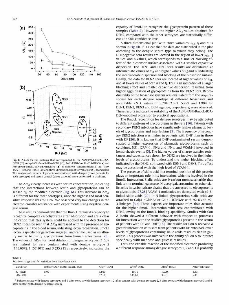
Fig. 6. DRCT% for the systems that corresponded to the AuNpPANI-BmoLL-BSA-DEN1 (h), AuNpPANI-BmoLL-BSA-DEN2 (s), AuNpPANI-BmoLL-BSA-DEN3 (N) andAuNpPANI-BmoLL-BSA-DENnegative (�) at different concentrations (1:25, 1:50,1:75, 1:100 and 1:150) (a); and three-dimensional plot for values of RCT, Q and n (b).The analyses of the sera of patients contaminated with dengue (three patients foreach serotype) and serum control (three patients) were performed in triplicate.
522 C.A.S. Andrade et al. / Journal of Colloid and Interface Science 362 (2011) 517–523
The DRCT clearly increases with serum concentration, indicatingthat the interactions between lectin and glycoproteins can besensed by the modified electrode (Fig. 6a). This increase in DRCT
is different for the three serotypes, since the highest and most sen-sitive response was to DEN2. We observed very low changes in theelectron-transfer resistance with experiments using negative den-gue serum.
These results demonstrate that the BmoLL retains its capacity torecognize complex carbohydrates after adsorption and are a clearindication that this system could be applied to the detection forDEN2. It can be seen that DRCT increased with the presence of gly-coproteins in the blood serum, indicating lectin recognition. BmoLLlectin is specific for galactose sugar [6] and can be used as an affin-ity matrix to purify glycoproteins from human colostrums [25].The values of DRCT, for fixed dilution of dengue serotypes (1:50),are highest for sera contaminated with dengue serotype 2(145.60%), 1 (57.10%) and 3 (35.91%), respectively, indicating the
Table 2Relative charge transfer variation from impedance data.
Condition Beforea (AuNpPANI-BmoLL-BSA) After1 DEN1
RCT (kX) 8.02 12.60DRCT (%) – 57.10
a Before contact with dengue serotypes and 1-after contact with dengue serotype 1, 2-after contact with dengue negative serum.
capacity of BmoLL to recognize the glycoprotein pattern of thesesamples (Table 2). However, the higher DRCT values obtained forDEN2, compared with the other serotypes, are statistically differ-ent at a 90% confidence level.
A three-dimensional plot with three variables, RCT, Q and n, isshown in Fig. 6b. It is clear that the data are distributed in the plotaccording to the dengue serum type to which they belong. TheDENnegative sera results are located in the region of lower RCT, Qvalues, and n values, which corresponds to a smaller blocking ef-fect of the biosensor surface associated with a smaller capacitivedispersion. The DEN1 and DEN3 sera results are distributed atintermediate values of RCT and higher values of Q and n, indicatingthe intermediate dispersion and blocking of the biosensor surface.Finally, the data for DEN2 sera are located at higher values of RCT,and at lower values of both n and Q. This is an indication of a largerblocking effect and smaller capacitive dispersion, resulting fromhigher agglutination of glycoproteins from the DEN2 sera. Repro-ducibility of the biosensor system was evaluated from the DRCT re-sponse for each dengue serotype at different biosensors andacceptable R.S.D. values of 5.70%, 2.33%, 5.28% and 1.99% forDEN1, DEN2, DEN3 and DENnegative, respectively, were observed.These results indicate the suitability of the AuNpPANI-BmoLL-BSA-DEN-modified biosensor to practical applications.
The BmoLL recognition for dengue serotypes may be attributedto different patterns of glycoproteins in the sera [16]. Patients withsecondary DEN2 infection have significantly higher plasmatic lev-els of glycoproteins and interleukins [3]. The frequency of second-ary DEN2 infection was higher in patients with DHF than in thosewith DF [26]. It is known that DHF-contaminated serum demon-strated a higher expression of plasmatic glycoproteins such ascytokines, NS1, ICAM-1, IFNa and IFNc, and VCAM-1 involved inhemorrhagic events [3]. The higher values of charge transfer resis-tances and capacitances shown by DEN2 are involved in the higherlevels of glycoproteins. To understand the higher blocking effectindicated by the DEN2, compared with DEN1 and DEN3, This effectmay be associated with the high level of VCAM-1.
The presence of sialic acid in a terminal position of this proteinplays an important role in its interaction, which is involved in theBmoLL interaction. Sialic acids are 9-carbon monosaccharide thatlink to the terminal galactose, N-acetylgalactosamine, or other sia-lic acids in carbohydrate chains that are attracted to glycoproteinsor glycolipids [27,28]. VCAM-1 molecules are decorated with a2-6-linked sialic acids [29]. In N-linked glycoproteins, sialic acids areattached to Galb1-4GlcNAc or Galb1-3GlcNAc with a2-6 and a2-3-linkages [30]. These aspects are important roles that accountfor the higher BmoLL interaction with sera contaminated withDEN2, owing to the BmoLL binding specificity. Studies with ConA lectin showed a different behavior with respect to processesfor interaction with the studied glycoproteins present in the serumof patients with DF and DHF [16]. The results for Con A revealed agreater interaction with sera from patients with DF, who had lowerlevels of glycoproteins containing sialic acids residues rich in gal-actose. This process was involved in the ability of Con A to interactspecifically with mannose and glucose residues.
Thus, the variable reaction of the modified electrode producinga different response among dengue serotypes 1, 2 and 3 is probably
After2 DEN2 After3 DEN3 After4 DENneg
19.70 10.09 8.43145.60 35.91 5.11
after contact with dengue serotype 2, 3-after contact with dengue serotype 3 and 4-

C.A.S. Andrade et al. / Journal of Colloid and Interface Science 362 (2011) 517–523 523
be due to the ability of BmoLL detect distinct glycosylation pat-terns. As previously reported [16], the changes in metabolismand diseases reveal different glycosylation in secreted glycopro-teins by infected cells [31,32], and our modified electrode maybe useful as a biosensing system for DEN2 diagnosis. Nonetheless,the proposed system could also be useful for predicting and mon-itoring the course of the dengue disease, and hence help guide theprescription of preventive therapy in dengue patients in endemicareas of the world by detecting unusual carbohydrate structuresby means of lectin [33].
4. Conclusions
The large specific surface area, excellent conductivity and sur-face charge of the AuNpPANI enable it to be used as a preeminentmatrix for protein immobilization. BmoLL lectin adsorption ontoAuNpPANI surfaces was mainly driven by the electrostatic interac-tion between the positively charged AuNpPANI and negatively-charged lectin residues. The AuNpPANI-BmoLL was able to detectglycoproteins from each of the three dengue virus serotypes witha high degree of specificity. Our results demonstrate that AuNpPA-NI-BmoLL interacts with glycoproteins of dengue serotype 2.AuNpPANI-BmoLL was used to manufacture a novel dengue glyco-protein biosensor. The biosensor exhibits excellent reproducibilityand shows good selectivity for the detection of dengue glycopro-teins in the sera samples. We believe that this study providesstrong evidence that naturally occurring BmoLL lectin recognitionfor dengue serotypes may be attributed to different patterns of gly-coproteins in the sera produced by the glycoprotein immunore-sponse from patients infected by the dengue virus.
Acknowledgments
The authors are grateful for the support provided by the RedeELINOR de Nanobiotecnologia/CAPES and FACEPE. CASA would liketo thank CAPES for a postdoctoral grant (BEX 3561/10-0) and XZlikes to thank NIH support 1R21EB006495.
Appendix A. Supplementary material
Supplementary data associated with this article can be found, inthe online version, at doi:10.1016/j.jcis.2011.07.013.
References
[1] G. Bock, J. Goode, New Treatment Strategies for Dengue and other FlaviviralDiseases, Novartis Foundation, John Wiley & Sons Ltd., Chichester, UK., 2006.
[2] E.M.B. Lemes, M.P. Miagostovicsh, A.M.B. Alves, S.M. Costa, A.M.B. Fillipis,G.R.G. Armoa, M.A.V. Araujo, J. Clin. Virol. 32 (2005) 305.
[3] R.F. Chen, K.D. Yang, L. Wang, J.W. Liu, C.C. Chiu, J.T. Cheng, Trans. R. Soc. Trop.Med. Hyg. 101 (2007) 1106.
[4] N. Sharon, Trends Biochem. Sci. 18 (1993) 221.[5] M.D.L. Oliveira, M.T.S. Correia, F.B. Diniz, Synth. Metals 159 (2009) 2162.[6] L.C.B.B. Coelho, M.B.R. da Silva, Phytochem. Anal. 11 (2000) 295.[7] M.L.R. Macedo, M.G.M. Freire, M.B.R. da Silva, L.C.B.B. Coelho, Comp. Biochem.
Physiol., Part A 146 (2007) 486.[8] V. Rosilio, M.M. Boissonnade, L.C.B.B. Coelho, N.S. Santos-Magalhaes, C.A.S.
Andrade, A. Baszkin, Colloids Surf., A 250 (2004) 491.[9] J. Wang, L.A. Luck, I.I. Suni, Electrochem. Solid-State Lett. 10 (2007) J33.
[10] F. Mizutani, Sens. Actuators, B 130 (2008) 14.[11] C. Sheng-Yun, P. Su-Moon, Synth. Met. 105 (1999) 91.[12] C.A.S. Andrade, A. Baszkin, N.S. Santos-Magalhães, L.C.B.B. Coelho, C.P. de Melo,
J. Colloids Interface Sci. 289 (2005) 371.[13] K. Mallick, M. Witcomb, M. Scurrell, A. Strydom, J. Phys. D 42 (2009) 095409.[14] W. Qiu, H. Huang, S. Zeng, T. Xue, J. Liu, J. Polym. Res. 18 (2011) 19.[15] H. Zhang, R. Liu, Q. Sheng, J. Zheng, Colloids Surf. B 82 (2011) 532.[16] M.D.L. Oliveira, M.T.S. Correia, F.B. Diniz, Biosens. Bioelectr. 25 (2009) 728.[17] A.G. Mantzila, C. Strongylis, V. Tsikaris, M.I. Prodromidis, Biosens. Bioelectr. 23
(2007) 362.[18] M.I. Prodromidis, Electrochim. Acta 55 (2010) 4227.[19] A. Mondini, R.V.M. Bronzoni, I.L.S. Cardeal, T.M.I.L. Santos, E. Lázaro, S.H.P.
Nunes, G.C.D. Silva, M.C.F.S. Madrid, P. Rahal, L.T. Figueiredo, F. ChiaravallotiNeto, M.L. Nogueira, J. Clin. Virol. 40 (2007) 84.
[20] C.P. de Melo, C.G. dos Santos, C.A.S. Andrade, Fluorescent nanoparticlecomposites themselves, process for the preparation of such composites, anduse in rapid diagnosis systems with affinity to biological molecules, PatentPCT/BR2009/000117, 2009.
[21] D.M.N. Luna, E.P.S. Falcao, S.J. Melo, C.A.S. Andrade, Colloids Surf. A 373 (2011)22.
[22] D. Necas, P. Klapetek, C. Anderson, Gwyddion, <http://www.gwyddion.net>2008.
[23] M.D.L. Oliveira, C.P. de Melo, O. Glaucius, C.A.S. Andrade, Colloids Surf., B 82(2011) 365.
[24] B.A. Boukamp, Solid State Ionics 18–19 (1986) 136.[25] E.G. Santana, Lectinas de sementes de Cratylia mollis (cramoll) e de folhas de
Bauhinia monandra (Bmoll): imobilizações e aplicações biotecnológicas, PhDThesis, Federal University of Pernambuco, Brazil, 2004.
[26] R.F. Chen, J.W. Liu, W.T. Yeh, L. Wang, J.C. Chang, H.R. Yu, J.T. Cheng, K.D. Yang,FEMS Immunol. Med. Microbiol. 44 (2005) 43.
[27] Y. Abe, C.W. Smith, J.P. Katkin, L.M. Thurmon, X. Xu, L.H. Mendoza, C.M.Ballantyne, J. Immunol. 1 (1999) 2867.
[28] A. Varki, FASEB J. 11 (1997) 248.[29] K. Hanasaki, A. Varki, L.D. Powell, J. Biol. Chem. 31 (1995) 7533.[30] S. Basu, M. Basu, S.S. Basu, in: A. Rosenberg (Ed.), Biological Specificity of
Sialyltransferases, in Biology of the Sialic Acids, Plenum Press, New York, 1995.[31] A. Azizan, J. Sweat, C. Espino, J. Gemmer, L. Stark, D. Kazanis, J. Virol. Methods
138 (2006) 211.[32] T.S. Raju, J.B. Briggs, S.M. Borge, A.J.S. Jones, Glycobiology 10 (2000) 477.[33] S. Sell, Human Pathol. 21 (1990) 1003.