Development of innovative liposome - Theses.fr
291
UNIVERSITÉ DE STRASBOURG ÉCOLE DOCTORALE DES SCIENCES DE LA VIE ET DE LA SANTE Laboratoire CAMB-UMR7199 CNRS/Université de Strasbourg THÈSE présentée par : Hanadi SALIBA soutenue le : 27 septembre 2017 pour obtenir le grade de : Docteur de l’Université de Strasbourg Discipline/ Spécialité : Aspects moléculaires et cellulaires de la biologie/ Immunologie Development of innovative liposome- based constructs for non-invasive cancer immunotherapy in humans THÈSE dirigée par : Mme FOURNEL Sylvie Professeur, Université de Strasbourg Mme CHAMAT Soulaïma Professeur, Université Libanaise RAPPORTEURS : Mme AUBERT Anne Maître de conférences, Université de Montpellier Mr FISSON Sylvain Professeur, Université d’Evry-Val-d’Essonne AUTRES MEMBRES DU JURY : Mme SOULAS Pauline Professeur, Université de Strasbourg Mr COURAUD Pierre-Olivier Directeur de recherches, Institut Cochin/Inserm
-
Upload
khangminh22 -
Category
Documents
-
view
0 -
download
0
Transcript of Development of innovative liposome - Theses.fr

UNIVERSITÉ DE STRASBOURG
ÉCOLE DOCTORALE DES SCIENCES DE LA VIE ET DE LA SAN TE
Laboratoire CAMB-UMR7199 CNRS/Université de Strasbo urg
THÈSE présentée par :
Hanadi SALIBA
soutenue le : 27 septembre 2017
pour obtenir le grade de : Docteur de l’Université de Strasbourg
Discipline/ Spécialité : Aspects moléculaires et cellulaires de la biologie/ Immunologie
Development of innovative liposome-based constructs for non-invasive cancer immunotherapy in humans
THÈSE dirigée par :
Mme FOURNEL Sylvie Professeur, Université de Strasbourg Mme CHAMAT Soulaïma Professeur, Université Libanaise
RAPPORTEURS :
Mme AUBERT Anne Maître de conférences, Université de Montpellier Mr FISSON Sylvain Professeur, Université d’Evry-Val-d’Essonne
AUTRES MEMBRES DU JURY : Mme SOULAS Pauline Professeur, Université de Strasbourg Mr COURAUD Pierre-Olivier Directeur de recherches, Institut Cochin/Inserm


REMERCIEMENTS ___________________________________________________________________________________________
Voilà ma thèse qui se termine; une page de ma vie qui tourne pour me mener à un nouveau
chapitre. Cette longue aventure m’a introduit à tellement de gens qui ont tous contribué,
chacun à sa manière, à rendre cette expérience plus enrichissante et plus précieuse, que ce
soit sur le plan scientifique ou humain. Vous avez tous été des perles sur mon chemin.
Je tiens tout d’abord à exprimer mes sincères remerciements aux membres de mon jury de
thèse pour l’honneur qu’ils m’ont accordé, Dr Anne Aubert, Pr Sylvain Fisson, Pr Pauline
Soulas, Pr Pierre-Olivier Couraud, ainsi que Dr Vincent Flacher et Dr Béatrice Heurtault pour
m’avoir fait l’honneur de juger mes travaux de thèse.
Une thèse est d’habitude dirigée par une ou deux personnes. De mon côté, j’ai eu la chance
d’en avoir plus… Je vous raconte.
Comment puis-je trouver les mots pour remercier les personnes qui ont inspiré et dirigé ce
travail, et qui, au-delà de cela, m’ont appris à comprendre les enjeux du monde de la recherche
et à y grandir? A mes directrices immunologistes, Soulaïma et Sylvie, et à ma directrice
galéniste, Béatrice (bien qu’il n’ait pas été possible d’avoir officiellement 3 co-directrices, et
que tu sois un membre invité du jury), mille mercis du fond du cœur. Dès le début, vous avez
été des accompagnatrices qui ont commencé par s’inquiéter de là où je vais dormir lors de
mon premier voyage en France, pour rester à mes côtés tout au long de cette thèse. En étant
toujours là pour me guider, mais tout en me laissant beaucoup de liberté, vous m’avez
donné l’opportunité d’acquérir une vision large et mature de la science. Merci beaucoup pour
votre encadrement, votre patience, votre compréhension, vos conseils précieux, votre
disponibilité, et vos encouragements.
Je vous demande de m’excuser si je métends longuement sur cette partie, mais derrière ma
thèse, il y a une histoire à raconter… L’histoire a commencé quand j’ai fini mon master et que
je cherchais à m’inscrire en thèse. C’est là où, Soulaïma, tu m’as prise comme protégée, tu
m’as soutenue sans faille et, grâce à toi, j’ai fait la connaissance de Sylvie et Béatrice. Depuis
lors, tu as tout fait pour que je puisse avoir un financement au Liban et mener à bien ma thèse
dans les meilleures conditions. Ça n’a pas toujours été facile ni évident. Au-delà de tout cela,

tu m’as été une grande sœur que je ne saurais pas assez remercier pour son accueil chaleureux
(j’ai pratiquement passé la dernière semaine de la rédaction chez toi!). Sylvie, cette thèse
n’aurait pas été possible sans ta confiance et ton engagement à m’assurer un financement en
France. J’ai appris de ta rigueur et ton dévouement, et tu as si bien su me booster quand la
situation devenait critique. Merci encore plus pour le plaisir que m’ont procuré toutes les
discussions que nous avons partagées après les longues journées de travail, et merci pour avoir
été si patiente et disponible. Que dire alors de toi Béatrice… Même si ton nom ne figure pas
officiellement en tant que directrice de cette thèse, tu l’as été. Tu as été aussi précieuse pour
moi que pour ce travail, et c’est grâce à toi que j’ai appris la galénique et que j’ai pu vivre une
expérience inter-disciplinaire très enrichissante.
Je tiens à remercier du fond du cœur les directeurs de mes deux laboratoires et équipes
d’accueil, Dr Jean Serge Remy (directeur de l’UMR7199), Dr Benoît Frisch (directeur de
l’équipe de Biovectorologie à l’UMR7199) et Dr Hasnaa Bouharoun-Tayoun (co-directrice du
laboratoire d’Immunologie de l’UL). Mille mercis pour m’avoir accueillie au sein de vos
laboratoires, pour votre confiance et pour vos conseils. Hasnaa, déjà avant que je commence
ma thèse, tu as été pour moi un mentor qui a vu une étincelle en moi et qui a su me guider
afin que je puisse me faire une place dans le monde de la recherche.
Au cours de cette expérience j’ai eu la chance de travailler avec Dr Vincent Flacher. Merci
Vincent pour m’avoir accueillie et formée, merci pour les expériences FACS, pour ta
disponibilité et pour tes conseils précieux. Merci également d’avoir participé à mon jury et pris
le temps de lire ce manuscrit.
Un grand merci aux stagiaires qui m’ont apporté de l’aide à la réalisation de certains travaux.
Merci à Nisrine Ballout, maintenant docteur, et à Joëlle Richa (pour votre aide pendant les
premières expériences sur les souris Hu-SPL-NSG), à Maroua Messous et à Yara El Murr (pour
votre aide dans la vaccination TC), et à Yohan Gerber (mille mercis pour les travaux sur les
liposomes fluorescents).
J’aurais difficilement pu terminer ma thèse dans de bonnes conditions sans le soutien et
l’encouragement de mes deux équipes d’accueil. Merci pour avoir partagé mon quotidien
durant cette longue expérience et de l’avoir enrichie. J’apprécie énormément la chance que

j’ai eue de nouer autant de relations humaines et de croiser autant de gens et de cultures. La
liste est longue et je vais essayer de n’oublier personne.
A l’équipe Biovec en France, merci à Célia pour ta compagnie, ta gentillesse, ton soutien et
pour ton aide pendant les jours fous des sacrifices. Merci à Zahra pour avoir formulé mes
premiers liposomes et m’avoir formée à préparer tous les suivants, et à Neïla pour nos
discussions sur les enjeux de la thèse et de la science. Merci à toutes les deux pour m’avoir
«adoptée» pendant mon premier séjour en France et pour votre soutien continu. Cendrine,
Anaïs et Evelyne, merci avant tout pour votre amitié, vos conseils et votre agréable compagnie.
Anaïs et Cendrine, merci encore pour votre aide dans les expériences et pour votre soutien
pendant les moments de forte déprime (Je n’ai pas oublié notre dernière longue discussion en
live à minuit, vous maitrisez incroyablement l’art de booster les gens!). Merci à toutes de vos
conseils judicieux. Je n’oublie pas de remercier Maria pour sa gentillesse, et Marcella, pour
avoir été une « co-bureau » si sympa, mais aussi pour les pauses gourmandes avec vous et les
cafés de 16h. Merci également à May (tu es un rayon de soleil), Alex (pour ton humour du
second degré), à Eya, Amina et Salif.
A quelques milliers de kilomètres, à mon laboratoire Libanais, mille mercis à Nabil (mon guide
lors de mes premiers pas dans la recherche), Stéphanie et Leila (mes «supporters» et les
copines de 24/7), Renée (nous avons partagé le même sort et avons été synchrones pendant
la thèse; qu’est-ce qu’il pourrait y avoir de plus encourageant?). Mille mercis à Catherine et
Najibé pour le plaisir de nos discussions matinales et pour votre soutien dans les expériences.
Merci à Sandy et Joëlle, à Elie, Silia, Elissa et Yara (encore), les jeunes motivés de la nouvelle
génération qui ont contribué au plaisir que j’ai eu à vivre en ce lieu.
Je tiens également à remercier toutes les personnes que j’ai rencontrées chez les « voisins»
(majoritairement des chimistes), pour toutes les pauses midi et les jeux de cartes. Un énorme
merci chaleureux aux incontournables Malak et Marianne pour leur soutien sans faille, leurs
conseils «logistiques» et leur compagnie. Merci à Fanny pour ses encouragements et sa bonne
humeur (tue me tues!). Merci à Murielle et Nour et tous les amis que je n’ai pas cités, pour
avoir vécu cette aventure avec moi.

Merci également à la faculté de médecine de l’Université Libanaise où j’ai eu mes premières
expériences d’encadrement d’étudiants, et qui m’a donné l’opportunité de poursuivre cette
thèse en parallèle. Je n’oublie pas de remercier la doyenne de la faculté de Santé Publique de
l’UL, Dr Nina Zeidan, et le doyen de l’ED 414 à l’Unistra pour leur acte de foi qu’ils ont fait à
l’égard de cette thèse.
Je ne serais certainement pas là à écrire ces lignes sans le soutien de mes parents, mon frère
Tanios et ma sœur Hiba, à qui je dédie cette thèse. Vous m’avez été un véritable soutien sur
tous les plans et avez cru en moi jusqu’au bout. L’ambition et la persévérance sont des qualités
que j’ai apprises de vous. Mille mercis surtout pour cette phase de rédaction où je risquais
d’être moins supportable que d’habitude (plutôt insupportable). Tanios et Hiba, n’oubliez pas,
«aim high and think big». Je suis fière de vous autant que vous êtes fiers de moi.
Si mes parents m’ont formée dès mon plus jeune âge à avoir de l’ambition et des rêves, alors
toi Fadi, «my other half», tu n’as hésité à aucun moment à renforcer ces traits de caractère en
moi, à me soutenir jusqu’au bout quelqu’en soit le coût et à m’aider à rebondir après chaque
chute. C’est grâce à toi que j’ai considéré la thèse avec un nouvel espoir après chaque
déprime… Ce n’est pas pour me plaindre, mais j’en ai eu beaucoup, et c’est grâce à toi, Fadi
que je me dis tous les matins que «tout ce qui ne te tue pas te rends plus fort ».
J’ai lu quelque part que « les remerciements sont la preuve que le scientifique n’est pas un
robot»… Oui… J’espère avoir pu, en ces quelques lignes, exprimer ma sincère gratitude à tous
les gens que j’ai croisés, et avec qui j’ai eu des relations aussi humaines que professionnelles
pendant toutes ces années. Ayant franchi cette étape parsemée de difficultés qui nous pousse
jusqu’à nos limites, je me retrouve prête à confronter d’autres défis…

i
TABLE OF CONTENTS ___________________________________________________________________________________________
RESUME ............................................................................................................................... ix
ABSTRACT ........................................................................................................................ xviii
LIST OF FIGURES ................................................................................................................ xxii
LIST OF TABLES ................................................................................................................. xxiii
LIST OF ABBREVIATIONS .................................................................................................. xxiv
FOREWORD .......................................................................................................................... 1
1. Overview of the immune system ................................................................................................. 2
1.1. Components of the innate and adaptive immune systems ................................... 2
1.2. Lymphocyte differentiation .................................................................................. 4
1.3. Innate immune response ...................................................................................... 5
1.4. Activation of adaptive immune responses ............................................................ 7
1.5. Effector phase of specific immunity .................................................................... 10
2. Cancer immunity and immunotherapy...................................................................................... 10
INTRODUCTION .................................................................................................................. 11
Chapter 1: Eliminating cancer cells using cancer vaccines ............................................................... 12
1. Overview on cancer development and treatments ................................................ 12
1.1. Oncogenesis: a historical perspective ................................................................. 12
1.2. Different approaches to cancer therapy ............................................................. 12
2. Evidence of cancer immunogenicity ....................................................................... 14
2.1. The first evidence of cancer immunogenicity ..................................................... 14
2.2. Immune response against tumors ...................................................................... 15
2.2.1. The immune system can fight tumors ...................................................... 15
2.2.2. Innate immunity to cancer: NK cells ......................................................... 18
2.2.3. Cancer adaptive immune response: the importance of T lymphocytes .... 22
2.2.4. Cancer adaptive immune response: the role of B lymphocytes ................ 32
3. Cancer immunoediting and other escape mechanisms .......................................... 32
3.1. The theory of immunoediting: tumors become less immunogenic ..................... 32
3.2. Tumors increase their own survival and evade destruction ................................ 33

ii
3.3. Tumors modulate tumor-specific immune response........................................... 33
3.4. The cancer-specific immune response is also modulated by inhibitory loops...... 35
3.5. Cancer progression: the three “Es” of tumor development ................................ 36
4. Therapeutic cancer vaccines .................................................................................. 37
4.1. Cancer vaccine requirements ............................................................................. 38
4.1.1. Cancer vaccines need immunostimulatory molecules .............................. 39
4.2. Recent advances in cancer vaccination ............................................................... 41
4.2.1. Tumor cell-based vaccines ....................................................................... 42
4.2.2. Nucleic acid-based vaccines ..................................................................... 44
4.2.3. DC-cell based vaccines ............................................................................. 45
4.2.4. Protein/peptide-based cancer vaccines ................................................... 45
Chapter 2: Liposomes as systems for the delivery of protein/peptide-based vaccines to DCs .... 48
1. Generalities ........................................................................................................... 48
1.1. A brief insight into liposomes ............................................................................. 48
1.1.1. Liposomes assemble from building blocks ............................................... 49
1.1.2. Liposomes structure affects their stability ............................................... 49
1.1.3. Cholesterol role in the stability of the lipid bilayer ................................... 50
1.1.4. Liposomes vary in composition and structure .......................................... 50
1.2. Liposome formulation techniques ...................................................................... 52
1.3. Physicochemical characteristics of liposomes ..................................................... 52
1.3.1. Liposome size .......................................................................................... 53
1.3.2. Liposome zeta potential ........................................................................... 53
2. Liposomes as vaccine delivery systems .................................................................. 54
2.1. Liposomes improve vaccine immunogenicity ...................................................... 56
2.1.1. Encapsulation protects molecules and increases their bioavailability ....... 56
2.1.2. Membrane display preserves epitopes in natural conformation .............. 56
2.2. Versatility of liposomal vaccines is crucial for tumor vaccination ........................ 57
2.2.1. Size and zeta potential control for a better vaccine: small changes make
big differences ....................................................................................................... 58
2.2.2. Targeting of liposomes to DCs for enhanced induction of tumor-specific
responses…. ........................................................................................................... 59
2.2.3. Liposomes favor cross-presentation ........................................................ 60
2.2.4. Liposomes can deliver vaccines through multiple routes ......................... 62

iii
Chapter 3. Delivering cancer vaccines to relevant DCs: the transcutaneous route:
RevEnhancing Tumor-Specific Immune Responses by Transcutaneous Vaccination (Review
article) .......................................................................................................................... 66
Abstract: ....................................................................................................................... 67
1. Introduction: .......................................................................................................... 68
2. Cancer immunity: challenges and vaccine design requirements ............................. 68
2.1. Protective tumor-specific immune response ...................................................... 68
2.2. Kinetics of tumor development and escape from immune response .................. 69
2.3. Therapies based on reversal of immune tolerance ............................................. 71
3. Vaccination via the skin ......................................................................................... 72
3.1. The skin immune system .................................................................................... 72
3.2. Skin DCs subsets ................................................................................................. 73
3.2.1. Langerhans cells: ..................................................................................... 73
3.2.2. Dermal dendritic cells: ............................................................................. 74
3.3. Antigen presentation potential of skin DCs ......................................................... 75
3.3.1. Endocytic receptors of skin DCs ............................................................... 75
3.3.2. Skin DC function in cellular immune response activation: relevance to
cancer vaccination? ............................................................................................... 75
4. Strategies of transcutaneous vaccination ............................................................... 77
4.1. Transcutaneous vaccination: making skin DCs the main vaccine recipients ........ 77
4.2. Barrier role of the stratum corneum ................................................................... 78
4.3. Physical barrier disruption .................................................................................. 79
4.4. Innovative vaccine formulations for skin barrier crossing ................................... 82
4.4.1. Peptide-based vaccines combined to adjuvants ....................................... 82
4.4.2. Nanoparticles for transcutaneous immunization ...................................... 82
4.4.3. Physicochemical properties influencing nanoparticles interaction with the
skin immune system ............................................................................................... 84
4.4.4. Potential of liposomes for transcutaneous immunization ......................... 86
4.4.5. Adapting nanoparticles for adequate skin DC targeting ........................... 87
5. Transcutaneous cancer vaccination using nanoparticles: where do we stand? ....... 88
5.1. Nanoparticle-based cancer vaccines in development ......................................... 90
5.1.1. ISCOM-based cancer vaccines .................................................................. 90
5.1.2. Liposome-based cancer vaccines .............................................................. 90

iv
5.2. Transcutaenous cancer vaccines in clinical trials ................................................. 92
6. Conclusion ............................................................................................................. 93
7. Expert commentary ............................................................................................... 93
8. Five-year view ........................................................................................................ 95
9. Key issues .............................................................................................................. 96
Bibliography .................................................................................................................. 97
Chapter 4: Evaluating cancer vaccines in humanized mouse models .......................................... 105
1. Development and evolution of the concept: from mouse immunodeficiency to a
humanized immune system ........................................................................................ 106
1.1. Evolution of immunodeficient mice: the first step towards a humanized model. ....
......................................................................................................................... 106
1.2. Engraftment of a functional immune system: from immunodeficiency to
humanization .......................................................................................................... 109
1.2.1. The Hu-PBL-SCID model ......................................................................... 109
1.2.2. The Hu-SRC-SCID model ......................................................................... 110
1.2.3. The BLT mouse model ............................................................................ 111
1.2.4. The Hu-SPL-SCID model ......................................................................... 111
2. Humanized mice in cancer vaccine research ........................................................ 112
2.1. Humanized mice for preclinical testing of vaccines immunogenicity ................. 113
2.2. Humanized mice in cancer vaccine research: evaluation of protection ............. 114
2.2.1. Humanized SCID mice can accept adoptive transfer of tumor xenografts
and reconstitute their natural microenvironment ................................................ 114
2.2.2. Humanized SCID mice as platforms for cancer vaccine evaluation ......... 114
OBJECTIVES .................................................................................................................. 116
EXPERIMENTAL RESULTS .............................................................................................. 123
Chapter 1: Transcutaneous immunization with liposome-based cancer vaccines induce CD4+ and
CD8+ T cell responses in BALB/c mice (Scientific article #1) ............................................................ 124
Introduction.............................................................................................................. 127
Material and methods .............................................................................................. 130
1. Formulation and characterization of liposomal constructs ............................... 130
1.1. Lipids and adjuvants .............................................................................. 130
1.2. Synthetic peptides ................................................................................. 130

v
1.3. Formulation of lipid vesicles .................................................................. 130
1.4. Peptide conjugation to Lp-SUV or Tf-SUV ............................................... 132
1.5. Physicochemical characterization of the constructs ............................... 132
2. Animals ............................................................................................................ 134
3. Immunization of mice ....................................................................................... 134
4. Immunogenicity of transcutaneously administered liposomal constructs ......... 134
4.1. Splenocyte and lymph node cell suspension .......................................... 134
4.2. Direct/ Standard IFNγ ELISPOT assay ...................................................... 135
4.3. Indirect IFNγ ELISPOT assay (cultured ELISpot, double stimulation) ....... 135
5. Tracking of DC skin migration to draining lymph nodes after TC application .... 136
5.1. Preparation of lymph node cell suspensions .......................................... 136
5.2. Flow cytometry analysis ......................................................................... 136
Results ...................................................................................................................... 138
1. Formulation and physicochemical characterization of different variants of
liposomal constructs ............................................................................................... 138
5.3. Formulation and characterization of conventional liposome-based
constructs ............................................................................................................ 138
5.4. Formulation and physicochemical characterization of different variants of
transfersome-based vaccines ............................................................................... 139
5.5. Formulation of fluorescent liposomes .................................................... 140
2. Systemic immune response induced by a physical mixture of ErbB2 and cholera
toxin: ErbB2 is immunogenic by the TC route. ......................................................... 141
3. Evaluation of the immune response induced by different ErbB2-bearing liposomal
constructs by TCI: proof of immunogenicity ............................................................ 142
3.1. Liposomes adjuvanted with a TLR2/6 agonist induce an ErbB2-specific
response after TCI ................................................................................................ 142
3.2. The TLR4 agonist is less suitable than the TLR2/6 agonist for T cell
activation after TCI with liposomal constructs ...................................................... 145
3.3. The DC targeting molecule has only a minor effect on ErbB2-specific
responses induced by TCI ..................................................................................... 147
3.4. Validation of the power of the ELISpot assay ......................................... 149
4. Influence of the lipid vesicle fluidity on the systemic immune response ........... 154
4.1. Transfersome-based formulations induce a T-cell response comparable but
not equal to that induced by their liposomal counterparts. ................................. 154
4.2. DC targeting molecules modulate the immunogenicity of transfersome-
based vaccines ..................................................................................................... 155

vi
4.3. Transfersome-based vaccines induce a high ErbB2-specific, but a low HA-
specific response rate .......................................................................................... 157
5. Liposomes induce skin DC migration to draining lymph nodes after TC
immunization .......................................................................................................... 159
Discussion ................................................................................................................. 162
References ................................................................................................................ 171
Chapter 2: NOD-SCID-Il2r Gamma null mice engrafted with human splenocytes show promise for
the evaluation of liposome-based cancer vaccines (scientific article #2) ..................................... 182
Introduction.............................................................................................................. 185
Material and methods .............................................................................................. 188
1. Immunodeficient animals ................................................................................. 188
2. Proteins and peptides....................................................................................... 188
3. Formulation and characterization of liposomal constructs ............................... 188
3.1. Lipids and adjuvants .............................................................................. 188
3.2. Formulation of liposomal SUVs (liposomes) ........................................... 188
3.3. Peptide conjugation to SUV ................................................................... 189
3.4. Physicochemical characterization of liposome constructs ...................... 190
4. Human spleen cells........................................................................................... 191
4.1. Human spleen cell sources ..................................................................... 191
4.2. Preparation and cryopreservation of human spleen cell suspensions .... 191
5. Cell proliferation assay ..................................................................................... 192
6. In vitro priming of human splenocytes with liposome-displayed peptides ........ 192
7. NSG mice reconstitution with primed human splenocytes................................ 192
8. Assessment of the engraftment of human leukocytes in the spleen of Hu-SPL-NSG
mice ........................................................................................................................ 193
9. Evaluation of the cellular immune response by ELISPOT ................................... 193
10. Measurement of total human immunoglobulin concentration and specific anti-
PAK IgG titer in hu-SPL-NSG serum by ELISA ............................................................ 194
11. Measurement of total human immunoglobulin concentration and specific anti-
PAK IgG titer in hu-SPL-NSG serum by dot blot ........................................................ 194
Results ...................................................................................................................... 196
1. Formulation and physicochemical characterization of different liposomal
constructs ............................................................................................................... 196

vii
2. Assessment of the safety profile of liposome-bound TLR agonists in vitro:
Pam2CAG is the molecule of choice ......................................................................... 197
3. Liposomes incorporating Pam2CAG at a molar ratio of 2% are toxic for Hu-SPL-
NSG mice ................................................................................................................. 198
4. Evaluation of liposomal constructs incorporating Pam2CAG 0.2% in Hu-SPL-NSG
mice ........................................................................................................................ 200
4.1. Liposomal constructs incorporating Pam2CAG 0.2% are not toxic .......... 200
4.2. Human cells remain functional and secrete IgG in the Hu-SPL-SCID mice 201
4.3. Human cells home to the spleens of NSG mice ...................................... 201
4.4. Immune response to liposomes incorporating PAK, HA and 0.2% Pam2CAG
204
Discussion................................................................................................................... 207
References .................................................................................................................. 213
GENERAL DISCUSSION AND PERSPECTIVES ................................................................... 218
1. A robust formulation technique that yields homogeneous liposome-based constructs ...... 219
2. Feasibility and immunogenicity of TC vaccination with liposome-based constructs ............. 221
2.1. The TC route is as potent as the SC route in inducing tumor-specific immune
responses: a proof of concept ..................................................................................... 221
2.2. Vaccine composition modulates the induced immune response ...................... 221
2.2.1. The TLR2/6 agonist Pam2CAG is superior to the TLR4 agonist MPLA for the TC
route 221
2.2.2. Skin DC targeting by mannose addition to liposomal constructs does not
significantly improve immunogenicity ..................................................................... 223
2.2.3. Transfersomes are not superior to conventional liposomes in TC vaccination .
.................................................................................................................. 224
2.3. Beyond this project: what other factors may influence the immune potential of
liposome-mediated TC vaccination?............................................................................ 225
2.4. Beyond this project: after the proof of immunogenicity of the liposomal
constructs by the TC route, it is time to assess their efficacy? ..................................... 227
2.5. Beyond this project: application of the TC vaccination with liposomal constructs
to melanoma .............................................................................................................. 228
3. Liposomal constructs are immunogenic in the Hu-SPL-NSG mouse model ........................... 229
3.1. The TLR2/6 agonist Pam2CAG is suitable to be used with human splenocytes .. 230
3.2. A model liposome-based construct induces a cellular but not a humoral immune
response in the Hu-SPL-NSG mouse ............................................................................ 231

viii
3.3. Beyond this project: evaluation of the efficacy of liposomal constructs against
cancer in the Hu-SPL-NSG mouse ................................................................................ 231
4. A humanized mouse model for TC cancer vaccination: time to think of the next generation
model ................................................................................................................................................. 232
BIBLIOGRAPHY ................................................................................................................. 235

ix
RESUME ___________________________________________________________________________________________
Développement de constructions liposomiques innovantes
pour l’immunothérapie humaine
Cette thèse est réalisée en co-direction entre Pr. Sylvie Fournel à l’UMR7199 CNRS à
l’Université de Strasbourg et Pr. Soulaima Chamat au Laboratoire d’Immunologie à l’Université
Libanaise.
Contexte
Les traitements antitumoraux classiques se basent pour la plupart sur la chimiothérapie et la
radiothérapie. En raison de leur faible spécificité pour les cellules tumorales, ces traitements
induisent de nombreux effets secondaires. La découverte que le système immunitaire du
patient pouvait éliminer les tumeurs en utilisant par exemple des lymphocytes T cytotoxiques
(CTL) a fait de l’immunothérapie anticancéreuse une stratégie attractive. Cette stratégie
thérapeutique se base sur la capacité des cellules présentatrices d’antigènes (CPA) et en
particulier des cellules dendritiques (DC), à capturer des antigènes associés aux tumeurs (TAA),
puis à migrer suite à leur maturation induite par un signal de danger (adjuvant) jusqu’aux
organes lymphoïdes secondaires pour y présenter des peptides issus des TAA aux lymphocytes
TCD4+ et TCD8+. Les premiers, lorsqu’ils sont différenciés en T helper 1 (Th1) procurent des
signaux de maturation sous forme de cytokines et de molécules de costimulation à la DC, qui
va alors être capable d’induire la différenciation des TCD8+ en lymphocytes T cytotoxiques
(CTL), principales cellules effectrices de la réponse antitumorale. La mise en place d’une
réponse immunitaire efficace contre les tumeurs nécessite donc 1) une activation de la DC par
des signaux de danger fournis par une molécule immunostimulatrice, comme, par exemple,
un agoniste de TLR, 2) l'activation de TCD4+ helper suite à la reconnaissance d'un épitope
TCD4+ présenté par la DC et 3) l'activation de TCD8+ cytotoxiques suite à la présentation par
une DC d’un épitope TCD8+.
Classiquement, l’administration d’un vaccin se fait à travers la peau, celle-ci étant un site
immunologique particulièrement riche en DC. En effet, l’épiderme comprend les cellules de

x
Langerhans (LCs) et le derme comprend plusieurs sous-populations de DCs dermiques (dDCs)
qui expriment ou pas la langerine. La voie d’administration cutanée la plus conventionnelle
est la voie sous-cutanée (SC). Toutefois, celle-ci implique un drainage du vaccin
de l’hypoderme qui est dépourvu de DC, vers les ganglions drainant la zone d’administration.
Alternativement, la voie intradermique est difficile à cibler. C’est pourquoi l’immunisation
transcutanée (TC) est envisagée comme voie intéressante qui cible préférentiellement les LC
et les dDCs. Les antigènes peptidiques sont adaptés, du fait de leur petite taille, au passage à
travers la peau. L’encapsulation de ces peptides dans des nanoparticules, tel que les
liposomes, augmente leur immunogenicité et leur absorption par la peau.
L’objectif général de mon projet de thèse est donc de developper des constructions
liposomiques anti-tumorales pour une administration TC chez l’homme.
Mon laboratoire d’accueil à l’Université de Strasbourg a développé des constructions
liposomiques peptidiques contenant tous les éléments indispensables à la réponse
immunitaire antitumorale (épitopes TCD4+, TCD8+, agoniste de TLR) qui induisent une réponse
immunitaire antitumorale après administration par voies SC et intranasale chez la souris. Pour
atteindre ce but, mon 1er objectif spécifique est donc d'optimiser ces constructions
vaccinales liposomiques pour induire une réponse immunitaire après administration par
voie TC.
Par ailleurs, les réponses immunitaires induites chez l’animal lors des essais précliniques des
vaccins divergent souvent de celles qui sont ensuite observées lors des essais cliniques, ce qui
rend nécessaire le développement de modèles animaux plus prédictifs de la réponse
immunitaire humaine aux vaccins. Un tel modèle a été développé dans mon laboratoire
d’accueil libanais. Il repose sur la reconstitution de souris immunodéficientes avec des cellules
immunitaires humaines provenant de la rate ou du sang périphérique humain, et est deisgné
par Hu-SPL-NSG. Le deuxième objectif de ce projet est donc de tester les constructions
vaccinales liposomiques, dans un modèle de souris humanisée afin de les optimiser pour
une application humaine ultérieure.

xi
Objectifs et stratégie de l’étude:
Objectif 1 : Développement de constructions liposomiques adaptées pour la vaccination TC
contre le cancer et évaluation de leur immunogenicité.
Les résultats de cette partie sont présentés dans l’article #1, en préparation.
1- Optimisation de la composition de la construction liposomique pour la vaccination TC à
partir de la construction précédemment validée au laboratoire, en optimisant 3 éléments :
la molécule immunostrimulatrice, la présence ou non d’une molécule de ciblage des DC,
et la nature de la vesicule lipidique. Nous avons aussi formulé des liposomes fluorescents
incorporant un fluorochrome lipophile dans leur bicouche lipidique. Cette stratégie nous
offre la possibilité de suivre les cellules qui internalisent les liposomes dans la peau, et leur
migration jusqu’aux ganglions lymphatiques.
2- Evaluation de la réponse immunitaire locale et systémique induite par les constructions.
Dans ce but nous avons d’abord évalué la réponse immunitaire induite par la construction
d’origine par la voie d’administration TC en comparaison à la voie SC. Nous avons ensuite
évalué l’influence de deux molécules immunostrimulatrices sur cette réponse, des
agonistes de TLR2/6 et de TLR4, ainsi que l’effet de l’addition du mannose. Nous avons
finalement évalué l’influence de la fluidité de la vésicule phospholipidique sur la réponse
immunitaire induite.
3- Evaluation de la migration des DC de la peau induite par une immunisation TC par les
formulations liposomiques, vers les ganglions lymphatiques drainant la zone d’application.
Objectif 2 : Evaluation de l’immunogénicité des liposomes dans le modèle Hu-SPL-NSG
Les résultats de cette partie sont présentés dans l’article #2, en préparation.
La capacité des souris humanisées à répondre à des formulations liposomiques n’est pas bien
établie dans la littérature. Pour cela nous avons choisi une formulation-modèle comprenant
un épitope B au lieu de l’épitope TCD8+, en addition a l’épitope TCD4+ et a un agoniste de TLR.
Ce choix nous a permis d’évaluer la capacité de suivre l’induction d’une réponse humorale
ainsi qu’une réponse cellulaire.

xii
1- Evaluation de la toxicité des agonistes de TLR vis-à-vis des splénocytes humains : Dans
une première étape, nous avons évalué les liposomes incorporant différents agonistes
de TLR vis-à-vis des splénocytes humains en culture pour leur capacité d’induire une
toxicité ou une prolifération. En plus, nous avons évalué leur effet sur la reconstitution
des souris Hu-SPL-NSG.
2- Evaluation de la capacité des liposomes à induire une réponse immunitaire chez la
souris Hu-SPL-NSG, contre l’épitope B et l’épitope T CD4+
Résultats :
1. Formulation et caractérisation des vaccins liposomiques
Pour répondre au 1er objectif, nous avons modifié des constructions vaccinales validées pour
des immunisations par voie SC pour formuler de nouvelles constructions potentiellement plus
adaptées pour la voie TC. Dans ces constructions, nous avons associé un peptide TCD4+ issu
de l’hémagglutinine du virus de la grippe (HA) et un peptide TCD8+ issu de la protéine ErbB2
humaine. Nous avons fait varier la nature de l’adjuvant en utilisant soit le Pam2CAG
(dipalmitoyl-cystéine-alanyl-glycine), agoniste de TLR2/6, soit le MPLA (monophosphoryl lipid
A), agoniste de TLR4. De plus, nous avons modifié la composition et les propriétés
physicochimiques de la vésicule lipidique, en utilisant soit des liposomes conventionnels soit
des liposomes ultradéformables, appelés transfersomes. Finalement, nous avons testé
l’avantage potentiel de l’addition du mannose (dioleyl glycérol-dimannose ou DOG-Man2),
molécule de ciblage connue pour cibler les DC et ainsi favoriser la capture de la construction
liposomique.
En addition, nous avons formulé des liposomes fluorescents (Lp DiI) en incorporant dans leur
bicouche lipidique un fluorochrome lipophile.
Pour répondre au 2ème objectif, nous avons préparé des liposomes incorporant uniquement
un agoniste de TLR. Nous avons varié la nature de cet agoniste en utilisant soit le MPLA
(agoniste de TLR4), soit Pam3CAG (tripalmitoyl-cystéine-alanyl-glycine), ligand de TLR2/1, soit
le Pam2CAG, ligand de TLR2/6. En se basant sur l’évaluation de l’effet de ces agonistes sur les

xiii
splénocytes humains en culture, nous avons choisi le Pam2CAG pour l’associer aux peptides B,
issu de la pilline de Pseudomonas euruginosa (PAK), et TCD4+ (HA).
Les liposomes et les transfersomes ont été préparés par la technique d’hydratation d’un film
lipidique à partir de phospholipides additionnés de l’adjuvant Pam2CAG ou MPLA et d’une
ancre amphiphile fonctionnalisée qui permet l’ancrage des épitopes peptidiques à la surface
de la construction. Lorsque nécessaire, les résidus mannose sont ajoutés au mélange de
départ. La suspension aqueuse obtenue, contenant des vésicules multi-lamellaires, a été
soniquée ou extrudée pour obtenir une population homogène de liposomes unilamellaires de
petite taille (SUV). Sur les SUV ainsi obtenus ont été ensuite greffés les peptides épitopiques
issus des protéines ErbB2 ou PAK, en addition à HA. La caractérisation physicochimique de ces
formulations a montré qu’elles présentent des diamètres moyens de l’ordre de 70 à 90 nm
avec une distribution étroite. Les indices de polydispersité étaient tous inférieurs à 0.3
indiquant une homogénéité des échantillons. Le rendement de couplage des épitopes était
entre 90 et 100% pour les liposomes et de l’ordre de 75% pour les transfersomes.
Ainsi, grâce à une technique de formulation robuste et maitrisée, nous avons préparé des
constructions liposomiques homogènes tout au long de ce travail, ce qui représente un atout
incontestable pour leur évaluation in vivo.
2. Evaluation des constructions vaccinales administrées par voie TC dans la
souris BALB/c
Pour évaluer la capacité des différentes constructions vaccinales à induire une réponse
immunitaire à médiation cellulaire après administration TC, les liposomes et les transfersomes
portant les épitopes peptidiques associés à un adjuvant (Pam2CAG ou MPLA), et portant ou
non une molécule de ciblage (DOG-Man2) ont été administrés par massage précédé d’une
application d’éthanol à des souris BALB/c (J0, J2, J8). Après 30 jours, le nombre de lymphocytes
T spléniques ou ganglionnaires spécifiques des peptides portés par les liposomes a été évalué
à l’aide d’un test ELISPOT mesurant la production d’IFN-γ.
En comparant la réponse immunitaire induite par la formulation d’origine par les 2 voies SC et
TC, nous avons démontré une sécrétion d’IFN-γ par les cellules de la rate et des ganglions. Ceci
montre que la voie TC est capable d’induire une réponse immunitaire aussi puissante que celle

xiv
induite par la voie SC. Cette preuve de concept constitue un rational qui nous permet
d’adapter nos formulations pour la voie TC.
Pour choisir le ligand TLR qui est le plus efficace par la voie TC, nous avons ensuite comparé
l’immunogénicité de constructions liposomiques incorporant différents agonistes de TLR, un
agoniste de TLR2/6 (Pam2CAG) et un agoniste de TLR4 (MPLA), pour leur effet
immunostimulateur par application TC chez la souris. Nos résultats ont montré que les
liposomes porteurs de Pam2CAG ont induit, en réponse aux peptides HA et ErbB2, une
sécrétion d'IFN-γ par les cellules ganglionnaires (réponse locale) aussi bien que par les cellules
de rate (réponse systémique). Par contre, les liposomes porteurs de MPLA ont induit une
sécrétion d’IFN-γ uniquement par les cellules de la rate. Ces résultats montrent que les deux
agonistes de TLR sont convenables pour une vaccination transcutanée, cependant, Pam2CAG
semble être meilleur que MPLA comme il induit à la fois une réponse locale et systémique.
Pour poursuivre l’optimisation de nos constructions liposomiques pour la voie TC, nous y
avons incorporé une molécule de ciblage des DC, le di-mannose, et nous évalué la réponse
induite par ces formulations. L’addition du di-mannose aux liposomes n’a pas
significativement amélioré la réponse immunitaire observée.
Pour vérifier si une augmentation de la déformabilité de la vésicule lipidique ne pouvait pas
améliorer la réponse induite par immunisation TC, nous avons remplacé, dans nos
constructions, les liposomes conventionnels par des transfersomes, et nous avons évalué leur
effet sur la réponse locale et systémique. De manière générale, les transfersomes n’ont pas
amélioré la réponse immunitaire observée. Chez les souris immunisées par les transfersomes,
nous avons noté dans les splénocytes et dans les ganglions une production d’IFN-γ en réponse
au peptide issu d’ErbB2 mais pas ou peu de réponse contre le peptide issu de HA. Ces résultats
suggèrent qu’en dépit de leur ultradéformabilité les formulations vaccinales à base de
transfersomes ont induit en TC une réponse immunitaire moins bonne que les liposomes
conventionnels.
Nos résultats montrent pour la première fois que ces constructions liposomiques sont
immunogènes par voie TC et qu’elles sont capable induire aussi bien une réponse CD8+ qu’une
réponse T CD4+. Ils montrent aussi que le Pam2CAG est supérieur au MPLA pour cette voie

xv
d’immunisation, puisqu’il induit à la fois une réponse immunitaire locale et une réponse
systémique. Toutefois, les transfersomes et le di-mannose ne semblent pas améliorer la réponse.
3. Etude de l’activation immunitaire locale induite par les constructions
liposomiques
Dans le but d’étudier la migration des DC de la peau vers les ganglions drainant la zone
d’application après immunisation TC, nous avons préparé des liposomes fluorescents
incorporant un fluorochrome dans leur bicouche lipidique. Des souris BALB/c ont reçu, par
massage précédé d’une application d’éthanol, ces liposomes incorporant ou non un ligand de
TLR. Cette partie du travail a été menée en parallèle à la première, donc comme nous n’avions
pas encore d’indications sur l’identité du meilleur agoniste de TLR, nous avons aléatoirement
choisi le MPLA. Les souris ont été sacrifiées après 48 heures pour l’analyse de la migration des
DC de la peau vers les ganglions brachiaux drainant la zone d’application. L’analyse en
cytométrie en flux des suspensions de cellules ganglionnaires a montré une absence de
fluorescence dans les ganglions. Toutefois, le nombre de DC provenant de la peau était
augmenté, indiquant ainsi que les constructions liposomiques sont capables d’induire la
migration des DC de la peau vers les ganglions après application TC. Les cellules qui migrent
préférentiellement sont les LCs et les dDCS lang-. Toutefois, nous avons observé que les
liposomes blancs sont également capables d’induire cette migration. Ceci pourrait être
expliqué par une contamination des liposomes blancs par des molécules pouvant induire la
migration de DC. Une autre hypothèse serait que même si les liposomes blancs sont capables
d’induire la migration des DC de la peau, seuls les liposomes incorporant un ligand de TLR sont
capables d’induire leur maturation.
4. Evaluation des constructions liposomiques dans les souris Hu-SPL-NSG
En parallèle, j’ai analysé au Liban l’immunogénicité des liposomes dans les souris humanisées
Hu-SPL-NSG, un autre modèle préclinique plus prédictif de la réponse immunitaire humaine
que le modèle murin classique. Dans ce modèle, des souris immunodéficientes sont
reconstituées par des splenocytes humains normaux, provenant de donneurs d’organes
décédés ou d’accidentés de route splenectomisés.

xvi
Dans un premier temps nous avons testé des liposomes incoprporant plusieurs agonistes de
TLR, MPLA, Pam2CAG et un agoniste de TLR2/1, Pam3AG, pour leur effet sur les cellules
spléniques humaines in vitro, en recherchant l’induction d’un effet toxique et/ou d’une
prolifération de ces cellules. Nous avons noté l’index de prolifération le plus élevé avec le
Pam2CAG, d’où il a été choisi pour incorporation dans les liposomes peptides à évaluer in vivo.
Pour avoir une preuve de concept, nous avons d’abord choisi une formulation modèle à
évaluer dans la souris Hu-SPL-NSG. Alors qu’une réponse cellulaire ne peut être analysée que
dans les organes lymphoïdes secondaires après euthanasie, une réponse humorale offre
l’opportunité d’être suivie en cours de l’expérience par ELISA dans les sérums des souris
immunisées. Pour cette raison, nous avons remplacé le peptide ErbB2 dans la formulation
d’origine par un peptide B, issu de la pilline de P.aerigunosa souche K (PAK), et nous avons
gardé le peptide HA, en addition au Pam2CAG.
Pour évaluer les liposomes chez la souris Hu-SPL-NSG, des splénocytes humains ont été
cultivés pendant 3 jours avec des constructions liposomiques puis injectés au J3 à des souris
NSG. Les souris ont reçu des injections de rappel par ces mêmes constructions par voie
intrapéritonéale aux J7 et J21. Aux jours 28 ou 35, les souris ont été sacrifiées et nous avons
déterminé la concentration des IgG humaines dans leur sérum (pour vérifier
« l’humanisation » des souris) ainsi que la réponse humaine contre HA de leurs cellules
spléniques et la reponse specifique anti-PAK dans leur serum.
Nous avons pu mettre en évidence que le sérum de toutes les souris Hu-SPL-NSG contenait
des IgG humaines, ce qui reflète une bonne reconstitution et indique que les cellules
humaines restent viables et fonctionnelles. Pour évaluer la circulation ciblée des splenocytes
humains vers les rates de ces souris, un test d’immunofluorescence indirecte (IFI) a été réalisé
et a démontré un «homing» des leucocytes humain vers cet organe. Ces résultats indiquent
que l’immunisation des souris Hu-SPL-NSG par les liposomes n’influence pas la viabilité et la
fonctionnalité des cellules humaines.
Nous avons ensuite évalué la response immunitaire spécifique induite par la construction
modèle chez la souris Hu-PSL-NSG. Un ELISpot réalisé à partir de cellules de rate a indiqué
une sécrétion d’IFN-γ humainen réponse au peptide HA, signalant ainsi l’induction d’une
réponse CD4+ spécifique aux constructions liposomiques. A notre connaissance, notre travail

xvii
figure parmi les premiers qui ont démontré l’immunogénicité de liposomes porteurs de
peptides épitopiques dans un modèle de souris humanisée.
Toutefois, nous n’avons pas pu détecter des anticorps spécifiques anti-PAK dans les sérums
de ces souris.
Ces résultats constituent une preuve de concept sur l’immunogénicité de la plateforme
liposomique sélectionnée dans la souris Hu-PSL-NSG, et reflètent l’utilité de ce modèle dans
leur l’évaluation. En plus, ils suggèrent un potentiel prometteur des liposomes comme
véhicule vaccinal anti-tumoral pour l’homme.
Conclusion
L’ensemble des résultats de ce projet a permis de démontrer, dans la souris BALB/c, la
faisabilité et l’immunogénicité de la vaccination anti-tumorale par la voie TC avec des
liposomes portant à leur surface des peptides T CD8+ de TAA et complétés par les éléments
nécessaires à l’activation des DC et de cellules Th1. Nos travaux nous ont également permis
de démontrer dans un modèle de souris humanisée que la plateforme vaccinale sélectionnée
dans les tests réalisés dans le modèle murin reste immunogène vis-à-vis des cellules
humaines. Ainsi, la vaccination TC de l’homme avec ce type de formulations pourrait
représenter une stratégie non invasive efficace et prometteuse pour l’immunothérapie active
antitumorale.
Ces résultats seront complétés par l’évaluation de la capacité de ces constructions à inhiber
la croissance de tumeurs exprimant la protéine ErbB2 humaine chez la souris BALB/c, ainsi
qu’à leur capacité d’induire une réponse CD8+ chez la souris Hu-SPL-NSG. Le but ultime de ce
travail étant le développement, à long terme, d’un modèle de vaccination TC chez la souris
humanisée.

xviii
ABSTRACT ___________________________________________________________________________________________
This thesis project is carried out in co-direction between Prof. Sylvie Fournel at the UMR7199
CNRS at the University of Strasbourg and Pr. Soulaima Chamat at the Laboratory of
Immunology at the Lebanese University.
Cancer immunotherapy is gaining more attention thanks to a better understanding of the
immune system’s role in fighting tumors. Tumor vaccines are intended to induce tumor
specific cytotoxic T lymphocytes (CTL) via 1- maturation of dendritic cells (DCs) by danger
signals provided by the immunostimulatory molecule, 2- activation of CD4+ T cells following
recognition of a CD4 epitope presented by the DC, iii) activation of CD8+ T cells following
recognition of a CD8 epitope presented by this DC.
The skin is an attractive route of tumor-specific vaccination because of its richness in dendritic
cells (DCs) and its capacity to induce robust CTL responses. Skin DCs internalize vaccines and
migrate to draining lymph nodes where they induce a systemic immune response. They are
especially endowed with the capacity to cross-present antigens to both naive CD4+ and CD8+
T cells, thus, resulting in the induction of a CTL response. Convenient targeting of skin DCs is
ensured by transcutaneous (TC) vaccination. However, the skin is impermeable for
conventional vaccine preparations. Therefore, peptide-based vaccines are desirable for TC
vaccination because their small size facilitates their diffusion through the skin. Additionally,
the use of various nanoparticles, such as liposomes and transfersomes, as peptide delivery
vectors increases their skin crossing and capture by DCs and subsequently, their
immunogenicity in presence of an immunostimulatory molecule.
Therefore, the general objective of my thesis was to develop these liposome-based
constructs adapted for cancer immunotherapy by the TC route in humans.
My host laboratory at the University Strasbourg developed highly versatile liposomal
constructs to co-deliver all the three crucial elements for an efficient tumor-specific immune
response (a CD4 epitope, a CD8 epitope and an adjuvant). These constructs were shown to
induce specific anti-tumor immune responses after subcutaneous injection in normal mice.

xix
The first specific objective of my work is to optimize these constructs to induce a potent
immune response after transcutaneous (TC) application.
In addition, responses induced in animal models may deviate partially or totally from those
observed later in clinical trials. In order to optimize these vaccine formulations for human
application, we proposed to evaluate them in an animal model which is more predictive of the
human immune response. This model is a humanized mouse developed by my host laboratory
at the Lebanese University, in which immunodeficient mice are engrafted with human
splenocytes in order to mimic human immune responses. These humanized mice are called
Hu-SPL-NSG mice. The second specific objective of my thesis was therefore to determine
whether liposome constructs that were previously validated in the conventional murine can
induce detectable human immune responses in the Hu-SPL-SCID model.
To meet the 1st objective, the previously developed vaccine constructs were optimized in
order to be more suitable for the TC route. These constructs express a universal CD4+ T cell
epitope-containing peptide from the influenza virus hemagglutinin (HA) and a CD8+ T cell
epitope-containing peptide from the human ErbB2 tumor antigen, in addition to an
immunostimulatory molecule (TLR2/6 agonist). The optimized vaccine constructs differ by the
TLR agonist and the physicochemical properties of the lipid vesicle, resulting in either
conventional liposomes or more flexible ones called transfersomes. A DC-targeting molecule,
di-mannose, could also be added.
Vaccine constructs were evaluated for their immunogenicity after TC application on previously
shaved dorsum of a normal mouse model. Liposomes bearing the peptides in combination
with a TLR2/6 (Pam2CAG) or a TLR4 agonist (MPLA) resulted in the induction of peptide-specific
cellular immune response. However, Pam2CAG seemed to be superior to MPLA, since it
induced an immune response both in the spleen (systemic response) and the lymph nodes
(local response) of the immunized mice. In contrast, MPLA-bearing liposomal constructs
induced only systemic responses. Di-mannose addition to the constructs did not improve the
immune response. Similarly, the replacement of the conventional liposomal vesicle with an
ultradeformable one, called transfersome, did not improve the immune response.
Transfersomes rather seemed to impair HA-specific responses. Our results show that the

xx
liposomal constructs are immunogenic by the TC route. Liposomes incorporating Pam2CAG as
an immunostimulatory molecule seem the most adapted for the TC route.
After confirming the constructs immunogenicity, we investigated their capability to induce
skin DC migration to the draining lymph nodes after TC immunization. Lymph node DCs were
analyzed by flow cytometry, and revealed that the liposomal constructs incorporating MPLA
as a danger molecule induced the migration of skin DCs. However, the same effect was
observed with the plain constructs, suggesting either a contamination of these constructs, or
a migration that is not accompanied by a maturation of the DCs.
We show herein for the first time that liposomal constructs are immunogenic by the TC route
and induce both a CD8 + response and a CD4+ T cell response.
To meet our 2nd objective, we first formulated liposomal constructs incorporating different
TLR agonists, namely MPLA, Pam2CAG, and a TLR2/1 agonist, Pam3CAG. The evaluation of their
safety profile in vitro towards human splenocytes indicated Pam2CAG to be the most
appropriate TLR agonist for in vivo evaluation.
The immunogenicity of a model liposomal constructs was then tested in the Hu-SPL-NSG
mouse model. Liposomes carrying a B cell epitope peptide instead of the ErbB2 peptide, the
HA peptide and Pam2CAG were injected intraperitoneally in NSG mice previously reconstituted
with human splenocytes. These liposomal constructs were shown to induce a specific human
immune response against HA, inducating that the liposomal constructs are able to induce a
specific CD4 + response. However, we were unable to detect specific anti-PAK antibodies in
the sera of these mice.
These results are a proof of concept on the immunogenicity of our liposomal platform in the
Hu-PSL-NSG mouse, and reflect the utility of the Hu-SPL-NSG model in their evaluation. In
addition, they indicate a potential of liposomes as an anti-tumor vaccine vehicle for humans.
In conclusion, all the results of this project demonstrated the feasibility and efficacy of tumor
vaccination by the TC route in BALB/c mice with liposomes carrying CD8 + TAA peptides on
their surface and incorporating the necessary elements for activation of DCs and Th1 cells. Our
work also allowed us to demonstrate in a humanized mouse model that the vaccine platform

xxi
selected in the tests carried out in the murine model remains immunogenic to human cells.
Thus, human TC vaccination with this type of formulations could represent an effective and
promising noninvasive strategy for anti-tumor active immunotherapy.
These results will be completed by evaluating the constructs ability to inhibit the growth of
tumors expressing the human ErbB2 protein in the BALB/c mosue as well as their ability to
induce a CD8+ response in the Hu-SPL-NSG mouse. On the long run, the ultimate goal of this
work is to develop a TC vaccination model in the Hu-SPL-NSG mouse.

xxii
LIST OF FIGURES ___________________________________________________________________________________________
Figure 1 : Cells and molecules of the innate and adaptive immune system ............................ 3
Figure 2 : Negative selection of B cells ................................................................................... 4
Figure 3: Positive and negative selection of T cells ................................................................. 5
Figure 4 : The two pathways of antigen processing and presentation. ................................... 6
Figure 5 : The induction of cellular adaptive immune response ............................................... 8
Figure 6: Milestones in molecular cancer research (upper part) and molecular biology (lower
part) ..................................................................................................................................... 13
Figure 7: Tumor infiltration with different immune cells ...................................................... 16
Figure 8: The role of NK cell in monoclonal antibody therapies (ADCC) ................................ 20
Figure 9: The dynamic regulation of NK cell effector function .............................................. 21
Figure 10: The reductionist view of cancer versus the integrative view of systems
immunology………………………………………………………………………………………………………………………. 23
Figure 11: Correspondence between the immune contexture and the Immunoscore. ......... 24
Figure 12: Immunoscore definition and methodology .......................................................... 24
Figure 13: The molecular mechanisms involved in CTL induction ......................................... 31
Figure 14: Natural selection of tumor variants in the generation of “tumor escape”
phenotypes. ......................................................................................................................... 33
Figure 15 : The effect of the inhibitory microenvironment on the tumor-specific immune
response. ............................................................................................................................. 34
Figure 16. The cancer specific immune response is downregulated by immune checkpoints,
such as the Cytotoxic T-Lymphocyte-Associated Antigen 4 (CTLA-4). ................................... 35
Figure 17: The three phases of cancer development ............................................................ 37
Figure 18: Cancer vaccines are intended to optimize the amplitude and the quality of the
tumor-specific immune response. ......................................................................................... 38
Figure 19 : Cancer vaccine approaches ................................................................................. 42
Figure 20: Representation of a phospholipid (a), the steric organization of a lipid bilayer (b)
and a liposome (c). ............................................................................................................... 49
Figure 21: Types of liposomes. ............................................................................................. 51
Figure 22: Interaction of vaccine components with the liposome. ........................................ 55
Figure 23: Mechanisms by which liposomes favor the induction of immune responses. ...... 57
Figure 24: Liposomes favor cross presentation. .................................................................... 61
Figure 25: Models for engraftment of human immune systems into SCID mice .................. 106
Figure 26: Members of the cytokine-receptor family bearing the common γ chain. ............ 108
Figure 27: Role of IL-7, IL-2 and IL-4 in the generation of T and B cells ................................ 109

xxiii
LIST OF TABLES __________________________________________________________________________________________
Table 1: Association of immune T cell infiltrates with prognosis in cancer (edited). ............. 18
Table 2: Antigenic targets, cancer indication and mechanism of action of the therapeutic
monoclonal antibodies currently approved by the Food and Drug Administration (FDA) for
cancer therapy that involve ADCC ........................................................................................ 19
Table 3: Classification and examples of TAAs based on molecular criteria. ........................... 29
Table 4: Humanized immunodeficient mouse models (edited) ........................................... 107
Table 5: Comparison of humanized immunodeficient mouse models. ................................ 112

xxiv
LIST OF ABBREVIATIONS __________________________________________________________________________________________
ADCC Antibody-Dependent Cell Cytotoxicity
AP Alkaline Phosphatase
APC Antigen Presenting Cell
Bcl B-cell lymphoma
BCR B cell receptor
BLT Bone marrow, Liver, Thymus
CDC Complement-Dependant Cytotoxicity
CEA Carcinoembryonic Antigen
Chol Cholesterol
ConA Concanavalin A
CT Core of the Tumor
CTL Cytotoxic T lymphocyte
CTLA-4 Cytotoxic T-Lymphocyte-Associated Antigen 4
CV Coefficient of variation
DAMP Damage-Associated Molecular Pattern
DC Dentritic Cells
dDC Dermal Dendritic cell
DiI 1,1'-dioctadecyl-3,3,3'3'-tetramethylindocarbocyanine perchlorate
DLS Dynamic Light Scattering
DMSO Dimethylsulfoxide
DNA Deoxyribonucleic
DOG-Man2: Dioleyglycerol-di-Mannose
DPG-mal Dipalmitoyl Glycerol-Maleimide
dSEARCH Dendrite Surveillance Extensions and Retraction Cycling Habitude
EDTA Ethylenediaminetetraacetic acid
EGFR Endothelial Growth Factor Receptor
ELISA Enzyme-Linked lmmunosorbent Assay
ELISpot Enzyme-Linked lmmunospot
FasL Fas Ligand
FBS Fetal Bovine Serum
FDA Food and Drug Administration
FITC Fluorescein isothiocyanate
G-CSF Granulocyte-Colony Stimulating Factor
GM-CSF Granulocyte Macrophage Colony-Stimulating Factor
gp glycoprotein
GVHD Graft Versus Host Disease
HA Hemaglutinin
HCl Hydrochloric acid
Her2/ErbB2/Neu Human epidermal growth factor Receptor-2
HIV Human Immunodeficiency Virus
HLA Human Leucocyte antigen

xxv
HPV Human Papilloma Virus
HRP Horseradish Peroxidase
HSC Hematopoietic Stem Cells
Hu Humanized
ID Intradermal
IDO Indolamine 2′3′-Dioxygenase
IFI Indirect Immunofluorescence
Ig Immunoglobulin
IL Interleukin
IM Intramuscular
IFN-γ Interferon Gamma
ISCOM Immune Stimulating Complex
Jak Janus kinase
KAR Killer Activating Receptor
KIR Killer-Cell Immunoglobuline-Like Receptors
Lang Langerin
LC Langerhans cell
LHRH Luteinizing-Hormone-Releasing Hormone
ln Natural log
Lp Liposome
LPS Lipopolysaccharides
mAb monoclonal antibody
MAGE-A3 Melanoma-Associated Antigen 3
MAMP Microbe-Associated Molecular Pattern
Man Mannose
MCA Methylcholanthreme
MDDC Monocyte-Derived Dendritic Cell
MDSC Myeloid-Derived Supressor Cells
MHC Major Histocompatibility Complex
MLV Multilamellar Vesicle
MPER Membrane Proximal External Region
MPLA Monophosphoryl Lipid A
MUC Mucin
MVV Multivesicular Vesicles
NK Natural Killer
NLR NOD-Like Receptor
NOD Non-Obese-Diabetic
NSG NOD-SCID-Gamma null
o/w oil-in-water
Ova Ovalbumin
PAK Pseudomona aeruginosa strain K
Pam2CAG Dipalmitoyil Cysteine-Alanyl-Glycine
Pam3CAG Tripalmitoyil Cysteine-Alanyl-Glycine
PAP Prostatic Acidic Phosphatase
PBL Peripheral Blood Lymphocytes

xxvi
PBMC Peripheral Blood Mononuclear Cells
PBS Phosphate Buffer Saline
PC Phosphatydilcholine
PDI Polydisrpersity Index
PD-L1 Programmed Death Protein Ligand 1
PG Phosphatydilglycerol
PGE2 Prostaglandine E2
PLGA Poly(Lactic-co-Glycolic Acid)
Poly-ICLC
Polyinosinic-Polycytidylic acid with Polylysine and
Carboxymethylcellulose
Prkdc Protein Kinase, DNA activated, Catalytic polypeptide
PRR Pattern-Recognition Receptor
PSA Prostate Specific Antigen
RAG Recombination-Activating Gene
RNA Ribonucleic
ROS Reactive Oxygen Species
SALT Skin-Associated Lymphoid Tissue
SC Subcutaneous
SCID Severe Combined ImmunoDeficiency
SDC Sodium Deoxycholate
SIS Skin Immune System
SPC Soy Phosphatydilcholine
SPL Splenocytes
SRC SCID Repopulating Cells
STAT Signal Transducer and Activator of Transcription
TAA Tumor Associated Antigen
TAM Tumor-Associated Macrophages
TBHSP70 Tuberculosis Heat Shock Protein 70
TBS Tris Buffer Saline
Tc Transition temperature
TC Transcutaneous
TCI Transcutaneous immunization
TCR T Cell Receptor
Tf Transfersome
TGF Transforming Growth Factor
Th1 T helper 1
Th2 T helper 2
TIL Tumor-Infiltrating Lymphocyte
TLR Toll-like receptor
TMB Trimethylbenzidine
TNF-α Tumor Necrosis Factor Alpha
TRAIL TNF-Related Apoptosis Inducing Ligand
Treg Regulatory T Cell
UV Unilamellar vesicle
VEGF Vascular Endothelial Growth Factor

1
FOREWORD __________________________________________________________________________________________
The role of the immune system in fighting
tumors

2
1. Overview of the immune system
The role of the immune system is not only to fight potential intruders from the external
environment, known as “non-self”, mainly pathogenic microbes, but also to control harmful
modifications within our own cells, that may arise following infection or cancerous
transformation, known as “modified self”. It comprises a multitude of cells and molecules that
cooperate in an integrated network. The immune system is divided broadly in two arms,
respectively the innate immune system and the adaptive immune system.
1.1.1.1.1.1.1.1. Components of the innate and adaptive immune systems Components of the innate and adaptive immune systems Components of the innate and adaptive immune systems Components of the innate and adaptive immune systems
The innate immune system is present in all taxa from cnidarians to mammals but with various
modalities. It includes mainly the epithelial barriers, phagocytes (macrophages and
neutrophils), dendritic cells (DCs) and different subsets of innate lymphoid cells (ILC) among
which the most important are the natural killer (NK) cells, as well as free molecules such as
the complement system (figure 1). The defense mechanisms of the innate immunity are
designed to respond rapidly to infections and cell transformations. To recognize danger, cells
of innate immunity rely only on a limited number of receptors that can bind to molecules
which are common to groups of related microbes (these are called microbial-associated
molecular patterns or MAMPs) or that are expressed or released by stressed or dying cells
(these are called danger-associated molecular patterns or DAMPs) but not by healthy cells.
These receptors, called pattern-recognition receptors or PRRs, are identical for all members
of the same animal species. Some members of the PRR family are called Toll-like receptors or
TLRs; their engagement with MAMPs or DAMPs leads to the activation of the immune cell.
The adaptive immune system is present in all taxa of the jawed Vertebrates. It comprises T
and B lymphocytes and antibodies secreted by plasma cells, which derive from activated B
cells (figure 1). Components of adaptive immunity rely on a huge number of receptors that are
extremely diversified and that recognize a virtually unlimited number of molecules of
microbial or non-microbial origin. Molecules recognized by T and B lymphocytes are called
antigens. T and B cells are clonally distributed, meaning that each cell acquires during its
differentiation a specific antigen receptor that can only recognize a single antigenic
determinant called epitope. The B cell receptor (BCR) recognizes “native” epitopes of
unprocessed antigens, while the T cell receptor (TCR) can only bind to small “degraded” linear

3
peptide epitopes generated by the “processing” of antigens within our own cells and loaded
on molecules of the major histocompatibility complex (MHC). B and T cells may be activated
only after encounter with their specific antigen. Following activation, a B or T cell proliferates,
giving rise to a clone of identical cells. Some differentiate into effector cells that participate in
the ongoing immune response while others are kept as “memory cells” to better respond to a
potential future reencounter with the same antigen. Therefore, while adaptive immunity is
delayed in comparison to innate immunity upon a first encounter with intruders, its memory
allows it to be more rapid and amplified upon re-challenge. This property is the cornerstone
of the principle of vaccination. A vaccine is an antigenic preparation that activates specific
lymphocytes and generates memory capable of mounting a more potent secondary antigen-
specific immune response.
B cells are in charge of the adaptive humoral immunity, as their main role is to secrete
antibodies after differentiation into plasma cells. T cells are in charge of the adaptive cellular
immunity. They are divided in three major effector populations, respectively “helper” T cells
(Th) which are responsible of secreting cytokines to initiate and regulate the adaptive immune
response and amplify innate immunity, cytotoxic T cells (Tc or CTL) that can kill target cells
expressing non-self antigens (mostly virally infected cells) or modified-self antigens
(transformed cells), and regulatory T cells (Treg) that downregulate other effectors of
immunity to avoid hypersensitivity and auto-immune diseases.
Figure 1 : Cells and molecules of the innate and adaptive immune system.
Antibodies
Effector T cells
Innate immunity Adaptive immunity
NK
B
B
B B Epithelial barriers
B lymphocytes Humoral immunity
T lymphocytes Cellular immunity
Phagocytes
T T
T T
NK cells
Cytokines Transmembrane
molecules
Hours Days
Complement
Denrtitic cell

4
1.2.1.2.1.2.1.2. LymphocLymphocLymphocLymphocyte yte yte yte differentiation differentiation differentiation differentiation
All cells of the immune system arise in the bone marrow. While B cells also differentiate before
being released in the circulation, T cells arise in the bone marrow but they differentiate in the
thymus. To prevent auto-immunity, lymphocyte differentiation involves acquisition of a
functional antigen receptor followed by elimination of auto-reactive lymphocytes whose
receptor can recognize self antigens. Since BCRs recognize native epitopes, B cell
differentiation implies only a negative selection of cells expressing an auto-reactive BCR
(figures 2). On the other hand, since TCRs recognize processed antigenic epitopes loaded on
MHC molecules, T cell differentiation includes 2 steps: the first is a positive selection of those
expressing a TCR capable of binding to self MHC molecules, the second is a negative selection
of those that have a high affinity for the complex made by a self-epitope loaded in the MHC
molecule (figure 3). There are 2 pathways of antigen processing (figure 4). Proteins present in
the cytosol, such as the proteins synthetized by the cell, are called endogenous antigens. They
are processed and their peptides are loaded on MHC class I molecule. T cells that are selected
on class I molecules differentiate to become CD8+ T cells, which turn mainly into cytotoxic T
cells. Proteins that derive from endocytosed or phagocytosed material are called exogenous
antigens. They are processed in the endosomal vesicles and their peptides are loaded on MHC
class II molecules. T cells that are selected on class II molecules differentiate to become CD4+
T cells, which turn mainly into helper T cells or regulatory T cells.
Figure 2 : Negative selection of B
cells. B cell differentiation implies
only a negative selection of cells
expressing an auto-reactive BCR
Pro-B
Bone
marrow
Binding to self-antigens
No
Migration to the circulation
Yes
Apoptosis
BCR gene rearrangement
B

5
Figure 3: Positive and negative selection of T cells. Since TCRs recognize processed antigenic epitopes
loaded on MHC molecules, T cell differentiation includes 2 steps: the first is a positive selection of those
expressing a TCR capable of binding to self MHC molecules, the second is a negative selection of those
that have a high affinity for the complex made by a self-epitope loaded in the MHC molecule
1.3.1.3.1.3.1.3. Innate immune response Innate immune response Innate immune response Innate immune response
The immune system is a powerful defense system that has to be very well controlled in order
to avoid inappropriate activation that may lead to harmful inflammation. Therefore, activation
of any component requires usually more than one signal, and “cross-talk” between the
different components is maintained at all times, mainly by a complex network of cytokines to
ensure targeted, effective yet balanced responses.
MHC II
Pro T
Binding to self MHC Yes No
Bone
marrow
Thymus
Positive
selection
Negative selection
High affinity to self MHC + self peptide
Yes No Yes No
CD4 CD8
TCR
TCR gene rearrangement
Migration to the
circulation Apoptosis Apoptosis Migration to the
circulation
MHCI

6
Figure 4 : The two pathways of antigen processing and presentation. In the exogenous or endocytic
pathway, proteins internalized by the cells are degraded in the endosome, and the derived peptides are
presented on MHC class II molecules. In the cytosolic or endogenous pathway, proteins which are
present in the cytosol, are degraded in the proteasome and the resulting peptides are loaded on MHC
class I molecules.
In case of infection or abnormal cell transformation, an immune response is triggered first
when the effectors of the innate immune system recognize danger by binding to MAMPS or
DAMPS. Innate immunity has 3 roles: 1) it provides an immediate defense line that eliminates
the source of danger or at least keeps it in check 2) it alerts and activates the effectors of the
adaptive immune system and 3) even when the adaptive immunity becomes effective, it keeps
contributing, in cooperation with lymphocytes and antibodies, to an optimal defense against
the intruder.
The type of elicited immune response depends on the nature of the triggering event. For
example, in case of viral infection or cell transformation, a prompt response may be provided
by NK cells. Indeed, in stressed cells, the amount of expressed MHC class I molecules is
Endosome
Lysosome
Endocytic processing pathway
Endosome fusion
with the lysosome
Exogenous
antigen
MHC class
II-peptide
Endogenous
antigen Degradation in
the proteasome
Loading on MHC
class II molecules
MHC class I-peptide
Cytosolic processing pathway
Endoplasmic
reticulum

7
typically reduced. This reduction disturbs the balance between activating and inhibiting signals
detected by NK cells and subsequently triggers them to exert a cytotoxic effect on the target.
Conversely, in case of infection with extracellular microbes, the most important effectors are
neutrophils, which can either ingest pathogens and kill them intracellularly or secrete digestive
enzymes and oxidative molecules that are cytotoxic to these pathogens.
Macrophages are present in most tissues. They do not only play a phagocytic role towards
microbes and cell debris, but they can also express antigenic peptides derived from
phagocytosed material on their MHC class II molecules, to be recognized and receive help from
Th1 cells, in order to increase their lytic activity. Finally, macrophages and other inflammatory
cells (mast cells, eosinophils, basophils) and molecules (cytokines, complement
components…) recruit more effectors to the “battle field” and induce the vascular changes
that are needed for this recruitment.
1.4.1.4.1.4.1.4. Activation of adaptive immune responses Activation of adaptive immune responses Activation of adaptive immune responses Activation of adaptive immune responses
The main immune cell players at the interface between the innate and the adaptive arms of
the immune system are the dendritic cells (DCs). Their main role is to be antigen presenting
cells (APCs). These cells are members of the innate immune system present in most tissues,
mainly the skin and the mucosa. They are endowed with long extensions called dendrites,
hence their name. Their main role is to constantly “sample” cellular and soluble components
from their environment, to endocytose them and to process them in order to present derived
peptides to T cells.
Processing of exogenous proteins in endocytic vesicles and loading of resulting peptides on
MHC class II molecules leads to antigen presentation to specific CD4+ T cells (figure 4). In the
DCs, some exogenous proteins can also leak from the endocytic vesicles and be degraded in
the cytosol, generating peptides that are loaded on MHC class I molecules. This unique
property of DCs is called cross-presentation of peptides on class I and class II molecules and is
crucial for the induction of CD8+ T cell responses. When a DC uptakes foreign material or cell
debris containing MAMPs or DAMPs, its PRRs, mainly TLRs, are engaged and mediate its
maturation, which is characterized by the expression of co-stimulatory molecules that can be
recognized by T cells. Mature DCs migrate to secondary lymphoid organs or tissues where they

8
present antigens to naïve CD4+ and CD8+ T cells in order to prime antigen-specific T cells (figure
5 A).
Figure 5 : The induction of cellular adaptive immune response. (A) DCs internalize cell debris, proteins
and microorganisms and cross-present them on MHC class I and II. Signaling through DAMP induces
their maturation: they express co-stimulatory molecules. (B) Binding of CD4+ T cells to MHCII-peptide
complexes and co-stimulatory molecules, and cytokines received by DCs induce their maturation into
TH1. (C) Th1 cytokines induce DCs to upregulate co-stimulatory molecules and secrete cytokines.
Binding of CD8+ T cells to DCs and signals received from Th1 and DC induce CTL differentiation
Differentiation of Th cells: CD4+ T cells are activated by receiving 2 signals from the DC, namely
by TCR binding to the class II MHC-peptide complex and by engagement of complementary
Co-stimulation Cytokines
Th1 cell
CD86
MHC I MHC II
CD28 TCR
TCR
Naïve
CD8+
T cell Cytotoxic T lymphocyte (CTL)
CD28 TCR
Cytokines
Cytokines
CTL
Th1
Dendritic cell MHC II
MHC I
Tumor-
specific naive
CD4+ T cell Th1 cell
Th1 Cytokines
Dendritic cell TAA uptake
and
processing
MHC II
MHC
I Co-stimulatory
molecule
(A) (B)
(C)
Dendritic cell
DAMPs
(Danger
signal)
PRR
Dendritic cell maturation and
peptide cross-presentation
Th1 cell differentiation
CTL differentiation
Co-stimulation
Co-stimulation

9
co-stimulatory molecules (figure 5 B). They secrete IL-2 and express IL-2 receptors, thus
inducing their own proliferation. Following their activation, CD4+ T cell differentiation into Th
will depend on the context of cytokines that are secreted by the DC, which is itself dictated by
the type of response needed. For instance, IL-12 secretion by the DC promotes mainly
differentiation of Th1 that secrete IFN-γ and activate an adaptive cellular immune response to
face intracellular infections and cell transformations. Conversely, in the presence of IL-4, Th
cells differentiate mainly into Th2 cells that secrete IL-4, IL-5 and IL-13 and activate an adaptive
humoral immune response to combat extracellular infections. Other differentiated Th cell
populations include, but are not restricted to, Th17 that secrete IL-17 and play an important
role in anti-infectious immune responses by activating neutrophils, and induced T regulatory
cells (iTreg) that secrete IL-10 and play an immunosuppressive role.
Differentiation of CTL: Differentiation of CTL requires two signals resulting from a triangular
engagement of CD4+ T cells, DCs, and CD8+ T cells.
1- Indeed, exogenous peptides are first cross-presented by the DC on MHC class I and
MHC class II molecules to CD8+ and CD4+ T cells respectively. The interaction of the
T cell receptor (TCR) of the CD8+ T cell with the peptide-MHC class I complex
induces the first signal.
2- The second signal is provided by co-stimulatory molecules and cytokines expressed
by the DC. These co-stimulatory molecules and cytokines are upregulated following
the interaction of this DC with a Th1 cell which previously differentiated from a
CD4+ T cell upon its interaction with a peptide-MHC class II complex.
Together, these two signals induce the differentiation of CD8+ T cells specifically bound, along
with the Th1 cell, to the same DC into CTL (figure 5 C). Then, CTL upregulate the production of
their apoptose-inducing molecules, including perforin and granzymes.
Activation of B cells and plasma cell differentiation: B cells that are selected through BCR
binding to native antigens that reach the secondary lymphoid organs or tissues usually need
Th cell help to be fully activated; these antigens are called T-dependent antigens. Only
complex carbohydrate antigens can activate B cells without TH help; they are referred to as T-
independent antigens. Activated B cells give rise to plasma cells that secrete antibodies.

10
1.5.1.5.1.5.1.5. Effector phase of specific immunity Effector phase of specific immunity Effector phase of specific immunity Effector phase of specific immunity
CTL can kill their target cells, usually virally infected or cancer cells, by inducing apoptosis, a
type of programmed cell death. Antibodies binding to soluble antigens help in their
elimination by forming immune complexes. Antibodies binding to cellular antigens bridge
between the target cell and phagocytes by binding to Fc receptors on the latter; this process
is called opsonization.
2. Cancer immunity and immunotherapy
Cancers arise from malignant cell transformations that lead to uncontrolled cell growth and
invasion of tissues. These are usually, but not always, accompanied by mutations in cellular
antigens, leading to expression of so-called tumor-associated antigens or TAA. Cancer cells are
poorly antigenic as the large majority of their proteins are unchanged as compared to the
normal cells from which they derive. Moreover, they usually grow in an immunosuppressive
microenvironment. As a result, they tend to be tolerated rather than induce an immune
response. Nonetheless, recent findings have proven that it is possible to enroll the immune
system in cancer therapy. Passive immunotherapy has been used for more than 20 years. It
relies on administration of tumor-specific monoclonal antibodies that control cell growth or
mediate killing of cancer cells by different effectors of the immune system. More recently,
attempts of cancer active immunotherapy are intended to boost the patient’s own immune
system to better fight the tumor. One strategy is to design cancer vaccines, which would have
all the minimum components needed to elicit a protective immune response. These would
include i) MAMPs or DAMPs that target and activate dendritic cells, ii) CD8 epitopes derived
from TAA to bind specific CD8+ T cells, and iii) CD4 epitopes to activate CD4+ T cells, in order
to ultimately initiate a tumor-specific CTL response.

11
INTRODUCTION
__________________________________________________________________________________________

12
Chapter 1: Eliminating cancer cells using cancer vaccines
1. Overview on cancer development and treatments
1.1.1.1.1.1.1.1. Oncogenesis: a historical perspectiveOncogenesis: a historical perspectiveOncogenesis: a historical perspectiveOncogenesis: a historical perspective
Throughout history, explanations of cancer and carcinogenesis have extensively changed.
From the old beliefs of Hippocrates in the “humoral” origin of tumors that stem from an
imbalance in the fluids of the body, to the discovery by Muller in 1838 that cancer originates
from cells, 19 centuries have elapsed. In the years that followed, multiple theories were
stated. Some believed that cancer was induced by trauma, others thought it was caused by
parasitic infections or even by chronic irritation. It is only after DNA was discovered by Watson
and Crick that the achievements of molecular biology lead to concrete discoveries about
cancer origin and development. The modern theories of carcinogenesis started with the
identification of oncogenes (figure 6). These are defined as genes involved in normal cells
growth, which, when mutated, cause an uncontrolled cell division, thus leading to cancer
development. Mutations may occur either spontaneously or because of carcinogens or
viruses, thus, mediating the initiation phase. The rapidly proliferating cancer cells grow into
tumors depending on various conditions that impose a certain rhythm, particular to each
cancer type and to each individual (progression phase). Growing tumors can subsequently
acquire an invasive potential and establish metastases in other tissues (invasion phase)
(Lonardo et al., 2015).
1.2.1.2.1.2.1.2. Different approaches to cancer therapyDifferent approaches to cancer therapyDifferent approaches to cancer therapyDifferent approaches to cancer therapy
The oldest treatments of cancer relied on total or partial surgical ablation of tumors. As of
today, ablation remains the first line treatment for solid tumors. Surgery is usually combined
with chemotherapy or radiotherapy, resulting in improved survival of cancer patients. Surgery
and radiation are used to locally treat cancer, whereas chemotherapy is systemically
administered, and therefore reaches cells that have spread throughout the body.
Chemotherapy involves cytotoxic drugs that drive cells into apoptosis through various
mechanisms, such as causing DNA damage and strand breakage in dividing cells, blocking
folate receptors and inhibiting key enzymes in folate metabolism or even by targeting death
receptors.

13
Figure 6: Milestones in molecular cancer research (upper part) and molecular biology (lower part)
(Lonardo et al., 2015). If the lower and the upper timelines are compared, it becomes quite evident that
the advancement of our knowledge of molecular processes was decisive in the elaboration of the
current understanding of tumor genetics. In particular, DNA structure discovery by Watson and Crick
was followed by the modern theories of carcinogenesis and the oncogenes identification. More recently,
genome wide sequencing projects and the –omics fields are allowing a broader molecular view of
cancer which is accessible to a wider population of the research community.

14
Since these mechanisms are poorly specific of tumor cells, they affect normal cells as well and,
therefore, they have multiple side effects. Additionally, drug-resistant tumor cells emerge
frequently. Combined therapies involving co-administration of two or more cytotoxic drugs
that achieve synergistic effects were recently found to improve the clinical outcome. Drugs
are chosen to have different mechanisms of action in order to minimize drug resistance and
overlapping toxicities while increasing tumor cell killing (Al-Lazikani et al., 2012). To cope with
emerging resistance and side effects, the search for alternative cancer therapies has been
ongoing for several decades with the aim of finding optimal drugs that would have high
efficacy and low intrinsic toxicity to normal tissues, improving the patients’ quality of life.
One of the considered alternative approaches is immunotherapy, a strategy that stems from
our growing understanding of the immunogenicity of cancer. Passive immunotherapy relies
on the administration of pre-formed antibodies targeting either proteins expressed by
malignant cells (such as Her2/ErbB2/neu, EGFR, CD20) or soluble growth factors required for
tumor growth (such as vascular endothelial growth factor), or on adoptive transfer of
autologous in-vitro modified tumor-specific lymphocytes, as well as on administrating
immunostimulatory molecules (such as TLR agonists) that activate the intrinsic immune
response against tumor. Active immunotherapy aims to induce the patient’s own immune
system to fight tumors by administrating cancer vaccines.
2. Evidence of cancer immunogenicity
2.1.2.1.2.1.2.1. The first evidence of cancer immunogenicityThe first evidence of cancer immunogenicityThe first evidence of cancer immunogenicityThe first evidence of cancer immunogenicity
The first reports on the immune system’s ability to fight established tumors were based on
the observation that febrile episodes were sometimes followed by complete spontaneous
remission in a number of cancer patients (Challis and Stam, 1990; Køstner et al., 2013). These
cases caught the attention of William Coley who made, in 1893, the first systematic attempt
of tumor immunotherapy. Coley administered to cancer patients a mixture of killed
Streptococcus pyogenes and Serratia marcescens known as “Coley’s toxin”, and reported that
it was able to induce complete remission in a number of soft and bone tissue cancer patients.
This “immunostimulant” was intended to induce a non-specific immune activation that favors
the induction of a tumor specific immune response. However, poor patient follow-up and
variable effectiveness of different Coley’s toxin preparations led to severe criticism of his work.

15
Yet, Coley’s theory for cancer immunotherapy was not abandoned. Throughout the following
Century, attempts to treat a wide range of cancers, like hepatocellular carcinoma and nodular
lymphoma, were carried out using the same strategy, without, however, reaching statistical
significance (McCarthy, 2006). Nowadays, bladder cancer is treated with an intra-cystic
injection of a variant of Coley’s toxin, the Bacillus Calmette-Guerin (BCG) vaccine, which has
proven to induce a beneficial clinical outcome by eliciting a local protective immune response
against tumor cells. Activation of the antitumor response appeared to be independent of the
BCG-specific response, but to rather rely on danger signals provided by the vaccine to the
immune system, thus inducing and amplifying the tumor-specific response (Redelman-Sidi et
al., 2014; Talat Iqbal and Hussain, 2014; Wu et al., 2015).
In 1909, Ehrlich was the first to propose the hypothesis of cancer immunosurveillance when
he suggested that in immunocompetent individuals host defense eliminates aberrant cells and
thus prevents them from turning into tumors. However, the lack of biotechnological tools at
that time prevented him from proving his hypothesis. It wasn’t until 50 years later that this
notion was brought back based on growing evidence in clinical practice and in experimental
settings, when Gross and Foley attempted to vaccinate mice against a sarcoma or chemically
induced tumors (Ribatti, 2016). It is only in 2001 that an elegant study was published by
Shankaran et al., demonstrating the role of the immune system, and more specifically, that of
T cells and IFN-γ , in protecting mice against chemically-induced and spontaneous tumors
(Shankaran et al., 2001).
2.2.2.2.2.2.2.2. Immune response against tumorsImmune response against tumorsImmune response against tumorsImmune response against tumors
2.2.1. The immune system can fight tumors
2.2.1.12.2.1.12.2.1.12.2.1.1 Immunodeficient patients display an increased frequency of malignanciesImmunodeficient patients display an increased frequency of malignanciesImmunodeficient patients display an increased frequency of malignanciesImmunodeficient patients display an increased frequency of malignancies
Individuals with congenital or acquired immunodeficiencies, or transplant recipients
undergoing chronic pharmacological immunosuppression, were found to exhibit an increased
incidence of various neoplasms (Boshoff and Weiss, 2002; Gatti and Good, 1971; Penn et al.,
1971). Congenital deficiency disorders in cellular and/or humoral immunity led to a cancer
incidence that could reach 15.4%, a percentage which is 128-fold higher than in age-matched
general population (Penn, 1981). Studies conducted on large cohorts of immunocompromised
transplant recipients over many decades and in several countries, such as Finland, Denmark,

16
Norway, Sweden, Australia and New Zealand revealed a constantly increased risk ratio of non-
virally-induced tumors including, bladder, colon, pancreas, kidney and ureter cancers,
melanoma, lymphomas and endocrine tumors (Dunn et al., 2004; Penn, 1996; Sheil, 1984,
1986). Altogether, these data suggested that cancer formation in these patients is tightly
related to the underlying immunodeficiency.
2.2.1.22.2.1.22.2.1.22.2.1.2 Cancer immune infiltration Cancer immune infiltration Cancer immune infiltration Cancer immune infiltration is associated with is associated with is associated with is associated with improved prognosisimproved prognosisimproved prognosisimproved prognosis
Solid tumors are sometimes found to be infiltrated with functionally distinct populations of
innate and adaptive immune cells. Among cells of the innate immune system, we can find
macrophages, NK cells and DCs, the only APCs capable of initiating adaptive responses. We
can also find cells of the adaptive immune response such as naïve and memory B lymphocytes
and effector T lymphocytes including Th1, Th2 and CTLs, as well as Treg lymphocytes (figure
7).
Figure 7: Tumor infiltration with different immune cells. The distribution of the different immune cells
is shown in the tumor core, invasive margin, tumor stroma, and the tertiary lymphoid structures
adjacent to the tumor zone. MØ: macrophage; DC: dendritic cell, NK: natural killer; Th: T helper
lymphocyte, CTL: cytotoxic T lymphocyte, B: B lymphocyte
Such immune infiltrates were found to be excessively heterogeneous and to distribute in
different locations in the tumor according to various tumor types and to different individuals
within the same tumor type. However, in the majority of cases, macrophages and DCs were
CTL
Th
CTL
Th
MØ
DC
MØ
DC
MØ
B Core of the
tumor
Invasive
margin
Stroma
Tertiary
lymphoid
structures
B NK

17
found to infiltrate the tumor core or to surround its invasive front. Lymphocytes appeared to
be less randomly distributed. NK cells were found to be located in the stroma, the supporting
matrix in which tumors grow and which is basically composed of connective tissue and
infiltrated by immune cells. T cells were found to colonize either the core of the tumor (CT) or
the invasive margin or both. Finally, B cells were found at the invasive margin and in the
tertiary lymphoid structures, which are lymphoid aggregates arising in the adjacent zone to
the tumor due to chronic immune stimulation (Fridman et al., 2012) (figure 7).
The phenotype, density and location of tumor infiltrating cells were found to largely influence
patient outcome. In particular, NK cells and CTLs are of a major importance since they both
are cytotoxic effectors against cancer cells. The activity of CTLs depends on a well-established
Th1 immunity, whereas it is downregulated by Treg cells. Table 1 summarizes a number of
studies that demonstrated associations between the different signatures of the immune
infiltrating T cells and prognosis in cancer.

18
Table 1: Association of immune T cell infiltrates with prognosis in cancer (edited) (Fridman et al.,
2012).
Cancer type Memory CD8+ T cells Th1 cells Th2 cells Treg cells
Melanoma Good
None/ Poor
Head and neck cancers Good
Good
Breast cancer Good Good/ None Good/ None None/ Poor
Bladder cancer Good Good
Good
Ovarian cancer Good Good Poor Good /Poor
Oesophageal cancer Good Good
Colorectal cancer Good Good None Good/None
Renal cell carcinoma Good /Poor Good
Poor
Prostatic adenocarcinoma Good
Lung carcinoma Good/ None Good
Poor
Pancreatic cancer Good
Poor Poor
Cervical cancer
Good
Anal squamous cell carcinoma
None
Brain cancer
None
Hepatocellular carcinoma Good /Poor Good
Poor
Gastric cancer
Good Poor
Medulloblastoma
Good
Merkel cell carcinoma Good
Urothelial cell carcinoma Good
Follicular lymphoma and
hodgkin’s lymphoma
Good Good/ None/
Poor
The majority of studies report a favorable influence of memory CD8+ and Th1 cell infiltration. Whereas
Th2 cells are less investigated, they may correlate with a good prognosis in certain histological types of
cancers, such as breast cancer.
2.2.2. Innate immunity to cancer: NK cells
2.2.2.1.2.2.2.1.2.2.2.1.2.2.2.1. Cancer immune infiltration with NK cellsCancer immune infiltration with NK cellsCancer immune infiltration with NK cellsCancer immune infiltration with NK cells
Natural Killer (NK) cells were first described as non-B and non-T lymphocytes that were
capable of killing certain cancer cells in vitro (Herberman et al., 1975). Early reports found that
tumor infiltration with NK cells was correlated with improved prognosis in different
malignancies. However, these studies used the phenotypic marker CD57, whose expression is
shared by NK and activated CTLs. Later reports using the NK-restricted phenotypic marker
NKp46 refuted these findings. Tumor-infiltrating NK cells were found to exhibit an anergic
state, suggesting that their role in immunosurveillance may be limited to the early stages of
cancer development (Fridman et al., 2012).

19
2.2.2.2.2.2.2.2.2.2.2.2.2.2.2.2. Do NK cells play a role in cancer immunosurveillance?Do NK cells play a role in cancer immunosurveillance?Do NK cells play a role in cancer immunosurveillance?Do NK cells play a role in cancer immunosurveillance?
Suggestions that NK cells contribute to cancer immunosurveillance relied on the observation
that NK cell-deficient mice injected with NK-sensitive tumors were found to exhibit an
accelerated tumor growth rate, a faster induction time and an increased metastatic tumor
spreading, as compared to normal mice (Kärre et al., 1983; Talmadge et al., 1980a, 1980b). By
contrast, NK cell depletion or blocking in mice bearing developed tumors did not seem to
influence tumor evolution, which strongly suggested that their role is limited to the early
stages of cancer development (Vesely et al., 2011). Interestingly, most of the therapeutic
antibodies clinically used in cancer treatment (anti-ErbB2, Anti-CD20, anti-CD30 etc…) (table
2) activate NK cell cytotoxicity. These antibodies bind to their cognate tumor-associated
antigen (TAA, see below paragraph 2.2.3.3), and subsequently engage their constant part (Fc)
with specific receptors on NK cells, thereby activating them to kill the target tumor cells (Wang
et al., 2015)(figure 8). This mechanism is called antibody-dependent cell cytotoxicity (ADCC).
Table 2: Antigenic targets, cancer indication and mechanism of action of the therapeutic monoclonal
antibodies currently approved by the Food and Drug Administration (FDA) for cancer therapy that
involve ADCC (Coulson et al., 2014)
Therapeutic
monoclonal antibody
Antigenic
target
Mode of action Main cancer indication(S)
RITUXIMAB CD20 ADCC, CDC, induces apoptosis Non-Hodgkin's lymphoma
ALEMTUZUMAB CD52 Induces apoptosis, CDC, ADCC Chronic lymphocytic
leukemia
TOSITUMOMAB CD20 ADCC, induces apoptosis Non-Hodgkin's lymphoma
CETUXIMAB EGFR ADCC, inhibition of EGFR signaling Colorectal cancer, head and
neck cancer
CATUMAXOMAB* EpCAM ADCC, T-cell mediated lysis,
phagocytosis via FcγR accessory
cells
Malignant ascites in patients
with EpCAM +positive
cancers
OFATUMUMAB CD20 ADCC, CDC Chronic lymphocytic
leukemia
TRASTUZUMAB
EMTANSINE
HER2 Inhibition of HER2 signalling, ADCC Breast cancer
*Approved by European Medicines Agency and undergoing trials in the USA.
ADCC: antibody-dependent cell cytotoxicity; CDC: complement dependent cytotoxicity; EGFR:
Endothelial Growth Factor Receptor; VEGF: Vascular Endothelial Growth Factor.

20
Figure 8: The role of NK cell in monoclonal antibody therapies (ADCC). Antibodies recognizing their
targets on cancer cells engage their constant part with their specific receptors on NK cells, and activate
them to kill their target.
2.2.2.3.2.2.2.3.2.2.2.3.2.2.2.3. How do NK cells How do NK cells How do NK cells How do NK cells attackattackattackattack cancer cells?cancer cells?cancer cells?cancer cells?
NK cells can differentiate between normal cells and transformed cells due to their expression
of a panel of activating and inhibitory receptors. The first identified NK inhibitory receptors,
termed Killer-cell Immunoglobulin-like Receptors (KIR), were described to recognize MHC class
I molecules expressed by normal cells, which are interestingly decreased in cancer cells (Deng
and Mariuzza, 2006). More recently, studies revealed that NK cell cytotoxicity is also inhibited
by other self-ligands such as E-cadherin (Gründemann et al., 2006), a transmembrane protein
that mediates cell-cell adhesion, and collagen, a component of the extracellular matrix
(Lebbink et al., 2006). NK cells were additionally found to possess activating receptors that can
recognize ubiquitous intracellular self-structures indicating abnormal cellular physiology upon
externalization. Other Killer Activating Receptors (KAR) recognize self-ligands that have low
NK cell
Activate Kill
Tumor cell
Cancer
antigen
Antibody Constant
part

21
expression level in most tissues, but are preferentially expressed or upregulated in case of
cellular distress (Marcus et al., 2014).
Therefore, the summation of activating and inhibitory signals determines whether NK
cytotoxic activity is stimulated or dampened upon interaction with a target cell (Vivier et al.,
2011). Normal cells expressing high levels of MHC I molecules and other self-ligands, together
with low levels of stress molecules inhibit NK cell activity and are spared (figure 9 A). In
contrast, cancer cells are killed because they usually express decreased levels of MHC class I
molecules (figure 9B). This is the principle of the “missing self-theory” (Kärre, 2008). They may
also be killed because of high expression of stress molecules (figure 9C).
Figure 9: The dynamic regulation of NK cell effector function. NK cells sense the density of various cell
surface molecules expressed at the surface of interacting cells. The integration of these distinct signals
dictates the quality and the intensity of the NK cell response. NK cells spare healthy cells that express
self-MHC class I molecules and low amounts of stress-induced self-molecules (A), whereas they
selectively kill target cells “in distress” that down-regulate MHC class I molecules (B) or up-regulate
stress-induced self-molecules (C). +, activating receptors; −, inhibitory receptors. (Vivier et al., 2011).
NK cells mediate cytotoxicity by inducing apoptosis of target cells, either by death receptor-
mediated pathways such as TNF-Related Apoptosis Inducing Ligand (TRAIL) and Fas ligand
(FasL) or through the perforin-granzyme pathway. When released perforin forms
transmembrane pores in the target cell, granzymes diffuse in to its cytosol and initiate

22
apoptosis. NK cells can additionally produce cytokines to activate other immune effectors. In
particular, they activate macrophages by secreting IFN-γ and induce their polarization towards
the pro-inflammatory M1 profile, which coordinates with the Th1 cell type and secretes IL-1,
IL-6, TNF-α and IL-12.
2.2.3. Cancer adaptive immune response: the importance of T
lymphocytes
2.2.3.1.2.2.3.1.2.2.3.1.2.2.3.1. Cancer immune infiltration with T Cancer immune infiltration with T Cancer immune infiltration with T Cancer immune infiltration with T lymphocytes: from the immune lymphocytes: from the immune lymphocytes: from the immune lymphocytes: from the immune
contexture to the immunoscorecontexture to the immunoscorecontexture to the immunoscorecontexture to the immunoscore
Tumor-infiltrating lymphocytes (TILs) correlate with protection. In various independent
studies, the number of TILs was found to be significantly and consistently correlated with
improved prognosis, such as in melanoma (Clemente et al., 1996) and in colorectal (Naito et
al., 1998) and ovarian (Sato et al., 2005; Zhang et al., 2003) cancer patients. In particular, CD8+
T lymphocytes, which differentiate into CTLs, are specialized in killing transformed, infected,
or damaged cells. Indeed, studies on colorectal cancer patients revealed that among TILs, the
CD8+ T cells were those that conferred protection (Naito et al., 1998). However, in epithelial
ovarian cancer, the beneficial effect of CD8+ T cell infiltration was found to be influenced by
the CD8+/CD4+ ratio (Sato et al., 2005; Zhang et al., 2003). Since CD4+ T cells can be either Th1
cells, associated with a CTL response, or Treg cells, which inhibit CTL function, the profile of
accompanying CD4+ T cells is decisive in defining the efficacy of CD8+ T cell infiltration.
A broad view of cancer immune infiltration: the immune contexture. The close interactions
between various immune cell types makes the conventional “reductionist” research approach,
which studies a limited number of elements at a time, insufficient to reveal the complex
interrelations arising between the various immune cells and the tumor. Therefore, the type,
density, location and functional orientation of immune cells within distinct tumor regions,
were altogether defined as the “immune contexture”, and their correlation with the clinical
outcome was investigated using robust studies of systems immunology (figure 10).To this end,
genomic profiling, and immunostaining were performed on resected tumors of large cohorts
of colon cancer patients by Gerome Galon and his collaborators. To assess the type of the
tumor-specific immune response in these patients, gene and marker clusters, such as a Th1
markers, inflammatory response markers or regulatory response markers, were chosen to
indicate the polarization of the immune response. To reveal the location of the immune cells,

23
samples from two distinct regions of the tumor were taken, namely the core of the tumor (CT)
or its invasive margin. Then, robust statistical analysis was conducted to visualize the
correlation between the expression of the different gene clusters and cell localization on one
hand, and patient outcome on the other hand (Galon et al., 2006, 2007, Pagès et al., 2005,
2009).
Figure 10: The reductionist view of cancer versus the integrative view of systems immunology. Tumor
cells do not exist alone in their microenvironment. They rather interact with a multitude of immune cells
that infiltrate the tumor and its stroma. Fibroblasts also have a major impact on tumor development
(not shown). Therefore, the study of the influence of any cell type cannot be performed alone, but has
to take into consideration all, or a large number, of accompanying cells, to elucidate the interrelations
that continuously arise between them. MØ: macrophage; DC: dendritic cell, NK: natural killer; Th: T
helper lymphocyte, CTL: cytotoxic T lymphocyte, B: B lymphocyte
From the immune contexture to the immunoscore. The results of this study have extensively
marked the course of our understanding of cancer immunosurveillance. Indeed, the authors
found a strong inverse correlation between the expression level of Th1 effector T-cell markers
in distinct tumor region on one hand, and tumor recurrence and early metastatic signs on the
other hand (Galon et al., 2006, 2007, Pagès et al., 2005, 2009). These findings indicate that a
well-established Th1 response mediates an efficient cytotoxic response, which, in turn,
controls cancer cells. Therefore, a new scoring system known as the “immunoscore” was
suggested to predict the clinical outcome of patients (figure 11). It is defined by the density of
two of the three important cell types, T cells (CD3+), CD8+ cells and effector memory T cells
(CD45RO+), together with their localization at the tumor center and invasive margin (Galon et
Reductionist approach Systems immunology
CTL
Th
CT Th
MØ
DC
MØ
DC
B
B
N MØ
Immune
cells
Tumor Tumor
One type
of cells

24
al., 2013). The immunoscore ranges from 0 (I0) when low densities are present in both tumor
regions, to 4 (I4) when high densities are found in both regions (Galon et al., 2014) (figure 12).
The potency of the immunoscore in predicting tumor recurrence and survival, along with the
failure of standard histopathological staging of tumors to do so, argued for its implementation
as a new component of tumor classification. To prove its feasibility and validate its major
prognostic power for routine use in colon cancer patients, a retrospective study involving
several thousands of tumors is currently ongoing in 23 different centers in 17 countries (Galon
et al., 2014).
Figure 11: Correspondence
between the immune
contexture and the
Immunoscore (Galon et al.,
2014).
Figure 12: Immunoscore definition and methodology (edited). (Galon et al., 2014). The immunoscore
ranges from 0 (I0) when low densities are present in both the core of the tumor (CT) and the invasive
margin (IM), to 4 (I4) when high densities are found in both regions.
2.2.3.2.2.2.3.2.2.2.3.2.2.2.3.2. Experimental evidence of the role of T cells in cancer immunosurveillanceExperimental evidence of the role of T cells in cancer immunosurveillanceExperimental evidence of the role of T cells in cancer immunosurveillanceExperimental evidence of the role of T cells in cancer immunosurveillance
Lymphocytes control cancer development in mice. Since the early phases of cancer research,
lymphocytes were experimentally shown to play a central role in controlling the distinct steps
of tumor development.

25
- Lymphocytes control early cancer development phases. Both normal mice and nude mice
(mice that lack T lymphocytes) develop tumors following subcutaneous or intramuscular
injections of carcinogens such as methylcholanthrene (MCA). Interestingly, monitoring of
the delay to sarcoma induction and tumor size showed that nude mice consistently
developed tumors more frequently and more rapidly than normal mice (Engel et al., 1997).
Despite that nude mice lack only T lymphocytes, they cannot mount B cell responses due
to the lack of the “help” of CD4+ T lymphocytes. Therefore, these results can be attributed
to a deficiency in lymphocyte functions. Later, studies were conducted on Recombination-
Activating Gene (RAG)-deficient mice, which are deficient for B, T and NKT cells. In a
hallmark study published in 2001, it was shown that RAG-2 deficient 129/SvEv mice were
more susceptible to develop both chemically-induced and spontaneous primary
malignancies, as compared to wild type 129/SvEv mice (Shankaran et al., 2001). These
findings are in agreement with the clinical data found in immunocompromised humans,
and clearly indicate that tumors can trigger an immune response capable of controlling
their development. They further show that this surveillance is lymphocyte-mediated and
particularly controls the first steps of tumor initiation.
- Properly-activated lymphocytes control the development of pre-established tumors.
Additionally, in vitro-activated lymphocytes were shown to control tumors established in
C57BL/6 mice by injection of the sarcoma cell line MCA 38. Adoptive transfer of TILs
activated in vitro in the presence of IL-2 was shown to confer immunity against pre-
developed tumor cells as it resulted in the cure of 50-100% of metastases-bearing mice
(Rosenberg et al., 1986). This protocol was adapted for clinical trials as early as 1988 in
melanoma patients and was found to induce tumor regression (Rosenberg et al., 1988).
These data conferred a comprehensive basis for the ability of lymphocytes to prevent
tumor outgrowth during progression and metastasis. As of today, extensive efforts are
being deployed in this branch of active cancer immunotherapy.
CTLs mediate tumor cell killing. Whereas transfer of serum could not confer immunity to
cancer, administration of in vitro generated CTLs specific for adenovirus-transformed cells into
tumor-bearing nude mice resulted in the destruction of established subcutaneous tumors
(Kast et al., 1989). Similarly, in vitro- generated CTLs from melanoma-bearing patients were
shown to exhibit a cytotoxic activity against autologous melanoma cells (Knuth et al., 1984).

26
Additional evidence of the role of CTL in tumor rejection was done in a mouse model of Simian
Virus 40-induced tumors in which their presence was correlated to protection against
osteosarcomas (Schell et al., 2000).
The role of Th1 cells and interferon (IFN)-γ. Strong evidence emerged from multiple studies
supporting a role for Th1 responses and IFN-γ in tumor immunosurveillance. Certain
observations indicated that the phenotype, but not the number, of CD4+ T cells in the lymph
node of tumor-bearing mice was correlated to tumor rejection. Indeed, syngeneic cells of the
P 815 tumor cell line injected in hind footpads of DBA/2 mice were found to regress only in
the presence of IFN-γ-producing CD4+ T cells in the tumor-draining lymph nodes (Fallarino et
al., 1996). Similarly, transfection of tumor cells with IL-2, which has a major role in the
establishment of Th1 responses, and immunization of mice with irradiated transfected cells,
proved to be efficient since it resulted in the regression of pre-established tumors (Fallarino
et al., 1997). Additional evidence supporting a major role for Th1 responses in tumor rejection
was obtained in mice that have Th1-biased immune responses due to the lack of IL-4 and IL-
13 (Th2 cytokines) signaling. These mice rejected immunogenic cancer cells that were
accepted by wild-type mice (Kacha et al., 2000).
A direct role for IFN-γ in tumor immunosurveillance was later evidenced. In contrast to
normal mice, those depleted in IFN-γ by monoclonal antibody administration prior to tumor
implantation failed to reject implanted tumors (Dighe et al., 1994). Similarly, in IFN-γ
insensitive mice, the frequency of tumor development was increased and the delay to tumor
onset was shortened as compared to normal mice (Kaplan et al., 1998).
Interestingly, beside its established role in inducing CTLs, IFN-γ was found to act on cancer
cells by enhancing their immunogenicity. When tumor cells were engineered to be IFN-γ
insensitive, they exhibited an enhanced in vivo tumorigenicity, as compared to IFN-γ sensitive
ones. Mice rejecting normal tumor cells were incapable of subsequently rejecting IFN-γ
insensitive tumors (Dighe et al., 1994; Kaplan et al., 1998).
Altogether, these data provide rational evidence for the role of IFN-γ producing Th1
cells and CTLs in fighting cancer initiation and development. Furthermore, they underline a

27
high level of cooperation that exists among these cell types and which is primordial for their
function.
2.2.3.3.2.2.3.3.2.2.3.3.2.2.3.3. How do lymphocytes kill tumor cells?How do lymphocytes kill tumor cells?How do lymphocytes kill tumor cells?How do lymphocytes kill tumor cells?
How are tumors recognized by lymphocytes? The role of Tumor Associated Antigens (TAAs).
Normal cells presenting endogenous peptides associated to MHC class I molecules on their
cell surface (figure 4) are not antigenic. Lymphocytes recognizing these self-peptides are either
eliminated during T lymphocytes differentiation in the thymus (foreword, figure 3) or
rendered tolerant, thus, preventing the induction of autoimmunity against these peptides. To
be able to induce efficient CTL responses in patients and immune rejection in genetically
compatible hosts, tumors were speculated to possess specific antigens that were recognized
by the immune system, thus triggering their immune-mediated destruction. Thus, to induce a
tumor-specific immune response, the first requirement is the expression of modified peptide
sequences that are recognized by the immune system. An additional requirement is the
presentation of these peptides in an immunostimulatory context (will be detailed in the next
paragraph).
Tumor-associated Antigens (TAA) were first identified in tumor transplantation experiments,
which prompted a continuous search for TAAs that can serve as targets for immunotherapy
(Baldwin, 1971; Van der Bruggen et al., 1991; Van den Eynde and Van der Bruggen, 1997). Up
to date, a plethora of TAAs has been identified and comprehensive overview of their
classification and their utility is provided by the “database for T-cell defined tumor antigens”
(Vigneron et al., 2013).
TAAs can classified according to several ways. Depending on the pattern of expression of the
parental gene, tumor specific antigens are divided into viral antigens, unique mutated
antigens and cancer germline antigens. Non-tumor specific antigens are either overexpressed
or tissue-specific antigens linked to a differentiation process.
1. Viral antigens derive from viral proteins synthesized inside virus-induced tumor cells, such
as in cervical carcinoma, nasopharyngeal carcinoma, hepatocarcinoma, and some
leukemias (Vigneron, 2015).

28
2. Unique mutated antigens derive from point mutations that usually change one amino acid
in the peptide sequence. Less frequently, point mutations produce totally new peptides
due to a frameshift (Vigneron, 2015).
3. Cancer germline antigens or cancer-testis antigens are expressed in several tumors but
not in most normal tissues due to the methylation of their genes. In some advanced
tumors, cancer germline gene promoters are demethylated due to the genome-wide
demethylation that takes place and therefore, they have their products expressed. The
exclusive expression of cancer germline antigens by tumors makes them attractive targets
for immunotherapy and especially for cancer vaccines (Vigneron, 2015).
4. Differentiation antigens are encoded by genes with a tissue-specific expression. They are
found on the normal tissue as well as on the derived tumor. They were identified mostly
on lymphoid and myeloid leukemia but also on melanoma cells (tyrosinase, gp100, Melan-
A/MART), on prostate cancer cells (prostate specific antigen PSA, prostatic acidic
phosphatase PAP) and on colorectal cancer cells (carcinoembryonic antigen CEA)
(Vigneron, 2015).
5. Overexpressed antigens are TAAs that have a low expression in normal cells, yet, as a
result of gene amplification or increased transcription, they are overexpressed in
malignant cells. Therefore, they are interesting cancer vaccine candidates. Examples
include the oncogene and growth factor receptor ErbB2 (HER2/NEU), which is
overexpressed in a number of epithelial tumors, such as ovarian and breast carcinoma
(Vigneron, 2015).

29
Table 3: Classification and examples of TAAs based on molecular criteria (Zarour et al., 2003).
CATEGORY EXAMPLE ANTIGEN CANCER HISTOLOGY
ONCOFETAL CEA Colorectal carcinoma TAG-72 Prostate carcinoma
ONCOVIRAL HPV E6, E7 Cervical carcinoma
OVEREXPRESSED/ACCUMULATED
BING-4 Melanoma 9D7 RCC Ep-CAM Breast carcinoma EphA3 Multi Her2/neu Multi Telomerase Multi Survivin Multi
CANCER-TESTIS
BAGE family Multi CAGE family Multi GAGE family Multi MAGE family Multi
CT9, CT10 NY-ESO-1/LAGE-1 Multi PRAME Multi SSX-2 Melanoma, Multi
LINEAGE RESTRICTED
Melan-A/MART-1 Melanoma Gp100/pmel17 Melanoma Tyrosinase Melanoma TRP-1/-2 Melanoma P.polypeptide Melanoma Prostate-specific antigen
Prostate
MUTATED
β-catenin Melanoma, Prostate, HCC BRCA1/2 Breast, ovarian carcinoma Fibronectin Multi MART-2 Melanoma p53 Multi Ras Multi
POSTTRANSLATIONALLY ALTERED MUC1 Ductal carcinoma, RCC
IDIOTYPIC Ig, TCR B, T leukemia, lymphoma, myeloma
Depicted are the various classes of TAAs. Beside the 1) oncoviral antigens, we find 2) unique mutated
antigens, 3) cancer germline antigens, which are cancer testis antigens, CT9 and CT10. We also find 4)
differentiation antigens, which may be oncofetal, idiotypic (found in hematological malignancies),
lineage-restricted antigens, or post-translationally altered antigens. Finally, 5) overexpressed antigens
such as Survivin are also shown. BRCA = breast cancer antigen; CDK4 = cyclin-dependent kinase-4; CEA
= carcino-embryonic antigen; CML66 = chronic myelogenous leukemia (antigen) 66; CT= cancer testis;
HPV = human papilloma virus; Ep-CAM = epithelial cell adhesion molecule; Ig = immunoglobulin; MART-
1/-2 = melanoma antigen recognized by T cells-1/-2; MC1R = melanocortin-1-receptor; SAP-1 = stomach
cancer- associated protein tyrosine phosphatase-1; TAG-72 = tumor antigen-72; TCR = T cell receptor;
TGF-βRII = transforming growth factor-β receptor II; TRP = tyrosinase-related protein.

30
How is a T-cell response mounted against the TAA? Tumor cell destruction depends on the
differentiation of CD8+ T cells into CTLs capable of specifically recognizing the TAAs. Upon
binding to their peptides expressed on the tumor cell surface, CTLs induce tumor cell apoptosis
through the perforin-granzyme pathway or the FasL pathway.
The initiation of the tumor specific cytotoxic response relies on the cooperation between 3
cell types: the DCs, the CD4+ T cells, and the CD8+ T cells. First, DCs internalize tumor cell debris
and constituents (such as lysates, apoptotic bodies, or exosomes), generated either by
spontaneous cancer cell lysis or NK cell-mediated destruction, or by nibbling from live cancer
cells. The internalized material is processed and the resulting TAAs are cross-presented as
peptide-MHC class II and MHC class I complexes to CD4+ and CD8+ T cells respectively.
In parallel, the DC receives from the tumor cell a danger signal that induces its maturation.
Danger signals are provided by endogenous molecules that have a physiological role in normal
cells, but serve as immunostimulatory molecules that indicate cell damage when released
from stressed cells, such as ATP and DNA, or when exposed on their surface, such as α-integrin
and phosphatidylserine. They are therefore termed DAMPs. Similar to MAMPs, usually sensed
by the organism to identify the presence of pathogens, DAMPs bind to pattern recognition
receptors (PRRs), like toll-like receptors (TLRs) and NOD-like receptors (NLRs). Mature DCs
upregulate the expression of MHC II molecules that present tumor cell derived-peptides and
express co-stimulatory molecules, such as B7.1 and B7.2 (also known as CD80 and CD86) (Kurts
et al., 2010) that subsequently engage with the CD28 co-receptor on T cells.
Binding of CD4+ and CD8+ T cells to their specific peptides presented by the mature DC induces
a cascade of events. First, engagement of CD4+ T cells with their specific peptide-MHC class II
complexes, together with the engagement of co-stimulatory molecules induce their
differentiation into Th1 cells. Subsequently, while Th1 cells remain bound to the DC, they ligate
their CD40 ligand (CD40L) with the CD40 receptor expressed on the DC. Additionally, they
secrete IFN-γ that acts on the DC by increasing its expression of co-stimulatory molecules and,
thus, license it to induce the differentiation of the CD8+ T cells into CTLs. For this differentiation
to happen, CD8+T that recognize their cognate peptide loaded on MHC class I molecules on
the DC also need IL-2 and IFN-γ secreted by the Th1 (figure 13). Finally, effector CTLs migrate,
infiltrate the tumor and recognize and lyse malignant cells expressing the TAA.

31
Therefore, the induction of an effective tumor-specific CTL response depends first on the
capacity of the DC to cross-present TAAs on MHC class I and class II molecules, a major
characteristic of DCs, and on its maturation state. In other words, it depends on the
simultaneous presence of CD4+ and CD8+ T cell epitopes along with a danger signal.
Figure 13: The molecular mechanisms involved in CTL induction. (1) Dendritic cells (DCs) uptake TAAs
in exosomes and apoptotic bodies released from cancer cells (not shown), and cross-present their
derived peptides to CD4+ T cells and CD8+ T cells on MHC class II and MHC class I molecules respectively.
A danger signal such as a TLR agonist induces the DC maturation and upregulates the expression of
MHC class II molecules and of co-stimulatory molecules.(2) Activated CD4+ T cells differentiate into Th1
cells. (3) that produce IL-2 and licence the DC for cross-priming through CD40 ligand (CD40L)–CD40
interactions. Licensed DCs upregulate their expression of co-stimulatory molecules, such as CD70, CD80
and CD86. Under the effect of its interaction with the DC, and of the IFN-γ and IL-2 cytokines, CD8+ T
cells differentiate into CTLs.
IFN-γ
Th1 cell
CD70/ CD80/ CD86
MHC I MHC II
CD27/ CD28
TCR
Naïve
CD8+
T cell Cytotoxic T
lymphocyte
(CTL)
CD40
TCR IL-2 IFN-γ
IFN-γ
CTL
Th1
Mature Dendritic
cell
MHC II MHC I
Tumor-
specific naive
CD4+ T cell
Th1 cell
Th1
Cytokines
Dendritic cell
TAA uptake
and
processing
MHC II MHC I CD80/CD86
(1) (2)
(3)
Mature Dendritic
cell
DAMPs
(Danger
signal)
PRR
CD80/CD86
CD28 TCR
CD40L TCR

32
2.2.4. Cancer adaptive immune response: the role of B lymphocytes
The potential contribution of natural B cell responses to cancer-specific immunosurveillance
is far less understood than that of the T cell immunity. Some reports documenting the
infiltration of tumors with TILs of the B-cell lineage (B-TILs) correlated it to better survival in
certain tumors (Iglesia et al., 2014; Schmidt et al., 2008). Additionally, B-cell depletion was
found to impair tumor-specific effector and memory IFN-γ-secreting CD4+ and CD8+ T cells in
a melanoma mouse model (DiLillo et al., 2010). By contrast, the occurrence of tumor-specific
antibodies was reported in a few cancer patients and was not frequently correlated with
protection (Toubi and Shoenfeld, 2007). Therefore, the favorable role of tumor infiltration
with B cells remains limited to certain histological types of tumors (table 1), and the
contribution of a natural tumor-specific humoral response to protection against tumor
development and spreading is not yet well understood.
3. Cancer immunoediting and other escape mechanisms
Immunosurveillance is an efficient process that constantly controls a great number of
transformed cells, preventing tumors from becoming apparent. Sometimes however, several
factors then lead to uncontrolled tumor growth. They include modulation of tumor
immunogenicity, or immunoediting, rapid tumor cell division and survival strategies,
modulation of the immune response and other escape mechanisms. Altogether, these
mechanisms result in a progressive shift of the cancer-immune system interplay from
successful elimination of cancer cells to evasion of the tumor.
3.1.3.1.3.1.3.1. The theory of immThe theory of immThe theory of immThe theory of immunoediting: tumors become less immunogenicunoediting: tumors become less immunogenicunoediting: tumors become less immunogenicunoediting: tumors become less immunogenic
The theory of tumor immunoediting first derived from the observation that tumors that
develop in immunodeficient mice were consistently more immunogenic than those in
immunocompetent hosts. Therefore, these tumors were more frequently rejected when
transplanted into normal mice than those initially grown into immunocompetent hosts
(Shankaran et al., 2001). These findings suggested that immunosurveillance leads to an
immunoselection phenomenon, during which the immune system destroys highly
recognizable or antigenic malignant cells, thus selecting non-antigenic ones and leaving them
to grow (Engel et al., 1997). The theory of immuno-editing postulates that tumors are edited

33
with time, thus becoming increasingly heterogeneous and allowing cancer cells to escape
immunosurveillance. During the multiplication process, a myriad of mutations and epigenetic
alterations accumulate and affect many levels of the tumor cell biology (figure 14). Above all,
they change the profile of TAA expression. Progressively, they exhibit lower levels of strong
antigens and escape detection by the immune system. This is usually accompanied by
downregulation of the antigen presentation machinery, and MHC and costimulatory
molecules expression, which all favor tumor cell escape from effector CD8+ T cells.
Figure 14: Natural selection of tumor variants in the generation of “tumor escape” phenotypes.
Genomic instability gives rise to genetic diversity in tumors. Natural selection of tumor variants occurs
by differential propagation of tumor subclones in their microenvironment. Reproduced from (Khong
and Restifo, 2002).
3.2.3.2.3.2.3.2. Tumors increase their own survival and evade destructionTumors increase their own survival and evade destructionTumors increase their own survival and evade destructionTumors increase their own survival and evade destruction
Tumors increase their survival and resistance to apoptosis by upregulating pro-survival and
growth factors, such as B-Cell Lymphoma (Bcl)-2 and Human Epidermal growth factor
Receptor (Her)-2. They also enhance their resistance mechanisms against cytotoxic effectors
of immunity, such as NK cells and CTLs. These mechanisms include the upregulation of the
Signal Transducer and Activator of Transcription (STAT)-3. Constitutive or inflammation-
induced activation of STAT-3 in tumor cells was reported to induce the secretion of Vascular
Endothelial Growth Factor (VEGF), a potent angiogenic factor responsible of tumor
neovascularization and the expression of immune inhibitory receptor ligands.
3.3.3.3.3.3.3.3. Tumors modulate tumorTumors modulate tumorTumors modulate tumorTumors modulate tumor----specific immune responsespecific immune responsespecific immune responsespecific immune response
Tumors establish an immunosuppressive microenvironment. To do this, they secrete
immunosuppressive cytokines, such as IL-10, which inhibits differentiation of DCs and
downregulates costimulatory molecules, and Transforming Growth Factor (TGF)-β, which
Initial
tumor
clone
Genomic instability Natural selection of
tumor variants

34
inhibits activation and proliferation of T cells and NK cells. They can also produce metabolic
factors such as Prostanglandin E2 (PGE2) and adenosine, as well as growth factors such as
VEGF, which inhibits differentiation and maturation of DCs (Teng et al., 2015) (figure 15).
This immunosuppressive microenvironment favors the induction and recruitment of
immunosuppressive cells, such as regulatory T cells (Treg), Myeloid-Derived Suppressor Cells
(MDSC) and macrophages (Teng et al., 2015) (figure 15). MDSCs are a heterogeneous class of
immature myeloid cells, defined, as their name suggests, by their ability to suppress immune
responses (Elliott et al., 2017). They impair T cell activation and trafficking between tumor site
and lymph nodes and reduce their viability. In addition, they induce Treg cells and negatively
influence macrophages (Safarzadeh et al.). Similarly, tumor-associated macrophages (TAM) in
the tumor microenvironment gradually shift from an M1 phenotype, which coordinates with
a cancer-protective Th1 immune response, to an M2 phenotype, which functions in
coordination with a Th2 immune response. They inhibit T cells and recruit Treg cells (Galdiero
et al., 2013; Guo et al., 2016).
Figure 15 : The effect of the inhibitory microenvironment on the tumor-specific immune response.
Depicted are examples of the role of the tumor microenvironment in impairing immune responses. The
tumor induces an inhibitory microenvironment through the production of inhibitory cytokines (IL-10,
TGF-β) and metabolic factors such as Prostaglandin E2 (PGE2). This microenvironment exerts direct
effects on NK cells and CTLs, and indirect effects by impairing DC maturation and recruiting or inducing
inhibitory cells. M2 macrophages, MDSCs, and Treg cells coordinate to inhibit the tumor-specific
immune response. MDSC: myeloid-derived suppressor cells.
TGF-β MDSCs M2 macrophages
Inhibits T and/or NK cell
activation and
proliferation
IL-10 Metabolic factors (PGE2)
TGF-β MDSCs M2 macrophages
MDSCs
Impairs T cell trafficking
between the lymph nodes
and the tumor
MDSCs
Inhibits DC maturation/
differentiation/
expression of co-
stimulatory molecules
Reduces viability of
activated T cells
Induces and recruits T reg
cells

35
3.4.3.4.3.4.3.4. The cancerThe cancerThe cancerThe cancer----specific immune response is also modulated by inhibitory loops specific immune response is also modulated by inhibitory loops specific immune response is also modulated by inhibitory loops specific immune response is also modulated by inhibitory loops
Modulation of the tumor specific immune response may occur as a result of negative
regulation following immune exhaustion. Indeed, at the time of the initial response to
antigens, T lymphocytes express CD28 that binds to its partners, B7.1 and B7.2 molecules,
expressed on DCs. CD28/B7 interaction provides co-stimulation to the TCR signaling and
results in the activation and differentiation of T cells. However, after a certain number of
cycles, CD28 is downregulated, and is replaced by the Cytotoxic T-Lymphocyte-Associated
Antigen 4 (CTLA-4). CTLA-4 binds to B7.1 and B7.2, thus providing a negative feedback that
downregulates effector activity, and prevents over-activation of the immune system and
bystander damaging effect on healthy tissues. It is part of what is called “Immune checkpoints”
(Pardoll, 2012; Teng et al., 2015) (figure 16). In the case of cancer, continuous stimulation
provided by tumor cells induce the upregulation of CTLA-4, thereby mediating the evasion of
tumor cells from immune destruction.
Figure 16. The cancer specific immune response is downregulated by immune checkpoints, such as
the Cytotoxic T-Lymphocyte-Associated Antigen 4 (CTLA-4). At the time of the initial response to
antigens, T lymphocytes express CD28 that binds to its partners, CD80 and CD86 (B7) molecules,
expressed on DCs. CD28/B7 interaction provides co-stimulation to the TCR signaling and results in the
activation and differentiation of T cells. However, after a certain number of cycles, CD28 is
downregulated, and is replaced by CTLA-4. CTLA-4 binds to B7.1 and B7.2, thus providing a negative
IFN-γ
CD4+ T cell
CD80/ CD86
MHC I MHC II
CTLA-4 TCR
CD8+
T cell
CTLA-4
TCR
IL-2 IFN-γ
IFN-γ
Mature dendritic cell
CD80/CD86
TCR
T cell effector function is downregulated

36
feedback that downregulates effector activity, and prevents over-activation of the immune system and
bystander damaging effect on healthy tissues.
Another immune checkpoint that is hijacked by the tumor cell is the PD1/PD-L1 pathway. PD-
1 is an inhibitory receptor that is upregulated on activated T cells upon the engagement of the
TCR by a cognate antigen. PD-1 has 2 ligands, PDL-1 and PDL-2. While PD-L2 expression is
restricted to DCs and macrophages, PDL-1 is additionally expressed on B cells and on cancer
cells. PD-L1 expression on cancer cells is upregulated by inflammatory signals. Binding of PD-
1 to its ligands generates an inhibition of the T cell activity. PD1/PDL-1 interactions occur in
the effector phases of the T cell response in the periphery and aims to prevent tissue damage
from excessive inflammation and to maintain self-tolerance. However, by upregulating PDL-1
expression, cancer cells hijack this pathway to evade immune destruction (Pardoll, 2012;
Ribas, 2015; Teng et al., 2015). It is reported that in some tumors, constitutive oncogenic
signaling can induce PD-L1 expression regardless of the inflammatory signals of the
microenvironment (Pardoll, 2012).
Despite its central role in tumor specific immune defense, IFN-γ may be a double-edged sword.
Indeed, it induces the production of indolamine 2′3′-dioxygenase (IDO) (Ribas, 2015), an
inhibitory metabolic enzyme, as well as the upregulation of 1 PD-L1 on tumor cells and tumor-
associated myeloid cells.
3.5.3.5.3.5.3.5. Cancer progression: the three “Es” of tumor development Cancer progression: the three “Es” of tumor development Cancer progression: the three “Es” of tumor development Cancer progression: the three “Es” of tumor development
Immunosurveillance and immune escape processes appear to co-exist in a dynamic
equilibrium that tips towards tumor evasion with time. When immunosurveillance is fully
effective, the immune system can basically destroy all immunogenic cancer cells. This is the
“elimination” phase. When it is incomplete, tumor cells continue to exist, yet, in a dynamic
“equilibrium” phase where the immune system can keep the tumor in a dormant state.
Progressively, the tumor is edited by a number of genetic and epigenetic mutations that arise
and accumulate during tumor proliferation, as well as by certain aspects of the immune
reaction itself. In this case, malignant cells may still be visible to the immune system, however
the selective pressure of the immune response originally aiming at eliminating them drives
them to spontaneously develop additional escape mechanisms. Cells with phenotypes that
favor tumor escape or resistance are maintained. Progressively, the immunosurveillance

37
mechanisms fade, the immune system has less control over the tumor, and the tumor enters
the “evasion” phase (Dunn et al., 2002).
Figure 17: The three phases of cancer development. During the elimination phase, innate and adaptive
immunity cells cooperate to destroy malignant cells. With time, genetic instability of tumors leads to
multiple tumor variants that will be naturally selected, during the equilibrium phase, by the selective
pressure of the immune response for those with low immunogenicity. The resulting tumor cells
propagate and progressively inhibit the adaptive immune response through multiple mechanisms, such
as the production of indolamine 2′3′-dioxygenase (IDO), an inhibitory metabolic enzyme, and the
induction and recruitment of regulatory T cells and Myeloid Derived suppressor cells (MDSCs). In turn,
these inhibitory cells negatively affect the function of effector CD8+ T cells and the tumor escapes the
immune response (the escape phase).
4. Therapeutic cancer vaccines
In order to restore tumor specific immunity in cancer patients, multiple strategies aim to
subvert the tumor-induced immunosuppression. In theory, each of the above mentioned steps
of immune response can be targeted, either passively or actively. Active cancer
immunotherapy relies heavily on therapeutic tumor vaccination. It is particularly interesting
because it reprograms the early steps of adaptive immune response to better fight tumors.
This can be achieved by reactivating pre-existing tumor-specific lymphocytes and by inducing
new ones. Cancer vaccines may alternatively skew the immune response by reprogramming
lymphocytes from a non-protective towards a protective profile (figure 18).
Elimination (Immunosurveillance)
Innate and adaptive
immune system
CD8 NK CD4
MØ NKT
CD8 NK CD4
MØ NKT
CD8 NK
Equilibrium
Genetic instability Selective pressure
Escape
IDO
CD8 Treg
MDSC
iNOS Arg1
TGFβ IL-10

38
Figure 18: Cancer vaccines are intended to optimize the amplitude and the quality of the tumor-
specific immune response. They either induce new cancer specific CTLs, or activate or reprogram
preexisting ones, thus mediating cancer cell killing.
4.1.4.1.4.1.4.1. Cancer vaccine requirements Cancer vaccine requirements Cancer vaccine requirements Cancer vaccine requirements
Cancer vaccines are intended to destroy tumor cells, while sparing normal tissues, and to
induce an immunological memory that would protect the host from subsequent tumor
initiation. Therefore, they aim to improve both the amplitude and the quality of tumor-specific
responses. The amplitude of a response is defined by the number of activated immune cells,
whereas its quality is defined by the profile of secreted chemokines, cytokines and mediators.
It is widely accepted that Th1 and CTL responses are the most protective effectors against
cancer. The benefit of B-cells and Th2 responses is by far less frequently evidenced (Fridman
et al., 2012).
Cancer vaccines should be able to efficiently reach large numbers of APCs, and particularly
DCs, to activate them by signaling via engagement of co-receptors and to turn them into
immunostimulatory DCs, which would present key protective cancer epitopes to B and T
lymphocytes. This requirement is based on two independent factors, namely the composition
of the vaccine and its delivery route:
i) the vaccine should either include, or induce the expression of CD4+ T cell epitopes in
combination with TAA-derived CD8+ epitopes. Additionally, an immunostimulatory molecule,
Killing of cancer cells
Cancer
vaccine
Inducing new cancer
specific CTLs
Activating/ Reprogramming
pre-existing CTLs
CTL CTL Naïve CD8+
T cells

39
such as a PRR ligand, is absolutely needed to provide danger signals necessary for the
maturation of the DC;
ii) the delivery route is crucial because it dictates which types of DCs are targeted. This will be
discussed in chapter 3.
4.1.1. Cancer vaccines need immunostimulatory molecules
4.1.1.1.4.1.1.1.4.1.1.1.4.1.1.1. Adjuvants: Adjuvants: Adjuvants: Adjuvants: conventional conventional conventional conventional immunostimulatory moleculesimmunostimulatory moleculesimmunostimulatory moleculesimmunostimulatory molecules
Conventional immunostimulatory molecules used in conventional vaccines are adjuvants that
come in the form of colloids or emulsions of mineral oils. Conventional adjuvants exert their
effect through their ability to 1) form a depot, thus ensuring slow sustained release of
antigens, 2) to favor uptake by DCs and 3) to provide danger signals to the DCs.
The traditional adjuvant used for human vaccination for more than 80 years is alum (Brewer,
2006). It contains a mixture of colloidal aluminum salts, mainly aluminum hydroxide but also
aluminum phosphate, in proportions that vary according to different manufacturers
(Lambrecht et al., 2009). Adsorption of soluble antigens to alum results in a particulate form
that is more efficiently internalized by APCs. After years during which the exact mode of action
of alum was not fully understood (Brewer, 2006), it was recently found that it activates
components of the inflammasome complex, by signaling through the NALP3, leading to the
release of pro-inflammatory cytokines such as IL1-β and IL-18 (Lambrecht et al., 2009).
Mineral oil-based adjuvants, such as complete and incomplete Freund’s adjuvants, are
extremely reactogenic therefore, they are not used in human vaccines. Recently, the base
material of this class of adjuvants has been highly refined to decrease their reactogenicity.
Incomplete Freund’s adjuvant, termed montanide in its clinical grade, is currently being added
to therapeutic cancer vaccines (Leroux-Roels, 2010).
4.1.1.2.4.1.1.2.4.1.1.2.4.1.1.2. NewNewNewNew----generation immunostimulatory moleculesgeneration immunostimulatory moleculesgeneration immunostimulatory moleculesgeneration immunostimulatory molecules
Alternatively, modern vaccines tend to replace the conventional adjuvants with well-defined
synthetic immunostimulatory ligands, such as TLR agonists (Jalali et al., 2012; Roth et al., 2004;
Thomann et al., 2011). Currently used TLR agonists are usually bacteria-derived. They can be
lipopolysaccharides, such as monophosphoryl lipid A (MPLA), a TLR 4 agonist, or lipopeptides

40
such as dipalmitoyil cysteine-alanyl-glycine (Pam2CAG), a TLR 2/6 agonist, tripalmitoyil
cysteine-alanyl-glycine (Pam3CAG), a TLR2/1 agonist, or DNA motifs such as unmethylated-
CPG, a TLR9 agonist, to name a few.
MPLA is currently one the leading innovative vaccine adjuvants. It is a chemically detoxified
form of the lipid A, the anchor moiety of lipopolysaccharides (LPS), a highly
immunostimulatory structure found on the outer cell surface of Gram negative bacteria
(Alving and Rao, 2008). MPLA is insoluble and prone to aggregation. Therefore, it is frequently
administered integrated into formulations that increase its efficacy and availability; for
example, it may be adsorbed on alum, or incorporated in oil-in-water (o/w) emulsions or even
integrated into a liposome (This will be detailed in chapter 2). These associations form what is
currently known as adjuvant systems (AS). They were developed by GlaxoSmithKline (Alving
and Rao, 2008; Alving et al., 2012a; Didierlaurent et al., 2009; Garçon et al., 2007). For
instance, AS04 contains MPLA adsorbed on alum, and is currently used in two licensed
vaccines, namely Cervarix, a vaccine against the Human Papilloma Virus (HPV), and Fendrix, a
vaccine against the Hepatitis B virus (Didierlaurent et al., 2009). AS02 contains MPLA in
addition to a purified fraction of Quil A Saponin (QS-21, Quillaja saponaria Molina fraction 21;
Antigenics Inc, a subsidiary of Agenus Inc., Lexington, MA, USA). AS01 contains MPLA and QS-
21 formulated into a liposome. The most potent is the AS15 comprising, in addition to the
AS01, the CpG adjuvant. It is currently in clinical development where it is being explored in
active immunotherapy of non-small lung cancer and melanoma (Leroux-Roels, 2010).
The formulation in which MPLA is presented to the DCs was found to orient the immune
response. When solubilized in water, it was found to skew the immune response towards
humoral responses, whereas when in liposomes or oil-in-water (o/w) emulsions, it triggered
preferably T cell responses. More specifically, a larger particle size within the formulation
induced a stronger CTL response. For instance, the AS01 adjuvant system was found to be
more potent than AS02 in eliciting CTL responses (Alving et al., 2012b).
Pam2CAG is a diacetylated lipopeptide derived from the N-terminal moiety of E-coli
lipoprotein. This molecule was found to induce the maturation of human monocyte-derived
DCs (MDDCs) in vitro, as indicated by the expression of CD80, CD83, CD86 and HLA-DR
molecules (Espuelas et al., 2005). When incorporated into liposomes with a CD4+ T cell

41
epitope, Pam2CAG was capable of inducing a humoral as well as a cytotoxic immune response.
The CTL response was also protective against tumor growth in mice (Heurtault et al., 2009;
Thomann et al., 2011).
As previously described in this manuscript, TLR agonists function as danger signals and are
responsible of DC maturation, a pivotal event for the induction of a specific immune response.
They induce the expression of co-stimulatory molecules and cytokines, thereby improving the
quality and amplitude of immune responses.
4.2.4.2.4.2.4.2. Recent advances in cancer vaccinationRecent advances in cancer vaccinationRecent advances in cancer vaccinationRecent advances in cancer vaccination
The development of efficient cancer immunity is hampered by the extreme diversity of tumor
origins, types and molecular characteristics and by the genetic diversity of patients. Each
histological type comprises multiple subtypes differing by the mutations that drive tumor
development, leading to various phenotypes and differences in response to treatment.
Despite the challenges, therapeutic cancer vaccines have proven their worth and achieved a
clinical proof-of-concept. One vaccine is already on the market, namely Sipuleucel-T
(Provenge®, Dendreon Corporation); it is used for the treatment of prostate cancer. Several
strategies for therapeutic cancer vaccination are currently being evaluated in clinical trials,
mainly related to the nature of the cancer vaccine itself, or to its administration route (the
administration route will be discussed in chapter 3). Cancer vaccines can be broadly assigned
to 4 groups, namely tumor cell-based, nucleic acid-based, DC-based and protein/peptide-
based vaccines, and will reviewed herein (figure 19).

42
Figure 19 : Cancer vaccine approaches. Recent advances in cancer vaccination led to the development
of multiple types of cancer vaccines that deliver TAAs and immunostimulatory molecules to the immune
system by different approaches, broadly assigned into 4 groups: irradiated autologous or allogeneic
tumor cells, autologous DCs pulsed in vitro with TAA, nucleic acid-based vaccines (DNA, RNA and
engineered viruses), in addition to whole proteins or TAA-derived peptides.
4.2.1. Tumor cell-based vaccines
Tumor cell-based vaccines are the homologs of classic attenuated or inactivated whole
microorganism vaccines, since they rely on the use of “inactivated cancer cells” to elicit anti-
tumor immunity. This strategy bypasses the need to define specific protective TAAs. In these
vaccines, a wide array of TAAs are provided by whole cancer cells, which are irradiated to stop
their proliferation yet retain their full antigenicity. Tumor cell-based cancer vaccines are said
to be autologous when the patients’ own cancer cells are harvested and expanded, or
DC Autologous DCs pulsed in vitro
with the TAA +/-
Immunostimulatory molecules
Autologous/ allogeneic cancer cells + Adjuvant/ cytokines/
immunostimulatory molecules
Nucleic acid DNA/ RNA Delivered naked, by
electroporation or
using nanoparticles
Virus expressing TAA
+/- immunostimulatory
molecule
Tumor derived
protein/peptide
Adjuvant and/or
immunostimulatory
molecule
+

43
allogeneic when pre-established cancer cell lines are used instead. Irradiated cancer cells are
injected into the patient, combined to an immunostimulatory molecule. However, tumor cell-
based vaccines can be very costly, and, in the case of autologous vaccines, it is difficult to
ensure adequate specimens of tumor cells (Disis, 2014): these cells may not only be technically
challenging to harvest in sufficient numbers, but they may also be poorly immunogenic, either
because the expression of neoantigens is reduced or totally suppressed, or because they lack
MHC class I and co-stimulatory molecules. Among the multiple strategies used to increase the
immune response to the vaccine, the most common one is tumor cell transduction to express
either the tumor antigen, or a costimulatory molecule, or an immunostimulatory mediator.
To date, one of the most successful genetically engineered tumor cell-based vaccines is GVAX
that was developed by Somatix in 1993. It consists of transfected tumor cells that express GM-
CSF. The broad platform vaccine is based on either autologous or allogeneic tumor cells. GM-
CSF is expressed either by the tumor cells themselves, or by bystander cells mixed with them.
GVAX was evaluated in phase 1 and phase 2 clinical trials for a number of tumors like prostate
cancer and non-small-cell lung carcinoma. Upon binding to its broadly expressed receptor on
DCs, GM-CSF induces upregulation of key cytokines and costimulatory molecules. GVAX has
resulted in an enhancement of lymphocytes infiltration of the tumor in most patients (Wong
et al., 2016), and even occasional clinical responses in a number of solid tumors. Indeed, in
prostate cancer patients, monthly injections of a GVAX vaccine comprising 3 irradiated
allogeneic prostate cell lines increased median survival by 38 weeks (Michael et al., 2005).
Another GVAX variant, comprising a mixture of irradiated autologous tumor cells and GM-CSF
producing “bystander cells”, was tested in patients with advanced chronic lymphoid leukemia.
Prior to vaccination, bone marrow-depleted patients were reconstituted with allogeneic
hematopoietic stem cells. Vaccine injection resulted in an increase in the number of tumor-
reactive CD8+ T cells (Burkhardt et al., 2013). In a phase I/II clinical trial in non-small-cell lung
carcinoma patients, a similar “bystander” GVAX platform induced anti-tumor immune
activation, however, unlike the original GVAX platform (where autologous or allogeneic cancer
cells are genetically modified to secrete GM-CSF), it failed to induce objective tumor response
(Nemunaitis et al., 2006).

44
4.2.2. Nucleic acid-based vaccines
Nucleic acid-based vaccines use different strategies to deliver genetic material coding for
antigens and/or immunostimulatory molecules into the patients’ DCs in situ. The intended
goal of these strategies is the transfection of the DCs with plasmid DNA or RNA or on their
infection with recombinant non virulent viruses, in order to induce expression of the encoded
proteins. For example, when DCs are transfected with a recombinant virus expressing TAAs
and immunostimulatory molecules they express these molecules, in addition to viral products
(including danger signals). Endogenous expression of the TAAs results in their presentation on
MHC class I molecules, thus favoring cross-presentation, while virus induced danger signals
and co-stimulatory molecules enhance DC maturation, resulting in an improved T cell cross-
priming.
Despite encouraging preclinical trials, clinical results of DNA vaccines have been less effective
than anticipated. Therefore, development of new technologies that aim to enhance their
efficiency is ongoing (Disis, 2014). RNA vaccines are thought to be safer than DNA vaccines,
since they do not risk integrating the human genome. Early phase I/II trials of RNA vaccines
conducted in patients with melanoma, prostate cancer and non-small-cell lung cancer proved
them to be well-tolerated and immunogenic (Disis, 2014; Kübler et al., 2015).
The development of virus-based vaccines is more advanced. Commonly used viruses are
retroviruses, poxviruses, adenoviruses and herpesviruses. A promising virus-based vaccine,
PROSTVAC-TRICOM, designed to treat advanced castration-resistant prostate cancer patients,
is currently under development. The initial formulation comprised two different Prostate
Specific Antigen (PSA)-expressing viruses, and was tested in a prime/boost regimen including
vaccinia virus for prime immunization and fowlpox virus for recall boosters. It was well
tolerated and resulted in encouraging clinical responses (Kaufman et al., 2004). The vaccine
was then improved by engineering the vectors to additionally encode three T co-stimulatory
molecules. The improved vaccine was named PROSTVAC-TRICOM. In a phase II clinical trial,
this vaccine increased overall survival time by 8-months (Singh et al., 2015; Wong et al., 2016)
It is currently in a phase III clinical trial, expected to be completed in 2018.

45
4.2.3. DC-cell based vaccines
These vaccines rely on the harvesting of the patient’s own DC and their culture in vitro in the
presence of the vaccine prior to their reinjection to the patient. The intended key
immunological events are vaccine uptake by DCs and their activation and maturation to trigger
the cancer immune response cascade. In order to reach this goal, a new tumor vaccine
approach was recently developed and is being extensively explored in clinical trials of active
cancer immunotherapy. In this approach, the patient’s monocyte-derived DCs are either
pulsed in vitro with tumor antigens or infected with viral vectors encoding these antigens. One
such vaccine, Sipuleucel-T (Provenge®), manufactured by Dendreon, is the first therapeutic
cancer vaccine approved by the US Food and Drug Administration (FDA) in 2010 for clinical
use. It ensures a 4 month-extension in overall survival of asymptomatic or minimally
symptomatic metastatic castration-resistant prostate cancer patients. This vaccine increases
recruitment of T cells to the tumor, and it could occasionally prolong PSA doubling time, a
parameter used to evaluate tumor progression. In this patient-tailored vaccine, autologous
PBMCs are collected by leukapheresis from the patient’s peripheral blood. Monocyte-derived
DCs are then incubated with a fusion protein, consisting of prostatic acid phosphatase (PAP)
and GM-CSF. GM-CSF helps overcoming the tumor induced immunosuppression by recruiting
new DCs to the tumor site and inducing their maturation (Johnson et al., 2015; Rehman et al.,
2016). It was also found to increase tumor infiltration with vaccine-induced CTLs through the
induction of two homing molecules (Clancy-Thompson et al., 2013). PAP is a highly prostate
cancer-specific TAA expressed in more than 95% of prostate adenocarcinomas. Mature APCs,
known as APC8015, are then reinfused into the patient (Graff and Chamberlain, 2015).
Despite their efficacy, DC-cell based vaccines are too costly and technically complex to be
produced on a large scale. Therefore, delivery of protein/peptide-based vaccines to the DCs in
vivo with a comparable efficacy are highly preferable.
4.2.4. Protein/peptide-based cancer vaccines
Protein/peptide-based cancer vaccines typically contain TAA-derived sequences, which vary
in size from whole proteins to single epitope peptides, associated with adjuvants. The rapid
expansion of the list of well characterized TAAs, in addition to our improved understanding of
tumor antigen presentation, and of the requirements of T cell activation facilitated the

46
development of this class of vaccines. Being highly specific, they exert a low risk of inducing
adverse reactions and are therefore very desirable. However, they are devoid of danger
signals, which implies a careful choice of strong adjuvants, in order to elicit a sufficiently
potent immune response (Guo et al., 2015).
One of the targets of peptide/protein-based vaccines is Melanoma Antigen E (MAGE)-A3, a
cancer-testis antigen expressed in a number of tumors, including non-small-cell lung
carcinoma and melanoma (Esfandiary and Ghafouri-Fard, 2015). The importance of the
adjuvant in peptide/protein-based vaccines was illustrated in a phase I clinical study, in which
patients were immunized either with full-length MAGE-A3 protein alone, or with full-length
MAGE-A3 protein combined to a saponin-based adjuvant containing MPLA (AS02B). The
majority of patients receiving the adjuvanted vaccine, termed MAGE-A3 Immunotherapeutics,
elicited strong immune responses. In contrast, only a very small proportion of those receiving
the non adjuvanted vaccine responded. Three years later, when all of these patients
subsequently received booster injections of MAGE-A3 Immunotherapeutics, only those who
were primed with the same vaccine could quickly re-establish high titer antibodies and vaccine
specific CD4+ and CD8+ T cells. In contrast, only 2 of 7 patients primed with the non-adjuvanted
MAGE-A3 protein elicited vaccine-specific antibodies, with a few vaccine specific-CD4+ T cells
and no CD8+ T cells (Atanackovic et al., 2008). MAGE-A3 Immunotherapeutics was also shown
to elicit antibody responses in patients with resected non-small cell lung carcinoma
(Vansteenkiste et al., 2013) and with melanoma. In the latter, it also exhibited a beneficial
clinical activity. Indeed, in a phase II study, adjuvanted full-length recombinant MAGE-A3 was
found to extend the overall survival, the disease free survival and to elicit complete responses
(Kruit et al., 2013). Despite these encouraging results, a large phase III clinical trial conducted
on non-small lung carcinoma patients was disappointing. In this trial, MAGE-A3
Immunotherapeutics did not yield any improvement in disease-free survival (Vansteenkiste et
al., 2016). Thus, the development of this vaccine was interrupted for NSCLC. However,
research is ongoing for other tumors.
Attempts for vaccination against MAGE-A3 expressing tumors also involved the development
of MAGE-A3-derived peptide-based vaccines. One of the first attempts was conducted using
a single CD8+ T cell epitope. This vaccine induced disease regression in 3 out of 25 patients,
although no CTL response was detectable against the vaccine peptide (Marchand et al., 1999).

47
Other attempts involving the incorporation of both MHC class I and MHC class II epitopes
resulted in the induction of vaccine specific T lymphocytes. However, these lymphocytes were
found to exhibit a regulatory activity (François et al., 2009). Lately, an improved MAGE-A3
peptide-based vaccine was developed by including in the peptide sequence a cell-penetrating
peptide derived from the HIV virus (HIV-TAT). This strategy aims to facilitate the vaccine
penetration to the endoplasmic reticulum and Golgi apparatus of DCs and thus to increase the
formation of MHC-peptide complexes and enhance presentation. Moreover, the epitopes
were linked via cleavable linkers that allow the release of individual peptides in the Golgi
apparatus. Finally, in addition to montanide, a strong adjuvant, the vaccine comprised GM-
CSF as a positive immunomodulatory (Zandberg et al., 2015). In a phase I clinical trial, this
vaccine elicited cellular and antibody responses in recurrent/metastatic squamous cell
carcinoma of the head and neck (Zandberg et al., 2015).

48
Chapter 2: Liposomes as systems for the delivery of
protein/peptide-based vaccines to DCs
Except for tumor cell-based and DC-based vaccines, cancer vaccines are generally based on
the delivery of small molecules, such as proteins, peptides or nucleic acids to DCs in vivo. The
therapeutic effect of these molecules depends heavily on their ability to cross biological
barriers in sufficient amounts. Nucleic acids need to be incorporated into the nucleus of the
target DC and to be transcribed and translated in order to induce the cellular expression of
the desired molecules (peptides, proteins, co-stimulatory molecule…). Proteins and peptides
need to be internalized by the appropriate DCs and to be presented to cancer-specific
lymphocytes in order to induce immune activation. In this chapter, we will focus on the
delivery of protein/peptide-based vaccines.
Recent advances in nanoparticle development have yielded several types of synthetic particles
with well-characterized biological functions that can serve as delivery vehicles of vaccines.
Liposomes are among the most investigated and the most promising examples of such
nanoparticles. It is now established that they can successfully deliver peptides or proteins,
along with immunostimulatory molecules, to target cells in vivo.
1. Generalities
1.1.1.1.1.1.1.1. A brief insight into liposomesA brief insight into liposomesA brief insight into liposomesA brief insight into liposomes
Liposomes were first discovered by Alec D Bangham and his colleagues in the 1960s (Bangham
and Horne, 1964; Bangham et al., 1974) during their research about lipid bilayers of plasma
membranes. The term liposome is composed of the 2 Greek words: “Lipos” meaning fat and
‘Soma” meaning body. Structurally, liposomes are concentric vesicles or capsules in which an
aqueous core is enclosed by one or more lipid bilayers. Due to this architecture, they belong
to the family of nanocapsules, which, in parallel with nanospheres, constitute the nanoparticle
family. Unlike nanocapsules, nanospheres are entirely composed of a matrix of polymers or
solid lipids. Both nanocapsules and nanospheres are dispersed in a medium, and therefore,
lead to colloidal suspensions.

49
1.1.1. Liposomes assemble from building blocks
Liposomes are mainly made of phospholipids that, upon dispersion in water, auto assemble in
closed structures or sacs. Due to their amphipathic nature, their hydrophilic heads are
attracted by the surrounding aqueous solvent, thus forming a surface or layer, while their long
hydrophobic tails, formed by acyl chains, line up and interact together. When the lipid layer is
formed by two surfaces of phospholipids, the tails of the two opposed layers, repelled by
water, face each other, thus forming an inner hydrophilic compartment (Bozzuto and Molinari,
2015). Altogether, these rearrangements form the lipid bilayer that constitutes a relatively
impermeable barrier preventing the passage of molecules from the aqueous core towards the
outer medium, and vice versa (Bozzuto and Molinari, 2015) (figure 20). When the lipid layer is
formed by a single surface of polar heads, the lipid chains are oriented towards the core of the
particle, thus forming micelles instead of liposomes, and the inner core is hydrophobic
Figure 20: Representation of a phospholipid (a), the steric organization of a lipid bilayer (b) and a
liposome (c). Due to their amphipathic nature, phospholipids assemble in lipid bilayers, where their
hydrophilic heads are attracted by the surrounding aqueous solvent and their long hydrophobic tails,
formed by acyl chains, line up and interact together. Lipid bilayers form spherical structures enclosing
an aqueous core, called liposomes.
1.1.2. Liposomes structure affects their stability
Besides hydrophobic interactions that form the lipid bilayers, the structure of liposomes is
maintained by Van der Waals forces that assemble the hydrophobic acyl chains in the inner
compartment of the lipid bilayer, and by hydrogen bonds and polar interactions between the
Hydrophobic
compartment
Hydrophilic head
Hydrophobic tail
(acyl chain)
Hydrophilic compartment
(a) (b) (c)

50
polar heads and the aqueous environment (Bozzuto and Molinari, 2015). The length of the
hydrophobic tail and its type affect the packing of the lipid bilayer and, thus, its stability. The
longer the tails are, the more space they have to interact with each other, and the less fluid
and more stable is the lipid bilayer. The type of the hydrophobic tail, specifically the degree of
saturation in the chain, predicts how the lipids bind together. Unsaturated double bonds
create a free space within the layer, allowing additional flexibility of the adjacent chains
(Hosta-Rigau et al., 2012).
It should be noted that these factors, namely the length and the type of the hydrophobic tail,
affect the fluidity of a lipid bilayer by affecting its transition temperature (Tc). At low
temperatures, the acyl chains are in a lamellar “solid” gel phase where they are preferentially
aligned and lateral diffusion is very slow. Therefore, the lipid bilayer is “rigid”. When the
temperature is raised and reaches Tc, the membrane undergoes a transition into the fluid
liquid crystalline phase, a disordered state in which the lipids are free to diffuse laterally. In
this case, the lipid bilayer is excessively fluid (Pentak, 2014). Therefore, the structure of
liposomes is far from being perfectly stable.
1.1.3. Cholesterol role in the stability of the lipid bilayer
Liposome fluidity can be modified by cholesterol addition to the lipid bilayer. On fluid bilayers,
cholesterol acts as a stabilizer: its rigid part intercalates between the hydrophilic heads, and
also between hydrophobic tails, thus partially reducing their flexibility and stabilizing the
membrane. Cholesterol also increases the degree of orientation of the apolar tails, thus
reducing the mobility of the lipid membrane (Hosta-Rigau et al., 2012). In contrast, the
addition of cholesterol to gel phase bilayers disrupts local packing orders, thus decreasing the
membrane stability.
1.1.4. Liposomes vary in composition and structure
While the first liposomes were solely composed of natural lipids, other components were later
integrated into their structure, such as synthetic lipids, surfactants, or even ethanol. The
resulting variants of liposomes will be further discussed in the chapter 3, section 4.4.4. The
phospholipid and cholesterol composition of classic liposomes makes them similar to
biological membranes and devoid of toxicity. Therefore, they are desirable for the delivery of
biologically active molecules.

51
Liposomes can be unilamellar, when a single lipid bilayer surrounds their aqueous core,
multilamellar when they have multiple concentric bilayers, or multivesicular when multiple
vesicles co-exist side by side in the core of a bigger one (figure 21). Their size ranges from the
micrometric (1-5 µm) to the nanometric (30-100 nm) scale. Since nanoparticles are defined as
ones having at least one of their dimensions < 100 nm, liposomes whose diameter is between
100 and 1000 nm are designated as sub-micrometric particles instead of nanoparticles.
Liposomes used in the medical field usually range from 50 to 450 nm (Bozzuto and Molinari,
2015). The liposomes that were formulated in this project were all nanometric, and therefore,
all the resulting liposome-based vaccine constructs are nanoparticular.
Figure 21: Types of liposomes. Liposomes can be composed either of a single lipid bilayer, and are
named unilamellar vesicles (UV), or of multiple concentric lipid bilayers, and are named multilamellar
vesicles (MLV). Finally, a less exploited type of liposomes is the one composed of multiple vesicles
enclosed in a large liposome, named multivesicular vesicles (MVV).
Unilamellar vesicle (UV) Multilamellar vesicle (MLV)
Multilvesicular vesicle (MVV)

52
1.2.1.2.1.2.1.2. Liposome formulation techniquesLiposome formulation techniquesLiposome formulation techniquesLiposome formulation techniques
Several methods can be used for liposome preparation, greatly affecting the characteristics of
the resulting vesicles, especially their size and lamellarity. Typically, a first step involves the
formation of large vesicles, which are often multilamellar. Post-formation energetic
processing is then necessary to break down the vesicles into homogeneous small oligolamellar
or small unilamellar vesicles (SUV).
The thin film hydration, or Bangham method (Bangham et al., 1967, 1974) figures among the
most widely used conventional methods for liposome preparation and will be used throughout
this work. It is relatively simple to implement and does not require sophisticated equipment.
Lipids are first dissolved in an organic solvent, forming a thin lipid film after solvent
evaporation. The lipid film is then hydrated in an aqueous medium resulting in the dispersion
of large heterogeneous MLVs (Bozzuto and Molinari, 2015; Patil and Jadhav, 2014). Other MLV
preparation methods involve for instance the reverse-phase evaporation and solvent injection
methods. In these methods, the aqueous solvent and the organic phase are injected one into
the other, before the organic solvent is finally eliminated by evaporation (Heurtault et al.,
2009; Patil and Jadhav, 2014).
Large vesicle breaking methods usually involve sonication or extrusion. Sonication is a
convenient and practical method. It consists in applying an ultrasonic irradiation that disrupts
MLVs into SUVs. Titanium dust resulting from the sonicator probe is easily removed through
a centrifugation step. Extrusion is also widely used. It consists in forcing the MLVs several times
through a membrane filter with a defined pore size, and yields homogeneous populations
(Bozzuto and Molinari, 2015). Both methods were used in our work.
Multiple technologies and variations were introduced in the process on liposome preparation
during the last years, such as microfluidics, especially for scaling up for industrial production,
with the advantage of increasing the control over the size, lamellarity and homogeneity of the
produced liposomes (Bozzuto and Molinari, 2015).
1.3.1.3.1.3.1.3. PPPPhysicochemical characteristicshysicochemical characteristicshysicochemical characteristicshysicochemical characteristics of liposomesof liposomesof liposomesof liposomes
The physicochemical characteristics of liposomes, including their shape, lamellarity and most
importantly, their size and charge (zeta potential) largely influence their biological behavior.

53
Therefore, a strict control over these parameters and their systematic characterization are
crucial for the development of liposome-based constructs.
1.3.1. Liposome size
The liposome size, or diameter, is mostly defined by the post-formation processing method,
such as the duration of MLV sonication and the number of passages through the membrane
filter during extrusion.
Several techniques are used for size determination, such as the dynamic light scattering (DLS)
method, also known as photon correlation spectroscopy. Particles in suspension undergo a
Brownian movement resulting from their collision with the solvent molecules. Briefly, when
these particles are hit by a laser beam, the random particle movement causes fluctuations in
the scattered light. The measured intensity of these fluctuations allows the determination of
a mean size of the liposomes and informs us about the size distribution within the liposomal
population. The homogeneity of the liposomal preparation is defined by an index, termed
polydispersity index (PDI) that indicates if the liposomes are mono- or polydispersed (Bozzuto
and Molinari, 2015). This technique is accurate and simple to perform, and was used for size
determination of our liposomal formulations in this work.
Other techniques involve electron microscopy and atomic force microscopy. These techniques
provide accurate determination of the size and shape of the liposomes, however, they are
very expensive and require highly specific equipment.
1.3.2. Liposome zeta potential
The zeta potential of a liposome is determined by the charge of its constituents. Whereas
cholesterol is neutral, phospholipids can be anionic, neutral, or cationic. For instance,
liposomes prepared with phosphatidylcholine, which is neutral, and phosphatidylglycerol,
which is negatively charged, in addition to cholesterol, are anionic.
The zeta potential of liposomes influences their stability in suspension and their interactions
with active molecules (adsorption on their surface) or biological components such as
negatively charged cell membranes. It has to be measured for each sample. The method of
zeta potential measurement uses DLS as for size measurement but with an applied electric
field (electrophoresis).

54
Cationic liposomes were reported to induce the maturation of DCs (Soema et al., 2015). They
were also reported to induce apoptosis and toxicity, and to exert a strong pro-inflammatory
effect by inducing the expression of pro-inflammatory mediators by DCs and macrophages,
such as reactive oxygen species (ROS) (reviewed by (Lonez et al., 2012). Besides exerting an
adjuvant effect, this pro-inflammatory role may induce non-desired off-target effects.
Therefore, anionic liposomes, lacking this effect, are desirable in this context. For this reason,
we chose them for the development of our liposomal constructs.
The possibility of controlling the physico-chemical properties of the liposomes confers them a
great versatility. Their size and charge can greatly influence their behavior. Therefore, using
the multiple formulation techniques and varying the composition of the liposomes will allow
a controlled modification of these characteristics, thus permitting the study of their influence
in a biological context.
2. Liposomes as vaccine delivery systems
Due to their physicochemical properties, such as their colloidal nature, size, versatility, ease
of preparation and capacity to carry a relatively big cargo of bioactive molecules, liposomes
are widely investigated as delivery vehicles.
Beyond their potential for drug delivery, liposomes rapidly gained attention for vaccine
delivery for several reasons. Their similarity to biological membranes shed the light on their
tolerability. Their lack of intrinsic immunogenicity is particularly interesting because they don’t
risk to hijack the vaccine-specific immune response or induce off-target responses. Moreover,
they can be designed to include the pathogen key components and other needed molecules
for induction of an immune response, through the incorporation of both proteins/peptides
and danger signals (as detailed in chapter 1).
The components to be delivered by liposomes can be either adsorbed on the lipid bilayer, or
incorporated in their structure, or even anchored on their surface (figure 22).
- For adsorption, a simple physical mixture of the compound with the liposomes is
performed taking advantages of Van der Waals or electrostatic interactions.
- If a molecule is to be incorporated into the liposome, it usually has to be added at
some stage during their preparation. Hydrophilic molecules are added in the hydration
fluid and are entrapped in the aqueous core, whereas hydrophobic or liposoluble
molecules are added to the organic lipid mixture, and thus, are incorporated in the

55
lipid bilayer. In our liposomal constructs, lipophilic immunostimulatory molecules,
namely TLR agonists, are incorporated into the lipid bilayer (Heurtault et al., 2009; Roth
et al., 2004).
- To anchor peptides on the liposome surface, an amphiphilic anchor, which is able to
chemically react with the peptide, is added to the composition of the lipid membrane.
This is the case of our liposomal constructs, where a functionalized lipid anchor
developed in our laboratory (Heurtault et al., 2009; Thomann et al., 2011) was first
incorporated into the lipid bilayer. After liposome formation, a coupling step served to
conjugated peptides through a covalent bond with the lipid anchor. Thus, the resulting
liposomal formulations display on their surface peptides that are conjugated to a lipid
anchor, which is itself inserted in the lipid bilayer.
Figure 22: Interaction of vaccine components with the liposome. Due to the physicochemical
properties of liposomes, vaccine compounds can either be incorporated in their core, or included in their
surrounding layer(s), or attached to or adsorbed on their surface
An additional application for liposomes is their use for tracking purposes. To this end,
fluorescent molecules can be added either in their aqueous core, and fluorescence can be
detected when the liposome opens and releases its content, or in their lipid bilayer, and the
liposome can be detected when it is intact.
- Encapsulation in the aqueous
core
- Display on the surface by
covalent fixation on a spacer
that is inserted in lipid bilayer
Hydrophilic molecules
Hydrophobic molecule
- Insertion in the lipid bilayer
- Adsorption on the lipid bilayer

56
2.1.2.1.2.1.2.1. Liposomes improve vaccine immunogenicityLiposomes improve vaccine immunogenicityLiposomes improve vaccine immunogenicityLiposomes improve vaccine immunogenicity
2.1.1. Encapsulation protects molecules and increases their bioavailability
Beyond the simple delivery of vaccines, liposomes can protect them from their environment.
Whereas protein antigens and certain adjuvants risk rapid degradation, liposomes can shield
them from their microenvironment, thus ensuring that increased amounts are delivered to
DCs, yielding higher immune responses (figure 23).
For example, microbial CpG DNA is a short single-stranded DNA molecule that acts as a TLR9
agonist when it is unmethylated and therefore, risks rapid degradation by nucleases (Malyala
et al., 2009). Its encapsulation into liposomes was shown to protect it, thus harnessing its full
immunostimulating potential. Indeed, liposomes presenting ovalbumin peptides and
encapsulating unmethylated CpG were found to increase IFN-γ and IL-6 secretion, Th1
cytokines and chemokines gene transcription, in addition to a cell-mediated ovalbumin (Ova)
specific immune response, as compared to the free CpG form (Erikçi et al., 2011).
2.1.2. Membrane display preserves epitopes in natural conformation
When cell-surface proteins or peptides are administered as soluble molecules, they are not in
their natural membrane environment. Therefore, they may lose important epitopes that might
be involved in protective humoral immunity. For instance, peptides that are normally found
close to transmembrane domains interact with the lipid membrane and adopt accordingly a
given specific conformation that is not preserved in the soluble molecule. An example is the
membrane proximal external region (MPER) of the glycoprotein gp41 of the human
immunodeficiency virus (HIV). When administered in oil emulsions or alum, MPER peptides
fail to induce a humoral response, even in the presence of a TLR agonist. However, when
anchored on the surface of a liposome, these peptides induce high-titer specific antibodies
(Hanson et al., 2015). For most cancer vaccines, conformation loss is not an issue, as protection
is mainly mediated by adaptive cellular immune responses against short linear peptides.
However, in some cancers, humoral immune responses are desirable. One such target is the
MUC-1, a protein that is highly expressed and aberrantly glycosylated in a number of cancers,
including adenocarcinomas. Vaccination with MUC-1 peptides fails to induce immune humoral
responses, whereas the display of these peptides on the surface of a liposome confers them a
conformation that induces potent humoral responses (Guan et al., 1998).

57
Figure 23: Mechanisms by which liposomes favor the induction of immune responses. Liposomes
immunostimulatory activity depends on multiple factors. (1) They protect the vaccine from degradation
and exert a depot effect. (2) They ensure passive targeting of DCs. (3) Their size and charge can
modulate their interaction with DCs. (4) Their particulate nature and composition enhances cross-
presentation by DCs. (5) Addition of DC targeting molecules can increase their binding and uptake by
DCs.
2.2.2.2.2.2.2.2. Versatility of liposomal vaccines Versatility of liposomal vaccines Versatility of liposomal vaccines Versatility of liposomal vaccines is crucial foris crucial foris crucial foris crucial for tumor vaccinationtumor vaccinationtumor vaccinationtumor vaccination
An undeniable key advantage of liposomal carriers is their versatility and plasticity. Multiple
parameters can be easily controlled to tailor their properties in view of an optimal immune
response. For instance, various formulation techniques yield liposomes of different sizes.
Depending on their composition, size, charge, and receptor interactions (which can all be
strictly controlled), their stability and behavior in the biological environment may also be
modified, thus controlling their distribution within the administration site, their retention and
trafficking, their uptake by DCs and finally, the subsequent immune response (figure 23).
Dendritic cells
Liposome-based vaccine
Ensure passive targeting of DCs
Protect vaccine components from
degradation, and increase vaccine
immunogenicity
Size and zeta potential improve DC
targeting
+ +
+ +
+ +
+
+
- -
- -
- -
-
-
Ensure active targeting of DCs
Favor cross-presentation
(1)
(2)
(3) (4)
(5)
DC targeting
molecule

58
2.2.1. Size and zeta potential control for a better vaccine: small changes
make big differences
The size of a liposomal vaccine, its structure as well as its zeta potential influence the way it
interacts with biological barriers, its stability and persistence at the administration site and its
immunostimulating activity. Studies conducted on liposomes are not abundant; therefore, the
data is mostly generated from studies conducted on various nanoparticles.
Particle size influences uptake by DCs, the ultimate recipient of cancer vaccines. Particles
ranging between 40 and 200 nm (Foged et al., 2005; Xiang et al., 2006; Zhao et al., 2014), or
even 500 nm are most efficient for uptake by DCs, with no significant differences in uptake
efficiency within this range (Chang et al., 2017). Particles larger than 500 nm are preferentially
taken up by macrophages instead of DCs (Xiang et al., 2006). Macrophages are not capable of
cross-presentation, and therefore, cannot induce tumor-specific CTL responses. The optimal
size for uptake by DCs seems similar to that for efficient draining. Smaller particles are drained
too fast and, therefore, do not interact with DCs, whereas larger ones are poorly drained to
lymph nodes (Fan and Moon, 2015).
Concerning the surface charge, or zeta potential, some authors found no difference in uptake
efficiency between cationic and anionic nanoparticles (Fromen et al., 2016), while others
reported that silica cationic nanoparticles exhibit increased internalization (Jambhrunkar et
al., 2014; Osaka et al., 2009) due to the electrostatic interactions between their positive
surface charge and the negatively charged cell membranes. However, other authors found
that this was only true for larger particles: 1 µm polystyrene or PLGA particles were more
efficiently taken up when positively charged, while, for polystyrene particle < 500 nm, the
degree of internalization is independent of the surface charge (Foged et al., 2005; Thiele et al.,
2003; Wischke et al., 2006).
The most optimal size range and charge for uptake by DCs and induction of an immune
response remain a matter of debate, thus indicating a certain degree of flexibility and an
influence of multiple parameters.

59
2.2.2. Targeting of liposomes to DCs for enhanced induction of tumor-
specific responses
2.2.2.1.2.2.2.1.2.2.2.1.2.2.2.1. Liposomes ensure passive targetingLiposomes ensure passive targetingLiposomes ensure passive targetingLiposomes ensure passive targeting of DCsof DCsof DCsof DCs
Despite statements in old reports based on in vitro studies (Pagano and Weinstein, 1978),
liposomes do not fuse with cell membranes, do not exchange lipids with them, nor do they
stably adsorb on them. After administration, liposomes are rapidly internalized by the
phagocytes, especially by DCs. Their internalization mechanism involves endocytosis, via
multiple pathways (endocytosis, micropinocytosis). Liposomes are rapidly coated by opsonins,
such as immunoglobulins and fibronectin, that help phagocytes to recognize and eliminate
them (Ishida et al., 2001). This may be deleterious for drug delivery, however, this property is
highly desirable for vaccine delivery since it ensures passive targeting that increases vaccine
uptake by DCs.
Additionally, the particulate nature of liposomes, as well as other nanoparticles, favors uptake
by DCs. Indeed, it leads to a depot effect that provides slow sustained release of the vaccine
antigens at the vaccination site, thus giving time for DCs to uptake these antigens.
2.2.2.2.2.2.2.2.2.2.2.2.2.2.2.2. Liposomes can be modified to actively target DCsLiposomes can be modified to actively target DCsLiposomes can be modified to actively target DCsLiposomes can be modified to actively target DCs
Due to their properties (structure, composition), liposomes can be “decorated” with a variety
of targeting molecules that enhance their uptake by DCs, thus optimizing DC activation and
sparing the host from off-target effects. Liposome targeting can also be controlled to deliver
the vaccine cargo to specific DC subsets.
One approach consists in displaying, on the liposome surface, ligands or monoclonal
antibodies specific for endocytose receptors, such as C-type lectin receptors or carbohydrate
receptors, expressed on DCs. Targeting of these receptors has been investigated by several
groups including ours. For instance, liposomes targeted to the C-type lectin receptor DCIR by
a monoclonal antibody (mAb) and delivering a TLR7 agonist were shown to induce potent
secretion of IL-12p70, IFN-α2a, and IFN-γ, all involved in protective tumor specific responses
(Klauber et al., 2017).Surface-modified liposomes expressing a glycan which is highly specific
for the C-type lectin receptor DC-SIGN were efficiently taken up by human monocyte-derived
DCs. Furthermore, they induced their maturation and the production of TNF-α and IL-6 (Boks
et al., 2015) and mediated antigen cross-presentation to CD4+ and CD8+ T cells (Fehres et al.,
2015a).

60
In previous works, our team has developed mono, di and tetramannosylated ligands for
anchoring in the liposome bilayer. An in vitro evaluation of liposomes incorporating these
ligands showed increased endocytosis by human DCs as compared to plain liposomes. The
difference was the most dramatic with the di- and tetramannosylated ligands that showed a
similar efficacy (Espuelas et al., 2003, 2008). Therefore, the di-mannosylated ligands were
incorporated into a liposomal vaccine carrying a TLR agonist, in addition to a CD8+ T cell
epitope and a CD4+ T cell epitope. Interestingly, di-mannose addition made it possible to
decrease the adjuvant amount by 100-fold without decreasing the vaccine efficacy (Thomann
et al., 2011).
Another approach consists in targeting liposomes to the Fcγ receptors that are expressed on
phagocytes and DCs. To this end, Fc fragments or IgG antibodies (Allen et al., 1995)(176) are
conjugated to a lipid anchor inserted in the liposome bilayer. These targeting strategies were
proven to be efficient, since liposomes incorporating IgG exhibited an increased uptake by DCs
and elicited an immune response against the model antigen Ovalbumin (Ova) (Kawamura et
al., 2006)(175). Similarly, Cruz et al. showed that liposomes expressing Fc fragments and
containing peptides derived from NY-ESO-1 (a cancer testis antigen), tetanus toxoid and an
adjuvant were shown to induce a potent immune response (Cruz et al., 2014). The same
research group pursued the development of similarly targeted vaccines against the
Luteinizing-Hormone-Releasing Hormone (LHRH). The results consistently indicated increased
DC maturation, cytokine production and subsequent lymphocyte activation (Rueda et al.).
2.2.3. Liposomes favor cross-presentation
2.2.3.1.2.2.3.1.2.2.3.1.2.2.3.1. CrossCrossCrossCross----presentation by cpresentation by cpresentation by cpresentation by conventionalonventionalonventionalonventional liposomesliposomesliposomesliposomes
Liposomes were found to induce CD8+ T cell responses (Alving et al., 2016; Chikh and Schutze-
Redelmeier, 2002; Filskov et al., 2017; Thomann et al., 2011), indicating that the delivered
components undergo cross-presentation on MHC class I molecules (figure 24). Among the
multiple studies investigating this phenomenon, confocal laser scanning microscopic analysis
revealed that when liposomes made of unsaturated fatty acids are internalized by
macrophages, ovalbumin peptides bound to their surface are associated to both MHC class I
and MHC class II molecules (Tanaka et al., 2010; Taneichi et al., 2006). In vivo evaluation of the
potential of these liposomes showed that they induce CTL responses and eradication of
tumors expressing the immunizing peptide (Taneichi et al., 2006).

61
A phenomenon of endosomal escape of conventional liposome-delivered antigens from
endosomes to the cytosol is speculated, however the molecular mechanisms that mediate this
escape remain poorly understood. Some studies also suggested that the nanometric size of
particles is crucial to prevent excessive acidification of the endosomes or phagosomes, as
compared to micrometric particles and soluble antigens. The slightly acid pH preserves
peptides from excessive degradation, thereby promoting cross-presentation (Chang et al.,
2017; Seydoux et al., 2014).
2.2.3.2.2.2.3.2.2.2.3.2.2.2.3.2. pHpHpHpH----sensitive liposomes sensitive liposomes sensitive liposomes sensitive liposomes enhanceenhanceenhanceenhance crosscrosscrosscross----presentationpresentationpresentationpresentation
In order to enhance cross-presentation through the cytosolic pathway, “intelligent” pH-
sensitive liposomes have been designed to further favor the endosomal escape process. These
liposomes keep their cargo in physiological conditions until they reach the acidic endocytic
vacuoles, where, depending on their composition, they either fuse with these vacuoles or
disrupt them, thereby releasing the encapsulated antigen in the cytosol (Fan and Moon, 2015;
Hu et al., 2015) (figure 24).
Figure 24: Liposomes favor cross presentation. Expected mechanism by which liposomes promote
cellular immune responses. Liposomes are taken up by endocytosis. Conventional liposomes prevent
excessive acidification of the endosome, favoring endosomal escape and cross-presentation. pH-
sensitive liposomes further promote this phenomenon: in the weakly acidic endosome environment,
fuse with and/or destabilize endosomes and release their cargo into the cytosol, which results in antigen
cross-presentation via MHC class I and II molecules and induction of a cytotoxic T lymphocyte (CTL)
response.
Conventionnal/
pH-sensitive
liposome
Dendritic cell
Endosome
Phagolysosome
Presentation on
MHC class I
molecules
Presentation on
MHC class II
molecules
Endosomal
escape Liposome fusion
with the
endosome

62
pH-sensitive fusion-active liposomes: To render them sensitive to low pH, liposomes can be
modified with either pH-sensitive polymers, such as linear or hyperbranched 3-
methylglutarylated poly(glycidol), or with pH-sensitive biodegradable polysaccharides, such as
the polysaccharide-based 3-methylglutarylated dextran derivative. When the endosomal pH
drops below 6, these liposomes are protonated, turning from hydrophilic to hydrophobic,
fusing with the endosome and releasing their cargo into the cytosol. As an example of the
validity of this strategy, fusion-active liposomes encapsulating ovalbumin (Ova) generated a
greater CTL response, as compared to unmodified liposomes, and protected mice from
challenge with an OVA-expressing tumor (Yuba et al., 2013, 2014). When these liposomes
were further modified to incorporate a cationic lipid, they were found to exhibit increased
sensitivity to pH variation and improved interaction with DCs (Yoshizaki et al., 2014).
pH-sensitive pore-forming liposomes: pH-sensitive pore-forming liposomes are based on
encapsulation of listeriolysin O, a member of the cytolysin family. Cytolysins mediate the
virulence of certain pathogens through the formation of pores in cell membranes, their
degradation or their solubilization. These functions provide the pathogen access to the cytosol
of the infected cell or mediate its escape from phagosomes. Listeria monocytogenes, for
instance, escapes immune defenses by lysing the phagosomal membrane, using listeriolysin
O. When listeriolysin O was co-encapsulated with ovalbumin into pH-sensitive liposomes, it
was found to promote endosomal escape of the antigen to the cytosol in primary cultures, to
increase CTL induction in vivo as compared to conventional liposomes and to confer protection
to mice against antigen-expressing tumors (Mandal and Lee, 2002).
2.2.4. Liposomes can deliver vaccines through multiple routes
The physicochemical properties of liposomes can also be manipulated to adapt them to
different delivery routes, including unconventional ones such as the mucosal and the
transcutaneous (TC) routes, which are increasingly investigated in novel vaccination
strategies. Indeed, because of their immune potential, these routes provide attractive
alternatives to conventional subcutaneous and intramuscular vaccination. The skin and the
mucosa are rich in immune cells, especially in DCs that can internalize antigens, migrate to
draining lymph nodes and induce adaptive immune responses. Currently, attempts are being
made to harness the immune potential of the skin through TC immunization. The specialized

63
DCs of the skin are Langerhans cells (LCs) and dermal DCs (dDCs). Various strategies are being
developed to target them using vectors specialized in skin delivery, for microbial and cancer
vaccination.
Conventional liposomes were reported to mediate TC passage of vaccines. For instance, it was
reported that TC administration of saponin-containing liposomes encapsulating ovalbumin
induced high titers of anti-OVA antibodies (Zhang et al., 2017). Additionally, it has been shown
that unsaturated lipid chains in the hydrophobic regions of phospholipids also mediate the TC
passage (Yokomizo and Sagitani, 1996). Variants of liposomes which are more adapted to the
TC route are developed, such as ultradeformable liposomes, or transfersomesTM. Their lipid
bilayers are designed either to transiently disrupt the architecture of the skin barrier, or to
squeeze into pores smaller than their size, in order to cross the skin barrier (Benson, 2006;
Cevc and Blume, 1992; Cevc et al., 1998; Rattanapak et al., 2012)

64
Chapter 3. Delivering cancer vaccines to relevant DCs: the
transcutaneous route
Transcutaneous (TC) cancer vaccination using liposomes
In the search for new and effective vaccine delivery strategies, special attention is given
nowadays to the transcutaneous route (TC), for its potential to induce immune activation. TC
vaccination, also known as epicutaneous or transdermal immunization, consists in a minimally
invasive application of the vaccine on intact or barrier-disrupted skin.
TC vaccines were first designed in the context of anti-infectious vaccination. Multiple studies
have since demonstrated their capacity to induce CD4+, CD8+ and B cell responses (Eypper et
al., 2013; Levin et al., 2015; Ng et al., 2012; Vassilieva et al., 2015) which are comparable to
those elicited by the traditional intramuscular or subcutaneous routes (Rouphael et al., 2017).
Being needle-free, pain-free, self-administered and suitable for children and elderly
immunization, TC vaccines are highly desirable, especially during epidemics. Also, the TC route,
in contrast to injections, provides sustained presence of antigen and adjuvant at the
immunization site, thus ensuring a prolonged presentation of the antigen to immune cells.
This effect is particularly marked with skin patch-based vaccines that can ensure the vaccine’s
persistence for several hours. Additionally, TC immunization offers the opportunity to target
DC subsets that are different from those targeted during intramuscular or subcutaneous
immunization (Karande and Mitragotri, 2010).
Shortly after its investigation for anti-infectious vaccines, the perspectives for the TC route
widened considerably, as its ability to induce systemic CD4+ and CD8+ T cell immunity
(Combadiere and Liard, 2011; Engelke et al., 2015; Ita, 2016; Levin et al., 2015) drew the
attention to its potential in cancer vaccination. Several preclinical and clinical findings
prompted further search for improved vaccine design for TC vaccination, for optimal strategies
that maximize the effectiveness of TC vaccine delivery and for a better understanding of the
role of different skin DC subsets.
ADP-ribosilating toxins, such as cholera toxin and E-coli thermolabile enterotoxin, were found
to be the most potent adjuvants for the TC route (Engelke et al., 2015; Partidos and Muller,
2005; Partidos et al., 2004). However, the use of active toxins in humans implies safety

65
concerns that prohibit its application. Indeed, an attempt was made to vaccinate healthy
volunteers with a Listeria monocytogenes vaccine adjuvanted with cholera toxin. In this trial,
the vaccine did not induce generalized serious side effects, however, it induced localized side
effects in all participants (Eypper et al., 2013). Therefore, in order to develop the TC route in
humans, finding an alternative adjuvant system that increases skin permeability and induces
a robust CTL response is a must.
In this section comprising the review article “Enhancing Tumor-Specific Immune Responses
by Transcutaneous Vaccination”, we will discuss the immunological features of the skin,
highlight the potential of TC vaccination in improving therapeutic cancer vaccines and provide
a comprehensive review of the technologies that make this vaccination possible, including
nanotechnology. A particular attention is dedicated to new liposome-based vaccine
formulations and the opportunity they provide for targeting potent skin DCs through the TC
route. Finally, we report the latest advances in clinical development that drive forward
transcutaneously delivered cancer vaccines.

66
Review article
Enhancing Tumor-Specific Immune Responses by
Transcutaneous Vaccination
Accepted in Expert Review of Vaccines, September 2017
Hanadi Saliba (1, 2), Béatrice Heurtault (1), Hasnaa Bouharoun-Tayoun (2), Vincent Flacher (3), Benoît
Frisch (1), Sylvie Fournel (1), Soulaima Chamat (2,4) *
Hanadi Saliba: PhD student, University of Strasbourg, Lebanese University.
Sylvie Fournel: PhD, Professor of Immunology, Life Science Faculty, University of Strasbourg.
Hasnaa Bouharoun-Tayoun: PhD, Professor of immunology, codirector of Laboratory of Immunology,
Faculty of Public Health, Lebanese University.
Vincent Flacher: PhD, Researcher, Laboratory of Immunopathology and Therapeutic Chemistry, CNRS
UPR 3572/Laboratory of Excellence MEDALIS, Institut de Biologie Moléculaire et Cellulaire.
Benoît Frisch: PhD, Director of biovectorology team, Laboratory of Design and Application of Bioactive
Molecules, UMR 7199CNRS / University of Strasbourg.
Béatrice Heurtault: PhD, PharmD, associate professor Faculty of Pharmacy, University of Strasbourg.
Soulaima Chamat, PhD: Professor of immunology, Faculty of Medicine, codirector of Laboratory of
Immunology, Faculty of Public Health, Lebanese University.
Affiliations: 1-Biovectorology team, Laboratory of Design and Application of bioactive molecules, UMR 7199CNRS / University of Strasbourg, Faculty of Pharmacy, 74 route du Rhin, 67401 Illkirch Cedex, France 2-Laboratory of Immunology, Faculty of Public Health, Lebanese University, Fanar, Lebanon. 3- Laboratory of Immunopathology and Therapeutic Chemistry, CNRS UPR 3572/Laboratory of Excellence MEDALIS, Institut de Biologie Moléculaire et Cellulaire, Strasbourg, France. 4-Faculty of Medicine, Lebanese University, Hadath, Lebanon Corresponding author: Soulaima Chamat, Tel: +961 3 315 780, E-mail: [email protected]

67
Abstract:
Introduction:
Whereas our understanding of the immune system involvement in cancer control has
increased over recent years, the development of cancer vaccines intended to reverse tumor-
induced immune tolerance remains slow as most current vaccine candidates exhibit limited
clinical efficacy. The skin is particularly rich with multiple subsets of dendritic cells (DCs) that
are involved to varying degrees in the induction of robust immune responses. Transcutaneous
administration of cancer vaccines may thus harness the immune potential of these DCs,
however, this approach is hampered by the impermeability of the stratum corneum.
Innovative vaccine formulations including various nanoparticles, such as liposomes, are
therefore needed to properly deliver cancer vaccine components to skin DCs.
Areas covered:
The recent insights into skin DC subsets and their functional specialization, the potential of
nanoparticle-based vaccines in transcutaneous cancer vaccination and, finally, the most
relevant clinical trial advances in liposomal and in cutaneous cancer vaccines will be discussed.
Expert commentary:
To define the optimal conditions for mounting protective skin DC-induced anti-tumor immune
responses, investigation of the cellular and molecular interplay that controls tumor
progression should be pursued in parallel with clinical development. The resulting knowledge
will then be translated into improved cancer vaccines that better target the most appropriate
immune players.
Keywords:
Cancer vaccine, liposome, nanoparticle, skin dendritic cell, transcutaneous vaccination

68
1. Introduction: The last decades have witnessed a gradual shift in cancer management from conventional
therapy (surgery, radiation therapy, chemotherapy and endocrine therapy) to
immunotherapy, mainly with monoclonal antibodies specific for tumor antigens. More
recently, targeted immunotherapies, intended to break the immune tolerance induced by
tumors or to actively stimulate the patient’s immune system against cancer cells, have
emerged. These approaches stem from our understanding that despite being antigenic and
often also immunogenic, most tumors fail to induce protective immunity because of their
immunosuppressive microenvironment. Immunotherapy is therefore intended to reverse this
microenvironment effect, thus harnessing the immune system to attack cancer cells.
2. Cancer immunity: challenges and vaccine design requirements
2.1. Protective tumor-specific immune response
A protective adaptive immune response against tumor cells should consist of several key steps,
including 1) Tumor Associated Antigen (TAA) expression by tumor cells, and release of these
antigens by dying cells. 2) Release of damage-associated molecular patterns (DAMPs) that
provide danger signals to dendritic cells (DCs) inducing their maturation. DAMPs are
recognized by specific receptors on DCs named Pattern Recognition Receptors (PRR). 3) Cross-
presentation of tumor antigens by mature DCs, on MHC class I and class II molecules, to tumor-
specific CD8+ and CD4+ T cells respectively. 4) Priming of tumor-specific T cells resulting in
cytotoxic T lymphocyte (CTL) differentiation. 5) Migration of effector T cells and infiltration of
the tumor and, finally, 6) recognition and killing of tumor cells by effector CTLs. Optimal CTL
differentiation requires, in addition to mature DCs, the presence of CD4+ IFN-γ -producing T
helper cells, named Th1 (figure 1).

69
Figure 1: Key steps of a protective adaptive tumor-specific immune response. (1) Tumor
associated antigens (TAAs) are expressed by tumor cells, presented on MHC class I molecules
and released from dying cells. (2) Damage-associated molecular patterns (DAMPs) are also
released from stressed or dying cells, and meet their specific receptors on DCs. (3) TAAs are
internalized by DCs and cross-presented on MHC class I and class II molecules. (4) TAA-specific
CD4+ and CD8+ T cells are primed by DCs. CD4+ T lymphocytes differentiate into Th1 cells and
primed CD8+ lymphocytes differentiate into CTLs. (5) Effector cells migrate to the tumor
where (6) CTL recognize and kill TAA-expressing cells. MHC: major histocompatibility complex,
TAA; Tumor associated antigen, DAMP: damage-associated patterns, DC: dendritic cell, PRR:
pattern recognition receptor, TCR: T cell receptor, CTL: Cytotoxic T lymphocyte.
2.2. Kinetics of tumor development and escape from immune response
During the initial tumor development stage, the tumor-specific immune response is capable
of eliminating all immunogenic cancer cells. Progressively, mutations decrease tumor cell
immunogenicity resulting in a dynamic “equilibrium phase”, where the immune system cannot
destroy all cancer cells, but only most of them, to keep the cancer in a dormant state. This
state will progressively fade, as specific tumor escape mechanisms, along with the exhaustion
(4)
Th1 cell
Dendritic cell
Expression and release of cancer antigens
Priming of tumor-specific T cells Migration of effector T cells and infiltration of the
tumor
Recognition and killing of tumor cells by effector CTLs.
Cytokines
MHC I
TAA uptake
and processing
MHC II
MHC I
CD86
MHC I MHC II
CD28 TCR
TCR
Naïve
CD8+ T cell
Cross-presentation of tumor antigens by
DCs on MHC class I and class II molecules
Cytotoxic T
lymphocyte
(CTL)
Dendritic cell
TAA Cell fragments,
apoptotic bodies,
exosomes
Tumor-
specific naive
CD4+ T cell
Release of damage associated patterns (DAMPs) that
will provide danger signal to dendritic cells (DCs). 2
CTL
CTL MHC I
TCR
TAA
DAMPs (Danger signal)
Cancer cells DAMPs
(Danger
signal)
PRR CD86
CD28
CD40
CD40L
Th1 cell
TCR
Cytokines
Cytokines
Cytokines
CTL
Th1
Th1
DAMPs
(Danger
signal)
PRR
TCR
MHC I
(1)
(2)
(3)
(5)
(6)

70
of lymphocytes, will render this immune response inefficient. At this stage, the balance
between the effector and regulatory immune compartments is seriously broken and the tumor
enters the “evasion phase” and develops more rapidly [1].
Tumor escape mechanisms were divided by Teng et al (2015) [2] into three major categories
(table 1). First, under the selective pressure of the immune system, a myriad of genetic and
epigenetic alterations occurs, resulting in several events referred to as immunoediting. They
include inhibition of antigen presenting machinery, expression of new TAAs, and
downregulation or loss of highly immunogenic TAAs and co-stimulatory molecules. Second,
tumor cells survival and resistance to apoptosis and to cytotoxic effectors of immunity is
enhanced. Third, tumors establish an immunosuppressive microenvironment by favoring the
induction and recruitment of immunosuppressive cells such as regulatory T cells (Tregs) and
myeloid-derived suppressor cells (MDSC). Therefore, the pattern of tumor infiltrating
lymphocyte subsets is a key criterion that drives disease progression.
In addition, chronic antigen exposure causes a continuous ligation of inhibitory receptors on
immune effector cells. This leads to an “exhausted” [2], characterized by Wherry et al as “a
poor effector function, a sustained expression of inhibitory receptors and a transcriptional
state distinct from that of functional effector or memory T cells” [3]. Under normal
physiological conditions, the immunosuppressive pathways described above are crucial for the
prevention of excessive immune responses and thus, the maintenance of self-tolerance by
ensuring a balance between inhibitory and co-stimulatory signaling. In the case of cancer,
however, these mechanisms shift the balance towards an inhibitory state [2].

71
Table 1: Tumor cells escape mechanisms according to Teng et al, 2015 [2].
Loss of tumor cell
immunogenicity
- Downregulation/ loss of strong antigens
- Downregulation /loss of co-stimulatory molecules
- Downregulation of MHC-I expression
Tumor-cell resistance
to apoptosis
- Upregulation of immune cytotoxicity resistance molecules
(STAT-3)
- Upregulation of prosurvival factor genes (Bcl-2, Her2…)
Establishment of an
immunosuppressive
microenvironment
- Production of immunosuppressive cytokines (IL-10, TGF-β)
- Production of immunosuppressive metabolic factors (PGE-2)
- Induction and recruitment of Tregs and MDSCs
- Adaptive immunity blockade by induction of exhaustion in T
cells
MHC: Major histocompatibility complex; STAT-3: Signal Transducer and Activator of Transcription-3;
Bcl-2: as B-cell lymphoma-2; Her2: Human epidermal growth factor receptor 2; IL-10: Interleukin-10;
TGF- β: Tumor growth factor-β; PGE-2: prostaglandin E2; Treg: Regulatory T cells; MDSC: myeloid-
derived suppressor cells.
2.3. Therapies based on reversal of immune tolerance
In theory, in order to reverse tumor-induced immune tolerance, the above-mentioned key
steps can be targeted using two different therapeutic approaches. On one hand,
administration of antagonists of inhibitory signals or agonists of co-stimulatory ones can be
used to inhibit immunosuppressive mechanisms and amplify antigen-specific T cell responses.
On the other hand, therapeutic cancer vaccines are intended to induce active cancer immunity
either by activating pre-existing host antitumor immune cells or by inducing the differentiation
of new ones.
Cancer vaccines are therapeutic preparations intended to enhance both the number and the
function of tumor-specific CTLs. They should therefore contain CD8+ T cell epitopes derived
from TAA of the targeted tumor type, as well as CD4+ T cell epitopes and a potent adjuvant.
The adjuvant, which is usually a PRR agonist, plays the role of a danger signal that activates
and drives maturation of DCs. Following uptake and epitope cross-presentation, mature DCs
would induce Th cells and tumor-specific CTLs. Besides its composition, the delivery route of
the vaccine is also crucial as it dictates the amount and type of DCs to be targeted. It may also
contribute to vaccine-induced inflammation that plays a role in DC maturation.

72
The most popular vaccination routes are the intramuscular (IM) and the subcutaneous (SC)
ones, mainly for their ease of administration, despite the scarcity of DCs in muscles and their
virtual absence in the hypodermis. Recently, the transcutaneous approach has been
considered because of the abundance of DCs in the skin.
3. Vaccination via the skin The skin is the main barrier that protects the body from the external environment, and
therefore, is continuously challenged by microbes, physical and chemical aggressions and
injuries. To face these challenges, it harbors a specialized, highly complex innate and adaptive
immune network, capable of mounting adequate immune responses. This ‘skin immune
system’ (SIS) consists of specialized skin-resident immune cells, along with immunocompetent
skin-trophic lymphocytes and DCs that constantly recirculate between the skin, the lymphatic
vessels, the skin-draining lymph nodes and, in the case of lymphocytes, the bloodstream.
3.1. The skin immune system
In many species, including humans and mice, the skin is anatomically composed of 3 layers,
namely, from the outer to the inner side, the epidermis, the dermis and the hypodermis. The
epidermis is comprised mainly of keratinocytes. Its outermost layer is the stratum corneum,
or horny layer, which is composed of 4-20 layers of dead corneocytes and confers the barrier
function of the skin. The immune cells of the epidermis are Langerhans cells (LCs) and effector
and memory CD8+ cytotoxic T cells. The dermis is a connective tissue composed of a fibroblast-
rich network of collagen and elastin fibers embedded in proteoglycans, providing strength and
elasticity to the skin [4]. It contains dermal dendritic cells (dDCs), natural killer (NK) cells,
memory B and T cells as well as mast cells and macrophages [4,5]. The hypodermis is also
called the subcutaneous (SC) tissue or adipose tissue. This layer of white fat is composed
mainly of fibroblasts and adipocytes and plays a role in fat reserve and thermal isolation.
Unlike the epidermis or the dermis, the hypodermis naturally lacks resident immune cells [4].
Finally, the skin contains appendages like hair follicles and sebaceous glands that together,
form pilosebaceous units. Hair follicles originate from the dermis, are surrounded by an
epidermal sheath [4] and are connected with a network of blood capillaries and nerve endings.
The epidermal sheath surrounding the follicle is a stratified epithelium that is continuous with
the epidermis. However, it is discontinued at the entrance of the sebaceous gland duct to the

73
hair canal [6]. Thus, hair follicles represent a potential entry port for pathogens and chemicals
(figure 2).
Figure 2: Skin layers, skin dendritic cells and their corresponding markers.
3.2. Skin DCs subsets
The skin contains a large number of DCs. These immune sentinels exhibit potent phagocytic,
macropinocytic and endocytic activity, thereby internalizing microorganisms, cell debris,
pathogen constituents and soluble molecules from their surroundings. Their role is to
constantly sample their microenvironment, process antigens and present them to T
lymphocytes.
3.2.1. Langerhans cells:
LCs are the only DC subset in the epidermis, accounting for 2-5% of all epidermal cells [4,7].
They are characterized by a high expression of langerin (CD207) and MCH class II, an
intermediate expression of CD11b (CD11bint) and the absence of the integrin alpha E chain
(CD103-) [8]. Overall, 2–3 % of LCs circulate naturally and continuously from the epidermis to
the lymph nodes, across the dermis [4].

74
LCs are specialized in epidermal immunosurveillance. Despite their scarcity, their extensive
network of dendrites covers the epidermis entirely and extends and retracts in a rhythmic
manner allowing them to sample the fluid in the intercellular spaces between keratinocytes
[7,9]. This behavior is termed ‘‘dendrite surveillance extension and retraction cycling
habitude’’ (dSEARCH) [7]. Activated LCs can migrate to draining lymph nodes to prime antigen-
specific T lymphocytes, thus initiating humoral and cellular immunity.
Additionally, upon sensing inflammatory signals, they can provide skin surface
immunosurveillance by increasing their dSEARCH motion and projecting their dendrites
through tight junctions between keratinocytes towards the stratum corneum [9,10]. Thereby,
they are able to collect extra-tight junctions pathogens/particles that have not yet breached
the epidermal barrier. Ouchi et al [11] have shown that after patch immunization of mice with
S. aureus-derived toxin, a high molecular weight molecule unable to cross the stratum
corneum barrier, a protective IgG1 antibody response was detectable in their sera. Similarly,
confocal microscopy experiments performed on immunostained human epidermal sheets,
showed that the dendrites of activated LCs, extend above the tight junctions [9,10], and
internalize topically applied proteins via endocytosis [10].
Whether LCs are capable of immunosurveillance of the dermis is still debatable. Using a mouse
model of dermal melanocytosis, Hemmi et al. suggested that epidermal LCs could not reach
down to the dermis as they failed to uptake melanocyte granules [12]. However, more
recently, Flacher et al. showed that monoclonal antibodies (mAb) targeting endocytic
receptors were efficiently taken up by LCs in human and mouse skin explants [13]. Moreover,
using in vivo experiments, they showed that these monoclonal antibodies are subsequently
transported by LCs to the draining lymph nodes [14]. To explain these results, two scenarios
can be proposed. It is most probable that the mAb have diffused across the basement
membrane separating dermis and epidermis. Yet, it cannot be excluded that LCs have reached
“down” to the dermis where they internalized them.
3.2.2. Dermal dendritic cells:
Dermal DCs (dDCs) are heterogeneous. Their markers vary between mice and humans. In
2005, Kissenpfennig et al showed [15] that langerin/CD207, first thought to be restricted to
LCs, was also expressed by some dDCs subpopulations. Based on the expression of CD207,

75
CD11b and CD103, Henri et al [8] identified in 2010 four distinct sub-populations of DCs from
digested skin: CD207high CD11bint CD103- cells corresponds to the epidermal LCs “en route”
towards lymph nodes, while the three remaining subsets are dermal resident subsets,
including CD207+ CD11blow CD103+, CD207-CD11b+ CD103- and CD207- CD11b- CD103-.
Similar to LCs, dDCs are all MHCIIhigh. They can present antigens to T cells following uptake,
maturation and migration to draining lymph nodes. Dermal DCs were shown to carry
Leishmania major antigens [16] or locally applied ovalbumin antigens [17] to draining lymph
nodes, where they induced antigen-specific T cell proliferation.
3.3. Antigen presentation potential of skin DCs
3.3.1. Endocytic receptors of skin DCs
Skin DCs are equipped with a panel of receptors that mediate pathogen/vaccine uptake and
tailor vaccine-induced immune responses. Among these, endocytic receptors of the C-type
lectin superfamily recognize pathogen-specific carbohydrate structures [18]. They therefore
offer the opportunity of targeting the endocytic pathway via their specific ligands. Examples
of endocytic receptors are DC-SIGN/CD209, Langerin/CD207, Clec9A/DNGR and the mannose
receptors family, including the mannose receptor MR/CD206, DEC-205/CD205, Endo180, and
the M-type phospholipase A2 receptor [19].
3.3.2. Skin DC function in cellular immune response activation: relevance to
cancer vaccination?
Protective immunity against cancer cells requires cross-presentation of exogenous antigenic
peptides on both MHC class I and class II to CD8+ and CD4+ T cells respectively, in order to drive
CTL differentiation. While it is established that under inflammatory conditions, both LCs and
dDCs have the ability to induce a specific immune response against foreign pathogens, their
selective capacity in initiating and driving cancer-specific immune response is largely debated
(Table 2).
Early studies suggested that only LCs were capable of cross-presentation. LCs differentiated in
vitro from human CD34+ hematopoietic progenitors were shown to sample necrotic/apoptotic
melanoma cells and efficiently prime CD8+ T cells thereby generating melanoma-specific CTLs
[20]. In another study, LCs that were induced to migrate from the epidermis in the presence

76
of external stimuli had the ability to cross-present both soluble and cell-bound protein
antigens on their MHC class I molecules and to induce CTLs capable of killing antigen-loaded
cells [13,21]. In vivo, it was reported that both intradermal and transcutaneous immunization
resulted in CD8+ T cell proliferation in draining lymph nodes [21,22].
On the other hand, LCs are believed to play an immunoregulatory role to promote tolerance
and prevent excessive inflammation. For example, they were shown to constitutively promote
local proliferation and activation of skin resident memory CD4+ regulatory T cells (Treg) and
to migrate to skin-draining lymph nodes where they present self-antigens to T cells [23,24].
Moreover, the depletion of LCs in a mouse model of contact hypersensitivity resulted in a
higher number of antigen-specific effector T cells, without affecting the Treg count [25,26].
Regarding dDCs, recent reports suggested that langerin+ CD103+ dDCs are particularly potent
in terms of cross-presenting antigens to CD8+ T cells [8,27]. Other dDC subpopulations, on the
other hand, fails to cross-present endogenous and viral antigens [8,27] and seems to mediate
mostly CD4+ T cell priming [28]. I should be noted, however, that cross-presenting langerin+
CD103+ dDCs represent a very small population (2.6%) of dDCs [28,29].
Another level of complexity was revealed when it was found that targeting a given C-type
lectin receptor does not invariably generate the same type of immune response in different
DCs; similarly, within the same DC population, signaling via different C-type lectin receptors
may lead to different outcomes. For example, early works suggested that targeting either DEC-
205/CD205 or langerin/CD207 results in efficient cross-presentation and proliferation of CD4+
and CD8+ T cells [22]. However, newer results indicated that these two receptors might not be
similarly involved in antigen presentation, depending on the DC subset that captures the
targeting antibody. Indeed, LCs targeted through DEC-205/CD205 seem to perform cross-
presentation and promote CD8+ T cell proliferation [13,30], while those targeted through
langerin rather tolerize CD8+ T cells for the antigen [13,29]. Conversely, antigen capture by
langerin+ CD103+ dDCs via either langerin of DEC-205 consistently leads to potent CD8+ T-cell
responses [13,29]. Further exploration of methods allowing selective targeting and stimulation
of LCs and dDCs is needed to achieve the most appropriate cross-presentation of vaccine
antigens.

77
Table 2: Role of skin DCs in immune activation and regulation.
Cell type Phenotypic
markers
Role in cellular immunity Reference
Langerhans cells
CD207 high
CD11bint
CD103-
In vitro and in vivo induction of T cell
proliferation
Induction of antigen-loaded cells killing by
CD8+ T cells
[14,22,23]
[22]
Dermal dendritic
cells
CD207+
CD11blow
CD103- CD207+ dDCs are particularly potent in
inducing a CD8+ response [15]
CD207+
CD11blow
CD103+
CD207-
CD11b+
CD103- Induction of a CD4+ T cell response [15]
CD207-
CD11b-
CD103-
Int: intermediate
Altogether, the current findings of skin immunobiology have so far proven the
undeniable skin potential of mounting immune responses. The various, often controversial
reports underline the skin DCs ability in driving the immune response either toward an
immunostimulatory or an immunoregulatory state, depending on specific conditions (type and
dose of antigen, danger signals, targeting receptor). This highlights not only the flexibility of
the skin-induced immune responses, but also the high specialization and cooperation between
different skin DC subsets. Further understanding of their activation conditions and their
respective contribution in T cell priming and CTL induction is still needed for the development
of improved skin-delivered vaccines.
4. Strategies of transcutaneous vaccination
4.1. Transcutaneous vaccination: making skin DCs the main vaccine recipients
Cutaneous vaccine delivery routes are distinguished as subcutaneous when the injection
targets the hypodermis, intradermal when the vaccine is delivered within the dermis and
transcutaneous (TC) when it is applied on the epidermis. Although subcutaneous injections

78
are widely used with acceptable results, this route ensures only suboptimal delivery of
vaccines since the hypodermis is naturally devoid of skin-resident DCs (LCs and dDCs).
Therefore, direct delivery of antigen to these cells appears as a potentially more efficient,
alternative for cancer vaccination. Since intradermal vaccination does not favour uptake by
LCs, the TC route is worth investigating. However, despite its promising potential, it is
hampered by the stratum corneum, the impermeable outermost skin layer. The ultimate goal
of TC vaccination is to ensure non-invasive antigen delivery through this barrier to the targeted
DCs in the internal skin layers.
4.2. Barrier role of the stratum corneum
The stratum corneum exhibits highly selective permeability dictated by the size and
lipophilicity of applied molecules. Depending on these parameters, TC passage of the vaccine
molecules implies their uptake through multiple ports of entry that can be, either
transepidermal, comprising the intercellular and the transcellular routes, or transfollicular
(Figure 3 a).
For the transepidermal passage, only molecules smaller than 500 Daltons have a chance to
cross the stratum corneum and reach internal skin layers [31]. When small molecules are
uncharged (relatively lipophilic), they penetrate through the intercellular route, while when
they are highly hydrophilic they are thought to prefer the transcellular route [32].
The stratum corneum is composed of keratin-rich dead corneocytes embedded in a lipid matrix
[33]. In normal conditions, the fluid fraction is minimal and both lipids and keratin are solid,
resulting in skin impermeability and elasticity. An increase in the fluid fraction can yield to mild
transient permeabilization of the stratum corneum towards polar and apolar compounds.
Achieving a hydration gradient can be performed by increasing the proportion of natural skin
moisturizing factors (e.g. urea, glycerol) [34], by skin hydration prior to immunization or by
applying occlusive bandage after vaccination. Once internalized, vaccine molecules diffuse
toward the higher hydration gradient presented by the circulation under the epidermis.
The transfollicular route contributes largely and in different ways to TC crossing. The
epithelium of the hair follicle infundibulum is immature, permitting the passage of soluble
antigens [35] and selective entry of small particles [36]. Hair follicles have a reservoir function,
however, they occupy less than 0.1% of the total skin surface, and their density varies

79
considerably according to body sites and individuals [35]. Besides, all hair follicles are not
available for particles penetration: only “active” hair follicles are “open” for transfollicular
passage, as hair growth and/or sebum production ensure removal of plugs formed by shed
corneocytes and excess sebum [6].
4.3. Physical barrier disruption
The physical properties of conventional vaccines are usually not adapted for the TC route. In
recent years, several barrier-disrupting and permeation-enhancing strategies have been
developed, in addition to innovative vaccine formulations. Two types of strategies are
currently used for stratum corneum barrier disruption: the first one relies on removal of one
or more layers prior to vaccine application and the second relies on driving the vaccine
components through the stratum corneum (Table 3).
Sandpapering, skin waxing and skin surface stripping are widely used to remove hair, excess
sebum and a few layers of the stratum corneum [37,38] (Figure 3 (b)). Skin surface stripping
was tested in preclinical trials and in humans to promote antigen penetration through the
transepidermal or the transfollicular route. The technique resulted in an improvement in the
immunogenicity of applied vaccines [4,39,40], but was uncomfortable to the patient. An
alternative cyanoacrylate skin surface stripping procedure on human skin [41] proved to be
more efficient and less uncomfortable.
Techniques using an external driving force include the use of jet injectors and micro-/nano-
needles to deposit the vaccine directly inside the live skin layers, or sonoporation,
electroporation and thermal poration to transiently and locally disrupt the stratum corneum
(Figure 3 b).
All these techniques are minimally invasive compared to conventional injection routes, while
they induce sufficient non-specific immunostimulation providing an adjuvant effect [42].

80
Table 3: Skin barrier disruption techniques.
Physical barrier
disruption technique Process Properties References
Abrasion, waxing,
skin surface
stripping
Removal of stratum corneum
Improve the immunogenicity
of applied vaccines
Uncomfortable when a high
number of strikes is needed
[4,37–39]
Jet injectors
Skin piercing with
compressed gas hitting the
skin with high velocity
Delivery of liquid or powder
vaccines
May cause pain, bruising, and
application-site burning
[42,43]
Microneedles Hollow or vaccine-coated
solid or dissolvable needles Painless and self-administered [44,45]
Thermal
microporation or
thermal ablation
Stratum corneum
vaporization with highly
focused thermal energy.
Induction of micron-sized
pores.
Delivery of hydrophilic molecules
Induce activation and migration
of LCs
[39,46]
Sonoporation/
electroporation
Transient molecular-scale
disruption of the cellular
plasma membrane
Expensive and needs a power
supply [39,42]
Permeation
enhancers, addition
of polar chains,
conjugation to cell
penetrating
peptides
Molecular interactions with
plasma membranes
Increase the permeability to
macromolecules [47,48]

81
Figure 3. (a) Transcutaneous passage involves transepidermal and transfollicular ports. (b)
Schematic representation of strategies of transcutaneous vaccination: jet injection of liquid
and powder vaccines, thermal poration, sonoporation, electroporation, microneedles,
permeation enhancers, nanoparticles and removal of stratum corneum and/or follicles
content.

82
4.4. Innovative vaccine formulations for skin barrier crossing
To enable TC antigen delivery, another attractive strategy relies on the vaccine formulation
itself.
4.4.1. Peptide-based vaccines combined to adjuvants
The current trend with vaccine design is to replace whole microorganism-based vaccines that
are often toxic and reactogenic with proteins or even small synthetic peptides. Given their
small size, these vaccines offer the additional advantage of being more suitable for the TC
route. However, unlike whole microorganism-based vaccines, they are devoid of Microbe
Associated Molecular Patterns (MAMPs) that provide danger signals to PRRs. Therefore, they
are poorly immunogenic and require the co-administation of adjuvants to provide these
signals and induce DC maturation. conventional adjuvants come in the form of emulsions (such
as MF59 and Incomplete Freund’s adjuvant, termed montanide when it is clinical grade) or
colloids (such aluminum hydroxide called alum and aluminum phosphate). When they are
injected, this physical form of results in a depot effect that ensures slow release of the vaccine
components at the administration site, thereby increasing their uptake and presentation to
DCs. Moreover, Alum induce danger signal by targeting the NALP3 PRR [49]. However,
emulsions and colloids are not suitable for TC administration since they are unable to cross
the cutaneous barrier [49]. Alternative modern adjuvants are pathogen-derived components,
chosen to function as MAMPs, like lipopeptides, recombinant proteins and nucleic acid
sequences.
4.4.2. Nanoparticles for transcutaneous immunization
Nanoparticles are well known for their capacity to permeate the skin and mediate delivery of
compounds of different sizes and polarities, therefore, their use has become a popular
strategy for TC vaccine delivery. Nanoparticles can effectively co-deliver the needed adjuvant
along with the vaccine antigen(s), and they improve the stability of the vaccine by protecting
it from the external environment and ensure its controlled slow release at the delivery site.
Moreover, their formulation techniques are flexible allowing addition of the needed adjuvant
and of various “ligand” molecules on their surface for targeted delivery. Their size and zeta
potential can be modified for optimal transdermal passage, uptake by DCs and subsequent
immune response.

83
“Nanoparticles” (NP) designate matricial as well as vesicular colloidal systems. Matricial
systems are nanospheres made of a matrix of polymers or of solid lipid(s), where the active
compounds are interspersed. Immunostimulating Complexes (ISCOMs) are one of the most
successful examples. These are spherical cage-like particles, approximately 30-40 nm in
diameter, made of cholesterol, phospholipids and glycosides (Quill A saponins), with a potent
adjuvant property. Vesicular systems can also be made of polymers or lipids (the bi-layered
lipide vesicles liposomes for example) but they have an inner liquid (aqueous or lipid) core.
Active components can be incorporated either in their core or in the surrounding layer(s), or
they can be attached or adsorbed on their surface (figure 4).
Figure 4: Interaction of active compounds with liposomes. Due to the physicochemical
properties of liposomes, active compounds can either be incorporated in their core, or
included in their surrounding layer(s), or be attached or adsorbed on their surface

84
4.4.3. Physicochemical properties influencing nanoparticles interaction with
the skin immune system
Size. Nanoparticle size is a critical parameter that does not only largely predict TC passage, but
also uptake by DCs and lymphatic draining efficiency. It was reported that the upper limit for
intact skin absorption was 20 nm, while barrier-disrupted skin allows passage of nanoparticles
up to 50 nm in diameter [50,51], and even 200 nm for ultradeformable ones [52]. Interestingly,
the optimal particle size for transfollicullar passage was reported to be in the 600 nm range,
allowing the highest penetration depth [53] (figure 5).
Nanoparticles ranging from 40 to 200 nm are optimal for fast and efficient uptake by DCs
[54,55], including LCs [56]. Such virus-sized particles enter cells either by receptor-mediated
endocytosis into clathrin-coated pits (<150nm), or through caveolae (50-80 nm). Particles 500-
5000 nm, considered to be bacteria-like in size, are preferentially taken up by macrophages
instead of DCs, through phagocytosis [54] (figure 5).
Regarding lymphatic draining, nanoparticles ranging from 10 to 100 nm seem to be most
optimal. Indeed, they can efficiently drain to regional lymph nodes while being sufficiently
retained in the vaccination site, thereby increasing the chance of antigen uptake and
presentation by DCs. Larger particles (>500 nm diameter) are poorly drained to lymph nodes,
whereas small ones (<10 nm) diffuse so rapidly that their chance to encounter DCs is
minimized [57] (figure 5).
An in vivo study conducted by Fifis et al showed that among intradermally injected polystyrene
nanoparticles ranging from 20 nm to 2000 nm, optimal immunogenicity was achieved by those
in the viral size range of 40-50nm [58]. A closer assessment of the influence of minute
differences in nanoparticle size showed that intradermal administration of 40-49 nm
nanobeads activates IFN-γ secreting CD8+ T cells, while that of 93-123 nm ones induces a CD4+
T cell response and IL-4 [59] (figure 5). These findings underline the influence of the particle
size on the cytokine profile and the type of elicited immune response, which may be of
particular importance in the case of transcutaneous vaccination against cancer that requires
CD8+ T cell activation and IFN-γ secretion.

85
Figure 5: Influence of particle diameter on skin barrier crossing (transepidermal and
transfollicular passage), drainage, uptake by dendritic cells (DCs) and immunogenicity. Other
physicochemical parameters such as hydrophily and zeta potential can also affect particle
behavior and properties.
Charge. The nanoparticle surface charge, reflected by its zeta potential, can largely affect its
capacity to penetrate the skin. As the skin is negatively charged, it is expected to be more
efficiently crossed by cationic or neutral nanoparticles. Indeed, such nanoparticles, like
liposomes, were found to be more efficient in drug delivery into deep skin layers [60].
However, Kohli et al reported that only negatively charged latex particles could permeate
through the skin. This unexpected finding was attributed to their passage via channels created
by the repulsive forces between them and negatively charged skin lipids [51].
When it comes to internalization into DCs, it was demonstrated that charge is only important
for larger particles: 1 µm polystyrene particles were more efficiently taken up when positively
charged, while for those < 500 nm, the degree of internalization is independent of the surface
charge [55,61].
How surface charge affects the induced immune response is still debatable. Nakanishi et al
reported that protein antigens encapsulated into cationic liposomes are best delivered to APC
cytosol and loaded on MHC class I, thus eliciting a cellular immune response [62]. Alternatively,
Cui and Mumper showed that anionic chitosan-based nanoparticles induce higher antibody
titers and cytokine production than cationic ones [63].

86
It is undeniable that these basic intrinsic properties can influence to a large extent
nanoparticle penetration into the skin, their uptake by DCs and their immune activation
properties. Nevertheless, current available data does not make it yet reasonable to draw final
conclusions. Additionally, initial nanoparticle properties may not be sufficient to predict their
behavior in vivo since they may be altered by their interaction with the skin lipids or with
physiological fluids: nanoparticles can possibly aggregate to the micron-scale, their charge
may change, etc.
Smart nanoparticle systems. These nanoparticles are designed to be applied through the
transfollicular route and to release their active components only upon specific stimuli in order
to increase follicular penetration of vaccine molecules. For example, bovine serum albumin
nanoparticles encapsulating active compounds can be applied simultaneously with protease.
Their subsequent enzymatic degradation ensures protease-triggered controlled release of
their content [64].
4.4.4. Potential of liposomes for transcutaneous immunization
Liposomes were the first nanoparticles to be developed, about 40 years ago, and they remain
the most investigated ones. They are the subject of a high number of patents and are available
on the market as vectors of vaccines [65–67] and transcutaneously delivered drugs [68,69].
These are nanometric vesicles, composed of natural or synthetic biodegradable, cholesterol
containing, phospholipid bilayers surrounding an aqueous core. Their zeta potential is defined
by the nature of their phospholipids. Their structure allows them to transport both hydrophilic
substances encapsulated in their core, and hydrophobic ones integrated in their lipid bilayers.
Because their composition is closely related to that of biological membranes, they are highly
tolerable and bear low intrinsic pro-inflammatory activity and are therefore among the most
attractive nanoparticles for vaccination [70]. Liposomes provide passive targeting of skin DCs.
Indeed, they are rapidly internalized by surrounding cells, ensuring a sufficient amount of
vaccine is collected by the DCs, while “non-packaged” vaccines are less stable and risk rapid
draining of their components.
Efficacy of liposomal carriers in TC crossing has been established since 1980, when they were
used for the first time for topical drug delivery [71]. In this study, they were shown to achieve
a four- to five-fold increase of the drug concentration in the epidermis and the dermis, as

87
compared to drug alone. Subsequent studies have however proposed their relatively rigid
structure to be suboptimal for skin penetration. Liposomes were thus proposed to enhance
drug deposition only in the upper layers of the stratum corneum, without reaching the internal
living layers [72]. Because of their unique versatility in composition and size, liposomes are
actively investigated as vehicles for TC vaccination. To increase skin penetration, many
variants have been developed including transfersomes and ethosomes.
The term “transfersomes” was introduced for the first time by Cevc and Blume in 1992 [73]
and is a trademark of IDEA AG, Munich, Germany. They are ultradeformable liposomes made
with small unsaturated soybean lecithin and an edge activator, usually a surfactant. They are
able to squeeze into pores much smaller than their size and to carry a remarkable amount of
lipid into the skin. Therefore, they were expected to enhance cutaneous vaccine delivery
through the stratum corneum with minimal barrier disruption [74]. However, results about
their potential remain contradictory [32,75].
Ethosomes are obtained by adding a high percentage of ethanol (up to 45%) to conventional
liposomes, thus significantly increasing their fluidity [75]. Rattanpack et al reported them to
be the most efficient vesicular carriers. It is also possible to combine ethanol and surfactants
in a single nanoparticle to maximize the fluidity enhancement effect. The resulting vesicles are
called transethosomes [52,76].
4.4.5. Adapting nanoparticles for adequate skin DC targeting
In order to benefit from the skin potential in mounting protective immune responses following
TC vaccination with nanoparticles, these particles should be able to reach, target and activate
the most appropriate skin DCs (epidermal LCs and/or dDCs) that would induce Th1 and CTL
responses. Targeting nanoparticles to receptors expressed on skin DCs improves the
interaction between them. Espuelas et al. showed that adding a mannose residue to liposomes
enhanced their uptake by human DCs through mannose receptor-mediated endocytosis [77].
When mannosylated liposomal cancer vaccines were injected SC in tumor-bearing mice, it was
possible to decrease the adjuvant dose up to 100-fold without any loss in the anti-tumoral
efficiency [78]. Translation of this strategy would be particularly interesting in TC vaccination
where the minimal amount of vaccine that crosses the stratum corneum barrier would be
compensated by a higher uptake by skin DCs.

88
5. Transcutaneous cancer vaccination using nanoparticles: where do we stand?
To date, the search by keywords “cancer vaccine” yields to more than 1800 returns in the
National Institute of Health database for clinical trials (www.clinicaltrials.gov). Out of these,
185 studies (10%) are in phase 3 and only two therapeutic cancer vaccines, Sipuleucel-T
(Provenge®)[79] and talimogene laherparepvec (IMLYGIC, Amgen, Inc.)[80], have been
licensed for clinical use. These numbers underline not only the great efforts that are being
dedicated to cancer vaccine research, but also the challenges faced in the development of
such vaccines. Despite their extensive diversity (purified peptides, proteins, antigen-loaded
autologous DCs, nanoparticle-based vaccines, etc), all of these vaccines aim to elicit protective
CTL responses. Various administration routes have been applied in clinical trials. While most
vaccines are delivered subcutaneously [81–88] a few are delivered intradermally, [89–91]. A
combination of both routes has also been tested and is known as intracutaneous [92,93].
However, cancer TC vaccination in humans has rarely been tested [94] and never with
nanoparticle-based vaccines (tables 4 and 5).

89
Table 4: Examples of the most promising cancer vaccines involving lipid-based carriers.
Vesicle
type Vaccine Adjuvant
Administ
ration
route
Traget
cancer
type
Clinical
trial Study outcome REF
Liposome Depovax (DPX)-
0907: 7 TAA-
derived Tc
peptides, and a
tetanus toxoid-
derived Th epitope
Polynucle
otide
based-
adjuvant
SC Breast,
ovarian,
prostate
cancer
Phase I - Specific CD8+ T cell
response (61%
response rate)
- Specific T cell memory
[82]
BLP-25 , or
Tecemotide, or
Stimuvax : BP 25
peptide of the
MUC1 protein
MPLA
(TLR 4
agonist)
SC Non-small-
cell lung
cancer
Phase III - MUC1 proliferative T
cell response
- No survival benefits,
unless with concurrent
chemotheraoy
[83,
85,
100]
ISCOM NY-ESO-1
ISCOMATRIX : Full
length NY-ESO-1
protein
ISCOM
vesicles
SC Melanoma Phase II - High titer NY-ESO-1
antibodies
- Circulating specific
CD4+ and CD8+ cells
- Response persistence
for 252-1155 days
- Absence of delayed
hypersensitivity
- No clinical benefits
[84,
86,
87]
Full length NY-ESO-
1 protein, and a
recombinant
fowlpox virus
Recombin
ant
fowlpox
virus
IM
(prime
boost
protocol)
Melanoma - - CD8+ T cell response in
3/18 patients
[88]
Table 5: The most promising cancer vaccines involving the TC administration route.
Vaccine Adjuvant Administration
route
Traget cancer
type
Clinical
trial Study outcome REF
CDX-1401 :
full length NY-
ESO-1-
protein, fused
to anti DEC-
2015 mAb
Resiquimod (TLR
7/8 agonist)
Or
Poly ICLC (TLR 3
agonist)
Various
combinations of
TC and SC
routes
melanoma,
sarcoma,
ovarian
cancer and
others
Phase I - NY-ESO-1-specific cellular
immunity of 56% of the
patients
- Disease stabilization in
13/56 patients, and
occasional disease
regression
[93]
Full length
NY-ESO-1
protein
Imiquimod (TLR 7
agonist)
Protein (ID) and
imiquimod (TC)
Melanoma Phase I - NY-ESO-1 specific
antibodies
- Absence of CD8+ response
[91]
Resiquimod (TLR
7/8 agonist) and
montanide
montanide-
emulsfied
protein (ID) and
resiquimod (TC)
Melanoma Phase II - NY-ESO-1 specific
antibodies in all subjects
- Specific CD4+ T cells and
CD8+ T cells (in 3/12
subjects)
[90]
Tumor-
derived
peptides/DM
SO
Absence of
adjuvant
molecule- Tape
tripping for skin-
barrier
disruption
TC Melanoma Phase I - Extension of overall
survival
[94]

90
5.1. Nanoparticle-based cancer vaccines in development
Various nanoparticles like nanoemulsions, polymeric and magnetite nanoparticles, ISCOMs
and liposomes have shown efficacy in vaccine preclinical trials, but only the ISCOMs and
liposomes have reached clinical studies.
5.1.1. ISCOM-based cancer vaccines
ISCOMs were first used to deliver viral and bacterial antigens. The promising results prompted
the development of ISCOM-based antitumoral vaccines. The cage-like matrix of ISCOM
nanoparticles is designated as ISCOMATRIX. An ISCOM-based cancer vaccine specific for NY-
ESO-1 has been tested in clinical trials. NY-ESO-1 is a cancer-testis antigen expressed in normal
testis but also in tumors of various tissues, including melanoma and ovarian cancer. In a phase
I clinical trial involving patients with resected melanoma, it elicited NY-ESO-1-specific antibody
responses, CD4+ and CD8+ T cells [84] and persistent memory [86]. In a phase II clinical trial
conducted on advanced metastatic melanoma patients, antibody responses were confirmed,
however patients failed to develop cellular immunity and had no clinical benefits [87]. In an
attempt to increase the NY-ESO-1 specific CD8+ T cell response, this vaccine was combined to
a recombinant NY-ESO-1 fowlpox virus in a heterologous prime-boost strategy. In a phase I
clinical trial, it gave positive results in 3/18 patients [88].
5.1.2. Liposome-based cancer vaccines
Cancer liposomal vaccines have been extensively used in preclinical studies where they have
shown variable efficacy. A vaccine was developed against hepatocellular carcinoma. It bears a
peptide derived from Glypican-3 (GPC3), a TAA overexpressed in this type of cancer. Although
devoid of an adjuvant molecule, this vaccine resulted in an inhibition of tumor growth [95].
Another vaccine expressing murine ErbB2-derived peptide and incorporating the TLR4 agonist
Monophosphoryl lipid A (MPLA) was evaluated in mice. Subcutaneous injection of this vaccine
resulted in the induction of a CTL response, yet, showed only partial protection against ErbB2-
expressing tumors [96]. We have designed a peptide-anchoring liposome-based vaccine
expressing a human ErbB2-derived CD8+ T cell epitope, a universal CD4+ T cell epitope and,
dipalmitoyil alanyl cysteine glycine (Pam2CAG), a potent TLR2/6 ligand. This vaccine was
evaluated in a mouse model bearing transgenic murine renal carcinoma cells expressing the
human ErbB2 protein. It induced a specific immune response against the ErbB2 peptide and

91
exhibited an efficient antitumoral effect after subcutaneous injection [78] and needle-free
airway administration [97,98].
One of the most promising liposome-based formulations that proved to be efficient in
preclinical trials was developed by Immunovaccine (Halifax, Canada) under the name of
VacciMax®. This vaccine-enhancement platform consists of a water-in-oil emulsion in which
liposomes are emulsified in Incomplete Freund's adjuvant. A more stable, water-free,
generation of VacciMax, called Depovax® (DPX) was developed for clinical trials. It consists of
lyophilized liposomes re-suspended in montanide immediately prior to vaccination [99]. DPX-
0907, one of the variants of Depovax®, contains a TLR ligand, a universal Th peptide derived
from tetanus toxoid, as well as seven human leukocyte antigen (HLA)-A2 restricted peptides
derived from various TAAs. These peptides are specifically presented by MHC class I on breast,
ovarian, and prostate cancer cells [81]. A phase I clinical trial of DPX-0907 increased the
frequency of CD8+ T cells in advanced-stage breast, ovarian and prostate cancer patients, with
61% immunological response rate and induced antigen-specific T cell memory [82]. This
clinical trial provides a rationale for further evaluation of the clinical benefits of DPX-0907,
especially in breast and ovarian cancer subjects. It is to be noted however, that the integrity
of the liposomes when suspended in a mineral oil such as montanide may have been affected
and the clinical benefit of this formulation cannot therefore be attributed with certainty to the
liposome formulation or to the adjuvant itself.
BLP25 or tecemotide, also known as Stimuvax®, is another promising liposomal anti-cancer
vaccine that has reached late clinical stages. It consists of a multilamellar liposome
incorporating a TLR4 ligand (MPLA), and BP25, a peptide derived of the mucin 1 (MUC1)
protein. MUC1 is a TAA overexpressed in more than 90% of adenocarcinomas including breast
and lung cancers. BP25 contains CD4+ and CD8+T cell epitopes. Phase I and II trials conducted
on non-small-cell lung cancer patients showed tolerability, induction of MUC1-specific T-cell
proliferation and cytokine production as well as extended median survival [83]. However, a
randomized phase III trial designated START (Stimulating Targeted Antigenic Responses to
NSCLC) did not exhibit any significant survival benefit [100]. Nonetheless, a prospective
analysis of the START trial showed that BLP-25 can be beneficial for patients treated with
concurrent chemotherapy, since it provides a 10.2 months extension of the median overall
survival [85].

92
5.2. Transcutaenous cancer vaccines in clinical trials
In the vast majority of cancer vaccine trials involving the cutaneous route, antigen
administration is performed either subcutaneously or intradermally, while the adjuvant is
applied topically. The goal of this strategy is to deliver sufficient amounts of the antigen in the
dermis and simultaneously activate LCs in the epidermis by the adjuvant.
This combination protocol was first used for a melanoma vaccine based on full length NY-ESO-
1 protein. In a phase I clinical trial where non emulsified NY-ESO-1 was injected ID, and
imiquimod, a TLR7 agonist was applied TC, a specific humoral response was elicited, however,
this vaccine failed to induce a CD8+ T cell response [91]. An improved vaccine in which NY-ESO-
1 protein was emulsified in montanide and imiquimod was replaced with resiquimod, a related
more potent TLR7/8 agonist, resulted in a potent humoral immune response and a CD8+ T cell
response was induced in 3/12 patients [90].
CDX-1401, another NY-ESO-1 based vaccine, is composed of the full length NY-ESO-1 protein
fused to a human mAb targeting the DEC-205 receptor expressed on DCs. It was tested in a
phase I clinical trial in 45 patients with diverse advanced malignancies, in combination with
various TLR ligands (Resiquimod targeting TLR7/8 and poly-ICLC targeting TLR 3), both by the
SC and the TC routes. Persistent cellular immunity and clinical benefits were observed in 56%
and 29% of the patients respectively, distributed to all study cohorts, independently of the
administration route [93].
As of today, only one clinical trial was conducted on a cancer vaccine administered exclusively
through the TC route. This vaccine consisted of a mixture of melanoma-derived peptides
dissolved in DMSO (Dimethylsulfoxide). Melanoma patients were vaccinated TC after skin
barrier disruption by tape stripping. The vaccine provided an overall survival of 55.8 months
for patients who responded to all vaccine peptides, compared to 20.3 months for partial
responders [94].
These studies provide irrevocable evidence of the efficiency of skin DCs in inducing tumor-
specific CD8+ T cell responses when they are activated under the appropriate conditions and
an additional rationale for the use of the TC route for cancer vaccination. Future studies will
likely explore more closely the exact factors that drive optimal immune responses against

93
topically applied vaccines and uncover more potent adjuvant molecules that can better
amplify tumor-specific CTL responses.
6. Conclusion
The key to successful cancer immunization resides in appropriate activation of DCs capable of
reversing the tumor-induced immune tolerance. The skin has a unique and potent immune
network, especially rich with DCs capable of inducing and tailoring immune responses. Skin
DCs have shown a potential for driving tumor-specific immune responses in mouse models
and in humans. Needle-free, nanoparticle-mediated, transcutaneous delivery of cancer
vaccines is therefore intended to target skin DCs including LCs and dDCs, in order to improve
tumor-specific immune response amplitude and quality. Several strategies acting on the level
of the vaccine formulation and on the TC vaccination techniques have been developed to
overcome the stratum corneum barrier. A careful choice of the vaccine carrier and adjuvant,
in addition to the use of DC targeting molecules are expected to drive the development of next
generation cancer vaccines.
7. Expert commentary
Cancer vaccination is far more challenging than microbial vaccination. While microbes express
a large panel of MAMPs and antigens that are strong activators of innate and adaptive
immunity, cancer cells express mostly self-antigens. TAAs are generally poorly immunogenic.
Additionally, within a single tumor, cancer cells may have different TAA expression profiles
and exhibit different escape mechanisms. Consequently, despite decades of efforts, cancer
vaccination has not yet reached its golden age. The major challenge is therefore to induce a
protective immune response against carefully selected TAA peptides properly presented by
adequate DCs. Only two cancer vaccines are currently in clinical use.
On the other hand, recent efforts have focused on harnessing the antigen presentation
potential of skin DCs. Several strategies have been developed to overcome the barrier of the
stratum corneum, including the use of nanoparticles. Encouraging results reported in clinical
trials of intradermal administration of a virosomal influenza vaccine are good proof of the
feasibility of this approach.

94
Unfortunately, our knowledge of skin immunobiology is still incomplete. A large number of
studies have been conducted, but their experimental settings varied extensively, thereby
leading to contradictory results. For instance, the respective roles of LCs and dDC
subpopulations in immune activation are far from being elucidated: the first studies suggested
that LCs were the only players in antigen cross-presentation, but later, it was found that
CD207/Lang+ dDC are also involved. Therefore, this role remains to be attributed to one or the
other or both populations.
Our knowledge about the ideal nanoparticles properties for TC vaccination is also limited. We
need better investigation of the influence of nanoparticles physicochemical characteristics
(size, charge, and composition) on skin barrier crossing, targeting and activation of adequate
antigen presenting cells. Reported studies have used a large variety of models: nanoparticles
were tested either in vivo or on human or porcine skin explants that may be frozen/thawed or
fresh. These tests lead obviously to non-coherent, often contradictive results.
The high number of variables should therefore be counterbalanced by the establishment of
common study design or, better, by organizing the laboratories into consortia. Centralization
of generated data is expected to elucidate the networks of cellular cooperation that arise
between these cells and identify optimal nanoparticle properties for specific targeting of the
desired DC subpopulation.
Another challenge resides in the choice of molecules to be incorporated in the nanoparticles.
Indeed, single epitope vaccination approaches are MHC dependent and would be effective
only in a subpopulation of cancer patients expressing the appropriate HLA genotype.
Therefore, multi-epitope vaccines might be more adequate. Moreover, when the selected
epitopes are derived from multiple TAAs, they decrease the risk of emergence of vaccine
resistance following TAA dowregulation by tumor cells. This argues for the need for additional
profiling of different malignancies, in order to identify the most relevant TAA peptides in each
cancer type. Further studies are also needed to identify optimal targeting and adjuvant
molecules to improve uptake and activation of DCs.
Finally, there is a need to improve the reproducibility of transcutaneous vaccination
techniques. Current practices may lead to variable results between individuals depending on

95
the zone, size and hair follicle density of the application site. Development of transcutaneous
vaccination devices that help standardizing the process deserves further attention.
For all these reasons, it will be many years before transcutaneous cancer vaccination
unleashes its full potential. The half way target that currently seems the most reachable is the
local treatment of melanomas, because lymphocytes primed by skin-derived DCs express skin
homing receptors and are sufficiently recruited to the vaccination site. Additionally,
combinatorial therapeutics, whether exclusively immunological, such as TC cancer vaccines
and immune checkpoint inhibitors, or mixed immunological-chemical, such as TC cancer
vaccines with concomitant chemotherapy, seem to be equally promising on the short-term.
8. Five-year view
This review of cancer vaccination strategies that are currently being investigated highlights the
exponential growth of our understanding in the recent years. Important discoveries have
been made in different converging fields, including tumor biology (TAA expression, escape
mechanisms), cancer specific immunity (immune checkpoints, antigen presentation),
vaccinology (adjuvantation, nanoparticles and cell targeting) as well as skin immune potential
(LC, dDC). They are expected to progressively bridge the gap in knowledge regarding optimal
TC cancer vaccine formulations and skin DC targeting strategies. As the respective roles of skin
DC subpopulations will be better understood, appropriate ways of targeting them through
nanoparticles and targeting molecules will be optimized. Identification of new adjuvants
adapted to TC delivery will further improve vaccine efficacy.
Many clinical trials are already in the pipeline, with the most advanced being for melanoma
treatment. Obviously, in the case of melanoma, topical application of the vaccine directly on
the cancer lesion is expected to induce local protective immunity, but this is not the only
intended effect. Indeed, similar to the currently used intratumoral melanoma vaccine (T-VEC),
TC melanoma vaccines are expected to induce also systemic immunity, leading to the
regression of metastatic lesions distant from the vaccination site. If such findings are
confirmed, they will strongly encourage the application of TC vaccination to other kinds of
tumors.
Finally, it will be interesting to evaluate in clinical trials the addition of TC vaccines to currently
validated therapies based on immune checkpoint modulators and/chemotherapeutic drugs.

96
Their purpose would be to tip the balance from immune tolerance of tumors toward tumor
rejection.
9. Key issues
• Cancers escape the immune system through many mechanisms, including
immunoediting. Despite the potential immunogenicity of tumor cells, protective
immune responses are rarely elicited and the balance is tipped towards tumor
tolerance, thus favoring cancer aggressiveness and progression.
• To restore efficient immune rejection, cancer vaccines must re-educate the immune
system to overcome tumor-induced tolerance.
• The skin harbors a complex network of dendritic cells. Langerhans cells and
CD207+/Langerin+ dermal dendritic cells are thought to be potent inducers of CTL
responses which are crucial for tumor specific immunity. Targeting these dendritic cells
is possible through the transcutaneous route, if the vaccine can cross the impermeable
stratum corneum barrier.
• Several strategies were recently developed to allow vaccine formulations to overcome
the stratum corneum barrier. They include the incorporation of vaccine components
into nanoparticles and the disruption of the skin barrier by microneedles and other
means.
• Research is currently focusing on determining critical nanoparticle properties, such as
size, charge and composition, for optimal delivery to skin dendritic cells and uptake.
Because of their versatility and their similarity with biological membranes, liposomes
are among the most promising nanoparticles adapted for transcutaneous
immunization.
• In liposome-based vaccines, antigenic molecules can be incorporated into the
liposome or expressed on its surface. Adapted adjuvants that are suitable for skin
barrier crossing can be added. Additionally, it is possible to insert, in the liposome
surface, DC targeting molecules whose receptors are differentially expressed on skin
DC subsets. This strategy allows the delivery of a greater vaccine cargo to the desired
cells. Sometimes, these receptors can also have an immunostimulatory role.

97
• Recently, several clinical trials have translated preclinical findings into human testing.
Liposome-based vaccines are under current clinical investigation by conventional
routes. On the other hand, peptide-based vaccines are being investigated by the TC
route. A combination of the key elements of these success stories is expected to drive
TC cancer vaccination using liposomal peptide vaccines into clinical development.
Bibliography
[1] Dunn GP, Bruce AT, Ikeda H, et al. Cancer immunoediting: from immunosurveillance to tumor
escape. Nat. Immunol. 2002;3:991–998.
** The first review to propose a comprehensive model of cancer immunoediting based on a
global insight into the historical and experimental basis of cancer immunosurveilance
[2] Teng MWL, Galon J, Fridman W-H, et al. From mice to humans: developments in cancer
immunoediting. J. Clin. Invest. 2015;125:3338.
* Provides an updated description of tumor resistance to immune effector mechanisms in mice
and humans, and in particular those targeting CD8+ T cells.
[3] Wherry EJ. T cell exhaustion. Nat. Immunol. 2011;12:492–499.
[4] Combadiere B, Liard C. Transcutaneous and intradermal vaccination. Human Vaccines.
2011;7:811–827.
[5] Kupper TS, Fuhlbrigge RC. Immune surveillance in the skin: mechanisms and clinical
consequences. Nat. Rev. Immunol. 2004;4:211–222.
[6] Wosicka H, Cal K. Targeting to the hair follicles: current status and potential. J. Dermatol. Sci.
2010;57:83–89.
[7] Nishibu A, Ward BR, Jester JV, et al. Behavioral responses of epidermal Langerhans cells in
situ to local pathological stimuli. J. Invest. Dermatol. 2006;126:787–796.
* Investigation of LCs ability to collect extra epidermal particles
[8] Henri S, Poulin LF, Tamoutounour S, et al. CD207+ CD103+ dermal dendritic cells cross-
present keratinocyte-derived antigens irrespective of the presence of Langerhans cells. J. Exp. Med.
2010;207:189–206.
* Shows that LCs may not be mandatory for cross-presentation while providing an elegant
characterization of skin DC subsets.
[9] Yoshida K, Kubo A, Fujita H, et al. Distinct behavior of human Langerhans cells and
inflammatory dendritic epidermal cells at tight junctions in patients with atopic dermatitis. J. Allergy
Clin. Immunol. 2014;134:856–864.
* Investigation of LCs ability to collect extra epidermal particles
[10] Kubo A, Nagao K, Yokouchi M, et al. External antigen uptake by Langerhans cells with
reorganization of epidermal tight junction barriers. J. Exp. Med. 2009;206:2937–2946.

98
* Investigation of LCs ability to collect extra epidermal particles
[11] Ouchi T, Kubo A, Yokouchi M, et al. Langerhans cell antigen capture through tight junctions
confers preemptive immunity in experimental staphylococcal scalded skin syndrome. J. Exp. Med.
2011;208:2607–2613.
[12] Hemmi H, Yoshino M, Yamazaki H, et al. Skin antigens in the steady state are trafficked to
regional lymph nodes by transforming growth factor-beta1-dependent cells. Int. Immunol.
2001;13:695–704.
[13] Flacher V, Tripp CH, Stoitzner P, et al. Epidermal Langerhans cells rapidly capture and present
antigens from C-type lectin-targeting antibodies deposited in the dermis. J. Invest. Dermatol.
2010;130:755–762.
** Reveals the importance of DC targeting in cutaneous delivery and how it increases uptake by
DCs
[14] Flacher V, Tripp CH, Haid B, et al. Skin langerin+ dendritic cells transport intradermally
injected anti-DEC-205 antibodies but are not essential for subsequent cytotoxic CD8+ T cell
responses. J. Immunol. Baltim. Md 1950. 2012;188:2146–2155.
** Demonstrates that LCs are not essential for CD8+ T cell priming.
[15] Kissenpfennig A, Henri S, Dubois B, et al. Dynamics and Function of Langerhans Cells In Vivo.
Immunity. 2005;22:643–654.
Immunity. 2005;22:643–654.
* Provides a complete dynamic view of LCs and dDCs under steady state and inflammatory
conditions.
[16] Ritter U, Meißner A, Scheidig C, et al. CD8α- and Langerin-negative dendritic cells, but not
Langerhans cells, act as principal antigen-presenting cells in leishmaniasis. Eur. J. Immunol.
2004;34:1542–1550.
[17] Lee C-H, Chen J-S, Chiu H-C, et al. Differential activation behavior of dermal dendritic cells
underlies the strain-specific Th1 responses to single epicutaneous immunization. J. Dermatol. Sci.
2016;
[18] Ebner S, Ehammer Z, Holzmann S, et al. Expression of C-type lectin receptors by subsets of
dendritic cells in human skin. Int. Immunol. 2004;16:877–887.
[19] East L, Isacke CM. The mannose receptor family. Biochim. Biophys. Acta. 2002;1572:364–386.
[20] Cao T, Ueno H, Glaser C, et al. Both Langerhans cells and interstitial DC cross-present
melanoma antigens and efficiently activate antigen-specific CTL. Eur. J. Immunol. 2007;37:2657–
2667.
[21] Stoitzner P, Tripp CH, Eberhart A, et al. Langerhans cells cross-present antigen derived from
skin. Proc. Natl. Acad. Sci. U. S. A. 2006;103:7783–7788.
[22] Idoyaga J, Cheong C, Suda K, et al. Cutting edge: langerin/CD207 receptor on dendritic cells
mediates efficient antigen presentation on MHC I and II products in vivo. J. Immunol. Baltim. Md
1950. 2008;180:3647–3650.

99
[23] Kaplan DH, Kissenpfennig A, Clausen BE. Insights into Langerhans cell function from
Langerhans cell ablation models. Eur. J. Immunol. 2008;38:2369–2376.
[24] Kautz-Neu K, Noordegraaf M, Dinges S, et al. Langerhans cells are negative regulators of the
anti- Leishmania response. J. Exp. Med. 2011;208:885–891.
[25] Bobr A, Olvera-Gomez I, Igyarto BZ, et al. Acute ablation of Langerhans cells enhances skin
immune responses. J. Immunol. Baltim. Md 1950. 2010;185:4724.
[26] Igyarto BZ, Jenison MC, Dudda JC, et al. Langerhans cells suppress CHS responses via cognate
CD4 interaction and LC-derived IL-10. J. Immunol. Baltim. Md 1950. 2009;183:5085.
[27] Bedoui S, Whitney PG, Waithman J, et al. Cross-presentation of viral and self antigens by skin-
derived CD103+ dendritic cells. Nat. Immunol. 2009;10:488–495.
[28] Brewig N, Kissenpfennig A, Malissen B, et al. Priming of CD8+ and CD4+ T cells in
experimental leishmaniasis is initiated by different dendritic cell subtypes. J. Immunol. Baltim. Md
1950. 2009;182:774–783.
[29] Flacher V, Tripp CH, Mairhofer DG, et al. Murine Langerin+ dermal dendritic cells prime CD8+
T cells while Langerhans cells induce cross-tolerance. EMBO Mol. Med. 2014;6:1191–1204.
[30] Dudziak D, Kamphorst AO, Heidkamp GF, et al. Differential antigen processing by dendritic
cell subsets in vivo. Science. 2007;315:107–111.
[31] Bos JD, Meinardi MM. The 500 Dalton rule for the skin penetration of chemical compounds
and drugs. Exp. Dermatol. 2000;9:165–169.
[32] Benson HAE. Transfersomes for transdermal drug delivery. Expert Opin. Drug Deliv.
2006;3:727–737.
[33] Lee M-Y, Shin M-C, Yang VC. Transcutaneous antigen delivery system. BMB Rep. 2013;46:17–
24.
[34] Pham QD, Björklund S, Engblom J, et al. Chemical penetration enhancers in stratum corneum
— Relation between molecular effects and barrier function. J. Controlled Release. 2016;232:175–187.
[35] Liu X, Grice JE, Lademann J, et al. Hair follicles contribute significantly to penetration through
human skin only at times soon after application as a solvent deposited solid in man. Br. J. Clin.
Pharmacol. 2011;72:768–774.
[36] Blume-Peytavi U, Vogt A. Human hair follicle: reservoir function and selective targeting. Br. J.
Dermatol. 2011;165 Suppl 2:13–17.
[37] Sloat BR, Kiguchi K, Xiao G, et al. Transcutaneous DNA immunization following waxing-based
hair depilation. J. Control. Release Off. J. Control. Release Soc. 2012;157:94–102.
[38] Frerichs DM, Ellingsworth LR, Frech SA, et al. Controlled, single-step, stratum corneum
disruption as a pretreatment for immunization via a patch. Vaccine. 2008;26:2782–2787.
[39] Kim Y-C, Prausnitz MR. Enabling skin vaccination using new delivery technologies. Drug Deliv.
Transl. Res. 2011;1:7–12.

100
[40] Glenn GM, Villar CP, Flyer DC, et al. Safety and immunogenicity of an enterotoxigenic
Escherichia coli vaccine patch containing heat-labile toxin: use of skin pretreatment to disrupt the
stratum corneum. Infect. Immun. 2007;75:2163–2170.
[41] Vogt A, Hadam S, Deckert I, et al. Hair follicle targeting, penetration enhancement and
Langerhans cell activation make cyanoacrylate skin surface stripping a promising delivery technique
for transcutaneous immunization with large molecules and particle-based vaccines. Exp. Dermatol.
2015;24:73–75.
* Proves the feasibility of TC vaccination in humans using a CSSS method.
[42] Nasr M, Abdel-Hamid S, Alyoussef AA. A highlight on lipid based nanocarriers for
transcutaneous immunization. Curr. Pharm. Biotechnol. 2015;16:371–379.
[43] Engelke L, Winter G, Hook S, et al. Recent insights into cutaneous immunization: How to
vaccinate via the skin. Vaccine. 2015;33:4663–4674.
[44] Esser ES, Romanyuk A, Vassilieva EV, et al. Tetanus vaccination with a dissolving microneedle
patch confers protective immune responses in pregnancy. J. Control. Release Off. J. Control. Release
Soc. 2016;236:47–56.
[45] van der Maaden K, Varypataki EM, Yu H, et al. Parameter optimization toward optimal
microneedle-based dermal vaccination. Eur. J. Pharm. Sci. Off. J. Eur. Fed. Pharm. Sci. 2014;64:18–25.
[46] Lee JW, Gadiraju P, Park J-H, et al. Microsecond thermal ablation of skin for transdermal drug
delivery. J. Control. Release Off. J. Control. Release Soc. 2011;154:58–68.
[47] Witting M, Boreham A, Brodwolf R, et al. Interactions of hyaluronic Acid with the skin and
implications for the dermal delivery of biomacromolecules. Mol. Pharm. 2015;12:1391–1401.
[48] Gautam A, Nanda JS, Samuel JS, et al. Topical Delivery of Protein and Peptide Using Novel Cell
Penetrating Peptide IMT-P8. Sci. Rep. 2016;6:26278.
[49] Khong H, Overwijk WW. Adjuvants for peptide-based cancer vaccines. J. Immunother. Cancer.
2016;4:56.
[50] Larese Filon F, Mauro M, Adami G, et al. Nanoparticles skin absorption: New aspects for a
safety profile evaluation. Regul. Toxicol. Pharmacol. 2015;72:310–322.
[51] Kohli AK, Alpar HO. Potential use of nanoparticles for transcutaneous vaccine delivery: effect
of particle size and charge. Int. J. Pharm. 2004;275:13–17.
[52] Ascenso A, Raposo S, Batista C, et al. Development, characterization, and skin delivery studies
of related ultradeformable vesicles: transfersomes, ethosomes, and transethosomes. Int. J.
Nanomedicine. 2015;10:5837–5851.
[53] Patzelt A, Richter H, Knorr F, et al. Selective follicular targeting by modification of the particle
sizes. J. Controlled Release. 2011;150:45–48.
[54] Xiang SD, Scholzen A, Minigo G, et al. Pathogen recognition and development of particulate
vaccines: does size matter? Methods San Diego Calif. 2006;40:1–9.

101
[55] Foged C, Brodin B, Frokjaer S, et al. Particle size and surface charge affect particle uptake by
human dendritic cells in an in vitro model. Int. J. Pharm. 2005;298:315–322.
[56] Vogt A, Combadiere B, Hadam S, et al. 40 nm, but not 750 or 1,500 nm, nanoparticles enter
epidermal CD1a+ cells after transcutaneous application on human skin. J. Invest. Dermatol.
2006;126:1316–1322.
[57] Fan Y, Moon JJ. Nanoparticle Drug Delivery Systems Designed to Improve Cancer Vaccines
and Immunotherapy. Vaccines. 2015;3:662–685.
[58] Fifis T, Gamvrellis A, Crimeen-Irwin B, et al. Size-dependent immunogenicity: therapeutic and
protective properties of nano-vaccines against tumors. J. Immunol. Baltim. Md 1950. 2004;173:3148–
3154.
[59] Mottram PL, Leong D, Crimeen-Irwin B, et al. Type 1 and 2 immunity following vaccination is
influenced by nanoparticle size: formulation of a model vaccine for respiratory syncytial virus. Mol.
Pharm. 2007;4:73–84.
* Demonstrates that particle size is a critical variable in immune response orientation
[60] Dragicevic-Curic N, Gräfe S, Gitter B, et al. Surface charged temoporfin-loaded flexible
vesicles: in vitro skin penetration studies and stability. Int. J. Pharm. 2010;384:100–108.
[61] Wischke C, Borchert H-H, Zimmermann J, et al. Stable cationic microparticles for enhanced
model antigen delivery to dendritic cells. J. Control. Release Off. J. Control. Release Soc.
2006;114:359–368.
[62] Nakanishi T, Kunisawa J, Hayashi A, et al. Positively charged liposome functions as an efficient
immunoadjuvant in inducing cell-mediated immune response to soluble proteins. J. Control. Release
Off. J. Control. Release Soc. 1999;61:233–240.
[63] Cui Z, Mumper RJ. Chitosan-based nanoparticles for topical genetic immunization. J. Control.
Release Off. J. Control. Release Soc. 2001;75:409–419.
[64] Patzelt A, Mak WC, Jung S, et al. Do nanoparticles have a future in dermal drug delivery? J.
Controlled Release. 2017;246:174–182.
[65] Ansaldi F, Orsi A, de Florentiis D, et al. Head-to-head comparison of an intradermal and a
virosome influenza vaccine in patients over the age of 60: evaluation of immunogenicity, cross-
protection, safety and tolerability. Hum. Vaccines Immunother. 2013;9:591–598.
* A clinical trial demonstrating the immunogenicity and the tolerability of a liposome-based
influenza vaccine in elderly
[66] Levin Y, Kochba E, Shukarev G, et al. A phase 1, open-label, randomized study to compare the
immunogenicity and safety of different administration routes and doses of virosomal influenza
vaccine in elderly. Vaccine. 2016;34:5262–5272.
* Another successful clinical trial demonstrating the superiority of the skin for the delivery of
liposome-based vaccines.
[67] Ambrosch F, Wiedermann G, Jonas S, et al. Immunogenicity and protectivity of a new
liposomal hepatitis A vaccine. Vaccine. 1997;15:1209–1213.

102
* A successful clinical trial for the evaluation of a liposome-based Hepatitis A vaccine.
[68] Jain S, Patel N, Shah MK, et al. Recent Advances in Lipid-Based Vesicles and Particulate
Carriers for Topical and Transdermal Application. J. Pharm. Sci. 2017;106:423–445.
[69] Fan Y, Zhang Q. Development of liposomal formulations: From concept to clinical
investigations. Asian J. Pharm. Sci. 2013;8:81–87.
[70] Schwendener RA. Liposomes as vaccine delivery systems: a review of the recent advances.
Ther. Adv. Vaccines. 2014;2:159.
[71] Mezei M, Gulasekharam V. Liposomes--a selective drug delivery system for the topical route
of administration. Lotion dosage form. Life Sci. 1980;26:1473–1477.
** The first report on the advantage of liposomes for the transcutaneous route.
[72] Elsayed MMA, Abdallah OY, Naggar VF, et al. Lipid vesicles for skin delivery of drugs:
reviewing three decades of research. Int. J. Pharm. 2007;332:1–16.
[73] Cevc G, Blume G. Lipid vesicles penetrate into intact skin owing to the transdermal osmotic
gradients and hydration force. Biochim. Biophys. Acta. 1992;1104:226–232.
** The original work that demonstrates and investigates the transfersomesTM ability to cross the
skin barrier.
[74] Matsuo K, Hirobe S, Okada N, et al. Frontiers of transcutaneous vaccination systems: novel
technologies and devices for vaccine delivery. Vaccine. 2013;31:2403–2415.
[75] Rattanapak T, Young K, Rades T, et al. Comparative study of liposomes, transfersomes,
ethosomes and cubosomes for transcutaneous immunisation: characterisation and in vitro skin
penetration. J. Pharm. Pharmacol. 2012;64:1560–1569.
[76] Song CK, Balakrishnan P, Shim C-K, et al. A novel vesicular carrier, transethosome, for
enhanced skin delivery of voriconazole: Characterization and in vitro/in vivo evaluation. Colloids Surf.
B Biointerfaces. 2012;92:299–304.
[77] Espuelas S, Thumann C, Heurtault B, et al. Influence of ligand valency on the targeting of
immature human dendritic cells by mannosylated liposomes. Bioconjug. Chem. 2008;19:2385–2393.
** Demonstrates an increased uptake of liposomes when they are targeted with ligands of
suitable valency.
[78] Thomann J-S, Heurtault B, Weidner S, et al. Antitumor activity of liposomal ErbB2/HER2
epitope peptide-based vaccine constructs incorporating TLR agonists and mannose receptor
targeting. Biomaterials. 2011;32:4574–4583.
** An elegant work demonstrating that minimal peptide-expressing liposomal vaccines induce
protective immunity against cancer
[79] Press Announcements - FDA Approves a Cellular Immunotherapy for Men with Advanced
Prostate Cancer [Internet]. [cited 2017 Jan 22]. Available from:
http://www.fda.gov/NewsEvents/Newsroom/PressAnnouncements/ucm210174.htm.
** The first cancer-vaccine improved by FDA for clinical use.

103
[80] Press Announcements - FDA approves first-of-its-kind product for the treatment of
melanoma [Internet]. [cited 2017 Jan 22]. Available from:
http://www.fda.gov/NewsEvents/Newsroom/PressAnnouncements/ucm469571.htm.
** The second cancer vaccine improved by FDA for clinical use
[81] Karkada M, Berinstein NL, Mansour M. Therapeutic vaccines and cancer: focus on DPX-0907.
Biol. Targets Ther. 2014;8:27.
[82] Berinstein NL, Karkada M, Morse MA, et al. First-in-man application of a novel therapeutic
cancer vaccine formulation with the capacity to induce multi-functional T cell responses in ovarian,
breast and prostate cancer patients. J. Transl. Med. 2012;10:156.
* A successful clinical trial of a cancer-specific liposomal based vaccine.
[83] Wurz GT, Kao C-J, Wolf M, et al. Tecemotide: an antigen-specific cancer immunotherapy.
Hum. Vaccines Immunother. 2014;10:3383–3393.
[84] Davis ID, Chen W, Jackson H, et al. Recombinant NY-ESO-1 protein with ISCOMATRIX adjuvant
induces broad integrated antibody and CD4+ and CD8+ T cell responses in humans. Proc. Natl. Acad.
Sci. U. S. A. 2004;101:10697.
[85] Mitchell P, Thatcher N, Socinski MA, et al. Tecemotide in unresectable stage III non-small-cell
lung cancer in the phase III START study: updated overall survival and biomarker analyses. Ann.
Oncol. 2015;26:1134–1142.
[86] Nicholaou T, Chen W, Davis ID, et al. Immunoediting and persistence of antigen-specific
immunity in patients who have previously been vaccinated with NY-ESO-1 protein formulated in
ISCOMATRIXTM. Cancer Immunol. Immunother. 2011;60:1625.
[87] Nicholaou T, Ebert LM, Davis ID, et al. Regulatory T-Cell–Mediated Attenuation of T-Cell
Responses to the NY-ESO-1 ISCOMATRIX Vaccine in Patients with Advanced Malignant Melanoma.
Clin. Cancer Res. 2009;15:2166–2173.
[88] Chen J-L, Dawoodji A, Tarlton A, et al. NY-ESO-1 specific antibody and cellular responses in
melanoma patients primed with NY-ESO-1 protein in ISCOMATRIX and boosted with recombinant NY-
ESO-1 fowlpox virus. Int. J. Cancer. 2015;136:E590–E601.
[89] Nemunaitis J, Jahan T, Ross H, et al. Phase 1/2 trial of autologous tumor mixed with an
allogeneic GVAX vaccine in advanced-stage non-small-cell lung cancer. Cancer Gene Ther.
2006;13:555–562.
[90] Sabado RL, Pavlick A, Gnjatic S, et al. Resiquimod as an immunologic adjuvant for NY-ESO-1
protein vaccination in patients with high-risk melanoma. Cancer Immunol. Res. 2015;3:278–287.
** A successful clinical trial showing that transcutaneous administration of a potent adjuvants is
crucial for the induction of tumor-specific CD8+ T cell responses against protein antigens.
[91] Adams S, O’Neill DW, Nonaka D, et al. Immunization of malignant melanoma patients with
full-length NY-ESO-1 protein using TLR7 agonist imiquimod as vaccine adjuvant. J. Immunol. Baltim.
Md 1950. 2008;181:776–784.
[92] Burkhardt UE, Hainz U, Stevenson K, et al. Autologous CLL cell vaccination early after
transplant induces leukemia-specific T cells. J. Clin. Invest. 2013;123:3756–3765.

104
[93] Dhodapkar MV, Sznol M, Zhao B, et al. Induction of antigen-specific immunity with a vaccine
targeting NY-ESO-1 to the dendritic cell receptor DEC-205. Sci. Transl. Med. 2014;6:232ra51.
[94] Fujiyama T, Oze I, Yagi H, et al. Induction of cytotoxic T cells as a novel independent survival
factor in malignant melanoma with percutaneous peptide immunization. J. Dermatol. Sci.
2014;75:43–48.
** The first and only successful clinical trial where the vaccine peptides and adjuvant are both
administred transcutaneously.
[95] Iwama T, Uchida T, Sawada Y, et al. Vaccination with liposome-coupled glypican-3-derived
epitope peptide stimulates cytotoxic T lymphocytes and inhibits GPC3-expressing tumor growth in
mice. Biochem. Biophys. Res. Commun. 2016;469:138–143.
[96] Shariat S, Badiee A, Jalali SA, et al. P5 HER2/neu-derived peptide conjugated to liposomes
containing MPL adjuvant as an effective prophylactic vaccine formulation for breast cancer. Cancer
Lett. 2014;355:54–60.
[97] Kakhi Z, Frisch B, Heurtault B, et al. Liposomal constructs for antitumoral vaccination by the
nasal route. Biochimie. 2016;130:14–22.
** Demonstrates the ability of well-designed liposome-based vaccines to induce a protective
tumor specific response after non invasive administration.
[98] Kakhi Z, Frisch B, Bourel-Bonnet L, et al. Airway administration of a highly versatile peptide-
based liposomal construct for local and distant antitumoral vaccination. Int. J. Pharm.
2015;496:1047–1056.
** Demonstrates the ability of well-designed liposome-based vaccines to induce a protective
tumor specific response after non invasive administration.
[99] Karkada M, Weir GM, Quinton T, et al. A liposome-based platform, VacciMax®, and its
modified water-free platform DepoVaxTM enhance efficacy of in vivo nucleic acid delivery. Vaccine.
2010;28:6176–6182.
[100] Butts C, Socinski MA, Mitchell PL, et al. Tecemotide (L-BLP25) versus placebo after
chemoradiotherapy for stage III non-small-cell lung cancer (START): a randomised, double-blind,
phase 3 trial. Lancet Oncol. 2014;15:59–68.
** A large phase III clinical trial for the evaluation of one of the first liposome-based cancer
vaccines

105
Chapter 4: Evaluating cancer vaccines in humanized mouse
models
Rodents have always been used for in vivo experimentation as surrogates to study human
biology. In addition to their genetic resemblance to humans (Walsh et al., 2017), their small
size, their ease of maintenance and handling, their prolific reproduction and their short
reproductive cycle are valuable qualities that prompted their wide use as model systems in
the biomedical field and especially in immunology. However, it is frequent that biological
products face partial or total failure in exerting their expected effects in clinical trials after
being successful in these murine preclinical models (Mestas and Hughes, 2004). Besides,
preclinical in vivo testing is of primordial importance, since candidate products cannot
obviously be tested in humans for ethical, financial and logistic reasons. Therefore, the
scientific community is in urgent need for a model that would better mimic the human biology
and be more predictive of the human response to treatment.
Humanized mouse models represent a valuable tool to study human biology ex homine. They
consist of mice with severe combined immunodeficiency (SCID) that are engrafted with human
cells or tissues, or of genetically engineered mice that carry human genes. SCID mice are
mostly engrafted with hematopoietic cells, such as the peripheral blood mononuclear cells
(PBMCs), splenocytes (SPL), or hematopoietic stem cells (HSCs) to reconstitute a human
immune system (figure 25). They can additionally be engrafted with fetal thymus or liver
fragments. Following engraftment, they can reconstitute a network of functional immune cells
capable of mounting innate and adaptive immune responses. The receptivity of SCID mice to
xenografts is conditioned, not only by the presence or absence of a residual activity of their
immune cells, such as B, T and NK lymphocytes, but also by the source of the transplanted
cells.

106
Figure 25: Models for engraftment of human immune systems into SCID mice (Walsh et al.,2017)
(edited). Immunodeficient mice support the engraftment and propagation of human immune cells of
multiple sources. They can be reconstituted with human Peripheral Blood Mononuclear cells (PBMC),
with splenocytes (SPL) or with Hematopoietic Stem Cells (HSC). The resulting humanized mice are
respectively designated as Hu-PBL-SCID, Hu-SPL-SCID and Hu-SRC-SCID. When they are implanted with
fetal liver and thymus fragments, they are termed BLT mice. Multiple injection routes have been
reported, such as the intrahepatic, intracardiac, intravenous or intraperitoneal routes. The routes
represented in this figure are indicative. BLT: Bone marrow Thymus Liver, Hu: Humanized, PBL:
Peripheral Blood Lymphocytes, SRC: SCID Reconstituting Cells, HSC: Hematopoietic Stem Cells, SPL:
Splenocytes
1. Development and evolution of the concept: from mouse immunodeficiency
to a humanized immune system
1.1.1.1.1.1.1.1. Evolution of immunodeficient mice: the first step towards a humanized Evolution of immunodeficient mice: the first step towards a humanized Evolution of immunodeficient mice: the first step towards a humanized Evolution of immunodeficient mice: the first step towards a humanized
modelmodelmodelmodel
The first key to a successful humanized mouse model is the receptivity of the host to the
xenograft, which is largely dependent on the extent of immunodeficiency. Numerous trials of
genetic modifications of normal mouse strains have resulted, according to Shultz et al (Shultz
Hu-SPL-SCID

107
et al., 2007), in three main breakthroughs that have revolutionized the field of humanized
mouse models (table 4).
Table 4: Humanized immunodeficient mouse models (edited) (Zhou et al., 2014)
Strain
name
Mutated
gene Advantage Disadvantage
The 1st
immunodeficient
mice
Nude Foxn1nu No T cells
NK activity high,
very low
engraftment of
human cells
1st breakthrough SCID Prkdcscid No functional T and B cells
NK activity high,
low engraftment of
human cells
2nd breakthrough NOD/SCID Prkdcscid
No functional T and B cells,
lowered NK level, promoted
engraftment of human cells and
tissues
Short lifespan, NK
activity still present
3rd breakthrough
NSG Prkdcscid
Il2rgtm1Wjl No functional T and B cells, no
NK cells, long lifespan, high
engraftment of human cells and
tissues
No human MHC, no
human cytokines
NOG Prkdcscid
Il2rgtm1Sug
NRG Rag2tm1Fwa
Il2rgtm1Sug
SCID: Severe Combined Immunodeficiency, NOD: Non-Obese Diabetic, NSG: NOD-SCID-Gamma null,
NRG: NOD-RAG null- Gamma null, NK: Natural Killer, MHC: Major Histocompatibility Complex.
After the nude athymic mouse that lacked functional T cells (Dwyer et al., 1971; Wortis, 1971;
Wortis et al., 1971), the first achievement in this field was the identification of an
immunodeficient mouse strain that carries a mutation in the protein kinase, DNA activated,
catalytic polypeptide (Prkdc) (Bosma et al., 1983) gene. The enzyme is involved in the V(D)J
recombination of the BCR and TCR genes. The mutated gene induces an arrest in B and T
lymphocyte development, leading to a severe combined immunodeficiency (SCID) phenotype
that results in acceptance of xenografts of human cells (Mosier et al., 1988). Later, mutations
were induced in the Recombination-Activating Genes 1 and 2 (RAG-1 and RAG-2) that have a
similar role in V(D)J recombination and result in a comparable phenotype (Mombaerts et al.,
1992; Shinkai et al., 1992). SCID and RAG- mice have however high levels of NK cells and exhibit
poor human cell engraftment rates.
The second breakthrough came along in 1995 when Non Obese Diabetic (NOD) mice were
backcrossed with the SCID mice, resulting in improved engraftment rates. This effect was

108
partially due to the decreased NK cell activity exhibited by NOD-SCID mice, but also to
additional defects in complement, DCs and macrophages (Shultz et al., 1995). However, the
engraftment rate was still less than optimal.
The third breakthrough was the targeted mutation of the gene encoding the IL-2 receptor γ
chain (IL-2rγ-/-). This hallmark greatly improved the humanized mouse model, driving a very
rapid development of this field. Indeed, since the IL-2rγ chain is shared with the receptors for
IL- 4, 7, 9, 15 and 21 (Nakajima et al., 1997) (figure 26), the mutated chain results in a severe
deficiency in T, B (figure 27) and NK cells, in addition to DCs and neutrophils. As one would
expect, these mice showed improved engraftment and functionality of human cells, especially
of T cells, as compared to the previous immunodeficient models (Ito et al., 2012; Lepus et al.,
2009).
Figure 26: Members of the cytokine-receptor family bearing the common γ γ γ γ chain. There are three
classes of IL-2 receptors, binding IL-2 with low affinity (IL-2Rα alone), intermediate affinity (IL-2Rβ + γc),
and high affinity (IL-2Rα + IL-2Rβ + γc); only the high affinity IL-2 receptor is shown in the figure (edited)
(Rochman et al., 2009).

109
Figure 27: Role of IL-7, IL-2 and IL-4 in the generation of T and B cells. IL-7 deficiency impairs the early
phases of T and B cell development, by inhibiting B and T cell progenitor differentiation. In addition to
IL-7, IL-2 and IL-4 are crucial for the generation of circulating B and T cells.
1.2.1.2.1.2.1.2. Engraftment of a functional immunEngraftment of a functional immunEngraftment of a functional immunEngraftment of a functional immune system: from immunodeficiency to e system: from immunodeficiency to e system: from immunodeficiency to e system: from immunodeficiency to
humanization humanization humanization humanization
The second key to the generation of a successfully humanized mouse, designated as Hu-SCID,
is the ability of the implanted immune cells to engraft in homing sites, to develop and to
remain functional in their exotic microenvironment. Therefore, the choice of the human
immune cell source has a great impact.
1.2.1. The Hu-PBL-SCID model
In this model, the implanted immune cells are the mononuclear cell fraction of the peripheral
blood (PBMC). Because of its numerous practical advantages, the Hu-PBL-SCID model is the
most widely used model for the assessment of human immune function and testing of vaccine
efficacy in humanized mice. Indeed, this source of human cells is relatively easy to secure from
blood donation byproducts, and abides to fewer ethical or legal restrictions than other
sources. However, the Hu-PBL-SCID model results in a poor engraftment of B cells as compared
to that of T cells, (Bazin et al., 1996; Wagar et al., 2000). Moreover, one of the important
pitfalls of this model is that the engrafted human CD4+ T cells are mostly reactive against MHC
molecules expressed on murine cells. Therefore, the engrafted lymphocytes attack the host
Lymphoid stem cell
CD8
CD4 T progenitor Thymocyte
IL-7
IL-7,
IL-2,
IL-4
IL-7
IL-4, IL-2
B
B progenitor

110
cells, causing a strong Graft Versus Host Disease (GVHD) (King et al., 2009; Tary-Lehmann et
al., 1994). Using this model, we have noticed premature death of about 40 % of the engrafted
mice (personal non-published data).
1.2.2. The Hu-SRC-SCID model
In the Hu-SRC-SCID model, immunodeficient mice are injected with hematopoietic stem cells
(HSC). Therefore, hematopoiesis occurs in the mouse, repopulating it for a long term with
multiple cell lineages, including B cells, T cells, T regs and DCs (Ishikawa et al., 2005; Shultz et
al., 2007).
Hu-HSC-SCID mice respond to antigen challenge by IgM and IgG pecific antibody production.
However, their T cell responses are low. Indeed, during their development, T cells undergo a
positive selection step in the thymus, where only T cells recognizing MHC molecules expressed
on thymic cells continue their development. In the Hu-SRC-SCID mouse model, the thymic
selection of human T cells occurs in the mouse thymus, resulting in T lymphocytes that
recognize the murine, but not the human MHC. This defect strongly impairs the activation of
these murine MHC-restricted T cells by human DCs expressing human MHC molecules
(Ishikawa et al., 2005; Shultz et al., 2007; Traggiai et al., 2004).
To improve the T cell thymic selection and generate T cells that are restricted to human MHC,
a human thymus can be engrafted along with the HSC (Lockridge et al., 2013). Another
alternative consists in genetic modifications of the NSG mice models to express human MHC
(Danner et al., 2011; Marron et al., 2002; Suzuki et al., 2012).
Typically, HSCs can be derived from umbilical cord blood, bone marrow or fetal liver.
Granulocyte-Colony Stimulating Factor (G-CSF) can also mobilize them into peripheral adult
blood. A comparative study of the engraftment rate of HSCs of several origins showed that,
while rates obtained with cells originating from fetal liver and umbilical cord blood are
acceptable, those with stem cells mobilized into adult blood are poor (Lepus et al., 2009).
Therefore, the Hu-HSC-SCID model is difficult to use for testing vaccine candidates, since the
sources of human cells repopulating the mice are not readily accessible.

111
1.2.3. The BLT mouse model
The NOD/SCID-hu BLT mouse model, abbreviated as BLT (Bone marrow, Liver, Thymus), is
generated by the engraftment of human fetal liver and thymus tissues under the renal capsule
of NOD-SCID mice, followed by the injection of CD34+ HSCs originating from the same fetal
liver. This model shows a good reconstitution of B cells, T cells, DCs and macrophages.
Additionally, T cell selection occurs exclusively in the implanted thymus (reviewed by (Cachat
et al., 2012; Lan et al., 2006; Shultz et al., 2007)). Therefore, the responses elicited in these
mice are restricted to human MHC. BLT mice secrete high IgG titers against T-cell dependent
antigens (Melkus et al., 2006). However, this model is laborious and, therefore, less frequently
used. It is not suitable for screening large numbers of vaccine candidates.
1.2.4. The Hu-SPL-SCID model
In this model, immunodeficient mice are transplanted by intraperitoneal injection of a
suspension of human splenocytes. Similarly to the Hu-PBL-SCID model, the mice are engrafted
with phenotypically differentiated cells. The Hu-SPL-SCID model seems to resolve the low
engraftment problem previously observed with the Hu-PBL-SCID model. Indeed, in
comparison to PBL, SPL showed better homing to the secondary lymphoid tissues, that
resulted in an improved engraftment, especially of T cells (Thirdborough et al., 1993).
Additionally, implanted splenocytes remained highly functional and were shown to mediate
rejection of skin allografts (Alegre et al., 1994). Hu-SPL-SCID mice were capable of mounting
an IgG mediated immune response specific to immunizing proteins, such as the F protein of
the Respiratory Syncitial Virus (Chamat et al., 1999), the horse ferritin (Brams et al., 1998) and
the Merozoite-surface-protein-3 of Plasmodium falciparum (Bouharoun-Tayoun et al., 2004).
Finally, this model does not imply remarkable GVHD reactions.
Reconstituting immunodeficient mice with spleen cells presents several advantages over the
use of the other sources of human cells cited above. Human spleen fragments may be
obtained following clinically indicated splenectomy or from deceased tissue donors. A high
number of splenocytes can be isolated from a single spleen specimen. For the purpose of
evaluating vaccine candidates, it is therefore possible to test several vaccines with the
splenocytes of the same donor as well as the response of several donors to the same vaccine.
From the experimental point of view, the model is easy to establish by simple injection of cell

112
suspensions in the peritoneal cavity and does not require surgical interventions. Finally, from
the regulatory point of view, it does not imply ethical and legal restrictions as stringent as
those imposed by the use of fetal liver and thymus tissue.
The different humanized immunodeficient mouse models are compared in Table 5.
Table 5: Comparison of humanized immunodeficient mouse models.
Humanized
mouse model
Human
immune cells
Advantage Disadvantage
Hu-PBL-SCID
Peripheral blood
mononuclear
cells (PBMCs)
- Accessible from blood donation
by-products
- Few ethical restrictions
- Engraftment of T cells
- Poor B cell engraftment
- CD4+ T cells are activated
against murine cells
- Strong Graft Versus Host
Disease
Hu- SRC-SCID Hematopoietic
stem cells (HSCs)
- Engraftment of multiple
immune cell lineages, including
T and B cells
- Antibody responses
- Less accessible than PBMCs
- Ethical restrictions
- T cells recognized antigen
presented on mouse MHC
SCID-hu or -BLT Fetal liver and
thymus tissues
- Similar to Hu-SRC-SCID
- T cells are restricted to human
MHC
- Less accessible than PBMCs and
HSCs
- Ethical restrictions
- Laborious
Hu-SPL-SCID Splenocytes
- Accessible from surgical waste
tissue
- Few ethical restrictions
- B and T cells engraftment
- Antibody responses
- Used by very few laboratories
- Les well characterized than the
remaining models
Hu: Humanized, PBL: Peripheral Blood Lymphocytes, SCID: Severe Combined Immunodeficiency, PBL:
Peripheral Blood Lymphocytes, SRC: SCID Repopulating Cells, BLT: Bone marrow Liver Thymus, SPL:
Splenocytes.
In my host laboratory at the Lebanese University, we have optimized the Hu-SPL-SCID model
by replacing SCID mice with NSG mice. The Hu-SPL-NSG model was used in this work.
2. Humanized mice in cancer vaccine research
In vivo evaluation of cancer vaccine candidates implies the investigation of their potential to
elicit immune responses of the desired profile, capable of inhibiting tumor growth. Because of
the intrinsic genetic differences between conventional animal models used in preclinical trials
and humans, immune responses observed in animals following vaccine administration may be
poorly predictive of those developed by the patients. Mice with a humanized immune system
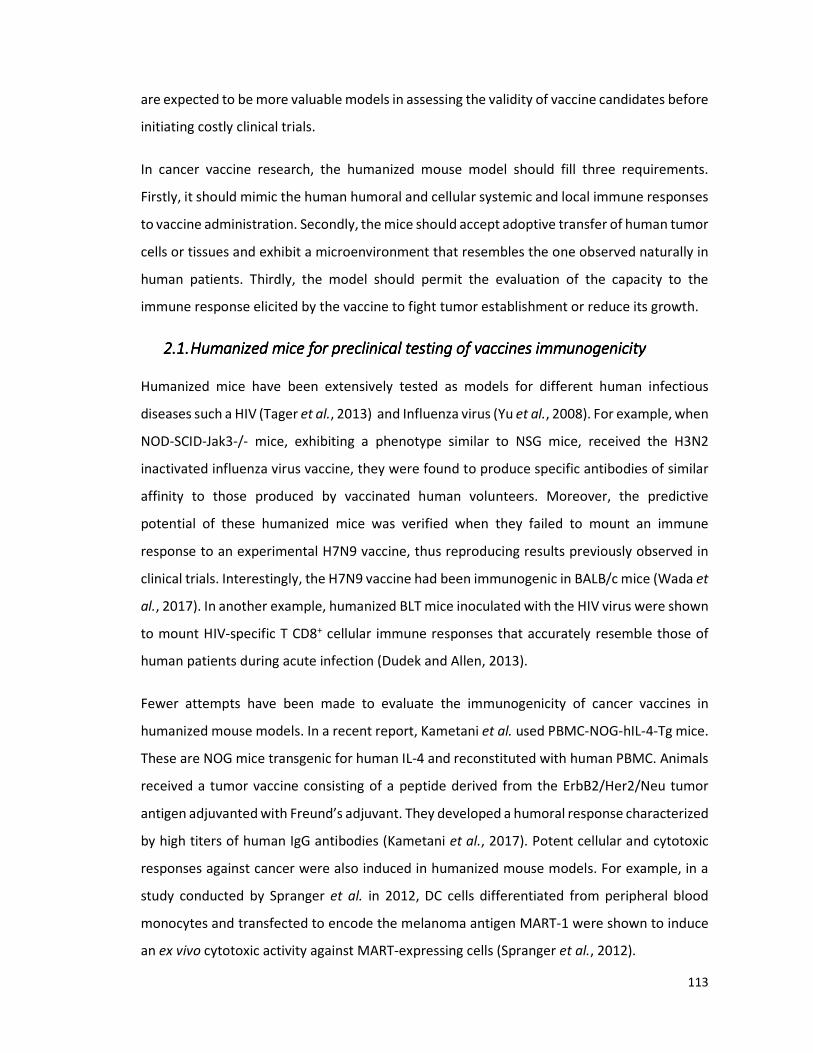
113
are expected to be more valuable models in assessing the validity of vaccine candidates before
initiating costly clinical trials.
In cancer vaccine research, the humanized mouse model should fill three requirements.
Firstly, it should mimic the human humoral and cellular systemic and local immune responses
to vaccine administration. Secondly, the mice should accept adoptive transfer of human tumor
cells or tissues and exhibit a microenvironment that resembles the one observed naturally in
human patients. Thirdly, the model should permit the evaluation of the capacity to the
immune response elicited by the vaccine to fight tumor establishment or reduce its growth.
2.1.2.1.2.1.2.1. Humanized mice for preclinical testing of vaccinesHumanized mice for preclinical testing of vaccinesHumanized mice for preclinical testing of vaccinesHumanized mice for preclinical testing of vaccines immunogenicityimmunogenicityimmunogenicityimmunogenicity
Humanized mice have been extensively tested as models for different human infectious
diseases such a HIV (Tager et al., 2013) and Influenza virus (Yu et al., 2008). For example, when
NOD-SCID-Jak3-/- mice, exhibiting a phenotype similar to NSG mice, received the H3N2
inactivated influenza virus vaccine, they were found to produce specific antibodies of similar
affinity to those produced by vaccinated human volunteers. Moreover, the predictive
potential of these humanized mice was verified when they failed to mount an immune
response to an experimental H7N9 vaccine, thus reproducing results previously observed in
clinical trials. Interestingly, the H7N9 vaccine had been immunogenic in BALB/c mice (Wada et
al., 2017). In another example, humanized BLT mice inoculated with the HIV virus were shown
to mount HIV-specific T CD8+ cellular immune responses that accurately resemble those of
human patients during acute infection (Dudek and Allen, 2013).
Fewer attempts have been made to evaluate the immunogenicity of cancer vaccines in
humanized mouse models. In a recent report, Kametani et al. used PBMC-NOG-hIL-4-Tg mice.
These are NOG mice transgenic for human IL-4 and reconstituted with human PBMC. Animals
received a tumor vaccine consisting of a peptide derived from the ErbB2/Her2/Neu tumor
antigen adjuvanted with Freund’s adjuvant. They developed a humoral response characterized
by high titers of human IgG antibodies (Kametani et al., 2017). Potent cellular and cytotoxic
responses against cancer were also induced in humanized mouse models. For example, in a
study conducted by Spranger et al. in 2012, DC cells differentiated from peripheral blood
monocytes and transfected to encode the melanoma antigen MART-1 were shown to induce
an ex vivo cytotoxic activity against MART-expressing cells (Spranger et al., 2012).

114
2.2.2.2.2.2.2.2. Humanized mice in cancer vaccine research: evaluation of protectionHumanized mice in cancer vaccine research: evaluation of protectionHumanized mice in cancer vaccine research: evaluation of protectionHumanized mice in cancer vaccine research: evaluation of protection
2.2.1. Humanized SCID mice can accept adoptive transfer of tumor
xenografts and reconstitute their natural microenvironment
The profound immunodeficiency of the SCID mice and their derivatives was found to facilitate
the engraftment of a large panel of human primary tumors and cell lines and to reconstitute
the tumor microenvironment (reviewed by (Ito et al., 2012; Zhou et al., 2014)).
Hu-PBL-SCID mice carrying human bladder cancer xenografts were shown to undergo a
successful reconstitution with human lymphocytes, to secrete human immunoglobulins and
to support tumor infiltration with T cells (Gong et al., 2015). Comparable results were obtained
in a model of cervical cancer carcinoma where the slowly growing tumors were found to be
infiltrated with lymphocytes (Ye et al., 2006). In a Hu-SRC-SCID model of breast cancer,
concurrent transplantation of human HSCs and breast cancer cells resulted both in the
engraftment of a functional immune system and in tumor development and dissemination.
The tumor was infiltrated with activated specific T-cells and NK cells (Wege et al., 2011).
2.2.2. Humanized SCID mice as platforms for cancer vaccine evaluation
The established capacity of humanized mouse models to mimic the human immune response
and the tumor microenvironment prompted their use for the development of cancer
immunotherapies (Kozlowska et al., 2016; Siegler et al., 2005; Trieu et al., 2004) and especially
cancer vaccines.
Among the multiple examples that illustrate their potential in this field, we cite a model of
colorectal carcinoma model in SCID/Beige nude mice, completely lacking NK cells that were
engrafted with HSCs. These mice were vaccinated with autologous in vitro-generated DCs,
transfected to express a co-stimulatory molecule, the CD137 ligand (CD137L), and pulsed with
tumor-cell antigens (Fu et al., 2017). CD137 is expressed on activated T cells, and the inducible
expression of its ligands is currently one of the targets of cancer immunotherapies (Vinay and
Kwon, 2014). This vaccination protocol was found to protect Hu-SRC-SCID mice from tumor
development. Indeed, mice showed a delayed tumor growth and a decreased tumor volume
and weight, as compared to non-vaccinated Hu-SRC-SCID mice, indicating that tumor rejection
was exclusively due to the vaccine, and not to HLA-mismatch between tumor cells and cancer
cells (Fu et al., 2017).

115
In another cancer vaccine trial, humanized mice were injected with MHC-matched melanoma
cells. They subsequently received an engineered oncolytic adenovirus encoding for GM-CSF,
on which MHC class I peptides of the MAGE-A1 melanoma antigen were adsorbed. The vaccine
was shown to eradicate the established tumors and to induce tumor-specific CD8+ T cells
(Capasso et al., 2016).
Humanized mouse models also served as platforms for a limited number of protein/peptide
based vaccine evaluation studies. In one of these studies, Hu-PBL-SCID mice were vaccinated
with a recombinant protein consisting of the two melanoma antigens MAGE-A1 and MAGE-
A2, in addition to the recombinant Mycobacterium tuberculosis heat shock protein 70
(TBHSP70) that served as a danger molecules. This protocol was shown to induce vaccine
specific lymphocytes, among which CTLs mediate the lysis of MAGE-expressing cancer cells in
vitro. In a prophylactic vaccination setting, Hu-PBL-SCID mice immunized with the MAGE-
A1/MAGE-A2/TBHSP70 showed slower tumor growth and higher survival rates as compared
to control mice (Junwei et al., 2016).
Altogether, the growing evidence of the capacity of the humanized mouse models to
mimic the human adaptive immune response argues for their use in vaccine development. In
particular, their capacity to induce CD8+ T cell responses against tumor antigens on one hand,
and to engraft and reconstitute tumor microenvironment on the other hand, strongly suggests
them as platforms for the evaluation of cancer vaccine immunogenicity and efficacy.

OBJECTIVES __________________________________________________________________________________________

117
Despite their improved efficacy in recent years, the majority of currently used cancer
treatments remain poorly specific for tumor cells and induce major side effects. For decades,
data has accumulated providing proof that tumors are immunogenic and capable of triggering
immune responses. These responses may, under certain circumstances, be able to control
them, but tumors usually escape destruction by undergoing genetic and epigenetic
modifications, inducing tolerization and immune response inhibition. Therefore, the idea of
developing therapeutic cancer vaccines that would reverse this context and initiate/amplify
the immune response against tumors is gaining increased attention. Translation of this
concept into a reality is rapidly evolving thanks to the recent discoveries in the field of cancer
immunobiology. It is today established that tumors display TAAs that may be presented by
DCs and recognized by CD4+ and CD8+ T cells. However, they usually do not activate an efficient
tumor-specific immune response because the tumor microenvironment is generally
immunosuppressive. A cancer vaccine should therefore include all the elements needed to
target and activate the DCs and generate tumor antigen presentation in an
immunostimulatory context. Ultimately, it should lead to the production of specific Th1 cells
to induce differentiation of CTLs that are capable of infiltrating the tumor and killing cancer
cells.
Currently, various clinical trials employing multiple strategies in the design and delivery of
cancer vaccines are ongoing and showing promising results. Extensive optimization efforts are
made to replace conventional approaches relying on the use of whole TAA proteins and
conventional adjuvants by innovative ones using minimal peptide sequences and small, less
reactogenic, stimulatory molecules derived from bacteria. These elements are generally
combined altogether in various vectors such as nanoparticles. Liposomes emerged as
interesting vectors for such vaccines. Indeed, they are biocompatible and their composition,
size, charge and surface functionalization can be easily manipulated, thereby offering a wide
range of potential strategies to optimize cancer vaccine delivery.
Regarding the vaccine delivery route, special attention has been recently devoted to the skin.
Being an immunological barrier that normally protects the organism from external
aggressions, the skin is rich in multiple DC subpopulations. The conventional subcutaneous
route, which has proven its efficacy in microbial vaccination, is currently being explored in
cancer vaccination. However, this route does not optimally deliver the vaccine to skin DCs

118
since the hypodermis is devoid of DCs, and leads to rapid vaccine draining to the lymph nodes.
A more efficient approach to target skin DCs may be to apply the vaccine on the skin surface
to reach the epidermis and the dermis. However, the skin is also a mechanical barrier with an
impermeable uppermost layer, the stratum corneum. Therefore, innovative solutions to cross
this barrier are needed. Fortunately, one of the advantages of liposomes is that they are
adapted to multiple delivery routes, including the transcutaneous one. Moreover, their
penetration in the skin can be further increased through modification of their physicochemical
properties.
In my host laboratory at the University of Strasbourg, liposomal constructs incorporating the
necessary elements for the induction of an efficient antitumor immune response were
previously developed. They incorporated a TLR 2/6 agonist (Pam2CAG) as immunostimulatory
molecule, a CD8+ T cell epitope peptide derived from a TAA expressed of cancer cells, as well
as a universal CD4+ T cell epitope peptide derived from the influenza virus hemagglutinin
protein. The immunogenicity and tumor-specific efficiency of these liposomes were
established following administration by the subcutaneous and the intranasal routes in tumor-
bearing conventional mice (Kakhi et al., 2015, 2016; Roth et al., 2005; Thomann et al., 2011).
In parallel, in the vaccine development field, it is not infrequent that vaccine candidates that
are originally selected and optimized based on preclinical experimentation in rodents yield
disappointing results in clinical trials. Severely immunodeficient mice reconstituted with
human immune cells offer the possibility to explore the human immune response to various
antigens. They may therefore be promising models to solve this issue, by allowing the in vivo
evaluation of vaccine candidates destined for humans.
In my host laboratory in the Lebanese University, a humanized mouse model was previously
developed, in which immunodeficient mice are reconstituted with human splenocytes (Hu-
SPL-SCID) (Bouharoun-Tayoun et al., 2004; Chamat et al., 1999). We have since adapted this
model to the NSG mouse. The Hu-SPL-NSG model proved to be superior to more conventional
models relying on the use of peripheral blood cells, yielding more potent primary human
immune responses against a variety of antigens, including the hepatitis B vaccine and
experimental vaccine candidates against Plasmodium falciparum. However, these antigens
were adjuvanted with conventional potent adjuvants (Ghosn, 2015).

119
In this context, the objective of my thesis project consisted in developing liposome-based
vaccines for transcutaneous cancer vaccination in humans. Therefore, we aimed at
optimizing a liposome vaccine that would induce a potent immune response after TC
administration to a conventional mouse model. Additionally, we aimed to verify that the
liposome constructs can induce an immune response by human immune cells in vivo, using a
humanized mouse model. To this end, the experimental work was divided into two parts, the
results of which will will be presented in two chapters, each corresponding to a scientific
article in preparation that answers one of these two general objectives.
The specific objectives are presented in detail below.
Objective 1: Development of liposome constructs adapted for TC cancer
vaccination and evaluation of their immune potential
The first specific objective of my thesis project consisted in developing and validating
liposome-based vaccines suitable for transcutaneous cancer vaccination. Specifically, we
aimed to refine the liposome-based vaccine composition previously validated in the BALB/c
model and to identify a formulation that induces a potent CD4+ and CD8+ T cell immune
response after TC administration in mouse models. The results of this part are presented in
the scientific article #1, in preparation.
1.1 Optimization of the composition of the liposomal constructs for TC immunization: Based
on the original liposomal vaccine developed in our team (Thomann et al., 2011), we
formulated new vaccine candidates for the TC route by modifying three elements:
- The immunostimulatory molecule: In order to choose the most optimal adjuvant for
the induction of vaccine-specific responses by the TC route, we replaced the original
immunostimulatory molecule, which was a TLR 2/6 agonist (Pam2CAG) by a TLR4
agonist (MPLA).
- The construct targeting to DCs: a bivalent mannose residue was previously developed
in our laboratory (Espuelas et al., 2003), and liposomal vaccines incorporating this
residue were shown to exhibit improved uptake by DCs (Espuelas et al., 2008),
permitting a reduction in the minimal dose of immunostimulatory molecule required

120
to induce an immune response (Thomann et al., 2011). Therefore, mannose residues
addition was tested in the constructs designated for TC vaccination.
- The flexibility/deformability of the lipid vesicle: The above mentioned elements were
incorporated in two types of vesicles, either conventional liposomes, or
ultradeformable liposomes or transfersomesTM which are assumed to provide better
penetration through the skin, to test whether the latter would induce improved
immune responses.
We also formulated fluorescent liposomes incorporating a hydrophobic fluorochrome in
their lipid bilayer. This strategy offers the opportunity to track the cells that internalize the
liposomes, including skin DCs, and assess their migration to draining lymph nodes
following TC application.
The formulated constructs had to fulfill the following requirements:
1- Lipid vesicle size:
a. Average diameter: we aimed to obtain lipid vesicles with an average diameter
of 100 nm, because in this size range various nanoparticles were reported to
be suitable for skin barrier crossing (Lilia Romero and Morilla, 2011; Rattanapak
et al., 2012)
b. Size distribution/homogeneity: we aimed to formulate liposomes with a
narrow size distribution and with a polydispersity index (PDI) < 0.3.
2- Peptide coupling rate: the peptide coupling rate to the preformed liposomes
incorporating a functionalized anchor had to be highly efficient in order to minimize
peptide loss.
3- Formulation technique: the reproducibility of the formulation technique was
essential to obtain homogenous formulations during the whole process. This allows
us to avoid the variations of the in vivo results that may be caused by a variation of
the vaccine formulation itself.
1.2 Evaluation of the local and systemic immune response induced by the constructs
In a first step, the original construct was evaluated by the SC and the TC routes to provide
a proof of concept of their immunogenicity after TC administration on one hand, and to

121
compare the immune response induced by the two administration routes on the other
hand.
We next compared the local and systemic immune responses induced by liposomes
incorporating the two different TLR agonists.
We finally investigated the influence of the lipid vesicle fluidity on the immune response
induced by the TC route.
1.3 Assessment of the migration of skin DCs after TC immunization
Because the liposomal constructs were shown to induce an immune response after TC
administration, we assessed their capability to induce the migration of skin DCs to lymph
nodes draining the application zone.
Objective 2: Evaluation of liposomes immunogenicity in the Hu-SPL-NSG model
The second specific objective of my thesis was therefore to determine whether liposome
constructs that were previously validated in murine models can induce detectable human
immune responses in the Hu-SPL-NSG model. The results of this part are presented in the
scientific article #2, in preparation.
To our knowledge, there are very few reports in the literature regarding administration of
liposome-based formulations to humanized mice and evaluation of their immunogenicity in
this model. Therefore, before attempting to test the capacity of TAA-expressing liposomes to
induce a tumor-specific CTL response in the Hu-SPL-NSG mouse model, several preliminary
steps were needed to establish the proof of concept of this approach. To this end we used
several alternative liposome-based vaccine formulations.
2.1. Assessment of the safety of the TLR agonists towards human splenocytes
In a first step, it was primordial to assess the safety of liposomes incorporating different TLR
agonists towards human cells. While their immunostimulatory effect is essential for activating
immune responses, excessive stimulation may lead to cell death or alter the capacity of human
cells to engraft in the NSG mouse.

122
Therefore, we first performed in vitro evaluation assays of liposomes incorporating different
TLR agonists. Then we completed the selected liposome by adding the vaccine epitopes and
tested it in vivo. We evaluated its effect on mice survival and on the homing of the injected
splenocytes to the mouse spleen and their capacity to secrete total human immunoglobulins.
2.1. Evaluation of the liposomes capacity to induce an immune response in the Hu-SPL-
NSG mouse model
We next aimed to determine whether the liposomal constructs can induce a detectable human
immune response in the Hu-SPL-NSG model. This approach faces several challenges. For
instance, the ability of the Hu-SPL-NSG mouse to mount cellular responses is far less well
established than that to mount humoral responses (Bouharoun-Tayoun et al., 2004; Brams et
al., 1998; Chamat et al., 1999). Moreover, cellular immune responses can be evaluated in
secondary lymphoid organs only after animal euthanasia, while humoral responses can be
assessed regularly in mice sera in the ongoing experiment. For all these reasons, we
formulated liposomal constructs incorporating, in addition to the HA peptide and the selected
TLR agonist, a B cell epitope rather than a T cell epitope. The B epitope is derived from
Pseudomonas aeruginosa, and has been previously evaluated in liposomal constructs
administered to immunocompetent mice by the intraperitoneal and the intranasal routes
(Heurtault et al., 2009). The humoral immune response was assessed by searching for anti-
PAK antibodies at several time points after immunization. The CD4+ T cell response was
evaluated in vitro in the mice spleen pools after sacrifice by detecting the production of IFN-
γ, IL-4 and IL-10 by ELISPOT.

EXPERIMENTAL
RESULTS __________________________________________________________________________________________

Chapter 1: Transcutaneous immunization with liposome-based
cancer vaccines induce CD4+ and CD8+ T cell responses in BALB/c
mice
My host team in the University of Strasbourg has developed innovative liposome-based
vaccines that co-deliver the crucial elements for a tumor-specific immune response. The
chosen liposomes are conventional ones, composed of phosphatidylcholine,
phosphatydilglycerol and cholesterol, the main constituents of cell membranes, and,
therefore, are tolerated by the immune system. The elements delivered by these liposomes
are namely a CD8+ T cell peptide epitope derived from the human TAA ErbB2, a universal CD4+
T cell peptide epitope derived from the hemagglutinin of the influenza virus, in addition to an
immunostimulatory molecule, a TLR2/6 agonist (Pam2CAG). In conventional mouse models,
these constructs were shown to induce efficient anti-tumor immune responses after
subcutaneous (Roth et al., 2005; Thomann et al., 2011) and intranasal (Kakhi et al., 2015, 2016)
administration in normal mice.
When I first joined the team, one previous attempt had been performed to deliver the
liposome-based constructs by the TC route and seemed to give promising results. In this
experiment, the mice were shaved on their back and were allowed to rest for 24h before
applying the formulation by massage. Therefore, we decided to adopt this same vaccination
strategy. As a preliminary step, we optimized the vaccine penetration through the skin by
dividing the vaccine dose into three subsequent sub-doses, and by massaging every sub-dose
for one minute. Then, to improve the immunogenicity of the liposomal formulations by the TC
route, we decided to benefit from their versatility by modifying their composition and their
physicochemical properties.
Because the immunostimulatory molecule plays a central role in the induction of an immune
response, we replaced the TLR2/6 agonist that was incorporated in the original formulation,
with a TLR 4 agonist (MPLA), and compared the resulting constructs. Additionally, in order to
allow a more efficient delivery of the vaccine components to the DC, we attempted to target
the liposome constructs to the skin DCs by incorporating a DC-targeting molecule, di-mannose.
Because the ability of the liposomes to cross the cutaneous barrier depends on the fluidity of

125
their lipid bilayer, we replaced the conventional liposome vesicle with an ultradeformable one,
called transfersome, in order to optimize the skin crossing ability of our formulations. Finally,
to evaluate the local and systemic immune response induced by these constructs variants, we
tested them in a conventional mouse model. The corresponding results are presented in the
following scientific article # 1 (in preparation).

126
Scientific article #1:
Transcutaneous immunization with liposome-based
cancer vaccines induce CD4 + and CD8+ T cell
responses in BALB/c mice
Hanadi Saliba, Vincent Flacher, Célia Jacoberger, Yohan Gerber, Maroua Messous,
Hasnaa Bouharoun-Tayoun, Benoît Frisch, Soulaima Chamat, Béatrice Heurtault,
Sylvie Fournel
In preparation

127
Introduction
The recent years have witnessed major research advances in therapeutic anti-tumor
vaccination, which is intended to induce and amplify the physiological immune responses
against cancer (reviewed by (Butterfield, 2015; Guo et al., 2015)), (Adams et al., 2008;
Berinstein et al., 2015; Chen et al., 2015; Dhodapkar et al., 2014; Fujiyama et al., 2014). These
advances were made possible by the identification of tumor-associated antigens (TAA) against
which an immune response may be induced, leading to the destruction of TAA-expressing
tumor cells (Baldwin, 1971; van der Bruggen et al., 1991; Van den Eynde and van der Bruggen,
1997; Vigneron, 2015). They have also highlighted the pivotal role played by dendritic cells
(DCs) in eliciting such protective immune responses (Palucka and Banchereau, 2013).
Tumor-specific responses are induced when dying cancer cells release exosomes and/or
apoptotic bodies containing TAAs and molecules that indicate cell damage and therefore play
the role of danger signals (Dhodapkar et al., 2007; Pathak et al., 2012; Tang et al., 2012).
Following TAA internalization by DCs, their processing leads to cross-presentation of derived
peptides on MHC class I and class II molecules to CD8+ and CD4+ T cells, respectively (Alloatti
et al., 2016; Joffre et al., 2012). Following recognition of TAA-derived epitope peptides by their
specific antigen receptors, CD4+ and CD8+ T cells differentiate into tumor specific helper (Th)
and cytotoxic (CTL) effector cells, respectively. The outcome of this differentiation depends
on the maturation state of the DC that is induced by danger signals provided by Microbe-
Associated Molecular Patterns (MAMPs) in infectious context or by Damage-Associated
Molecular Patterns (DAMPs) in non-infectious context such as cancer. These molecules engage
specific receptors on the DC surface, named PRR for Pattern Recognition Receptors (PRRs),
such as Toll-Like-Receptors (TLRs (Dudek et al., 2013; Pathak et al., 2012). Beside danger
signals, optimal differentiation of CTLs needs complementary signals provided by Th cells,
especially IFNγ producing Th1 cells, after recognition of their specific antigen presented by the
same DC that is presenting the antigen to the CTL. Following recognition of TAA-derived
peptides on the cancer cells surface, CTLs mediate their apoptosis. Therefore, to induce a
cancer-specific response, a cancer vaccine should incorporate three elements, namely a i)
CD4+ T cell epitope, a ii) CD8+ T cell epitope derived from the TAA, both necessary for the
activation of cancer-specific T cells, and iii) a danger signal that induces DC maturation.

128
One of the most extensively used vaccination sites is the skin, and more specifically through
the subcutaneous route. Recently, the transcutaneous (TC) route has emerged as a tempting
alternative (DeMuth et al., 2013; Glenn et al., 2000; Kim and Prausnitz, 2011). Indeed, while
the hypodermis is virtually devoid of DCs, the epidermis and the dermis harbor several DC
populations. The epidermal Langerhans cells (LCs), characterized by their high expression of
langerin/CD207, and the dermal dendritic cells (dDCs), comprised of different subpopulations
that can express or not langerin/CD207, normally ensure skin immunosurveillance (Malissen
et al., 2014; Merad et al., 2002, 2008). After sampling the constituents of their environment,
activated skin DCs that sense danger signals migrate to the skin draining lymph nodes where
they activate lymphocytes to mount an adaptive immune response (Worbs et al., 2017). Upon
activated T cell recirculation, an adaptive immune response is elicited locally and systemically
(Hopkins and McConnell, 1984; Masopust and Schenkel, 2013; Thomas et al., 2012). However,
for a vaccine to be efficient by transcutaneous immunization (TCI), it must efficiently cross the
stratum corneum, which is the impermeable uppermost layer of the epidermis (Marks, 2004).
One of the advances in vaccine technology resulted in the emergence of innovative liposome-
based formulations (Bangham and Horne, 1964; Bangham et al., 1974) that are suitable for
skin barrier crossing (Ashtikar et al., 2016; Hansen and Lehr, 2012) and can deliver all the
minimum vaccine key elements to the skin DCs (Chikh and Schutze-Redelmeier, 2002; Iwama
et al., 2016; Roth et al., 2005; Shariat et al., 2014; Thomann et al., 2011). Liposomes are
versatile phospholipid vesicles. It is possible to modulate each of their physicochemical
characteristics to adapt their use for different applications. In particular, it is possible to
increase their fluidity to optimize them for TC delivery. Liposomes with ultradeformable
bilayers, called transfersomesTM, are reported to exhibit increased passage capacities through
mildly disrupted skin barriers, by squeezing into pores smaller than their size (Ascenso et al.,
2015; Benson, 2006; Cevc et al., 1998)
Our team has previously developed several liposomal formulations that co-deliver a CD4+ T
cell epitope derived from the hemagglutinin of the influenza virus (HA 307–319,
PKYVKQNTLKLAT-C) (O’Sullivan et al., 1991) and a CD8+ T cell epitope derived from the human
TAA ErbB2 (p63–71, CG-TYLPTNASL) (Nagata et al., 1997), in addition to TLR agonists that
functions as danger signal (Roth et al., 2005; Thomann et al., 2011). Different TLR agonists
have been tested, including S-[2,3-bis(palmitoyloxy)-(2R)-propyl]-(R)-cysteinyl-alanyl-glycine

129
(Pam2CAG), a diacetylated lipopeptide derived from the N-terminal moiety of E. coli
lipoprotein that interacts with TLR 2/6 heterodimers (Oliveira-Nascimento et al., 2012; Omueti
et al., 2005). When incorporated into liposomal constructs, this TLR agonist was found to
induce the maturation of human monocyte-derived DCs in vitro, as indicated by the expression
of CD80, CD83, CD86 and HLA-DR molecules (Espuelas et al., 2005a). Additionally, the above-
mentioned liposomes incorporating the T CD4+ and T CD8+ epitopes trigger protective tumor-
specific responses in vivo (Heurtault et al., 2009; Thomann et al., 2011). GlaxoSmithKline has
recently designed adjuvant systems consisting of liposomes incorporating monophosphoryl
lipid A (MPLA), a TLR4 agonist (Alving and Rao, 2008; Alving et al., 2012a). MPLA is a chemically
detoxified form of the lipid A, the anchor moiety of lipopolysaccharides (LPS) (a highly
immunostimulatory structure found on the outer cell surface of Gram negative bacteria).
These adjuvant systems are currently used in multiple vaccines, especially those where CTL
responses are needed (Alving et al., 2012b).
Addition of targeting molecules such as mannose residues to the liposomes can facilitate their
uptake into the DC through interaction with mannose-specific receptors (East and Isacke,
2002; Espuelas et al., 2008; Markov et al., 2015). Our team has reported that, when adding di-
mannose residues to liposome surface, it is possible to reduce the amount of incorporated TLR
agonist without losing their immune-activating potential (Thomann et al., 2011).
In the present work, our aim was to optimize liposomal constructs previously developed in our
laboratory for TC cancer vaccination. We formulated several variants that differ for the TLR
agonist, the presence of a DC targeting molecule and the lipid vesicle composition. We
evaluated their immune potential after TC immunization of BALB/c mice by testing their
capacity to elicit an HA-specific CD4+ and ErbB2-specific CD8+ T cell response. Moreover, we
tested the ability of the liposomal constructs to induce skin DC migration to draining lymph
nodes.

130
Material and methods
1. Formulation and characterization of liposomal constructs
1.1.1.1.1.1.1.1. Lipids Lipids Lipids Lipids and adjuvants and adjuvants and adjuvants and adjuvants
Egg yolk L-α-phosphatidylcholine (PC), soy PC (SPC) and cholesterol (Chol, recristallized in
methanol) were purchased from Sigma-Aldrich (St. Quentin Fallavier, France) and their
purities exceeded 99%. L-α-phosphatidyl-DL-glycerol transesterified from egg yolk PC (PG) was
purchased from Avanti Polar lipids (Alabama, USA). The lipopeptide S-[2,3-bis(palmitoyloxy)-
(2R)-propyl]-(R)-cysteinyl-alanyl-glycine (Pam2CAG), the thiol-functionalized lipid anchor
dipalmitoylglycerol maleimide (DPGMal) and the mannosylated lipid anchor dioleylglycerol di-
mannose (DOG-Man2) were synthesized at the laboratory as previously described (Espuelas et
al., 2003, 2008; Heurtault et al., 2009; Roth et al., 2004). The lipopolysaccharide derivative
Monophosphoryl lipid A (MPLA) was purchased from Invivogen (San Diego, CA). All reagents
were conserved under argon at -20 °C.
1.2.1.2.1.2.1.2. Synthetic peptidesSynthetic peptidesSynthetic peptidesSynthetic peptides
The peptides ErbB2 p63–71 (CG-TYLPTNASL, MW = 1139 g/mol) (CTL eptitope) (Nagata et al.,
1997), and influenza virus haemagglutinin-derived HA 307–319 (PKYVKQNTLKLAT-C, MW =
1606 g/mol) (TCD4+ epitope) (O’Sullivan et al., 1991) were obtained from Genosphere
Biotechnologies (Paris, France). Their purity, as assessed by HPLC, was > 85%.
1.3.1.3.1.3.1.3. Formulation of lipid vesicles Formulation of lipid vesicles Formulation of lipid vesicles Formulation of lipid vesicles
1.3.1. Formulation of fluorescent liposomal SUVs (DiI-liposomes)
Multilamellar fluorescent vesicles (DiI-MLV) were prepared by lipid film hydration technique.
Briefly, a chloroform/methanol (9/1 v/v) solution containing PC, PG, Chol, adjuvant, DOG-
Man2 and DiI (1,1'-dioctadecyl-3,3,3'3'-tetramethylindocarbocyanine perchlorate) were mixed
in a round-bottom Pyrex tube, and slowly evaporated under a continuous flow of argon. The
molar proportions of each of the constituents is shown in table 1. The resulting lipid film was
completely dried under high vacuum for 1 hour. It was then hydrated in 10 mM Hepes buffer
(pH 7.4) containing 5% w/v sorbitol by rigorous vortex mixing, to yield a phospholipid
concentration of 15 mM. The resulting DiI-MLV suspension was sonicated (1s cycle every 3 s)
for 1 hour at room temperature under a continuous flow of argon, using a Vibra Cell 75041
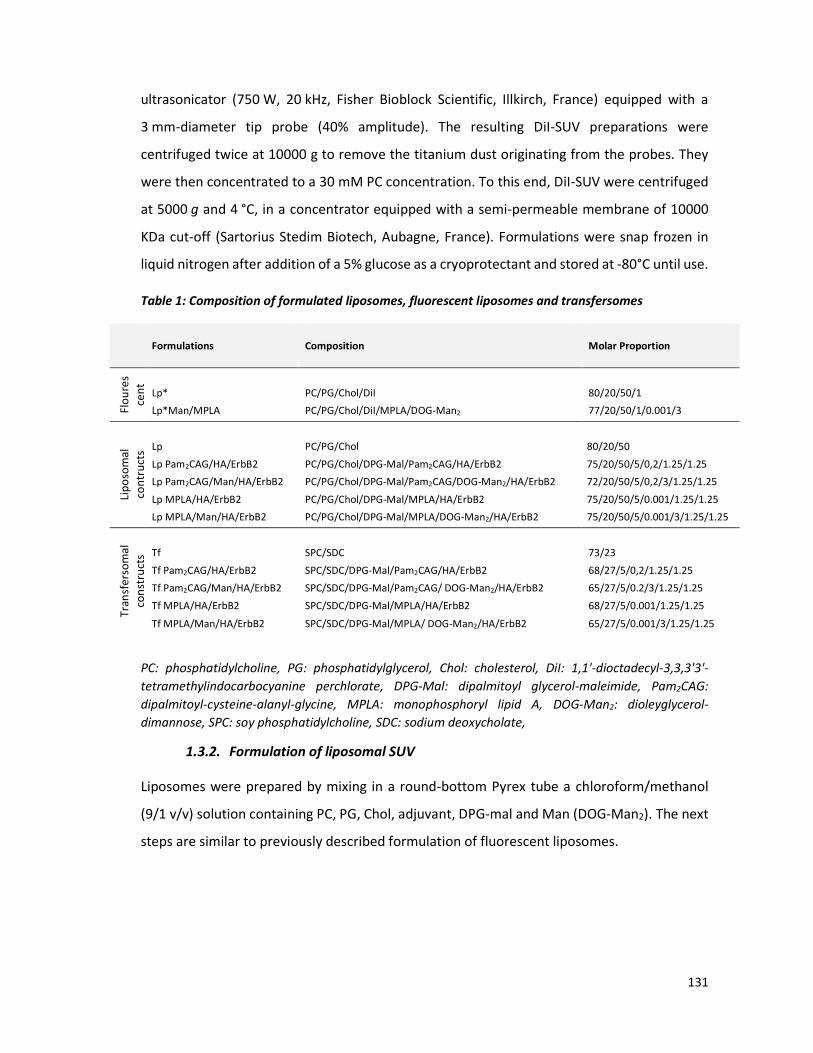
131
ultrasonicator (750 W, 20 kHz, Fisher Bioblock Scientific, Illkirch, France) equipped with a
3 mm-diameter tip probe (40% amplitude). The resulting DiI-SUV preparations were
centrifuged twice at 10000 g to remove the titanium dust originating from the probes. They
were then concentrated to a 30 mM PC concentration. To this end, DiI-SUV were centrifuged
at 5000 g and 4 °C, in a concentrator equipped with a semi-permeable membrane of 10000
KDa cut-off (Sartorius Stedim Biotech, Aubagne, France). Formulations were snap frozen in
liquid nitrogen after addition of a 5% glucose as a cryoprotectant and stored at -80°C until use.
Table 1: Composition of formulated liposomes, fluorescent liposomes and transfersomes
Formulations Composition Molar Proportion
Flo
ure
s
cen
t
Lp* PC/PG/Chol/DiI 80/20/50/1
Lp*Man/MPLA PC/PG/Chol/DiI/MPLA/DOG-Man2 77/20/50/1/0.001/3
Lip
oso
ma
l
con
tru
cts
Lp PC/PG/Chol 80/20/50
Lp Pam2CAG/HA/ErbB2 PC/PG/Chol/DPG-Mal/Pam2CAG/HA/ErbB2 75/20/50/5/0,2/1.25/1.25
Lp Pam2CAG/Man/HA/ErbB2 PC/PG/Chol/DPG-Mal/Pam2CAG/DOG-Man2/HA/ErbB2 72/20/50/5/0,2/3/1.25/1.25
Lp MPLA/HA/ErbB2 PC/PG/Chol/DPG-Mal/MPLA/HA/ErbB2 75/20/50/5/0.001/1.25/1.25
Lp MPLA/Man/HA/ErbB2 PC/PG/Chol/DPG-Mal/MPLA/DOG-Man2/HA/ErbB2 75/20/50/5/0.001/3/1.25/1.25
Tra
nsf
ers
om
al
con
stru
cts
Tf SPC/SDC 73/23
Tf Pam2CAG/HA/ErbB2 SPC/SDC/DPG-Mal/Pam2CAG/HA/ErbB2 68/27/5/0,2/1.25/1.25
Tf Pam2CAG/Man/HA/ErbB2 SPC/SDC/DPG-Mal/Pam2CAG/ DOG-Man2/HA/ErbB2 65/27/5/0.2/3/1.25/1.25
Tf MPLA/HA/ErbB2 SPC/SDC/DPG-Mal/MPLA/HA/ErbB2 68/27/5/0.001/1.25/1.25
Tf MPLA/Man/HA/ErbB2 SPC/SDC/DPG-Mal/MPLA/ DOG-Man2/HA/ErbB2 65/27/5/0.001/3/1.25/1.25
PC: phosphatidylcholine, PG: phosphatidylglycerol, Chol: cholesterol, DiI: 1,1'-dioctadecyl-3,3,3'3'-
tetramethylindocarbocyanine perchlorate, DPG-Mal: dipalmitoyl glycerol-maleimide, Pam2CAG:
dipalmitoyl-cysteine-alanyl-glycine, MPLA: monophosphoryl lipid A, DOG-Man2: dioleyglycerol-
dimannose, SPC: soy phosphatidylcholine, SDC: sodium deoxycholate,
1.3.2. Formulation of liposomal SUV
Liposomes were prepared by mixing in a round-bottom Pyrex tube a chloroform/methanol
(9/1 v/v) solution containing PC, PG, Chol, adjuvant, DPG-mal and Man (DOG-Man2). The next
steps are similar to previously described formulation of fluorescent liposomes.

132
1.3.3. Formulation of ultradeformable liposomes or transfersomes™
Uf-SUVs were prepared as described before by Cevc et al (Cevc and Blume, 1992; Cevc et al.,
1998). Briefly, appropriate proportions of SPC, DPG-Mal, adjuvant and DOG-Man2 were
dissolved in chloroform/methanol (9/1 v/v). Solvent was evaporated under high vacuum in a
Pyrex tube for 1 h. The resulting lipid film was dispersed in ethanol (8.5% (v/v) at a final
phospholipid concentration of 15 mM) and mixed vigorously for 2 min. A 10 mM Hepes buffer
(pH 6.5) containing 5% w/v sorbitol and 5 mM SDC was then added to the suspension to obtain
a final concentration of 4 mM of the surfactant. The mixture was stirred vigorously for 2 min.
The resulting MLV suspension was manually extruded (LiposoFast-Basic, Avestin, Canada) for
20 cycles through a 100-nm pore size polycarbonate mesh, in order to obtain Tf-SUV.
Formulations were snap frozen in liquid nitrogen after addition of 10% glucose as a
cryoprotectant and stored at -80°C until use.
1.4.1.4.1.4.1.4. Peptide conjugation to LpPeptide conjugation to LpPeptide conjugation to LpPeptide conjugation to Lp----SUV or TfSUV or TfSUV or TfSUV or Tf----SUVSUVSUVSUV
Potential disulfide bonds of cysteine residues between peptides that may result in peptide
dimerisation, were reduced with 0.7 M eq. tris(2-carboxyethyl) phosphine (Interchim,
Montluçon, France), for 15 minutes under argon. Equimolar quantities of CD4+ and CD8+ T cell
epitope peptides were then coupled to freshly prepared SUVs by Michael addition in 10 mM
Hepes buffer (pH 6.5) containing 5% (w/v) sorbitol (0.5 molar eq of each peptide vs surface
accessible thiol-reactive maleimide functions). After an incubation of 3 hr under argon at room
temperature, a 10-fold excess of β-mercaptoethanol was added for 30 minutes to inactivate
all unreacted maleimide groups on internal or external surface of SUVs. Then, the formulation
was extensively dialysed (Spectra/Por, exclusion limit of 12–14 kDa, Spectrum laboratories,
DG Breda, Netherlands) against a 10 mM Hepes buffer (pH 7.4) containing 5% (w/v) sorbitol
to eliminate unreacted reagents and peptides.
1.5.1.5.1.5.1.5. Physicochemical characterization of the constructsPhysicochemical characterization of the constructsPhysicochemical characterization of the constructsPhysicochemical characterization of the constructs
1.5.1. Nanoparticle size measurement by dynamic light scattering
The average size of formulated Lp-SUVs and Tf-SUVs was measured by dynamic light scattering
using a Zetasizer Nano-ZS (Malvern instruments, Orsay, France) with the following

133
specifications: sampling time, 30 s; viscosity, 1.014 cP; refractive index, 1.34; scattering angle,
90°; temperature, 25°C. SUVs were diluted at 1/100 in 10 mM Hepes buffer (pH 7.4) containing
5% (w/v) sorbitol, and the results were the average of three consecutive measurements. Data
were analyzed using the multimodal number distribution software included with the
instrument. Particle size is expressed in intensity. Sample are considered monodispersed when
the polydispersity index (PDI) is < 0.3.
1.5.2. Phosphatidylcholine content
The PC content of formulated Lp-SUVs and Tf-SUVs was determined using an enzymatic assay
with the LabAssay™ Phospholipid kit (Wako Pure Chemical industries Ltd, Richmond, VA).
Briefly, 1-2 μL of SUV or Tf preparation were incubated in triplicates in a 96-well plate with
200 μL of the enzymatic reagent. The reagent contains a phospholipase C (0.47 U/mL) that
releases the choline, which, by its turn, is oxidized by the choline oxidase. The reaction
produces hydrogen peroxide needed by the peroxidase (2.16 U/mL) to convert a chromogen
into a blue product. After 10 min at 37 °C, absorbance was measured at 590 nm using a
microplate reader (Safas SP2000, Xenius 5801, Monaco). A standard curve of choline chloride
served to establish a calibration curve.
1.5.3. Quantification of conjugated peptides
The quantification of conjugated peptides in each formulation was performed after acid
hydrolysis using a fluorometric assay with fluorescamine (4-phenyl-spiro [furan-2(3H), 1′-
phthalan] -3,3′ –dione, Sigma-Aldrich) (Boeckler et al., 1999; Böhlen et al., 1973). Briefly,
amino acids were generated after formulation hydrolysis at 110 °C for 12 h in a heating
module (Pierce Reacti Therm III™). After neutralization by the addition of sodium hydroxide,
40 μL of the hydrolysis solution was added to 1.5 mL of 50 mM sodium borate buffer (pH 9),
followed by the addition of 500 μL of fluorescamine solution in dioxane (300 mg/mL).
Fluorescence was measured immediately at λexcitation = 400 nm and λemission = 480 nm. A
physical mixture of plain liposomes and peptides served to establish a calibration curve.
Coupling yields were calculated relative to the quantity of surface-exposed maleimide
functions.

134
2. Animals
For the evaluation of the immunogenicity of CD8+ T cell epitope-bearing liposomes, specific-
pathogen-free 6-8 week old female BALB/c mice (Charles River, Laboratories Saint-Germain-
sur-l’Arbresle, France) were used. In vivo experiments were performed in full compliance with
the CEE directive 2010/63 of September 22nd, 2010 relating to the protection of animals used
for experimental purposes and in compliance with the French law (décret n° 2013–118 of
February 1st, 2013). Moreover the experiments were performed in accordance with the
guidelines of Animal Care and Use Committee of Alsace (authorization numbers:
AL/106/113/02/13).
3. Immunization of mice
Mice were shaved on the dorsum and allowed to rest for 20 h. Then the shaved skin was
swabbed with ethanol, and 100 µl of constructs were applied in three steps (three doses: 50
µl, 30 µl and 20 µl approximately) by massage for 1 min for each dose (total 3min). Mice were
housed individually for the following 24 hours in order to avoid grooming.
4. Immunogenicity of transcutaneously administered liposomal constructs
4.1.4.1.4.1.4.1. Splenocyte and lymph node cell suspensionSplenocyte and lymph node cell suspensionSplenocyte and lymph node cell suspensionSplenocyte and lymph node cell suspension
Mice were euthanized at day 30 after immunization by cervical dislocation. Spleen and lymph
nodes draining the application zone (axillary, inguinal and brachial) were harvested in ice-cold
RPMI 1640 culture medium, supplemented with 100 UI/mL penicillin, 100 mg/mL
streptomycin, 10% heat-inactivated Fetal Bovine Serum (FBS), and 10 μM β-mercaptoéthanol.
All culture medium reagents were purchased from Sigma Aldrich, Saint-Louis, USA. Spleens
and lymph nodes of mice of the same group were pooled. Organs were dissociated onto a
70 μm nylon mesh cell strainer (BD FalconTM, Le pont de Claix, France). Resulting cell
suspensions were centrifuged at 220 g for 5min. The lymph node cell pellet was resuspended
in RPMI medium, where FBS is replaced with 2 % normal mouse serum (Dominique Dutscher,
Brumath, France). To lyse red blood cells, the spleen cell pellet was resuspended in
ammonium-chloride-potassium (ACK) lysing buffer (0.15 M NH4Cl, 1 M K2CO3, and 0.1 M EDTA,

135
pH 7.4) and incubated at room temperature for 30 sec. To stop the lysis, ACK was diluted 10
times in RPMI, and the cells were centrifuged. The spleen cell pellet was resuspended in RPMI
2% normal mouse serum. Cells were counted and incubated in enzyme-linked lmmunospot
(ELISpot) plates at a 4.106 cells/mL.
4.2.4.2.4.2.4.2. Direct/ Standard Direct/ Standard Direct/ Standard Direct/ Standard IFNγIFNγIFNγIFNγ ELISPOT assayELISPOT assayELISPOT assayELISPOT assay
PVDF-bottomed plates (Multiscreen® HTS filter plates, Merck Millipore, Molsheim, France)
were activated with 35% ethanol for 1 min, followed by three washes with sterile PBS. All
plates were coated with 100 μL/well of purified anti-mouse IFNγ antibody (BD Pharmingen™,
Le pont de Claix, France), clone AN-18, at 15 µg/mL in PBS overnight at 4 °C. After three
washes, membranes were blocked with RPMI 10 % FBS for at least 2h at 37 °C, and washed
again with RPMI without FBS. Splenocytes or lymph node cell suspension were cultured in
triplicates (4.105 cells per well), in presence of recombinant human IL-2 (30 UI/well), and HA
or ErbB2 peptides (10 µg/mL). Cells cultured with medium, and cells cultured with 5 µg/mL of
concanavalin-A (Con A, Sigma Aldrich, St. Quentin Fallavier, France) served as negative and
positive controls, respectively. After 20-24 h at 37°C, 5% CO2, cells were removed by 6 washes
with PBS 0.01% Tween 20 (washing buffer). A volume of 100 µl of biotinylated anti-IFNγ
antibody (BD Pharmingen™, Le pont de Claix, France), clone R4-6A2, was diluted at 1 µg/mL in
PBS 0.01% tween 20 and applied for 2h at 37 °C. Plates were then washed 6 times with washing
buffer and alkaline phosphatase-conjugated extravidin (Sigma-Aldrich, 1/5000, St. Quentin
Fallavier, France) was added. After 45 min incubation at 37 °C, three washes in washing buffer,
and three washes in PBS alone, spots were revealed by the addition of 100 µL of BCIP/NBT
substrate (Sigma Aldrich). Coloration was allowed to develop for 30-60 minutes. To stop the
reaction, plates were extensively washed with water and dried overnight before analysis
(Bioreader 4000 PRO-S, Biosys, Karben, Germany). The number of spots/well was normalized
per 106 cells and averaged for each replicate.
4.3.4.3.4.3.4.3. Indirect IFNIndirect IFNIndirect IFNIndirect IFNγγγγ ELISPOT assay (cultured ELISpot, double stimulation)ELISPOT assay (cultured ELISpot, double stimulation)ELISPOT assay (cultured ELISpot, double stimulation)ELISPOT assay (cultured ELISpot, double stimulation)
To allow the proliferation of peptide specific T cells, spleen cells and lymph nodes cells were
cultured in a 6-well flat-bottom plate (4-5.106 cells/mL) in RPMI 10% FBS, in the presence of HA
or ErbB2 peptide (10 µg/mL) at 37°C in a humidified 5% CO2 atmosphere for 3 days. On day 2,

136
half of the supernatant was replaced with fresh culture medium containing 20 UI/mL
recombinant human IL-2. Cells were harvested on day 3, centrifuged at 220 g for 5min. The cell
pellet was re-suspended in RPMI medium, where FBS is replaced with 2 % normal mouse
serum. A standard ELISpot assay was then performed.
5. Tracking of DC skin migration to draining lymph nodes after TC application
This part of the work was performed in collaboration with Drs Christopher Mueller and Vincent
Flacher (CNRS UPR 3572, IBMC). Flacher (CNRS UPR 3572, IBMC). Flacher (CNRS UPR 3572, IBMC). Flacher (CNRS UPR 3572, IBMC).
5.1.5.1.5.1.5.1. Preparation of lymph node cell suspensionsPreparation of lymph node cell suspensionsPreparation of lymph node cell suspensionsPreparation of lymph node cell suspensions
After immunization by TC route (#2.3), mice were euthanized by cervical dislocation 48h after
immunization. Lymph nodes draining the immunization sites, namely brachial lymph nodes,
were harvested in ice-cold RPMI 1640 medium supplemented with 10% FBS. They were then
dissociated using thin forceps in RPMI 1640 medium supplemented with 2% FBS and
containing the following enzymes: 160 µg/mL Collagenase D (Roche, Roche Applied Science,
Hamburg, Germany) and 120µg/mL DNAse I (Roche, bovine pancreas grade II). After digestion
for 1 h at 37°C with stirring, the lymph node cell suspensions was filtered on 100 µm nylon
mesh cell strainer and washed with PBS. Finally, cells were counted before being re-suspended
in PBS with 2% FBS and 5mM EDTA.
5.2.5.2.5.2.5.2. Flow cytometry analysisFlow cytometry analysisFlow cytometry analysisFlow cytometry analysis
Cells were stained with a Fixable Viability Dye (eBioscience, San Diego, USA) then incubated
with FcR Blocking Reagent (Miltenyi-Biotec, Auburn, USA). Antibody panels used for cell-
surface staining are detailed in table 2. Intracytoplasmic staining with anti-langerin mAb was
carried out using the Cytofix/CytopermTM kit (Becton-Dickinson, Le Pont-de-Clays, France)
according to the manufacturer’s instructions. Briefly, cells were washed with PBS before being
resuspended in the BD Cytofix/CytopermTM solution. After an incubation step of 20 min at 4°C,
cells were washed with the BD Perm/WashTM buffer stained with anti-Langerin. All incubations
with mAb were performed in a volume of 100 µL at 4°C in the dark. Experimental data were
acquired on a Gallios flow cytometer (Beckman-Coulter, Brea, CA) and analyzed with the
FlowJo software (Version 7.6.5 Treestar, Ashland, OR).

137
Table 2: Dyes and antibodies used for flow cytometry staining.
Staining Fluorochrome
Clone Final
concentration / Dilution
Provider Incubation Time
Viability Fixable Viability
Dye eFluor780
- 1/1000 eBioscience 15 minutes
Fc-receptor Blocking
FcR Blocking Reagent
None
Not indicated
1/100 Miltenyi-biotec 15 minutes
Extra-cellular
I-A/I-E (MHC II)
Alexa Fluor 700
M5/114.15.2 1µg/mL Biolegend 15 minutes
Anti-CD11c
PE-Cy7
HL3 2 µg/mL BD Biosciences 15 minutes
Anti-CD103
PE
M290 2 µg/mL BD Biosciences 15 minutes
Intra-cytoplasmic
Anti-Langerin
Alexa Fluor 488
929F3 2 µg/mL Dendritics 20 minutes
138
Results
1. Formulation and physicochemical characterization of different variants of liposomal
constructs
1.1.1.1.1.1.1.1. Formulation and characterization of conventional liposomeFormulation and characterization of conventional liposomeFormulation and characterization of conventional liposomeFormulation and characterization of conventional liposome----based constructsbased constructsbased constructsbased constructs
In a previous work, our team designed liposome-based constructs that co-deliver ErbB2 (T
CD8+ epitope) and HA (T CD4+ epitope) peptides and incorporate a TLR2/6 agonist (Pam2CAG)
as a danger signal. It was demonstrated that subcutaneous delivery (SC) of these constructs
induces a potent protective antitumor response against ErbB2-overexpressing tumors in mice
(Espuelas et al., 2005b; Thomann et al., 2011). To evaluate whether similar formulations can
induce a tumor specific immune response by the TC route, multiple variants of the original
liposome-based vaccine (Lp Pam2CAG/HA/ErbB2) were prepared. They incorporated in
addition to the two epitope peptides a TLR4 agonist (MPLA) or a TLR2/6 agonist (Pam2CAG) as
danger signal. They also incorporated or not di-mannose (Man) as a DC targeting molecule.
Liposome-based SUVs, resulting from the sonication of MLVs, had a mean diameter of 60-70
nm, with a narrow distribution (low CV) and a polydispersity index always lower than 0.3
reflecting the monodispersity of the liposome diameter distribution. Their size was
independent of the coupling. In all cases, the peptide coupling yield to the surface-exposed
maleimide function exceeded 88% (table 3). Size and coupling were affected neither by the
replacement of Pam2CAG in the original formulation by MPLA nor by the addition of mannose.
The reproducibility of our formulation technique ensures a constant amount of peptides was
delivered per vaccine administration dose, equivalent to 15 μg of ErbB2 and 20 μg of HA/100
µL of liposome suspension.

139
Table 3: Physicochemical characteristics of the liposome-based constructs.
Composition
Average diameter ±
width (nm)
(% of the population)
CV (%) PDI
Peptide
coupling rate
(%)
PC/PG/Chol 69 ± 12
(96%) 8% 0.185 -
PC/PG/Chol/DPG-Mal/Pam2CAG 69 ± 10
(93%) 8% 0.216 91
PC/PG/Chol/DPG-Mal/Pam2CAG/DOG-Man2 62 ± 11
(91%) 17% 0.218 94
PC/PG/Chol/DPG-Mal/MPLA 66 ± 8
(92%) 13% 0.222 88
PC/PG/Chol/DPG-Mal/MPLA/DOG-Man2 68 ± 12
(92%) 14% 0.214 95
The liposome average size was measured by the dynamic light scattering method, and the peptide
coupling rate was determined after acid hydrolysis using a fluorometric assay with fluorescamine (n=4
preparations, with 3 measurements on each preparation). CV: Coefficient of variation, PDI:
polydispersity index, PC: phosphatidyl choline, PG: phosphatidyl glycerol, Chol: cholesterol, DPG-Mal:
dipalmitoyl glycerol-maleimide, Pam2CAG: dipalmitoyl-cysteine-alanyl-glycine, MPLA: monophosphoryl
lipid A, DOG-Man2: dioleyglycerol-dimannose
1.2.1.2.1.2.1.2. Formulation and physicochemical characterization of different variants of Formulation and physicochemical characterization of different variants of Formulation and physicochemical characterization of different variants of Formulation and physicochemical characterization of different variants of
transfersometransfersometransfersometransfersome----based vaccinesbased vaccinesbased vaccinesbased vaccines
TransfersomesTM (Tf) are flexible deformable vesicles known for their capacity to squeeze into
pores smaller than their own diameter (Cevc and Blume, 1992, 2001; Cevc et al., 1998).
Therefore, they are described as highly efficient for skin barrier crossing. In this context, we
formulated and evaluated formulations incorporating the same elements as the conventional
liposomes, but replaced the liposomal vesicle by an ultradeformable vesicle (transfersomeTM,
Tf). SUV-Tf resulting from the extrusion of MLV-Tf had a mean diameter of 80-90 nm and a
narrow size distribution (PDI<0.3). Similarly to conventional liposomes, Tf that incorporated a
TLR2/6 agonist (Pam2CAG) or a TLR4 agonist (MPLA), as well as those that bore the DC
targeting molecule, mannose, did not exhibit any remarkable variation in their
physicochemical properties. Their mean diameter remained <100 nm (80-90 nm), with a
narrow distribution (low CV) and a polydispersity index always lower than 0.3 reflecting the
monodispersity of the transfersome diameter distribution. The peptide coupling rates were
slightly decreased compared to those previously obtained with the liposomes in this work, but
always exceeded 78 % (table 4). Thus, we have repeatedly formulated transfersome-based
peptide-expressing constructs, incorporating a danger signal and a DC targeting molecule.

140
Table 4: Physicochemical characteristics of the transfersome-based vaccines.
Composition
Average diameter ±
width (nm)
(% of the population)
% CV PDI Peptide coupling
rate (%)
SPC/SDC 91 ± 11
(97%) 7% 0.136 -
SPC/SDC/DPG-Mal/Pam2CAG 92 ± 9
(99%) 6% 0.150 83
SPC/SDC/DPG-Mal/Pam2CAG/ DOG-Man2 91 ± 10
(99%) 4% 0.238 83
SPC/SDC/DPG-Mal/MPLA 88 ± 11
(95%) 15% 0.237 78
SPC/SDC/DPG-Mal/MPLA/ DOG-Man2 80 ± 11
(100%) 6% 0.124 87
Average size was measured by the dynamic light scattering method, and the peptide coupling rate was
determined after acid hydrolysis using a fluorometric assay with fluorescamine. (n=3 preparations, with
3 measurements on each preparation). CV: Coefficient of variation, PDI: polydispersity index, SPC: soy
phosphatidyl choline, SDC: sodium deoxycholate, DPG-Mal: dipalmitoyl glycerol-maleimide, Pam2CAG:
dipalmitoyl-cysteine-alanyl-glycine, MPLA: monophosphoryl lipid A, DOG-Man2: dioleyglycerol-di-
mannose
1.3.1.3.1.3.1.3. Formulation of fluorescent liposomesFormulation of fluorescent liposomesFormulation of fluorescent liposomesFormulation of fluorescent liposomes
To follow the development of the immune response by tracking the liposomes in the lymph
nodes of immunized mice, we formulated fluorescent liposomes by incorporating a
fluorescent molecule, DiI, in their lipid bilayer. The formulations also comprised i) the danger
molecule (MPLA) to allow DC maturation and migration to the draining lymph nodes, and ii)
mannose residues for optimal DC targeting. Fluorescent liposomes had a mean diameter of 60
nm. DiI incorporation did not alter neither their narrow distribution (low CV) nor their
polydispersity index (<0.3) thus reflecting the monodispersity of the fluorescent liposome
diameter distribution (table 5).

141
Table 5: Physicochemical characteristics of the fluorescent liposome-based constructs.
Composition Average diameter ± width (nm)
(% of the population) CV (%) PDI
PC/PG/Chol/DiI 58 ± 10
(94%) 8.9% 0.204
PC/PG/Chol/DiI /Pam2CAG/DOG-Man2 57 ± 4
(96%) 3% 0.210
The liposome average size was measured by the dynamic light scattering method (n=3 preparations,
with 3 measurements on each preparation). CV: Coefficient of variation, PDI: polydispersity index, PC:
phosphatidyl choline, PG: phosphatidyl glycerol, Chol: cholesterol, DiI: 1,1'-dioctadecyl-3,3,3'3'-
tetramethylindocarbocyanine perchlorate ,MPLA: monophosphoryl lipid A, DOG-Man2: dioleyglycerol-
di-mannose
2. Systemic immune response induced by a physical mixture of ErbB2 and cholera toxin:
ErbB2 is immunogenic by the TC route.
Before evaluating the immunogenicity of liposome formulations administered by the TC route,
we investigated whether the potential CD8+ T cell peptide epitope ErbB2 would be capable of
eliciting a cellular immune response when administered in a physical mixture with cholera
toxin, according to a previously validated robust transcutaneous (TC) immunization protocol.
Cholera toxin has been described, along with other ADB-ribosilating toxins, as a potent
adjuvant for inducing CD8+ T cell responses in TC immunization (Glenn et al., 1999; Olvera-
Gomez et al., 2012; Partidos et al., 2004). Following TC application of a physical mixture of
cholera toxin and the target peptide, cholera toxin ensures a strong DC activation and
sufficient skin permeation to elicit potent responses (Olvera-Gomez et al., 2012). We
therefore immunized mice by applying a physical mixture of ErbB2 peptide and cholera toxin
on ethanol-wet, previously shaved dorsum. After sacrifice, spleen and lymph node cells were
cultured in the presence of the ErbB2 peptide; the number of ErbB2-specific IFNγ secreting
lymphocytes was measured by ELISpot. As shown in figure 1, an ErbB2-specific cellular
immune response was induced in spleen and lymph node cell cultures of immunized mice after
re-stimulation with ErbB2 peptide, but not with the HA peptide used as negative control.
These results indicate that TC delivery of ErbB2 on ethanol-treated skin induces a local and
systemic specific cellular immune response.

142
Figure 1: ErbB2-specific IFNγ-response to transcutaneous immunization with a physical mixture of
cholera toxin and ErbB2 peptide. Immunizations were carried out at days 0, 4 and 8, where mice
received a TC application of cholera toxin (20 µg) mixed with ErbB2 peptide (15 µg). Mice were sacrificed
on day 28. Pools of spleen or lymph node cells were prepared for each experimental group (n=5), and
cultured either alone (Background, B) or in the presence of ErbB2 or with HA (10 µg/mL) as a negative
control peptide (peptide, P). The number of IFNγ-secreting cells/106 cells was measured by ELISpot
assay. Data from one representative experiment among 5 is shown. Results are expressed as mean+/-
SEM of ELISpot triplicates. (A) and (B) display the number of spots specific to each peptide antigen (P-
B) in the spleen and lymph nodes respectively.
3. Evaluation of the immune response induced by different ErbB2-bearing liposomal
constructs by TCI: proof of immunogenicity
3.1.3.1.3.1.3.1. Liposomes adjuvanted with a TLR2/6 agonist induce an ErbB2Liposomes adjuvanted with a TLR2/6 agonist induce an ErbB2Liposomes adjuvanted with a TLR2/6 agonist induce an ErbB2Liposomes adjuvanted with a TLR2/6 agonist induce an ErbB2----specific response after specific response after specific response after specific response after
TCITCITCITCI
To evaluate if liposomal constructs are suitable for TCI, we compared the immunogenicity of
the original formulation (Lp Pam2CAG/HA/ErbB2) after TCI and after SC injection.
For this, mice received 3 doses of Lp Pam2CAG/HA/ErbB2 formulation, either through the TC
route or by SC injection and the number of T cells producing IFNγ in response to the peptide
presented by the liposome was evaluated by ELISPOT in the spleen (systemic immune
response) and the draining lymph nodes (local immune response).
As expected, SC administration of liposome constructs containing peptides and danger
molecules induces a systemic and local specific cellular immune response as shown by the
(A)
HA ErbB2
0
50
100
150
200
250N
um
be
r o
f IF
N- γγ γγ
-pro
du
cin
g c
ell
s /1
06
cell
s
In vitro stimulating peptide
S p l e e n
0
20
40
60
80
100
120
Nu
mb
er
of
IFN
- γγ γγ-p
rod
uci
ng
ce
lls
/10
6
cell
s
In vitro stimulating peptide
Ly m p h n o d e
HA ErbB2 HA ErbB2
(A) (B)

143
increased number of IFNγ-producing cells in comparison with administration of plain
liposomes (figure 2). It is worth noting that the observed response is weak against ErbB2 in
the lymph nodes. Interestingly, similar results were obtained after TC administration with an
increase of the specific immune response as compared to plain liposomes. These results show
that Lp Pam2CAG/HA/ErbB2 formulations delivered by the TC route, as well as by the SC route,
induce an immune activation in the skin that elicits ErbB2 and HA-specific T cell proliferation
and differentiation. Furthermore, they indicate that the TC immunization route is comparably
efficient to the SC route.

144
Figure 2: ErbB2 and HA-specific IFNγ-production induced by transcutaneous (TC) or subcutaneous (SC)
immunization with the original (Lp Pam2CAG/HA/ErbB2) formulation. Mice received on days 0, 4 and
8 a TC application of plain liposomes (lp) (white) or Lp Pam2CAG/HA/ErbB2 (pink) or a SC injection of Lp
Pam2CAG/HA/ErbB2 (green). Spleen and lymph node cells were collected on day 28 and cultured either
alone (Background, B) or in the presence of ErbB2 or HA (peptide, P, 10 µg/mL). The number of IFNγ-
secreting cells/106 cells was measured by direct ELISpot assay. Data from one representative
experiment of five independent ones, using 5 mice per group each, is shown. Results are expressed as
mean+/- SEM of ELISpot triplicates. (A) and (B) display the number of spots specific to each peptide
antigen (P-B) in the spleen and lymph nodes of immunized mice, respectively. Lp: liposome, Pam2CAG:
dipalmitoyl-cysteine-alanyl-glycine.
0
10
20
30
40
50
60
70
80
HA ErbB2
Nu
mb
er
of
IFN
- γγ γγ-p
rod
uci
ng
ce
lls
/10
6ce
lls
In vitro stimulatiing peptide
Spleen
Lp
Lp Pam2CAG/HA/ErbB2 TC
Lp Pam2CAG/HA/ErbB2 SC
(A)
TC
SC
0
10
20
30
40
50
60
70
80
HA ErbB2
Nu
mb
er
of
IFN
- γγ γγ-p
rod
uci
ng
ce
lls
/10
6
cell
s
In vitro stimulating peptide
Lymph nodes
Lp
Lp Pam2CAG/HA/ErbB2 TC
Lp Pam2CAG/HA/ErbB2 SC
(B)
TC
SC

145
3.2.3.2.3.2.3.2. The TLR4 agonist is less suitable than the TLR2/6 agonist for T cell activation after TCI The TLR4 agonist is less suitable than the TLR2/6 agonist for T cell activation after TCI The TLR4 agonist is less suitable than the TLR2/6 agonist for T cell activation after TCI The TLR4 agonist is less suitable than the TLR2/6 agonist for T cell activation after TCI
with with with with liposomal constructsliposomal constructsliposomal constructsliposomal constructs
In an attempt to optimize the vaccine for TCI, the TLR2/6 agonist Pam2CAG was replaced in
the liposome construct by a TLR4 agonist, MPLA. Liposome-incorporated MPLA is known for
its ability to favor the induction of CTL responses (Alving et al., 2012a), suitable for anti-
tumoral immune response. Mice were immunized with liposomes-ErbB2-HA-MPLA by the TC
route and the immune response was evaluated by IFNγ ELISpot as described above.
Whereas the immune response induced by both constructs was similar in the spleen, the TLR4
agonist bearing construct did not induce any specific immune response in the lymph nodes
(figure 3). These results show that DC activation by TLR4 elicits a systemic immune response
but is less effective in inducing a local one.

146
Figure 3: ErbB2 and HA-specific IFNγ response induced by TC immunization of mice with Pam2CAG
(TLR2/6 agonist) or MPLA (TLR4 agonist) adjuvanted liposomes. Plain liposomes (lp) (white), Lp
Pam2CAG/HA/ErbB2 (pink), or Lp MPLA/HA/ErbB2 (blue) were applied to the skin of 3 mouse groups (5
mice/group). Immunizations were carried out at days 0, 4 and 8. Spleen and lymph node cells were
collected on day 28 and cultured either alone (Background, B) or in the presence of ErbB2 or HA
(peptide, P, 10 µg/mL). The number of IFNγ-secreting cells/106 cells was measured by direct ELISpot
assay. Data from one representative experiment of two independent ones are shown. Results are
expressed as mean+/- SEM of ELISpot triplicates. (A) and (B) show the number of spots specific to each
peptide (P-B) in the spleens and lymph nodes respectively. Lp: liposomes, Pam2CAG: dipalmitoyl-
cysteine-alanyl-glycine, MPLA: monophosphoryl lipid A.
0
5
10
15
20
25
30
35
40
45
HA ErbB2
Nu
mb
er
of
IFN
- γγ γγ-p
rod
uci
ng
ce
lls
/10
6
cell
s
In vitro stimulation peptide
Spleen
Lp
Lp Pam2CAG/HA/ErbB2
Lp MPLA/HA/ErbB2
(A)
0
10
20
30
40
50
60
70
80
HA ErbB2
Nu
mb
er
of
IFN
−− −− γγ γγ-p
rod
uci
ng
ce
lls
/10
6
cell
s
In vitro stimulation peptide
Lymph nodes
Lp
Lp Pam2CAG/HA/ErbB2
Lp MPLA/HA/ErbB2
(B)

147
3.3.3.3.3.3.3.3. The DC targeting molecule has only a minor effect on ErbB2The DC targeting molecule has only a minor effect on ErbB2The DC targeting molecule has only a minor effect on ErbB2The DC targeting molecule has only a minor effect on ErbB2----sssspecific responses induced pecific responses induced pecific responses induced pecific responses induced
by TCIby TCIby TCIby TCI
Another approach to optimize the liposomal constructs for TCI consisted in targeting them to
receptors expressed on the surface of DCs via the mannose receptor. We have previously
observed that when mannose was incorporated in the lipid bilayers of the Lp, it dramatically
increased their uptake by DCs in vitro (Espuelas et al., 2008). In vivo, targeting of SC injected
Lp-based vaccines allowed us to decrease 100-fold the danger molecule, without affecting the
amplitude of the immune response. Similarly, it allowed 100-fold dilution of the injected
formulation without affecting its protective effect in tumor-bearing mice (Thomann et al.,
2011).
To assess the efficacy of DC targeting in TC vaccination, mice were immunized with the same
liposomes described in paragraph 3.2, to which we added or not mannose as a DC targeting
molecule. The immune response was evaluated by IFNγ ELISpot as described above.
As expected, the amplitude of ErbB2-specific responses induce by TCI was slightly increased in
the spleen by the addition of the DC targeting molecule on constructs bearing TLR 2/6 as well
as TLR4 agonists, indicating a slight potentiating effect of DC targeting on the systemic immune
response (figure 4).
In contrast, in the lymph nodes, ErbB2 specific responses induced by TLR2/6 agonist bearing
formulations were decreased and those induced by TLR4 agonist formulations remained
unchanged, suggesting that the addition of a DC targeting molecule did not improve the local
specific immune response (figure 4).
Neither the local nor the systemic immune response to HA was affected by mannose addition
to the formulations (figure 4).

148
Figure 4: ErbB2 and HA-specific IFNγ response induced by TC immunization of mice with DC targeted
liposomal vaccines. Plain liposomes (lp) (white), adjuvanted liposomes Lp Pam2CAG/HA/ErbB2 (pink),
or Lp MPLA/HA/ErbB2 (light blue), and targeted adjuvanted liposomes, Lp Pam2CAG/HA/ErbB2/Man
(red), or Lp MPLA/HA/ErbB2/Man (dark blue), were applied to the skin of 5 mouse groups (5
mice/group). Immunizations were carried out at days 0, 4 and 8. Spleen and lymph node cells were
collected on day 28 and cultured either alone (Background, B) or in the presence of ErbB2 or HA
(peptide, P, 10 µg/mL). The number of IFNγ-secreting cells/106 cells was measured by direct ELISpot
assay. Data from one representative experiment of two independent ones are shown. Results are
expressed as mean+/- SEM of ELISpot triplicates. (A) and (B) show the number of spots specific to each
peptide (P-B) in the spleens and lymph nodes respectively. Lp: liposome, Pam2CAG: dipalmitoyl-
cysteine-alanyl-glycine, MPLA: monophosphoryl lipid A, Man: mannose.
0
10
20
30
40
50
60
70
HA ErbB2
Nu
mb
er
of
IFN
- γγ γγ-p
rod
uci
ng
ce
lls
/10
6
cell
s
In vitro stimulation peptide
Spleen
Lp
Lp Pam2CAG/HA/ErbB2
Lp Pam2CAG/HA/ErbB2/Man
Lp MPLA/HA/ErbB2
Lp MPLA/HA/ErbB2/Man
(A)
0
10
20
30
40
50
60
70
80
HA ErbB2
Nu
mb
er
of
IFN
- γγ γγ-p
rod
uci
ng
ce
lls
/10
6
cell
s
In vitro stimulation peptide
Lymph nodes
Lp
Lp Pam2CAG/HA/ErbB2
Lp Pam2CAG/HA/ErbB2/Man
Lp MPLA/HA/ErbB2
Lp MPLA/HA/ErbB2/Man
(B)

149
3.4.3.4.3.4.3.4. Validation of the Validation of the Validation of the Validation of the power of the ELISpot assay power of the ELISpot assay power of the ELISpot assay power of the ELISpot assay
Once the immunogenicity of liposomal vaccines by the TC route was established, it became
important to address the reproducibility of the T-cell responses in our model. However, in all
the above results, the total number of peptide-specific IFNγ producing cells remained
generally low with all the tested formulations, resulting in timid margins in mice immunized
with peptide and adjuvant-bearing liposomes as compared to mice immunized with plain
liposomes. Therefore, a robust method of analysis was needed to ensure high sensitivity while
maintaining a minimal risk of false positive responses. Indeed, even if IFNγ ELISpot has
emerged as a powerful tool to detect rare antigen-specific T cells, the interpretation of ELISpot
data becomes problematic when the signal is low. Several empirical methods were developed
and employed for analyzing ELISpot responses in clinical trials for infectious diseases and
cancer. Empirical methods are dependent on the reagents, the settings of the test and on
populations that are being tested, and therefore, are internally set up. A rational step-by-step
empirical method was illustrated by Dubey et al., to set and validate positivity criteria for the
ELISpot assay (Dubey et al., 2007). This method was adopted herein, and was used to analyze
the results of the direct as well as the indirect ELISpot assay.
3.4.1. Dubey's method: how to define the positivity cut-off?
The power of Dubey's method is to establish a cut-off to compare results obtained in different
ex vivo experiments while avoiding the variations inherent to animal experimentation. This
cut-off determines the number of spots above which a complete liposome construct
(containing peptides + adjuvant) is considered to induce a specific response in comparison
with the the plain liposome taken as a negative control. To establish this positivity cut-off, we
retrospectively analyzed IFNγ ELISpot data of mice immunized with the plain construct in all
our experiments. A two-dimensional distribution was generated by the natural log (ln) of spot
numbers in unstimulated cultures (b = ln B) against that of the peptide-stimulated culture (p
= ln P). Since in each immunization experiment, the immune response had been analyzed in
two types of ELISpot assays, known as direct and indirect ELISpot, two different two-
dimensional distributions were generated, one for each ELISpot type.
Positivity regions were then defined in such a way that they did not include any data point
from control mice, by setting a minimum spot number for peptide-stimulated cultures and a

150
threshold ratio for peptide/background spot number. For the direct ELISpot, the Dubey’s
method defines 2 positivity regions according to the number of spots in peptide-stimulated
cultures. When this number was low (5 ≤ P < 20 spots/106 cells) the ratio threshold was set at
R1 = P/B ≥ 1.6; when it was high (P2 ≥ 20 spots/106 cells), we selected a lower threshold (R2 ≥
1.4) (Figure 5 (A). For the indirect ELISpot assay, positive results were defined as a combination
of a minimum peptide-stimulated spot count P ≥ 20 spots/106 cells and a R ≥ 1.3

151
Figure 5: ELISpot positivity criteria are defined by the responses of control mice vaccinated with plain
formulations. IFNγ spots of spleen and lymph node cell pools from mice immunized with the plain
formulations are plotted in a 2-dimensional distribution. The ln of spot numbers in peptide-stimulated
cultures (p = ln P) is plotted against the ln of spot numbers in unstimulated cultures (b = ln B). The region
of positivity is obtained by defining a minimal spot count (P ≥ P0) and a peptide/background ratio r. (A)
For direct ELISpot, 2 positivity regions were defined (5 ≤ P1 < 20 and R1 ≥ 1.6 ; P2 ≥ 20 and R2 ≥ 1.4.) (B)
For indirect ELISpot, the positivity region was defined by P0 ≥ 20, and R0 ≥ 1.3.
3.4.2. Dubey's method: how to analyze ELISpot responses
To evaluate whether the peptide-induced spot counts observed in mice immunized with the
various liposome constructs reflect a significantly positive response, we retrospectively
analyzed the peptide/background spot number ratios (R=P/B) of the vaccine constructs
according to the positivity criteria set above. Table 6 summarizes Dubey’s analysis of the
-1.0
0.0
1.0
2.0
3.0
4.0
5.0
-1.0 0.0 1.0 2.0 3.0 4.0 5.0
b=
ln B
p = ln P
LN (Mean ErbB2)
LN (Mean HA)
P=5
P/B=1.6
P=20P/B=1.4
(A)
0.0
1.0
2.0
3.0
4.0
5.0
6.0
7.0
8.0
0.0 1.0 2.0 3.0 4.0 5.0 6.0 7.0 8.0
b=
ln B
p=ln P
LN (Mean ErbB2)
LN (Mean HA)
P/B=1.3
P=20(B)
Positivity region 1 Positivity region 2
Positivity region

152
experiments represented in the figures 2, 3 and 4, where the responses were observed in a
direct ELISpot assay and the number of peptide-stimulated spots exceeded 20 spots/106 cells
(P2 ≥ 20/106 cells). Therefore, the positivity threshold as R2 ≥ 1.4.
Responses that were apparently positive in figures 2, 3 and 4 were all significant according to
Dubey’s positivity criteria, with the exception of HA-stimulated spleen-cultures of mice
immunized with Lp MPLA/HA/ErbB2/Man. Dubey’s method proved to be concordant with the
"visual” interpretation of ELISpot results. Therefore, Dubey's method is valid to be used in the
rest of this work.
Table 6: Analysis of ELISpot responses to (A) HA and (B) ErbB2 peptides in mice immunized with
mannose-targeted and untargeted Lp Pam2CAG/HA/ErbB2 (TLR2/6 agonist) or Lp MPLA/HA/ErbB2
(TLR4 agonist), according to Dubey’s method.
Spleen and lymph node cells of immunized animals were cultured either alone (background) or in the
presence of ErbB2 or HA peptide. IFNγ production was assayed by direct ELISPOT. Since the number of
peptide-stimulated spots P consistently exceeded 20 spots/106 cells, the peptide/background ratio R ≥
1.4 was adopted. Lp: liposome, Pam2CAG: dipalmitoyl-cysteine-alanyl-glycine, SC: subcutaneous, TC:
transcutaneous, MPLA: monophosphoryl lipid A.
3.4.3. Dubey's method: our results are reproducible and show that the liposome
constructs induce a specific immune response by the TC route
We then retrospectively analyzed the results of all the ex vivo experiments using the Dubey’s
method. A response was considered positive if it met the positivity criteria defined by Dubey’s
(A) HA-specific
response
Lp Pam2CAG/HA/ErbB2
(SC) Lp Pam2CAG/HA/ErbB2
(TC)
Lp MPLA/HA/ErbB2
(TC)
No mannose No mannose + mannose No mannose + mannose
Sp
lee
n Peptide/background
ratio 2.4 1.5 1.4 1.5 1.3
Interpretation Positive Positive Positive Positive Negative
Lym
ph
no
de
s Peptide/background
ratio 1.4 1.6 1.5 1.0 0.9
Interpretation Positive Positive Positive Negative Negative
(B) ErbB2-specific
response
Lp Pam2CAG/HA/ErbB2
(SC) Lp Pam2CAG/HA/ErbB2
(TC)
Lp MPLA/HA/ErbB2
(TC)
No mannose No mannose + mannose No mannose + mannose
Sp
lee
n Peptide/background
ratio 1.8 1.7 1.8 1.5 1.5
Interpretation Positive Positive Positive Positive Positive
Lym
ph
no
de
s Peptide/background
ratio 1.4 2.0 1.5 0.9 1.2
Interpretation Positive Positive Positive Negative Negative
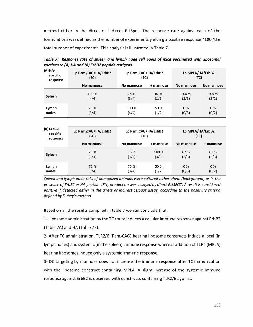
153
method either in the direct or indirect ELISpot. The response rate against each of the
formulations was defined as the number of experiments yielding a positive response *100 /the
total number of experiments. This analysis is illustrated in Table 7.
Table 7: Response rate of spleen and lymph node cell pools of mice vaccinated with liposomal
vaccines to (A) HA and (B) ErbB2 peptide antigens.
Spleen and lymph node cells of immunized animals were cultured either alone (background) or in the
presence of ErbB2 or HA peptide. IFNγ production was assayed by direct ELISPOT. A result is considered
positive if detected either in the direct or indirect ELISpot assay, according to the positivity criteria
defined by Dubey’s method.
Based on all the results compiled in table 7 we can conclude that:
1- Liposome administration by the TC route induces a cellular immune response against ErbB2
(Table 7A) and HA (Table 7B).
2- After TC administration, TLR2/6 (Pam2CAG) bearing liposome constructs induce a local (in
lymph nodes) and systemic (in the spleen) immune response whereas addition of TLR4 (MPLA)
bearing liposomes induce only a systemic immune response.
3- DC targeting by mannose does not increase the immune response after TC immunization
with the liposome construct containing MPLA. A slight increase of the systemic immune
response against ErbB2 is observed with constructs containing TLR2/6 agonist.
(A) HA-
specific
response
Lp Pam2CAG/HA/ErbB2
(SC)
Lp Pam2CAG/HA/ErbB2
(TC)
Lp MPLA/HA/ErbB2
(TC)
No mannose No mannose + mannose No mannose No mannose
Spleen 100 %
(4/4)
75 %
(3/4)
67 %
(2/3)
100 %
(3/3)
100 %
(2/2)
Lymph
nodes
75 %
(3/4)
100 %
(4/4)
50 %
(1/2)
0 %
(0/3)
0 %
(0/2)
(B) ErbB2-
specific
response
Lp Pam2CAG/HA/ErbB2
(SC)
Lp Pam2CAG/HA/ErbB2
(TC)
Lp MPLA/HA/ErbB2
(TC)
No mannose No mannose + mannose No mannose + mannose
Spleen 75 %
(3/4)
75 %
(3/4)
100 %
(3/3)
67 %
(2/3)
67 %
(2/3)
Lymph
nodes
75 %
(3/4)
75 %
(3/4)
50 %
(1/2)
0 %
(0/3)
0 %
(0/2)

154
These results indicated a high reproducibility of the immune response in our TC vaccination
model, despite the technical challenges of TC immunization. Thus, liposome-based
formulations are promising TC vaccines that deserve further attention in tumor
immunotherapy.
4. Influence of the lipid vesicle fluidity on the systemic immune response
4.1.4.1.4.1.4.1. TransfersomeTransfersomeTransfersomeTransfersome----based formulations induce a Tbased formulations induce a Tbased formulations induce a Tbased formulations induce a T----cell response comparable but not equal to cell response comparable but not equal to cell response comparable but not equal to cell response comparable but not equal to
that induced by their liposomal counterparts.that induced by their liposomal counterparts.that induced by their liposomal counterparts.that induced by their liposomal counterparts.
To assess the immunogenicity of the Tf-based constructs after TCI, and to address the
influence of the danger signal in these constructs, the same immunization protocol that was
adopted for conventional liposomes was followed, using TLR2/6 agonist-containing
transfersomes (Tf Pam2CAG/HA/ErbB2) or TLR4 agonist-containing transfersomes (Tf
MPLA/HA/ErbB2).
In contrast to their liposomal counterparts, TLR2/6- and TLR4-containing transfersomes
induced poorly reproducible HA-specific immune responses (not shown). Therefore, we
compared their immunostimulatory potential according to ErbB2-induced responses.
As shown in figure 6, whereas Tf incorporating a TLR2/6 agonist induced an ErbB2-specific
immune response in the spleen and lymph nodes, the TLR4 agonist-bearing Tf induced an
immmune response only in the lymph nodes. Interestingly, these results came in contrast with
those obtained with TLR4 agonist-incorporating conventional Lp induced an immune response
only in the spleen, not in the lymph nodes.
These results suggest that similarly to their liposomal counterparts, transfersome-based
formulations trigger a specific T cell response. Interestingly, the vesicle type (Lp or Tf) seems
to affect the localization of the immune response induced by certain adjuvants like the TLR4
agonist (MPLA).

155
Figure 6: ErbB2-specific IFNγ response in mice immunized with Pam2CAG (TLR2/6 agonist) or MPLA
(TLR4 agonist) adjuvanted transfersomes. Plain transfersomes (Tf) (white), Tf Pam2CAG/HA/ErbB2
(pink), or Tf MPLA/HA/ErbB2 (blue) were applied to the skin of 3 mouse groups (5 mice/group).
Immunizations were carried out at days 0, 4 and 8. Spleen and lymph node cells were collected on day
28 and cultured either alone (Background, B) or in the presence of ErbB2 (Peptide, P). The number of
IFNγ-secreting cells/106 cells was measured by direct ELISpot assay. Data from one representative
experiment of two independent ones are shown. Results are expressed as mean+/- SEM of ELISpot
triplicates. (A) and (B) show the number of ErbB2-specific spots (P-B) in the spleens and lymph nodes
respectively. Lp: liposomes, Pam2CAG: dipalmitoyl-cysteine-alanyl-glycine, MPLA: monophosphoryl
lipid A.
4.2.4.2.4.2.4.2. DC targeting molecules modulate the immunogenicity of transfersomeDC targeting molecules modulate the immunogenicity of transfersomeDC targeting molecules modulate the immunogenicity of transfersomeDC targeting molecules modulate the immunogenicity of transfersome----based vaccinesbased vaccinesbased vaccinesbased vaccines
Targeting the conventional Lp to DCs did not seem to influence their immunogenicity by TCI.
To check if this is the case with Tf too, mice received a TC application of Tf incorporating or
not the DC targeting molecule, di-mannose.
As seen in figure 7, addition of a DC targeting molecule to TLR2/6 agonist-bearing Tf has a
deleterious effect on the systemic immune response in spleen but a favorable one on the local
immune response in lymph nodes. These results came in contrast to those obtained with
targeted liposomal vaccines where mannose addition induced only a slight effect and in the
opposite way.
0
2
4
6
8
10
12
14
16
18
20
ErbB2
Nu
mb
er
of
IFN
- γγ γγ-p
rod
uci
ng
ce
lls
/10
6ce
lls
In vitro stimulation peptide
Spleen
Tf
Tf
Pam2CAG/HA/ErbB2
Tf MPLA/HA/ErbB2
(A)
0
2
4
6
8
10
12
ErbB2
Nu
mb
er
of
IFN
- γγ γγ-p
rod
uci
ng
ce
lls
/10
6ce
lls
In vitro stimulation peptide
Lymph nodes
Tf
Tf
Pam2CAG/HA/ErbB2
Tf MPLA/HA/ErbB2
(B)

156
Figure 7: ErbB2-specific IFNγresponse in mice immunized with non-targeted or DC targeted
transfersomal vaccines adjuvanted with Pam2CAG. Plain transfersomes (Tf) (white), adjuvanted
transfersomes, Tf Pam2CAG/HA/ErbB2 (pink), and targeted adjuvanted transfersomes, Tf
Pam2CAG/HA/ErbB2/Man (red), were applied to the skin of 5 mouse groups (5 mice/group).
Immunizations were carried out at days 0, 4 and 8. Spleen and lymph node cells were collected on day
28 and cultured either alone (Background, B) or in the presence of ErbB2 (Peptide, P). The number of
IFNγ-secreting cells/106 cells was measured by direct ELISpot assay. Data from one representative
experiment of two independent ones are shown. Results are expressed as mean+/- SEM of ELISpot
triplicates. (A) and (B) show the number of spots specific to each peptide (P-B) in the spleens and lymph
nodes respectively. Tf: transfersome, Pam2CAG: dipalmitoyl-cysteine-alanyl-glycine, Man: dioleyil-
glycerol-di-mannose (DOG-Man2).
In contrast, addition of a DC targeting molecule to TLR4 agonist-bearing transfersomes allows
them to induce a systemic immune response and seems to slightly increase the amplitude of
the local immune response. (figure 8).
0
5
10
15
20
25
30
35
40
45
50
ErbB2
Nu
mb
er
of
IFN
- γγ γγ-p
rod
uci
ng
ce
lls
/10
6ce
lls
In vitro stimulation peptide
Lymph nodesTf
Tf Pam2CAG/HA/ErbB2
Tf Pam2CAG/HA/ErbB2/Man
(B)
0
2
4
6
8
10
12
14
16
ErbB2Nu
mb
er
of
IFN
- γγ γγ-p
rod
uci
ng
ce
lls
/10
6ce
lls
In vitro stimulation peptide
SpleenTf
Tf Pam2CAG/HA/ErbB2
Tf Pam2CAG/HA/ErbB2/Man
(A)

157
Figure 8: ErbB2-specific IFNγ response in mice immunized with DC targeted liposomal vaccines
adjuvanted with MPLA. Plain transfersomes (Tf) (white), adjuvanted transfersomes, Tf
MPLA/HA/ErbB2 (light blue), and targeted adjuvanted transfersomes, Tf MPLA/HA/ErbB2/Man ((dark
blue) were applied to the skin of 5 mouse groups (5 mice/group). Immunizations were carried out on
days 0, 4 and 8. Spleen and lymph node cells were collected on day 28 and cultured either alone
(Background, B) or in the presence of ErbB2 (Peptide, P). The number of IFNγ-secreting cells/106 cells
was measured by an indirect ELISpot assay. Data from one representative experiment of two
independent ones are shown. Results are expressed as mean+/- SEM of ELISpot triplicates. (A) and (B)
show the number of spots specific to each peptide (P-B) in the spleens and lymph nodes respectively.
Tf: transfersome, MPLA: monophosphoryl lipid A, Man: dioleyl-glycerol-di-mannose (DOG-Man2).
Altogether, these results suggest that DC targeting has a more significant effect in
transfersomes than in liposomes. Moreover, its role seems to depend on the danger molecule
type and varies according to the localization of the immune response. Indeed, when the
systemic ErbB2-specific response is suppressed by mannose association with a TLR2/6 agonist
(Pam2CAG), and increased by its association with a TLR2/4 agonist (MPLA). The local immune
response is increased by mannose addition to both TLR agonists.
4.3.4.3.4.3.4.3. TransfersomeTransfersomeTransfersomeTransfersome----based vaccines induce a high ErbB2based vaccines induce a high ErbB2based vaccines induce a high ErbB2based vaccines induce a high ErbB2----specific, but a low HAspecific, but a low HAspecific, but a low HAspecific, but a low HA----specific specific specific specific
response rateresponse rateresponse rateresponse rate
To evaluate the reproducibility of the experiments performed with transfersomes, we
retrospectively evaluated the peptide/background spot number ratios (R=P/B), according to
the positivity criteria set above, in 4 different experiments. The results were analyzed by the
0
20
40
60
80
100
120
140
160
180
200
ErbB2
Nu
mb
er
of
IFN
- γγ γγ-p
rod
uci
ng
ce
lls
/10
6ce
lls
In vitro stimulation peptide
Lymph nodes Tf
Tf MPLA/HA/ErbB2
Tf MPLA/HA/ErbB2/Man
(B)
0
20
40
60
80
100
120
ErbB2
Nu
mb
er
of
IFN
- γγ γγ-p
rod
uci
ng
ce
lls
/10
6ce
lls
In vitro stimulation peptide
Spleen Tf
Tf MPLA/HA/ErbB2
Tf MPLA/HA/ErbB2/Man
(A)

158
method used in paragraph 3.4.2 for the conventional liposome (see table 6). All the results are
compiled in table 8.
Table 8: Response rate of spleens and lymph node cell pools of mice vaccinated
with transfersome-based vaccines to (A) HA and (B) ErbB2 peptide antigens.
Kjhgfdsdfghjklkjhgfdfghjkjhgfd
(B) HA-
specific
response
Tf Pam2CAG/HA/ErbB2
(TC)
Tf MPLA/HA/ErbB2
(TC)
No mannose + mannose No mannose + mannose
Spleen 50 %
(2/4)
25 %
(1/4)
25 %
(1/4)
50 %
(2/4)
Lymph nodes 0 %
(0/4)
75 %
(3/4)
25 %
(1/4)
25 %
(1/4)
Spleen and lymph node cell pools of immunized animals were cultured either alone
(background) or in the presence of ErbB2 or HA peptide. IFNγproduction was
assayed by direct ELISPOT. A result is judged positive if it was detected either in the
direct or indirect ELISpot assay, according to the positivity criteria defined by Dubey’s
empirical method.
By analyzing these results, we first observed that the response rates induced by transfersomes
were generally lower and more variable than those observed with liposomes. However, we
could draw some preliminary conclusions on the cellular immune response induced by TC
immunization with transfersomes.
1- Transfersome administration by the TC route induces a cellular immune response against
ErbB2 (Table 8A), but almost none against HA (Table 8B)
2- Incorporation of a TLR2/6 agonist (Pam2CAG) in transfersome-based constructs
administered by the TC route induces a systemic immune response (in the spleen) against both
peptides and a local response (in lymph nodes) only against ErbB2. In contrast, the TLR4
agonist (MPLA) induces only infrequent immune responses (systemic and local) against both
peptides.
(A) ErbB2-
specific
response
Tf Pam2CAG/HA/ErbB2
(TC)
Tf MPLA/HA/ErbB2
(TC)
No mannose + mannose No mannose + mannose
Spleen 50 %
(2/4)
25 %
(1/4)
25 %
(1/4)
75 %
(3/4)
Lymph nodes 100 %
(4/4)
100 %
(4/4)
50 %
(2/4)
75 %
(3/4)

159
3- DC targeting by mannose increased the systemic as well as the local immune responses,
mainly the local HA-specific response by the TLR2/6 agonist-bearing Tf.
This study shows that TCI with transfersome-based vaccines incorporating a TLR agonist and
expressing a CD8+ (ErbB2) and a CD4+ (HA) T cell epitope induce an immune response which
seems less consistent than that of similar liposome-based formulations. Additionally, mannose
targeting seems to improve the reproducibility of these responses.
5. Liposomes induce skin DC migration to draining lymph nodes after TC immunization
To elicit an immune response, a vaccine should be able to induce the migration of DCs towards
lymph nodes that drain the vaccination site, where an adaptive immune response is initiated.
We therefore investigated the capacity of the liposome-based constructs to induce DC
migration to the brachial lymph nodes that drain the immunization site, after TC application.
We started the analysis with the constructs incorporating MPLA. This TLR agonist was selected
before the experiments comparing MPLA and Pam2CAG efficacy were completed. To this end,
BALB/c mice received liposomes incorporating MPLA and mannose, or plain liposomes, both
labeled with the Dil fluorochrome, or the buffer in which the liposomes are dispersed. Brachial
lymph nodes were harvested 48 hours later, and different DC subpopulations, including Lang+
dDCs, Lang- dDCs and Langerhans cells, were analyzed by flow cytometry.
We first attempted to determine in each of these skin DC subpopulations the percentage of
DiI+ cells, as this would indicate that they have internalized the fluorescent liposomes. The
results are shown in figure 9. They indicate that the percentage of DiI+ cells in all 3 skin DC
subpopulations present in the lymph node is almost nil.

160
Figure 9: Skin-DCs found in the lymph nodes 48h after immunization with fluorescent liposomes do
not show any fluorescence. Liposome dispersion buffer, plain fluoresecent liposomes (Lp DiI) (yellow)
and fluorescent liposomes incorporating MPLA and mannose (Lp DiI/MPLA/Man) (brown) were applied
to the skin of 5 BALB/c mice. After 48 h, brachial lymph nodes were harvested and different DC
subpopulations were analyzed by flow cytometry. Every spot represents the % of DiI+ DCs in one mouse,
after the mean percentage of DiI+ DCs obtained in mice immunized with the dispersion buffer was
substracted. The mean and standard deviation were also added to the figure. Lp: liposome, DiI: 1,1'-
dioctadecyl-3,3,3'3'-tetramethylindocarbocyanine perchlorate , MPLA: monophosphoryl lipid A, Man:
mannose
To determine whether the absence of fluorescence is caused by a lack of skin DC migration or
because DiI fluorescence fades due to liposome internalization and processing in the
endosome for 48 hours, we compared the total number of cells of the different skin DC
subpopulation in the lymph nodes of mice that received the different liposomal constructs or
the negative control. Figure 10 shows that in control mice that received buffer, a low number
or skin DCs can be detected in lymph node. In contrast, both liposomal constructs application
induces migration of skin DCs, independently of whether they incorporate or not MPLA and
mannose. However, all the skin DC subpopulations do not seem to migrate similarly, since LCs
and langerin – dDCs seem to migrate in higher numbers, as compared to langerin+ dDCs.
Lp DiI
Lp DiI/MPLA/Man
Lp DiI Lp DiI/MPLA
/Man
Lang- dDCs
Lp DiI Lp DiI/MPLA
/Man
LCs
Lp DiI Lp DiI/MPLA
/Man
Lang+ dDCs

161
These results show that our liposomal constructs induce the migration of skin DCs after TC
immunization. However, it is quite surprising that the plain liposomes, which are devoid of
danger signal, induce skin DC migration in the same way as those incorporating MPLA and
mannose.
Figure 10: Liposomal constructs induce skin DC migration towards lymph nodes. Liposome dispersion
buffer, plain fluorescent liposomes (Lp DiI) (yellow) and fluorescent liposomes incorporating MPLA and
mannose (Lp DiI/MPLA/Man) (brown) were applied to the skin of 10 BALB/c mice. After 48 h, brachial
lymph nodes were harvested and different DC subpopulations were analyzed by flow cytometry. After
48 h, brachial lymph nodes were harvested and different DC subpopulations were analyzed by flow
cytometry. Every spot represents the % of DCs in one mouse. The mean and standard deviation were
also added to each condition. n= 10 mice, in 2 different experiments. Test ANOVA, **=p<0.005 and
***= p<0.0005. Lp: liposome, DiI: 1,1'-dioctadecyl-3,3,3'3'-tetramethylindocarbocyanine perchlorate ,
MPLA: monophosphoryl lipid A, Man: mannose
LCs Lang- dDCs Lang+ dDCs
Buffer Lp DiI Lp DiI/
MPLA/
Man
Buffer Lp DiI Lp DiI/
MPLA/
Man
Buffer Lp DiI Lp DiI/
MPLA/
Man
Buffer
Lp DiI
Lp DiI/MPLA/Man
Ce
ll c
ou
nt/
lym
ph
no
de
(x
10
3)

162
Discussion
The variable efficacy of therapeutic cancer vaccines that aim to fight established cancers
highlights the need to improve tumor-directed T cell responses in the patient.
A liposome-based cancer vaccine has been previously developed by our team. It allows the
delivery of all vaccine components needed by the immune system to induce a cancer specific
response. To induce DC maturation, the liposomal constructs incorporate a danger signal
provided by a TLR 2/6 agonist (Pam2CAG), and to activate T lymphocytes they incorporate a
CD4+ (HA 307-319) and a CD8+ (ErbB2 63-71) epitope peptides. These constructs proved to be
immunogenic when delivered by the subcutaneous (Roth et al., 2005; Thomann et al., 2011)
and intranasal routes (Kakhi et al., 2015, 2016), and, most importantly, they protected mice
against tumor challenge in therapeutic vaccination settings.
The skin is an immunological barrier rich in multiple DC subpopulations that can mount
adaptive immune responses and protect the organism from external aggressions. Given the
central role of DCs in the antitumor immune response, the skin appears as a highly suitable
administration site of cancer vaccines. Application of these vaccines on skin surface, termed
transcutaneous (TC) vaccination, is expected to target skin DCs in the best way to harness their
immunostimulatory potential. The TC immunization route has recently made its first steps
towards clinical use by the success of a phase I clinical trial of an influenza vaccine delivered
to the skin using a microneedle array. The vaccine has proven to be safe and as immunogenic
as the intramuscularly delivered one, providing a proof of concept and a strong rationale for
the investment of further efforts in the development of vaccines for the TC route (Rouphael
et al., 2017).
Because the physicochemical properties of liposomes are reported to favor their TC passage
through the skin, we aimed in the present work to deliver our liposomal cancer vaccines by
the TC route and to assess their capability to induce potent cellular adaptive immune
responses. We also attempted to increase the potency of the immune response by optimizing
their composition and physicochemical characteristics.
In a first step, we chose to use an easy-to-use vaccination technique that consists in applying
the vaccine dose by massage on previously shaved, ethanol-wiped and dry skin. Ethanol

163
application on the skin (Hirschberg et al., 2012) is intended to solubilize skin lipids and induce
disorder in the lipid structure of the stratum corneum, thus mildly disrupting the skin barrier
and increasing its fluidity and permeability. Another strategy for skin barrier disruption is the
use of tape stripping, a more aggressive technique that removes few layers of the stratum
corneum, thus favoring the penetration of applied vaccine (Inoue et al., 2005; Loan Honeywell-
Nguyen et al., 2002). However, tape stripping induces cellular damage. Upon epidermis
disruption, danger signals and subsequent signaling through PRR induce the secretion of
proinflammatory cytokines such as IL-1α, IL-1β, tumor necrosis factor (TNF)-α and
granulocyte/macrophage-colony-stimulating factor (GM-CSF) (Clausen and Stoitzner, 2015;
Kaurav et al., 2016; Li et al., 2011; Partidos and Muller, 2005). By contrast, our immunization
technique involving a very mild stratum corneum disruption using ethanol avoids these
uncontrolled inflammatory effects.
To assess the development of a systemic and local peptide-specific cellular immune
response, we investigated the presence of specific T cells producing IFNγ, , , , the surrogate of T
lymphocyte responses of Th1 and CTL profiles, using an ELISpot assay. In addition to the
spleen, we tested the lymph nodes that drain the skin application zone, namely the inguinal,
axillary and brachial lymph nodes. The ELISpot technique is a sensitive and powerful tool that
allows the detection of rare antigen-specific lymphocytes to the single-cell level, as rare as 3
cells per well (Karulin et al., 2015). Moreover, the sensitivity of the standard ELISpot assay can
be increased by culturing the cells in vitro during a longer incubation time that can reach 12
days, in presence of antigen and IL-2, to induce the expansion of antigen-specific lymphocytes.
This increased-sensitivity ELISpot variant is known as indirect ELISpot assay, or cultured
ELISpot assay (Calarota and Baldanti, 2013). In the present work, because the induced immune
responses were low, we assessed their presence using both a standard and an indirect ELISpot
assay.
The interpretation of ELISpot data becomes problematic when the signal is low, and is
accompanied by a variable background spot count. In our case, the spot count in antigen-
stimulated wells was frequently increased as compared to control wells. However, the
amplitude of the detected response remained low, indicating that a statistical analysis of the
significance of these results is not suitable. Alternatively, several empirical methods, or

164
empirical rules (“ER”), usually based on observations from a specific study were developed
and employed for detecting positive ELISpot signals in clinical trials for infectious diseases and
cancer (Dubey et al., 2007; Lai et al., 2011; Lewis et al., 2000; Moodie et al., 2010). A step-by-
step empirical method was illustrated by Dubey et al., to set and validate positivity criteria for
an ELISpot assay for the detection of responses against a set of HIV peptides (Dubey et al.,
2007). Since empirical methods are dependent on the reagents, the test settings and the
populations that are being tested, they are internally set up. Therefore, we followed Dubey’s
steps to generate positivity criteria that can be used in our conditions to analyze the direct
and indirect ELISpot assay.
Before evaluating the immunogenicity of liposome formulations administered by the TC
route, we investigated whether the potential CD8+ T cell peptide epitope ErbB2 can elicit a
cellular immune response following a previously validated robust TCI protocol. As described
in the literature, cholera toxin is a powerful adjuvant (Kawamura et al., 2003; Schnitzler et al.,
2007) that induces potent humoral as well as cellular responses by the TC route, including CTL
responses (Glenn et al., 1998, 1999; Kahlon et al., 2003). However, due to its toxicity, cholera
toxin cannot be used in humans and exerts multiple secondary effect and, therefore, is used
in the present study only as a positive control for TC immunization. The mechanism by which
cholera exerts its adjuvant effect is debated. While some studies reported that its
immunostimulatory potential is unconventional and independent of TLR signaling (Kahlon and
Dutz, 2003; Olvera-Gomez et al., 2012), others suggest it to strongly depend on the activation
of TLR4 (Liu et al., 2016; Phongsisay et al., 2015). Indeed, our results showed that
administration of a physical mixture of cholera toxin and ErbB2 by massage on ethanol-wiped
and dried skin induces a potent local and systemic immune response, thus indicating that
ErbB2 is suitable to be administered by the TC route. Therefore, we prepared liposomal
constructs incorporating this peptide associated to a T CD4+ epitope and a danger signal.
We formulated conventional liposomes (Lp-SUV) as well as ultradeformable ones, or
transfersomes (Tf-SUV). Tf were chosen because the presence of ethanol and a surfactant in
their composition increases their fluidity and makes them ultradeformable. The surfactant
decreases their resistance at tension points and allows them to deform, thus squeezing into
pores smaller than their size. Therefore, they were reported to cross the stratum corneum

165
barrier more efficiently than conventional liposomes (Cevc and Gebauer, 2003; Cevc et al.,
1998).
Lp-SUV were obtained by sonication of Lp-MLV prepared from PC, PG and Chol, in proportions
similar to those previously described by our team (Espuelas et al., 2008; Kakhi et al., 2015,
2016; Thomann et al., 2011). Tf-SUV were obtained by manual extrusion of Tf-MLV prepared
with SPC, SDC and ethanol (Cevc and Blume, 1992; Kakhi et al., 2016). These formulation
techniques allowed us to reproducibly obtain monodispersed liposomal constructs, with a size
< 100 nm, which is suitable for skin barrier crossing and uptake by DCs.
In order to display the peptides on the surface of Lp-SUVs and Tf-SUVs, an amphiphilic thiol
reactive anchor, DPGmal, was added to the vaccine composition and was inserted in the lipid
bilayer. Cysteine-containing peptides were subsequently covalently linked to the maleimide
group of the anchor by the Michael addition (Schelté et al., 2000). This soft coupling step in
aqueous medium preserves the narrow size distribution of the preformed liposomes resulting
in reproducible liposome-based constructs.
Beside epitope peptides, immunostimulatory molecules, namely a TLR 2/6 agonist and a TLR
4 agonist (Pam2CAG and MPLA), in addition to a DC-targeting molecule (DOG-Mann2, or
mannose) were incorporated in the lipid bilayer of SUVs. In order to maintain the overall
charge of formulations, additions of amphiphilic molecules in a proportion > 1% was
compensated by an equivalent decrease in PC proportion. Through its diacylglycerol moiety,
Pam2CAG a diacetylated lipopeptide derived from the N-terminal moiety of E-coli lipoprotein),
interacts with TLR 2/6 heterodimers (Oliveira-Nascimento et al., 2012; Omueti et al., 2005).
MPLA, a chemically detoxified form of the lipid A, the anchor moiety of lipopolysaccharides
(LPS) is currently one of the leading innovative vaccine adjuvants (Alving and Rao, 2008).
Because the adjuvant molecule is a decisive factor of the immune response induced by a
vaccine, we compared both adjuvants in order to choose the most appropriate one for this
route of administration.
Mannose was chosen to be a di-antennary molecule, because previous work in our team
comparing the efficacy of mono-, di-, and tetramannosylated ligands showed that both di- and
tetramannosylated onces drastically improve liposome uptake by DCs with no significant

166
difference among them (Espuelas et al., 2008). Because dimannnose is easier to synthetize, it
was chosen to be used in the present work,
In a proof of concept, we first compared the immune response induced by administration of
the original liposomal construct incorporating the TLR2/6 agonist by the TC route and the SC
route. Our results show that TC application of this construct triggers local (in the lymph nodes)
and systemic (in the spleen) immune responses against the CD4+ and the CD8+ T-cell peptides
that are similar to those induced by subcutaneous injection. Efficacy of liposomal carriers in
TC barrier crossing was established in 1980 when they were used for the first time for topical
drug delivery and were found to achieve a four- to five-fold increase of the drug concentration
in the epidermis and the dermis, as compared to drug alone (Mezei and Gulasekharam, 1980).
However, several studies report that liposomes fuse on the stratum corneum surface instead
of penetrating it, as shown by the observation of the behavior of bilayer-forming surfactant L-
595 (sucrose laurate ester) rigid vesicles applied on the skin under an electron microscope and
their absence in the viable layers (Loan Honeywell-Nguyen et al., 2002). Interestingly, Trauer
et al found that massage may improve the delivery of particulate substances, including
conventional liposomes, into hair follicles, which constitute a port of entry to the internal skin
layers (Trauer et al., 2014). Therefore, the induction of an immune response after TC
application of our liposome-based constructs indicates that liposomes were indeed capable of
reaching the internal skin layers and to be taken up by skin DCs in order to induce T cell
responses. We believe that this may be facilitated by our massage vaccination techniques. To
our knowledge, TC immunization with conventional liposomes delivering peptides/proteins
has been limited to encapsulated model-antigens such as ovalbumin or tetanus toxoid, where
the humoral response, but rarely the cellular response was assessed (Gupta et al., 2014; Zhang
et al., 2017). In this report, we show that liposomes displaying simple epitopes on their surface
are able to induce a primary immune response via the TC route.
For the development of efficient vaccines, the determination of the most optimal
immunostimulatory molecule is essential and may influence the type of the induced
immune response. Therefore, the first attempt to improve our liposome-based formulations
was by replacing the TL2/6 agonist (Pam2CAG) with a TLR4 agonist (MPLA). Pam2CAG induces
the maturation of human monocyte-derived DCs in vitro, as indicated by the expression of
CD80, CD83, CD86 and HLA-DR molecules (Espuelas et al., 2005b), and triggers protective

167
tumor-specific responses in vivo (Heurtault et al., 2009; Thomann et al., 2011), whereas
liposome- incorporated MPLA triggers T cell responses (Alving et al., 2012b). The comparison
of the immune response induced after TCI with the liposomal constructs incorporating
Pam2CAG or MPLA showed that both constructs can induce a systemic immune response,
however, only Pam2CAG-bearing construct could induce a detectable local immune response.
While TLR2, TLR4 and TLR6 are expressed on LCs (Fehres et al., 2015; de Koning et al., 2010),
their functionality seems to be debated. Indeed, Flacher et al. reported that while TLR2 is
functional and its engagement induces LC maturation, TLR4 signaling is impaired (Flacher et
al., 2006). Accordingly, intradermal administration of several TLR agonists revealed that
signaling through TLR2, but not through TLR4, induces a local inflammatory response by LCs
(Oosterhoff et al., 2013). On the other hand, dDCs express both TLR2 and TLR4 (Aar et al.,
2007; Rozis et al., 2008). The ability of our TLR4 agonist-bearing liposomes to induce systemic
responses suggests that either TLR4 is functional on LCs, in contrast to other findings in the
literature, or that dDCs alone are responsible for the induction of the observed immune
response.
Targeting of vaccines to C-lectin type receptors, such as mannose receptors, has been shown
to increase uptake by DCs (Espuelas et al., 2008; Jeong et al., 2014; Markov et al., 2015;
Thomann et al., 2011; Vyas et al., 2010), thus resulting in an improved DC activation, and
vaccine-specific T cell priming and proliferation. The most widely targeted receptor is the
mannose receptor. When the liposome-based constructs developed by our team were
targeted to DCs by mannose addition, they were found to conserve their immunostimulatory
potential despite a 100-fold reduction of the adjuvant amount, and to conserve their
antitumor efficacy despite a 10-fold reduction of the vaccine dose (Thomann et al., 2011).
Therefore, in an attempt to increase the immunogenicity of our liposome-based constructs in
the present work, we targeted them to DCs by adding a DC targeting molecule to their
composition, namely, di-mannose. Liposome targeting to DCs had variable effects. It slightly
increased the amplitude of the cellular response against ErbB2 peptide in the spleen, while it
was at most ineffective for local immune responses in the lymph nodes. The mannose receptor
is expressed on dDCs, however its expression on LCs seems controversial. Some studies report
that it is expressed on LCs in normal skin (Condaminet et al., 1998; de Koning et al., 2010),
while others report it to be only induced under inflammatory conditions (Fehres et al., 2015;

168
Plzáková et al., 2004; Polak et al., 2014; Wollenberg et al., 2002). Therefore, improved
immunogenicity following mannose addition to our liposomal constructs does not provide
enough information regarding the targeted DC subpopulation in the light of current evidence.
For improved targeting of LCs and/or dDCs, further studies should be pursued to investigate
the expression of mannose receptor on LCs. Choosing another receptor to target, such DEC-
205, is also an interesting alternative. DEC-205 is expressed on both LCs and dDCs, even if its
expression is low on LCs, and its targeting has resulted in potent immune response induction
(Stoitzner et al., 2014).
Because the flexibility of the vesicular carrier affects its passage through the skin barrier, it
also modulates its activity. Despite their use for TC drug delivery (Jain et al., 2017), liposomes
were reported by some authors to be suboptimal for TC delivery and to enhance deposition
in the upper layers of the skin rather than transdermal penetration, thus mediating topical
and not TC delivery (Ashtikar et al., 2016). Therefore, modified liposomes were formulated to
improve TC passage such transfersomes (Cevc and Blume, 1992; Cevc et al., 1998). In the
present work, we aimed to optimize the liposomal constructs for the TC route by replacing the
conventional liposomal vesicle with a transfersome. Surprisingly, transfersome-based
constructs did not improve the induced cellular immune response. On the contrary, their
almost failed to induce reproducible HA-specific responses.
Comparative studies of transfersomes and liposomes were mostly conducted in the context
of drug delivery. A few studies assessed their immunostimulatory capacity, in which the results
were sometimes discordant. Cationic ultradeformable liposomes delivering HBs antigen DNA
were found to induce potent cellular and humoral immune responses that were superior to
those induced by conventional liposomes, and comparable to intramuscular injection of naked
DNA (Wang et al., 2007). Gupta et al found that transfersomes induce higher anti-TT titers as
compared to conventional liposomes (Gupta et al., 2014). Similarly, transdermal immunization
with ultradeformable liposomes was found to induce stronger cellular and humoral immunity
against merozoite surface protein-1 (PfMSP-119) of Plasmodium falciparum, as compared to
conventional liposomes (Tyagi et al., 2016). By contrast, other studies showed that
transfersomes exert the same effect as conventional liposomes. A comparative in vitro skin
permeation study, conducted by Rattanpack et al. on transfersomes and liposomes, showed

169
that transfersomes do not significantly increase penetration as compared to conventional
liposomes (Rattanapak et al., 2012). Accordingly, transfersomes were found to induce the
same antibody titers as those induced by liposomes entrapping the model antigens Ova (Zhang
et al., 2017).
A possible explanation of the decreased reproducibility of responses with the transfersomes
is that their slightly bigger size could have negatively influenced their TC passage. When
insufficient vaccine amount is delivered to the DC, a less potent immune response is induced.
However, this hypothesis is unlikely to be true, since the size difference is less than 20 nm. To
confirm it, transfersomes of the same size as liposomes (60 nm) have to be evaluated.
Another possible explanation is that their ultradeformability could have negatively influenced
their stability, resulting in a decreased depot effect at the administration site. Since one of the
most important assets of nanoparticles for their immunogenicity is their potential to exert
depot effect, this results in a decreased immunostimulation.
In parallel to the experiments aiming to detect local and systemic immunity induced by the
TC delivery of our liposomal constructs, we used a mechanistic approach to investigate the
migration of skin DCs subtypes to the lymph nodes. Fluorescent liposomes were obtained by
addition of a lipophilic dye, DiI, in the lipid bilayer. DiI is routinely used for long term tracking
of cells and liposomes (Litzinger et al., 1994; Shah et al., 2015; Yefimova et al., 2014). When
fluorescent liposomes are taken up by DCs, the cells become fluorescent. In addition to
mannose which aims at preferentially targeting DCs, fluorescent liposomes incorporated a TLR
agonist. MPLA was chosen arbitrarily since the result of the immunogenicity of the constructs
were not clear yet.
No fluorescent skin cells could be detected in the lymph nodes. Interestingly, we could detect
fluorescent skin DCs in the epidermis as well as in the dermis (data not shown) confirming that
our results were not due to a lack of liposome uptake in the skin. Besides, the increased
numbers of skin DCs in the lymph nodes 48h after TC application of liposomes, confirmed that
the lack of fluorescence was not due to an absence of migration of DC that have internalized
the fluorescent liposomes either. Therefore, we speculate that DiI dye could be unstable at
the endosomal pH for long periods of time.

170
Skin DC subsets that were found to preferentially migrate to the lymph nodes are LCs and
Lang- dDCs. Both of these populations are capable of antigen cross-presentation to CD8+ T
cells, and were reported to induce CTL differentiation. Stoitzner et al. showed that LCs can
induce OVA-specific CTLs (Stoitzner et al., 2006) . Lang– as well as lang+ dDCs were found to be
involved in T cell cross-priming (Nizza and Campbell, 2014). In humans, LCs and in vitro-
generated dDCs were found to capture and cross-present melanoma antigens to CD8+ T
cells(Cao et al., 2007). Naturally occurring human LCs and Lang– dDCs were also found to
induce CD8+ T cell priming against two different melanoma antigen peptides (the melanoma
differentiation antigen MART-1 peptide, and the glycoprotein gp100 peptide). However, LC
were found to be more efficient since the generated CTL had an increased cytotoxic activity
(Klechevsky et al., 2008).
Surprisingly, both plain liposomes and those incorporating MPLA and mannose induced the
migration of skin DCs to the lymph nodes in similar numbers. Two interpretations are possible.
The first hypothesis involves a technical problem that may have occurred during the
formulation step, and that may have resulted in liposome contamination with molecules
capable of inducing DC activation, such as bacterial products. This hypothesis will be verified
by repeating the experiment under more rigorous conditions to ensure the liposomes are
devoid of any DC activating molecule. Another hypothesis is that liposome constituents may
have the capacity to activate DCs. However, in contrast to cationic liposomes capable of
inducing DC maturation and secretion of pro-inflammatory cytokines (Lonez et al., 2012;
Soema et al., 2015), it is improbable that our anionic constructs are capable of exerting such
effects. Previous studies in our laboratory have shown that they do not induce the maturation
of human DCs in vitro (Espuelas et al., 2005b, 2008). This hypothesis may be verified by
investigating the maturation state of the migrating skin DCs, by assessing the expression of
maturation markers, such as CD80 and CD86. LCs and dDCs were described to have the
capacity to migrate to draining lymph nodes after antigen uptake, without acquiring a mature
state. (Sparber et al., 2010). Therefore, we speculate that even if plain liposomes induce the
migration of skin DCs to the lymph nodes, for an unknown reason yet, they do not induce their
maturation, in contrast to TLR agonist-containing liposomes.
In conclusion, we report herein the development of a TC vaccination strategy involving the
use of liposome-based constructs and resulting in the induction of local and systemic immune

171
responses. Altogether, our results provide a rationale for the use of the liposome technology
to develop and improve cancer vaccines for TC delivery.
References:
Aar, A.M.G. van der, Sylva-Steenland, R.M.R., Bos, J.D., Kapsenberg, M.L., Jong, E.C. de, and
Teunissen, M.B.M. (2007). Cutting Edge: Loss of TLR2, TLR4, and TLR5 on Langerhans Cells Abolishes
Bacterial Recognition. J. Immunol. 178, 1986–1990.
Adams, S., O’Neill, D.W., Nonaka, D., Hardin, E., Chiriboga, L., Siu, K., Cruz, C.M., Angiulli, A., Angiulli,
F., Ritter, E., et al. (2008). Immunization of malignant melanoma patients with full-length NY-ESO-1
protein using TLR7 agonist imiquimod as vaccine adjuvant. J. Immunol. Baltim. Md 1950 181, 776–
784.
Alloatti, A., Kotsias, F., Magalhaes, J.G., and Amigorena, S. (2016). Dendritic cell maturation and
cross-presentation: timing matters! Immunol. Rev. 272, 97–108.
Alving, C.R., and Rao, M. (2008). Lipid A and liposomes containing lipid A as antigens and adjuvants.
Vaccine 26, 3036–3045.
Alving, C.R., Rao, M., Steers, N.J., Matyas, G.R., and Mayorov, A.V. (2012a). Liposomes containing
lipid A: an effective, safe, generic adjuvant system for synthetic vaccines. Expert Rev. Vaccines 11,
733–744.
Alving, C.R., Peachman, K.K., Rao, M., and Reed, S.G. (2012b). ADJUVANTS FOR HUMAN VACCINES.
Curr. Opin. Immunol. 24, 310.
Ascenso, A., Raposo, S., Batista, C., Cardoso, P., Mendes, T., Praça, F.G., Bentley, M.V.L.B., and
Simões, S. (2015). Development, characterization, and skin delivery studies of related
ultradeformable vesicles: transfersomes, ethosomes, and transethosomes. Int. J. Nanomedicine 10,
5837–5851.
Ashtikar, M., Nagarsekar, K., and Fahr, A. (2016). Transdermal delivery from liposomal formulations -
Evolution of the technology over the last three decades. J. Control. Release Off. J. Control. Release
Soc.
Baldwin, R.W. (1971). Tumour-associated antigens and tumour-host interactions. Proc. R. Soc. Med.
64, 1039.
Bangham, A.D., and Horne, R.W. (1964). NEGATIVE STAINING OF PHOSPHOLIPIDS AND THEIR
STRUCTURAL MODIFICATION BY SURFACE-ACTIVE AGENTS AS OBSERVED IN THE ELECTRON
MICROSCOPE. J. Mol. Biol. 8, 660–668.
Bangham, A.D., Hill, M.W., and Miller, N.G.A. (1974). Preparation and Use of Liposomes as Models of
Biological Membranes. In Methods in Membrane Biology, (Springer, Boston, MA), pp. 1–68.
Benson, H.A.E. (2006). Transfersomes for transdermal drug delivery. Expert Opin. Drug Deliv. 3, 727–
737.

172
Berinstein, N.L., Karkada, M., Oza, A.M., Odunsi, K., Villella, J.A., Nemunaitis, J.J., Morse, M.A.,
Pejovic, T., Bentley, J., Buyse, M., et al. (2015). Survivin-targeted immunotherapy drives robust
polyfunctional T cell generation and differentiation in advanced ovarian cancer patients.
Oncoimmunology 4, e1026529.
Boeckler, C., Dautel, D., Schelté, P., Frisch, B., Wachsmann, D., Klein, J.P., and Schuber, F. (1999).
Design of highly immunogenic liposomal constructs combining structurally independent B cell and T
helper cell peptide epitopes. Eur. J. Immunol. 29, 2297–2308.
Böhlen, P., Stein, S., Dairman, W., and Udenfriend, S. (1973). Fluorometric assay of proteins in the
nanogram range. Arch. Biochem. Biophys. 155, 213–220.
van der Bruggen, P., Traversari, C., Chomez, P., Lurquin, C., De Plaen, E., Van den Eynde, B., Knuth, A.,
and Boon, T. (1991). A gene encoding an antigen recognized by cytolytic T lymphocytes on a human
melanoma. Science 254, 1643–1647.
Butterfield, L.H. (2015). Cancer vaccines. BMJ 350, h988.
Calarota, S.A., and Baldanti, F. (2013). Enumeration and characterization of human memory T cells by
enzyme-linked immunospot assays. Clin. Dev. Immunol. 2013, 637649.
Cao, T., Ueno, H., Glaser, C., Fay, J.W., Palucka, A.K., and Banchereau, J. (2007). Both Langerhans cells
and interstitial DC cross-present melanoma antigens and efficiently activate antigen-specific CTL. Eur.
J. Immunol. 37, 2657–2667.
Cevc, G., and Blume, G. (1992). Lipid vesicles penetrate into intact skin owing to the transdermal
osmotic gradients and hydration force. Biochim. Biophys. Acta 1104, 226–232.
Cevc, G., and Blume, G. (2001). New, highly efficient formulation of diclofenac for the topical,
transdermal administration in ultradeformable drug carriers, Transfersomes. Biochim. Biophys. Acta
1514, 191–205.
Cevc, G., and Gebauer, D. (2003). Hydration-Driven Transport of Deformable Lipid Vesicles through
Fine Pores and the Skin Barrier. Biophys. J. 84, 1010–1024.
Cevc, G., Gebauer, D., Stieber, J., Schätzlein, A., and Blume, G. (1998). Ultraflexible vesicles,
Transfersomes, have an extremely low pore penetration resistance and transport therapeutic
amounts of insulin across the intact mammalian skin. Biochim. Biophys. Acta BBA - Biomembr. 1368,
201–215.
Chen, J.-L., Dawoodji, A., Tarlton, A., Gnjatic, S., Tajar, A., Karydis, I., Browning, J., Pratap, S., Verfaille,
C., Venhaus, R.R., et al. (2015). NY-ESO-1 specific antibody and cellular responses in melanoma
patients primed with NY-ESO-1 protein in ISCOMATRIX and boosted with recombinant NY-ESO-1
fowlpox virus. Int. J. Cancer 136, E590–E601.
Chikh, G., and Schutze-Redelmeier, M.-P. (2002). Liposomal delivery of CTL epitopes to dendritic cells.
Biosci. Rep. 22, 339–353.
Clausen, B.E., and Stoitzner, P. (2015). Functional Specialization of Skin Dendritic Cell Subsets in
Regulating T Cell Responses. Front. Immunol. 6, 534.

173
Condaminet, B., Péguet-Navarro, J., Stahl, P.D., Dalbiez-Gauthier, C., Schmitt, D., and Berthier-
Vergnes, O. (1998). Human epidermal Langerhans cells express the mannose-fucose binding
receptor. Eur. J. Immunol. 28, 3541–3551.
DeMuth, P.C., Li, A.V., Abbink, P., Liu, J., Li, H., Stanley, K.A., Smith, K.M., Lavine, C.L., Seaman, M.S.,
Kramer, J.A., et al. (2013). Vaccine delivery with microneedle skin patches in nonhuman primates.
Nat. Biotechnol. 31, 1082–1085.
Dhodapkar, M.V., Dhodapkar, K.M., and Palucka, A.K. (2007). Interactions of tumor cells with
dendritic cells: balancing immunity and tolerance. Cell Death Differ. 15, 39–50.
Dhodapkar, M.V., Sznol, M., Zhao, B., Wang, D., Carvajal, R.D., Keohan, M.L., Chuang, E., Sanborn,
R.E., Lutzky, J., Powderly, J., et al. (2014). Induction of antigen-specific immunity with a vaccine
targeting NY-ESO-1 to the dendritic cell receptor DEC-205. Sci. Transl. Med. 6, 232ra51.
Dubey, S., Clair, J., Fu, T.-M., Guan, L., Long, R., Mogg, R., Anderson, K., Collins, K.B., Gaunt, C.,
Fernandez, V.R., et al. (2007). Detection of HIV vaccine-induced cell-mediated immunity in HIV-
seronegative clinical trial participants using an optimized and validated enzyme-linked immunospot
assay. J. Acquir. Immune Defic. Syndr. 1999 45, 20–27.
Dudek, A.M., Martin, S., Garg, A.D., and Agostinis, P. (2013). Immature, Semi-Mature, and Fully
Mature Dendritic Cells: Toward a DC-Cancer Cells Interface That Augments Anticancer Immunity.
Front. Immunol. 4.
East, L., and Isacke, C.M. (2002). The mannose receptor family. Biochim. Biophys. Acta 1572, 364–
386.
Espuelas, S., Haller, P., Schuber, F., and Frisch, B. (2003). Synthesis of an amphiphilic tetraantennary
mannosyl conjugate and incorporation into liposome carriers. Bioorg. Med. Chem. Lett. 13, 2557–
2560.
Espuelas, S., Roth, A., Thumann, C., Frisch, B., and Schuber, F. (2005a). Effect of synthetic
lipopeptides formulated in liposomes on the maturation of human dendritic cells. Mol. Immunol. 42,
721–729.
Espuelas, S., Roth, A., Thumann, C., Frisch, B., and Schuber, F. (2005b). Effect of synthetic
lipopeptides formulated in liposomes on the maturation of human dendritic cells. Mol. Immunol. 42,
721–729.
Espuelas, S., Thumann, C., Heurtault, B., Schuber, F., and Frisch, B. (2008). Influence of ligand valency
on the targeting of immature human dendritic cells by mannosylated liposomes. Bioconjug. Chem.
19, 2385–2393.
Fehres, C.M., Bruijns, S.C.M., Sotthewes, B.N., Kalay, H., Schaffer, L., Head, S.R., Gruijl, T.D. de,
Garcia-Vallejo, J.J., and Kooyk, Y. van (2015). Phenotypic and Functional Properties of Human Steady
State CD14+ and CD1a+ Antigen Presenting Cells and Epidermal Langerhans Cells. PLoS ONE 10.
Flacher, V., Bouschbacher, M., Verronèse, E., Massacrier, C., Sisirak, V., Berthier-Vergnes, O., de
Saint-Vis, B., Caux, C., Dezutter-Dambuyant, C., Lebecque, S., et al. (2006). Human Langerhans cells
express a specific TLR profile and differentially respond to viruses and Gram-positive bacteria. J.
Immunol. Baltim. Md 1950 177, 7959–7967.

174
Fujiyama, T., Oze, I., Yagi, H., Hashizume, H., Matsuo, K., Hino, R., Kamo, R., Imayama, S., Hirakawa,
S., Ito, T., et al. (2014). Induction of cytotoxic T cells as a novel independent survival factor in
malignant melanoma with percutaneous peptide immunization. J. Dermatol. Sci. 75, 43–48.
Glenn, G.M., Rao, M., Matyas, G.R., and Alving, C.R. (1998). Skin immunization made possible by
cholera toxin. Nature 391, 851.
Glenn, G.M., Scharton-Kersten, T., Vassell, R., Matyas, G.R., and Alving, C.R. (1999). Transcutaneous
Immunization with Bacterial ADP-Ribosylating Exotoxins as Antigens and Adjuvants. Infect. Immun.
67, 1100.
Glenn, G.M., Taylor, D.N., Li, X., Frankel, S., Montemarano, A., and Alving, C.R. (2000).
Transcutaneous immunization: a human vaccine delivery strategy using a patch. Nat. Med. 6, 1403–
1406.
Guo, F., Wang, J., Ma, M., Tan, F., and Li, N. (2015). Skin targeted lipid vesicles as novel nano-carrier
of ketoconazole: characterization, in vitro and in vivo evaluation. J. Mater. Sci. Mater. Med. 26, 175.
Gupta, V., Dhote, V., Paul, B.N., and Trivedi, P. (2014). Development of novel topical drug delivery
system containing cisplatin and imiquimod for dual therapy in cutaneous epithelial malignancy. J.
Liposome Res. 24, 150–162.
Hansen, S., and Lehr, C.-M. (2012). Nanoparticles for transcutaneous vaccination. Microb. Biotechnol.
5, 156–167.
Heurtault, B., Gentine, P., Thomann, J.-S., Baehr, C., Frisch, B., and Pons, F. (2009). Design of a
Liposomal Candidate Vaccine Against Pseudomonas aeruginosa and its Evaluation in Triggering
Systemic and Lung Mucosal Immunity. Pharm. Res. 26, 276–285.
Hirschberg, H., van Kuijk, S., Loch, J., Jiskoot, W., Bouwstra, J., Kersten, G., and Amorij, J.-P. (2012). A
combined approach of vesicle formulations and microneedle arrays for transcutaneous immunization
against hepatitis B virus. Eur. J. Pharm. Sci. Off. J. Eur. Fed. Pharm. Sci. 46, 1–7.
Hopkins, J., and McConnell, I. (1984). Immunological aspects of lymphocyte recirculation. Vet.
Immunol. Immunopathol. 6, 3–33.
Inoue, J., Yotsumoto, S., Sakamoto, T., Tsuchiya, S., and Aramaki, Y. (2005). Changes in immune
responses to antigen applied to tape-stripped skin with CpG-oligodeoxynucleotide in mice. J.
Controlled Release 108, 294–305.
Iwama, T., Uchida, T., Sawada, Y., Tsuchiya, N., Sugai, S., Fujinami, N., Shimomura, M., Yoshikawa, T.,
Zhang, R., Uemura, Y., et al. (2016). Vaccination with liposome-coupled glypican-3-derived epitope
peptide stimulates cytotoxic T lymphocytes and inhibits GPC3-expressing tumor growth in mice.
Biochem. Biophys. Res. Commun. 469, 138–143.
Jain, S., Patel, N., Shah, M.K., Khatri, P., and Vora, N. (2017). Recent Advances in Lipid-Based Vesicles
and Particulate Carriers for Topical and Transdermal Application. J. Pharm. Sci. 106, 423–445.
Jeong, H.-S., Na, K.S., Hwang, H., Oh, P.-S., Kim, D.H., Lim, S.T., Sohn, M.-H., and Jeong, H.-J. (2014).
Effect of space length of mannose ligand on uptake of mannosylated liposome in RAW 264.7 cells: In
vitro and in vivo studies. J. Biomed. Mater. Res. A 102, 4545–4553.

175
Joffre, O.P., Segura, E., Savina, A., and Amigorena, S. (2012). Cross-presentation by dendritic cells.
Nat. Rev. Immunol. 12, 557–569.
Kahlon, R., and Dutz, J.P. (2003). Skin immune responses to peptide and protein antigen are TLR4
independent. Cell. Immunol. 226, 116–123.
Kahlon, R., Hu, Y., Orteu, C.H., Kifayet, A., Trudeau, J.D., Tan, R., and Dutz, J.P. (2003). Optimization of
epicutaneous immunization for the induction of CTL. Vaccine 21, 2890–2899.
Kakhi, Z., Frisch, B., Bourel-Bonnet, L., Hemmerlé, J., Pons, F., and Heurtault, B. (2015). Airway
administration of a highly versatile peptide-based liposomal construct for local and distant
antitumoral vaccination. Int. J. Pharm. 496, 1047–1056.
Kakhi, Z., Frisch, B., Heurtault, B., and Pons, F. (2016). Liposomal constructs for antitumoral
vaccination by the nasal route. Biochimie 130, 14–22.
Karulin, A.Y., Caspell, R., Dittrich, M., and Lehmann, P.V. (2015). Normal Distribution of CD8+ T-Cell-
Derived ELISPOT Counts within Replicates Justifies the Reliance on Parametric Statistics for
Identifying Positive Responses. Cells 4, 96–111.
Kaurav, M., Minz, S., Sahu, K., Kumar, M., Madan, J., and Pandey, R.S. (2016). Nanoparticulate
mediated transcutaneous immunization: Myth or reality. Nanomedicine Nanotechnol. Biol. Med. 12,
1063–1081.
Kawamura, Y.I., Kawashima, R., Shirai, Y., Kato, R., Hamabata, T., Yamamoto, M., Furukawa, K.,
Fujihashi, K., McGhee, J.R., Hayashi, H., et al. (2003). Cholera toxin activates dendritic cells through
dependence on GM1-ganglioside which is mediated by NF-kappaB translocation. Eur. J. Immunol. 33,
3205–3212.
Kim, Y.-C., and Prausnitz, M.R. (2011). Enabling skin vaccination using new delivery technologies.
Drug Deliv. Transl. Res. 1, 7–12.
Klechevsky, E., Morita, R., Liu, M., Cao, Y., Coquery, S., Thompson-Snipes, L., Briere, F., Chaussabel,
D., Zurawski, G., Palucka, A.K., et al. (2008). Functional specializations of human epidermal
Langerhans cells and CD14+ dermal dendritic cells. Immunity 29, 497–510.
de Koning, H.D., Rodijk-Olthuis, D., van Vlijmen-Willems, I.M.J.J., Joosten, L.A.B., Netea, M.G.,
Schalkwijk, J., and Zeeuwen, P.L.J.M. (2010). A comprehensive analysis of pattern recognition
receptors in normal and inflamed human epidermis: upregulation of dectin-1 in psoriasis. J. Invest.
Dermatol. 130, 2611–2620.
Lai, C.-C., Tan, C.-K., Lin, S.-H., Liu, W.-L., Liao, C.-H., Huang, Y.-T., and Hsueh, P.-R. (2011). Diagnostic
value of an enzyme-linked immunospot assay for interferon-γ in cutaneous tuberculosis. Diagn.
Microbiol. Infect. Dis. 70, 60–64.
Lewis, J.J., Janetzki, S., Schaed, S., Panageas, K.S., Wang, S., Williams, L., Meyers, M., Butterworth, L.,
Livingston, P.O., Chapman, P.B., et al. (2000). Evaluation of CD8(+) T-cell frequencies by the Elispot
assay in healthy individuals and in patients with metastatic melanoma immunized with tyrosinase
peptide. Int. J. Cancer 87, 391–398.
Li, N., Peng, L.-H., Chen, X., Nakagawa, S., and Gao, J.-Q. (2011). Transcutaneous vaccines: novel
advances in technology and delivery for overcoming the barriers. Vaccine 29, 6179–6190.
176
Litzinger, D.C., Buiting, A.M.J., van Rooijen, N., and Huang, L. (1994). Effect of liposome size on the
circulation time and intraorgan distribution of amphipathic poly(ethylene glycol)-containing
liposomes. Biochim. Biophys. Acta BBA - Biomembr. 1190, 99–107.
Liu, T., Wei, Y., Liu, G., Shi, B., Giovanni, S., Peterson, J.W., and Chopra, A.K. (2016). A mutated
cholera toxin without the ADP-ribosyltransferase activity induces cytokine production and inhibits
apoptosis of splenocytes in mice possibly via toll-like receptor-4 signaling. Mol. Immunol. 75, 21–27.
Loan Honeywell-Nguyen, P., de Graaff, A.M., Wouter Groenink, H.W., and Bouwstra, J.A. (2002). The
in vivo and in vitro interactions of elastic and rigid vesicles with human skin. Biochim. Biophys. Acta
BBA - Gen. Subj. 1573, 130–140.
Lonez, C., Vandenbranden, M., and Ruysschaert, J.-M. (2012). Cationic lipids activate intracellular
signaling pathways. Adv. Drug Deliv. Rev. 64, 1749–1758.
Malissen, B., Tamoutounour, S., and Henri, S. (2014). The origins and functions of dendritic cells and
macrophages in the skin. Nat. Rev. Immunol. 14, 417–428.
Markov, O.V., Mironova, N.L., Shmendel, E.V., Serikov, R.N., Morozova, N.G., Maslov, M.A., Vlassov,
V.V., and Zenkova, M.A. (2015). Multicomponent mannose-containing liposomes efficiently deliver
RNA in murine immature dendritic cells and provide productive anti-tumour response in murine
melanoma model. J. Control. Release Off. J. Control. Release Soc. 213, 45–56.
Marks, R. (2004). The stratum corneum barrier: the final frontier. J. Nutr. 134, 2017S–2021S.
Masopust, D., and Schenkel, J.M. (2013). The integration of T cell migration, differentiation and
function. Nat. Rev. Immunol. 13, 309–320.
Merad, M., Manz, M.G., Karsunky, H., Wagers, A., Peters, W., Charo, I., Weissman, I.L., Cyster, J.G.,
and Engleman, E.G. (2002). Langerhans cells renew in the skin throughout life under steady-state
conditions. Nat. Immunol. 3, 1135–1141.
Merad, M., Ginhoux, F., and Collin, M. (2008). Origin, homeostasis and function of Langerhans cells
and other langerin-expressing dendritic cells. Nat. Rev. Immunol. 8, 935–947.
Mezei, M., and Gulasekharam, V. (1980). Liposomes--a selective drug delivery system for the topical
route of administration. Lotion dosage form. Life Sci. 26, 1473–1477.
Moodie, Z., Price, L., Gouttefangeas, C., Mander, A., Janetzki, S., Löwer, M., Welters, M.J.P.,
Ottensmeier, C., van der Burg, S.H., and Britten, C.M. (2010). Response definition criteria for ELISPOT
assays revisited. Cancer Immunol. Immunother. CII 59, 1489–1501.
Nagata, Y., Furugen, R., Hiasa, A., Ikeda, H., Ohta, N., Furukawa, K., Nakamura, H., Furukawa, K.,
Kanematsu, T., and Shiku, H. (1997). Peptides derived from a wild-type murine proto-oncogene c-
erbB-2/HER2/neu can induce CTL and tumor suppression in syngeneic hosts. J. Immunol. 159, 1336–
1343.
Nizza, S.T., and Campbell, J.J. (2014). CD11b+ migratory dendritic cells mediate CD8 T cell cross-
priming and cutaneous imprinting after topical immunization. PloS One 9, e91054.
Oliveira-Nascimento, L., Massari, P., and Wetzler, L.M. (2012). The Role of TLR2 in Infection and
Immunity. Front. Immunol. 3.

177
Olvera-Gomez, I., Hamilton, S.E., Xiao, Z., Guimaraes, C.P., Ploegh, H.L., Hogquist, K.A., Wang, L., and
Jameson, S.C. (2012). Cholera toxin activates nonconventional adjuvant pathways that induce
protective CD8 T-cell responses after epicutaneous vaccination. Proc. Natl. Acad. Sci. U. S. A. 109,
2072.
Omueti, K.O., Beyer, J.M., Johnson, C.M., Lyle, E.A., and Tapping, R.I. (2005). Domain exchange
between human toll-like receptors 1 and 6 reveals a region required for lipopeptide discrimination. J.
Biol. Chem. 280, 36616–36625.
Oosterhoff, D., Heusinkveld, M., Lougheed, S.M., Kosten, I., Lindstedt, M., Bruijns, S.C.M., Es, T. van,
Kooyk, Y. van, Burg, S.H. van der, and Gruijl, T.D. de (2013). Intradermal Delivery of TLR Agonists in a
Human Explant Skin Model: Preferential Activation of Migratory Dendritic Cells by Polyribosinic-
Polyribocytidylic Acid and Peptidoglycans. J. Immunol. 190, 3338–3345.
O’Sullivan, D., Arrhenius, T., Sidney, J., Del Guercio, M.F., Albertson, M., Wall, M., Oseroff, C.,
Southwood, S., Colón, S.M., and Gaeta, F.C. (1991). On the interaction of promiscuous antigenic
peptides with different DR alleles. Identification of common structural motifs. J. Immunol. Baltim. Md
1950 147, 2663–2669.
Palucka, K., and Banchereau, J. (2013). Dendritic-cell-based therapeutic cancer vaccines. Immunity
39, 38–48.
Partidos, C.D., and Muller, S. (2005). Decision-making at the surface of the intact or barrier disrupted
skin: potential applications for vaccination or therapy. Cell. Mol. Life Sci. CMLS 62, 1418–1424.
Partidos, C.D., Beignon, A.-S., Briand, J.-P., and Muller, S. (2004). Modulation of immune responses
with transcutaneously deliverable adjuvants. Vaccine 22, 2385–2390.
Pathak, S.K., Sköld, A.E., Mohanram, V., Persson, C., Johansson, U., and Spetz, A.-L. (2012). Activated
Apoptotic Cells Induce Dendritic Cell Maturation via Engagement of Toll-like Receptor 4 (TLR4),
Dendritic Cell-specific Intercellular Adhesion Molecule 3 (ICAM-3)-grabbing Nonintegrin (DC-SIGN),
and β2 Integrins. J. Biol. Chem. 287, 13731–13742.
Phongsisay, V., Iizasa, E., Hara, H., and Yoshida, H. (2015). Evidence for TLR4 and FcRγ–CARD9
activation by cholera toxin B subunit and its direct bindings to TREM2 and LMIR5 receptors. Mol.
Immunol. 66, 463–471.
Plzáková, Z., Chovanec, M., Smetana, K., Plzák, J., Stork, J., and Saeland, S. (2004). Comparison of the
expression of Langerin and 175 kD mannose receptor in antigen-presenting cells in normal human
skin and basal cell carcinoma. Folia Biol. (Praha) 50, 71–73.
Polak, M.E., Thirdborough, S.M., Ung, C.Y., Elliott, T., Healy, E., Freeman, T.C., and Ardern-Jones, M.R.
(2014). Distinct Molecular Signature of Human Skin Langerhans Cells Denotes Critical Differences in
Cutaneous Dendritic Cell Immune Regulation. J. Invest. Dermatol. 134, 695–703.
Rattanapak, T., Young, K., Rades, T., and Hook, S. (2012). Comparative study of liposomes,
transfersomes, ethosomes and cubosomes for transcutaneous immunisation: characterisation and in
vitro skin penetration. J. Pharm. Pharmacol. 64, 1560–1569.
Roth, A., Espuelas, S., Thumann, C., Frisch, B., and Schuber, F. (2004). Synthesis of Thiol-Reactive
Lipopeptide Adjuvants. Incorporation into Liposomes and Study of Their Mitogenic Effect on Mouse
Splenocytes. Bioconjug. Chem. 15, 541–553.

178
Roth, A., Rohrbach, F., Weth, R., Frisch, B., Schuber, F., and Wels, W.S. (2005). Induction of effective
and antigen-specific antitumour immunity by a liposomal ErbB2/HER2 peptide-based vaccination
construct. Br. J. Cancer 92, 1421–1429.
Rouphael, N.G., Paine, M., Mosley, R., Henry, S., McAllister, D.V., Kalluri, H., Pewin, W., Frew, P.M.,
Yu, T., Thornburg, N.J., et al. (2017). The safety, immunogenicity, and acceptability of inactivated
influenza vaccine delivered by microneedle patch (TIV-MNP 2015): a randomised, partly blinded,
placebo-controlled, phase 1 trial. The Lancet 390, 649–658.
Rozis, G., Benlahrech, A., Duraisingham, S., Gotch, F., and Patterson, S. (2008). Human Langerhans’
cells and dermal-type dendritic cells generated from CD34 stem cells express different toll-like
receptors and secrete different cytokines in response to toll-like receptor ligands. Immunology 124,
329–338.
Schelté, Philippe, Boeckler, C., Frisch, B., and Schuber, F. (2000). Differential Reactivity of Maleimide
and Bromoacetyl Functions with Thiols: Application to the Preparation of Liposomal Diepitope
Constructs. Bioconjug. Chem. 11, 118–123.
Schnitzler, A.C., Burke, J.M., and Wetzler, L.M. (2007). Induction of Cell Signaling Events by the
Cholera Toxin B Subunit in Antigen-Presenting Cells. Infect. Immun. 75, 3150.
Shah, S.M., Ashtikar, M., Jain, A.S., Makhija, D.T., Nikam, Y., Gude, R.P., Steiniger, F., Jagtap, A.A.,
Nagarsenker, M.S., and Fahr, A. (2015). LeciPlex, invasomes, and liposomes: A skin penetration study.
Int. J. Pharm. 490, 391–403.
Shariat, S., Badiee, A., Jalali, S.A., Mansourian, M., Yazdani, M., Mortazavi, S.A., and Jaafari, M.R.
(2014). P5 HER2/neu-derived peptide conjugated to liposomes containing MPL adjuvant as an
effective prophylactic vaccine formulation for breast cancer. Cancer Lett. 355, 54–60.
Soema, P.C., Willems, G.-J., Jiskoot, W., Amorij, J.-P., and Kersten, G.F. (2015). Predicting the
influence of liposomal lipid composition on liposome size, zeta potential and liposome-induced
dendritic cell maturation using a design of experiments approach. Eur. J. Pharm. Biopharm. 94, 427–
435.
Sparber, F., Tripp, C.H., Hermann, M., Romani, N., and Stoitzner, P. (2010). Langerhans cells and
dermal dendritic cells capture protein antigens in the skin: possible targets for vaccination through
the skin. Immunobiology 215, 770–779.
Stoitzner, P., Tripp, C.H., Eberhart, A., Price, K.M., Jung, J.Y., Bursch, L., Ronchese, F., and Romani, N.
(2006). Langerhans cells cross-present antigen derived from skin. Proc. Natl. Acad. Sci. U. S. A. 103,
7783–7788.
Stoitzner, P., Schaffenrath, S., Tripp, C.H., Reider, D., Komenda, K., Del Frari, B., Djedovic, G., Ebner,
S., and Romani, N. (2014). Human skin dendritic cells can be targeted in situ by intradermal injection
of antibodies against lectin receptors. Exp. Dermatol. 23, 909–915.
Tang, D., Kang, R., Coyne, C.B., Zeh, H.J., and Lotze, M.T. (2012). PAMPs and DAMPs: signal 0s that
spur autophagy and immunity. Immunol. Rev. 249, 158–175.
Thomann, J.-S., Heurtault, B., Weidner, S., Brayé, M., Beyrath, J., Fournel, S., Schuber, F., and Frisch,
B. (2011). Antitumor activity of liposomal ErbB2/HER2 epitope peptide-based vaccine constructs
incorporating TLR agonists and mannose receptor targeting. Biomaterials 32, 4574–4583.

179
Thomas, N., Matejovicova, L., Srikusalanukul, W., Shawe-Taylor, J., and Chain, B. (2012). Directional
Migration of Recirculating Lymphocytes through Lymph Nodes via Random Walks. PLOS ONE 7,
e45262.
Trauer, S., Richter, H., Kuntsche, J., Büttemeyer, R., Liebsch, M., Linscheid, M., Fahr, A., Schäfer-
Korting, M., Lademann, J., and Patzelt, A. (2014). Influence of massage and occlusion on the ex vivo
skin penetration of rigid liposomes and invasomes. Eur. J. Pharm. Biopharm. Off. J.
Arbeitsgemeinschaft Pharm. Verfahrenstechnik EV 86, 301–306.
Tyagi, R.K., Garg, N.K., Dalai, S.K., and Awasthi, A. (2016). Transdermal immunization of P. falciparum
surface antigen (MSP-119) via elastic liposomes confers robust immunogenicity. Hum. Vaccines
Immunother. 12, 990–992.
Van den Eynde, B.J., and van der Bruggen, P. (1997). T cell defined tumor antigens. Curr. Opin.
Immunol. 9, 684–693.
Vigneron, N. (2015). Human Tumor Antigens and Cancer Immunotherapy. BioMed Res. Int. 2015.
Vyas, S.P., Goyal, A.K., and Khatri, K. (2010). Mannosylated Liposomes for Targeted Vaccines Delivery.
SpringerLink 177–188.
Wang, J., Hu, J., Li, F., Liu, G., Zhu, Q., Liu, J., Ma, H., Peng, C., and Si, F. (2007). Strong cellular and
humoral immune responses induced by transcutaneous immunization with HBsAg DNA–cationic
deformable liposome complex. Exp. Dermatol. 16, 724–729.
Wollenberg, A., Oppel, T., Schottdorf, E.-M., Günther, S., Moderer, M., and Mommaas, M. (2002).
Expression and Function of the Mannose Receptor CD206 on Epidermal Dendritic Cells in
Inflammatory Skin Diseases. J. Invest. Dermatol. 118, 327–334.
Worbs, T., Hammerschmidt, S.I., and Förster, R. (2017). Dendritic cell migration in health and disease.
Nat. Rev. Immunol. 17, 30–48.
Yefimova, S.L., Kurilchenko, I.Y., Tkacheva, T.N., Kavok, N.S., Todor, I.N., Lukianova, N.Y., Chekhun,
V.F., and Malyukin, Y.V. (2014). Microspectroscopic Study of Liposome-to-cell Interaction Revealed by
Förster Resonance Energy Transfer. J. Fluoresc. 24, 403–409.
Zhang, Y., Ng, W., Feng, X., Cao, F., and Xu, H. (2017). Lipid vesicular nanocarrier: Quick encapsulation
efficiency determination and transcutaneous application. Int. J. Pharm. 516, 225–230

180
The work reported in this first chapter allowed us to ascertain some keypoints usefull for
future investigation:
First, the formulation process is well controlled across newly introduced variations. All our
liposome construct variants were prepared using the lipid film hydration technique. They
differed by the TLR agonist (TLR2/6 agonist, Pam2CAG, or TLR 4 agonist, MPLA), the presence
of a DC-targeting molecule (di-mannose) and the physicochemical properties of the lipid
vesicle, resulting in either conventional liposomes or more flexible ones called transfersomes.
All the formulations were monodispersed with diameter < 100 nm, and the peptide coupling
rates were high, indicating that our formulation technique is robust and reproducible.
Second, TC administration of our constructs proved to induce similar immune responses to
those induced by the SC route. Our attempts to further optimize the liposome formulation
showed the TLR2/6 agonist to be superior to the TLR4 agonist, since it induced a local and a
systemic immune response, whereas the latter induced only a systemic one. Both di-mannose
addition to the constructs, and the replacement of the conventional liposome vesicle with a
transfersome, did not improve the immune response.
Interestingly, we showed through these findings that our liposomal constructs are promising
tumor-specific vaccines for the TC route and defined the composition of the most optimal
formulation in our model, thereby, reaching the first objective of this work.
To pursue the development of our liposome-based vaccines for the TC tumor-specific
vaccination, their tumor-specific efficacy by the TC route has to be evaluated in mice bearing
ErbB2-expressing tumors. On another level, it would be interesting to optimize the vaccine
formulation by varying further one or more of its elements. The danger molecule can be
optimized by combining multiple agonists which may have a synergistic effect. The CD4+ T cell
epitope can be varied by choosing peptides derived from the target TAA, and, finally, the
carrier liposomal vesicle can be replaced with a lipid nanocapsule which currently seems
promising for TC immunization.
To further develop the liposome constructs in the view of an ultimate human use, we first
need further insights into the different immune activation potentials of the various
formulations. Therefore, it appears necessary for us to run comparative studies of the

181
migration of skin DC subpopulations induced by liposomes and transfersomes. They are
expected to inform us about the identity, the number and the maturation state of migrating
skin DC subpopulation in addition to the kinetics of this migration. Correlation of this
information with the induced immune response may contribute to further understanding of
the differences observed in the detected immune responses. Responses induced in animal
models may deviate partially or totally from those observed later in clinical trials. Therefore,
always in the view of a human use, evaluating the liposome constructs in a context that is
more predictive of the human immune response is crucial.

Chapter 2: NOD-SCID-IL2r gamma null mice engrafted with
human splenocytes show promise for the evaluation of liposome-
based candidate vaccines
My host laboratory in the Lebanese University has significantly contributed to the
development of a humanized mouse model in which immunodeficient NSG mice are engrafted
with normal human splenocytes (Hu-SPL-NSG). These splenocytes derive from a bank
generated from spleens of organ donors or of individuals who have undergone clinically
indicated splenectomy. In previous works, the Hu-SPL-NSG mouse model had proven its
capability to mount primary human immune responses. It was found to respond to
immunization with the Merozoite Surface Protein (MSP)-3 of Plasmodium falciparum and to
the F protein of the Respiratory Syncytial Virus (RSV) by secreting specific human antibodies
(Bouharoun-Tayoun et al., 2004; Chamat et al., 1999). Therefore we chose the Hu-SPL-NSG
mouse model as a surrogate for the evaluation of the human immune response and tested the
immunogenicity of our liposome-based constructs in it.
The ultimate goal of this part of my thesis was to verify whether our liposomal constructs can
induce a CD8+ T cell response in the Hu-SPL-NSG mouse. However, the work in this model faces
several challenges and implies various difficulties and complications. From choosing precious
human spleens that have a suitable MHC class II genotype capable of presenting the CD4+ T
cell peptide epitope to euthanizing the Hu-SPL-NSG mice and verifying their reconstitution and
the induction of a human immune response, more than two months may run out. In addition,
the ability of the Hu-SPL-NSG mouse model to mount human immune responses is established
mostly for the humoral responses. Finally, humoral responses can be assessed regularly in
mice sera in the ongoing experiment whereas cellular immune responses can be evaluated in
secondary lymphoid organs only after animal euthanasia. For all these reasons, in a first proof
of concept, we replaced the CD8+ T cell epitope with a B cell epitope in our liposomal platform
incorporating a CD4+ T cell epitope and a TLR agonist and verified its ability to induce human
humoral and CD4+ T cell immune responses in the Hu-SPL-NSG mouse after administration via
a conventional vaccination route. The chosen B cell epitope was derived from the pilin of
Pseudomonas aeruginosa strain K (PAK), and the B cell epitope-bearing-constructs were

183
previously evaluated in a conventional mouse model (Heurtault et al., 2009). The results of
this section are presented in the following scientific article#2 (in preparation).

184
Scientific article #2:
NOD-SCID-IL2r gamma null mice engrafted with human
splenocytes show promise for the evaluation of liposome-
based candidate vaccines
Hanadi Saliba, Hasnaa Bouharoun-Tayoun, Benoît Frisch, Zahra Kakhi, Béatrice
Heurtault, Sylvie Fournel, Soulaima Chamat.
In preparation

185
Introduction
In the preclinical development stage of biomedical products, including vaccines, the rodent
models are the most widely used ones because they are convenient, easy to manipulate and
they largely mirror the human biology at different levels, including the immune system.
However, divergence of clinical results from the ones expected based on preclinical trials
frequently interrupts the vaccine development pipeline, at a high logistic and financial loss
(Mestas and Hughes, 2004). Such occurrences underline the fact that murine immune
responses do not always reflect the human ones. Therefore, even though mice have always
been and will remain an attractive in vivo model for preliminary studies of human
immunology, the development of complementary models for further investigation of
promising candidates is of a primordial importance. Mice with severe combined
immunodeficiency (SCID) reconstituted with human cells have been used to study the human
immune response to a wide variety of antigens. Our team has contributed to the development
of a humanized mouse model, in which SCID mice are engrafted with human splenocytes (Hu-
SPL-SCID) (Brams et al., 1998, 1998; Chamat et al., 1999). We have shown that in this model it
is possible to induce primary human responses against several antigens, including the F
protein of the Respiratory Syncitial Virus (Chamat et al., 1999),the horse ferritin (Brams et al.,
1998) and the Merozoite-surface-protein-3 of Plasmodium falciparum (Bouharoun-Tayoun et
al., 2004). Nonetheless, the immunization protocol relied on the use of whole proteins or large
peptides mixed with potent adjuvants such as complete Freund’s adjuvant, which cannot be
used in clinic.
The current trend in modern vaccination is to avoid the use of large antigens and replace them
with minimal pathogen-derived antigenic sequences that would reduce the risk of vaccine
toxicity, reactogenicity and off-target effects. These peptide-based vaccines require the co-
administration of adjuvants that provide danger signals needed for dendritic cell (DC)
maturation, the latter being central for the induction of adaptive immune responses. Modern
adjuvants are pathogen-derived components, chosen to be Microbe-Associated Molecular
Patterns (MAMPs), such as bacterial lipopeptides (Tang et al., 2012). To deliver the vaccine
peptides and the adjuvant simultaneously to the same DCs, a new strategy using lipid
nanoparticles such as liposomes is rapidly developing (Schwendener, 2014).

186
Liposomes have been used as viral vaccine delivery vectors for more than 15 years in clinic and
are, in this case, termed virosomes. Epaxal, a hepatitis A vaccine consisting of virosomes
displaying inactivated viral particles on their lipid bilayers, was shown to induce rapid and long
lasting immunity (Ambrosch et al., 1997; Bovier, 2008). Another virosome-based vaccine,
Inflexal, directed against influenza, was recently approved for clinical use. It proved to be more
immunogenic than the conventional influenza vaccine (Fan and Zhang, 2013).
Our team has developed liposome-based vaccine candidates that co-deliver a target peptide
containing a B cell or a CD8+T cell epitope, as well as a universal CD4+ T cell epitope derived
from the hemagglutinin of the influenza virus (HA), and a TLR agonist as an adjuvant (Heurtault
et al., 2009; Thomann et al., 2011). One of these vaccines targets Pseudomonas aeruginosa,
an opportunistic human pathogen that causes respiratory and urinary tract infections in
patients with impaired immunity. The adherence of P. aeruginosa to the host cells is mediated
by polar pili on the bacterial surface (Campbell et al., 2003; Salter, 2015). Therefore, antibodies
directed against certain regions of these pili are capable of blocking bacterial infections.
The liposome vaccine contains a 17 aminoacid B cell epitope peptide derived from the C-
terminal receptor-binding region (residues 128–144) of P. aeruginosa pilin protein strain K
(PAK) (Campbell et al., 2003; Lee et al., 1989). The native peptide PAKcys
(KCTSDWDEQFIPKGCSK) contains two cysteine residues and therefore, is cyclic and yields low
coupling rates on functionalized liposomes (Heurtault et al., 2009). In a previous work, our
team has found that replacing one of the two cysteine residues with a serine, resulting in a
linear PAKser peptide (KCTSDWDEQFIPKGSSK) yields improved coupling rates. In BALB/c mice,
we have shown that intraperitoneal administration of PAK peptide-containing constructs
resulted in the induction of high anti-PAK IgG titers with both PAKcys and PAKser peptides,
while intranasal administration induced IgA antibodies. Additionally, PAKser induced higher
specific antibody titers as compared to PAKcys, and those antibodies were found to recognize
to native peptide Therefore, we chose PAKser to incorporate in our construct during this work
(Heurtault et al., 2009).
In the present work, we aimed to evaluate the suitability of a humanized mouse model for
preclinical testing of liposome-based vaccines, using the liposomal PAK vaccine candidate. We
used NOD-SCID-γnull (NSG) mice, which are more severely imunodeficient that SCID mice and

187
therefore more receptive for human xenografts (Ito et al., 2012; Lepus et al., 2009). We first
assessed the safety of different liposome formulations incorporating diverse TLR agonists
towards human splenocytes in vitro. The most suitable TLR agonist was then incorporated into
liposomes bearing HA and PAK peptides, and used to immunize Hu-SPL-NSG mice. We
evaluated the effect of concomitant liposomes injection on the capacity of human splenocytes
to survive in the mice, home to the spleen and remain functional. Finally, the liposomes
potential to induce humoral and cellular responses was determined.

188
Material and methods
1. Immunodeficient animals
NOD.Cg-Prkdcscid-IL2rgtm1WjI /SzJ (NSG) mice were obtained from The Jackson Laboratories (Bar
Harbor, ME) and housed and bred in sterile microisolator cages in our facilities at the Lebanese
University according to the US animal care and experimentation guidelines. All food, water,
caging and bedding was autoclaved before use. Six- to 8-week old NSG mice were included in
the experiments.
2. Proteins and peptides
The following peptides and conjugates were obtained from Genosphere (Paris, France): P.
aeruginosa strain K (PAK) pilin-derived PAKSer (KCTSDWDEQFIPKGSSK) peptide (Campbell et
al., 2003); influenza virus haemagglutinin-derived HA 307–319 (PKYVKQNTLKLAT-C)
(O’Sullivan et al., 1991); Keyhole limpet hemocyanin-conjugated PAK (PAK-KLH); Bovine serum
albumin-conjugated PAK (PAK-BSA). All products purity, as assessed by HPLC, was > 80%.
3. Formulation and characterization of liposomal constructs
3.1.3.1.3.1.3.1. Lipids and adjuvants Lipids and adjuvants Lipids and adjuvants Lipids and adjuvants
Egg yolk L-α-phosphatidylcholine (PC) and cholesterol (Chol, recrystallized in methanol) were
purchased from Sigma-Aldrich (St. Quentin Fallavier, France); their purity exceeded 99%. L-α-
phosphatidyl-DL-glycerol transesterified from egg yolk PC (PG) was purchased from Avanti
Polar lipids (Alabaster, AL). The lipopeptides S-[2,3-bis(palmitoyloxy)-(2R)-propyl]-(R)-
cysteinyl-alanyl-glycine (Pam2CAG) and S-[2,3-bis (palmitoyloxy)-(2R)-propyl]-N-palmitoyl-(R)-
cysteinyl-alanyl-glycine (Pam3CAG) and the thiol-functionalized lipid anchor
dipalmitoylglycerol maleimide (DPGMal) were synthesized in our laboratory as previously
described (Espuelas et al., 2003, 2008; Heurtault et al., 2009; Roth et al., 2004). The
lipopolysaccharide derivative Monophosphoryl Lipid A (MPLA) was purchased from Invivogen
(San Diego, CA). All reagents were conserved under argon at -20°C.
3.2.3.2.3.2.3.2. Formulation of Formulation of Formulation of Formulation of liposomaliposomaliposomaliposomal SUVs (liposomes)l SUVs (liposomes)l SUVs (liposomes)l SUVs (liposomes)
Multilamellar vesicles (MLVs) were prepared by lipid film hydration technique. Briefly,
chloroform/methanol (9/1 v/v) solutions containing PC, PG, Chol, DPG-mal and adjuvant (table
1) were mixed in a round-bottom Pyrex tube, and slowly evaporated under a continuous flow

189
of argon. The molar proportions of each of the constituents is shown in table 1. The resulting
lipid film was completely dried under high vacuum for 1 h. It was then hydrated in 10 mM
Hepes buffer (pH 7.4) containing 5% w/v sorbitol by rigorous vortex mixing, to yield a
phospholipid concentration of 15 mM. The resulting MLV suspension was sonicated (1 sec
cycle every 3 sec) for 1 hour at room temperature under a continuous flow of argon, using a
Vibra Cell 75041 ultrasonicator (750 W, 20 kHz, Fisher Bioblock Scientific, Illkirch, France)
equipped with a 3 mm-diameter tip probe (40% amplitude). The resulting SUV preparations
were centrifuged twice at 10,000 g to remove the titanium dust originating from the probes.
Formulations were snap frozen in liquid nitrogen after addition of 5% glucose as
cryoprotectant and stored at -80°C until use.
3.3.3.3.3.3.3.3. Peptide conjugation to SUVPeptide conjugation to SUVPeptide conjugation to SUVPeptide conjugation to SUV
Potential disulfide bonds of cysteine residues between peptides that may result in peptide
dimerisation, were reduced with 0.7 M eq. tris(2-carboxyethyl) phosphine (Interchim,
Montluçon, France), for 15 minutes under argon. Equimolar quantities of CD4+ and CD8+ T cell
epitope peptides were then coupled to freshly prepared SUVs by Michael addition in 10 mM
Hepes buffer (pH 6.5) containing 5% (w/v) sorbitol (0.5 molar eq of each peptide vs surface
accessible thiol-reactive maleimide functions) according to a two-step procedure. In a first
step, 0.5 molar eq. of B epitope peptide (PAK) vs surface accessible thiol-reactive maleimide
function of DPGMal (final molar ratio of 2.5%) were added and incubated for 2 h. In a second
step, the PAK-coupled formulations were incubated with the CD4+ T cell epitope (HA) for 2 h.
Incubations were performed under argon at room temperature. A 10-fold excess of β-
mercaptoethanol was then added for 30 minutes to inactivate all unreacted maleimide groups
on internal and external surfaces of SUVs. Then, the formulation was extensively dialyzed
(Spectra/Por, exclusion limit of 12–14 kDa, Spectrum laboratories, DG Breda, Netherlands)
against a 10 mM Hepes buffer (pH 7.4) containing 5% (w/v) sorbitol to eliminate unreacted
reagents and peptides.

190
Table 1: Composition of formulated liposomes.
Formulations Composition Molar Proportion
Non-peptide
incorporating
constructs
Lp MPLA 2% PC/PG/Chol/MPLA 78/20/50/2
Lp Pam3CAG 2% PC/PG/Chol/ Pam3CAG 78/20/50/2
Lp Pam2CAG 2% PC/PG/Chol/ Pam2CAG 78/20/50/2
Lp Pam2CAG 0.2% PC/PG/Chol/ Pam2CAG 78/20/50/0.2
Peptide
incorporating
constructs
Lp Pam2CAG 2%/HA/PAK PC/PG/Chol/DPG-Mal/Pam2CAG/ HA/PAK 73/20/50/5/2/1.25/1.25
Lp Pam2CAG 0.2%/HA/PAK PC/PG/Chol/DPG-Mal/Pam2CAG/ HA/PAK 75/20/50/5/0.2/1.25/1.25
PC: phosphatidylcholine, PG: phosphatidylglycerol, Chol: cholesterol, DPG-Mal: dipalmitoyl glycerol-
maleimide, MPLA: monophosphoryl lipid A, Pam2CAG: dipalmitoyl-cysteine-alanyl-glycine, Pam3CAG:
tripalmitoyl-cysteine-alanyl-glycine.
3.4.3.4.3.4.3.4. Physicochemical characterization of liposome constructsPhysicochemical characterization of liposome constructsPhysicochemical characterization of liposome constructsPhysicochemical characterization of liposome constructs
3.4.1. Nanoparticle size measurement by dynamic light scattering
The average size of formulated SUVs was measured by dynamic light scattering using a
Zetasizer Nano-ZS (Malvern instruments, Orsay, France) with the following specifications:
sampling time: 30 sec; viscosity: 1.014 cP; refractive index: 1.34; scattering angle: 90°;
temperature: 25°C. SUVs were diluted at 1/100 in 10 mM Hepes buffer (pH 7.4) containing 5%
(w/v) sorbitol, and the results were the average of three consecutive measurements. Data
were analyzed using the multimodal number distribution software included with the
instrument. Particle size is expressed in intensity. Samples are considered monodispersed
when the polydispersity index (PDI) is <0.3.
3.4.2. Phosphatidylcholine content
The PC content of formulated SUVs was determined using an enzymatic assay with the
LabAssay™ Phospholipid kit (Wako Pure Chemical Industries Ltd, Richmond, VA). Briefly, 1-2
μL of SUV preparation were incubated in triplicates in a 96-well plate with 200 μL of the
enzymatic reagent. The reagent contains a phospholipase C (0.47 U/mL) that releases the
choline, which, in turn, is oxidized by the choline oxidase. The reaction produces hydrogen
peroxide needed by the peroxidase (2.16 U/mL) to convert a chromogen into a blue product.
After 10 min at 37 C, absorbance was measured at 590 nm using a microplate reader (Safas
SP2000, Xenius 5801, Monaco). A standard curve of choline chloride served to establish a
calibration curve.

191
3.4.3. Quantification of conjugated peptides
The quantification of conjugated peptides in each formulation was performed after acid
hydrolysis using a fluorometric assay with fluorescamine (4-phenyl-spiro [furan-2(3H), 1′-
phthalan] -3,3′ –dione (Sigma-Aldrich) (Boeckler et al., 1999; Böhlen et al., 1973). Briefly,
amino acids were generated after formulation hydrolysis at 110 C for 12 h in a heating module
(Pierce Reacti Therm III™, Pierce, Breviere, France). After neutralization by the addition of
sodium hydroxide, 40 μL of the hydrolysis solution was added to 1.5 mL of 50 mM sodium
borate buffer (pH 9), followed by the addition of 500 μL of fluorescamine solution in dioxane
(300 mg/mL. Fluorescence was measured immediately at λexcitation = 400 nm and λemission = 480
nm. A physical mixture of plain liposomes and peptides served to establish a calibration curve.
Coupling yields were calculated relative to the quantity of surface-exposed maleimide
functions.
4. Human spleen cells
4.1.4.1.4.1.4.1. Human spleen cell sources Human spleen cell sources Human spleen cell sources Human spleen cell sources
Anonymized human spleen fragments from deceased organ transplant donors were provided,
following an ethical agreement by the National Organization for Organ and Tissues Donation
and Transplantation (NOOTDT), a governmental organization affiliated to the Lebanese
Ministry of Health. The informed consent to donate organs for transplantation or scientific
research was signed by the donors themselves during their lifetime or by their parents
following their death.
4.2.4.2.4.2.4.2. Preparation anPreparation anPreparation anPreparation and cryopreservation of human spleen cell suspensionsd cryopreservation of human spleen cell suspensionsd cryopreservation of human spleen cell suspensionsd cryopreservation of human spleen cell suspensions
Donors were screened for HIV, hepatitis B and C and syphilis. The spleen fragments were
processed within 24 hours after surgical excision as previously described (Bouharoun-Tayoun
et al., 2004). Briefly, splenic tissue was dissected and forced through a stainless steel mesh.
Red blood cells were lyzed in Gey’s solution for 5-10 min at 25⁰C. The leukocyte-enriched cell
suspension was washed and suspended in ice-cold medium consisting of 37% fetal calf serum
(FCS), 10% culture-grade dimethyl sulfoxide (DMSO) and 53% RPMI 1640 (all purchased from
Sigma Aldrich, Saint Louis, MA), and stored in liquid nitrogen until use.

192
5. Cell proliferation assay
After thawing, spleen cells were washed twice with RPMI-10%FCS, distributed at 5.105
cells/well in a 96-well plate in complete culture medium consisting of RPMI1640
supplemented with 2 mM glutamine, 2 mM sodium pyruvate, non-essential amino acids, 50
μg/mL gentamicin and 10% Fetal Calf Serum (Sigma). Different liposomal constructs
incorporating only Pam2CAG (TLR2/6 agonist), Pam3CAG (TLR2/1 agonist) or MPLA (TLR4
agonist) were added to a final concentration of 0.1, 1 and 10 µM of each TLR agonist. Cells
cultured with medium served as negative control, and cells cultured with 10 µg/mL of
concanavalin-A (Con A, Sigma Aldrich) or lipopolysaccharide (LPS, Invivogen) served as positive
controls for T cell proliferation and B cell proliferation, respectively. Cultures were made in
triplicates in a final volume of 200 µL. After incubation for 72 hours at 37°C, 5% CO2, an MTS
assay was performed according to the manufacturer protocol (Promega, Madison, WI). Briefly,
150 µL of the supernatant were discarded, and 20 µL/well MTS were added for 4 h at 37°C, 5%
CO2. The optical density was assessed at 490 nm using a microplate spectrophotometer
(Multiskan FC, Thermo Scientific, Ratastie, Finland).
6. In vitro priming of human splenocytes with liposome-displayed peptides
Antigen priming was done essentially as described previously, with minor modifications
(Bouharoun-Tayoun et al., 2004; Brams et al., 1998; Chamat et al., 1999). Briefly, splenocytes
were cultured at 4 x 10⁶ cells/mL (day 0) for three days in complete culture medium either
alone or in the presence of 1 μg/mL PAK-KLH or with liposomes. Liposomes incorporating only
Pam2CAG (Lp Pam2CAG), serving as negative control, or liposomes incorporating Pam2CAG and
peptides, (complete peptide constructs, Lp Pam2CAG/HA/PAK) serving as vaccine constructs
were added at a dilution of 1/150. This results in a final concentration or 0.1 μM of Pam2CAG
with control or peptide constructs, and of 1 μg/mL PAK and 0.8 μg/mL of HA with peptide
constructs. On day 1, recombinant human IL-2 was added at 25 IU/mL (Gibco Invitrogen,
Carlsbad, CA).
7. NSG mice reconstitution with primed human splenocytes
After 3 days, antigen-primed splenocytes were washed and resuspended in Hank’s balanced
salt solution (HBSS, Sigma-Aldrich). NSG mice were reconstituted by an intraperitoneal
injection (ip) of 30 x 106 primed splenocytes. Four days later (day 7), the reconstituted Hu-SPL-

193
NSG mice received a booster ip injection of the same type of antigen that was used in the
priming step. They received either 10 μg of PAK-KLH antigen in 200 μL of HBSS- Montanide ISA
720 adjuvant (v/v), or of 100 μL of liposomes. Lp Pam2CAG 2% and 0.2% liposome doses
contain 25 μg and 2.5 μg of Pam2CAG respectively, whereas Lp-Pam2CAG-HA-PAK contain, in
addition to, 12 μg of HA and 15 μg of PAK. A second booster was performed 2 weeks later (day
21). Mice were tail-bled one week after each booster and the serum collected from clotted
blood was tested for total human IgG concentration and anti-PAK IgG antibody titer.
8. Assessment of the engraftment of human leukocytes in the spleen of Hu-SPL-NSG mice
Hu-SPL-NSG mice were euthanized at day 28. Spleens were harvested in ice-cold complete
culture medium and spleens of the same mouse group were pooled. Single cell suspensions
were obtained after organs were crushed and dissociated onto a 70 μm nylon mesh cell
strainer. After washing, the spleen cell pellets were resuspended in HBSS containing 1% bovine
serum albumin (Sigma-Aldrich), in addition to mouse antibodies specific for human cell surface
antigens CD45 (leukocyte marker) (Abcam, Cambridge, UK) or CD3 (T cell marker) (Abcam).
After 1h on ice, cells were washed and the secondary antibody, fluorescein isothiocyanate
(FITC)-coupled goat anti-mouse Ig (Invitrogen) was added. The percentage of fluorescent
human leukocytes was evaluated by fluorescence microscopy.
9. Evaluation of the cellular immune response by ELISPOT
Nitrocellulose-bottomed ELISPOT 96-well plates were coated overnight at 4° C with 5 μg/mL
monoclonal antibodies specific for human cytokine IFN-γ, IL-4 or IL-10. The spleen cells were
added in triplicates at 3 x 10⁵ cells/well and incubated in presence of HA peptide (5 μg/mL),
concanavalin A (Con A, 5 μg/mL) (Sigma-Aldrich) or complete culture medium alone. After 36h
at 37° C, 5% CO2, plates were washed twice with deionized H2O and 3 times with PBS
containing 0.05% Tween® 20, and the corresponding biotinylated anti-human cytokine
antibody was added for 2h at room temperature. After washing, horseradish peroxidase-
conjugated streptavidin was added at 1/100. After 1 h incubation, the spots were revealed by
the addition of the horseradish peroxidase substrate 3-amino-9-ethylcarbazole. The
colometric reaction was allowed to develop for 30-60 minutes. To stop the reaction, plates
were extensively washed with water and dried overnight before analysis. The reaction volume
was 100 µL/well. All reagents were purchased from BD Biosciences (San Jose, CA).

194
10. Measurement of total human immunoglobulin concentration and specific anti-PAK IgG
titer in hu-SPL-NSG serum by ELISA
For the detection of total human Ig concentration or specific anti-PAK IgG titer in mouse sera,
flat-bottomed microassay plates (Nunc-Thermo Scientific) were coated overnight at 4°C with
2 μg/mL of purified goat anti-human IgG (H + L) (Invitrogen) or PAK-BSA antigen at 2.5 μg/mL
respectively, in 0.1 M sodium carbonate buffer, pH 9.5. After washing (PBS, pH 7.2), the plates
were saturated with PBS containing 3% skimmed milk (Regilait) for the determination of total
human Ig concentration, or containing 3% human serum albumin (Sigma) for the detection of
anti-PAK antibodies (dilution buffer). Test sera were then added, two-fold serially diluted in
PBS containing 0.05% Tween 20 and either 3% skimmed milk for the determination of total
human Ig concentration, or 3% human serum albumin for the detection of anti-PAK antibodies
(dilution buffer). Negative controls consisted of preimmune mouse serum of the same
animals, whereas positive controls consisted of a standard human IgG solution (Zymed,
Invitrogen) for the total human Ig detection, or of mouse anti-PAK serum revealed with
appropriate secondary antibody for antibody titers. After an incubation of 1 h, bound human
Igs were revealed by subsequent addition of horseradish peroxidase-conjugated goat anti-
human IgG (H + L) (Invitrogen) for 1 h, followed by the peroxidase substrate
Tetramethylbenzidin (Amresco, Solon, OH). The colorimetric reaction was stopped with 3 M
HCL. All incubation steps were performed at room temperature, and the reaction volume was
50 µL/well. The plates were read at 450 nm with substraction of readings at 492 nm (Multiskan
FC, Thermo Scientific).
11. Measurement of total human immunoglobulin concentration and specific anti-PAK IgG
titer in hu-SPL-NSG serum by dot blot
For the detection of anti-PAK and anti-KLH IgG antibodies, two microliters of a 0.125 µg/µL
solution of PAK-BSA or KLH-HA respectively were spotted onto nitrocellulose bands and
allowed to dry. The bands were saturated with TBS containing 5% skimmed milk (Regilait). Test
sera were then diluted in TBS (Tris 100 mM, NaCl 1.5 M) containing 5% skimmed milk and
0.05% Tween 20 (dilution buffer) to a concentration of 0.025 mg/mL of human IgG for anti-
KLH detection, and 1mg/mL of human IgG for anti-PAK detection. Negative controls consisted
of preimmune mouse serum of non-reconstituted, and of mouse serum from animal
reconstituted with cells from the same spleen donor. After an incubation of 1 h, bound human

195
Igs were revealed by subsequent addition of alkaline phosphatase-conjugated goat anti-
human IgG (H+L) diluted at 1/7500 (Promega) for 1 hr, followed by the addition of NBT/BCIP
(Nitro-Blue Tetrazolium / 5-Bromo-4-Chloro-3'-Indolyphosphate) (Promega) diluted in
revelation buffer (Tris 100mM, NaCl 100 mM, MgCl2 50mM, pH 9.5). The colorimetric reaction
was stopped by washing the strips in distilled water. All incubation steps were performed at
room temperature. The color intensity was then evaluated using the GelAnalyzer 2010a.

196
Results
Our team has previously designed a liposome-based Pseudomonas aeruginosa construct that
co-delivers 2 peptides, namely PAK (a B cell epitope) and HA (a CD4+ T cell epitope), and
incorporates a TLR2/6 agonist (Pam2CAG) as a danger signal. In BALB/c mice, these constructs
were found to induce the production of anti-PAK antibodies when administered by the
intranasal or by the intraperitoneal route (Heurtault et al., 2009). Our objective in this part of
the work was to evaluate whether a liposome-based vaccine expressing PAK and HA epitopes
is able to induce a human cellular and/or humoral immune response in the Hu-SPL-NSG model.
In our experimental approach, we first prepared various liposomes which incorporated only a
TLR agonist; they differed by the nature and the concentration of the TLR agonist on their
surface. We made a primary assessment of the safety profile of these preparations by testing
their toxicity towards human splenocytes in vitro. Selected preparations were completed by
incorporating the PAK and HA epitopes and were investigated in Hu-SPL-NSG mice for cellular
and humoral immune response.
1. Formulation and physicochemical characterization of different liposomal constructs
To identify the most suitable TLR agonist for the analysis of human immune response in Hu-
SPL-NSG-mice, we first formulated liposomes incorporating either the original TLR2/6 agonist
(Pam2CAG), previously tested in the Balb/c mouse, a TLR4 agonist (MPLA), or a TLR2/1 agonist
(Pam3CAG), all at 2% mol/mol (2 mol of TLR agonist to 100 mol of phospholipids). The results
of the in vitro toxicity test (figure 1) suggested Pam2CAG to be the most appropriate
immunostimulatory molecule for incorporation into the liposomal constructs for in vivo
evaluation. Therefore, we subsequently prepared formulations incorporating Pam2CAG at two
different molar ratios, either 2% or 0.2 %, and expressing or not PAK and HA peptides. The
physico-chemical characteristics of all these constructs are summarized in table 2.
Liposome-based SUVs, resulting from the sonication of MLVs, had a mean diameter of 59-85
nm, with a polydispersity index always lower than 0.3, reflecting the monodispersity of the
liposome diameter distribution. Their size does not seem to be affected neither by the TLR
agonist nor by the peptide coupling step. Liposomes incorporating Pam2CAG at a molar ratio
of 2% exhibited a mean diameter of 82-85 nm, and those incorporating Pam2CAG at a molar
ratio of 0.2% had a mean diameter of 59-64 nm. Peptide coupling yield to the surface-exposed

197
maleimide function was 100% Physico-chemical characteristics of the liposome-based constructs
(n=3)
Table 2: Physicochemical characteristics of the liposome-based constructs (n=3)
Composition Average diameter
± width (nm) PDI
Peptide coupling
rate (%)
PC/PG/Chol/MPLA 2% 74 ± 12 0.210 -
PC/PG/Chol/Pam3CAG 2% 68 ± 9 0.230 -
PC/PG/Chol/Pam2CAG 2% 85 ± 13 0.187 -
PC/PG/Chol/Pam2CAG 2%/ DPG-Mal/HA/PAK 82 ± 17 0.179 100
PC/PG/Chol/Pam2CAG 0.2% 64 ± 13 0.200 -
PC/PG/Chol/Pam2CAG 0.2%/ DPG-Mal/HA/PAK 59 ± 9 0.238 100
Average size was measured by the dynamic light scattering method, and the peptide coupling rate was
determined after acid hydrolysis using a fluorometric assay with fluorescamine. (n=3 preparations, with
3 measurements on each preparation). PDI: polydispersity index, PC: phosphatidylcholine, PG:
phosphatidylglycerol, Chol: cholesterol, DPG-Mal: dipalmitoyl glycerol-maleimide, MPLA:
monophosphoryl lipid A, Pam2CAG: dipalmitoyl-cysteine-alanyl-glycine, Pam3CAG: tripalmitoyl-
cysteine-alanyl-glycine.
Our results indicate that the formulation technique is reproducible and robust. Such a
characteristic is of a primordial importance in our work because it allows us to obtain
comparable constructs, thus, ensuring that a constant amount of peptides is delivered per
vaccine dose. These amounts are equivalent to 15 μg of PAK and 12 μg of HA/100 µL of
liposome suspension.
2. Assessment of the safety profile of liposome-bound TLR agonists in vitro: Pam2CAG is
the molecule of choice
To identify the most suitable TLR agonist for the analysis of human immune response in Hu-
SPL-NSG mice, we first assessed the in vitro safety profile of liposomes incorporating the
different TLR agonists. For this, splenocytes from different donors were cultured for 72 h
either alone or in the presence of liposomes incorporating 2% Pam2CAG, Pam3CAG, or MPLA
at a TLR agonist at final concentration of 0.1, 1 or 10 µM. At a concentration of 10 µM, the
three molecules were found to be toxic, inducing a high mortality rate (data not shown). At
0.1 µM and 1 µM, all liposomes addition resulted in an increase in the number of viable cells,
suggesting a polyclonal proliferation of the splenocytes. The proliferation index (OD with
liposomes/OD without liposomes) was the highest for the TLR2/6 agonist (Pam2CAG). Indeed,

198
addition of these liposomes at 0.1 and and 1 µM final resulted in an index equal to 1.5 (figure
1).
These results suggested Pam2CAG to be the most adapted TLR agonist for evaluation in the
Hu-SPL-NSG mouse model. Concentrations of 0.1 and 1 µM are both suitable for the in vitro
priming of human splenocytes.
Figure 1: Proliferation of human spleen cells in the presence of liposomes incorporating different TLR
agonists. Spleen cells of different donors were incubated for 72 hours at 37° C with liposomes (Lp)
incorporating MPLA (2 donors), Pam3CAG or Pam2CAG (5 donors), at a final concentration of 0.1 µM or
1 µM. The number of viable cells in each culture was indirectly estimated using the MTS assay, which
was revealed by measuring the optic density at 490 nm. The proliferation index was obtained by
dividing the optic density values of each culture condition over that of cells cultured in the absence of
liposomes. Results are expressed as mean+/- SD. MPLA: monophosphoryl lipid A, Pam2CAG: dipalmitoyl-
cysteine-alanyl-glycine, Pam3CAG: tripalmitoyl-cysteine-alanyl-glycine.
3. Liposomes incorporating Pam2CAG at a molar ratio of 2% are toxic for Hu-SPL-NSG mice
To evaluate the specific human immune response induced by the liposomal constructs
containing Pam2CAG as a danger signal, NSG mice were reconstituted with splenocytes from
one donor (donor #1) and immunized with liposomal constructs incorporating PAK and HA
peptides, in addition to the TLR 2/6 agonist (Lp Pam2CAG 2%/HA/PAK) at a molar ratio of 2%.
Negative controls received liposomes that incorporated only 2% Pam2CAG (Lp Pam2CAG2%).
1.0
1.1
1.2
1.3
1.4
1.5
1.6
1.7
1.8
1.9
Lp MPLA Lp Pam3CAG Lp Pam2CAG
Pro
life
rati
on
in
de
x
0.1 µM
1 µM
Lp MPLA 2% Lp-Pam3CAG 2% Lp-Pam2CAG 2%

199
To this end, human splenocytes from donor #1 were primed in vitro with Lp Pam2CAG
2%/HA/PAK or Lp Pam2CAG 2% at a final concentration of 1 µM of Pam2CAG in spleen cell
cultures. After reconstitution with primed splenocytes, NSG mice received intraperitoneal
booster injections of the same constructs that were used in the in vitro priming at days 7 and
21 (figure2).
Figure 2: Reconstitution and immunization of Hu-SPL-NSG mice. Human splenocytes are primed in
vitro with the liposomal constructs for 3 days. After human IL-2 addition at day one, activated
splenocytes are harvested and injected into the NSG mice at day 3. The resulting humanized mice are
named Hu-SPL-NSG, and receive booster injections of the same construct that was used in the priming
step at days 7 and 21. Hu-SPL-NSG mice are killed at day 28 or 35 and their spleens and blood are
harvested to evaluate their reconstitution and the human immune response.
Unexpectedly, these constructs induced significant morbidity and a high mortality rate in the
Hu-SPL-NSG mice, regardless of the presence of peptides in the construct (table 3). Clinical
signs appeared after the 2nd boost injection, and included weight loss, anemia, hunched
posture, fur loss and reduced mobility. Only 53% of the animals remained alive at day 35.
Human IgG concentrations in their serum were low (0.1-0.5 mg/mL) suggesting mortality of
the injected human cells. Therefore, the evaluation of Lp Pam2CAG 2%/HA-/PAK formulations
in Hu-SPL-NSG mice was discontinued and the amount of TLR agonist was reduced ten-fold in
the following experiments.
In vitro priming of
human splenocytes Harvesting and i.p.
injection of activated
splenocytes
I.p. booster injection
with the Liposomal
constructs
Hu-SPL-NSG
IL-2 addition
Liposomal
constructs IL-2
Human splenocytes Human splenocytes
Day 0
Priming Day 1 Day 3
Reconstitution Days 7 and 21
Immunization Days 28 or 35
Euthanasia
NSG
Euthanasia-
Harvesting of
spleen and blood

200
Table 3: Mortality of Hu-SPL-NSG mice immunized with liposomal constructs incorporating Pam2CAG
2%.
Liposomal construct Engrafted mice (n) Mortality (%)
Lp Pam2CAG 2% 7 4 (57%)
Lp Pam2CAG 2%/HA/PAK 8 3 (37.5%)
TOTAL 15 7 (47%)
Mortality rates refer to mice that either died spontaneously, or were morbid and euthanized for
humane reasons before the experiment end point. Pam2CAG: dipalmitoyl-cysteine-alanyl-glycine,
4. Evaluation of liposomal constructs incorporating Pam2CAG 0.2% in Hu-SPL-NSG mice
4.1.4.1.4.1.4.1. LiposomalLiposomalLiposomalLiposomal constructs incorporating Pamconstructs incorporating Pamconstructs incorporating Pamconstructs incorporating Pam2222CAG 0.2% are not toxicCAG 0.2% are not toxicCAG 0.2% are not toxicCAG 0.2% are not toxic
Subsequent immunization experiments were performed with liposomal constructs
incorporating PAK and HA peptides in addition to the TLR 2/6 agonist (Pam2CAG), at a molar
ratio of 0.2%. The negative control consisted of liposomes that incorporated only 0.2%
Pam2CAG. The positive control consisted of the PAK peptide covalently coupled to the carrier
protein KLH (PAK-KLH) and adjuvanted with montanide.
The HA peptide contains 12 amino acids and consists in a single CD4+ T cell epitope. This
epitope can bind to the DR1, DR2, DR5 and DR7 alleles of the MHC DRB1 gene (O’Sullivan et
al., 1991). Therefore, human spleens were first genotyped and those with one or two alleles
recognizing the HA peptide were included in the study.
NSG mice received an intraperitoneal injection of primed human splenocytes as described in
the “Material and Methods” section, followed by 2 booster injections of liposomes or PAK-
KLH at days 7 and 21 (figure 2). No signs of morbidity or mortality were recorded in 25 mice
reconstituted with splenocytes of 4 donors (donors #2 to #5) and immunized with liposomes
incorporating 0.2% Pam2CAG, nor in 9 mice reconstituted with splenocytes of 2 donors (donors
#4 and #5) and immunized with PAK-KLH.

201
4.2.4.2.4.2.4.2. Human cells remain functional and secrete IgG in the HuHuman cells remain functional and secrete IgG in the HuHuman cells remain functional and secrete IgG in the HuHuman cells remain functional and secrete IgG in the Hu----SPLSPLSPLSPL----SCID miceSCID miceSCID miceSCID mice
The Hu-SPL-SCID mice were bled at days 14, 28 and 35. Human IgG were detectable in the
serum of all the mice, and their concentration increased with time. Furthermore, variations
were observed. For instance, at day 28, in mice reconstituted with cells of donor # 1, the serum
concentration was 8.2 mg/mL, whereas in those reconstituted with cells of donor # 4, it was
0.76 mg/mL. Results for day 28 are shown in figure 3.
Figure 3: Concentration of human IgG in Hu-SPL-NSG mice sera at day 28 (D28). NSG mice engrafted
with in vitro-primed splenocytes of 4 different donors received booster injections of Lp Pam2CAG
0.2%/HA/PAK or PAK-KLH at days 7 and 21. Spleen cells of donor #2 were used to engraft 6 mice. Those
of donor# 3 were used to reconstitute 7 mice. Spleen cells of donor #3 were engrafted in 10 mice. Finally,
cells of donor #4 were used to engraft 11 mice. The concentration of human IgG was evaluated by ELISA
using a standard of human IgG. Results are expressed as mean +/- SD of values obtained in sera of mice
engrafted with the same human spleen cells. Pam2CAG: dipalmitoyl-cysteine-alanyl-glycine.
All these results indicate that the human cells remained alive and functional in the
reconstituted animals. Additionally, we noted that the human IgG concentration varied among
different spleen donors.
4.3.4.3.4.3.4.3. HuHuHuHuman cells home to the spleens of NSG mice man cells home to the spleens of NSG mice man cells home to the spleens of NSG mice man cells home to the spleens of NSG mice
Previous results reported by our team indicate that successful reconstitution of
immunodeficient mice with human splenocytes is dependent on efficient homing of the
0
1
2
3
4
5
6
7
Donor #2 Donor #3 Donor #4 Donor # 5 Donor #4 Donor # 5
Lp Pam2CAG 0.2%/HA/PAK (D28) PAK-KLH (D28)
Hu
ma
n I
gG
co
nce
ntr
ati
on
(m
g/m
L)

202
human cells to the animal’s spleen following their intraperitoneal injection. Indeed, the spleen
architecture provides the optimal environment for the cooperation between different cell
populations needed for mounting an efficient immune response (Ghosn, 2015). Therefore, we
investigated the presence of viable human splenocytes in the spleens of reconstituted mice.
NSG mice engrafted with spleen cells of the 4 different donors (donor #2: 13 mice; donor # 3:
12 mice; donor #4: 6 mice; donor #5: 6 mice) were euthanized at day 28 or 35 and their spleens
were harvested for analysis.
Macroscopic observation revealed significant increase in the spleen size: before engraftment
of human cells, spleens of NSG mice are very thin and less than 1 cm long, while almost one
month after reconstitution, we noticed that the spleens of the Hu-SPL-NSG mice became
noticeably thicker and reached 1.5 to 2.5 cm long (figure 4).
Figure 4: Spleens of (A) a non-reconstituted NSG mouse and (B) a Hu-SPL-NSG mouse. One month
after engraftment, the spleens of Hu-SPL-NSG mice increased in size, suggesting a homing of human
splenocytes.
Spleens of animals of the same group were then pooled. Cell suspensions were tested in
indirect immunofluorescence to evaluate the percentage of human leucocytes and T
lymphocytes. Cells were incubated with FITC-labeled anti-human CD45 and anti-human CD3
antibodies and observed under a fluorescent microscope. In Figure 5, panels A and B show the
fluorescent human cells in a representative experiment. Figure 5C represents the percentage
of human leucocytes and human T lymphocytes in the spleens of Hu-SPL-NSG mice
(A) (B)

203
reconstituted with splenocytes of the 4 different donors. These spleens were found to harbor
35-55% of human leucocytes (CD45+), among which the majority are T cells (CD3+, 26-35% of
total cells).
These results show that human splenocytes remain viable one month after engraftment and
home to the spleens of Hu-SPL-NSG mice. The homing of leucocytes, and especially of T
lymphocytes seems independent of the human donor.
Figure 5: Human leucocytes and T lymphocytes in the spleens of Hu-SPL-NSG mice. Spleens of Hu-SPL-
NSG mice reconstituted with human splenocytes were harvested at day 28 or 35. Pooled splenocytes
were then labeled with FITC-conjugated anti-human CD45 and anti-human CD3 antibodies. (A) and (B)
corresponds to a representative experiment showing fluorescence of a spleen cell suspension of Hu-SPL-
NSG for CD45 and CD3, respectively. (C) Percentage of human leucocytes and T lymphocytes in the
spleens Hu-SPL-NSG mice. Results are expressed as mean +/- SD of experiments performed with 4
different human spleen cell donors.
(B) (A)
0
10
20
30
40
50
60
70
Donor #2 Dono #3 Donor # 4 Donor #5
Human spleen
Pe
rce
nta
ge
of
sple
no
cyte
s
CD45+
CD3+
(C)

204
4.4.4.4.4.4.4.4. Immune response to liposomes incorporating PAK, HA and 0.2% PamImmune response to liposomes incorporating PAK, HA and 0.2% PamImmune response to liposomes incorporating PAK, HA and 0.2% PamImmune response to liposomes incorporating PAK, HA and 0.2% Pam2222CAG CAG CAG CAG
4.4.1. Liposomes HA-PAK-Pam2CAG 0.2% induce a CD4+ T cell immune response in
Hu-SPL-NSG mice
To assess the capacity of our liposomal constructs to induce a human cellular immune
response in the Hu-SPL-NSG mouse model, pooled spleen cells of animals immunized either
with the Lp Pam2CAG 0.2%/HA/PAK or with the negative control Lp Pam2CAG 0.2% were
challenged in vitro with the HA peptide. ELISPOT assays were performed to evaluate the
number of human T cells producing IFN-γ, IL-4 or IL-10. Mice reconstituted with the cells of
donor # 4, Lp Pam2CAG 0.2% /HA/PAK elicited a high number of HA-specific IFN-γ producing
cells, as compared to Pam2CAG-bearing liposomes (figure 6). These results suggest that Lp
Pam2CAG 0.2%/HA/PAK formulations are able to induce a cellular immune response in the Hu-
SPL-NSG mouse model with a Th1 profile. No specific secretion of IL-4 (Th2 profile) or IL-10
(Treg profile) was detectable.
Figure 6. HA-specific IFN-γ, , , , IL-10 and IL-4-production induced by the liposomal constructs in the Hu-
SPL-NSG mouse model. NSG mice were reconstituted with spleen cells of donor #4 and immunized at
days 7 and 21 with Lp Pam2CAG 0.2% or Lp Pam2CAG/HA/PAK formulations. Spleens were collected on
day 28 and cells were cultured either alone or in the presence of HA (5 µg/mL). The number of IFN-γ-
secreting cells/106 cells was measured by direct ELISpot assay. Results are expressed as mean +/- SEM
of ELISpot triplicates. IFN: interferon, IL: interleukin, Lp: liposome, Pam2CAG: dipalmitoyl-cysteine-
alanyl-glycine.
0
20
40
60
80
100
120
140
160
180
200
IFN-γ IL-10 IL-4
Nu
mb
er
of
cyto
kin
e-p
rod
uci
ng
ce
lls
/10
6ce
lls
Lp Pam2CAG 0.2%
Lp Pam2CAG 0.2%/HA/PAK

205
4.4.2. Liposomes Pam2CAG 0.2%/HA/PAK do not induce detectable human anti-PAK
antibodies in Hu-SPL-NSG mice
To evaluate the capacity of our liposomal constructs to induce a specific humoral immune
response in the Hu-SPL-NSG mouse model, we assessed by ELISA the presence of anti-PAK
antibodies in the sera of mice immunized with either Lp Pam2CAG 0.2%/HA/PAK or PAK-
KLH/montanide.
Unexpectedly, and despite a successful engraftment of functional human splenocytes in the
NSG mice, no humoral response against PAK peptide was detected, neither in mice immunized
with the liposomal constructs, nor in those receiving PAK-KLH. Even more surprisingly, the
search for antibodies directed against the carrier protein KLH in mice immunized with PAK-
KLH was also negative by ELISA (table 4). These results prompted further investigations of the
validity of the antibody detection technique. Therefore, we assessed the presence of these
antibodies using a different method, the dot blot. This method revealed the presence of anti-
KLH and anti-PAK antibodies in sera of mice immunized with PAK-KLH. However, sera of mice
immunized with Lp-Pam2CAG 0.2%/HA/PAK did not show any signal (figure 7).
Table 4: Reconstitution and immune response of mice immunized with Lp Pam2CAG 0.2%/HA/PAK
or PAK-KLH.
Immunizing
preparation
Spleen
cell
donor
Number of
immunized
Hu-SPL-NSG
mice
HA-specific
IFN-γ -
production
[Human
IgG] at D28
(mg/mL)
ELISA Dot-blot
Anti-
PAK
Anti-
KLH
Anti-
PAK
Anti-
KLH
Lp Pam2CAG
0.2%/HA/PAK
#2 3 - 3.4 - 6.3 - / - /
#3 4 - 1.6 – 4.2 - / - /
#4 3 + 4.2-4.6 - / - /
#5 3 - 1.2-1.5 - / - /
PAK-KLH/
Montanide
#4 4 / 2.4-3 - - + +
#5 5 / 0.7-0.9 - - + +

206
Figure 7. Human anti-KLH and anti-PAK
antibodies in Hu-SPL-NSG mice immunized
with (A) PAK-KLH, or with (B) Lp Pam2CAG
0.2%/HA/PAK. NSG mice engrafted wi-
primed splenocytes of donor #4 received
booster injections of Lp PAK-KLH or
Pam2CAG 0.2%/HA/PAK at days 7 and 21.
Sera were collected at day 28 and the
presence of anti-KLH and anti-PAK
antibodies was assessed by dot blot. Results
are representative of two different
experiments. Red numbers under the dots
represent the coloration intensity recorded
by the GelAnalyzer 2010 a.
The dot blot results show that the ELISA technique used for the detection of anti-KLH and anti-
PAK antibodies is not reliable. They further confirm that the Hu-SPL-NSG can mount a primary
humoral response against PAK peptide when it is coupled to a carrier protein and administered
with a strong adjuvant, however, our liposomal constructs do not seem to be able to induce a
humoral immune response against this small peptide in the Hu-SPL-NSG mouse model.

207
Discussion:
Despite its extensive use and utility as a first-line model for the evaluation of candidate
vaccines, the classical rodent model presents a major pitfall in that immune responses elicited
in mice frequently diverge from those observed in humans (Mestas and Hughes, 2004). The
humanized mouse model appears as an attractive alternative for testing vaccine candidates in
development. The NSG mouse reconstituted with human splenocytes has proven its worth
and ability to mount human responses to highly immunogenic antigen preparations
(Bouharoun-Tayoun et al., 2004; Brams et al., 1998; Chamat et al., 1999) and to be more
predictive of human responses to vaccine candidates than the classical murine model (Ghosn
et al., in preparation). However, there are very few reports in the literature about its potential
in evaluating modern vaccine candidates containing minimal antigen structures and mild
adjuvants.
Our team has developed a liposome-based construct which seems promising as a vaccine
candidate against P. aeruginosa. This construct contains a B cell epitope of P. aeruginosa
(PAK), a universal CD4+ T cell epitope (HA) and a TLR2/6 agonist (Pam2CAG) at a proportion of
0.2% mol/mol. It induces a specific anti-PAK humoral immune response after intranasal and
intraperitoneal administration to BALB/c mice (Heurtault et al., 2009). Our objective was to
determine whether the Hu-SPL-NSG model is suitable for preclinical testing of this construct
and applicable for evaluation of liposome-based vaccines in general.
Our first aim was to select the TLR agonist with the most optimal potential as a danger signal
to be used as an adjuvant of liposome constructs in human vaccines. Variants of the original
liposome were designed, carrying three different immunostimulatory molecules, namely a TLR
4 agonist (MPLA), a TLR2/1 agonist (Pam3CAG) or the original TLR 2/6 agonist (Pam2CAG).
MPLA is the nontoxic derivative of bacterial lipopolysaccharides (LPS) and is currently
incorporated into adjuvant systems developed by GlaxoSmithKline. It has been incorporated
into liposomes (Boks et al., 2015; Cluff, 2010) and evaluated in several vaccine clinical trials,
where it proved to efficiently induce both humoral and cellular immune responses by human
cells (Alving and Rao, 2008; Alving et al., 2012). Pam2CAG and Pam3CAG are di- and tri-acylated
derivatives of the N-terminal moiety of E. coli lipoprotein that interact with TLR2/6 and TLR2/1
heterodimers, respectively (Espuelas et al., 2005; Oliveira-Nascimento et al., 2012; Omueti et
al., 2005; Roth et al., 2004; Takeda et al., 2002). When incorporated into liposomal constructs,

208
they were both found to induce human monocyte-derived DCs maturation in vitro, as revealed
by the expression of CD80, CD83, CD86 and HLA-DR molecules (Espuelas et al., 2005).
Pam2CAG and Pam3CAG were both found to promote protective tumor-specific responses in
vivo in conventional preclinical mouse models (Heurtault et al., 2009; Thomann et al., 2011).
In our constructs, peptides were anchored to the liposome surface using an amphiphilic thiol
reactive anchor inserted in the lipid bilayer. Cysteine-containing peptides were subsequently
covalently linked to the maleïmide group of the anchor by the Michael addition (Schelté et al.,
2000). This soft coupling step in aqueous medium preserves the narrow size distribution of
the preformed liposomes resulting in reproducible liposome-based constructs.
The physicochemical characteristics of our formulations were highly controlled and
reproducible. This is advantageous for their in vivo evaluation, especially in Hu-SPL-NSG mice.
Since the animals are reconstituted with human spleen cells of donors having different genetic
backgrounds, the homogeneity of the formulations is essential to minimize the variations
inherent to the experimental conditions.
We first assessed the safety profile of the 3 different TLR agonists toward human
splenocytes by adding different concentrations of liposomes incorporating these TLR
agonists to spleen cell cultures. At the high concentration of 10 µM, all three TLR agonists
were found to be toxic, inducing high mortality rates in human splenocyte cultures. In a
previous report, human monocyte-derived DCs were cultured in presence of liposomal
Pam3CAG or a functionalized Pam2CAG derivative at a lipopeptide concentration of up to 50
µM and no toxic effect was reported (Espuelas,2005). Similarly, free water-soluble analogs of
Pam2CAG and Pam3CAG could be added at concentrations of 5 µM to a murine DC cell line
without inducing a toxic effect (Spanneda 2010). These results suggest that the toxic effect we
noted is exerted on a different cell population than DCs in the spleen cell suspension, probably
the lymphocytes.
At lower concentrations, we observed that MPLA, Pam3CAG and Pam2CAG exhibit a TLR
agonist activity by inducing stimulation indexes >1. Pam2CAG seemed be the most appropriate
TLR agonist because it elicited the highest antigen independent proliferation rate. It is to be
noted that TLRs, including TLR2 and TLR4 are also expressed on T (Kabelitz, 2007) and B
lymphocytes, even if TLR4 expression is low on B lymphocytes (Buchta and Bishop, 2014). It is

209
well documented that signaling through these TLRs induces B cell antigen-independent
proliferation (Buchta and Bishop, 2014). Concerning T cells, it is widely accepted that the role
of TLR signaling is restricted to amplifying responses induced by TCR engagement (Rahman et
al., 2009). Our results agree with previous observations in mice in which Pam2 C-type
lipopeptides appeared to be more efficient than the Pam3-C type ones in inducing the
proliferation of murine splenocytes (Metzger et al., 1995). Conversely, Spanneda et al. and
Boeglin et al. found no differences on the proliferation of murine B cells cultured with either
Pam2C-type or Pam3C-type lipopeptides (Boeglin et al., 2011; Spanedda et al., 2010). Boeglin
et al additionally reported that signaling through TLR4 results in the same proliferation index
(Boeglin et al., 2011).
Based on these results we selected Pam2CAG-incorporating complete liposomes for in vivo
evaluation in Hu-SPL-NSG mice. According to our immunization protocol, human splenocytes
were primed with complete liposomes in vitro before being engrafted in the NSG mice and the
animals received 2 boosters of those same constructs. Liposomal formulations incorporating
Pam2CAG at a 2% molar ratio (equivalent to the in vitro experimental condition of 1 µM) were
first used in the hope to elicit the most potent response. However, these constructs were
found to be toxic, as they resulted in the death of approximately 50% of the immunized mice.
Hu-SPL-NSG mice immunized with complete liposomes incorporating 0.2% Pam2CAG had a
very high viability, indicating the lack of toxicity of this adjuvant at the chosen concentration.
Moreover, the engraftment rate of human leucocytes in their spleens was reproducibly high,
with the majority of these cells being CD3+ T cells. In parallel, a spontaneous production of
high amounts of human IgG, with a serum concentration always exceeding 1 mg/mL, was
noted in all animals. Altogether, these results indicate a successful engraftment and
functionality of human splenocytes in the Hu-SPL-NSG mice. They are in accordance with
results usually obtained with classical vaccine preparations in our team (Ghosn, 2015). We can
therefore conclude that the concomitant intraperitoneal administration of liposomal
constructs incorporating Pam2CAG 0.2% in addition to a CD4+ and a B cell epitope together
with the human cells to the NSG mice does not alter the injected human cell viability or
behavior and, specifically, does not impair their homing to the spleen. A recently published
paper by Majji et al. shows that NOD RAG-deficient γ-null (NRG) mice humanized with
hematopoietic stem cells and vaccinated with liposomes that express a peptide antigen and

210
incorporate MPLA, undergo a successful engraftment of injected cells and generate human
CD4+, CD8+ and B cells (Majji et al., 2016). However, to our knowledge, there are no reports
on engraftment of differentiated human lymphocytes in immunodeficient mice in the
presence of liposomes. This information is therefore important for further consideration of
the Hu-SPL-NSG model for evaluation of liposome based vaccines.
In our experiments, we immunized mice reconstituted with the spleen cells of 4 different
donors. We assessed the capacity of our liposomal constructs to induce a human cellular
immune response by evaluating the number of human lymphocytes specific for the CD4+ T
cell peptide (HA) in pooled spleens of mice of each experimental group. Our results show a
potent cellular immune response in the spleen pool of mice reconstituted with splenocytes of
one donor. These results seem promising because the detected immune response is
characterized by the production of IFN-γ, which corresponds to a Th1 profile. This response
profile is particularly desirable in the case of vaccines against cancer and intracellular
pathogens.
Retrospectively, we suspect that our experimental conditions may have led to an
underestimation of the response rate (1/4 donors). Indeed, recent results obtained in our
laboratory indicate that a higher sensitivity may be obtained by testing each mouse spleen cell
suspension separately with the CD4+ peptide in the ELISpot assay. Since we tested the
responses of pooled spleens of mouse groups, we speculate that low individual responses
might have been diluted and became undetectable when the spleens wee pooled. Moreover,
we can improve the sensitivity of CD4+ T cell response detection by performing an indirect or
cultured ELISpot. In this assay, the cells are cultured for up to 12 days in the presence of the
peptide and human IL-2 to induce the proliferation of antigen-specific lymphocytes before
performing the ELISpot assay.
In accordance with our results, the study reported by Majji et al. shows that immunization of
humanized mice with liposomes encapsulating an influenza A matrix protein-derived peptide
and incorporating MPLA elicits vaccine specific CD8+ T cells. It is to be noted that in our
liposome constructs, each vaccine dose delivers 15 µg of the B peptide, 12 µg of the HA
peptide and 2.5 µg of Pam2CAG, while their vaccine dose comprises more than 7-fold the
amount of the immunizing peptide (200 µg) (Majji et al., 2016). In another recent study

211
published in June 2017, immunodeficient mice reconstituted with human hematopoietic stem
cells received an intranasal administration of cationic liposomes encapsulating a complete
Mycobacterium tuberculosis protein and CpG oligodeoxynucleotide (ODN), a nucleotide-based
adjuvant that signals through TLR9. These mice responded by producing CD4+ and CD8+ T cells
secreting cytokines known to be required for protective immunity. In addition, the elicited
immune response limited infection in these mice upon bacterial challenge (Grover et al.,
2017). Interestingly, the administered vaccine dose comprised only 2 µg of Mycobacterium
tuberculosis protein, as compared to 15 and 12 µg of our single-epitope B and CD4+ T cell
peptides, respectively.
The ability of liposomal constructs to induce a humoral immune response in the humanized
mouse model has been described in a few reports. An early study conducted in 1995 described
the immunization of SCID mice reconstituted with human peripheral blood lymphocytes with
a liposome encapsulating a ganglioside and the tetanus toxoid (TT) protein. The authors
reported production of high titers of ganglioside-specific IgG and IgM antibodies but indicated
that a pre-existing immune memory against TT was required for the induction of this response
(Ifversen et al., 1995). Another study in which Hu-PBL-SCID mice were immunized with
multilamellar liposomes entrapping the model protein ovalbumin also showed their capability
to produce high IgG and IgM titers (Walker and Gallagher, 1994).
Surprisingly, we failed to detect anti-PAK antibodies in the sera of immunized mice, in both
ELISA and dot blot techniques. We extensively attempted to optimize the ELISA assay, for
instance by testing different coating conditions in order to increase the sensitivity of the test,
and several blocking reagents in order to minimize background signal. However, no specific
signal could be detected for anti-PAK antibodies.
The ability of human splenocytes to respond against the PAK peptide was confirmed by
immunizing the mice with the PAK peptide coupled to the carrier protein keyhole limpet
hemocyanin (PAK-KLH). Mice were found to produce human anti-PAK and anti-KLH antibodies,
that were detectable by dot blot but not by ELISA.
These results suggest that the liposomal constructs are unable to induce PAK-specific humoral
responses in the Hu-SPL-NSG mouse model, however this conclusion should be considered

212
with caution due to the confirmed invalidity of the ELISA assay for the detection of anti-PAK
antibodies, in addition to the low sensitivity of the dot blot technique.
In conclusion, we demonstrated through this work that minimalist liposome-based constructs
incorporating a B-cell and a CD4+ T cell peptides and a TLR2/6 agonist are safe in the Hu-SPL-
NSG mouse model up to a ratio of 0.2% Pam2CAG mole /mole of phospholipid. Moreover, the
present paper is one of the first reports that provide a proof of concept for the ability of such
constructs to induce primary cellular immune responses in the Hu-SPL-NSG model.

213
References
Alegre, M.L., Peterson, L.J., Jeyarajah, D.R., Weiser, M., Bluestone, J.A., and Thistlethwaite, J.R.
(1994). Severe combined immunodeficient mice engrafted with human splenocytes have
functional human T cells and reject human allografts. J. Immunol. 153, 2738–2749.
Alving, C.R., and Rao, M. (2008). Lipid A and liposomes containing lipid A as antigens and
adjuvants. Vaccine 26, 3036–3045.
Alving, C.R., Peachman, K.K., Rao, M., and Reed, S.G. (2012). ADJUVANTS FOR HUMAN
VACCINES. Curr. Opin. Immunol. 24, 310.
Ambrosch, F., Wiedermann, G., Jonas, S., Althaus, B., Finkel, B., Glück, R., and Herzog, C.
(1997). Immunogenicity and protectivity of a new liposomal hepatitis A vaccine. Vaccine 15,
1209–1213.
Boeckler, C., Dautel, D., Schelté, P., Frisch, B., Wachsmann, D., Klein, J.P., and Schuber, F.
(1999). Design of highly immunogenic liposomal constructs combining structurally
independent B cell and T helper cell peptide epitopes. Eur. J. Immunol. 29, 2297–2308.
Boeglin, E., Smulski, C.R., Brun, S., Milosevic, S., Schneider, P., and Fournel, S. (2011). Toll-like
receptor agonists synergize with CD40L to induce either proliferation or plasma cell
differentiation of mouse B cells. PloS One 6, e25542.
Böhlen, P., Stein, S., Dairman, W., and Udenfriend, S. (1973). Fluorometric assay of proteins in
the nanogram range. Arch. Biochem. Biophys. 155, 213–220.
Boks, M.A., Ambrosini, M., Bruijns, S.C., Kalay, H., van Bloois, L., Storm, G., Garcia-Vallejo, J.J.,
and van Kooyk, Y. (2015). MPLA incorporation into DC-targeting glycoliposomes favours anti-
tumour T cell responses. J. Controlled Release 216, 37–46.
Bouharoun-Tayoun, H., Noun, G., Druilhe, P., Nakhlé, C., Haddad, N., and Chamat, S. (2004).
Plasmodium falciparum: production of human antibodies specific for the MSP-3 protein in the
Hu-SPL-SCID mouse. Exp. Parasitol. 108, 47–52.
Bovier, P.A. (2008). Epaxal ® : a virosomal vaccine to prevent hepatitis A infection. Expert Rev.
Vaccines 7, 1141–1150.
Brams, P., Nguyen, M.L., Chamat, S., Royston, I., and Morrow, P.R. (1998). Antigen-specific IgG
responses from naive human splenocytes: in vitro priming followed by antigen boost in the
SCID mouse. J. Immunol. Baltim. Md 1950 160, 2051–2058.
Buchta, C.M., and Bishop, G.A. (2014). Toll-like receptors and B cells: functions and
mechanisms. Immunol. Res. 59, 12–22.
Campbell, A.P., Spyracopoulos, L., Wong, W.Y., Irvin, R.T., and Sykes, B.D. (2003). Interaction
of a peptide from the receptor-binding domain of Pseudomonas aeruginosa pili strain PAK
with a cross-reactive antibody: changes in backbone dynamics induced by binding.
Biochemistry (Mosc.) 42, 11334–11346.

214
Chamat, S., Walsh, E.E., Anderson, D., Osta, M., Awaraji, C., Pan, L.-Z., Ochi, J., Shuey, S., and
Brams, P. (1999). Human Monoclonal Antibodies Isolated from Spontaneous Epstein-Barr
Virus—Transformed Tumors of Hu-SPL-SCID Mice and Specific for Fusion Protein Display Broad
Neutralizing Activity Toward Respiratory Syncytial Virus. J. Infect. Dis. 180, 268–277.
Cluff, C.W. (2010). Monophosphoryl lipid A (MPL) as an adjuvant for anti-cancer vaccines:
clinical results. Adv. Exp. Med. Biol. 667, 111–123.
Espuelas, S., Haller, P., Schuber, F., and Frisch, B. (2003). Synthesis of an amphiphilic
tetraantennary mannosyl conjugate and incorporation into liposome carriers. Bioorg. Med.
Chem. Lett. 13, 2557–2560.
Espuelas, S., Roth, A., Thumann, C., Frisch, B., and Schuber, F. (2005). Effect of synthetic
lipopeptides formulated in liposomes on the maturation of human dendritic cells. Mol.
Immunol. 42, 721–729.
Espuelas, S., Thumann, C., Heurtault, B., Schuber, F., and Frisch, B. (2008). Influence of ligand
valency on the targeting of immature human dendritic cells by mannosylated liposomes.
Bioconjug. Chem. 19, 2385–2393.
Fan, Y., and Zhang, Q. (2013). Development of liposomal formulations: From concept to clinical
investigations. Asian J. Pharm. Sci. 8, 81–87.
Ghosn, S., (2015) Développement d’un modèle animal prédictif de la réponse immunitaire de
l’homme aux vaccins
Grover, A., Troy, A., Rowe, J., Troudt, J.M., Creissen, E., McLean, J., Banerjee, P., Feuer, G., and
Izzo, A.A. (2017). Humanized NOG mice as a model for tuberculosis vaccine-induced immunity:
a comparative analysis with the mouse and guinea pig models of tuberculosis. Immunology
152, 150–162.
Heurtault, B., Gentine, P., Thomann, J.-S., Baehr, C., Frisch, B., and Pons, F. (2009). Design of a
Liposomal Candidate Vaccine Against Pseudomonas aeruginosa and its Evaluation in
Triggering Systemic and Lung Mucosal Immunity. Pharm. Res. 26, 276–285.
Ifversen, P., Mårtensson, C., Danielsson, L., Ingvar, C., Carlsson, R., and Borrebaeck, C.A.
(1995). Induction of primary antigen-specific immune reponses in SCID-hu-PBL by coupled T-
B epitopes. Immunology 84, 111.
Ito, R., Takahashi, T., Katano, I., and Ito, M. (2012). Current advances in humanized mouse
models. Cell. Mol. Immunol. 9, 208.
Kabelitz, D. (2007). Expression and function of Toll-like receptors in T lymphocytes. Curr. Opin.
Immunol. 19, 39–45.
Lee, K.K., Doig, P., Irvin, R.T., Paranchych, W., and Hodges, R.S. (1989). Mapping the surface
regions of Pseudomonas aeruginosa PAK pilin: the importance of the C-terminal region for
adherence to human buccal epithelial cells. Mol. Microbiol. 3, 1493–1499.

215
Lepus, C.M., Gibson, T.F., Gerber, S.A., Kawikova, I., Szczepanik, M., Hossain, J., Ablamunits,
V., Kirkiles-Smith, N., Herold, K.C., Donis, R.O., et al. (2009). Comparison of human fetal liver,
umbilical cord blood, and adult blood hematopoietic stem cell engraftment in NOD-
scid/gammac-/-, Balb/c-Rag1-/-gammac-/-, and C.B-17-scid/bg immunodeficient mice. Hum.
Immunol. 70, 790–802.
Majji, S., Wijayalath, W., Shashikumar, S., Pow-Sang, L., Villasante, E., Brumeanu, T.D., and
Casares, S. (2016). Differential effect of HLA class-I versus class-II transgenes on human T and
B cell reconstitution and function in NRG mice. Sci. Rep. 6, srep28093.
Mestas, J., and Hughes, C.C.W. (2004). Of Mice and Not Men: Differences between Mouse and
Human Immunology. J. Immunol. 172, 2731–2738.
Metzger, J.W., Beck-Sickinger, A.G., Loleit, M., Eckert, M., Bessler, W.G., and Jung, G. (1995).
Synthetic S-(2,3-dihydroxypropyl)-cysteinyl peptides derived from the N-terminus of the
cytochrome subunit of the photoreaction centre of Rhodopseudomonas viridis enhance
murine splenocyte proliferation. J. Pept. Sci. Off. Publ. Eur. Pept. Soc. 1, 184–190.
Oliveira-Nascimento, L., Massari, P., and Wetzler, L.M. (2012). The Role of TLR2 in Infection
and Immunity. Front. Immunol. 3.
Omueti, K.O., Beyer, J.M., Johnson, C.M., Lyle, E.A., and Tapping, R.I. (2005). Domain exchange
between human toll-like receptors 1 and 6 reveals a region required for lipopeptide
discrimination. J. Biol. Chem. 280, 36616–36625.
O’Sullivan, D., Arrhenius, T., Sidney, J., Del Guercio, M.F., Albertson, M., Wall, M., Oseroff, C.,
Southwood, S., Colón, S.M., and Gaeta, F.C. (1991). On the interaction of promiscuous
antigenic peptides with different DR alleles. Identification of common structural motifs. J.
Immunol. Baltim. Md 1950 147, 2663–2669.
Rahman, A.H., Taylor, D.K., and Turka, L.A. (2009). The contribution of direct TLR signaling to
T cell responses. Immunol. Res. 45, 25.
Roth, A., Espuelas, S., Thumann, C., Frisch, B., and Schuber, F. (2004). Synthesis of Thiol-
Reactive Lipopeptide Adjuvants. Incorporation into Liposomes and Study of Their Mitogenic
Effect on Mouse Splenocytes. Bioconjug. Chem. 15, 541–553.
Salter, S.J. (2015). Keeping an eye on P. aeruginosa. Nat. Rev. Microbiol. 13, 69–69.
Schelté, Philippe, Boeckler, C., Frisch, B., and Schuber, F. (2000). Differential Reactivity of
Maleimide and Bromoacetyl Functions with Thiols: Application to the Preparation of
Liposomal Diepitope Constructs. Bioconjug. Chem. 11, 118–123.
Schwendener, R.A. (2014). Liposomes as vaccine delivery systems: a review of the recent
advances. Ther. Adv. Vaccines 2, 159.
Spanedda, M.V., Heurtault, B., Weidner, S., Baehr, C., Boeglin, E., Beyrath, J., Milosevic, S.,
Bourel-Bonnet, L., Fournel, S., and Frisch, B. (2010). Novel powerful water-soluble lipid

216
immunoadjuvants inducing mouse dendritic cell maturation and B cell proliferation using TLR2
pathway. Bioorg. Med. Chem. Lett. 20, 1869–1872.
Takeda, K., Takeuchi, O., and Akira, S. (2002). Recognition of lipopeptides by Toll-like
receptors. J. Endotoxin Res. 8, 459–463.
Tang, D., Kang, R., Coyne, C.B., Zeh, H.J., and Lotze, M.T. (2012). PAMPs and DAMPs: signal 0s
that spur autophagy and immunity. Immunol. Rev. 249, 158–175.
Thomann, J.-S., Heurtault, B., Weidner, S., Brayé, M., Beyrath, J., Fournel, S., Schuber, F., and
Frisch, B. (2011). Antitumor activity of liposomal ErbB2/HER2 epitope peptide-based vaccine
constructs incorporating TLR agonists and mannose receptor targeting. Biomaterials 32, 4574–
4583.
Walker, W., and Gallagher, G. (1994). The in vivo production of specific human antibodies by
vaccination of human-PBL-SCID mice. Immunology 83, 163.

217
The liposomes used in this part of the work were prepared using the same techniques as in
the chapter 1 and had similar physicochemical properties. Our liposomal platform proved to
be immunogenic in the Hu-SPL-NSG mouse since it induced a cellular immune response against
the CD4+ T cell epitope. However, it was unable to induce a humoral response against the
chosen B cell epitope.
Since the humoral response was not the main goal of the current part of this work, the
presence of a CD4+ T cell response justifies further evaluation of the capacity of the liposomal
formulations to induce a CD8+ T cell response in the Hu-SPL-NSG mouse model. Subsequently,
the efficacy of this immune response against tumors has to be evaluated. To this end, human
tumor models expressing the TAA from which the CD8+ T cell epitope is derived have to be
developed.

GENERAL DISCUSSION
AND
PERPECTIVES __________________________________________________________________________________________

219
Despite extensive efforts joined worldwide to develop efficient cancer vaccines, there is only
one such vaccine available on the market nowadays, which is Sipuleucel-T, an approved DC-
based vaccine against prostate cancer. The development of these vaccines is hampered mainly
by the lack of a reliable predictive animal model, indeed many candidates have revealed no
efficacy in clinical trials, despite promising preclinical data. These facts highlight the need to
improve many elements of the preclinical evaluation phase. To address this issue, we aimed
in this work to assess three main factors that influence vaccines efficacy, namely the vaccine
administration route, the vaccine composition, which is partly dictated by the administration
route, and the preclinical model in which these vaccine preparations are evaluated.
To this end, we first chose liposome-based formulations that were previously developed by
our team (Thomann et al., 2011). They incorporate a TLR2/6 agonist (Pam2CAG), a universal
CD4+ T cell epitope peptide derived from the hemagglutinin of the influenza virus (HA 307–
319, PKYVKQNTLKLAT-C) (O’Sullivan et al., 1991), in addition to a target CD8+ T cell peptide
derived from the human TAA ErbB2 (p63–71, CG-TYLPTNASL) (Nagata et al., 1997), expressed
on a number of cancers of epithelial origin (Penault-Llorca, 2003). Since these formulations
have proved their efficacy by the SC and the intranasal routes in the BALB/c mouse, we aimed
to evaluate their immune potential by TC administration. To assess whether it is possible to
use a more predictive preclinical model of the immune response to evaluate liposome-based
constructs, we administered a model liposomal formulation to Hu-SPL-NSG mice (Bouharoun-
Tayoun et al., 2004). In this formulation, the CD8+ T cell epitope peptide was replaced with a
B cell peptide derived from the pilin of P. aeruginosa (PAK, 128–144, KCTSDWDEQFIPKGSSK)
(Campbell et al., 2003; Lee et al., 1989), in order to allow the evaluation of both cellular and
humoral immunity. We then evaluated the liposomes for their effect on mice reconstitution
on one hand, and for their immune potential on the other hand.
1.1.1.1. A robust formulation technique that yields homogeneous liposomeA robust formulation technique that yields homogeneous liposomeA robust formulation technique that yields homogeneous liposomeA robust formulation technique that yields homogeneous liposome----based based based based
constructs constructs constructs constructs
In order to adapt the original formulation for the TC route, we first optimized the
immunostimulatory molecule. Therefore, we replaced the previously evaluated TLR2/6
agonist, Pam2CAG, with a TLR4 agonist, MPLA. We also investigated the effect of the addition
of a DC targeting molecule, di-mannose, and that of the lipid vesicle fluidity by replacing the

220
conventional liposomal vesicle with a transfersome. Additionally, to evaluate the model
construct in the Hu-SPL-NSG mouse, we also optimized the immunostimulatory molecule.
Therefore, we incorporated in the liposomes either MPLA, or Pam2CAG, or Pam3CAG, a TLR2/1
agonist
To formulate the liposomal constructs, we adopted the lipid hydration technique that results
in MLVs, followed by sonication and peptide conjugation of the SUV surface. In the case of
transfersomes, it has been demonstrated by our team that Tf-MLV sonication results in
polydisperse formulations (Kakhi, 2015). Therefore, sonication was replaced with manual
extrusion. Extrusion is currently one of the most common methods that produce controlled
monodisperse SUVs, and allow a strict control of the resulting SUV size through the choice of
the pore size of the filter across which the vesicles are forced, and of the number of passages
through this filter (Lapinski et al., 2007). Our formulation technique allowed us to obtain
homogenous populations (<100nm) that were also monodisperse and showed a narrow size
distribution (PDI<0.3 and low CV). Thus, we succeeded at maintaining virtually constant
physicochemical properties all along our work, independently of the lipid vesicle composition,
of the incorporation of several types of immunostimulatory molecules and of peptide addition
to the formulation. Concerning the peptide anchoring strategy, it was found to be more
efficient than the conventional encapsulation method (non-published observations) and has
previously proved its efficacy in several works (Heurtault et al., 2009; Roth et al., 2005;
Thomann et al., 2011)
In conclusion, the preparation of various liposomal constructs, with homogeneous physico-
chemical properties all along this work highlighted the robustness of the employed strategy,
comprising the formulation technique and the peptide conjugation. This is a particularly
interesting asset for their in vivo evaluation that minimizes the variations inherent to the
experimental conditions.

221
2.2.2.2. Feasibility and immunogenicity of TC vaccination with liposomeFeasibility and immunogenicity of TC vaccination with liposomeFeasibility and immunogenicity of TC vaccination with liposomeFeasibility and immunogenicity of TC vaccination with liposome----based based based based
constructsconstructsconstructsconstructs
2.1. The TC route is as potent as the SC route in inducing tumor-specific
immune responses: a proof of concept
First, we compared the immune responses induced by the TC and SC administration of the
original liposomal formulation. Our results showed similar amplitudes of the local and
systemic immune responses against both vaccine peptides, meaning that our liposomal
constructs are able to induce similar responses whether by the subcutaneous or TC routes.
This indicates that applying conventional liposomes by massage on intact skin is followed by
the passage of these liposomes through the skin barrier towards internal skin layers rich in
DCs. These results provide one more undeniable evidence that the TC route is capable, under
the right conditions, to drive the cutaneous immune system to mount a potent cancer-specific
immune response.
Previous reports have proven the ability of the TC route to induce tumor-specific responses.
Using a model CTL epitope derived from ovalbumin, Stein et al. have shown that TCI induces
a potent response which is, at least, as potent as that induced by the subcutaneous and the
oral routes (Stein et al., 2014), that resulted in the rejection of the antigen-expressing tumor
cells. Similarly, Rechtsteiner et al found that TC immunization can induce potent immune
responses characterized by a cytotoxic activity against cancer cells (Rechtsteiner et al., 2005).
However, this work is among the first reports where liposomes where used a vaccine delivery
vectors for the TC route. This proof of concept substantiates further optimization of the
formulations.
2.2. Vaccine composition modulates the induced immune response
2.2.1. The TLR2/6 agonist Pam2CAG is superior to the TLR4 agonist MPLA
for the TC route
Comparing the response induced by liposomes expressing TLR2/6 and TL4 agonists by the TC
route, we proved the efficacy of both liposomal TLR2/6 agonist (Pam2CAG) and TLR4 (MPLA)
agonist to induce cellular responses of the Th1 type, which is known to be protective against

222
cancer. As for the choice of the TLR agonist, we showed that the TLR2/6 agonist is potent in
eliciting both local and systemic responses, whereas the TLR4 agonist induced only systemic
responses. These results are of a great interest since they can be applied in different ways in
cancer vaccination. Induction of a systemic response means that liposomes bearing both TLR
agonist are theoretically worth investigating for the treatment of internal tumors. Various
cancers of epithelial origin expressing ErbB2, such as breast cancer and ovarian cancer, can be
targeted by TC application of our liposome-based formulations. Moreover, ErbB2 is chosen
herein as a model TAA and, therefore, can be replaced with any other target TAA.
However, local immune responses are particularly needed in the case of melanoma, where
the lymphocytes need to be sufficiently recruited to the vaccination site. Several attempts of
TC vaccination against melanoma have been conducted in preclinical models and melanoma
TC vaccination was recently translated into clinical testing (Ott et al., 2014; Ozao-Choy et al.,
2014). For instance, a TC vaccine consisting of a mixture of melanoma-derived peptides
dissolved in DMSO administered to melanoma patients after barrier disruption by tape
stripping was found to increase overall survival (55.8 months for patients who responded to
all vaccine peptides vs 20.3 months for partial responders) (Fujiyama et al., 2014). Another
attempt was conducted against the NY-ESO-1 melanoma TAA, where combination of TC
administered immunostimulatory molecules, such as resiquimod, a potent TLR7/8 agonist,
with intradermal administration of NY-ESO-1 protein emulsified in montanide resulted in CD8+
T cell responses in 3/12 patients (Sabado et al., 2015). Therefore, it seems that the TLR2/6
agonist is superior to the TLR4 agonist.
The different efficacy profiles of these 2 TLR agonists also suggest that they target different
DC populations. It is admitted that TLR2/6 is expressed on both LCs in the epidermis and dDCs.
However, it has been frequently reported that TLR4 functionality is impaired on LCs, but not
on dDCs (Flacher et al., 2006; Oosterhoff et al., 2013). Our results suggest therefore that the
liposomal constructs have reached the dermis to activate dDCs through TLR signaling.
Alternatively, TLR4 functionality on LCs may not be impaired as it is thought to be.
Interestingly, using flow cytometric analysis of the DC sub-populations after TCI with
liposomes incorporating theTLR4 agonist in the lipid bilayer, we showed that they induce the
migration of skin DCs, preferentially LCs and Lang- dDCs. However there was no difference

223
between their effect and that of the plain formulations. This means that MPLA might not be
capable of inducing efficient skin DC migration and is in line with our previous result.
- What other TLR agonist are strong candidates for TC vaccination? After providing the proof
of concept about the feasibility and the immune potential of TC vaccination using TLR2/6 and
TLR4 agonists, it is interesting to expand our investigation to include other immunostimulatory
molecules in the liposomal constructs. Therefore, it appears necessary for us to incorporate in
their composition a TLR agonist of the imidazoquinoline family. Imidazoquinolines are potent
TLR agonists that seem promising for application in cancer adjuvant immunotherapy (Shukla
et al., 2012, 2012; Vasilakos and Tomai, 2013). Imiquimod, an imidazoquinoline that signals
through the TLR7, has been approved by the FDA for the therapy of basal cell carcinoma and
genital warts (Vacchelli et al., 2012), and is currently investigated in several studies in
preclinical and clinical development (Chi et al., 2017). Imiquimod is used as a topical cream
which is applied alone on the cancerous lesion. It induces a non-specific immune activation
that promotes tumor-specific responses thus, resulting in tumor regression. Indeed, it was
shown to enhance tumor-specific CD4+ and CD8+ immune responses and to induce objective
responses in melanoma, in various skin-involving metastatic cancers such as breast cancer and
in intravesical therapy of bladder cancer (Adams et al., 2012; Dewan et al., 2012; Hayashi et
al., 2010; Narayan et al., 2012). Due to his potential, imiquimod is being tested as a cancer
vaccine adjuvant. For instance, a bivalent therapeutic vaccine for Human Papillomavirus (HPV)
delivered intradermally was adjuvanted with transcutaneously applied imiquimod. This
vaccine was found to induce HPV-specific CD8+ T cell responses and tumor eradication in
C57BL/6 mice (Esquerré et al., 2017). Therefore, we expect imiquimod incorporation into our
liposomal constructs to amplify the specific immune responses against vaccine-peptide,
especially that it is adapted for the TC route.
2.2.2. Skin DC targeting by mannose addition to liposomal constructs does
not significantly improve immunogenicity
Next we evaluated whether mannose addition to the liposomal constructs delivered through
the TC route would increase their immunogenicity by improving their uptake. Previous results
of SC immunizations had shown that mannose addition increases vaccine immunogenicity
when the amount of TLR agonist is very low (Thomann et al., 2011). In TC immunization, a
favorable effect of mannose was far from being as evident. Indeed, mannose addition was

224
found to exert variable and slightly significant effect on the local and systemic immune
response. While mannose receptor expression is demonstrated on dDCs, it is still debatable
on LCs (Condaminet et al., 1998; de Koning et al., 2010; Plzáková et al., 2004; Polak et al., 2014;
Wollenberg et al., 2002). According to our results, we may speculate that the mannose
receptor is indeed absent on LCs, or that it is not suitable for skin DC targeting. Additionally,
its expression on keratinocytes (Szolnoky et al., 2001) may result in a preferential uptake of
mannose-expressing liposomes by the numerous keratinocytes instead of the rare DCs.
Therefore, we conclude that adding a mannose residue to our liposomal construct does not
provide a beneficial effect in targeting skin DCs.
- How to improve skin DC targeting? Several authors have addressed the utility of targeting
vaccines or proteins to skin DCs through endocytic receptors, such as langerin, DEC-
205/CD205 and Dendritic Cell-Specific ICAM-3 Grabbing non-Integrin (DC-SIGN). These C-type
lectin receptors are not expressed equally on all skin DCs. For instance, langerin expression is
restricted to LCs (Valladeau et al., 2000) and a small subset of dDCs (Henri et al., 2010), while
DC-SIGN expression is mostly on dDCs (Fehres et al., 2015b). DEC-205/CD205 expression is
high on DCs and low on LCs and its targeting through monoclonal antibodies injected in the
dermis was found to induce preferential uptake by dDCs (Fehres et al., 2015c; Stoitzner et al.,
2014), but also by LCs (Flacher et al., 2006, 2009, 2010). Interestingly, targeting of LCs through
the langerin receptor, but not through the DEC-205/CD205 receptor was suggested to induce
tolerance (Flacher et al., 2010; Idoyaga et al., 2008). Altogether, these findings can be of
interest for future improvements of the formulations, using 2 different strategies. For
analytical purposes, adding only one of these molecules to the liposome constructs would
target one or few subsets of skin DCs and would reveal their differential contribution to the
induced immune response. Alternatively, several molecules may be combined in one vaccine
formulation to simultaneously target a broader selection of skin DC subpopulations.
2.2.3. Transfersomes are not superior to conventional liposomes in TC
vaccination
We next assessed the immune potential of transfersome-based vaccines. We speculated that,
being ultradeformable, transfersomes would be better able to cross the skin barrier, thus
reaching skin DCs in increased amounts and inducing a more potent immune response.
Indeed, transfersomes are characterized by their capacity to increase skin penetration and are
225
drawing a growing attention to be used for TC delivery of drugs and vaccines (Benson, 2006,
2009). Our results show however that transfersome-based constructs do not exert an
improved immunostimulatory potential as compared to their liposomal counterparts. On the
contrary, they seem to have a negative influence by impairing the CD4+ T cell responses. It
may possible that the conditions in which we performed the TCI with the nanoparticles, i.e
ethanol application on the mouse skin and massage of the formulation, are not favorable for
transfersomes to fully exert their skin crossing activity. Alternatively, another reason why
transfersomes were inferior to liposomes may reside elsewhere than in their skin barrier
crossing potential. Due to their ultradeformability, transfersomes are less stable than
liposomes (Kakhi, 2015), which results in a shorter residence time inside the skin layers (depot
effect) leading to decreased interactions with the DCs. Therefore, we conclude that
transfersomes are not superior to liposomes in our vaccination conditions.
- Can other lipid vesicles than transfersomes improve the constructs immunogenicity? In the
future experiments it would be interesting to test another type of ultrafluid variants of
liposomes, called ethosomes. They contain an high amount of ethanol in their composition,
up to 50 % v/v, which increases the fluidity of their lipid bi-layers (Touitou et al., 2000). Their
efficiency in skin barrier crossing is being progressively established over the last few years.
Ethosomes were proved to have a high capacity for drug delivery through the skin barrier
(Bragagni et al., 2012; Ghanbarzadeh and Arami, 2013; Zhang et al., 2012), and are currently
being investigated in TC vaccine delivery. For instnace, Zhang et al. reported that ovalbumin
encapsulation inside ethosomes induces more potent humoral responses than in other lipid
vesicles (Zhang et al., 2017). Therefore, incorporating the three elements of our liposomal
constructs into ethosomes may also constitute a promising approach to optimize them for TCI
purposes.
2.3. Beyond this project: what other factors may influence the immune
potential of liposome-mediated TC vaccination?
Besides vaccine composition, several factors may influence the immune potential of liposome-
based vaccines delivered by the TC route and need to be addressed in the future.
As a first-line TC application technique, we have chosen the massage technique for its
convenience and simplicity. Now that we have evaluated the effect of TLR agonists, mannose

226
targeting molecules, and the lipid vesicles on the immune response, it is worth considering
another application technique than massage, such as the use of occlusive patches or
microneedles. It has been reported that TC vaccination using occlusive patches, increases
vaccine penetration by creating a hydration gradient through the skin that is associated with
an improved penetration of hydrophilic molecules and liposomes (Trauer et al., 2014). The
efficacy of this approach has been proved in preclinical as well in clinical experiments of TC
delivery. For instance, a Respiratory Syncytial Virus vaccine was administered using a patch to
mice and conferred them protection against viral challenge (Hervé et al., 2016). In a clinical
trial of TC vaccination against tetanus toxoid and diphtheria toxoid, a patch was used to deliver
the two vaccine components and was shown to induce high titer antibodies (Hirobe et al.,
2012). For all these reasons, it appears necessary for us to investigate the immune potential
of our transfersome-based constructs under occlusive conditions or using patches, in an
attempt to harness their full potential. The administration of the conventional liposomes using
these same strategies seems also interesting to perform.
The efficacy of TC vaccination can further improved by the use of microneedles that would
better deliver the vaccine into the internal skin layers. Since a few years, various types of
microneedle are being developed for TC vaccine delivery (DeMuth et al., 2013; Esser et al.,
2016; Kim and Prausnitz, 2011; van der Maaden et al., 2014), among which self-resorbing
biodegradable microneedles are the most adapted for clinical use. Interestingly, the efficacy
of microneedle-mediated TC vaccination has recently proved its worth, especially with the
recent success of a phase I clinical trial of an influenza vaccine which is delivered through the
TC route using a microneedle patch (Rouphael et al., 2017).
In the first steps of TC immunization testing, we chose to apply the formulations in their
original fluid state. Now that we have demonstrated the proof of concept, it appears necessary
for us to optimize their physical form. Semi solid formulations, or gels, are more convenient
and practical for TC application and increase the residence time of the formulations on the
skin, thus improving TC penetration (Boyapalle et al., 2012; Priprem et al., 2016; Sardana et
al., 2017). Our liposome-based constructs were previously found to be suitable for
incorporation into a hydroxypropyl methylcellulose (HPMC) gel, without alteration of their
physicochemical properties (Kakhi, 2015). Therefore, incorporating the currently optimized

227
liposomes into semisolid, HPMC or other gel formulations, is expected to enhance their TC
passage.
In conclusion, by combining different strategies that have each proven to be efficient in
partially enhancing skin penetration, it would probably be possible to obtain an additive or
synergistic effect that greatly enhances vaccine delivery to skin DCs, resulting in high efficacy
of liposome vaccination by the TC route
2.4. Beyond this project: after the proof of immunogenicity of the
liposomal constructs by the TC route, it is time to assess their efficacy?
The present work is one of the first reports that demonstrate the ability of liposomal
constructs incorporating two single-epitope peptides and a TLR agonist delivered by the TC
route to induce local and systemic immune responses. Because the ultimate goal is to develop
a cancer vaccine with a therapeutic potential, our results justify the evaluation of our
liposomal constructs capacity to provide protection against cancer progression in tumor-
bearing mice following TC immunization. We have already initiated this part of the project.
My host team at the University of Strasbourg has a mouse renal carcinoma cell line transfected
to express the human ErbB2 protein (RenCa-ErbB2), the target TAA of our liposomal
preparations. It has been demonstrated that following intravenous injection, RenCa-ErbB2
cells migrate to the mouse lungs where they form pulmonary tumors. This cell line has been
used to evaluate the protective effect of ErbB2-bearing constructs administered by the
subcutaneous and the intranasal routes. In the current project, we evaluated the potential of
liposomal constructs administered (D 2, 6 and 10) therapeutically via the TC route to inhibit
tumor growth in BALB/s mice previously injected with RenCa-ErbB2 cells (D0). Unfortunately,
we noticed that a behavioral divergence had occurred in the tumor cell line leading to variable
aggressivity. With the same number of injected cells, the number of pulmonary nodules varied
extensively from a very low count, to an overwhelming number of adjacent nodules that led
to rapid death of the mice before the experiment end point. Nonetheless, in one experiment
where the number of lung tumors was within the desired range, liposomal constructs
incorporating the TLR4 agonist MPLA showed a partial efficacy. Interestingly, the size of the

228
pulmonary nodules was also reduced (data not shown). These results correlate with MPLA
capacity to promote a systemic immune response following TCI. It is to be noted however, that
production of IFN-γ in the spleen was more important in mice which received the TLR2/6
agonist, without a noticeable effect on tumor development. At this stage, these results have
not yet been reproduced and we are still unable to confirm their significance.
We are currently addressing this difficulty by subcloning the RenCa-ErbB2 cells to obtain a
stable reliable cell line before repeating the tests.
2.5. Beyond this project: application of the TC vaccination with
liposomal constructs to melanoma
The first cancer type that may be targeted with TC vaccination is, obviously, melanoma. In this
case, TC vaccination is expected to induce a protective local immunity in which the tumor-
specific lymphocytes home to the skin. Indeed, following their priming in the secondary
lymphoid organs by DCs, lymphocytes express homing molecules to the sites where these DC
encountered the antigen for the first time This has been described with DCs of several origins,
including cutaneous, intestinal and pulmonary DCs (Mikhak et al., 2013; Mora et al., 2005).
Additionally, melanoma cells express a set of TAAs which are melanoma differentiated
antigens. Because of their high specificity, these TAA constitute attractive targets for cancer
vaccination that have been already investigated in a number of clinical trials using different
strategies (Adams et al., 2008; Blanchard et al., 2013; Chen et al., 2015; Ott et al., 2014; Sabado
et al., 2015).
For all these reasons, it appears interesting to use the liposome-based vaccine platform that
we have adapted for the TC route in order to develop a melanoma vaccine. To this end, the
first step will be the choice of a CD8+ target peptide, derived from a melanoma differentiation
antigen, to be incorporated into our liposomal formulations. An attractive mouse model to be
used is the B16 melanoma model, which has been adapted for the evaluation of other
melanoma vaccines. It consists of C57BL/6 mice injected with B16 melanoma cells, either
subcutaneously to form local tumors, or intravenously to form pulmonary tumors (Damsky
and Bosenberg, 2010; Fedosova et al., 2015; Li et al., 2007; Overwijk and Restifo, 2001).

229
Evaluating the efficacy of our liposome constructs following TC immunization towards local
and pulmonary tumors will not only be interesting for its therapeutic potential but will also
provide important information the immune mechanisms underlying the protective effect.
3.3.3.3. Liposomal constructs are immunogenic Liposomal constructs are immunogenic Liposomal constructs are immunogenic Liposomal constructs are immunogenic in the Huin the Huin the Huin the Hu----SPLSPLSPLSPL----NSG mouse modelNSG mouse modelNSG mouse modelNSG mouse model
After evaluating several liposome-based constructs in the conventional murine model, the
promising results prompted us to further investigate their potential for human vaccination.
The discrepancy between mouse and human immune responses to antigens can be a major
limiting factor in vaccine development. This is particularly true for peptide vaccines that rely
on recognition of single epitopes by lymphocyte antigen receptors, given that these receptors
are species specific.
For this evaluation, we selected the Hu-NSG-SPL model, in which NSG mice are reconstituted
with human splenocytes. As discussed in the chapter 4 of the introduction, this model presents
several advantages over more common humanized models for the evaluation of vaccine
candidates. Indeed, in mice reconstituted with peripheral blood lymphocytes (Hu-PBL-SCID),
a strong GVHD occurs at high rates leading to premature death and decreasing the experiment
time window (Ito et al., 2009; King et al., 2009; Rijn et al., 2003). Moreover, a massive
activation of human PBL against mouse xenoantigens (King et al., 2009; Tary-Lehmann et al.,
1994)masks specific responses induced by weak immunizing peptides. In the humanized
mouse model where mice are reconstituted with hematopoietic stem cells (HSC), ethical and
practical considerations limit the access to these cells. Additionally, this model presents T cells
that are restricted to murine MHC, resulting in the induction of weak responses (Ishikawa et
al., 2005; Shultz et al., 2007; Traggiai et al., 2004). Moreover, the Hu-SPL-NSG model that we
used represents several advantages: a large amount of cells is obtained from one human
spleen, thus allowing to repeat the experiment several times with cells of the same donor.
Additionally, engrafted mice do not develop signs of strong graft versus host disease (GVHD)
(Ghosn, 2015) and the human T cells are restricted to the human MHC.
Since the potential of TC immunization with liposomes has not yet been significantly
investigated in humanized immunodeficient mice, we selected to use, prior to the cancer

230
specific liposome constructs another model liposome in which a B epitope is inserted instead
of the CD8+ epitope ErbB2. Our rationale for this choice was that an induced humoral immune
response would be easier to follow up than the cellular immune response. We selected for
liposomes expressing, besides the CD4+ peptide (HA), the PAK peptide specific for P.
aeruginosa.
3.1. The TLR2/6 agonist Pam2CAG is suitable to be used with human
splenocytes
We carried our experiments on 3 different agonists, namely a TLR4 agonist (MPLA), a TLR 2/1
agonist (Pam3CAG) and a TLR2/6 agonsit (Pam2CAG). In vitro comparison of their potential to
induce splenocyte proliferation allowed us to select Pam2CAG for further investigation in vivo.
Intraperitoneal injection of the liposomes incorporating Pam2CAG to the humanized mice
induced HA-specific CD4+ IFN-γ secreting cells indicating a Th1 profile immune response. This
is one of the few reports on the capability of liposomes incorporating single-epitope peptide
to induce detectable responses in the humanize moue model.
Regarding the comparison of the three TLR agonists, the selection of the Pam2CAG was made
based upon the in vitro preliminary assays. Indeed, when splenocytes of different donors were
cultured in presence of liposomes incorporating the three immunostimulatory molecules,
Pam2CAG resulted in the highest proliferation rate. These results are somehow in agreement
with our findings for the TC immunization of BALB/c mice, where Pam2CAG was superior to
MPLA.
It has been shown that TLR agonists induce antigen-independent B cell, but not T cell,
proliferation. Since MPLA is a TLR4 agonist, and this receptor is weakly expressed on B cells
(Buchta and Bishop, 2014), this explains the low proliferation index induced by MPLA. It is to
be noted that MPLA is currently used in a number of vaccines such as the viral vaccines Fendrix
and Cervarix, respectively against Hepatitis B and Human Papillomavirus, in addition to a
number of cancer vaccine candidates in clinical trial development pipeline (Didierlaurent et
al., 2009). MPLA efficacy in viral and cancer vaccines stems from its capacity to induce Th1
responses. Therefore, for the purpose of evaluating a cancer-specific liposomes in the
humanized mice, MPLA remains a good immunostimulatory candidate.

231
3.2. A model liposome-based construct induces a cellular but not a
humoral immune response in the Hu-SPL-NSG mouse
Concerning the specific immune response of the Hu-APL-NSG mice to the liposomes
incorporating PAK, HA and Pam2CAG, we detected a high-amplitude response of the Th1
profile against the CD4+ T cell peptide. To our knowledge, we are the first to demonstrate that
liposomal constructs delivering epitope peptides and adjuvanted with a TLR agonist can elicit
a potent cellular immune response in the Hu-SPL-NSG mouse model. Concerning other
humanized models, very few reports have addressed this issue (Majji et al., 2016).
No humoral response to the PAK peptide was, however, detected. Two explanations are
possible for this finding:
a) PAK peptide contains a single B-cell epitope, meaning that we cannot expect high
specific antibody titers in the Hu-SPL-NSG mouse model.
b) The induced immune response is of the Th1 profile, which is less efficient in inducing
antibodies than the Th2 profile. For potential application of this liposomal model in
vaccine trials in which a humoral immune response in desired, it is worth investigating
other TLR agonists more appropriate for the desired profile.
In previous works that demonstrated the ability of the humanized mice to respond to
immunizations with liposomes, whole proteins were usually used (Ifversen et al., 1995; Walker
and Gallagher, 1994), or small peptides in excessively high amounts (Majji et al., 2016).
Therefore, the ability of the Hu-SPL-NSG mouse model to response to single-epitope peptides
incorporated in liposomal constructs makes it a strong candidate prototype for liposomal
cancer vaccine evaluation. For this purpose, it needs to be further investigated for its capability
to induce CD8+ T cell responses following liposome administration.
3.3. Beyond this project: evaluation of the efficacy of liposomal
constructs against cancer in the Hu-SPL-NSG mouse
To evaluate whether it is possible to elicit cancer specific CTL responses in this model, mice
will be immunized with liposomes bearing, in addition to a CD4+ T cell epitope peptide and a
TLR agonist, a CD8+ T cell peptide (ErbB2) instead of the B cell epitope peptide.

232
As for the choice of CD4+ T cell epitope to be included in the liposomes, new alternatives
should be explored. Throughout this thesis work, all tested liposomal vaccines HA peptide
derived from the hemagglutinin protein of the influenza virus. This peptide was selected
because virtually all humans are expected to have memory CD4+ T cells to the hemagglutinin
protein. , since influenza virus infection is ubiquitous. Additionally, the CD4+ T cell epitope
(307–319, PKYVKQNTLKLAT-C) is promiscuous and can bind to MHC DRB1 molecules of several
haplotypes (O’Sullivan et al., 1991). However, we were only able to detect a cellular response
in mice reconstituted with cells of one spleen donor out of four. Therefore, it might be
beneficial to replace HA with another universal CD4+ peptide, such as a tetanus toxoid-derived
peptide. According to the literature, peptides derived from the tetanus toxoid are frequently
used to provide help for vaccine formulations lacking CD4+ epitope sequences (Cruz et al.,
2014; Rueda et al.)
To subsequently evaluate the protective effect of the liposomal constructs, a Hu-SPL-NSG-
tumor model is needed. We are currently preliminary experiments in order to select a suitable
human tumor cell line that highly expresses ErbB2 and can induce solid tumors. Two cell lines
candidates are being considered, namely MCF-7 and SKBR-3, both derived from breast cancer
adenocarcinoma (Comşa et al., 2015; Holliday and Speirs, 2011).
4.4.4.4. A humanized mouse model for TC cancer vaccination: time to think of the next A humanized mouse model for TC cancer vaccination: time to think of the next A humanized mouse model for TC cancer vaccination: time to think of the next A humanized mouse model for TC cancer vaccination: time to think of the next
generation modelgeneration modelgeneration modelgeneration model
The ultimate goal of the current project is to develop a humanized Hu-SPL-NSG model for TC
immunization, in which animals are engrafted both with human skin explants and human
immune cells, and in which immune responses following vaccine application on the human
skin graft can be evaluated. The two major requirements for the success of this model are first
to conserve the human skin architecture and physiological conditions in the host and, second,
to develop an immune cell engraftment protocol that allows lymphocyte priming by the skin
dendritic cells.
Models of human skin transplantation into humanized mice are currently being developed in
the aim of studying skin-related conditions, such as psoriasis, or organ graft rejection. Soria et

233
al. transplanted NSG mice with human skin and assessed the architectural, immune, and
functional integrity of the transplanted grafts. They demonstrated that the skin retains its
integrity and its revascularization for several weeks after transplantation. Additionally, they
showed that the skin preserves its immune architecture, with a persistence of LCS, dDCs, and
dermal macrophages. Using intradermally injected fluorescent nanoparticles, they found that
various skin DCs are capable of nanoparticle uptake. Moreover, both LCs and dDCs were found
to conserve their capacity of antigen presentation. Indeed, after an intradermal injection of
an attenuated vaccinia virus coding for the gag protein of human immunodeficiency virus, the
human specimen was excised and it was demonstrated that activated LCs and dDCs were both
capable of priming gag-specific lymphocytes in vitro (Soria et al., 2014).
Our goal is to further develop this model to assess the immune response to the liposomal
vaccine candidates in vivo. Therefore, the two human tissues, respectively the spleen and the
skin, will need to derive from the same donor. The skin explants will need to be obtained from
deceased spleen donor. Alternatively, skin explants may be obtained from surgical waste
tissue following plastic surgeries. In the latter case, to prevent skin allograft rejection, MHC-
matching would be needed.
Another challenge would be to avoid interference of the mouse innate immune system.
Indeed, upon skin transplantation, NSG mice show high levels of mouse cellular infiltrates in
the graft, mostly comprised of neutrophils (Kirkiles-Smith et al., 2009). Racki et al have
efficiently reduced this infiltration, which is detrimental for the graft survival, by
administrating to the Hu-PBL-NSG mice antibodies targeting, GR1, a neutrophil marker. This
strategy dramatically decreased skin graft infiltration with murine granulocytes, without
affecting the subsequent engraftment of allogeneic human PBL (Racki et al., 2010).
In conclusion, we proved in this work the feasibility and the efficacy of TC vaccination
using versatile liposome-based constructs that serve as platforms for designing innovative
vaccines, thus providing a further rationale for the development of cancer vaccines for TC
delivery. We also found that the Hu-SPL-NSG mouse model can indeed be suitable for the
evaluation of liposome-based vaccines. Therefore, the ultimate perspective of this work

234
resides in developing a humanized mouse model for TC vaccination that complements the
conventional mouse models, thus facilitating the selection of the best candidate cancer
vaccines for human use.

BIBLIOGRAPHY __________________________________________________________________________________________

236
Adams, S., O’Neill, D.W., Nonaka, D., Hardin, E., Chiriboga, L., Siu, K., Cruz, C.M., Angiulli, A., Angiulli,
F., Ritter, E., et al. (2008). Immunization of malignant melanoma patients with full-length NY-
ESO-1 protein using TLR7 agonist imiquimod as vaccine adjuvant. J. Immunol. Baltim. Md 1950
181, 776–784.
Adams, S., Kozhaya, L., Martiniuk, F., Meng, T.-C., Chiriboga, L., Liebes, L., Hochman, T., Shuman, N.,
Axelrod, D., Speyer, J., et al. (2012). Topical TLR7 agonist imiquimod can induce immune-
mediated rejection of skin metastases in patients with breast cancer. Clin. Cancer Res. Off. J.
Am. Assoc. Cancer Res. 18, 6748.
Alegre, M.L., Peterson, L.J., Jeyarajah, D.R., Weiser, M., Bluestone, J.A., and Thistlethwaite, J.R. (1994).
Severe combined immunodeficient mice engrafted with human splenocytes have functional
human T cells and reject human allografts. J. Immunol. 153, 2738–2749.
Al-Lazikani, B., Banerji, U., and Workman, P. (2012). Combinatorial drug therapy for cancer in the
post-genomic era. Nat. Biotechnol. 30, 679–692.
Allen, T.M., Brandeis, E., Hansen, C.B., Kao, G.Y., and Zalipsky, S. (1995). A new strategy for
attachment of antibodies to sterically stabilized liposomes resulting in efficient targeting to
cancer cells. Biochim. Biophys. Acta 1237, 99–108.
Alving, C.R., and Rao, M. (2008). Lipid A and liposomes containing lipid A as antigens and adjuvants.
Vaccine 26, 3036–3045.
Alving, C.R., Rao, M., Steers, N.J., Matyas, G.R., and Mayorov, A.V. (2012a). Liposomes containing lipid
A: an effective, safe, generic adjuvant system for synthetic vaccines. Expert Rev. Vaccines 11,
733–744.
Alving, C.R., Peachman, K.K., Rao, M., and Reed, S.G. (2012b). ADJUVANTS FOR HUMAN VACCINES.
Curr. Opin. Immunol. 24, 310.
Alving, C.R., Beck, Z., Matyas, G.R., and Rao, M. (2016). Liposomal adjuvants for human vaccines.
Expert Opin. Drug Deliv. 13, 807–816.
Atanackovic, D., Altorki, N.K., Cao, Y., Ritter, E., Ferrara, C.A., Ritter, G., Hoffman, E.W., Bokemeyer,
C., Old, L.J., and Gnjatic, S. (2008). Booster vaccination of cancer patients with MAGE-A3
protein reveals long-term immunological memory or tolerance depending on priming. Proc.
Natl. Acad. Sci. U. S. A. 105, 1650.
Baldwin, R.W. (1971). Tumour-associated antigens and tumour-host interactions. Proc. R. Soc. Med.
64, 1039.
Bangham, A.D., and Horne, R.W. (1964). NEGATIVE STAINING OF PHOSPHOLIPIDS AND THEIR
STRUCTURAL MODIFICATION BY SURFACE-ACTIVE AGENTS AS OBSERVED IN THE ELECTRON
MICROSCOPE. J. Mol. Biol. 8, 660–668.
Bangham, A.D., De Gier, J., and Greville, G.D. (1967). Osmotic properties and water permeability of
phospholipid liquid crystals. Chem. Phys. Lipids 1, 225–246.

237
Bangham, A.D., Hill, M.W., and Miller, N.G.A. (1974). Preparation and Use of Liposomes as Models of
Biological Membranes. In Methods in Membrane Biology, (Springer, Boston, MA), pp. 1–68.
Bazin, R., Chevrier, M.C., Delage, R., and Lemieux, R. (1996). Evidence for restricted diversity of
antigen-specific human antibodies in immunized hu-PBL-SCID mice. Hum. Antibodies
Hybridomas 7, 129–134.
Benson, H.A.E. (2006). Transfersomes for transdermal drug delivery. Expert Opin. Drug Deliv. 3, 727–
737.
Benson, H.A.E. (2009). Elastic liposomes for topical and transdermal drug delivery. Curr. Drug Deliv.
6, 217–226.
Blanchard, T., Srivastava, P.K., and Duan, F. (2013). Vaccines against advanced melanoma. Clin.
Dermatol. 31, 179–190.
Boks, M.A., Ambrosini, M., Bruijns, S.C., Kalay, H., van Bloois, L., Storm, G., Garcia-Vallejo, J.J., and van
Kooyk, Y. (2015). MPLA incorporation into DC-targeting glycoliposomes favours anti-tumour T
cell responses. J. Controlled Release 216, 37–46.
Boshoff, C., and Weiss, R. (2002). Aids-related malignancies. Nat. Rev. Cancer 2, 373–382.
Bosma, G.C., Custer, R.P., and Bosma, M.J. (1983). A severe combined immunodeficiency mutation in
the mouse. Nature 301, 527–530.
Bouharoun-Tayoun, H., Noun, G., Druilhe, P., Nakhlé, C., Haddad, N., and Chamat, S. (2004).
Plasmodium falciparum: production of human antibodies specific for the MSP-3 protein in the
Hu-SPL-SCID mouse. Exp. Parasitol. 108, 47–52.
Boyapalle, S., Mohapatra, S., and Mohapatra, S. (2012). Nanotechnology Applications to HIV Vaccines
and Microbicides. J. Glob. Infect. Dis. 4, 62–68.
Bozzuto, G., and Molinari, A. (2015). Liposomes as nanomedical devices. Int. J. Nanomedicine 10, 975.
Bragagni, M., Mennini, N., Maestrelli, F., Cirri, M., and Mura, P. (2012). Comparative study of
liposomes, transfersomes and ethosomes as carriers for improving topical delivery of
celecoxib. Drug Deliv. 19, 354–361.
Brams, P., Nguyen, M.L., Chamat, S., Royston, I., and Morrow, P.R. (1998). Antigen-specific IgG
responses from naive human splenocytes: in vitro priming followed by antigen boost in the
SCID mouse. J. Immunol. Baltim. Md 1950 160, 2051–2058.
Brewer, J.M. (2006). (How) do aluminium adjuvants work? Immunol. Lett. 102, 10–15.
van der Bruggen, P., Traversari, C., Chomez, P., Lurquin, C., De Plaen, E., Van den Eynde, B., Knuth, A.,
and Boon, T. (1991). A gene encoding an antigen recognized by cytolytic T lymphocytes on a
human melanoma. Science 254, 1643–1647.
Buchta, C.M., and Bishop, G.A. (2014). Toll-like receptors and B cells: functions and mechanisms.
Immunol. Res. 59, 12–22.

238
Burkhardt, U.E., Hainz, U., Stevenson, K., Goldstein, N.R., Pasek, M., Naito, M., Wu, D., Ho, V.T.,
Alonso, A., Hammond, N.N., et al. (2013). Autologous CLL cell vaccination early after transplant
induces leukemia-specific T cells. J. Clin. Invest. 123, 3756–3765.
Cachat, A., Villaudy, J., Rigal, D., Gazzolo, L., and Dodon, M.D. (2012). Les souris ne sont pas des
hommes et pourtant… - Ce que les souris humanisées nous apprennent sur les maladies
infectieuses. médecine/sciences 28, 63–68.
Campbell, A.P., Spyracopoulos, L., Wong, W.Y., Irvin, R.T., and Sykes, B.D. (2003). Interaction of a
peptide from the receptor-binding domain of Pseudomonas aeruginosa pili strain PAK with a
cross-reactive antibody: changes in backbone dynamics induced by binding. Biochemistry
(Mosc.) 42, 11334–11346.
Capasso, C., Hirvinen, M., Garofalo, M., Romaniuk, D., Kuryk, L., Sarvela, T., Vitale, A., Antopolsky, M.,
Magarkar, A., Viitala, T., et al. (2016). Oncolytic adenoviruses coated with MHC-I tumor
epitopes increase the antitumor immunity and efficacy against melanoma. Oncoimmunology
5.
Cevc, G., and Blume, G. (1992). Lipid vesicles penetrate into intact skin owing to the transdermal
osmotic gradients and hydration force. Biochim. Biophys. Acta 1104, 226–232.
Cevc, G., Gebauer, D., Stieber, J., Schätzlein, A., and Blume, G. (1998). Ultraflexible vesicles,
Transfersomes, have an extremely low pore penetration resistance and transport therapeutic
amounts of insulin across the intact mammalian skin. Biochim. Biophys. Acta BBA - Biomembr.
1368, 201–215.
Challis, G.B., and Stam, H.J. (1990). The spontaneous regression of cancer. A review of cases from
1900 to 1987. Acta Oncol. Stockh. Swed. 29, 545–550.
Chamat, S., Walsh, E.E., Anderson, D., Osta, M., Awaraji, C., Pan, L.-Z., Ochi, J., Shuey, S., and Brams,
P. (1999). Human Monoclonal Antibodies Isolated from Spontaneous Epstein-Barr Virus—
Transformed Tumors of Hu-SPL-SCID Mice and Specific for Fusion Protein Display Broad
Neutralizing Activity Toward Respiratory Syncytial Virus. J. Infect. Dis. 180, 268–277.
Chang, T.Z., Stadmiller, S.S., Staskevicius, E., and Champion, J.A. (2017). Effects of ovalbumin protein
nanoparticle vaccine size and coating on dendritic cell processing. Biomater. Sci. 5, 223–233.
Chen, J.-L., Dawoodji, A., Tarlton, A., Gnjatic, S., Tajar, A., Karydis, I., Browning, J., Pratap, S., Verfaille,
C., Venhaus, R.R., et al. (2015). NY-ESO-1 specific antibody and cellular responses in melanoma
patients primed with NY-ESO-1 protein in ISCOMATRIX and boosted with recombinant NY-
ESO-1 fowlpox virus. Int. J. Cancer 136, E590–E601.
Chi, H., Li, C., Zhao, F.S., Zhang, L., Ng, T.B., Jin, G., and Sha, O. (2017). Anti-tumor Activity of Toll-Like
Receptor 7 Agonists. Front. Pharmacol. 8.
Chikh, G., and Schutze-Redelmeier, M.-P. (2002). Liposomal delivery of CTL epitopes to dendritic cells.
Biosci. Rep. 22, 339–353.

239
Clancy-Thompson, E., King, L.K., Nunnley, L.D., Mullins, I.M., Slingluff, C.L., Jr, and Mullins, D.W.
(2013). Peptide vaccination in Montanide adjuvant induces and GM-CSF increases CXCR3 and
Cutaneous Lymphocyte Antigen expression by tumor antigen-specific CD8 T cells. Cancer
Immunol. Res. 1.
Clemente, C.G., Mihm, M.C., Bufalino, R., Zurrida, S., Collini, P., and Cascinelli, N. (1996). Prognostic
value of tumor infiltrating lymphocytes in the vertical growth phase of primary cutaneous
melanoma. Cancer 77, 1303–1310.
Combadiere, B., and Liard, C. (2011). Transcutaneous and intradermal vaccination. Hum. Vaccin. 7,
811–827.
Comşa, Ş., Cîmpean, A.M., and Raica, M. (2015). The Story of MCF-7 Breast Cancer Cell Line: 40 years
of Experience in Research. Anticancer Res. 35, 3147–3154.
Condaminet, B., Péguet-Navarro, J., Stahl, P.D., Dalbiez-Gauthier, C., Schmitt, D., and Berthier-
Vergnes, O. (1998). Human epidermal Langerhans cells express the mannose-fucose binding
receptor. Eur. J. Immunol. 28, 3541–3551.
Coulson, A., Levy, A., and Gossell-Williams, M. (2014). Monoclonal Antibodies in Cancer Therapy:
Mechanisms, Successes and Limitations. West Indian Med. J. 63, 650–654.
Cruz, L.J., Rueda, F., Simón, L., Cordobilla, B., Albericio, F., and Domingo, J.C. (2014). Liposomes
containing NY‑ESO‑1/tetanus toxoid and adjuvant peptides targeted to human dendritic cells
via the Fc receptor for cancer vaccines. Nanomed. 9, 435–449.
Damsky, W.E., and Bosenberg, M. (2010). Mouse melanoma models and cell lines. Pigment Cell
Melanoma Res. 23, 853–859.
Danner, R., Chaudhari, S.N., Rosenberger, J., Surls, J., Richie, T.L., Brumeanu, T.-D., and Casares, S.
(2011). Expression of HLA Class II Molecules in Humanized NOD.Rag1KO.IL2RgcKO Mice Is
Critical for Development and Function of Human T and B Cells. PLoS ONE 6.
DeMuth, P.C., Li, A.V., Abbink, P., Liu, J., Li, H., Stanley, K.A., Smith, K.M., Lavine, C.L., Seaman, M.S.,
Kramer, J.A., et al. (2013). Vaccine delivery with microneedle skin patches in nonhuman
primates. Nat. Biotechnol. 31, 1082–1085.
Deng, L., and Mariuzza, R.A. (2006). Structural basis for recognition of MHC and MHC-like ligands by
natural killer cell receptors. Semin. Immunol. 18, 159.
Dewan, M.Z., Vanpouille-Box, C., Kawashima, N., DiNapoli, S., Babb, J.S., Formenti, S.C., Adams, S.,
and Demaria, S. (2012). Synergy of topical Toll-Like Receptor 7 agonist with radiation and low
dose cyclophosphamide in a mouse model of cutaneous breast cancer. Clin. Cancer Res. Off.
J. Am. Assoc. Cancer Res. 18, 6668.
Didierlaurent, A.M., Morel, S., Lockman, L., Giannini, S.L., Bisteau, M., Carlsen, H., Kielland, A., Vosters,
O., Vanderheyde, N., Schiavetti, F., et al. (2009). AS04, an Aluminum Salt- and TLR4 Agonist-
Based Adjuvant System, Induces a Transient Localized Innate Immune Response Leading to
Enhanced Adaptive Immunity. J. Immunol. 183, 6186–6197.

240
Dighe, A.S., Richards, E., Old, L.J., and Schreiber, R.D. (1994). Enhanced in vivo growth and resistance
to rejection of tumor cells expressing dominant negative IFNγ receptors. Immunity 1, 447–
456.
DiLillo, D.J., Yanaba, K., and Tedder, T.F. (2010). B Cells Are Required for Optimal CD4+ and CD8+ T
Cell Tumor Immunity: Therapeutic B Cell Depletion Enhances B16 Melanoma Growth in Mice.
J. Immunol. Baltim. Md 1950 184, 4006.
Disis, M.L. (2014). Mechanism of action of immunotherapy. Semin. Oncol. 41 Suppl 5, S3-13.
Dudek, T.E., and Allen, T.M. (2013). HIV-Specific CD8+ T-Cell Immunity in Humanized Bone Marrow–
Liver–Thymus Mice. J. Infect. Dis. 208, S150.
Dunn, G.P., Bruce, A.T., Ikeda, H., Old, L.J., and Schreiber, R.D. (2002). Cancer immunoediting: from
immunosurveillance to tumor escape. Nat. Immunol. 3, 991–998.
Dunn, G.P., Old, L.J., and Schreiber, R.D. (2004). The three Es of cancer immunoediting. Annu. Rev.
Immunol. 22, 329–360.
Dwyer, J.M., Mason, S., Warner, N.L., and Mackay, I.R. (1971). Antigen binding lymphocytes in
congenitally athymic (nude) mice. Nature. New Biol. 234, 252–253.
Elliott, L.A., Doherty, G.A., Sheahan, K., and Ryan, E.J. (2017). Human Tumor-Infiltrating Myeloid Cells:
Phenotypic and Functional Diversity. Front. Immunol. 8.
Engel, A.-M., Svane, I.M., Rygaard, J., and Werdelin, O. (1997). MCA Sarcomas Induced in scid Mice
are More Immunogenic than MCA Sarcomas Induced in Congenic, Immunocompetent Mice.
Scand. J. Immunol. 45, 463–470.
Engelke, L., Winter, G., Hook, S., and Engert, J. (2015). Recent insights into cutaneous immunization:
How to vaccinate via the skin. Vaccine 33, 4663–4674.
Erikçi, E., Gursel, M., and Gürsel, I. (2011). Differential immune activation following encapsulation of
immunostimulatory CpG oligodeoxynucleotide in nanoliposomes. Biomaterials 32, 1715–
1723.
Esfandiary, A., and Ghafouri-Fard, S. (2015). MAGE-A3: an immunogenic target used in clinical
practice. Immunotherapy 7, 683–704.
Espuelas, S., Haller, P., Schuber, F., and Frisch, B. (2003). Synthesis of an amphiphilic tetraantennary
mannosyl conjugate and incorporation into liposome carriers. Bioorg. Med. Chem. Lett. 13,
2557–2560.
Espuelas, S., Roth, A., Thumann, C., Frisch, B., and Schuber, F. (2005). Effect of synthetic lipopeptides
formulated in liposomes on the maturation of human dendritic cells. Mol. Immunol. 42, 721–
729.
Espuelas, S., Thumann, C., Heurtault, B., Schuber, F., and Frisch, B. (2008). Influence of ligand valency
on the targeting of immature human dendritic cells by mannosylated liposomes. Bioconjug.
Chem. 19, 2385–2393.

241
Esquerré, M., Bouillette-Marussig, M., Goubier, A., Momot, M., Gonindard, C., Keller, H., Navarro, A.,
and Bissery, M.-C. (2017). GTL001, a bivalent therapeutic vaccine against human
papillomavirus 16 and 18, induces antigen-specific CD8+ T cell responses leading to tumor
regression. PLoS ONE 12.
Esser, E.S., Romanyuk, A., Vassilieva, E.V., Jacob, J., Prausnitz, M.R., Compans, R.W., and Skountzou,
I. (2016). Tetanus vaccination with a dissolving microneedle patch confers protective immune
responses in pregnancy. J. Control. Release Off. J. Control. Release Soc. 236, 47–56.
Eypper, E.H., Johnson, P.V., Purro, E.I., and Hohmann, E.L. (2013). Transcutaneous immunization of
healthy volunteers with an attenuated Listeria monocytogenes vaccine strain and cholera
toxin adjuvant. Vaccine 31.
Fallarino, F., Uyttenhove, C., Boon, T., and Gajewski, T.F. (1996). Endogenous IL-12 is necessary for
rejection of P815 tumor variants in vivo. J. Immunol. Baltim. Md 1950 156, 1095–1100.
Fallarino, F., Ashikari, A., Boon, T., and Gajewski, T.F. (1997). Antigen-specific regression of established
tumors induced by active immunization with irradiated IL-12- but not B7-1-transfected tumor
cells. Int. Immunol. 9, 1259–1269.
Fan, Y., and Moon, J.J. (2015). Nanoparticle Drug Delivery Systems Designed to Improve Cancer
Vaccines and Immunotherapy. Vaccines 3, 662–685.
Fedosova, N.I., Voeykova, I.M., Karaman, О.М., Symchych, T.V., Didenko, G.V., Lisovenko, G.S.,
Evstratieva, L.М., and Potebnia, G.P. (2015). Cytotoxic activity of immune cells following
administration of xenogeneic cancer vaccine in mice with melanoma B-16. Exp. Oncol. 37,
130–134.
Fehres, C.M., Bruijns, S.C.M., Sotthewes, B.N., Kalay, H., Schaffer, L., Head, S.R., Gruijl, T.D. de, Garcia-
Vallejo, J.J., and Kooyk, Y. van (2015a). Phenotypic and Functional Properties of Human Steady
State CD14+ and CD1a+ Antigen Presenting Cells and Epidermal Langerhans Cells. PLoS ONE
10.
Fehres, C.M., van Beelen, A.J., Bruijns, S.C.M., Ambrosini, M., Kalay, H., Bloois, L. van, Unger, W.W.J.,
Garcia-Vallejo, J.J., Storm, G., de Gruijl, T.D., et al. (2015b). In situ Delivery of Antigen to DC-
SIGN+CD14+ Dermal Dendritic Cells Results in Enhanced CD8+ T-Cell Responses. J. Invest.
Dermatol. 135, 2228–2236.
Fehres, C.M., Kalay, H., Bruijns, S.C.M., Musaafir, S.A.M., Ambrosini, M., van Bloois, L., van Vliet, S.J.,
Storm, G., Garcia-Vallejo, J.J., and van Kooyk, Y. (2015c). Cross-presentation through langerin
and DC-SIGN targeting requires different formulations of glycan-modified antigens. J.
Controlled Release 203, 67–76.
Filskov, J., Mikkelsen, M., Hansen, P.R., Christensen, J.P., Thomsen, A.R., Andersen, P., Bukh, J., and
Agger, E.M. (2017). Broadening CD4(+) and CD8(+) T Cell Responses against Hepatitis C Virus
by Vaccination with NS3 Overlapping Peptide Panels in Cross-Priming Liposomes. J. Virol. 91.
Flacher, V., Bouschbacher, M., Verronèse, E., Massacrier, C., Sisirak, V., Berthier-Vergnes, O., de Saint-
Vis, B., Caux, C., Dezutter-Dambuyant, C., Lebecque, S., et al. (2006). Human Langerhans cells

242
express a specific TLR profile and differentially respond to viruses and Gram-positive bacteria.
J. Immunol. Baltim. Md 1950 177, 7959–7967.
Flacher, V., Sparber, F., Tripp, C.H., Romani, N., and Stoitzner, P. (2009). Targeting of epidermal
Langerhans cells with antigenic proteins: attempts to harness their properties for
immunotherapy. Cancer Immunol. Immunother. CII 58, 1137–1147.
Flacher, V., Tripp, C.H., Stoitzner, P., Haid, B., Ebner, S., Del Frari, B., Koch, F., Park, C.G., Steinman,
R.M., Idoyaga, J., et al. (2010). Epidermal Langerhans cells rapidly capture and present
antigens from C-type lectin-targeting antibodies deposited in the dermis. J. Invest. Dermatol.
130, 755–762.
Foged, C., Brodin, B., Frokjaer, S., and Sundblad, A. (2005). Particle size and surface charge affect
particle uptake by human dendritic cells in an in vitro model. Int. J. Pharm. 298, 315–322.
François, V., Ottaviani, S., Renkvist, N., Stockis, J., Schuler, G., Thielemans, K., Colau, D., Marchand,
M., Boon, T., Lucas, S., et al. (2009). The CD4+ T-Cell Response of Melanoma Patients to a
MAGE-A3 Peptide Vaccine Involves Potential Regulatory T Cells. Cancer Res. 69, 4335–4345.
Fridman, W.H., Pagès, F., Sautès-Fridman, C., and Galon, J. (2012). The immune contexture in human
tumours: impact on clinical outcome. Nat. Rev. Cancer 12, 298–306.
Fromen, C.A., Rahhal, T.B., Robbins, G.R., Kai, M.P., Shen, T.W., Luft, J.C., and DeSimone, J.M. (2016).
Nanoparticle Surface Charge Impacts Distribution, Uptake and Lymph Node Trafficking by
Pulmonary Antigen-Presenting Cells. Nanomedicine Nanotechnol. Biol. Med. 12, 677.
Fu, Z.-X., Han, J.-S., Liu, F., Zhao, Z.-L., Li, D.-B., Shi, L., Dong, J.-T., Zhou, Y., and Cai, J.-H. (2017). The
study on specific umbilical blood Dc vaccine for Beige nude mice loaded human colorectal
carcinoma to induce anti-tumor immunity. Eur. Rev. Med. Pharmacol. Sci. 21, 2364–2371.
Fujiyama, T., Oze, I., Yagi, H., Hashizume, H., Matsuo, K., Hino, R., Kamo, R., Imayama, S., Hirakawa,
S., Ito, T., et al. (2014). Induction of cytotoxic T cells as a novel independent survival factor in
malignant melanoma with percutaneous peptide immunization. J. Dermatol. Sci. 75, 43–48.
Galdiero, M.R., Bonavita, E., Barajon, I., Garlanda, C., Mantovani, A., and Jaillon, S. (2013). Tumor
associated macrophages and neutrophils in cancer. Immunobiology 218, 1402–1410.
Galon, J., Costes, A., Sanchez-Cabo, F., Kirilovsky, A., Mlecnik, B., Lagorce-Pagès, C., Tosolini, M.,
Camus, M., Berger, A., Wind, P., et al. (2006). Type, Density, and Location of Immune Cells
Within Human Colorectal Tumors Predict Clinical Outcome. Science 313, 1960–1964.
Galon, J., Fridman, W.-H., and Pagès, F. (2007). The Adaptive Immunologic Microenvironment in
Colorectal Cancer: A Novel Perspective. Cancer Res. 67, 1883–1886.
Galon, J., Angell, H.K., Bedognetti, D., and Marincola, F.M. (2013). The continuum of cancer
immunosurveillance: prognostic, predictive, and mechanistic signatures. Immunity 39, 11–26.

243
Galon, J., Mlecnik, B., Bindea, G., Angell, H.K., Berger, A., Lagorce, C., Lugli, A., Zlobec, I., Hartmann,
A., Bifulco, C., et al. (2014). Towards the introduction of the “Immunoscore” in the
classification of malignant tumours. J. Pathol. 232, 199.
Garçon, N., Chomez, P., and Van Mechelen, M. (2007). GlaxoSmithKline Adjuvant Systems in vaccines:
concepts, achievements and perspectives. Expert Rev. Vaccines 6, 723–739.
Gatti, R.A., and Good, R.A. (1971). Occurrence of malignancy in immunodeficiency diseases: A
literature review. Cancer 28, 89–98.
Ghanbarzadeh, S., and Arami, S. (2013). Enhanced transdermal delivery of diclofenac sodium via
conventional liposomes, ethosomes, and transfersomes. BioMed Res. Int. 2013, 616810.
Gong, Z., Xu, H., Su, Y., Wu, W., Hao, L., and Han, C. (2015). Establishment of a Novel Bladder Cancer
Xenograft Model in Humanized Immunodeficient Mice. Cell. Physiol. Biochem. 37, 1355–1368.
Graff, J.N., and Chamberlain, E.D. (2015). Sipuleucel-T in the treatment of prostate cancer: an
evidence-based review of its place in therapy. Core Evid. 10, 1–10.
Gründemann, C., Bauer, M., Schweier, O., von Oppen, N., Lässing, U., Saudan, P., Becker, K.-F., Karp,
K., Hanke, T., Bachmann, M.F., et al. (2006). Cutting edge: identification of E-cadherin as a
ligand for the murine killer cell lectin-like receptor G1. J. Immunol. Baltim. Md 1950 176, 1311–
1315.
Guan, H.H., Budzynski, W., Koganty, R.R., Krantz, M.J., Reddish, M.A., Rogers, J.A., Longenecker, B.M.,
and Samuel, J. (1998). Liposomal formulations of synthetic MUC1 peptides: effects of
encapsulation versus surface display of peptides on immune responses. Bioconjug. Chem. 9,
451–458.
Guo, F., Wang, J., Ma, M., Tan, F., and Li, N. (2015). Skin targeted lipid vesicles as novel nano-carrier
of ketoconazole: characterization, in vitro and in vivo evaluation. J. Mater. Sci. Mater. Med.
26, 175.
Guo, Q., Jin, Z., Yuan, Y., Liu, R., Xu, T., Wei, H., Xu, X., He, S., Chen, S., Shi, Z., et al. (2016). New
Mechanisms of Tumor-Associated Macrophages on Promoting Tumor Progression: Recent
Research Advances and Potential Targets for Tumor Immunotherapy. J. Immunol. Res. 2016.
Hanson, M.C., Abraham, W., Crespo, M.P., Chen, S.H., Liu, H., Szeto, G.L., Kim, M., Reinherz, E.L., and
Irvine, D.J. (2015). Liposomal vaccines incorporating molecular adjuvants and intrastructural
T-cell help promote the immunogenicity of HIV membrane-proximal external region peptides.
Vaccine 33, 861–868.
Hayashi, T., Crain, B., Corr, M., Chan, M., Cottam, H.B., Maj, R., Barberis, A., Leoni, L., and Carson, D.A.
(2010). Intravesical Toll-like receptor 7 agonist R-837: Optimization of its formulation in an
orthotopic mouse model of bladder cancer. Int. J. Urol. Off. J. Jpn. Urol. Assoc. 17.
Henri, S., Poulin, L.F., Tamoutounour, S., Ardouin, L., Guilliams, M., de Bovis, B., Devilard, E., Viret, C.,
Azukizawa, H., Kissenpfennig, A., et al. (2010). CD207+ CD103+ dermal dendritic cells cross-

244
present keratinocyte-derived antigens irrespective of the presence of Langerhans cells. J. Exp.
Med. 207, 189–206.
Herberman, R.B., Nunn, M.E., and Lavrin, D.H. (1975). Natural cytotoxic reactivity of mouse lymphoid
cells against syngeneic acid allogeneic tumors. I. Distribution of reactivity and specificity. Int.
J. Cancer 16, 216–229.
Hervé, P.-L., Descamps, D., Deloizy, C., Dhelft, V., Laubreton, D., Bouguyon, E., Boukadiri, A.,
Dubuquoy, C., Larcher, T., Benhamou, P.-H., et al. (2016). Non-invasive epicutaneous vaccine
against Respiratory Syncytial Virus: Preclinical proof of concept. J. Control. Release Off. J.
Control. Release Soc. 243, 146–159.
Heurtault, B., Gentine, P., Thomann, J.-S., Baehr, C., Frisch, B., and Pons, F. (2009). Design of a
Liposomal Candidate Vaccine Against Pseudomonas aeruginosa and its Evaluation in
Triggering Systemic and Lung Mucosal Immunity. Pharm. Res. 26, 276–285.
Hirobe, S., Matsuo, K., Quan, Y.-S., Kamiyama, F., Morito, H., Asada, H., Takaya, Y., Mukai, Y., Okada,
N., and Nakagawa, S. (2012). Clinical study of transcutaneous vaccination using a hydrogel
patch for tetanus and diphtheria. Vaccine 30, 1847–1854.
Holliday, D.L., and Speirs, V. (2011). Choosing the right cell line for breast cancer research. Breast
Cancer Res. 13, 215.
Hosta-Rigau, L., Zhang, Y., Teo, B.M., Postma, A., and Städler, B. (2012). Cholesterol – a biological
compound as a building block in bionanotechnology. Nanoscale 5, 89–109.
Hu, Y., Zhao, Z., Ehrich, M., Fuhrman, K., and Zhang, C. (2015). In vitro controlled release of antigen in
dendritic cells using pH-sensitive liposome-polymeric hybrid nanoparticles. Polymer 80, 171.
Idoyaga, J., Cheong, C., Suda, K., Suda, N., Kim, J.Y., Lee, H., Park, C.G., and Steinman, R.M. (2008).
Cutting edge: langerin/CD207 receptor on dendritic cells mediates efficient antigen
presentation on MHC I and II products in vivo. J. Immunol. Baltim. Md 1950 180, 3647–3650.
Ifversen, P., Mårtensson, C., Danielsson, L., Ingvar, C., Carlsson, R., and Borrebaeck, C.A. (1995).
Induction of primary antigen-specific immune reponses in SCID-hu-PBL by coupled T-B
epitopes. Immunology 84, 111.
Iglesia, M.D., Vincent, B.G., Parker, J.S., Hoadley, K., Carey, L.A., Perou, C.M., and Serody, J.S. (2014).
Prognostic B-Cell Signatures using mRNA-Seq in Patients with Subtype-Specific Breast and
Ovarian Cancer. Clin. Cancer Res. Off. J. Am. Assoc. Cancer Res. 20, 3818.
Ishida, T., Harashima, H., and Kiwada, H. (2001). Interactions of liposomes with cells in vitro and in
vivo: opsonins and receptors. Curr. Drug Metab. 2, 397–409.
Ishikawa, F., Yasukawa, M., Lyons, B., Yoshida, S., Miyamoto, T., Yoshimoto, G., Watanabe, T., Akashi,
K., Shultz, L.D., and Harada, M. (2005). Development of functional human blood and immune
systems in NOD/SCID/IL2 receptor γ chainnull mice. Blood 106, 1565.

245
Ita, K. (2016). Transdermal delivery of vaccines – Recent progress and critical issues. Biomed.
Pharmacother. 83, 1080–1088.
Ito, R., Katano, I., Kawai, K., Hirata, H., Ogura, T., Kamisako, T., Eto, T., and Ito, M. (2009). Highly
Sensitive Model for Xenogenic Gvhd Using Severe Immunodeficient Nog Mice. Transplantation
87, 1654–1658.
Ito, R., Takahashi, T., Katano, I., and Ito, M. (2012). Current advances in humanized mouse models.
Cell. Mol. Immunol. 9, 208.
Jalali, S.A., Sankian, M., Tavakkol-Afshari, J., and Jaafari, M.R. (2012). Induction of tumor-specific
immunity by multi-epitope rat HER2/neu-derived peptides encapsulated in LPD Nanoparticles.
Nanomedicine Nanotechnol. Biol. Med. 8, 692–701.
Jambhrunkar, S., Qu, Z., Popat, A., Yang, J., Noonan, O., Acauan, L., Ahmad Nor, Y., Yu, C., and
Karmakar, S. (2014). Effect of surface functionality of silica nanoparticles on cellular uptake
and cytotoxicity. Mol. Pharm. 11, 3642–3655.
Johnson, D.B., Puzanov, I., and Kelley, M.C. (2015). Talimogene laherparepvec (T-VEC) for the
treatment of advanced melanoma. Immunotherapy 7, 611–619.
Junwei, W., Xiumin, Z., Jing, Y., Shoujing, Y., and Zengshan, L. (2016). In vivo enhancement of the
MAGE-specific cellular immune response by a recombinant MAGE1-MAGE3-TBHSP70 tumor
vaccine. Cancer Cell Int. 16, 45.
Kacha, A.K., Fallarino, F., Markiewicz, M.A., and Gajewski, T.F. (2000). Cutting edge: spontaneous
rejection of poorly immunogenic P1.HTR tumors by Stat6-deficient mice. J. Immunol. Baltim.
Md 1950 165, 6024–6028.
Kakhi, Z., Frisch, B., Bourel-Bonnet, L., Hemmerlé, J., Pons, F., and Heurtault, B. (2015). Airway
administration of a highly versatile peptide-based liposomal construct for local and distant
antitumoral vaccination. Int. J. Pharm. 496, 1047–1056.
Kakhi, Z., Frisch, B., Heurtault, B., and Pons, F. (2016). Liposomal constructs for antitumoral
vaccination by the nasal route. Biochimie 130, 14–22.
Kametani, Y., Katano, I., Miyamoto, A., Kikuchi, Y., Ito, R., Muguruma, Y., Tsuda, B., Habu, S., Tokuda,
Y., Ando, K., et al. (2017). NOG-hIL-4-Tg, a new humanized mouse model for producing tumor
antigen-specific IgG antibody by peptide vaccination. PLoS ONE 12.
Kaplan, D.H., Shankaran, V., Dighe, A.S., Stockert, E., Aguet, M., Old, L.J., and Schreiber, R.D. (1998).
Demonstration of an interferon γ-dependent tumor surveillance system in immunocompetent
mice. Proc. Natl. Acad. Sci. U. S. A. 95, 7556.
Karande, P., and Mitragotri, S. (2010). Transcutaneous immunization: an overview of advantages,
disease targets, vaccines, and delivery technologies. Annu. Rev. Chem. Biomol. Eng. 1, 175–
201.
Kärre, K. (2008). Natural killer cell recognition of missing self. Nat. Immunol. 9, 477–480.

246
Kärre, K., Klein, G.O., Kiessling, R., and Klein, G. (1983). Genetic control of in vitro NK-activity and in
vivo resistance to tumors. Tokai J. Exp. Clin. Med. 8, 429–448.
Kast, W.M., Offringa, R., Peters, P.J., Voordouw, A.C., Meloen, R.H., van der Eb, A.J., and Melief, C.J.M.
(1989). Eradication of adenovirus E1-induced tumors by E1A-specific cytotoxic T lymphocytes.
Cell 59, 603–614.
Kaufman, H.L., Wang, W., Manola, J., DiPaola, R.S., Ko, Y.-J., Sweeney, C., Whiteside, T.L., Schlom, J.,
Wilding, G., and Weiner, L.M. (2004). Phase II randomized study of vaccine treatment of
advanced prostate cancer (E7897): a trial of the Eastern Cooperative Oncology Group. J. Clin.
Oncol. Off. J. Am. Soc. Clin. Oncol. 22, 2122–2132.
Kawamura, K., Kadowaki, N., Suzuki, R., Udagawa, S., Kasaoka, S., Utoguchi, N., Kitawaki, T., Sugimoto,
N., Okada, N., Maruyama, K., et al. (2006). Dendritic Cells That Endocytosed Antigen-
containing Igg-liposomes Elicit Effective Antitumor Immunity. J. Immunother. 29, 165–174.
Khong, H.T., and Restifo, N.P. (2002). Natural selection of tumor variants in the generation of “tumor
escape” phenotypes. Nat. Immunol. 3, 999–1005.
Kim, Y.-C., and Prausnitz, M.R. (2011). Enabling skin vaccination using new delivery technologies. Drug
Deliv. Transl. Res. 1, 7–12.
King, M.A., Covassin, L., Brehm, M.A., Racki, W., Pearson, T., Leif, J., Laning, J., Fodor, W., Foreman,
O., Burzenski, L., et al. (2009). Human peripheral blood leucocyte non-obese diabetic-severe
combined immunodeficiency interleukin-2 receptor gamma chain gene mouse model of
xenogeneic graft-versus-host-like disease and the role of host major histocompatibility
complex. Clin. Exp. Immunol. 157, 104–118.
Kirkiles-Smith, N.C., Harding, M.J., Shepherd, B.R., Fader, S.A., Yi, T., Wang, Y., McNiff, J.M., Snyder,
E.L., Lorber, M.I., Tellides, G., et al. (2009). Development of a humanized mouse model to
study the role of macrophages in allograft injury. Transplantation 87, 189–197.
Klauber, T.C.B., Laursen, J.M., Zucker, D., Brix, S., Jensen, S.S., and Andresen, T.L. (2017). Delivery of
TLR7 agonist to monocytes and dendritic cells by DCIR targeted liposomes induces robust
production of anti-cancer cytokines. Acta Biomater. 53, 367–377.
Knuth, A., Danowski, B., Oettgen, H.F., and Old, L.J. (1984). T-cell-mediated cytotoxicity against
autologous malignant melanoma: analysis with interleukin 2-dependent T-cell cultures. Proc.
Natl. Acad. Sci. U. S. A. 81, 3511.
de Koning, H.D., Rodijk-Olthuis, D., van Vlijmen-Willems, I.M.J.J., Joosten, L.A.B., Netea, M.G.,
Schalkwijk, J., and Zeeuwen, P.L.J.M. (2010). A comprehensive analysis of pattern recognition
receptors in normal and inflamed human epidermis: upregulation of dectin-1 in psoriasis. J.
Invest. Dermatol. 130, 2611–2620.
Køstner, A.H., Johansen, R.F., Schmidt, H., and Mølle, I. (2013). Regression in cancer following fever
and acute infection. Acta Oncol. 52, 455–457.

247
Kozlowska, A.K., Kaur, K., Topchyan, P., and Jewett, A. (2016). Adoptive transfer of osteoclast-
expanded natural killer cells for immunotherapy targeting cancer stem-like cells in humanized
mice. Cancer Immunol. Immunother. CII 65, 835–845.
Kruit, W.H.J., Suciu, S., Dreno, B., Mortier, L., Robert, C., Chiarion-Sileni, V., Maio, M., Testori, A.,
Dorval, T., Grob, J.-J., et al. (2013). Selection of Immunostimulant AS15 for Active
Immunization With MAGE-A3 Protein: Results of a Randomized Phase II Study of the European
Organisation for Research and Treatment of Cancer Melanoma Group in Metastatic
Melanoma. J. Clin. Oncol. 31, 2413–2420.
Kübler, H., Scheel, B., Gnad-Vogt, U., Miller, K., Schultze-Seemann, W., Vom Dorp, F., Parmiani, G.,
Hampel, C., Wedel, S., Trojan, L., et al. (2015). Self-adjuvanted mRNA vaccination in advanced
prostate cancer patients: a first-in-man phase I/IIa study. J. Immunother. Cancer 3, 26.
Kurts, C., Robinson, B.W.S., and Knolle, P.A. (2010). Cross-priming in health and disease. Nat. Rev.
Immunol. 10, 403–414.
Lambrecht, B.N., Kool, M., Willart, M.A., and Hammad, H. (2009). Mechanism of action of clinically
approved adjuvants. Curr. Opin. Immunol. 21, 23–29.
Lan, P., Tonomura, N., Shimizu, A., Wang, S., and Yang, Y.-G. (2006). Reconstitution of a functional
human immune system in immunodeficient mice through combined human fetal thymus/liver
and CD34+ cell transplantation. Blood 108, 487–492.
Lapinski, M.M., Castro-Forero, A., Greiner, A.J., Ofoli, R.Y., and Blanchard, G.J. (2007). Comparison of
Liposomes Formed by Sonication and Extrusion: Rotational and Translational Diffusion of an
Embedded Chromophore. Langmuir 23, 11677–11683.
Lebbink, R.J., de Ruiter, T., Adelmeijer, J., Brenkman, A.B., van Helvoort, J.M., Koch, M., Farndale,
R.W., Lisman, T., Sonnenberg, A., Lenting, P.J., et al. (2006). Collagens are functional, high
affinity ligands for the inhibitory immune receptor LAIR-1. J. Exp. Med. 203, 1419–1425.
Lee, K.K., Doig, P., Irvin, R.T., Paranchych, W., and Hodges, R.S. (1989). Mapping the surface regions
of Pseudomonas aeruginosa PAK pilin: the importance of the C-terminal region for adherence
to human buccal epithelial cells. Mol. Microbiol. 3, 1493–1499.
Lepus, C.M., Gibson, T.F., Gerber, S.A., Kawikova, I., Szczepanik, M., Hossain, J., Ablamunits, V.,
Kirkiles-Smith, N., Herold, K.C., Donis, R.O., et al. (2009). Comparison of human fetal liver,
umbilical cord blood, and adult blood hematopoietic stem cell engraftment in NOD-
scid/gammac-/-, Balb/c-Rag1-/-gammac-/-, and C.B-17-scid/bg immunodeficient mice. Hum.
Immunol. 70, 790–802.
Leroux-Roels, G. (2010). Unmet needs in modern vaccinology: Adjuvants to improve the immune
response. Vaccine 28, Supplement 3, C25–C36.
Levin, C., Perrin, H., and Combadiere, B. (2015). Tailored immunity by skin antigen-presenting cells.
Hum. Vaccines Immunother. 11, 27–36.

248
Li, Q., Gao, J.-Q., Qiu, L.-Y., Cui, F.-D., and Jin, Y. (2007). Enhanced immune responses induced by
vaccine using Sendai virosomes as carrier. Int. J. Pharm. 329, 117–121.
Lilia Romero, E., and Morilla, M.J. (2011). Topical and mucosal liposomes for vaccine delivery. Wiley
Interdiscip. Rev. Nanomed. Nanobiotechnol. 3, 356–375.
Lockridge, J.L., Zhou, Y., Becker, Y.A., Ma, S., Kenney, S.C., Hematti, P., Capitini, C.M., Burlingham,
W.J., Gendron-Fitzpatrick, A., and Gumperz, J.E. (2013). Mice Engrafted With Human Fetal
Thymic Tissue And Hematopoietic Stem Cells Develop Pathology Resembling Chronic GVHD.
Biol. Blood Marrow Transplant. J. Am. Soc. Blood Marrow Transplant. 19, 1310–1322.
Lonardo, A.D., Nasi, S., and Pulciani, S. (2015). Cancer: We Should Not Forget The Past. J. Cancer 6,
29.
Lonez, C., Vandenbranden, M., and Ruysschaert, J.-M. (2012). Cationic lipids activate intracellular
signaling pathways. Adv. Drug Deliv. Rev. 64, 1749–1758.
van der Maaden, K., Varypataki, E.M., Yu, H., Romeijn, S., Jiskoot, W., and Bouwstra, J. (2014).
Parameter optimization toward optimal microneedle-based dermal vaccination. Eur. J. Pharm.
Sci. Off. J. Eur. Fed. Pharm. Sci. 64, 18–25.
Majji, S., Wijayalath, W., Shashikumar, S., Pow-Sang, L., Villasante, E., Brumeanu, T.D., and Casares,
S. (2016). Differential effect of HLA class-I versus class-II transgenes on human T and B cell
reconstitution and function in NRG mice. Sci. Rep. 6, srep28093.
Malyala, P., O’Hagan, D.T., and Singh, M. (2009). Enhancing the therapeutic efficacy of CpG
oligonucleotides using biodegradable microparticles. Adv. Drug Deliv. Rev. 61, 218–225.
Mandal, M., and Lee, K.-D. (2002). Listeriolysin O-liposome-mediated cytosolic delivery of
macromolecule antigen in vivo: enhancement of antigen-specific cytotoxic T lymphocyte
frequency, activity, and tumor protection. Biochim. Biophys. Acta BBA - Biomembr. 1563, 7–
17.
Marchand, M., van Baren, N., Weynants, P., Brichard, V., Dréno, B., Tessier, M.-H., Rankin, E.,
Parmiani, G., Arienti, F., Humblet, Y., et al. (1999). Tumor regressions observed in patients with
metastatic melanoma treated with an antigenic peptide encoded by gene MAGE-3 and
presented by HLA-A1. Int. J. Cancer 80, 219–230.
Marcus, A., Gowen, B.G., Thompson, T.W., Iannello, A., Ardolino, M., Deng, W., Wang, L., Shifrin, N.,
and Raulet, D.H. (2014). Recognition of tumors by the innate immune system and natural killer
cells. Adv. Immunol. 122, 91.
Marron, M.P., Graser, R.T., Chapman, H.D., and Serreze, D.V. (2002). Functional evidence for the
mediation of diabetogenic T cell responses by HLA-A2.1 MHC class I molecules through
transgenic expression in NOD mice. Proc. Natl. Acad. Sci. U. S. A. 99, 13753.
McCarthy, E.F. (2006). The Toxins of William B. Coley and the Treatment of Bone and Soft-Tissue
Sarcomas. Iowa Orthop. J. 26, 154.

249
Melkus, M.W., Estes, J.D., Padgett-Thomas, A., Gatlin, J., Denton, P.W., Othieno, F.A., Wege, A.K.,
Haase, A.T., and Garcia, J.V. (2006). Humanized mice mount specific adaptive and innate
immune responses to EBV and TSST-1. Nat. Med. 12, 1316–1322.
Mestas, J., and Hughes, C.C.W. (2004). Of Mice and Not Men: Differences between Mouse and Human
Immunology. J. Immunol. 172, 2731–2738.
Michael, A., Ball, G., Quatan, N., Wushishi, F., Russell, N., Whelan, J., Chakraborty, P., Leader, D.,
Whelan, M., and Pandha, H. (2005). Delayed Disease Progression after Allogeneic Cell
Vaccination in Hormone-Resistant Prostate Cancer and Correlation with Immunologic
Variables. Clin. Cancer Res. 11, 4469–4478.
Mikhak, Z., Strassner, J.P., and Luster, A.D. (2013). Lung dendritic cells imprint T cell lung homing and
promote lung immunity through the chemokine receptor CCR4. J. Exp. Med. 210, 1855–1869.
Mombaerts, P., Iacomini, J., Johnson, R.S., Herrup, K., Tonegawa, S., and Papaioannou, V.E. (1992).
RAG-1-deficient mice have no mature B and T lymphocytes. Cell 68, 869–877.
Mora, J.R., Cheng, G., Picarella, D., Briskin, M., Buchanan, N., and von Andrian, U.H. (2005). Reciprocal
and dynamic control of CD8 T cell homing by dendritic cells from skin- and gut-associated
lymphoid tissues. J. Exp. Med. 201, 303–316.
Mosier, D.E., Gulizia, R.J., Baird, S.M., and Wilson, D.B. (1988). Transfer of a functional human immune
system to mice with severe combined immunodeficiency. Nature 335, 256–259.
Nagata, Y., Furugen, R., Hiasa, A., Ikeda, H., Ohta, N., Furukawa, K., Nakamura, H., Furukawa, K.,
Kanematsu, T., and Shiku, H. (1997). Peptides derived from a wild-type murine proto-
oncogene c-erbB-2/HER2/neu can induce CTL and tumor suppression in syngeneic hosts. J.
Immunol. 159, 1336–1343.
Naito, Y., Saito, K., Shiiba, K., Ohuchi, A., Saigenji, K., Nagura, H., and Ohtani, H. (1998). CD8+ T Cells
Infiltrated within Cancer Cell Nests as a Prognostic Factor in Human Colorectal Cancer. Cancer
Res. 58, 3491–3494.
Nakajima, H., Shores, E.W., Noguchi, M., and Leonard, W.J. (1997). The Common Cytokine Receptor
γ Chain Plays an Essential Role in Regulating Lymphoid Homeostasis. J. Exp. Med. 185, 189.
Narayan, R., Nguyen, H., Bentow, J.J., Moy, L., Lee, D.K., Greger, S., Haskell, J., Vanchinathan, V.,
Chang, P.-L., Tsui, S., et al. (2012). Immunomodulation by imiquimod in patients with high-risk
primary melanoma. J. Invest. Dermatol. 132, 163–169.
Nemunaitis, J., Jahan, T., Ross, H., Sterman, D., Richards, D., Fox, B., Jablons, D., Aimi, J., Lin, A., and
Hege, K. (2006). Phase 1/2 trial of autologous tumor mixed with an allogeneic GVAX vaccine
in advanced-stage non-small-cell lung cancer. Cancer Gene Ther. 13, 555–562.
Ng, H.-I., Fernando, G.J.P., and Kendall, M.A.F. (2012). Induction of potent CD8+ T cell responses
through the delivery of subunit protein vaccines to skin antigen-presenting cells using densely
packed microprojection arrays. J. Controlled Release 162, 477–484.

250
Oosterhoff, D., Heusinkveld, M., Lougheed, S.M., Kosten, I., Lindstedt, M., Bruijns, S.C.M., Es, T. van,
Kooyk, Y. van, Burg, S.H. van der, and Gruijl, T.D. de (2013). Intradermal Delivery of TLR
Agonists in a Human Explant Skin Model: Preferential Activation of Migratory Dendritic Cells
by Polyribosinic-Polyribocytidylic Acid and Peptidoglycans. J. Immunol. 190, 3338–3345.
Osaka, T., Nakanishi, T., Shanmugam, S., Takahama, S., and Zhang, H. (2009). Effect of surface charge
of magnetite nanoparticles on their internalization into breast cancer and umbilical vein
endothelial cells. Colloids Surf. B Biointerfaces 71, 325–330.
O’Sullivan, D., Arrhenius, T., Sidney, J., Del Guercio, M.F., Albertson, M., Wall, M., Oseroff, C.,
Southwood, S., Colón, S.M., and Gaeta, F.C. (1991). On the interaction of promiscuous
antigenic peptides with different DR alleles. Identification of common structural motifs. J.
Immunol. Baltim. Md 1950 147, 2663–2669.
Ott, P.A., Fritsch, E.F., Wu, C.J., and Dranoff, G. (2014). Vaccines and Melanoma. Hematol. Oncol. Clin.
North Am. 28, 559–569.
Overwijk, W.W., and Restifo, N.P. (2001). B16 as a mouse model for human melanoma. Curr. Protoc.
Immunol. Chapter 20, Unit 20.1.
Ozao-Choy, J., Lee, D.J., and Faries, M.B. (2014). Melanoma Vaccines: Mixed Past, Promising Future.
Surg. Clin. North Am. 94, 1017.
Pagano, R.E., and Weinstein, J.N. (1978). Interactions of liposomes with mammalian cells. Annu. Rev.
Biophys. Bioeng. 7, 435–468.
Pagès, F., Berger, A., Camus, M., Sanchez-Cabo, F., Costes, A., Molidor, R., Mlecnik, B., Kirilovsky, A.,
Nilsson, M., Damotte, D., et al. (2005). Effector Memory T Cells, Early Metastasis, and Survival
in Colorectal Cancer. N. Engl. J. Med. 353, 2654–2666.
Pagès, F., Kirilovsky, A., Mlecnik, B., Asslaber, M., Tosolini, M., Bindea, G., Lagorce, C., Wind, P.,
Marliot, F., Bruneval, P., et al. (2009). In Situ Cytotoxic and Memory T Cells Predict Outcome
in Patients With Early-Stage Colorectal Cancer. J. Clin. Oncol. 27, 5944–5951.
Pardoll, D.M. (2012). The blockade of immune checkpoints in cancer immunotherapy. Nat. Rev.
Cancer 12, 252–264.
Partidos, C.D., and Muller, S. (2005). Decision-making at the surface of the intact or barrier disrupted
skin: potential applications for vaccination or therapy. Cell. Mol. Life Sci. CMLS 62, 1418–1424.
Partidos, C.D., Beignon, A.-S., Briand, J.-P., and Muller, S. (2004). Modulation of immune responses
with transcutaneously deliverable adjuvants. Vaccine 22, 2385–2390.
Patil, Y.P., and Jadhav, S. (2014). Novel methods for liposome preparation. Chem. Phys. Lipids 177, 8–
18.
Penault-Llorca, F. (2003). [Epidermal growth factor receptors: status evaluation methods and tumor
expression]. Bull. Cancer (Paris) 90 Spec No, S186-191.
Penn, I. (1981). Depressed immunity and the development of cancer. Clin. Exp. Immunol. 46, 459.

251
Penn, I. (1996). Malignant Melanoma In Organ Allograft Recipients1,2. Transplantation 61, 274–278.
Penn, I., Halgrimson, C.G., and Starzl, T.E. (1971). De novo malignant tumors in organ transplant
recipients. Transplant. Proc. 3, 773–778.
Pentak, D. (2014). Physicochemical properties of liposomes as potential anticancer drugs carriers.
Interaction of etoposide and cytarabine with the membrane: Spectroscopic studies.
Spectrochim. Acta. A. Mol. Biomol. Spectrosc. 122, 451–460.
Plzáková, Z., Chovanec, M., Smetana, K., Plzák, J., Stork, J., and Saeland, S. (2004). Comparison of the
expression of Langerin and 175 kD mannose receptor in antigen-presenting cells in normal
human skin and basal cell carcinoma. Folia Biol. (Praha) 50, 71–73.
Polak, M.E., Thirdborough, S.M., Ung, C.Y., Elliott, T., Healy, E., Freeman, T.C., and Ardern-Jones, M.R.
(2014). Distinct Molecular Signature of Human Skin Langerhans Cells Denotes Critical
Differences in Cutaneous Dendritic Cell Immune Regulation. J. Invest. Dermatol. 134, 695–703.
Priprem, A., Janpim, K., Nualkaew, S., and Mahakunakorn, P. (2016). Topical Niosome Gel of Zingiber
cassumunar Roxb. Extract for Anti-inflammatory Activity Enhanced Skin Permeation and
Stability of Compound D. AAPS PharmSciTech 17, 631–639.
Racki, W.J., Covassin, L., Brehm, M., Pino, S., Ignotz, R., Dunn, R., Laning, J., Graves, S.K., Rossini, A.A.,
Shultz, L.D., et al. (2010). NOD-scid IL2rgamma(null) mouse model of human skin
transplantation and allograft rejection. Transplantation 89, 527–536.
Rattanapak, T., Young, K., Rades, T., and Hook, S. (2012). Comparative study of liposomes,
transfersomes, ethosomes and cubosomes for transcutaneous immunisation: characterisation
and in vitro skin penetration. J. Pharm. Pharmacol. 64, 1560–1569.
Rechtsteiner, G., Warger, T., Osterloh, P., Schild, H., and Radsak, M.P. (2005). Cutting edge: priming
of CTL by transcutaneous peptide immunization with imiquimod. J. Immunol. Baltim. Md 1950
174, 2476–2480.
Redelman-Sidi, G., Glickman, M.S., and Bochner, B.H. (2014). The mechanism of action of BCG therapy
for bladder cancer[mdash]a current perspective. Nat. Rev. Urol. 11, 153–162.
Rehman, H., Silk, A.W., Kane, M.P., and Kaufman, H.L. (2016). Into the clinic: Talimogene
laherparepvec (T-VEC), a first-in-class intratumoral oncolytic viral therapy. J. Immunother.
Cancer 4, 53.
Ribas, A. (2015). Adaptive immune resistance: How cancer protects from immune attack. Cancer
Discov. 5, 915.
Ribatti, D. (2016). The concept of immune surveillance against tumors. The first theories. Oncotarget.
Rijn, R.S. van, Simonetti, E.R., Hagenbeek, A., Hogenes, M.C.H., Weger, R.A. de, Dijk, M.R.C., Weijer,
K., Spits, H., Storm, G., Bloois, L. van, et al. (2003). A new xenograft model for graft-versus-
host disease by intravenous transfer of human peripheral blood mononuclear cells in RAG2-/-
γc-/- double-mutant mice. Blood 102, 2522–2531.

252
Rochman, Y., Spolski, R., and Leonard, W.J. (2009). New insights into the regulation of T cells by
gamma(c) family cytokines. Nat. Rev. Immunol. 9, 480–490.
Rosenberg, S.A., Packard, B.S., Aebersold, P.M., Solomon, D., Topalian, S.L., Toy, S.T., Simon, P., Lotze,
M.T., Yang, J.C., Seipp, C.A., et al. (1988). Use of Tumor-Infiltrating Lymphocytes and
Interleukin-2 in the Immunotherapy of Patients with Metastatic Melanoma. N. Engl. J. Med.
319, 1676–1680.
Roth, A., Espuelas, S., Thumann, C., Frisch, B., and Schuber, F. (2004). Synthesis of Thiol-Reactive
Lipopeptide Adjuvants. Incorporation into Liposomes and Study of Their Mitogenic Effect on
Mouse Splenocytes. Bioconjug. Chem. 15, 541–553.
Roth, A., Rohrbach, F., Weth, R., Frisch, B., Schuber, F., and Wels, W.S. (2005). Induction of effective
and antigen-specific antitumour immunity by a liposomal ErbB2/HER2 peptide-based
vaccination construct. Br. J. Cancer 92, 1421–1429.
Rouphael, N.G., Paine, M., Mosley, R., Henry, S., McAllister, D.V., Kalluri, H., Pewin, W., Frew, P.M.,
Yu, T., Thornburg, N.J., et al. (2017). The safety, immunogenicity, and acceptability of
inactivated influenza vaccine delivered by microneedle patch (TIV-MNP 2015): a randomised,
partly blinded, placebo-controlled, phase 1 trial. The Lancet 390, 649–658.
Rueda, F., Eich, C., Cordobilla, B., Domingo, P., Acosta, G., Albericio, F., Cruz, L.J., and Domingo, J.C.
Effect of TLR ligands co-encapsulated with multiepitopic antigen in nanoliposomes targeted to
human DCs via Fc receptor for cancer vaccines. Immunobiology.
Sabado, R.L., Pavlick, A., Gnjatic, S., Cruz, C.M., Vengco, I., Hasan, F., Spadaccia, M., Darvishian, F.,
Chiriboga, L., Holman, R.M., et al. (2015). Resiquimod as an immunologic adjuvant for NY-ESO-
1 protein vaccination in patients with high-risk melanoma. Cancer Immunol. Res. 3, 278–287.
Safarzadeh, E., Orangi, M., Mohammadi, H., Babai, F., and Baradaran, B. Myeloid-derived Suppressor
Cells: important contributors to tumor progression and metastasis. J. Cell. Physiol. n/a-n/a.
Sardana, V., Burzynski, J., and Zalzal, P. (2017). Safety and efficacy of topical ketoprofen in
transfersome gel in knee osteoarthritis: A systematic review. Musculoskeletal Care 15, 114–
121.
Sato, E., Olson, S.H., Ahn, J., Bundy, B., Nishikawa, H., Qian, F., Jungbluth, A.A., Frosina, D., Gnjatic, S.,
Ambrosone, C., et al. (2005). Intraepithelial CD8+ tumor-infiltrating lymphocytes and a high
CD8+/regulatory T cell ratio are associated with favorable prognosis in ovarian cancer. Proc.
Natl. Acad. Sci. U. S. A. 102, 18538.
Schell, T.D., Knowles, B.B., and Tevethia, S.S. (2000). Sequential Loss of Cytotoxic T Lymphocyte
Responses to Simian Virus 40 Large T Antigen Epitopes in T Antigen Transgenic Mice
Developing Osteosarcomas. Cancer Res. 60, 3002–3012.
Schmidt, M., Böhm, D., Törne, C. von, Steiner, E., Puhl, A., Pilch, H., Lehr, H.-A., Hengstler, J.G., Kölbl,
H., and Gehrmann, M. (2008). The Humoral Immune System Has a Key Prognostic Impact in
Node-Negative Breast Cancer. Cancer Res. 68, 5405–5413.

253
Seydoux, E., Rothen-Rutishauser, B., Nita, I.M., Balog, S., Gazdhar, A., Stumbles, P.A., Petri-Fink, A.,
Blank, F., and von Garnier, C. (2014). Size-dependent accumulation of particles in lysosomes
modulates dendritic cell function through impaired antigen degradation. Int. J. Nanomedicine
9, 3885–3902.
Shankaran, V., Ikeda, H., Bruce, A.T., White, J.M., Swanson, P.E., Old, L.J., and Schreiber, R.D. (2001).
IFNγ and lymphocytes prevent primary tumour development and shape tumour
immunogenicity. Nature 410, 1107–1111.
Sheil, A.G. (1984). Cancer in organ transplant recipients: part of an induced immune deficiency
syndrome. Br. Med. J. Clin. Res. Ed 288, 659.
Sheil, A.G. (1986). Cancer after transplantation. World J. Surg. 10, 389–396.
Shinkai, Y., Rathbun, G., Lam, K.P., Oltz, E.M., Stewart, V., Mendelsohn, M., Charron, J., Datta, M.,
Young, F., and Stall, A.M. (1992). RAG-2-deficient mice lack mature lymphocytes owing to
inability to initiate V(D)J rearrangement. Cell 68, 855–867.
Shukla, N.M., Salunke, D.B., Balakrishna, R., Mutz, C.A., Malladi, S.S., and David, S.A. (2012). Potent
adjuvanticity of a pure TLR7-agonistic imidazoquinoline dendrimer. PloS One 7, e43612.
Shultz, L.D., Schweitzer, P.A., Christianson, S.W., Gott, B., Schweitzer, I.B., Tennent, B., McKenna, S.,
Mobraaten, L., Rajan, T.V., and Greiner, D.L. (1995). Multiple defects in innate and adaptive
immunologic function in NOD/LtSz-scid mice. J. Immunol. 154, 180–191.
Shultz, L.D., Ishikawa, F., and Greiner, D.L. (2007). Humanized mice in translational biomedical
research. Nat. Rev. Immunol. 7, 118–130.
Siegler, U., Kalberer, C.P., Nowbakht, P., Sendelov, S., Meyer-Monard, S., and Wodnar-Filipowicz, A.
(2005). Activated natural killer cells from patients with acute myeloid leukemia are cytotoxic
against autologous leukemic blasts in NOD/SCID mice. Leukemia 19, 2215–2222.
Singh, P., Pal, S.K., Alex, A., and Agarwal, N. (2015). Development of PROSTVAC immunotherapy in
prostate cancer. Future Oncol. 11, 2137–2148.
Soema, P.C., Willems, G.-J., Jiskoot, W., Amorij, J.-P., and Kersten, G.F. (2015). Predicting the influence
of liposomal lipid composition on liposome size, zeta potential and liposome-induced dendritic
cell maturation using a design of experiments approach. Eur. J. Pharm. Biopharm. 94, 427–
435.
Soria, A., Boccara, D., Chonco, L., Yahia, N., Dufossée, M., Cardinaud, S., Moris, A., Liard, C., Joulin-
Giet, A., Julithe, M., et al. (2014). Long-term maintenance of skin immune system in a NOD-
Scid IL2rγ(null) mouse model transplanted with human skin. Exp. Dermatol. 23, 850–852.
Spranger, S., Frankenberger, B., and Schendel, D.J. (2012). NOD/scid IL-2Rgnull mice: a preclinical
model system to evaluate human dendritic cell-based vaccine strategies in vivo. J. Transl. Med.
10, 30.

254
Stein, P., Gogoll, K., Tenzer, S., Schild, H., Stevanovic, S., Langguth, P., and Radsak, M.P. (2014). Efficacy
of imiquimod-based transcutaneous immunization using a nano-dispersed emulsion gel
formulation. PloS One 9, e102664.
Stoitzner, P., Schaffenrath, S., Tripp, C.H., Reider, D., Komenda, K., Del Frari, B., Djedovic, G., Ebner,
S., and Romani, N. (2014). Human skin dendritic cells can be targeted in situ by intradermal
injection of antibodies against lectin receptors. Exp. Dermatol. 23, 909–915.
Suzuki, M., Takahashi, T., Katano, I., Ito, R., Ito, M., Harigae, H., Ishii, N., and Sugamura, K. (2012).
Induction of human humoral immune responses in a novel HLA-DR-expressing transgenic
NOD/Shi-scid/γcnull mouse. Int. Immunol. 24, 243–252.
Szolnoky, G., Bata-Csörgö, Z., Kenderessy, A.S., Kiss, M., Pivarcsi, A., Novák, Z., Nagy Newman, K.,
Michel, G., Ruzicka, T., Maródi, L., et al. (2001). A mannose-binding receptor is expressed on
human keratinocytes and mediates killing of Candida albicans. J. Invest. Dermatol. 117, 205–
213.
Tager, A.M., Pensiero, M., and Allen, T.M. (2013). Recent advances in humanized mice: accelerating
the development of an HIV vaccine. J. Infect. Dis. 208 Suppl 2, S121-124.
Talat Iqbal, N., and Hussain, R. (2014). Non-specific immunity of BCG vaccine: A perspective of BCG
immunotherapy. Trials Vaccinol. 3, 143–149.
Talmadge, J.E., Meyers, K.M., Prieur, D.J., and Starkey, J.R. (1980a). Role of natural killer cells in tumor
growth and metastasis: C57BL/6 normal and beige mice. J. Natl. Cancer Inst. 65, 929–935.
Talmadge, J.E., Meyers, K.M., Prieur, D.J., and Starkey, J.R. (1980b). Role of NK cells in tumour growth
and metastasis in beige mice. Nature 284, 622–624.
Tanaka, Y., Taneichi, M., Kasai, M., Kakiuchi, T., and Uchida, T. (2010). Liposome-Coupled Antigens
Are Internalized by Antigen-Presenting Cells via Pinocytosis and Cross-Presented to CD8+ T
Cells. PLOS ONE 5, e15225.
Taneichi, M., Ishida, H., Kajino, K., Ogasawara, K., Tanaka, Y., Kasai, M., Mori, M., Nishida, M.,
Yamamura, H., Mizuguchi, J., et al. (2006). Antigen Chemically Coupled to the Surface of
Liposomes Are Cross-Presented to CD8+ T Cells and Induce Potent Antitumor Immunity. J.
Immunol. 177, 2324–2330.
Tary-Lehmann, M., Lehmann, P.V., Schols, D., Roncarolo, M.G., and Saxon, A. (1994). Anti-SCID mouse
reactivity shapes the human CD4+ T cell repertoire in hu-PBL-SCID chimeras. J. Exp. Med. 180,
1817–1827.
Teng, M.W.L., Galon, J., Fridman, W.-H., and Smyth, M.J. (2015). From mice to humans: developments
in cancer immunoediting. J. Clin. Invest. 125, 3338.
Thiele, L., Merkle, H.P., and Walter, E. (2003). Phagocytosis and phagosomal fate of surface-modified
microparticles in dendritic cells and macrophages. Pharm. Res. 20, 221–228.

255
Thirdborough, S.M., Chamberlain, J.C., Swift, S.M., James, R.F., and London, N.J. (1993). The kinetics
of SCID-hu reconstitution: a comparison between human splenocytes and peripheral blood
leucocytes. Cell Transplant. 2, 339–343.
Thomann, J.-S., Heurtault, B., Weidner, S., Brayé, M., Beyrath, J., Fournel, S., Schuber, F., and Frisch,
B. (2011). Antitumor activity of liposomal ErbB2/HER2 epitope peptide-based vaccine
constructs incorporating TLR agonists and mannose receptor targeting. Biomaterials 32, 4574–
4583.
Toubi, E., and Shoenfeld, Y. (2007). Protective autoimmunity in cancer (review). Oncol. Rep. 17, 245–
251.
Touitou, E., Dayan, N., Bergelson, L., Godin, B., and Eliaz, M. (2000). Ethosomes - novel vesicular
carriers for enhanced delivery: characterization and skin penetration properties. J. Control.
Release Off. J. Control. Release Soc. 65, 403–418.
Traggiai, E., Chicha, L., Mazzucchelli, L., Bronz, L., Piffaretti, J.-C., Lanzavecchia, A., and Manz, M.G.
(2004). Development of a Human Adaptive Immune System in Cord Blood Cell-Transplanted
Mice. Science 304, 104–107.
Trauer, S., Richter, H., Kuntsche, J., Büttemeyer, R., Liebsch, M., Linscheid, M., Fahr, A., Schäfer-
Korting, M., Lademann, J., and Patzelt, A. (2014). Influence of massage and occlusion on the
ex vivo skin penetration of rigid liposomes and invasomes. Eur. J. Pharm. Biopharm. Off. J.
Arbeitsgemeinschaft Pharm. Verfahrenstechnik EV 86, 301–306.
Trieu, Y., Wen, X.-Y., Skinnider, B.F., Bray, M.R., Li, Z., Claudio, J.O., Masih-Khan, E., Zhu, Y.-X., Trudel,
S., McCart, J.A., et al. (2004). Soluble Interleukin-13Rα2 Decoy Receptor Inhibits Hodgkin’s
Lymphoma Growth in Vitro and in Vivo. Cancer Res. 64, 3271–3275.
Vacchelli, E., Galluzzi, L., Eggermont, A., Fridman, W.H., Galon, J., Sautès-Fridman, C., Tartour, E.,
Zitvogel, L., and Kroemer, G. (2012). Trial watch: FDA-approved Toll-like receptor agonists for
cancer therapy. Oncoimmunology 1, 894.
Valladeau, J., Ravel, O., Dezutter-Dambuyant, C., Moore, K., Kleijmeer, M., Liu, Y., Duvert-Frances, V.,
Vincent, C., Schmitt, D., Davoust, J., et al. (2000). Langerin, a novel C-type lectin specific to
Langerhans cells, is an endocytic receptor that induces the formation of Birbeck granules.
Immunity 12, 71–81.
Van den Eynde, B.J., and van der Bruggen, P. (1997). T cell defined tumor antigens. Curr. Opin.
Immunol. 9, 684–693.
Vansteenkiste, J., Zielinski, M., Linder, A., Dahabreh, J., Gonzalez, E.E., Malinowski, W., Lopez-Brea,
M., Vanakesa, T., Jassem, J., Kalofonos, H., et al. (2013). Adjuvant MAGE-A3 Immunotherapy
in Resected Non–Small-Cell Lung Cancer: Phase II Randomized Study Results. J. Clin. Oncol. 31,
2396–2403.
Vansteenkiste, J.F., Cho, B.C., Vanakesa, T., De Pas, T., Zielinski, M., Kim, M.S., Jassem, J., Yoshimura,
M., Dahabreh, J., Nakayama, H., et al. (2016). Efficacy of the MAGE-A3 cancer
immunotherapeutic as adjuvant therapy in patients with resected MAGE-A3-positive non-

256
small-cell lung cancer (MAGRIT): a randomised, double-blind, placebo-controlled, phase 3
trial. Lancet Oncol. 17, 822–835.
Vasilakos, J.P., and Tomai, M.A. (2013). The use of Toll-like receptor 7/8 agonists as vaccine adjuvants.
Expert Rev. Vaccines 12, 809–819.
Vassilieva, E.V., Kalluri, H., McAllister, D., Taherbhai, M.T., Esser, E.S., Pewin, W.P., Pulit-Penaloza,
J.A., Prausnitz, M.R., Compans, R.W., and Skountzou, I. (2015). Improved immunogenicity of
individual influenza vaccine components delivered with a novel dissolving microneedle patch
stable at room temperature. Drug Deliv. Transl. Res. 5, 360–371.
Vesely, M.D., Kershaw, M.H., Schreiber, R.D., and Smyth, M.J. (2011). Natural Innate and Adaptive
Immunity to Cancer. Annu. Rev. Immunol. 29, 235–271.
Vigneron, N. (2015). Human Tumor Antigens and Cancer Immunotherapy. BioMed Res. Int. 2015.
Vigneron, N., Stroobant, V., Eynde, B.J.V. den, and Bruggen, P. van der (2013). Database of T cell-
defined human tumor antigens: the 2013 update. Cancer Immun. 13.
Vinay, D.S., and Kwon, B.S. (2014). 4-1BB (CD137), an inducible costimulatory receptor, as a specific
target for cancer therapy. BMB Rep. 47, 122–129.
Vivier, E., Raulet, D.H., Moretta, A., Caligiuri, M.A., Zitvogel, L., Lanier, L.L., Yokoyama, W.M., and
Ugolini, S. (2011). Innate or Adaptive Immunity? The Example of Natural Killer Cells. Science
331, 44.
Wada, Y., Nithichanon, A., Nobusawa, E., Moise, L., Martin, W.D., Yamamoto, N., Terahara, K.,
Hagiwara, H., Odagiri, T., Tashiro, M., et al. (2017). A humanized mouse model identifies key
amino acids for low immunogenicity of H7N9 vaccines. Sci. Rep. 7.
Wagar, E.J., Cromwell, M.A., Shultz, L.D., Woda, B.A., Sullivan, J.L., Hesselton, R.M., and Greiner, D.L.
(2000). Regulation of human cell engraftment and development of EBV-related
lymphoproliferative disorders in Hu-PBL-scid mice. J. Immunol. Baltim. Md 1950 165, 518–527.
Walker, W., and Gallagher, G. (1994). The in vivo production of specific human antibodies by
vaccination of human-PBL-SCID mice. Immunology 83, 163.
Walsh, N., Kenney, L., Jangalwe, S., Aryee, K.-E., Greiner, D.L., Brehm, M.A., and Shultz, L.D. (2017).
Humanized mouse models of clinical disease. Annu. Rev. Pathol. 12, 187.
Wang, W., Erbe, A.K., Hank, J.A., Morris, Z.S., and Sondel, P.M. (2015). NK Cell-Mediated Antibody-
Dependent Cellular Cytotoxicity in Cancer Immunotherapy. Front. Immunol. 6, 368.
Wege, A.K., Ernst, W., Eckl, J., Frankenberger, B., Vollmann-Zwerenz, A., Männel, D.N., Ortmann, O.,
Kroemer, A., and Brockhoff, G. (2011). Humanized tumor mice—A new model to study and
manipulate the immune response in advanced cancer therapy. Int. J. Cancer 129, 2194–2206.
Wischke, C., Borchert, H.-H., Zimmermann, J., Siebenbrodt, I., and Lorenzen, D.R. (2006). Stable
cationic microparticles for enhanced model antigen delivery to dendritic cells. J. Control.
Release Off. J. Control. Release Soc. 114, 359–368.

257
Wollenberg, A., Oppel, T., Schottdorf, E.-M., Günther, S., Moderer, M., and Mommaas, M. (2002).
Expression and Function of the Mannose Receptor CD206 on Epidermal Dendritic Cells in
Inflammatory Skin Diseases. J. Invest. Dermatol. 118, 327–334.
Wong, K.K., Li, W.A., Mooney, D.J., and Dranoff, G. (2016). Advances in Therapeutic Cancer Vaccines.
Adv. Immunol. 130, 191–249.
Wortis, H.H. (1971). Immunological responses of “nude” mice. Clin. Exp. Immunol. 8, 305–317.
Wortis, H.H., Nehlsen, S., and Owen, J.J. (1971). Abnormal development of the thymus in “nude” mice.
J. Exp. Med. 134, 681–692.
Wu, Y., Enting, D., Rudman, S., and Chowdhury, S. (2015). Immunotherapy for urothelial cancer: from
BCG to checkpoint inhibitors and beyond. Expert Rev. Anticancer Ther. 15, 509–523.
Xiang, S.D., Scholzen, A., Minigo, G., David, C., Apostolopoulos, V., Mottram, P.L., and Plebanski, M.
(2006). Pathogen recognition and development of particulate vaccines: does size matter?
Methods San Diego Calif 40, 1–9.
Ye, F., Chen, H., Liang, Z., Lu, W., Cheng, Q., and Xie, X. (2006). Establishment of a cervical cancer
model via inoculating SiHa Cells into humanized severe combined immunodeficient mice. Eur.
J. Gynaecol. Oncol. 27, 566–572.
Yokomizo, Y., and Sagitani, H. (1996). The effects of phospholipids on the percutaneous penetration
of indomethacin through the dorsal skin of guinea pig in vitro. 2. The effects of the
hydrophobic group in phospholipids and a comparison with general enhancers. J. Controlled
Release 42, 37–46.
Yoshizaki, Y., Yuba, E., Sakaguchi, N., Koiwai, K., Harada, A., and Kono, K. (2014). Potentiation of pH-
sensitive polymer-modified liposomes with cationic lipid inclusion as antigen delivery carriers
for cancer immunotherapy. Biomaterials 35, 8186–8196.
Yu, C.I., Gallegos, M., Marches, F., Zurawski, G., Ramilo, O., García-Sastre, A., Banchereau, J., and
Palucka, A.K. (2008). Broad influenza-specific CD8+ T-cell responses in humanized mice
vaccinated with influenza virus vaccines. Blood 112, 3671–3678.
Yuba, E., Harada, A., Sakanishi, Y., Watarai, S., and Kono, K. (2013). A liposome-based antigen delivery
system using pH-sensitive fusogenic polymers for cancer immunotherapy. Biomaterials 34,
3042–3052.
Yuba, E., Tajima, N., Yoshizaki, Y., Harada, A., Hayashi, H., and Kono, K. (2014). Dextran derivative-
based pH-sensitive liposomes for cancer immunotherapy. Biomaterials 35, 3091–3101.
Zandberg, D.P., Rollins, S., Goloubeva, O., Morales, R.E., Tan, M., Taylor, R., Wolf, J.S., Schumaker,
L.M., Cullen, K.J., Zimrin, A., et al. (2015). A Phase I Dose Escalation Trial of MAGE-A3 and
HPV16 Specific Peptide Immunomodulatory Vaccines in Patients with Recurrent / Metastatic
(RM) Squamous Cell Carcinoma of the Head and Neck (SCCHN). Cancer Immunol. Immunother.
CII 64, 367.

258
Zarour, H.M., DeLeo, A., Finn, O.J., and Storkus, W.J. (2003). Categories of Tumor Antigens.
Zhang, J.-P., Wei, Y.-H., Zhou, Y., Li, Y.-Q., and Wu, X.-A. (2012). Ethosomes, binary ethosomes and
transfersomes of terbinafine hydrochloride: a comparative study. Arch. Pharm. Res. 35, 109–
117.
Zhang, L., Conejo-Garcia, J.R., Katsaros, D., Gimotty, P.A., Massobrio, M., Regnani, G., Makrigiannakis,
A., Gray, H., Schlienger, K., Liebman, M.N., et al. (2003). Intratumoral T Cells, Recurrence, and
Survival in Epithelial Ovarian Cancer. N. Engl. J. Med. 348, 203–213.
Zhang, Y., Ng, W., Feng, X., Cao, F., and Xu, H. (2017). Lipid vesicular nanocarrier: Quick encapsulation
efficiency determination and transcutaneous application. Int. J. Pharm. 516, 225–230.
Zhao, L., Seth, A., Wibowo, N., Zhao, C.-X., Mitter, N., Yu, C., and Middelberg, A.P.J. (2014).
Nanoparticle vaccines. Vaccine 32, 327–337.
Zhou, Q., Facciponte, J., Jin, M., Shen, Q., and Lin, Q. (2014). Humanized NOD-SCID IL2rg–/– mice as
a preclinical model for cancer research and its potential use for individualized cancer
therapies. Cancer Lett. 344, 13–19.

Hanadi SALIBA
Development of innovative liposome -based constructs for
non-invasive cancer immunotherapy in humans
Résumé La voie d’administration d’un vaccin et le modèle préclinique dans le lequel il est évalué sont des facteurs majeurs qui contribuent à son succès chez l’homme. Dans ce contexte, la découverte que la voie transcutanée (TC) induit une réponse immunitaire puissante a fait de la vaccination antitumorale TC une stratégie prometteuse. Une évaluation complémentaire du candidat vaccin dans un modèle de souris humanisée (Hu-SPL-NSG), plus prédictif de la réponse humaine, est aussi nécessaire.
L’objectif de cette thèse est i) d'optimiser des constructions liposomiques peptidiques incorporant un agoniste de TLR pour la voie TC et ii) d’évaluer leur immunogénicité dans le modèle Hu-SPL-NSG.
Ainsi, nous avons fait varier la nature de l’agoniste de TLR et la déformabilité de la vésicule liposomique, et avons rajouté une molécule de ciblage des cellules dendritiques. L’immunogenicité de ces formulations par voie TC a ensuite été évaluée chez la souris. Enfin, nous avons testé la capacité d’une construction liposomique modèle à induire une réponse cellulaire et humorale dans le modèle Hu-SPL-NSG.
L’ensemble de ces travaux a fourni une première preuve de concept sur la faisabilité de la vaccination antitumorale TC par des liposomes et de son applicabilité chez l’homme.
Mots-clés : vaccin antitumoral, liposome, voie transcutanée, souris humanisée
Résumé en anglais
A vaccine administration route and the preclinical model in which it is evaluated are major factors that contribute to its success in humans. In this context, the discovery that the transcutaneous (TC) route induces a powerful immune response has made theTC tumor-specific vaccination a promising strategy. Further evaluation of candidate vaccines in a humanized mouse model (Hu-SPL-NSG), more predictive of the human response, is also needed.
The objective of this thesis is to (i) optimize liposomal constructs incorporating peptides and a TLR agonist for the TC pathway and (ii) evaluate their immunogenicity in the Hu-SPL-NSG model.
Thus, we have varied the nature of the TLR agonist and the deformability of the liposomal vesicle, and have added a dendritic cell targeting molecule. Immunogenicity of these formulations by the TC route was then evaluated in mice. Finally, we tested the ability of a model liposomal construct to induce a cellular and humoral response in the Hu-SPL-NSG model.
All of this work provided a first proof of concept on the feasibility of TC tumor-specific vaccination by liposomes and its applicability in humans.
Keywords: tumor-specific vaccine, liposome, transcutaneous route, humanized mouse