Delayed disease onset and extended survival in the SOD1G93A rat model of amyotrophic lateral...
14
Neurobiology of Disease Delayed Disease Onset and Extended Survival in the SOD1 G93A Rat Model of Amyotrophic Lateral Sclerosis after Suppression of Mutant SOD1 in the Motor Cortex Gretchen M. Thomsen, 1 X Genevieve Gowing, 1 Jessica Latter, 1 Maximus Chen, 1 X Jean-Philippe Vit, 2,3 Kevin Staggenborg, 1 Pablo Avalos, 1 Mor Alkaslasi, 1 Laura Ferraiuolo, 4 Shibi Likhite, 4 X Brian K. Kaspar, 4,5 and Clive N. Svendsen 1,2 1 Board of Governors Regenerative Medicine Institute, 2 Department of Biomedical Sciences, and 3 Biobehavioral Research Core, Cedars-Sinai Medical Center, Los Angeles, California 90048, 4 Center for Gene Therapy, Research Institute at Nationwide Children’s Hospital, Columbus, Ohio 43205, and 5 Department of Neuroscience, Ohio State University, Columbus, Ohio 43210 Sporadic amyotrophic lateral sclerosis (ALS) is a fatal disease with unknown etiology, characterized by a progressive loss of motor neurons leading to paralysis and death typically within 3–5 years of onset. Recently, there has been remarkable progress in understanding inherited forms of ALS in which well defined mutations are known to cause the disease. Rodent models in which the superoxide dismutase-1 (SOD1) mutation is overexpressed recapitulate hallmark signs of ALS in patients. Early anatomical changes in mouse models of fALS are seen in the neuromuscular junctions (NMJs) and lower motor neurons, and selective reduction of toxic mutant SOD1 in the spinal cord and muscle of these models has beneficial effects. Therefore, much of ALS research has focused on spinal motor neuron and NMJ aspects of the disease. Here we show that, in the SOD1 G93A rat model of ALS, spinal motor neuron loss occurs presymptomatically and before degeneration of ventral root axons and denervation of NMJs. Although overt cell death of corticospinal motor neurons does not occur until disease endpoint, we wanted to establish whether the upper motor neuron might still play a critical role in disease progression. Surprisingly, the knockdown of mutant SOD1 in only the motor cortex of presymptomatic SOD1 G93A rats through targeted delivery of AAV9 –SOD1–shRNA resulted in a significant delay of disease onset, expansion of lifespan, enhanced survival of spinal motor neurons, and maintenance of NMJs. This datum suggests an early dysfunction and thus an important role of the upper motor neuron in this animal model of ALS and perhaps patients with the disease. Key words: ALS; amyotrophic lateral sclerosis; motor neuron disease; neurodegenerative disorder; RNAi; SOD1 Introduction Amyotrophic lateral sclerosis (ALS) is characterized by progres- sive loss of upper and lower motor neurons, typically leading to muscle atrophy, paralysis, and death within 3–5 years. Disease etiology remains unclear, with multiple causes of cell death under investigation (Ling et al., 2013). There is no effective treatment, and several promising drugs have failed recently in clinical trials (Goyal and Mozaffar, 2014). Most ALS cases present as sporadic, and, of the remaining familial cases, 20% are linked to muta- tions in the superoxide dismutase-1 (SOD1) gene. In addition, there is great interest in the recently discovered C9orf72 mutation (DeJesus-Hernandez et al., 2011; Renton et al., 2011). However, little is known about how this mutation causes disease, and there is no established animal model. Instead, transgenic rodents over- expressing mutant human SOD1 (SOD1 G93A ) recapitulate the hallmarks of ALS. This knock-in model and the fact that the absence of SOD1 does not cause motor neuron disease provide evidence for an acquired toxicity of mutant SOD1 (Gurney et al., 1994; Turner and Talbot, 2008). Reducing mutant SOD1 can delay disease onset and extend survival in transgenic SOD1 mice. Nonretrogradely transported lentivirus-encoded siRNA to SOD1, injected into the muscle of transgenic mice, does not affect disease onset or progression (Miller et al., 2006). In contrast, retrogradely transported RNAi, injected into the muscle (Miller et al., 2005; Ralph et al., 2005) or directly into the spinal cord (Raoul et al., 2005; Foust et al., 2013) to knock down mutant SOD1 in spinal motor neurons, provides improved functional outcome, delayed disease onset, and ex- tended survival. However, these experiments did not assess retrograde knockdown of SOD1 in the motor cortex nor sur- vival of corticospinal motor neurons, leaving open the possi- bility that mutant SOD1 was significantly knocked down in Received May 15, 2014; revised Sept. 26, 2014; accepted Oct. 2, 2014. Author contributions: G.M.T., G.G., S.L., B.K.K., and C.N.S. designed research; G.M.T., J.L., M.C., J.-P.V., K.S., P.A., M.A., L.F., and S.L. performed research; G.M.T., G.G., L.F., and B.K.K. contributed unpublished reagents/analytic tools; G.M.T., J.-P.V., M.A., L.F., and S.L. analyzed data; G.M.T. and C.N.S. wrote the paper. This work was supported by the Board of Governors Regenerative Medicine Institute (C.N.S.), NIH Grant RO1 NS644912 (B.K.K.), and the ALS Association (C.N.S. and B.K.K.). We thank Dr. Soshana Svendsen for critical review and manuscript editing, Dr. Patrick Card, Dr. Lynn Enquist, and the Center for Neuroanatomy with Neurotropic Viruses (National Institutes of Health Grant P40 OD010996) for providing pseudorabies virus, and Dr. Jean-Pierre Julien for providing D3H5 antibody. Correspondence should be addressed to Clive Svendsen, Board of Governors Regenerative Medicine Institute, Cedars-Sinai Medical Center, 8700 Beverly Boulevard, Los Angeles, CA 90048. E-mail: [email protected]. DOI:10.1523/JNEUROSCI.2037-14.2014 Copyright © 2014 the authors 0270-6474/14/3415587-14$15.00/0 The Journal of Neuroscience, November 19, 2014 • 34(47):15587–15600 • 15587
Transcript of Delayed disease onset and extended survival in the SOD1G93A rat model of amyotrophic lateral...
Neurobiology of Disease
Delayed Disease Onset and Extended Survival in theSOD1G93A Rat Model of Amyotrophic Lateral Sclerosis afterSuppression of Mutant SOD1 in the Motor Cortex
Gretchen M. Thomsen,1 X Genevieve Gowing,1 Jessica Latter,1 Maximus Chen,1 X Jean-Philippe Vit,2,3
Kevin Staggenborg,1 Pablo Avalos,1 Mor Alkaslasi,1 Laura Ferraiuolo,4 Shibi Likhite,4 XBrian K. Kaspar,4,5
and Clive N. Svendsen1,2
1Board of Governors Regenerative Medicine Institute, 2Department of Biomedical Sciences, and 3Biobehavioral Research Core, Cedars-Sinai MedicalCenter, Los Angeles, California 90048, 4Center for Gene Therapy, Research Institute at Nationwide Children’s Hospital, Columbus, Ohio 43205, and5Department of Neuroscience, Ohio State University, Columbus, Ohio 43210
Sporadic amyotrophic lateral sclerosis (ALS) is a fatal disease with unknown etiology, characterized by a progressive loss of motorneurons leading to paralysis and death typically within 3–5 years of onset. Recently, there has been remarkable progress in understandinginherited forms of ALS in which well defined mutations are known to cause the disease. Rodent models in which the superoxidedismutase-1 (SOD1) mutation is overexpressed recapitulate hallmark signs of ALS in patients. Early anatomical changes in mouse modelsof fALS are seen in the neuromuscular junctions (NMJs) and lower motor neurons, and selective reduction of toxic mutant SOD1 in thespinal cord and muscle of these models has beneficial effects. Therefore, much of ALS research has focused on spinal motor neuron andNMJ aspects of the disease. Here we show that, in the SOD1 G93A rat model of ALS, spinal motor neuron loss occurs presymptomaticallyand before degeneration of ventral root axons and denervation of NMJs. Although overt cell death of corticospinal motor neurons doesnot occur until disease endpoint, we wanted to establish whether the upper motor neuron might still play a critical role in diseaseprogression. Surprisingly, the knockdown of mutant SOD1 in only the motor cortex of presymptomatic SOD1 G93A rats through targeteddelivery of AAV9 –SOD1–shRNA resulted in a significant delay of disease onset, expansion of lifespan, enhanced survival of spinal motorneurons, and maintenance of NMJs. This datum suggests an early dysfunction and thus an important role of the upper motor neuron inthis animal model of ALS and perhaps patients with the disease.
Key words: ALS; amyotrophic lateral sclerosis; motor neuron disease; neurodegenerative disorder; RNAi; SOD1
IntroductionAmyotrophic lateral sclerosis (ALS) is characterized by progres-sive loss of upper and lower motor neurons, typically leading tomuscle atrophy, paralysis, and death within 3–5 years. Diseaseetiology remains unclear, with multiple causes of cell death underinvestigation (Ling et al., 2013). There is no effective treatment,and several promising drugs have failed recently in clinical trials(Goyal and Mozaffar, 2014). Most ALS cases present as sporadic,and, of the remaining familial cases, �20% are linked to muta-tions in the superoxide dismutase-1 (SOD1) gene. In addition,
there is great interest in the recently discovered C9orf72 mutation(DeJesus-Hernandez et al., 2011; Renton et al., 2011). However,little is known about how this mutation causes disease, and thereis no established animal model. Instead, transgenic rodents over-expressing mutant human SOD1 (SOD1 G93A) recapitulate thehallmarks of ALS. This knock-in model and the fact that theabsence of SOD1 does not cause motor neuron disease provideevidence for an acquired toxicity of mutant SOD1 (Gurney et al.,1994; Turner and Talbot, 2008).
Reducing mutant SOD1 can delay disease onset and extendsurvival in transgenic SOD1 mice. Nonretrogradely transportedlentivirus-encoded siRNA to SOD1, injected into the muscle oftransgenic mice, does not affect disease onset or progression(Miller et al., 2006). In contrast, retrogradely transported RNAi,injected into the muscle (Miller et al., 2005; Ralph et al., 2005) ordirectly into the spinal cord (Raoul et al., 2005; Foust et al., 2013)to knock down mutant SOD1 in spinal motor neurons, providesimproved functional outcome, delayed disease onset, and ex-tended survival. However, these experiments did not assessretrograde knockdown of SOD1 in the motor cortex nor sur-vival of corticospinal motor neurons, leaving open the possi-bility that mutant SOD1 was significantly knocked down in
Received May 15, 2014; revised Sept. 26, 2014; accepted Oct. 2, 2014.Author contributions: G.M.T., G.G., S.L., B.K.K., and C.N.S. designed research; G.M.T., J.L., M.C., J.-P.V., K.S., P.A.,
M.A., L.F., and S.L. performed research; G.M.T., G.G., L.F., and B.K.K. contributed unpublished reagents/analytictools; G.M.T., J.-P.V., M.A., L.F., and S.L. analyzed data; G.M.T. and C.N.S. wrote the paper.
This work was supported by the Board of Governors Regenerative Medicine Institute (C.N.S.), NIH Grant RO1NS644912 (B.K.K.), and the ALS Association (C.N.S. and B.K.K.). We thank Dr. Soshana Svendsen for critical reviewand manuscript editing, Dr. Patrick Card, Dr. Lynn Enquist, and the Center for Neuroanatomy with NeurotropicViruses (National Institutes of Health Grant P40 OD010996) for providing pseudorabies virus, and Dr. Jean-PierreJulien for providing D3H5 antibody.
Correspondence should be addressed to Clive Svendsen, Board of Governors Regenerative Medicine Institute,Cedars-Sinai Medical Center, 8700 Beverly Boulevard, Los Angeles, CA 90048. E-mail: [email protected].
DOI:10.1523/JNEUROSCI.2037-14.2014Copyright © 2014 the authors 0270-6474/14/3415587-14$15.00/0
The Journal of Neuroscience, November 19, 2014 • 34(47):15587–15600 • 15587
the brain at an early age to contribute to the observed benefi-cial effects.
ALS research has primarily focused on the lower motor neu-rons and connectivity with the neuromuscular junction (NMJ),with little focus on upper motor neuron pathology. TheSOD1 G93A mouse model shows that NMJs are denervated early indisease progression (Fischer et al., 2004). However, transcranialmagnetic stimulation of familial and SOD1-carrying ALS pa-tients has established that cortical hyperexcitability occurs earlyin disease and even at presymptomatic stages and is linked toneurodegeneration (Vucic and Kiernan, 2006; Vucic et al., 2008,2011). Furthermore, the mouse ALS model has shown that cor-ticospinal motor neurons display early, presymptomatic signs ofdegeneration (Ozdinler et al., 2011). However, this aspect ofdisease onset and progression remains unclear. Given this sug-gestion of upper motor neuron involvement with ALS, we per-formed a detailed study of the progression of disease pathology inboth upper and lower motor neurons using another larger rodentmodel of ALS: the SOD1 G93A rat (Howland et al., 2002). Addi-tionally, we used short hairpin RNA (shRNA) to knock downmutant SOD1 expression in only the motor cortex to establishhow this region including the upper motor neurons may contrib-ute to disease. This resulted in delayed disease onset, extendedsurvival, and amelioration of both spinal motor neuron deathand denervation of NMJs. Our findings highlight the importanceof the motor cortex in ALS.
Materials and MethodsAnimals. Wild-type (WT) and SOD1 G93A transgenic rats (SpragueDawley background) were housed under National Institute of Healthguidelines, and all experiments were conducted in accordance with theCedars-Sinai Institutional Animal Care and Use Committee (IACUCProtocol 4260) and the Guide for the Care and Use of Laboratory Animals.This colony provides later onset and endpoint than the original modelpublished by Howland et al. (2002). Reminiscent of human pathology,disease onset in hindlimbs and/or forelimbs is unpredictable in thismodel with overt paresis progressing to complete paralysis. Significantatrophy of trunk and neck muscles is also observed in some animals. Nosignificant differences were observed in WT rats over time, so data werepooled. Male and female SOD1 G93A rats showed no significant differ-ences, so data were also pooled.
High-copy SOD1 G93A mice were obtained from The Jackson Labora-tory and bred within the laboratory of B.K.K. All procedures performedwere in accordance with the National Institutes of Health guidelines andapproved by the Research Institute at Nationwide Children’s Hospital(IACUC Protocol AR11-00100). Animals were genotyped before the treat-ment to obtain SOD1G93A-expressing mice and their WT littermates.
Assessment of motor behavior. The Basso, Beattie, and Bresnahan (BBB)locomotor rating scale (Basso et al., 1995) is used commonly to assess ananimal’s ability to walk around its environment and has been used toquantify the degree of limb paralysis in SOD1 G93A rats. The 21-pointBBB scoring is an open-field locomotor test of limb function, with a 21score indicating coordinated limb movement, consistent toe clearance,and parallel paw placement, and a 0 score indicating no observable limbmovement. BBB locomotor ratings provide an indication of when paral-ysis starts in any limb and the degree of progression continuing until theanimal’s endpoint.
SOD1 G93A female and male rats (n � 6) were randomized into groupsfor postnatal day 90 (P90), P120, early symptomatic (defined as whenrats have lost 10 –15% of their peak body weight), and endpoint (definedas when rats can no longer right themselves within 25 s after being placedon their side). Starting at �100 d and continuing until disease endpoint,an observer blinded for genotype and treatment assessed animals once ortwice weekly for BBB scores and body weights to determine the averageage of onset and survival (endpoint group only). If left and right limbscores were different, both scores were recorded but the average for each
(hind/fore) limb were taken for the rat’s overall score. Disease onset wasclassified as when an animal displayed a BBB score of �15.
Tissue collection. Tissue for anatomical characterization was collectedfrom animals euthanized with a ketamine/xylazine mixture and transcar-dially perfused with 0.9% saline, followed by 4% paraformaldehyde(PFA). Brain, spinal cord, dorsal/ventral roots (lumbar L3, L4, and L5),and muscle tissue were postfixed in PFA overnight and stored in 30%sucrose. Brains and spinal cords were sectioned at 35 �m using a mi-crotome and collected as free-floating sections for immunohistochemis-try. Muscle tissue was sectioned, using a cryostat, directly onto precoatedslides at 25 �m for immunohistochemistry.
Analysis of corticospinal motor neurons. To identify corticospinal motorneurons, sections were stained for COUP-TF-interacting protein 2(CTIP2; 1:250; Abcam) that stains the nuclei of motor neurons in layer Vand fluorescent Nissl (Neurotrace; Life Technologies) to stain cell bodies.Before stereological analysis, fluorogold injections in the lumbar spinalcord were performed on a subset of animals to define hindlimb repre-sentation regions within the motor cortex. Based on fluorogold labelingin the cortex, stereological analysis of CTIP2-expressing (CTIP2 �) cellswas performed on three 35 �m sections at 420 �m apart, starting at 1 mmposterior to bregma. For each section, a standard size rectangular con-tour (dimensions, 300 �m height � 1400 �m width) was drawn encom-passing layer V of the cortex (as determined by initial fluorogoldexperiments). The optical fractionator method was used with a countingframe of 100 � 100 �m and grid size of 200 � 200 �m (yielding 14 –18counting sites per section) with Gunderson coefficient of error �0.08.CTIP2 � cells were counted and cell bodies were traced to measure cellsize using Neurotrace (fluorescent Nissl) staining and the nucleatormethod (MBF Bioscience software).
Analysis of spinal motor neurons, astrocytes, and microglia. Spinal cordsections were immunostained using primary antibodies against cholineacetyltransferase (ChAT; goat, 1:250; Millipore) for spinal motor neurons,glial fibrillary acidic protein (GFAP; mouse, 1:1000; Millipore) for astrocytesand ionized calcium binding adaptor molecule 1 (Iba1; rabbit, 1:1000;Wako) for microglia, followed by appropriate secondary antibodies. One20� image stack per section (eight sections total, 420 �m apart) was cap-tured encompassing the lateral ventral horn of the lumbar (L4–L5) spinalcord. ChAT� cells were counted to determine total spinal motor neuronnumbers, and cell body size was measured using NIH Image J software.
Analysis of L5 ventral root axons. L5 ventral root axons were collectedand postfixed in 3% glutaraldehyde for at least 2 d before staining forosmium tetroxide (2%). Samples were washed, dehydrated, embedded,sectioned at 1 �m, and stained for toluidine blue. Axons were quantifiedstereologically at 63� (using a counting frame of 75 � 75 �m and gridsize of 150 � 150 �m) and classified as healthy (as defined by intactmyelin sheath and clear lumen) or unhealthy based on the appearance ofdegeneration.
Analysis of NMJs. NMJs were stained using �-bungarotoxin (1:100; LifeTechnologies) to label acetylcholine receptors located at the subsynapticmembrane and an antibody against synaptic vesicle protein 2 (SV2; mouse,1:25; Developmental Studies Hybridoma Bank) to label synaptic vesicle pro-teins. NMJs were classified as fully innervated, semi-innervated, or dener-vated based on the extent of overlap of �-bungarotoxin and SV2 staining.
Pseudorabies virus. For transneuronal tracing of the pathway providinginput to gastrocnemius motor neurons, we used a transgenic recombi-nant of an attenuated pseudorabies virus strain (PRV-614). PRV-614 isderived from PRV-Bartha, which is an attenuated form of the parentalstrain PRV-Becker. PRV-614 carries the gene coding for red fluorescentprotein (RFP) at the gG locus, which is constitutively expressed undercontrol of the cytomegalovirus immediate early promoter (Smith et al.,2000). Previous studies have demonstrated that this virus strain is trans-ported trans-synaptically in a retrograde manner (Card et al., 1993,1998). Viral recombinants, kindly provided by Dr. Lynn Enquist (Prince-ton University, Princeton, NJ; Virus Center Grant P40RR018604), wereharvested from pig kidney cell cultures at a titer of 10 8 pfu/ml, and asingle 30 �l injection was made into the right gastrocnemius of WT andSOD1 G93A rats at �90 and 120 d of age.
Injections of AAV9 –GFP–SOD1–shRNA. Presymptomatic rats at P70were injected with adeno-associated virus serotype 9 containing the
15588 • J. Neurosci., November 19, 2014 • 34(47):15587–15600 Thomsen et al. • Suppression of Mutant SOD1 in the Rat Motor Cortex
green fluorescent reporter protein (AAV9 –GFP) detailed by Foust et al.(2009) or with AAV9 –GFP–SOD1–shRNA (AAV9 –SOD1–shRNA;SOD1–shRNA: –CATGGATTCCATGTTCATGA–) detailed by Foust etal. (2013). Virus particles at 5 � 10 12 were injected in 2 �l of viralsolution per site using a glass micropipette attached to a 10 �l Hamiltonsyringe, controlled by a Hamilton microinjection pump. For the initialknockdown experiment, bilateral injections were made at lateral � me-dial stereotaxic coordinates from bregma as follows: (1) 2 � 1 mm; (2)2 � 0 mm; (3) 2 � �1 mm; (4) 2 � �2 mm; (5) 3 � 1 mm; (6) 3 � 0 mm;(7) 3 � �1 mm; and (8) 3 � �2 mm. These 16 injections were admin-istered at a depth of 1.4 mm, relative to the brain surface. The secondknockdown experiment consisted of 20 total injections through addi-tional bilateral coordinates: (9) 2 � 2 mm; and (10) 3 � 2 mm.
To detect possible anterograde transport of AAV9 from brain to spinalcord, spinal cord sections were immunostained using an antibodyagainst GFP (rabbit polyclonal or goat polyclonal, 1:500; Abcam).ChAT � motor neurons, GFAP � astrocytes, and Iba1 � microglia cellpopulations (in eight sections as described above) were quantified forGFP expression to assess the extent of viral transduction. Knockdown ofmisfolded SOD1 protein was assessed in brain and spinal cord sectionsusing a D3H5 antibody (1:250; kindly supplied by Dr. Jean-Pierre Julien,Quebec, Quebec, Canada).
Laser-capture microdissection. Lumbar spinal cord and brain frozen sec-tions (12 �m) were collected onto polyethylene naphthalate membraneslides (Zeiss) and stained with 1% cresyl violet (Sigma) in methanol. Sectionswere air dried and stored at �80°C. After thawing, motor neurons werecollected within 30 min from staining using the laser-capture microdissector(PALM Robo3; Zeiss) with the following settings: cut energy, 48; laser powercontroller (LPC) energy, 20; cut focus, 80/81; LPC focus, 1; position speed,100; cut speed, 50. Approximately 300 lumbar motor neurons were collectedper animal from the ventral horn, and �500 motor neurons were collectedfrom layer V in the motor cortex.
qRT-PCR. RNA was isolated from pooled laser-captured cells usingthe RNaqueous Micro kit (Ambion) according to manufacturer instruc-tions. The RT2 HT First Strand kit was used for RT of RNA into cDNA(SABiosciences). A total of 12.5 ng of RNA were used in each qPCRreaction using SYBR Green (Invitrogen) to establish the relative quantityof human SOD1 and GFP transcripts in animals who had received theAAV9 –SOD1–shRNA compared with control animals. Each sample wasrun in triplicate, and relative concentration was calculated using the ddCtvalues normalized to endogenous actin transcript.
Viral genome particles quantification. DNA was isolated from �500pooled laser-captured corticospinal motor neurons from rats injectedwith AAV9 –SOD1–shRNA and controls using the QIAamp DNA Minikit. Absolute quantification of viral genome (vg) particles per micro-grams of sample DNA was calculated interpolating the cycle threshold Ctvalues obtained with a standard curve ranging from 25 to 2 � 10 6 copiesof viral plasmid.
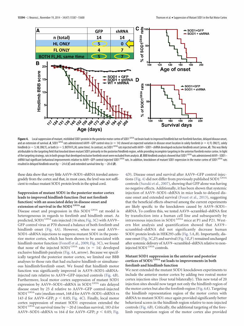
Inclusion criteria for behavioral and histological analysis. Injections inthe first knockdown experiment targeted the entire hindlimb motorregion and not the entire forelimb motor region. It was not surprisingthat analysis of forelimb motor function revealed no differences inonset or behavioral function. However, there was evidence for de-layed hindlimb onset and improved hindlimb behavioral function inAAV9 –SOD1–shRNA-injected rats versus AAV9 –GFP controls.However, this was not significant, likely because of the inclusion ofdata from rats exhibiting initial forelimb onset (data not shown).Because of the lack of target injections to the entire forelimb motorcortex region, we excluded rats (in both AAV9 –GFP and AAV9 –SOD1–shRNA groups) from behavioral analyses that showed exclu-sive forelimb onset. Therefore, SOD1 G93A rats included in analysis forhindlimb BBB, onset, and survival either exhibited onset in hindlimbonly (n � 5, exclusively the AAV9 –GFP control group) or simulta-neous forelimb/hindlimb onset (AAV9 –GFP controls, n � 5; andAAV9 –SOD1–shRNA, n � 7) for a total n � 10 AAV9 –GFP controlsand n � 7 AAV9 –SOD1–shRNA. For the second knockdown experi-ment targeting both forelimb and hindlimb motor regions, in eachgroup, only rats that had reached peak body weight and had begun toconsistently lose weight were included in the behavioral and histolog-
ical analyses (n � 4 AAV9 –SOD1–shRNA, n � 4 non-injectedcontrols).
AAV9-scrambled vector construction. A scrambled shRNA (–GACTT-GACCGACTTATGTAT–) construct was generated using Life Technol-ogies design tool. The shRNA construct was first cloned into pRNA–H1neo vector under human H1 promoter and tested in vitro. Scrambled–shRNA–SOD1 along with the H1 promoter was then PCR amplified andcloned at Kpn1 sites in AAV9 –CBA–GFP vector carrying GFP under achicken �-actin promoter.
Transfection. Human embryonic kidney 293 (HEK293) cells weretransfected using the calcium phosphate method with AAV9 –scram-bled–SOD1–shRNA plasmid to determine the effect on human SOD1protein levels. Cell lysates were harvested 72 h after transfection andanalyzed for the levels of human SOD1 protein by Western blot analysis(rabbit anti-hSOD1 at 1:750, Cell Signaling Technology; mouse anti-GAPDH at 1:1000, Millipore; anti-rabbit horse radish peroxidase (HRP)at 1:25000 or anti-mouse HRP at 1:10000, Jackson ImmunoResearch).AAV9 –CBA–GFP and AAV9 –SOD1–shRNA were used as negative andpositive controls for SOD1 downregulation, respectively, and GAPDHwas used for protein normalization.
Mouse injections. For neonatal mouse injections, P1 SOD1 G93A pups(n � 5) were used. A total volume of 50 �l containing 5 � 10 11 (3.6 �10 14 vg/kg) DNase-resistant viral particles of AAV9 –scrambled–SOD1–shRNA (Virapur) was injected through the temporal vein as describedpreviously (Foust et al., 2009). After a correct injection was verified byblanching of the vein, pups were returned to their cage. For adult tail veininjections, SOD1 G93A mice (n � 4) at P21 were placed in a restraint thatpositioned the tail in a lighted, heated groove. The tail was swabbed withalcohol and then injected intravenously with 200 �l viral solution con-taining 2 � 10 12 DNase-resistant viral particles of AAV9 –scrambled–SOD1–shRNA, for an average dose of 1.7 � 10 14 vg/kg.
Mouse behavior and survival analysis. Treated and non-injected con-trol SOD1 G93A mice were monitored throughout the disease coursecompared with non-injected control mice (n � 7). Mice were subjectedto weekly assessment of hindlimb grip strength using a grip-strengthmeter (Columbus Instruments). Each weekly session consisted of threetests per animal. Onset and survival analyses were performed using Ka-plan–Meier analyses. Endpoint was defined as the death point when ananimal could no longer regain their upright position within 30 s afterbeing placed on their back. Disease onset was determined from retro-spective analysis of the data and defined as the age at which the animalreached its peak weight.
Statistical analysis. Statistical analyses were performed using Prismsoftware (GraphPad Software). Student’s t tests, and one-way and two-way ANOVA using Bonferroni’s post hoc analyses were performed todetermine standard error of the mean (SEM) with a 95% confidencelevel. Kaplan–Meier survival analyses were analyzed by the log-rank test,and comparisons of median disease durations and survival times wereanalyzed by the Wilcoxon’s signed-rank test.
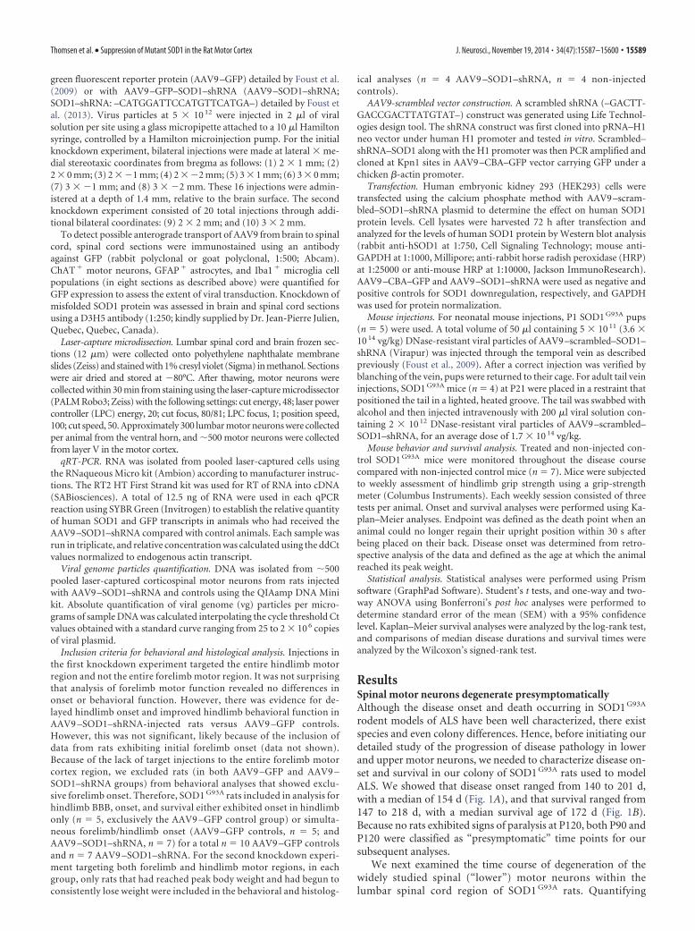
ResultsSpinal motor neurons degenerate presymptomaticallyAlthough the disease onset and death occurring in SOD1 G93A
rodent models of ALS have been well characterized, there existspecies and even colony differences. Hence, before initiating ourdetailed study of the progression of disease pathology in lowerand upper motor neurons, we needed to characterize disease on-set and survival in our colony of SOD1 G93A rats used to modelALS. We showed that disease onset ranged from 140 to 201 d,with a median of 154 d (Fig. 1A), and that survival ranged from147 to 218 d, with a median survival age of 172 d (Fig. 1B).Because no rats exhibited signs of paralysis at P120, both P90 andP120 were classified as “presymptomatic” time points for oursubsequent analyses.
We next examined the time course of degeneration of thewidely studied spinal (“lower”) motor neurons within thelumbar spinal cord region of SOD1 G93A rats. Quantifying
Thomsen et al. • Suppression of Mutant SOD1 in the Rat Motor Cortex J. Neurosci., November 19, 2014 • 34(47):15587–15600 • 15589
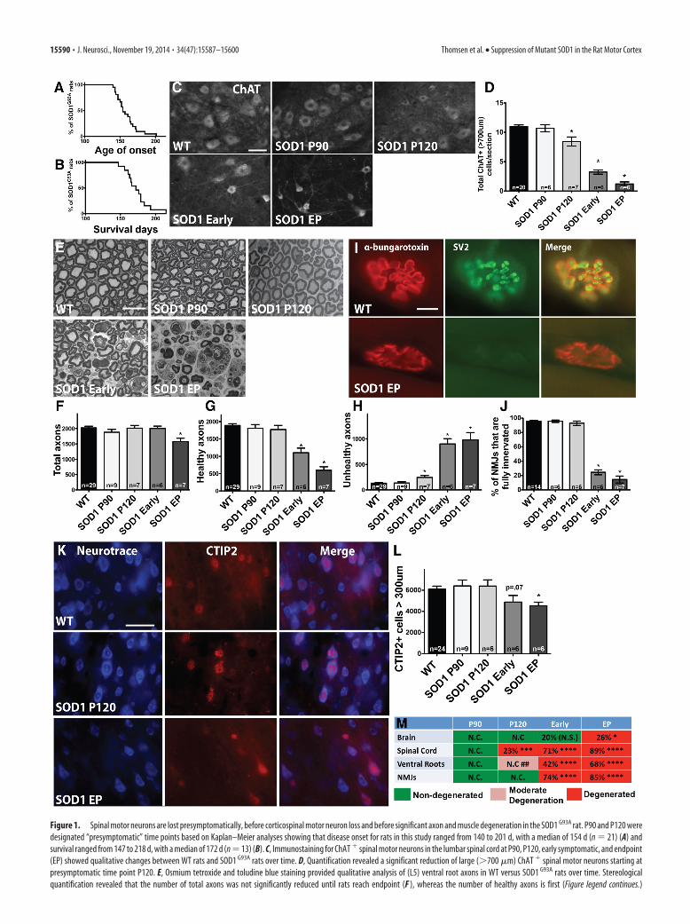
Figure 1. Spinal motor neurons are lost presymptomatically, before corticospinal motor neuron loss and before significant axon and muscle degeneration in the SOD1 G93A rat. P90 and P120 weredesignated “presymptomatic” time points based on Kaplan–Meier analyses showing that disease onset for rats in this study ranged from 140 to 201 d, with a median of 154 d (n � 21) (A) andsurvival ranged from 147 to 218 d, with a median of 172 d (n � 13) (B). C, Immunostaining for ChAT � spinal motor neurons in the lumbar spinal cord at P90, P120, early symptomatic, and endpoint(EP) showed qualitative changes between WT rats and SOD1 G93A rats over time. D, Quantification revealed a significant reduction of large (�700 �m) ChAT � spinal motor neurons starting atpresymptomatic time point P120. E, Osmium tetroxide and toludine blue staining provided qualitative analysis of (L5) ventral root axons in WT versus SOD1 G93A rats over time. Stereologicalquantification revealed that the number of total axons was not significantly reduced until rats reach endpoint (F ), whereas the number of healthy axons is first (Figure legend continues.)
15590 • J. Neurosci., November 19, 2014 • 34(47):15587–15600 Thomsen et al. • Suppression of Mutant SOD1 in the Rat Motor Cortex
the total number of ChAT � cells in the ventral horn showedthat spinal motor neurons in SOD1 G93A rats were significantlyreduced starting presymptomatically at P120 with an 11% re-duction relative to WT rats and that there was an additionalloss to 35% by endpoint (data not shown). Quantifying onlythe large (�700 �m) subpopulation of total spinal motor neu-rons in SOD1 G93A rats showed an even more dramatic losswith a significant 23% decrease starting at P120 relative to WTrats and an 89% depletion by endpoint (Fig. 1C,D). We alsoassessed the cell size of spinal motor neurons that were slightlydecreased presymptomatically at P90 and P120, reaching sig-nificance with 32% decrease at early symptomatic and 52%decrease at endpoint (data not shown).
Ventral root axons show degeneration and NMJs aredenervated only after spinal motor neuron loss insymptomatic ratsWe next assessed degeneration in L5 ventral root axons over timein SOD1 G93A rats. Sections of ventral root axons stained withosmium tetroxide and toluidine blue (Fig. 1E) were quantifiedfor total numbers of healthy and unhealthy axons. We found thatthere was no significant loss in the total number of axons(healthy plus unhealthy) relative to WT until disease endpoint(Fig. 1F ). Numbers of healthy axons (as defined by intactmyelin sheath and clear lumen) did not decrease until the earlysymptomatic stage (Fig. 1G). However, we did observe a sig-nificant increase in the number of apparently degenerating,“unhealthy” axons (damaged myelin sheath, dark lumen) be-ginning at P120 (Fig. 1H ).
We also examined NMJs in the gastrocnemius over time bystaining for �-bungarotoxin and SV2. NMJs were classified asfully innervated, semi-innervated, or denervated based on theextent of overlap of these two markers (Fig. 1I). At presymptom-atic time points, NMJs in SOD1 G93A rats were still fully inner-vated relative to WT rats (Fig. 1J). At P120, when a significant lossof spinal motor neurons was observed, 93% of NMJs were stillfully innervated in SOD1 G93A rats, which was similar to the96% seen in WT rats. However, by the time SOD1 G93A rats hadreached the early symptomatic stage, there was a significant74% decrease in the percentage of fully innervated NMJs, andthis continued to drop off as SOD1 G93A rats reached endpoint.This careful temporal analysis provided the unexpected find-ing that ventral root axons were still intact and appeared gen-erally healthy and that NMJs remained fully innervated at thepresymptomatic time point when spinal motor neurons were
already lost. We next turned to understanding the health andsurvival of corticospinal motor neurons in SOD1 G93A rat.
Corticospinal motor neurons are not lost untildisease endpointCorticospinal motor neurons reside in layer V of the motor cor-tex and express the CTIP2 protein (Arlotta et al., 2005). To de-termine the time course of corticospinal (“upper”) motor neurondegeneration, we quantified the number of large CTIP2� cellspresent in this defined region over time. A fluorescent Nissl stainwas used to visualize cell size, followed by stereological analysis tocalculate the number of large (�300 �m) CTIP2� cells (Fig. 1K).Results showed that the number of larger corticospinal motorneurons in the SOD1 G93A rat brain was similar during presymp-tomatic time periods (P90 and P120) relative to WT rats (Fig. 1L).Total numbers of large corticospinal motor neurons did not de-cline until rats were in the early symptomatic stage, and this onlybecame significant when rats had reached clinical endpoint (26%reduction in CTIP2� cells relative to WT at endpoint; Fig. 1L).Additionally, overall numbers of CTIP2� cells in layer V, regard-less of cell size, were not significantly different relative to WT orover time in SOD1 G93A rats (data not shown). Collectively, re-sults from the spinal cord, ventral roots, NMJs, and now the brainsuggest that the death of motor neurons starts at P120 in thespinal cord and radiates out to the muscle and brain over time(Fig. 1M).
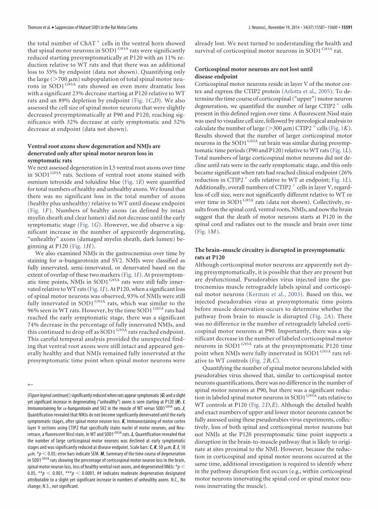
The brain–muscle circuitry is disrupted in presymptomaticrats at P120Although corticospinal motor neurons are apparently not dy-ing presymptomatically, it is possible that they are present butare dysfunctional. Pseudorabies virus injected into the gas-trocnemius muscle retrogradely labels spinal and corticospi-nal motor neurons (Kerman et al., 2003). Based on this, weinjected pseudorabies virus at presymptomatic time pointsbefore muscle denervation occurs to determine whether thepathway from brain to muscle is disrupted (Fig. 2A). Therewas no difference in the number of retrogradely labeled corti-cospinal motor neurons at P90. Importantly, there was a sig-nificant decrease in the number of labeled corticospinal motorneurons in SOD1 G93A rats at the presymptomatic P120 timepoint when NMJs were fully innervated in SOD1 G93A rats rel-ative to WT controls (Fig. 2 B, C).
Quantifying the number of spinal motor neurons labeled withpseudorabies virus showed that, similar to corticospinal motorneurons quantifications, there was no difference in the number ofspinal motor neurons at P90, but there was a significant reduc-tion in labeled spinal motor neurons in SOD1 G93A rats relative toWT controls at P120 (Fig. 2D,E). Although the detailed healthand exact numbers of upper and lower motor neurons cannot befully assessed using these pseudorabies virus experiments, collec-tively, loss of both spinal and corticospinal motor neurons butnot NMJs at the P120 presymptomatic time point supports adisruption in the brain-to-muscle pathway that is likely to origi-nate at sites proximal to the NMJ. However, because the reduc-tion in corticospinal and spinal motor neurons occurred at thesame time, additional investigation is required to identify wherein the pathway disruption first occurs (e.g., within corticospinalmotor neurons innervating the spinal cord or spinal motor neu-rons innervating the muscle).
4
(Figure legend continued.) significantly reduced when rats appear symptomatic (G) and a slightyet significant increase in degenerating (“unhealthy”) axons is seen starting at P120 (H). I,Immunostaining for �-bungarotoxin and SV2 in the muscle of WT versus SOD1 G93A rats. J,Quantification revealed that NMJs do not become significantly denervated until the earlysymptomatic stages, after spinal motor neuron loss. K, Immunostaining of motor cortexlayer V sections using CTIP2 that specifically stains nuclei of motor neurons, and Neu-rotrace, a fluorescent Nissl stain, in WT and SOD1 G93A rats. L, Quantification revealed thatthe number of large corticospinal motor neurons was declined at early symptomaticstages and was significantly reduced at disease endpoint. Scale bars: C, K, 50 �m; E, I, 10�m. *p � 0.05; error bars indicate SEM. M, Summary of the time course of degenerationin SOD1 G93A rats showing the percentage of corticospinal motor neuron loss in the brain,spinal motor neuron loss, loss of healthy ventral root axons, and degenerated NMJs: *p �0.05, **p � 0.001, ***p � 0.0001, ## indicates moderate degeneration designatedattributable to a slight yet significant increase in numbers of unhealthy axons. N.C., Nochange; N.S., not significant.
Thomsen et al. • Suppression of Mutant SOD1 in the Rat Motor Cortex J. Neurosci., November 19, 2014 • 34(47):15587–15600 • 15591
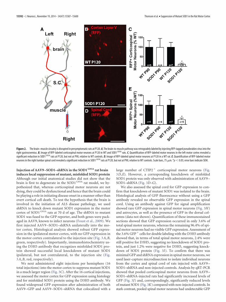
Injection of AAV9 –SOD1–shRNA in the SOD1 G93A rat braininduces local suppression of mutant, misfolded SOD1 proteinAlthough our initial anatomical studies did not show that thebrain is first to degenerate in the SOD1 G93A rat model, we hy-pothesized that, whereas corticospinal motor neurons are notdying, they could be dysfunctional and hence that the brain couldbe playing a role in initiating disease onset in a manner other thanovert cortical cell death. To test the hypothesis that the brain isinvolved in the initiation of ALS disease pathology, we usedshRNA to knock down mutant SOD1 expression in the motorcortex of SOD1 G93A rats at 70 d of age. The shRNA to mutantSOD1 was fused to the GFP reporter, and both genes were pack-aged in AAV9, known to infect neurons (Foust et al., 2009). Wefirst injected AAV9 –SOD1–shRNA unilaterally into the mo-tor cortex. Histological analysis showed robust GFP expres-sion in the ipsilateral motor cortex, with no GFP expression inthe motor cortex contralateral to the injection site (Fig. 3 A, B,green, respectively). Importantly, immunohistochemistry us-ing the D3H5 antibody that recognizes misfolded SOD1 pro-tein showed successful local knockdown of mutant SOD1ipsilateral, but not contralateral, to the injection site (Fig.3 A, B, red, respectively).
We next administered eight injections per hemisphere (16total injections) into the motor cortex to suppress mutant SOD1in a much larger region (Fig. 3C). After the 16 cortical injections,we assessed the motor cortex for GFP expression using histologyand for misfolded SOD1 protein using the D3H5 antibody. Wefound widespread GFP expression after administration of bothAAV9 –GFP and AAV9 –SOD1–shRNA that colocalized with a
large number of CTIP2� corticospinal motor neurons (Fig.3D,E). However, a corresponding knockdown of misfoldedSOD1 protein was only observed with administration of AAV9 –SOD1–shRNA (Fig. 3D–G).
We also assessed the spinal cord for GFP expression to con-firm that knockdown of mutant SOD1 was isolated to the brain.Histological analysis of GFP fluorescence without using a GFPantibody revealed no observable GFP expression in the spinalcord. Using an antibody against GFP for signal amplificationshowed rare GFP expression in spinal motor neurons (Fig. 3H)and astrocytes, as well as the presence of GFP in the dorsal col-umns (data not shown). Quantification of these immunostainedsections showed that GFP expression occurred in only 3.6% oftotal spinal motor neurons, whereas the remaining 96.4% of spi-nal motor neurons had no visible GFP expression. Assessment ofthe 3.6% GFP� cells for double labeling with the D3H5 antibodyshowed that, in terms of total spinal motor neurons, 2.4% werestill positive for D3H5, suggesting no knockdown of SOD1 pro-tein, and just 1.2% were negative for D3H5, suggesting knock-down of SOD1 protein (Fig. 3I). To confirm that there wasminimal GFP and shRNA expression in spinal motor neurons, weused laser-capture microdissection to isolate individual neuronsfrom the cortex and spinal cord of rats injected with AAV9 –SOD1–shRNA and non-injected controls. Analysis by qRT-PCRshowed that pooled corticospinal motor neurons from AAV9 –SOD1–shRNA-injected rats had significantly increased levels ofGFP (Fig. 3J) and, correspondingly, significantly reduced levelsof mutant SOD1 (Fig. 3K) compared with non-injected controls. Instark contrast, pooled spinal motor neurons had undetectable GFP
Figure 2. The brain–muscle circuitry is disrupted in presymptomatic rats at P120. A, The brain-to-muscle pathway was retrogradely labeled by injecting RFP-tagged pseudorabies virus into theright gastrocnemius. B, Image of RFP-labeled corticospinal motor neurons at P120 in WT and SOD1 G93A rats. C, Quantification of RFP-labeled motor neurons in the left motor cortex revealed asignificant reduction in SOD1 G93A rats at P120, but not at P90, relative to WT controls. D, Image of RFP-labeled spinal motor neurons at P120 in a WT rat. E, Quantification of RFP-labeled motorneurons in the right lumbar spinal cord revealed a significant reduction in SOD1 G93A rats at P120, but not at P90, relative to WT controls. Scale bars, 75 �m. *p � 0.05; error bars indicate SEM.
15592 • J. Neurosci., November 19, 2014 • 34(47):15587–15600 Thomsen et al. • Suppression of Mutant SOD1 in the Rat Motor Cortex
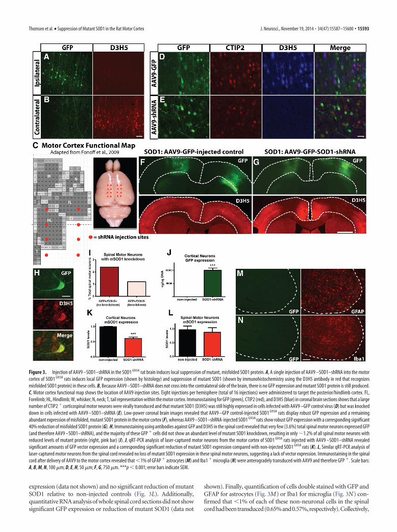
expression (data not shown) and no significant reduction of mutantSOD1 relative to non-injected controls (Fig. 3L). Additionally,quantitative RNA analysis of whole spinal cord sections did not showsignificant GFP expression or reduction of mutant SOD1 (data not
shown). Finally, quantification of cells double stained with GFP andGFAP for astrocytes (Fig. 3M) or Iba1 for microglia (Fig. 3N) con-firmed that �1% of each of these non-neuronal cells in the spinalcordhadbeentransduced(0.65%and0.57%,respectively).Collectively,
Figure 3. Injection of AAV9 –SOD1–shRNA in the SOD1 G93A rat brain induces local suppression of mutant, misfolded SOD1 protein. A, A single injection of AAV9 –SOD1–shRNA into the motorcortex of SOD1 G93A rats induces local GFP expression (shown by histology) and suppression of mutant SOD1 (shown by immunohistochemistry using the D3H5 antibody in red that recognizesmisfolded SOD1 protein) in these cells. B, Because AAV9 –SOD1–shRNA does not cross into the contralateral side of the brain, there is no GFP expression and mutant SOD1 protein is still produced.C, Motor cortex functional map shows the location of AAV9 injection sites. Eight injections per hemisphere (total of 16 injections) were administered to target the posterior/hindlimb cortex. FL,Forelimb; HL, Hindlimb; W, whisker; N, neck; T, tail representation within the motor cortex. Immunostaining for GFP (green), CTIP2 (red), and D3H5 (blue) in coronal brain sections shows that a largenumber of CTIP2 � corticospinal motor neurons were virally transduced and that mutant SOD1 (D3H5) was still highly expressed in cells infected with AAV9 –GFP control virus (D) but was knockeddown in cells infected with AAV9 –SOD1–shRNA (E). Low-power coronal brain images revealed that AAV9 –GFP control-injected SOD1 G93A rats display robust GFP expression and a remainingabundant expression of misfolded, mutant SOD1 protein in the motor cortex (F), whereas AAV9 –SOD1–shRNA-injected SOD1 G93A rats show robust GFP expression with a corresponding significant40% reduction of misfolded SOD1 protein (G). H, Immunostaining using antibodies against GFP and D3H5 in the spinal cord revealed that very few (3.6%) total spinal motor neurons expressed GFP(and therefore AAV9 –SOD1–shRNA), and the majority of these GFP � cells did not show an abundant level of mutant SOD1 knockdown, resulting in only �1.2% of all spinal motor neurons withreduced levels of mutant protein (right, pink bar) (I). J, qRT-PCR analysis of laser-captured motor neurons from the motor cortex of SOD1 G93A rats injected with AAV9 –SOD1–shRNA revealedsignificant amounts of GFP vector expression and a corresponding significant reduction of mutant SOD1 expression compared with non-injected SOD1 G93A rats (K). L, Similar qRT-PCR analysis oflaser-captured motor neurons from the spinal cord revealed no loss of mutant SOD1 expression in these spinal motor neurons, suggesting a lack of vector expression. Immunostaining in the spinalcord after delivery of AAV9 to the motor cortex revealed that �1% of GFAP � astrocytes (M) and Iba1 � microglia (N) were anterogradely transduced with AAV9 and therefore GFP �. Scale bars:A, B, M, N, 100 �m; D, E, H, 50 �m; F, G, 750 �m. ***p � 0.001; error bars indicate SEM.
Thomsen et al. • Suppression of Mutant SOD1 in the Rat Motor Cortex J. Neurosci., November 19, 2014 • 34(47):15587–15600 • 15593
these data show that very little AAV9–SOD1–shRNA traveled antero-gradely from the cortex and that, in most cases, the level was not suffi-cient to reduce mutant SOD1 protein levels in the spinal cord.
Suppression of mutant SOD1 in the posterior motor cortexleads to improved hindlimb function (but not forelimbfunction) with an associated delay in disease onset andextension of survival in the SOD1 G93A ratDisease onset and progression in this SOD1 G93A rat model isheterogeneous in regards to forelimb and hindlimb onset. Aspredicted, SOD1 G93A rats injected (16 sites; Fig. 3C) with AAV9 –GFP control virus at P70 showed a balance of both forelimb andhindlimb onset (Fig. 4A). However, when we used AAV9 –SOD1–shRNA injections to suppress mutant SOD1 in the poste-rior motor cortex, which has been shown to be associated withhindlimb motor function (Fonoff et al., 2009; Fig. 3C), we foundthat none of the injected SOD1 G93A rats (n � 14) developedexclusive hindlimb paralysis (Fig. 4A, arrow). Because we specif-ically targeted the posterior motor cortex, we limited our BBBanalyses to those rats that had exclusive hindlimb or simultane-ous hindlimb/forelimb onset. We found that hindlimb motorfunction was significantly improved in AAV9 –SOD1–shRNA-injected rats relative to AAV9 –GFP-injected controls (Fig. 4B).Furthermore, local motor cortex suppression of mutant SOD1expression by AAV9 –SOD1–shRNA in SOD1 G93A rats delayeddisease onset by 25 d relative to AAV9 –GFP control-injectedSOD1 G93A rats (median onset, 168 d for AAV9 –SOD1–shRNA vs143 d for AAV9 –GFP; p � 0.05; Fig. 4C). Finally, local motorcortex suppression of mutant SOD1 expression extended theSOD1 G93A rat survival time by �20 d (median survival, 183 d forAAV9 –SOD1–shRNA vs 164 d for AAV9 –GFP; p � 0.05; Fig.
4D). Disease onset and survival after AAV9 –GFP control injec-tions (Fig. 4) did not differ from previously published SOD1 G93A
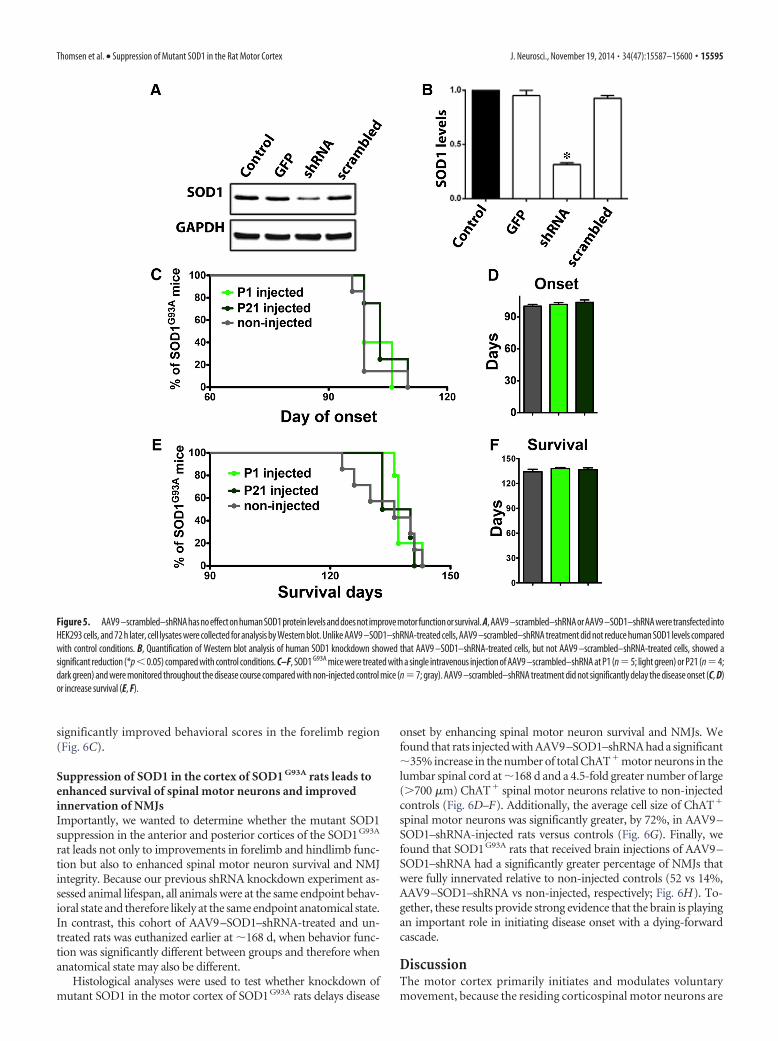
controls (Suzuki et al., 2007), showing that GFP alone was havingno negative effects. Additionally, it has been shown that systemicinjection of AAV9 –SOD1–shRNA in mice leads to delayed dis-ease onset and extended survival (Foust et al., 2013), suggestingthat the beneficial effects observed among the current experimentsare likely specific to the knockdown caused by AAV9–SOD1–shRNA. To confirm this, we tested AAV9–scrambled–shRNA firstby transfection into a human cell line and subsequently byintravenous injection in SOD1 G93A mice at P1 and P21. West-ern blot analysis and quantification showed that AAV9 –scrambled–shRNA did not significantly decrease humanSOD1 protein levels in HEK293 cells (Fig. 5A,B). Importantly, dis-ease onset (Fig. 5C,D) and survival (Fig. 5E,F) remained unchangedafter systemic delivery of AAV9–scrambled–shRNA relative to non-injected SOD1G93A controls.
Mutant SOD1 suppression in the anterior and posteriorcortices of SOD1 G93A rat leads to improvements in bothforelimb and hindlimb functionWe next extended the mutant SOD1 knockdown experiments toinclude the anterior motor cortex by adding two rostral motorcortex injection sites (four total bilaterally). This new total of 20injection sites should now target not only the hindlimb region ofthe motor cortex but also the forelimb region (Fig. 6A). Targetingthe hindlimb representation region of the motor cortex withshRNA to mutant SOD1 once again provided significantly betterbehavioral scores in the hindlimb region relative to non-injectedcontrols (Fig. 6B). Critically, the additional targeting of the fore-limb representation region of the motor cortex also provided
Figure 4. Local suppression of mutant, misfolded SOD1 protein in the posterior motor cortex of SOD1 G93A rat brain leads to improved hindlimb but not forelimb function, delayed disease onset,and an extension of survival. A, SOD1 G93A rats administered AAV9 –GFP control virus (n � 14) showed an expected variation in disease onset location in solely forelimb (n � 4; FL ONLY), solelyhindlimb (n � 5; HL ONLY), or both (n � 5; BOTH FL,HL same time). In contrast, no SOD1 G93A rats injected with AAV9 –SOD1–shRNA developed exclusive hindlimb onset (arrow, A). This was likelyattributable to the targeting field that knocked down mutant SOD1 primarily in the posterior/hindlimb region, while providing incomplete targeting in the anterior/forelimb motor cortex. In lightof the targeting strategy, rats in both groups that developed exclusive forelimb onset were excluded from analysis. B, BBB hindlimb analysis showed that SOD1 G93A rats administered AAV9 –SOD1–shRNA had significant behavioral improvements relative to AAV9 –GFP control-injected SOD1 G93A rats. In addition, knockdown of mutant SOD1 expression in the motor cortex of SOD1 G93A ratsresulted in delayed hindlimb onset by �24 d (C) and extended survival time by �20 d (D).
15594 • J. Neurosci., November 19, 2014 • 34(47):15587–15600 Thomsen et al. • Suppression of Mutant SOD1 in the Rat Motor Cortex
significantly improved behavioral scores in the forelimb region(Fig. 6C).
Suppression of SOD1 in the cortex of SOD1 G93A rats leads toenhanced survival of spinal motor neurons and improvedinnervation of NMJsImportantly, we wanted to determine whether the mutant SOD1suppression in the anterior and posterior cortices of the SOD1G93A
rat leads not only to improvements in forelimb and hindlimb func-tion but also to enhanced spinal motor neuron survival and NMJintegrity. Because our previous shRNA knockdown experiment as-sessed animal lifespan, all animals were at the same endpoint behav-ioral state and therefore likely at the same endpoint anatomical state.In contrast, this cohort of AAV9–SOD1–shRNA-treated and un-treated rats was euthanized earlier at �168 d, when behavior func-tion was significantly different between groups and therefore whenanatomical state may also be different.
Histological analyses were used to test whether knockdown ofmutant SOD1 in the motor cortex of SOD1G93A rats delays disease
onset by enhancing spinal motor neuron survival and NMJs. Wefound that rats injected with AAV9–SOD1–shRNA had a significant�35% increase in the number of total ChAT� motor neurons in thelumbar spinal cord at �168 d and a 4.5-fold greater number of large(�700 �m) ChAT� spinal motor neurons relative to non-injectedcontrols (Fig. 6D–F). Additionally, the average cell size of ChAT�
spinal motor neurons was significantly greater, by 72%, in AAV9–SOD1–shRNA-injected rats versus controls (Fig. 6G). Finally, wefound that SOD1G93A rats that received brain injections of AAV9–SOD1–shRNA had a significantly greater percentage of NMJs thatwere fully innervated relative to non-injected controls (52 vs 14%,AAV9–SOD1–shRNA vs non-injected, respectively; Fig. 6H). To-gether, these results provide strong evidence that the brain is playingan important role in initiating disease onset with a dying-forwardcascade.
DiscussionThe motor cortex primarily initiates and modulates voluntarymovement, because the residing corticospinal motor neurons are
Figure 5. AAV9 –scrambled–shRNAhasnoeffectonhumanSOD1protein levelsanddoesnot improvemotorfunctionorsurvival. A,AAV9 –scrambled–shRNAorAAV9 –SOD1–shRNAweretransfectedintoHEK293 cells, and 72 h later, cell lysates were collected for analysis by Western blot. Unlike AAV9 –SOD1–shRNA-treated cells, AAV9 –scrambled–shRNA treatment did not reduce human SOD1 levels comparedwith control conditions. B, Quantification of Western blot analysis of human SOD1 knockdown showed that AAV9 –SOD1–shRNA-treated cells, but not AAV9 –scrambled–shRNA-treated cells, showed asignificant reduction (*p�0.05) compared with control conditions. C–F, SOD1 G93A mice were treated with a single intravenous injection of AAV9 –scrambled–shRNA at P1 (n�5; light green) or P21 (n�4;dark green) and were monitored throughout the disease course compared with non-injected control mice (n�7; gray). AAV9 –scrambled–shRNA treatment did not significantly delay the disease onset (C, D)or increase survival (E, F).
Thomsen et al. • Suppression of Mutant SOD1 in the Rat Motor Cortex J. Neurosci., November 19, 2014 • 34(47):15587–15600 • 15595
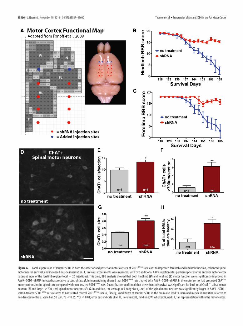
Figure 6. Local suppression of mutant SOD1 in both the anterior and posterior motor cortices of SOD1 G93A rats leads to improved forelimb and hindlimb function, enhanced spinalmotor neuron survival, and increased muscle innervation. A, Previous experiments were repeated, with two additional AAV9 injection sites per hemisphere to the anterior motor cortexto target more of the forelimb region (total � 20 injections). This time, BBB analysis showed that both hindlimb (B) and forelimb (C) motor function were significantly improved inAAV9 –SOD1–shRNA-injected rats relative to control rats. D, Immunostaining showed that SOD1 G93A rats treated with AAV9 –SOD1–shRNA in the motor cortex had preserved ChAT �
motor neurons in the spinal cord compared with non-treated SOD1 G93A rats. Quantification confirmed that the enhanced survival was significant for both total ChAT � spinal motorneurons (E) and large (�700 �m) spinal motor neurons (F). G, In addition, the average cell body size (�m 2) of the spinal motor neurons was significantly larger in AAV9 –SOD1–shRNA-treated SOD1 G93A rats relative to nontreated control SOD1 G93A rats. H, Finally, knockdown of mutant SOD1 in the brain also lead to increased muscle innervation relative tonon-treated controls. Scale bar, 50 �m. *p � 0.05, **p � 0.01; error bars indicate SEM. FL, Forelimb; HL, hindlimb; W, whisker; N, neck; T, tail representation within the motor cortex.
15596 • J. Neurosci., November 19, 2014 • 34(47):15587–15600 Thomsen et al. • Suppression of Mutant SOD1 in the Rat Motor Cortex
responsible for collecting, integrating, translating, and transfer-ring information to the spinal cord. If these cells do not functionproperly, system breakdown occurs. Because the motor neuroncircuitry clearly degenerates over time in ALS patients and animalmodels, it is surprising that the motor cortex containing the ini-tiating component (corticospinal motor neurons) has receivedlimited attention among the ALS research community.
Here, we have shown for the first time that suppression ofmutant SOD1 at the “top” of the motor neuron pathway (e.g., themotor cortex) results in a significant delay in ALS disease onset
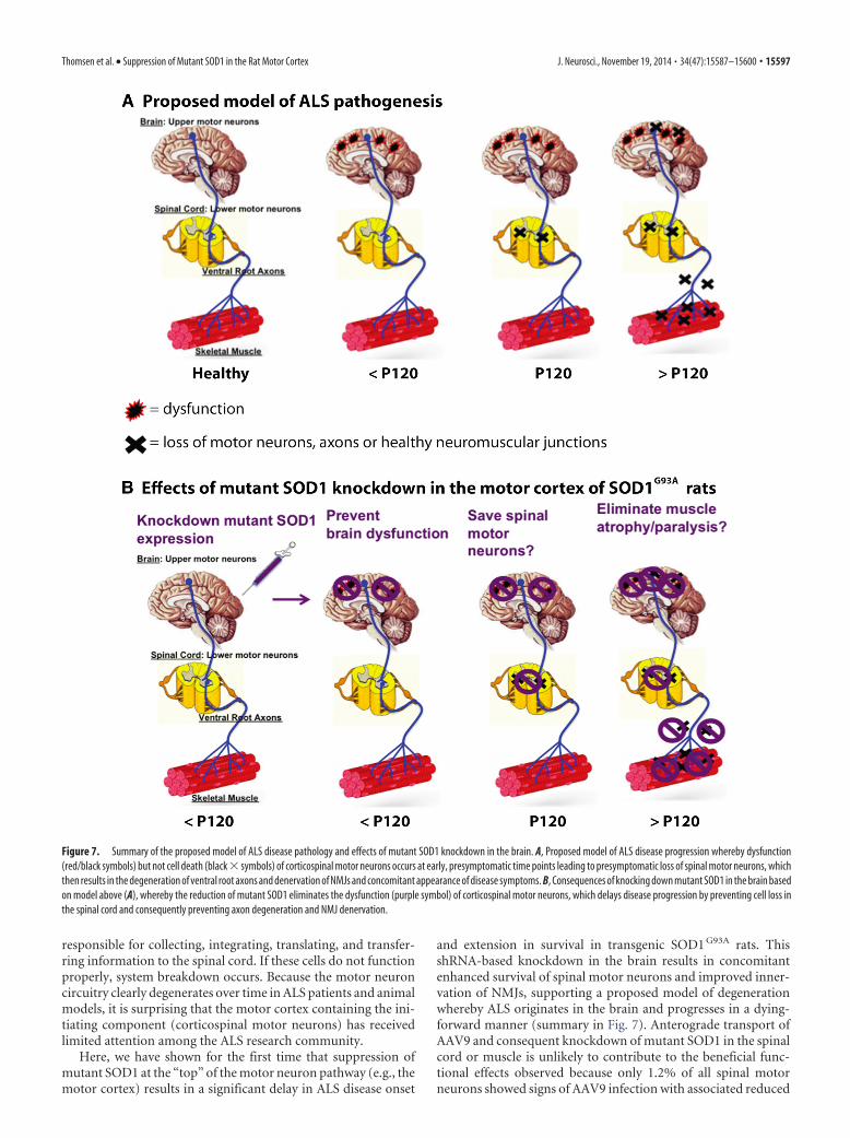
and extension in survival in transgenic SOD1 G93A rats. ThisshRNA-based knockdown in the brain results in concomitantenhanced survival of spinal motor neurons and improved inner-vation of NMJs, supporting a proposed model of degenerationwhereby ALS originates in the brain and progresses in a dying-forward manner (summary in Fig. 7). Anterograde transport ofAAV9 and consequent knockdown of mutant SOD1 in the spinalcord or muscle is unlikely to contribute to the beneficial func-tional effects observed because only 1.2% of all spinal motorneurons showed signs of AAV9 infection with associated reduced
Figure 7. Summary of the proposed model of ALS disease pathology and effects of mutant SOD1 knockdown in the brain. A, Proposed model of ALS disease progression whereby dysfunction(red/black symbols) but not cell death (black � symbols) of corticospinal motor neurons occurs at early, presymptomatic time points leading to presymptomatic loss of spinal motor neurons, whichthen results in the degeneration of ventral root axons and denervation of NMJs and concomitant appearance of disease symptoms. B, Consequences of knocking down mutant SOD1 in the brain basedon model above (A), whereby the reduction of mutant SOD1 eliminates the dysfunction (purple symbol) of corticospinal motor neurons, which delays disease progression by preventing cell loss inthe spinal cord and consequently preventing axon degeneration and NMJ denervation.
Thomsen et al. • Suppression of Mutant SOD1 in the Rat Motor Cortex J. Neurosci., November 19, 2014 • 34(47):15587–15600 • 15597
levels of mutant SOD1, and even �1% of non-neuronal cellsshowed infection. The findings from these studies provide criticalsupport for a cortical basis in the development of ALS, providingscientists and drug screeners with a novel focus for future ALSresearch and treatments.
A wide range of factors are likely to contribute to ALSpathogenesis, including mitochondrial dysfunction, proteinmisfolding/aggregation, oxidative damage, neuronal excitotoxicity,non-cell-autonomous effects/neuroinflammation, axonal transportdefects, neurotrophin depletion, effects from extracellular mutantSOD1, and aberrant RNA processing (Eisen et al., 1992; Chouand Norris, 1993; Williamson, 1999; Kieran et al., 2005; De Win-ter et al., 2006; Parkhouse et al., 2008; Schmidt et al., 2009;Bilsland et al., 2010; Brockington et al., 2010; Li et al., 2010;Dadon-Nachum et al., 2011). However, the extent to which eachfactor is involved and the overall pathology of the disease are stillnot understood. A common feature of ALS is glutamate toxicityin the spinal cord, suggesting that upper motor neuron hyperex-citability may induce anterior horn degeneration via an antero-grade glutamatergic mechanism (Eisen et al., 1992; Trotti et al.,1999; Rothstein et al., 2005). Hyperexcitability has also beenshown recently to be a feature of induced pluripotent stem cell-derived motor neurons from ALS patients harboring SOD1 mu-tations (Wainger et al., 2014). Because cortical hyperexcitabilityand signs of corticospinal motor neuron degeneration have beenshown to be early events in ALS patients and mice (Vucic et al.,2008; Ozdinler et al., 2011; Menon et al., 2014), future studiesfocusing on cortical dysfunction will likely yield insight into theimportance of the brain in disease pathology.
Electrical recording techniques and low-resolution imagingare used to assess global changes in ALS patient cortical functionbut cannot successfully determine changes at the cellular level,such as motor neuron death at early time points. Using detailedhistological analysis of SOD1 G93A rats over time, we showed thatthe first signs of cell death occurred in the spinal cord, with thisloss of spinal motor neurons preceding significant degenerationof distal ventral root axons and denervation of NMJs. The de-crease in spinal motor neuron cell bodies before an equal loss ofNMJs suggested that there is a window of time in this rat model ofALS when the cell body is cleared and the NMJs appear intact.Although this was an unexpected finding, it mirrors transectionmodels of axon degeneration that show a latency period afteracute axonal injury when distal axons may remain morphologi-cally intact and functionally excitable for brief periods of time(Lubinska, 1982; Tsao et al., 1994). Indeed, changing the physio-logical conditions can alter the timeframe during which injuredaxons are in a vulnerable yet functional state (George et al., 1995;Tsao et al., 1999; Conforti et al., 2000). In the current report, asurvival or trophic factor within the distal axons may be main-tained at a sufficient level to preserve the distal axons even whenspinal motor neurons are lost (for review, see Wang et al., 2012).
Our anatomical studies showed no evidence that corticospinalmotor neurons are lost early on at presymptomatic time points.This is in contrast to the presymptomatic death of corticospinalmotor neurons observed in the SOD1 G93A mouse, with associ-ated structural differences in apical dendrites of these layer Vpyramidal cells (Ozdinler et al., 2011; Jara et al., 2012). The lack ofovert cell death within the motor cortex at early time points in thetransgenic rat compared with the transgenic mouse can likely beattributed to differences in animal models. Indeed, many prom-ising preclinical trials using mouse models have not successfullytranslated to humans (Gordon et al., 2007; Berry and Cudkowicz,2011; Philips and Robberecht, 2011). Relative to the transgenic
mouse model of ALS, the rat model is larger and the timing andlocation of disease onset is less predictable. Interestingly, patientsalso show variable location of onset and progression rates. Al-though the rat model is valuable for additional studies investigat-ing ALS pathogenesis and therapeutic approaches, ALS mousemodels are more amenable to transgenic strategies that may elu-cidate fundamental mechanisms of the disease. For instance, theUCHL1–eGFP mouse was generated recently in which corticospinalmotor neurons selectively expressed GFP under the control of theubiquitin C-terminal hydrolase-L1 (UCHL1) promoter (Yasvoina etal., 2013). Crossbreeding UCHL1–GFP and SOD1G93A mice gener-ated hSOD1G93A–U–GFP mice that recapitulated the reduced num-bers of corticospinal motor neurons with disease progressionreported in the SOD1G93A mice (Ozdinler et al., 2011).
It is well established that ALS involves non-cell-autonomoustoxicity and that unknown toxic factors secreted by mutantSOD1 astrocytes and microglia are capable of killing healthy mo-tor neurons (Clement et al., 2003; Di Giorgio et al., 2008; Haidet-Phillips et al., 2011; Sargsyan et al., 2011; Frakes et al., 2014; Re etal., 2014). shRNA knockdown of SOD1 in astrocytes eliminatesthis toxicity, confirming that this is mediated through glial SOD1synthesis (Haidet-Phillips et al., 2011; Frakes et al., 2014). It hasbeen proposed that a prion-like mechanism is involved in thespread of ALS and that perhaps toxicity is transferred through anunidentified misfolded SOD1 initiator that may trigger prion-like aggregation of normal SOD1 in neighboring cells (for review,see Polymenidou and Cleveland, 2011; Grad and Cashman,2014). We found that shRNA-based knockdown of mutant SOD1in the posterior motor cortex provided functional improvementsin the hindlimb and increased lifespan. Interestingly, unlike lo-calized injections targeting only the hindlimb region, addingshRNA injection sites to knockdown SOD1 in an increased areathat included the forelimb motor cortex resulted in additionalfunctional improvements within the forelimb region. AlthoughSOD1 knockdown in specific regions of the motor cortex did notcompletely prevent disease onset and progression, it is quite pos-sible that targeting the entire motor cortex to eliminate a greaterarea of toxicity and the potential for toxic spread may lead to aneven better outcome. The presence of GFP expression and theabsence of mutant SOD1 expression within layers I–VI both sug-gest that AAV9 –SOD1–shRNA transduced all cells in this regionof the motor cortex, which includes corticospinal motor neurons,astrocytes, and interneurons. Although our data strongly suggestthat the brain plays an important role in ALS pathology, the exactmechanisms and involvement of various cell types require addi-tional investigation, such as experiments using promoter-specificexpression or knockdown of mutant SOD1. It is likely that non-cell-autonomous toxicity occurs in the brain as in the spinal cord.It has taken years of research by numerous laboratory groups toshow a central role of the spinal cord in ALS, and we hope thatthese novel data will initiate a new era of experiments that focuson understanding the mechanisms underlying the critical contri-bution of the brain to ALS.
AAV9 –SOD1–shRNA might be a valid candidate to translateAAV9-mediated suppression of SOD1 synthesis to the humansetting for clinical trials in ALS. Indeed, direct cerebrospinal fluidinfusion at the lumbar level in nonhuman primates has beendeemed safe and has been shown to produce substantial SOD1reduction by targeting both motor neurons and non-neuronalcells (Foust et al., 2013). However, caution is still required givendifferences not only between rodent models and the human con-dition but also given differences between the motor neuron poolsmost affected with distinct human mutations (Cudkowicz et al.,
15598 • J. Neurosci., November 19, 2014 • 34(47):15587–15600 Thomsen et al. • Suppression of Mutant SOD1 in the Rat Motor Cortex
1998; Lopate et al., 2010). In addition, it remains controversialwhether SOD1 toxicity is involved in both sporadic and familialcases of ALS (Haidet-Phillips et al., 2011; Re et al., 2014). Clearly,SOD1 toxicity involvement in both sporadic and familial caseswould substantially broaden the application of this therapeuticapproach for ALS. Although the exact mechanism of ALS is stillunclear and is likely dependent on multiple factors, we show herethat a toxic/dysfunctional brain environment can trigger down-stream disruption of the motor neuron pathway. Thus, our studyfurther highlights the importance of the upper motor neurons inALS.
ConclusionsThere is a large body of evidence to suggest that the onset ofclinical symptoms in ALS patients and animal models is precededby a long presymptomatic period (Eisen et al., 2014). It is verylikely that dysfunction at a cellular level (without cell death) existsand goes undetected for years before clinical diagnosis. It is onlyafter reaching and exceeding a threshold that extensive motorneuron circuitry failure occurs and clinical expression is ob-served. The current lack of successful therapeutic options for ALSpatients may be attributed, in part, to the timing of treatmentinitiation, because attempts at slowing disease progression typi-cally occur after diagnosis, which is after an irreversible cascade ofmotor neuron circuitry failure has set in. Therefore, it is essentialto identify the key players in early dysfunction in order to identifyand treat those at high risk of developing ALS. Because it is be-coming clear that the motor cortex plays a significant role in ALSpathology, it will be essential to focus future studies on identify-ing the mechanisms and key molecular/cellular componentswithin the brain that may initiate system breakdown and theonset of ALS.
ReferencesArlotta P, Molyneaux BJ, Chen J, Inoue J, Kominami R, Macklis JD (2005)
Neuronal subtype-specific genes that control corticospinal motor neurondevelopment in vivo. Neuron 45:207–221. CrossRef Medline
Basso DM, Beattie MS, Bresnahan JC (1995) A sensitive and reliable loco-motor rating scale for open field testing in rats. J Neurotrauma 12:1–21.CrossRef Medline
Berry JD, Cudkowicz ME (2011) New considerations in the design of clini-cal trials for amyotrophic lateral sclerosis. Clin Investig (Lond) 1:1375–1389. CrossRef Medline
Bilsland LG, Sahai E, Kelly G, Golding M, Greensmith L, Schiavo G (2010)Deficits in axonal transport precede ALS symptoms in vivo. Proc NatlAcad Sci U S A 107:20523–20528. CrossRef Medline
Brockington A, Heath PR, Holden H, Kasher P, Bender FL, Claes F, Lam-brechts D, Sendtner M, Carmeliet P, Shaw PJ (2010) Downregulation ofgenes with a function in axon outgrowth and synapse formation in motorneurones of the VEGFdelta/delta mouse model of amyotrophic lateralsclerosis. BMC Genomics 11:203. CrossRef Medline
Card JP, Levitt P, Enquist LW (1998) Different patterns of neuronal infec-tion after intracerebral injection of two strains of pseudorabies virus.J Virol 72:4434 – 4441. Medline
Card JP, Rinaman L, Lynn RB, Lee BH, Meade RP, Miselis RR, Enquist LW(1993) Pseudorabies virus infection of the rat central nervous system:ultrastructural characterization of viral replication, transport, and patho-genesis. J Neurosci 13:2515–2539. Medline
Chou SM, Norris FH (1993) Amyotrophic lateral sclerosis: lower motorneuron disease spreading to upper motor neurons. Muscle Nerve 16:864 –869. CrossRef Medline
Clement AM, Nguyen MD, Roberts EA, Garcia ML, Boillee S, Rule M, Mc-Mahon AP, Doucette W, Siwek D, Ferrante RJ, Brown RH Jr, Julien JP,Goldstein LS, Cleveland DW (2003) Wild-type nonneuronal cells ex-tend survival of SOD1 mutant motor neurons in ALS mice. Science 302:113–117. CrossRef Medline
Conforti L, Tarlton A, Mack TG, Mi W, Buckmaster EA, Wagner D, Perry
VH, Coleman MP (2000) A Ufd2/D4Cole1e chimeric protein and over-expression of Rbp7 in the slow Wallerian degeneration (WldS) mouse.Proc Natl Acad Sci U S A 97:11377–11382. CrossRef Medline
Cudkowicz ME, McKenna-Yasek D, Chen C, Hedley-Whyte ET, Brown RH Jr(1998) Limited corticospinal tract involvement in amyotrophic lateralsclerosis subjects with the A4V mutation in the copper/zinc superoxidedismutase gene. Ann Neurol 43:703–710. CrossRef Medline
Dadon-Nachum M, Melamed E, Offen D (2011) The “dying-back” phe-nomenon of motor neurons in ALS. J Mol Neurosci 43:470 – 477.CrossRef Medline
De Winter F, Vo T, Stam FJ, Wisman LA, Bar PR, Niclou SP, van MuiswinkelFL, Verhaagen J (2006) The expression of the chemorepellent Sema-phorin 3A is selectively induced in terminal Schwann cells of a subset ofneuromuscular synapses that display limited anatomical plasticity andenhanced vulnerability in motor neuron disease. Mol Cell Neurosci 32:102–117. CrossRef Medline
DeJesus-Hernandez M, Mackenzie IR, Boeve BF, Boxer AL, Baker M, Ruth-erford NJ, Nicholson AM, Finch NA, Flynn H, Adamson J, Kouri N,Wojtas A, Sengdy P, Hsiung GY, Karydas A, Seeley WW, Josephs KA,Coppola G, Geschwind DH, Wszolek ZK, Feldman H, Knopman DS,Petersen RC, Miller BL, Dickson DW, Boylan KB, Graff-Radford NR,Rademakers R (2011) Expanded GGGGCC hexanucleotide repeat innoncoding region of C9ORF72 causes chromosome 9p-linked FTD andALS. Neuron 72:245–256. CrossRef Medline
Di Giorgio FP, Boulting GL, Bobrowicz S, Eggan KC (2008) Human embry-onic stem cell-derived motor neurons are sensitive to the toxic effect ofglial cells carrying an ALS-causing mutation. Cell Stem Cell 3:637– 648.CrossRef Medline
Eisen A, Kiernan M, Mitsumoto H, Swash M (2014) Amyotrophic lateral sclerosis:a long preclinical period? J Neurol Neurosurg Psychiatry. Advance online publi-cation. Retrieved October 14, 2014. doi:10.1136/jnnp-2013-307135.
Eisen A, Kim S, Pant B (1992) Amyotrophic lateral sclerosis (ALS): a phylo-genetic disease of the corticomotoneuron? Muscle Nerve 15:219 –224.CrossRef Medline
Fischer LR, Culver, DG, Tennant P, Davis AA, Wang M, Castellano-SanchezA, Khan J, Polak MA, Glass JD (2004) Amyotrophic lateral sclerosis is adistal axonopathy: evidence in mice and man. Exp Neurol 185:232–240.CrossRef Medline
Fonoff ET, Pereira JF Jr, Camargo LV, Dale CS, Pagano RL, Ballester G,Teixeira MJ (2009) Functional mapping of the motor cortex of the ratusing transdural electrical stimulation. Behav Brain Res 202:138 –141.CrossRef Medline
Foust KD, Nurre E, Montgomery CL, Hernandez A, Chan CM, Kaspar BK(2009) Intravascular AAV9 preferentially targets neonatal neurons andadult astrocytes. Nat Biotechnol 27:59 – 65. CrossRef Medline
Foust KD, Salazar DL, Likhite S, Ferraiuolo L, Ditsworth D, Ilieva H, Meyer K,Schmelzer L, Braun L, Cleveland DW, Kaspar BK (2013) TherapeuticAAV9-mediated suppression of mutant SOD1 slows disease progressionand extends survival in models of inherited ALS. Mol Ther 21:2148 –2159.CrossRef Medline
Frakes AE, Ferraiuolo L, Haidet-Phillips AM, Schmelzer L, Braun L, MirandaCJ, Ladner KJ, Bevan AK, Foust KD, Godbout JP, Popovich PG, GuttridgeDC, Kaspar BK (2014) Microglia induce motor neuron death via theclassical NF-kappaB pathway in amyotrophic lateral sclerosis. Neuron81:1009 –1023. CrossRef Medline
George EB, Glass JD, Griffin JW (1995) Axotomy-induced axonal degener-ation is mediated by calcium influx through ion-specific channels. J Neu-rosci 15:6445– 6452. Medline
Gordon PH, Moore DH, Miller RG, Florence JM, Verheijde JL, Doorish C,Hilton JF, Spitalny GM, MacArthur RB, Mitsumoto H, Neville HE, Boy-lan K, Mozaffar T, Belsh JM, Ravits J, Bedlack RS, Graves MC, McCluskeyLF, Barohn RJ, Tandan R (2007) Efficacy of minocycline in patients withamyotrophic lateral sclerosis: a phase III randomised trial. Lancet Neurol6:1045–1053. CrossRef Medline
Goyal NA, Mozaffar T (2014) Experimental trials in amyotrophic lateralsclerosis: a review of recently completed, ongoing and planned trials usingexisting and novel drugs. Expert Opin Investig Drugs 23:1541–1551.CrossRef Medline
Grad LI, Cashman NR (2014) Prion-like activity of Cu/Zn superoxide dis-mutase: implications for amyotrophic lateral sclerosis. Prion 8:33– 41.CrossRef Medline
Gurney ME, Pu H, Chiu AY, Dal Canto MC, Polchow CY, Alexander DD,
Thomsen et al. • Suppression of Mutant SOD1 in the Rat Motor Cortex J. Neurosci., November 19, 2014 • 34(47):15587–15600 • 15599
Caliendo J, Hentati A, Kwon YW, Deng HX (1994) Motor neuron de-generation in mice that express a human Cu, Zn superoxide dismutasemutation. Science 264:1772–1775. CrossRef Medline
Haidet-Phillips AM, Hester ME, Miranda CJ, Meyer K, Braun L, Frakes A,Song S, Likhite S, Murtha MJ, Foust KD, Rao M, Eagle A, Kammesheidt A,Christensen A, Mendell JR, Burghes AH, Kaspar BK (2011) Astrocytesfrom familial and sporadic ALS patients are toxic to motor neurons. NatBiotechnol 29:824 – 828. CrossRef Medline
Howland DS, Liu J, She Y, Goad B, Maragakis NJ, Kim B, Erickson J, Kulik J,DeVito L, Psaltis G, DeGennaro LJ, Cleveland DW, Rothstein JD (2002)Focal loss of the glutamate transporter EAAT2 in a transgenic rat model ofSOD1 mutant-mediated amyotrophic lateral sclerosis (ALS). Proc NatlAcad Sci U S A 99:1604 –1609. CrossRef Medline
Jara JH, Villa SR, Khan NA, Bohn MC, Ozdinler PH (2012) AAV2 mediatedretrograde transduction of corticospinal motor neurons reveals initialand selective apical dendrite degeneration in ALS. Neurobiol Dis 47:174 –183. CrossRef Medline
Kerman IA, Enquist LW, Watson SJ, Yates BJ (2003) Brainstem substrates ofsympatho-motor circuitry identified using trans-synaptic tracing withpseudorabies virus recombinants. J Neurosci 23:4657– 4666. Medline
Kieran D, Hafezparast M, Bohnert S, Dick JR, Martin J, Schiavo G, Fisher EM,Greensmith L (2005) A mutation in dynein rescues axonal transportdefects and extends the life span of ALS mice. J Cell Biol 169:561–567.CrossRef Medline
Ling SC, Polymenidou M, Cleveland DW (2013) Converging mechanismsin ALS and FTD: disrupted RNA and protein homeostasis. Neuron 79:416 – 438. CrossRef Medline
Li Q, Vande Velde C, Israelson A, Xie J, Bailey AO, Dong MQ, Chun SJ, RoyT, Winer L, Yates JR, Capaldi RA, Cleveland DW, Miller TM (2010)ALS-linked mutant superoxide dismutase 1 (SOD1) alters mitochondrialprotein composition and decreases protein import. Proc Natl Acad SciU S A 107:21146 –21151. CrossRef Medline
Lopate G, Baloh RH, Al-Lozi MT, Miller TM, Fernandes Filho JA, Ni O,Leston A, Florence J, Schierbecker J, Allred P (2010) Familial ALS withextreme phenotypic variability due to the I113T SOD1 mutation. Amy-otroph Lateral Scler 11:232–236. CrossRef Medline
Lubinska L (1982) Patterns of Wallerian degeneration of myelinated fibresin short and long peripheral stumps and in isolated segments of ratphrenic nerve. Interpretation of the role of axoplasmic flow of the trophicfactor. Brain Res 233:227–240. CrossRef Medline
Menon P, Kiernan MC, Vucic S (2014) Cortical dysfunction underlies thedevelopment of the split-hand in amyotrophic lateral sclerosis. PLoS One9:e87124. CrossRef Medline
Miller TM, Kaspar BK, Kops GJ, Yamanaka K, Christian LJ, Gage FH, Cleve-land DW (2005) Virus-delivered small RNA silencing sustains strengthin amyotrophic lateral sclerosis. Ann Neurol 57:773–776. CrossRefMedline
Miller TM, Kim SH, Yamanaka K, Hester M, Umapathi P, Arnson H, Rizo L,Mendell JR, Gage FH, Cleveland DW, Kaspar BK (2006) Gene transferdemonstrates that muscle is not a primary target for non-cell-autonomous toxicity in familial amyotrophic lateral sclerosis. Proc NatlAcad Sci U S A 103:19546 –19551. CrossRef Medline
Ozdinler PH, Benn S, Yamamoto TH, Guzel M, Brown RH Jr, Macklis JD (2011)Corticospinal motor neurons and related subcerebral projection neurons un-dergo early and specific neurodegeneration in hSOD1G(9)(3)A transgenic ALSmice. J Neurosci 31:4166–4177. CrossRef Medline
Parkhouse WS, Cunningham L, McFee I, Miller JM, Whitney D, Pelech SL,Krieger C (2008) Neuromuscular dysfunction in the mutant superoxidedismutase mouse model of amyotrophic lateral sclerosis. Amyotroph Lat-eral Scler 9:24 –34. CrossRef Medline
Philips T, Robberecht W (2011) Neuroinflammation in amyotrophic lateralsclerosis: role of glial activation in motor neuron disease. Lancet Neurol10:253–263. CrossRef Medline
Polymenidou M, Cleveland DW (2011) The seeds of neurodegeneration:prion-like spreading in ALS. Cell 147:498 –508. CrossRef Medline
Ralph GS, Radcliffe PA, Day DM, Carthy JM, Leroux MA, Lee DC, Wong LF,Bilsland LG, Greensmith L, Kingsman SM, Mitrophanous KA, MazarakisND, Azzouz M (2005) Silencing mutant SOD1 using RNAi protectsagainst neurodegeneration and extends survival in an ALS model. NatMed 11:429 – 433. CrossRef Medline
Raoul C, Abbas-Terki T, Bensadoun JC, Guillot S, Haase G, Szulc J, Hender-son CE, Aebischer P (2005) Lentiviral-mediated silencing of SOD1
through RNA interference retards disease onset and progression in amouse model of ALS. Nat Med 11:423– 428. CrossRef Medline
Re DB, Le Verche V, Yu C, Amoroso MW, Politi KA, Phani S, Ikiz B, Hoff-mann L, Koolen M, Nagata T, Papadimitriou D, Nagy P, Mitsumoto H,Kariya S, Wichterle H, Henderson CE, Przedborski S (2014) Necropto-sis drives motor neuron death in models of both sporadic and familialALS. Neuron 81:1001–1008. CrossRef Medline
Renton AE, Majounie E, Waite A, Simon-Sanchez J, Rollinson S, Gibbs JR,Schymick JC, Laaksovirta H, van Swieten JC, Myllykangas L, Kalimo H,Paetau A, Abramzon Y, Remes AM, Kaganovich A, Scholz SW, Duckworth J,Ding J, Harmer DW, Hernandez DG, et al. (2011) A hexanucleotide repeatexpansion in C9ORF72 is the cause of chromosome 9p21-linked ALS-FTD.Neuron 72:257–268. CrossRef Medline
Rothstein JD, Patel S, Regan MR, Haenggeli C, Huang YH, Bergles DE, Jin L,Dykes Hoberg M, Vidensky S, Chung DS, Toan SV, Bruijn LI, Su ZZ,Gupta P, Fisher PB (2005) Beta-lactam antibiotics offer neuroprotec-tion by increasing glutamate transporter expression. Nature 433:73–77.CrossRef Medline
Sargsyan SA, Blackburn DJ, Barber SC, Grosskreutz J, De Vos KJ, Monk PN,Shaw PJ (2011) A comparison of in vitro properties of resting SOD1transgenic microglia reveals evidence of reduced neuroprotective func-tion. BMC Neurosci 12:91. CrossRef Medline
Schmidt ER, Pasterkamp RJ, van den Berg LH (2009) Axon guidance pro-teins: novel therapeutic targets for ALS? Prog Neurobiol 88:286 –301.CrossRef Medline
Smith BN, Banfield BW, Smeraski CA, Wilcox CL, Dudek FE, Enquist LW,Pickard GE (2000) Pseudorabies virus expressing enhanced green fluo-rescent protein: A tool for in vitro electrophysiological analysis of trans-synaptically labeled neurons in identified central nervous system circuits.Proc Natl Acad Sci U S A 97:9264 –9269. CrossRef Medline
Suzuki M, Tork C, Shelley B, McHugh J, Wallace K, Klein SM, Lindstrom MJ,Svendsen CN (2007) Sexual dimorphism in disease onset and progres-sion of a rat model of ALS. Amyotroph Lateral Scler 8:20 –25. CrossRefMedline
Trotti D, Rolfs A, Danbolt NC, Brown RH Jr, Hediger MA (1999) SOD1mutants linked to amyotrophic lateral sclerosis selectively inactivate aglial glutamate transporter. Nat Neurosci 2:427– 433. CrossRef Medline
Tsao JW, Brown MC, Carden MJ, McLean WG, Perry VH (1994) Loss of thecompound action potential: an electrophysiological, biochemical andmorphological study of early events in axonal degeneration in the C57BL/Ola mouse. Eur J Neurosci 6:516 –524. CrossRef Medline
Tsao JW, George EB, Griffin JW (1999) Temperature modulation revealsthree distinct stages of Wallerian degeneration. J Neurosci 19:4718 – 4726.Medline
Turner BJ, Talbot K (2008) Transgenics, toxicity and therapeutics in rodentmodels of mutant SOD1-mediated familial ALS. Prog Neurobiol 85:94 –134. CrossRef Medline
Vucic S, Kiernan MC (2006) Novel threshold tracking techniques suggestthat cortical hyperexcitability is an early feature of motor neuron disease.Brain 129:2436 –2446. CrossRef Medline
Vucic S, Nicholson GA, Kiernan MC (2008) Cortical hyperexcitability mayprecede the onset of familial amyotrophic lateral sclerosis. Brain 131:1540 –1550. CrossRef Medline
Vucic S, Cheah BC, Yiannikas C, Kiernan MC (2011) Cortical excitabilitydistinguishes ALS from mimic disorders. Clin Neurophysiol 122:1860 –1866. CrossRef Medline
Wainger BJ, Kiskinis E, Mellin C, Wiskow O, Han SS, Sandoe J, Perez NP,Williams LA, Lee S, Boulting G, Berry JD, Brown RH Jr, Cudkowicz ME,Bean BP, Eggan K, Woolf CJ (2014) Intrinsic membrane hyperexcitabil-ity of amyotrophic lateral sclerosis patient-derived motor neurons. CellRep 7:1–11. CrossRef Medline
Wang JT, Medress ZA, Barres BA (2012) Axon degeneration: molecularmechanisms of a self-destruction pathway. J Cell Biol 196:7–18. CrossRefMedline
Williamson TAC (1999) Slowing of axonal transport is a very early event inthe toxicity of ALS-linked SOD1 mutants to motor neurons.
Yasvoina MV, Genc B, Jara JH, Sheets PL, Quinlan KA, Milosevic A, ShepherdGM, Heckman CJ, Ozdinler PH (2013) eGFP expression under UCHL1promoter genetically labels corticospinal motor neurons and a subpopu-lation of degeneration-resistant spinal motor neurons in an ALS mousemodel. J Neurosci 33:7890 –7904. CrossRef Medline
15600 • J. Neurosci., November 19, 2014 • 34(47):15587–15600 Thomsen et al. • Suppression of Mutant SOD1 in the Rat Motor Cortex