Multidirectional Memory and the Implicated Subject: On Sebald and Kentridge
Defining the role of a FYVE domain in the localization andactivity of a cAMP phosphodiesterase implicated inosmoregulation in Trypanosoma cruzi
Alejandra C. Schoijet1,3,4, Kildare Miranda3,4,5, Lia Carolina Soares Medeiros4,Wanderley de Souza4,5, Mirtha M. Flawiá1,2, Héctor N. Torres1,2, Omar P. Pignataro6,7,Roberto Docampo3,*, and Guillermo D. Alonso1,2,**1Instituto de Investigaciones en Ingeniería Genética y Biología Molecular, Consejo Nacional deInvestigaciones Científicas y Técnicas, Vuelta de Obligado 2490, C1428ADN Buenos Aires,Argentina2Departamento de Fisiología, Biología Molecular y Celular, Facultad de Ciencias Exactas yNaturales, Universidad de Buenos Aires, Buenos Aires, Argentina3Center for Tropical and Emerging Global Diseases and Department of Cellular Biology,University of Georgia, Athens, Georgia 306024Laboratório de Ultraestrutura Celular Hertha Meyer, Instituto de Biofísica Carlos Chagas Filho,and Instituto Nacional de Ciência e Tecnologia em Biologia Estrutural e Bioimagens,Universidade Federal do Rio de Janeiro, Rio de Janeiro, Brazil5Diretoria de Programas, Instituto Nacional de Metrologia, Normalização e Qualidade Industrial –INMETRO, Rio de Janeiro, Brazil6Laboratorio de Endocrinología Molecular y Transducción de Señales, Instituto de Biología yMedicina Experimental, Consejo Nacional de Investigaciones Científicas y Técnicas, Vuelta deObligado 2490, CP 1428 Buenos Aires, Argentina7Departamento de Química Biológica, Facultad de Ciencias Exactas y Naturales, Universidad deBuenos Aires, Buenos Aires, Argentina
SummaryIntracellular levels of cyclic nucleotide second messengers are regulated predominantly by a largesuperfamily of phosphodiesterases. Trypanosoma cruzi, the causative agent of Chagas disease,encodes four different phosphodiesterase (PDE) families. One of these PDEs, T. cruziphosphodiesterase C2 (TcrPDEC2) has been characterized as a FYVE-domain containing protein.Here, we report a novel role for TcrPDEC2 in osmoregulation in T. cruzi and reveal the relevanceof its FYVE domain. Our data show that treatment of epimastigotes with TcrPDEC2 inhibitorsimproves their regulatory volume decrease, whereas cells overexpressing this enzyme areunaffected by the same inhibitors. Consistent with these results, TcrPDEC2 localizes to thecontractile vacuole complex, showing strong labeling in the region corresponding to thespongiome. Furthermore, transgenic parasites overexpressing a truncated version of TcrPDEC2without the FYVE domain show a failure in its targeting to the contractile vacuole complex and amarked decrease in phosphodiesterase activity, supporting the importance of this domain to thelocalization and activity of TcrPDEC2. Taking together, the results here presented are consistent
*For correspondence: [email protected]; Tel.: 706-542-8104; Fax: 706-542-9493.. **[email protected]; Tel.: 54-11-4783-2871;Fax: 54-11-4786-8578..
NIH Public AccessAuthor ManuscriptMol Microbiol. Author manuscript; available in PMC 2012 January 1.
Published in final edited form as:Mol Microbiol. 2011 January ; 79(1): 50–62. doi:10.1111/j.1365-2958.2010.07429.x.
NIH
-PA Author Manuscript
NIH
-PA Author Manuscript
NIH
-PA Author Manuscript
with the importance of the cyclic AMP signaling pathway in regulatory volume decrease andimplicate TcrPDEC2 as a specifically localized phosphodiesterase involved in osmoregulation inT. cruzi.
KeywordscAMP signaling; FYVE domain; osmoregulation; Chagas disease
IntroductionDegradation of the second messengers cAMP and cGMP is achieved through theirhydrolysis by cyclic nucleotide phosphodiesterases (PDEs). Human Class I PDEs have beenclassified into 11 families based on sequence homology, enzymatic properties, andsensitivity to inhibitors (Conti, 2000, Lugnier, 2001, Omori & Kotera, 2007). Given thecomplexity of the PDE families, it is now accepted that, although there may be someredundancy among isoenzymes, most of the different PDE variants play specificphysiological functions (Bender & Beavo, 2006). In fact, PDEs can associate with otherproteins, allowing them to be strategically anchored throughout the cell (Cooper, 2005,Houslay & Adams, 2003, Scott, 2006). In this regard, precise cellular expression andcompartmentalization of these enzymes allows the specific control of cAMP gradients incells and permits the integration with other signaling pathways (Baillie, 2009).
The data on the completed trypanosome genome (Berriman et al., 2005, El-Sayed et al.,2005, Ivens et al., 2005) have allowed the identification of four PDE families inTrypanosoma cruzi, Trypanosoma brucei and Leishmania major, which belong to the ClassI PDEs. In trypanosomatids, cAMP is involved in the control of several processes includingproliferation, cell differentiation, response to oxidative damage, and osmoregulation(Bhattacharya et al., 2009, Flawia et al., 1997, Laxman & Beavo, 2007, Rohloff &Docampo, 2008). In T. cruzi, the etiological agent of Chagas disease, previous studies on theresponse to hyposmotic stress have shown that both insect and vertebrate stages possess astrong regulatory volume decrease (RVD) mechanism that completely reverses cell swelling(Rohloff & Docampo, 2008). Moreover, it has been reported that cAMP levels increasewhen epimastigotes are subjected to hyposmotic stress, stimulating the traffic of anaquaporin from the acidocalcisomes to the contractile vacuole complex (CVC).Concomitantly, an increase in osmotically active metabolites occurs in the acidocalcisomes,which are transferred to the CVC creating an osmotic gradient that stimulates water entryinto this organelle. Finally the excess of water is released out of the cell through the flagellarpocket (Rohloff & Docampo, 2008, Rohloff et al., 2004). In this regard, the CVC, which iscomposed of a bladder and a series of numerous vesicles and tubules known as thespongiome, is a crucial organelle for the adaptation of this parasite to the osmoticfluctuations that occur throughout its life cycle.
Our laboratory has previously described three cAMP-specific phosphodiesterases in T. cruzi:TcrPDEB2, which was the first PDE cloned and biochemically characterized in this parasiteand proved to be strongly associated to the flagellum (D'Angelo et al., 2004); TcrPDEA1,which was singularly resistant to the typical PDE inhibitors (Alonso et al., 2007) andTcrPDEC2, whose activity was associated to the membrane fraction in mutant yeast (Alonsoet al., 2006). TcrPDEC2 has also been distinguished by the presence of a FYVE domainclose to its N-terminus (amino acid positions 8–74). The FYVE domain is an approx. 60–70amino acid zinc binding finger involved in membrane trafficking, which specificallyrecognizes phosphatidylinositol 3-phosphate (PI 3-P) and recruits many proteins to PI 3-Penriched membranes (Kutateladze, 2007). The mechanism of membrane anchoring of the
Schoijet et al. Page 2
Mol Microbiol. Author manuscript; available in PMC 2012 January 1.
NIH
-PA Author Manuscript
NIH
-PA Author Manuscript
NIH
-PA Author Manuscript
FYVE domain involves several features including non-specific electrostatic contacts withacidic lipids besides PI 3-P (Diraviyam et al., 2003, Kutateladze et al., 2004, Stahelin et al.,2002), hydrophobic insertion into the bilayers (Blatner et al., 2004, Kutateladze & Overduin,2001) and in some cases oligomerization (Callaghan et al., 1999, Dumas et al., 2001). Inaddition, it has recently been reported that targeting of Early Endosome Antigen 1 (EEA1)to PI 3-P membranes is also dependent on pH and regulated by a histidine switch (Lee et al.,2005).
Because of the correlation mentioned above between cAMP and osmoregulation, we studiedthe functional role of TcrPDEC2 in T. cruzi. We found that inhibition of TcrPDEC2 resultedin stimulation of the regulatory volume decrease after hyposmotic stress. Moreover, wedemonstrated that TcrPDEC2 is localized to the CVC in epimastigotes of T. cruzi andrevealed the importance of its FYVE domain for the subcellular targeting and catalyticactivity of this enzyme.
ResultsCharacterization of pTREX-TcrPDEC2 transfected parasites
In order to investigate the role of TcrPDEC2 in T. cruzi, we overexpressed the TcrPDEC2gene in epimastigotes. The pTREX-TcrPDEC2 construct was transfected into CL Brenerepimastigotes generating stable cell lines after 60 days of selection with G418. The presenceof an additional gene copy was confirmed by Southern blot analysis, where the extrahybridization bands observed in the pTREX-TcrPDEC2 expressing parasites indicate asuccessful integration of this construct (Fig. 1A). Northern blot analysis indicatedsignificantly increased levels of the specific mRNA in the transfected parasites as comparedwith the wild type cells (Fig. 1B). Additionally, the overexpression of TcrPDEC2 at theprotein level was analyzed by western blot (Fig. 1C). As we reported previously (Alonso etal., 2006), a band with the expected apparent molecular weight (103 kDa) was detected inthe membrane fraction (P100) in both wild type and pTREX-TcrPDEC2 expressing parasiteextracts using a polyclonal antiserum raised against the recombinant TcrPDEC2 expressedin E. coli. Moreover, a 4-fold increase in the intensity of the band was observed in P100extracts of transgenic versus wild type parasites. Finally, phosphodiesterase specific activitywas measured in P100 and S100 fractions of wild type and pTREX-TcrPDEC2 expressingparasites (Fig. 1D). In agreement with the results obtained by western blot analysis,TcrPDEC2 activity was mainly detected in the particulate fraction (Fig. 1D, black bars) andwas 4.6-fold higher in extracts from pTREX-TcrPDEC2 expressing cells than in those fromwild type cells.
TcrPDEC2 is involved in volume recovery in epimastigotesA previous report has shown that cAMP is involved in osmoregulation in T. cruzi and thatintracellular levels of this second messenger increase when epimastigotes are subjected tohyposmotic stress (Rohloff et al., 2004). In addition, cAMP analogs stimulated volumerecovery in epimastigotes (Rohloff et al., 2004). To study whether TcrPDEC2 is involved inosmoregulation in T. cruzi, wild type CL Brener and pTREX-TcrPDEC2 expressingepimastigotes were pre-incubated for 30 min with either 50 μM etazolate (triangles) or 50μM rolipram (squares) and then subjected to hyposmotic stress (150 mOsm). Volumerecovery was followed for 10 min by light scattering and compared with untreated controls(circles). As shown in Fig. 2A, etazolate and rolipram increased approximately 20 and 30%,respectively, the rate of recovery (defined under Experimental procedures) in wild typecells. On the other hand, these inhibitors had no effect on pTREX-TcrPDEC2 expressingparasites (Fig. 2B). Additionally, wild type cells treated with PDE inhibitors reached a finalvolume slightly lower than untreated cells. At the same time, approximately a 30% decrease
Schoijet et al. Page 3
Mol Microbiol. Author manuscript; available in PMC 2012 January 1.
NIH
-PA Author Manuscript
NIH
-PA Author Manuscript
NIH
-PA Author Manuscript
in the rate of recovery of TcrPDEC2 overexpressing cells was observed as compared withwild type cells (0.0116 ± 0.0015 (SEM) and 0.0171 ± 0.0028 (SEM), respectively, n = 5).The treatment did not interfere with amino acid efflux, which still occurred, indicating thatonly the contractile vacuole complex contribution of the RVD was affected by the inhibitors.In addition, the integrity of the plasma membrane, evaluated by ethidium bromide uptake,was not affected during hyposmotic stress, excluding the possibility of toxic side effects dueto the treatment with these inhibitors (data not shown). Taken together, these results suggestan active role for TcrPDEC2 in the parasite response to hyposmotic stress. It is important tonotice that no differences were observed when comparing wild type cells with thosetransfected with pTREX-GFP vector (results not shown).
To evaluate if the PDE inhibitors used could be affecting other phosphodiesterases, weinvestigated the in vitro phosphodiesterase activity in wild type and pTREX-TcrPDEC2expressing parasite extracts in the presence of 50 μM etazolate or rolipram. According to theresults of volume recovery, we observed that both compounds inhibited thephosphodiesterase activity in wild type cells (39.2% ± 0.91 (SEM) and 33.8% ± 1.1 (SEM)for etazolate and rolipram, respectively), whereas they had a minor effect in pTREX-TcrPDEC2 expressing parasites (18.5% ± 2.0 (SEM) and 14.2% ± 1.8 (SEM) for etazolateand rolipram, respectively).
TcrPDEC2 associates with the CVC in epimastigotesIn order to investigate the subcellular localization of TcrPDEC2, a fusion constructcontaining the enhanced GFP at the C-terminus of TcrPDEC2 was generated in pTEX vector(TcrPDEC2-GFP). Immunofluorescence microscopy of mid-log T. cruzi epimastigotesexpressing this fusion protein, showed a localization of TcrPDEC2-GFP in a region close tothe flagellar pocket (Fig. 3B) which was similar to the localization observed when wild typeparasites were incubated with a specific antiserum against TcrPDEC2 (Anti-TcrPDEC2)generated in rabbit (Fig. 3D), suggesting that the TcrPDEC2-GFP pattern is not an artifact ofprotein overexpression and/or mistargeting. Since in trypanosomatids the flagellar pocket islocated close to the CVC (Montalvetti et al., 2004) we explored the possibility thatTcrPDEC2 could be located in this latter structure in T. cruzi. Epimastigotes that wereincubated with either an anti-TcrPDEC2 or an antiserum against calmodulin (Anti-CAM), apreviously reported marker for the contractile vacuole (Rohloff et al., 2004,Zhu & Clarke,1992) showed co-localization of these proteins suggesting an association of TcrPDEC2 withthe CVC (Fig. 3F–H). To further corroborate this result, we used epimastigotes expressing aversion of TcAQP cloned in pTEX vector fused to the enhanced GFP at its C-terminalregion, which serves as a suitable fluorescent marker for the contractile vacuole (TcAQP-GFP) (Montalvetti et al., 2004). A strong co-localization of TcrPDEC2 with TcAQP-GFP inthe contractile vacuole was detected (Fig. 3L), while no signal was observed when using thepre-immune serum or the secondary antibody alone (data not shown).
In all organisms examined to date, the CVC consists of a large central bladder surroundedby a diffuse radial network of tubules and vesicles known as the spongiome (Montalvetti etal., 2004). To define the precise localization of TcrPDEC2 in the CVC, cryo-immunogoldultrastructural localization of this protein was performed in epimastigotes of T. cruzi. AsFig. 4 shows, most of the labeling was localized to the CVC with an accumulation at thespongiome vesicles and tubules that surround the bladder. Quantitative analysis of theparticle density at the spongiome, bladder and cytosol, confirmed a higher density ofTcrPDEC2 in the two first compartments, with prevalence in the spongiome (Fig. 4, right-upper panel).
Schoijet et al. Page 4
Mol Microbiol. Author manuscript; available in PMC 2012 January 1.
NIH
-PA Author Manuscript
NIH
-PA Author Manuscript
NIH
-PA Author Manuscript
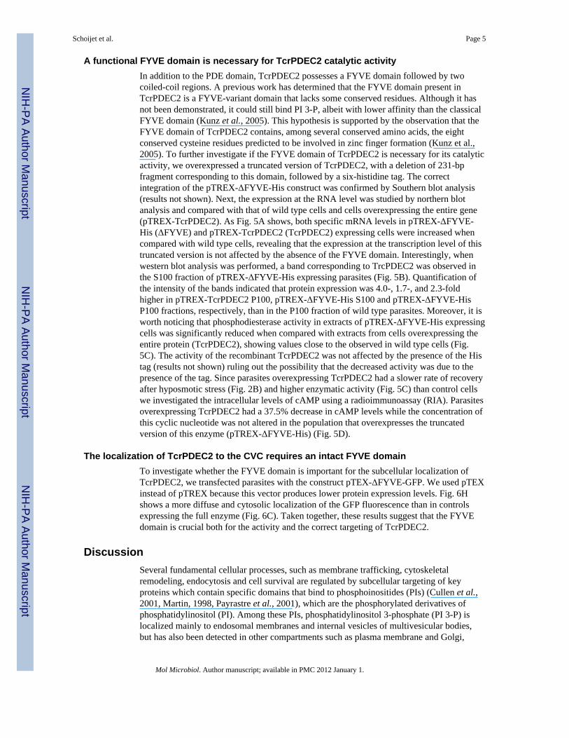
A functional FYVE domain is necessary for TcrPDEC2 catalytic activityIn addition to the PDE domain, TcrPDEC2 possesses a FYVE domain followed by twocoiled-coil regions. A previous work has determined that the FYVE domain present inTcrPDEC2 is a FYVE-variant domain that lacks some conserved residues. Although it hasnot been demonstrated, it could still bind PI 3-P, albeit with lower affinity than the classicalFYVE domain (Kunz et al., 2005). This hypothesis is supported by the observation that theFYVE domain of TcrPDEC2 contains, among several conserved amino acids, the eightconserved cysteine residues predicted to be involved in zinc finger formation (Kunz et al.,2005). To further investigate if the FYVE domain of TcrPDEC2 is necessary for its catalyticactivity, we overexpressed a truncated version of TcrPDEC2, with a deletion of 231-bpfragment corresponding to this domain, followed by a six-histidine tag. The correctintegration of the pTREX-ΔFYVE-His construct was confirmed by Southern blot analysis(results not shown). Next, the expression at the RNA level was studied by northern blotanalysis and compared with that of wild type cells and cells overexpressing the entire gene(pTREX-TcrPDEC2). As Fig. 5A shows, both specific mRNA levels in pTREX-ΔFYVE-His (ΔFYVE) and pTREX-TcrPDEC2 (TcrPDEC2) expressing cells were increased whencompared with wild type cells, revealing that the expression at the transcription level of thistruncated version is not affected by the absence of the FYVE domain. Interestingly, whenwestern blot analysis was performed, a band corresponding to TrcPDEC2 was observed inthe S100 fraction of pTREX-ΔFYVE-His expressing parasites (Fig. 5B). Quantification ofthe intensity of the bands indicated that protein expression was 4.0-, 1.7-, and 2.3-foldhigher in pTREX-TcrPDEC2 P100, pTREX-ΔFYVE-His S100 and pTREX-ΔFYVE-HisP100 fractions, respectively, than in the P100 fraction of wild type parasites. Moreover, it isworth noticing that phosphodiesterase activity in extracts of pTREX-ΔFYVE-His expressingcells was significantly reduced when compared with extracts from cells overexpressing theentire protein (TcrPDEC2), showing values close to the observed in wild type cells (Fig.5C). The activity of the recombinant TcrPDEC2 was not affected by the presence of the Histag (results not shown) ruling out the possibility that the decreased activity was due to thepresence of the tag. Since parasites overexpressing TcrPDEC2 had a slower rate of recoveryafter hyposmotic stress (Fig. 2B) and higher enzymatic activity (Fig. 5C) than control cellswe investigated the intracellular levels of cAMP using a radioimmunoassay (RIA). Parasitesoverexpressing TcrPDEC2 had a 37.5% decrease in cAMP levels while the concentration ofthis cyclic nucleotide was not altered in the population that overexpresses the truncatedversion of this enzyme (pTREX-ΔFYVE-His) (Fig. 5D).
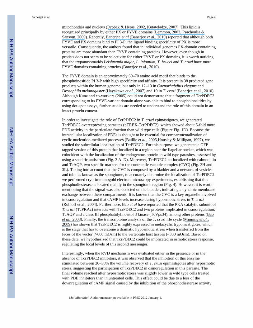
The localization of TcrPDEC2 to the CVC requires an intact FYVE domainTo investigate whether the FYVE domain is important for the subcellular localization ofTcrPDEC2, we transfected parasites with the construct pTEX-ΔFYVE-GFP. We used pTEXinstead of pTREX because this vector produces lower protein expression levels. Fig. 6Hshows a more diffuse and cytosolic localization of the GFP fluorescence than in controlsexpressing the full enzyme (Fig. 6C). Taken together, these results suggest that the FYVEdomain is crucial both for the activity and the correct targeting of TcrPDEC2.
DiscussionSeveral fundamental cellular processes, such as membrane trafficking, cytoskeletalremodeling, endocytosis and cell survival are regulated by subcellular targeting of keyproteins which contain specific domains that bind to phosphoinositides (PIs) (Cullen et al.,2001, Martin, 1998, Payrastre et al., 2001), which are the phosphorylated derivatives ofphosphatidylinositol (PI). Among these PIs, phosphatidylinositol 3-phosphate (PI 3-P) islocalized mainly to endosomal membranes and internal vesicles of multivesicular bodies,but has also been detected in other compartments such as plasma membrane and Golgi,
Schoijet et al. Page 5
Mol Microbiol. Author manuscript; available in PMC 2012 January 1.
NIH
-PA Author Manuscript
NIH
-PA Author Manuscript
NIH
-PA Author Manuscript
mitochondria and nucleus (Drobak & Heras, 2002, Kutateladze, 2007). This lipid isrecognized principally by either PX or FYVE domains (Lemmon, 2003, Psachoulia &Sansom, 2009). Recently, Banerjee et al (Banerjee et al., 2010) reported that although bothFYVE and PX domains bind to PI 3-P, the ligand binding specificity of PX is moreversatile. Consequently, the authors found that in individual genomes PX-domain containingproteins are more abundant than FYVE containing proteins. However, even though inprotists does not seem to be selectivity for either FYVE or PX domains, it is worth noticingthat the trypanosomatids Leishmania major, L. infantum, T. brucei and T. cruzi have moreFYVE domains containing proteins (Banerjee et al., 2010).
The FYVE domain is an approximately 60–70 amino acid motif that binds to thephosphoinositide PI 3-P with high specificity and affinity. It is present in 38 predicted geneproducts within the human genome, but only in 12–13 in Caenorhabditis elegans andDrosophila melanogaster (Hayakawa et al., 2007) and 19 in T. cruzi (Banerjee et al., 2010).Although Kunz and co-workers (2005) could not demonstrate that a fragment of TcrPDEC2corresponding to its FYVE-variant domain alone was able to bind to phosphoinositides byusing dot-spot assays, further studies are needed to understand the role of this domain in anintact protein context.
In order to investigate the role of TcrPDEC2 in T. cruzi epimastigotes, we generatedTcrPDEC2 overexpressing parasites (pTREX-TcrPDEC2), which showed about 5-fold morePDE activity in the particulate fraction than wild type cells (Figure Fig. 1D). Because theintracellular localization of PDEs is thought to be essential for compartmentalization ofcyclic nucleotide-mediated processes (Baillie et al., 2005,Houslay & Milligan, 1997), westudied the subcellular localization of TcrPDEC2. For this purpose, we generated a GFPtagged version of this protein that localized in a region near the flagellar pocket, which wascoincident with the localization of the endogenous protein in wild type parasites, assessed byusing a specific antiserum (Fig. 3 A–D). Moreover, TcrPDEC2 co-localized with calmodulinand TcAQP, two specific markers for the contractile vacuole complex (CVC) (Fig. 3H and3L). Taking into account that the CVC is composed by a bladder and a network of vesiclesand tubules known as the spongiome, to accurately determine the localization of TcrPDEC2we performed cryo-immunogold electron microscopy experiments, establishing that thisphosphodiesterase is located mainly in the spongiome region (Fig. 4). However, it is worthmentioning that the signal was also detected on the bladder, indicating a dynamic membraneexchange between these compartments. It is known that the CVC is a key organelle involvedin osmoregulation and that cAMP levels increase during hyposmotic stress in T. cruzi(Rohloff et al., 2004). Furthermore, Bao et al have reported that the PKA catalytic subunit ofT. cruzi (TcPKAc) interacts with TcrPDEC2 and two proteins implicated in osmoregulation:TcAQP and a class III phosphatidylinositol 3 kinase (TcVps34), among other proteins (Baoet al., 2008). Finally, the transcriptome analysis of the T. cruzi life cycle (Minning et al.,2009) has shown that TcrPDEC2 is highly expressed in metacyclic trypomastigotes, whichis the stage that has to overcome a dramatic hyposmotic stress when transferred from thefeces of the vector (>600 mOsm) to the vertebrate host tissues (~330 mOsm). Based onthese data, we hypothesized that TcrPDEC2 could be implicated in osmotic stress response,regulating the local levels of this second messenger.
Interestingly, when the RVD mechanism was evaluated either in the presence or in theabsence of TcrPDEC2 inhibitors, it was observed that the inhibition of this enzymestimulated between 20–30% the volume recovery of T. cruzi epimastigotes after hyposmoticstress, suggesting the participation of TcrPDEC2 in osmoregulation in this parasite. Thefinal volume reached after hyposmotic stress was slightly lower in wild type cells treatedwith PDE inhibitors than in untreated cells. This effect could be due to a loss of thedownregulation of cAMP signal caused by the inhibition of the phosphodiesterase activity.
Schoijet et al. Page 6
Mol Microbiol. Author manuscript; available in PMC 2012 January 1.
NIH
-PA Author Manuscript
NIH
-PA Author Manuscript
NIH
-PA Author Manuscript
Moreover, the variation of intracellular cAMP levels in wild type, pTREXTcrPDEC2 andpTREX-ΔFYVE-His expressing parasites was directly determined by RIA, showing asignificant decrease only in pTREX-TcrPDEC2 expressing parasites. These resultscorrelated with the RVD results, and with the increased phosphodiesterase activity. We havereported before that an amino acid efflux mechanism accounts for approximately 50% of theregulatory volume decrease in epimastigote, trypomastigote and amastigote stages submittedto hyposmotic stress (Rohloff et al., 2003) while K+ efflux in epimastigotes accounts foronly about 7% of the regulatory volume decrease (Rohloff & Docampo, 2008). Only thedifference (about 40%) would be accounted for by the cyclic AMP-mediated CVCmechanism (Rohloff & Docampo, 2008, Rohloff et al., 2004). However, this mechanismcould also be important under isosmotic conditions. In this regard, the contractile vacuole ofepimastigotes has periodic contractions under isosmotic conditions with a pulsation periodbetween 1 min and 1 min and 15 s (Clark, 1959). It is well established that in many species,such as Paramecium, a CVC is necessary to maintain their water balance under normalenvironmental conditions as well as during dramatic changes in their environment (Allen etal., 2009). We recently reported that chemically validated inhibitors of TcrPDEC could belethal to intracellular stages of the parasite under isosmotic conditions (King-Keller et al.).On the basis of these results we propose that the cAMP signalling pathway is necessary notonly to overcome dramatic changes in osmolarity but also under isosmotic steady-stateconditions. It is tempting to speculate that TcrPDEC would be an important mechanism tocontrol cAMP oscillations responsible for the periodic function of the CVC.
Class I PDEs have three functional domains, a catalytic core, a C-terminal conserveddomain, and an N-terminal domain that is involved in the regulation and subcellularlocalization of each different phosphodiesterase (Conti & Beavo, 2007, Halpin, 2008). Tofurther study the relevance of the localization of TcrPDEC2 at the CVC and the role of itsFYVE domain in determining this targeting, we generated parasites overexpressing atruncated version of TcrPDEC2, which lacks its aforementioned domain (pTREX-ΔFYVE).While parasites overexpressing the full version of TcrPDEC2 (pTREX-TcrPDEC2)significantly enhanced their PDE activity in the membrane fraction, pTREX-ΔFYVEexpressing parasites showed no difference on PDE activity levels as compared with wildtype cells, both in soluble and particulate fractions (Fig. 5C). These results could beexplained by the necessity of an interaction between TcrPDEC2 and a specific membranefor enzymatic activity or because the deletion mutant is not able to acquire the right foldingin the absence of its N-terminal region. Moreover, pTEX-ΔFYVE expressing parasitesshowed a diffuse cytosolic localization (Fig. 6). This result might suggest that the FYVEdomain plays a role in determining the final localization of TcrPDEC2, although thepresence of other signals that may contribute to the correct targeting of this protein cannotbe rule out. In this regard, it has been reported that the Early Endosome Antigen 1 (EEA1)requires both its FYVE domain and the interaction with the small GTPase Rab5 for itstargeting to endosomal membranes (Lawe et al., 2000, Simonsen et al., 1998).
Previous reports have shown that cAMP plays a crucial role in osmoregulation in T. cruzi. Inthe present work we demonstrate that TcrPDEC2 is a key enzyme controlling the localcAMP levels at the CVC and therefore regulating the RVD process in epimastigotes of T.cruzi. Furthermore, we showed that the subcellular localization of TcrPDEC2 requires its N-terminal FYVE domain, which may also be necessary for its catalytic activity.
Experimental proceduresChemicals and Reagents
All radiochemicals used in this work were purchased from Dupont NEN Life ScienceProducts Inc., Boston, MA and restriction endonucleases were from New England Biolabs
Schoijet et al. Page 7
Mol Microbiol. Author manuscript; available in PMC 2012 January 1.
NIH
-PA Author Manuscript
NIH
-PA Author Manuscript
NIH
-PA Author Manuscript
Inc., Beverly, MA. Bacto-tryptose and liver infusion were from Difco Laboratories, Detroit,MI. Goat polyclonal antibody raised against an N-terminal 19 amino acid peptide fromhuman calmodulin was purchased from Santa Cruz Biotechnology (Santa Cruz, CA). Goatanti-rabbit Alexa 488 nm conjugate and rabbit anti-goat Alexa 546 nm conjugate were fromMolecular Probes. All other reagents were purchased from SIGMA Chemical Co., St. Louis,MO.
Cell cultures and extractsT. cruzi epimastigote forms (CL Brener strain) were cultured at 28°C for 7 days in LITmedium (5 g l−1 liver infusion, 5 g l−1 bacto-tryptose, 68 mM NaCl, 5.3 mM KCl, 22 mMNa2PO4, 0.2% (w/v) glucose, 0.002% (w/v) hemin) supplemented with 10% (v/v) newborncalf serum, 100 units ml−1 penicillin and 100 mg l−1 streptomycin. Cell viability wasassessed by direct microscopic examination. For T. cruzi extracts, 108 epimastigotes wereharvested by centrifugation at 1,500 g for 10 min and washed twice with phosphate-bufferedsaline (PBS). Cell pellets were then resuspended in lysis buffer (50 mM HEPES buffer, pH7.3, containing 0.01 mg ml−1 leupeptin, 25 U ml−1 aprotinin, 0.5 mM phenylmethylsulfonylfluoride and 14 mM 2-mercaptoethanol), and lysed by six cycles of freezing in liquid N2 andthawing at 4°C. The total extracts were further centrifuged for 1 h at 100,000 g to obtainparticulate (P100) and soluble (S100) fractions.
Southern, northern and western blot analysesGenomic DNA was purified as described by Pereira et al. (Pereira et al., 2000). Totalcellular RNA was isolated from 108 epimastigotes in the exponential growth phase usingTRIzol reagents, as described by the manufacturer (Invitrogen). Southern and northern blotanalyses were performed as described by Alonso et al. (Alonso et al., 2007). The productswere revealed with a specific 1,070-bp TcrPDEC2 probe obtained by digestion of the 2,772-bp fragment with BamHI and SacII. All probes were labeled with [α-32P]dCTP using thePrime-a-Gene kit (Promega, Madison, WI) following the manufacturer's instructions.
TcrPDEC2 antiserum was developed in rabbit by intraperitoneal immunization with 800 μgof recombinant protein, expressed in E. coli, plus Freund's adjuvant followed by two moresubsequent injections every 15 days without Freund's adjuvant.
For western blot analysis, proteins were solved in 8% (w/v) SDS-polyacrylamide gelelectrophoresis as described by Laemmli (Laemmli, 1970) and electrotransferred to Hybond-C membranes (Amersham Pharmacia Biotech, Piscataway, USA). The membranes wereblocked with 5% (w/v) non-fat milk suspension in TBS-Tween for 2 h. After overnightincubation with 1:1,000 dilution of the rabbit anti-TcrPDEC2 antiserum, detection wascarried out by incubating with a 1:5,000 dilution of a goat anti-rabbit IgG labeled withperoxidase (KPL Inc., Gaithersburg, MA). The latter was developed with the ECL PlusTMWestern Blotting Detection System (NEN Life Science Products Inc., Boston, MA). Theintensity of the bands obtained was quantified using the Gene Tools Software of Syngeneimages capture systems. To control for sample loading, the blot was also probed with a1:2,500 dilution of rabbit anti-T. cruzi hexokinase (Caceres et al., 2003).
TcrPDEC2 constructs and parasite transfectionThe constructs pTREX-TcrPDEC2, pTEX-TcrPDEC2-GFP and pTEX-ΔFYVE-GFP weregenerated by PCR amplification from T. cruzi genomic DNA using the primers PDEC2-Fw-pTrex-XbaI 5′-TCTAGAATGTCGGAGGACGCTGGGCTT-3′ and PDEC2-Rv-pTREX-XhoI 5′-CTCGAGTCAGCACTGCGTCAACAGAGT-3′, the primers PDEC2-Fw-pTEXGFP-BamHI 5′-GGATCCATGTCGGAGGACGCTGGGCTT-3′ and PDEC2-Rv-pTEXGFP-BamHI 5′-GGATCCGTGCACTGCGTCAACAGAGTGGT-3′ and the primers
Schoijet et al. Page 8
Mol Microbiol. Author manuscript; available in PMC 2012 January 1.
NIH
-PA Author Manuscript
NIH
-PA Author Manuscript
NIH
-PA Author Manuscript
PDEC2-Fw-ΔFYVE-pTEXGFP-BamHI 5′-GGATCCATGCGGCGACGCGTGGAGCCGGA-3′ and PDEC2-Rv-pTEXGFP-BamHI 5′-GGATCCGTGCACTGCGTCAACAGAGTGGT-3′ respectively. The PCR products werethen cloned into pGEM-T Easy plasmid and subcloned into the pTREX integration(Vazquez & Levin, 1999) or pTEX-GFP episomal (Wilkinson et al., 2002) expression vectoras applicable. The truncated form of TcrPDEC2 lacking the 231-bp fragment correspondingto the N-terminal FYVE domain and 6xHis tagged at its C-terminus (pTREX-ΔFYVE-His)was amplified from the construct pET22-TcrPDEC2 by using the primers ΔFYVE-Fw-XbaI5′-TCTAGAATGGTGGAGCCGGATGAGGGATCAT-3 ′ and pET-Rv-X baI 5 ′-TCTAGACTTCCTTTCGGGCTTTGTTAGCA-3′. The full length TcrPDEC2 tagged at itsC-terminus with 6xHis (pTREX-TcrPDEC2-His) was amplified in the same way thatpTREX-ΔFYVE-His but using the primers PDEC2-Fw-pTrex-XbaI and pET-Rv-XbaI. BothPCR products were then cloned into pGEM-T Easy plasmid and subcloned into the pTREXexpression vector (for detail of the constructs see Fig. S1). T. cruzi epimastigotes of CLBrener strain were transfected as described previously (Vazquez & Levin, 1999). Parasitestransfected with pTREX-GFP or pTEX-GFP constructs were monitored by fluorescencemicroscopy as selection controls. Stable cell lines were achieved after 60 days of treatmentwith 500 μg ml−1 of G418 (Gibco BRL, Carlsbad, CA) and the transgenic condition wasconfirmed by Southern blot, northern blot and phosphodiesterase activity analyses. Selectionof pTEX transfectants were carried out beginning with 100 μg ml−1 of G418 and once thosecultures were stabilized and growing properly, G418 concentration was increased up to 500μg ml−1.
Regulatory volume decreaseEpimastigotes were collected and washed twice with isotonic chloride buffer (Iso-Cl buffer,137 mM NaCl, 4 mM KCl, 1.5 mM KH2PO4, 8.5 mM Na2PO4, 20 mM HEPES, 11 mMglucose, 1 mM CaCl2, 0.8 mM MgSO4, pH 7.4). The osmolarity of the buffer was adjustedto 300 ± 5 mOsm as verified by an Advanced Instruments 3D3 Osmometer (Norwood, MA).The washed cells were resuspended in Iso-Cl buffer to a cell density of 1 × 10⁸ cells ml−1.The cells were distributed in 96-well plates with 150 μl per well in triplicates andhyposmotic stress was induced by 1:1 dilution of cell suspension with sterile deionizedwater, resulting in a final osmolarity of 150 mOsm. The absorbance changes at 550 nm wererecorded every 20 s for 10 min using a SpectraMax M2e plate reader (Molecular Devices)(Rohloff et al., 2003). To further study the recovery capability after hyposmotic stress, wedefined the rate of recovery as the slope corresponding to the linear region of the curve,comprised between the maximum swelling (2.33 min) and the plateau where the parasitesstabilized their volume (5.66 min).
Membrane integrity and amino acid releaseAfter treatment with the inhibitors etazolate and rolipram, membrane integrity wasdetermined by ethidium bromide exclusion as described by Rohloff et al (Rohloff et al.,2003). Total amino acid analysis of the cell extracts and the supernatant fraction ofepimastigote cells exposed to hypotonic or isotonic buffer were determined as describedbefore (Rohloff et al., 2003).
Immunofluorescence microscopyFor immunofluorescence, cells were fixed in 0.1 M sodium cacodylate buffer (pH 7.2)containing 4% paraformaldehyde and washed twice in Dulbecco's phosphate-bufferedsaline, pH 7.2, adhered to poly-l-lysine-coated coverslips, and permeabilized for 2 min with0.3% Triton X-100. Cells were blocked for 20 min in 50 mM NH4Cl and 3% bovine serumalbumin in phosphate-buffered saline, pH 8.0, and incubated for 1 h with rabbit polyclonalanti-TcrPDEC2 at 1:1,000 and goat polyclonal anti-calmodulin antibody (Santa Cruz
Schoijet et al. Page 9
Mol Microbiol. Author manuscript; available in PMC 2012 January 1.
NIH
-PA Author Manuscript
NIH
-PA Author Manuscript
NIH
-PA Author Manuscript
Biotechnology) at 1:50. Cells were then washed in 3% bovine serum albumin, incubatedwith secondary antibodies anti-rabbit Alexa 546 at 1:500 and anti-goat Alexa 488 conjugateat 1:500 containing 5 μg ml−1 DAPI and mounted with Vectashield (Vector Laboratories).Cells were observed alternative in an Olympus IX-71 fluorescence microscope with aPhotometrix CoolSnapHQ CCD (charge-coupled device) camera driven by Delta Visionsoftware (Applied Precision), an Olympus BX41 fluorescence microscope or axioplan Zeissmicroscope.
Cryo-immunogold electron microscopyFor cryo-immuno electron microscopy, cells were fixed in 0.1 M sodium cacodylate buffer(pH 7.2) containing 4% paraformaldehyde and 0.1% glutaraldehyde for 1 h at 4°C. Cellswere washed twice in sodium cacodylate buffer, pelleted by centrifugation, embedded in25% polyvinylpyrrolidone plus 2.3 M sucrose and frozen in liquid nitrogen. Thin sections offrozen cells were obtained using a Leica EM FC6 cryoultramicrotome. Sections wereblocked for 30 min in 50 mM NH4Cl and 3% bovine serum albumin in phosphate-bufferedsaline, pH 8.0 and incubated with anti-TcrPDEC2 at 1:100 for 1 h followed by goat anti-rabbit 15-nm gold at 1:100.
cAMP phosphodiesterase assaysPDE activity was determinate as described previously (Alonso et al., 2007). The reactionswere performed using 30 μg of P100 or S100 fractions of parasite extracts in the presence of20 mM Tris–HCl, pH 7.5, 5 mM Mg2+ and 50 μM [3H]cAMP. Incubations were carried outat 30°C for 20 min in a total volume of 100 μl. For inhibition studies, rolipram and etazolatewere used at a final concentration of 50 μM. Results represent means ± standard error(SEM) of three experiments (n = 3).
Determination of intracellular cAMPCells were suspended at a concentration of 5 × 10⁷ cells ml−1 and incubated with 1 mMIBMX for 10 min in Dulbecco's PBS supplemented with 5 mM glucose. Then they werecentrifuged, and resuspended in 100 μl of PBS and 400 μl of 50 mM sodium acetate, pH 5.5,preheated to 95°C. These samples were heated at 95°C for 5 min, centrifuged, and thesupernatants collected and stored at −20°C. Samples and standards were acetylated andassayed by RIA using the method described previously (Del Punta et al., 1996, Mondillo etal., 2009).
Supplementary MaterialRefer to Web version on PubMed Central for supplementary material.
AcknowledgmentsWe wish to thank Dr. Sumana Banerjee, from Department of Biological Sciences, Indian Institute of ScienceEducation and Research for helpful information about the GI-numbers of FYVE and PX containing proteins inTrypanosomatids. This work was supported in part by the Consejo Nacional de Investigaciones Científicas yTécnicas (CONICET, Argentina); University of Buenos Aires (Argentina); Agencia Nacional de PromociónCientífica y Tecnológica (Argentina), and the U.S. National Institute of Health (grant AI-68647 to R.D). G.D.A.,M.M.F., H.N.T. and OPP are members of the Scientific Investigator Career of CONICET, Argentina. A.C.S. is afellow from the same institution and was supported in part by a training grant of the Ellison Medical Foundation tothe Center for Tropical and Emerging Global Diseases. WS, KM and LCSM were supported by MCT-CNPq andFAPERJ.
Schoijet et al. Page 10
Mol Microbiol. Author manuscript; available in PMC 2012 January 1.
NIH
-PA Author Manuscript
NIH
-PA Author Manuscript
NIH
-PA Author Manuscript
ReferencesAlonso GD, Schoijet AC, Torres HN, Flawia MM. TcPDE4, a novel membrane-associated cAMP-
specific phosphodiesterase from Trypanosoma cruzi. Mol Biochem Parasitol 2006;145:40–49.[PubMed: 16225937]
Alonso GD, Schoijet AC, Torres HN, Flawia MM. TcrPDEA1, a cAMP-specific phosphodiesterasewith atypical pharmacological properties from Trypanosoma cruzi. Mol Biochem Parasitol2007;152:72–79. [PubMed: 17222469]
Allen, RD.; Tominaga, T.; Naitoh, Y. The contractile vacuole complex and cell volume regulation. In:Evans, DH., editor. Osmotic and Ionic Regulation. CRC Press, Taylor & Francis Group; 2009. p.69-105.
Baillie GS. Compartmentalized signalling: spatial regulation of cAMP by the action ofcompartmentalized phosphodiesterases. Febs J 2009;276:1790–1799. [PubMed: 19243430]
Baillie GS, Scott JD, Houslay MD. Compartmentalisation of phosphodiesterases and protein kinase A:opposites attract. FEBS Lett 2005;579:3264–3270. [PubMed: 15943971]
Banerjee S, Basu S, Sarkar S. Comparative genomics reveals selective distribution and domainorganization of FYVE and PX domain proteins across eukaryotic lineages. BMC genomics2010;11:83. [PubMed: 20122178]
Bao Y, Weiss LM, Braunstein VL, Huang H. Role of protein kinase A in Trypanosoma cruzi. InfectImmun 2008;76:4757–4763. [PubMed: 18694966]
Bender AT, Beavo JA. Cyclic nucleotide phosphodiesterases: molecular regulation to clinical use.Pharmacol Rev 2006;58:488–520. [PubMed: 16968949]
Berriman M, Ghedin E, Hertz-Fowler C, Blandin G, Renauld H, Bartholomeu DC, et al. The genomeof the African trypanosome Trypanosoma brucei. Science 2005;309:416–422. [PubMed: 16020726]
Bhattacharya A, Biswas A, Das PK. Role of a differentially expressed cAMP phosphodiesterase inregulating the induction of resistance against oxidative damage in Leishmania donovani. FreeRadic Biol Med 2009;47:1494–1506. [PubMed: 19733234]
Blatner NR, Stahelin RV, Diraviyam K, Hawkins PT, Hong W, Murray D, Cho W. The molecularbasis of the differential subcellular localization of FYVE domains. J Biol Chem 2004;279:53818–53827. [PubMed: 15452113]
Caceres AJ, Portillo R, Acosta H, Rosales D, Quinones W, Avilan L, et al. Molecular and biochemicalcharacterization of hexokinase from Trypanosoma cruzi. Mol Biochem Parasitol 2003;126:251–262. [PubMed: 12615324]
Callaghan J, Simonsen A, Gaullier JM, Toh BH, Stenmark H. The endosome fusion regulator early-endosomal autoantigen 1 (EEA1) is a dimer. Biochem J 1999;338(Pt 2):539–543. [PubMed:10024533]
Clark TB. Comparative morphology of four genera of trypanosomatidae. J. Protozool 1959;6:227–232.Conti M. Phosphodiesterases and cyclic nucleotide signaling in endocrine cells. Mol Endocrinol
2000;14:1317–1327. [PubMed: 10976911]Conti M, Beavo J. Biochemistry and physiology of cyclic nucleotide phosphodiesterases: essential
components in cyclic nucleotide signaling. Annu Rev Biochem 2007;76:481–511. [PubMed:17376027]
Cooper DM. Compartmentalization of adenylate cyclase and cAMP signalling. Biochem Soc Trans2005;33:1319–1322. [PubMed: 16246108]
Cullen PJ, Cozier GE, Banting G, Mellor H. Modular phosphoinositide-binding domains--their role insignalling and membrane trafficking. Curr Biol 2001;11:R882–893. [PubMed: 11696348]
D'Angelo MA, Sanguineti S, Reece JM, Birnbaumer L, Torres HN, Flawia MM. Identification,characterization and subcellular localization of TcPDE1, a novel cAMP-specificphosphodiesterase from Trypanosoma cruzi. Biochem J 2004;378:63–72. [PubMed: 14556647]
Del Punta K, Charreau EH, Pignataro OP. Nitric oxide inhibits Leydig cell steroidogenesis.Endocrinology 1996;137:5337–5343. [PubMed: 8940355]
Diraviyam K, Stahelin RV, Cho W, Murray D. Computer modeling of the membrane interaction ofFYVE domains. J Mol Biol 2003;328:721–736. [PubMed: 12706728]
Schoijet et al. Page 11
Mol Microbiol. Author manuscript; available in PMC 2012 January 1.
NIH
-PA Author Manuscript
NIH
-PA Author Manuscript
NIH
-PA Author Manuscript
Drobak BK, Heras B. Nuclear phosphoinositides could bring FYVE alive. Trends Plant Sci2002;7:132–138. [PubMed: 11906837]
Dumas JJ, Merithew E, Sudharshan E, Rajamani D, Hayes S, Lawe D, Corvera S, Lambright DG.Multivalent endosome targeting by homodimeric EEA1. Mol Cell 2001;8:947–958. [PubMed:11741531]
El-Sayed NM, Myler PJ, Bartholomeu DC, Nilsson D, Aggarwal G, Tran AN, et al. The genomesequence of Trypanosoma cruzi, etiologic agent of Chagas disease. Science 2005;309:409–415.[PubMed: 16020725]
Flawia MM, Tellez-Inon MT, Torres HN. Signal transduction mechanisms in Trypanosoma cruzi.Parasitol Today 1997;13:30–33. [PubMed: 15275164]
Halpin DM. ABCD of the phosphodiesterase family: interaction and differential activity in COPD. IntJ Chron Obstruct Pulmon Dis 2008;3:543–561. [PubMed: 19281073]
Hayakawa A, Hayes S, Leonard D, Lambright D, Corvera S. Evolutionarily conserved structural andfunctional roles of the FYVE domain. Biochem Soc Symp 2007:95–105. [PubMed: 17233583]
Houslay MD, Adams DR. PDE4 cAMP phosphodiesterases: modular enzymes that orchestratesignalling cross-talk, desensitization and compartmentalization. Biochem J 2003;370:1–18.[PubMed: 12444918]
Houslay MD, Milligan G. Tailoring cAMP-signalling responses through isoform multiplicity. TrendsBiochem Sci 1997;22:217–224. [PubMed: 9204709]
Ivens AC, Peacock CS, Worthey EA, Murphy L, Aggarwal G, Berriman M, et al. The genome of thekinetoplastid parasite, Leishmania major. Science 2005;309:436–442. [PubMed: 16020728]
King-Keller S, Li M, Smith A, Zheng S, Kaur G, Yang X, Wang B, Docampo R. Chemical validationof phosphodiesterase C as a chemotherapeutic target in Trypanosoma cruzi, the etiological agentof Chagas' disease. Antimicrobial agents and chemotherapy 54:3738–3745. [PubMed: 20625148]
Kunz S, Oberholzer M, Seebeck T. A FYVE-containing unusual cyclic nucleotide phosphodiesterasefrom Trypanosoma cruzi. Febs J 2005;272:6412–6422. [PubMed: 16336277]
Kutateladze T, Overduin M. Structural mechanism of endosome docking by the FYVE domain.Science 2001;291:1793–1796. [PubMed: 11230696]
Kutateladze TG. Mechanistic similarities in docking of the FYVE and PX domains tophosphatidylinositol 3-phosphate containing membranes. Prog Lipid Res 2007;46:315–327.[PubMed: 17707914]
Kutateladze TG, Capelluto DG, Ferguson CG, Cheever ML, Kutateladze AG, Prestwich GD, OverduinM. Multivalent mechanism of membrane insertion by the FYVE domain. J Biol Chem2004;279:3050–3057. [PubMed: 14578346]
Laemmli UK. Cleavage of structural proteins during the assembly of the head of bacteriophage T4.Nature 1970;227:680–685. [PubMed: 5432063]
Lawe DC, Patki V, Heller-Harrison R, Lambright D, Corvera S. The FYVE domain of early endosomeantigen 1 is required for both phosphatidylinositol 3-phosphate and Rab5 binding. Critical role ofthis dual interaction for endosomal localization. J Biol Chem 2000;275:3699–3705. [PubMed:10652369]
Laxman S, Beavo JA. Cyclic nucleotide signaling mechanisms in trypanosomes: possible targets fortherapeutic agents. Mol Interv 2007;7:203–215. [PubMed: 17827441]
Lee SA, Eyeson R, Cheever ML, Geng J, Verkhusha VV, Burd C, Overduin M, Kutateladze TG.Targeting of the FYVE domain to endosomal membranes is regulated by a histidine switch. ProcNatl Acad Sci U S A 2005;102:13052–13057. [PubMed: 16141328]
Lemmon MA. Phosphoinositide recognition domains. Traffic 2003;4:201–213. [PubMed: 12694559]Lugnier C. Cyclic nucleotide phosphodiesterase families in intracellular signaling and diabetes. Adv
Exp Med Biol 2001;498:253–261. [PubMed: 11900376]Martin TF. Phosphoinositide lipids as signaling molecules: common themes for signal transduction,
cytoskeletal regulation, and membrane trafficking. Annu Rev Cell Dev Biol 1998;14:231–264.[PubMed: 9891784]
Minning TA, Weatherly DB, Atwood J 3rd, Orlando R, Tarleton RL. The steady-state transcriptome ofthe four major life-cycle stages of Trypanosoma cruzi. BMC genomics 2009;10:370. [PubMed:19664227]
Schoijet et al. Page 12
Mol Microbiol. Author manuscript; available in PMC 2012 January 1.
NIH
-PA Author Manuscript
NIH
-PA Author Manuscript
NIH
-PA Author Manuscript
Mondillo C, Pagotto RM, Piotrkowski B, Reche CG, Patrignani ZJ, Cymeryng CB, Pignataro OP.Involvement of nitric oxide synthase in the mechanism of histamine-induced inhibition of Leydigcell steroidogenesis via histamine receptor subtypes in Sprague-Dawley rats. Biol Reprod2009;80:144–152. [PubMed: 18768916]
Montalvetti A, Rohloff P, Docampo R. A functional aquaporin co-localizes with the vacuolar protonpyrophosphatase to acidocalcisomes and the contractile vacuole complex of Trypanosoma cruzi. JBiol Chem 2004;279:38673–38682. [PubMed: 15252016]
Omori K, Kotera J. Overview of PDEs and their regulation. Circ Res 2007;100:309–327. [PubMed:17307970]
Payrastre B, Missy K, Giuriato S, Bodin S, Plantavid M, Gratacap M. Phosphoinositides: key playersin cell signalling, in time and space. Cell Signal 2001;13:377–387. [PubMed: 11384836]
Pereira CA, Alonso GD, Paveto MC, Iribarren A, Cabanas ML, Torres HN, Flawia MM. Trypanosomacruzi arginine kinase characterization and cloning. A novel energetic pathway in protozoanparasites. J Biol Chem 2000;275:1495–1501. [PubMed: 10625703]
Psachoulia E, Sansom MS. PX- and FYVE-mediated interactions with membranes: simulation studies.Biochemistry 2009;48:5090–5095. [PubMed: 19408958]
Rohloff P, Docampo R. A contractile vacuole complex is involved in osmoregulation in Trypanosomacruzi. Exp Parasitol 2008;118:17–24. [PubMed: 17574552]
Rohloff P, Montalvetti A, Docampo R. Acidocalcisomes and the contractile vacuole complex areinvolved in osmoregulation in Trypanosoma cruzi. J Biol Chem 2004;279:52270–52281.[PubMed: 15466463]
Rohloff P, Rodrigues CO, Docampo R. Regulatory volume decrease in Trypanosoma cruzi involvesamino acid efflux and changes in intracellular calcium. Mol Biochem Parasitol 2003;126:219–230.[PubMed: 12615321]
Scott JD. Compartmentalized cAMP signalling: a personal perspective. Biochem Soc Trans2006;34:465–467. [PubMed: 16856833]
Simonsen A, Lippe R, Christoforidis S, Gaullier JM, Brech A, Callaghan J, et al. EEA1 links PI(3)Kfunction to Rab5 regulation of endosome fusion. Nature 1998;394:494–498. [PubMed: 9697774]
Stahelin RV, Long F, Diraviyam K, Bruzik KS, Murray D, Cho W. Phosphatidylinositol 3-phosphateinduces the membrane penetration of the FYVE domains of Vps27p and Hrs. J Biol Chem2002;277:26379–26388. [PubMed: 12006563]
Vazquez MP, Levin MJ. Functional analysis of the intergenic regions of TcP2beta gene loci allowedthe construction of an improved Trypanosoma cruzi expression vector. Gene 1999;239:217–225.[PubMed: 10548722]
Wilkinson SR, Meyer DJ, Taylor MC, Bromley EV, Miles MA, Kelly JM. The Trypanosoma cruzienzyme TcGPXI is a glycosomal peroxidase and can be linked to trypanothione reduction byglutathione or tryparedoxin. J Biol Chem 2002;277:17062–17071. [PubMed: 11842085]
Zhu Q, Clarke M. Association of calmodulin and an unconventional myosin with the contractilevacuole complex of Dictyostelium discoideum. J Cell Biol 1992;118:347–358. [PubMed: 1629238]
Schoijet et al. Page 13
Mol Microbiol. Author manuscript; available in PMC 2012 January 1.
NIH
-PA Author Manuscript
NIH
-PA Author Manuscript
NIH
-PA Author Manuscript
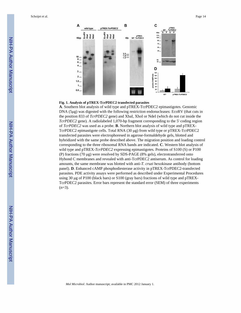
Fig. 1. Analysis of pTREX-TcrPDEC2 transfected parasitesA. Southern blot analysis of wild type and pTREX-TcrPDEC2 epimastigotes. GenomicDNA (5μg) was digested with the following restriction endonucleases: EcoRV (that cuts inthe position 833 of TcrPDEC2 gene) and XbaI, XhoI or NdeI (which do not cut inside theTcrPDEC2 gene). A radiolabeled 1,070-bp fragment corresponding to the 5' coding regionof TcrPDEC2 was used as a probe. B. Northern blot analysis of wild type and pTREX-TcrPDEC2 epimastigote cells. Total RNA (30 μg) from wild type or pTREX-TcrPDEC2transfected parasites were electrophoresed in agarose-formaldehyde gels, blotted andhybridized with the same probe described above. The migration position and loading controlcorresponding to the three ribosomal RNA bands are indicated. C. Western blot analysis ofwild type and pTREX-TcrPDEC2 expressing epimastigotes. Proteins of S100 (S) or P100(P) fractions (70 μg) were resolved by SDS-PAGE (8% gels), electrotransferred ontoHybond C membranes and revealed with anti-TcrPDEC2 antiserum. As control for loadingamounts, the same membrane was blotted with anti-T. cruzi hexokinase antibody (bottompanel). D. Enhanced cAMP phosphodiesterase activity in pTREX-TcrPDEC2-transfectedparasites. PDE activity assays were performed as described under Experimental Proceduresusing 30 μg of P100 (black bars) or S100 (gray bars) fractions of wild type and pTREX-TcrPDEC2 parasites. Error bars represent the standard error (SEM) of three experiments(n=3).
Schoijet et al. Page 14
Mol Microbiol. Author manuscript; available in PMC 2012 January 1.
NIH
-PA Author Manuscript
NIH
-PA Author Manuscript
NIH
-PA Author Manuscript
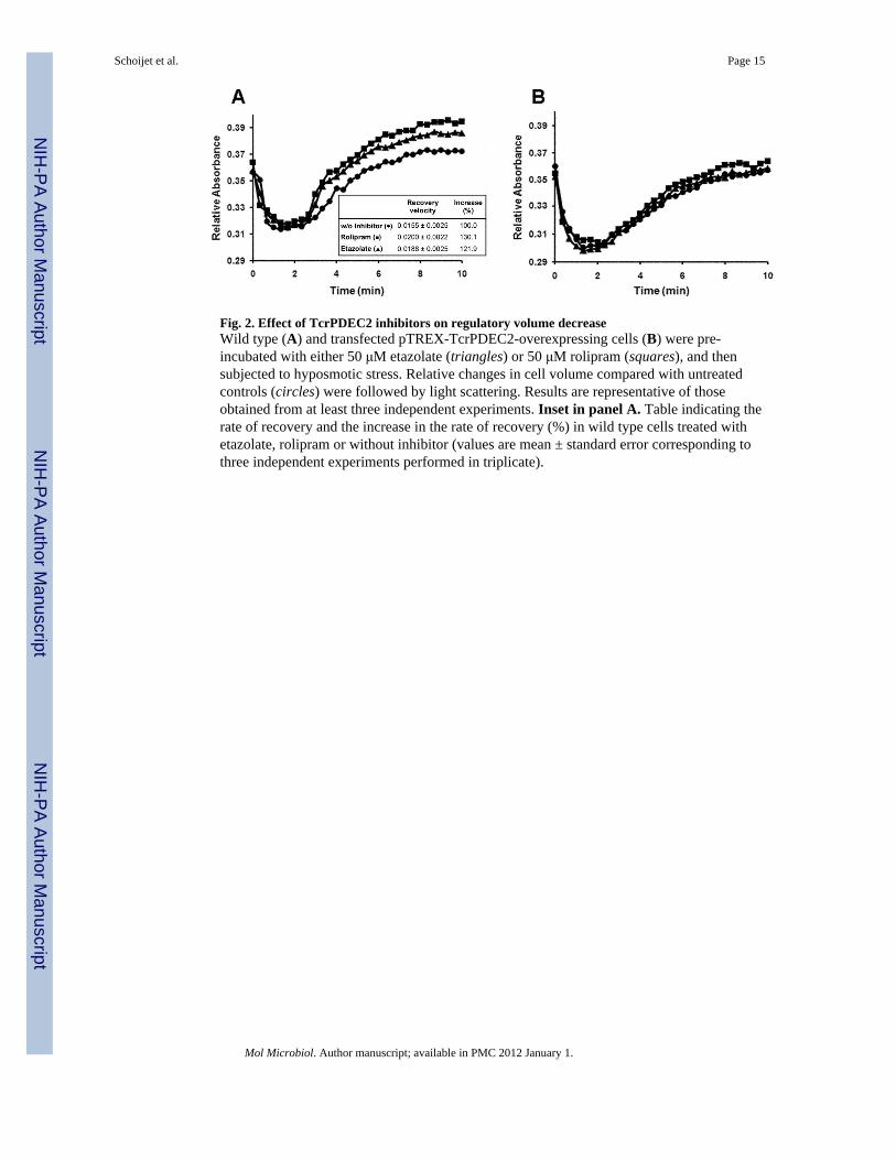
Fig. 2. Effect of TcrPDEC2 inhibitors on regulatory volume decreaseWild type (A) and transfected pTREX-TcrPDEC2-overexpressing cells (B) were pre-incubated with either 50 μM etazolate (triangles) or 50 μM rolipram (squares), and thensubjected to hyposmotic stress. Relative changes in cell volume compared with untreatedcontrols (circles) were followed by light scattering. Results are representative of thoseobtained from at least three independent experiments. Inset in panel A. Table indicating therate of recovery and the increase in the rate of recovery (%) in wild type cells treated withetazolate, rolipram or without inhibitor (values are mean ± standard error corresponding tothree independent experiments performed in triplicate).
Schoijet et al. Page 15
Mol Microbiol. Author manuscript; available in PMC 2012 January 1.
NIH
-PA Author Manuscript
NIH
-PA Author Manuscript
NIH
-PA Author Manuscript
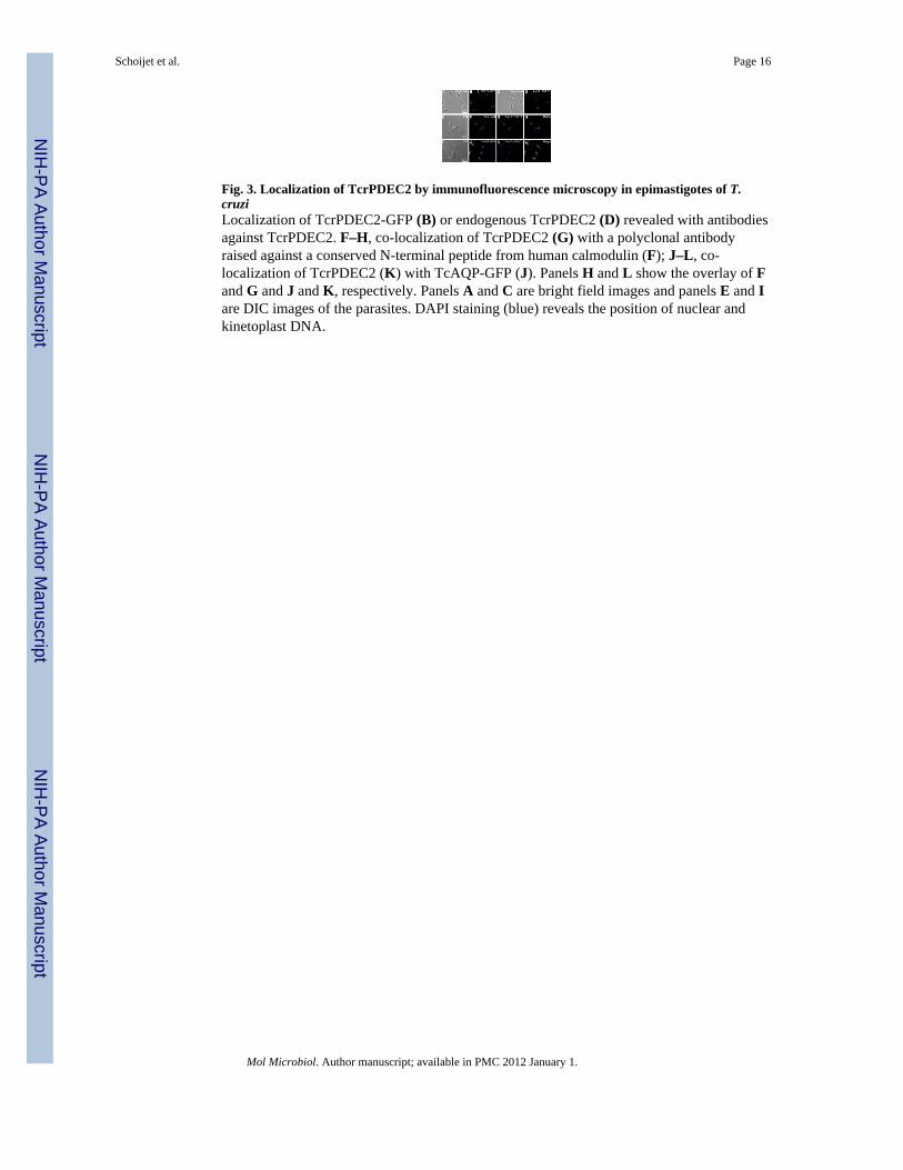
Fig. 3. Localization of TcrPDEC2 by immunofluorescence microscopy in epimastigotes of T.cruziLocalization of TcrPDEC2-GFP (B) or endogenous TcrPDEC2 (D) revealed with antibodiesagainst TcrPDEC2. F–H, co-localization of TcrPDEC2 (G) with a polyclonal antibodyraised against a conserved N-terminal peptide from human calmodulin (F); J–L, co-localization of TcrPDEC2 (K) with TcAQP-GFP (J). Panels H and L show the overlay of Fand G and J and K, respectively. Panels A and C are bright field images and panels E and Iare DIC images of the parasites. DAPI staining (blue) reveals the position of nuclear andkinetoplast DNA.
Schoijet et al. Page 16
Mol Microbiol. Author manuscript; available in PMC 2012 January 1.
NIH
-PA Author Manuscript
NIH
-PA Author Manuscript
NIH
-PA Author Manuscript
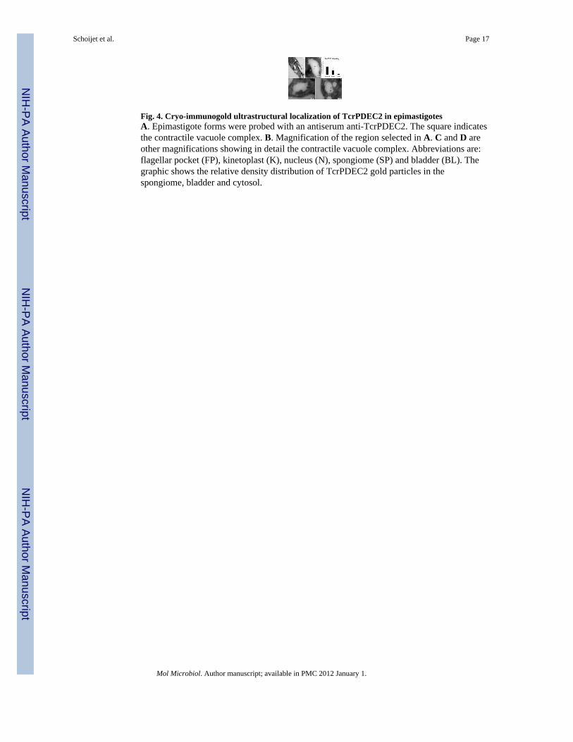
Fig. 4. Cryo-immunogold ultrastructural localization of TcrPDEC2 in epimastigotesA. Epimastigote forms were probed with an antiserum anti-TcrPDEC2. The square indicatesthe contractile vacuole complex. B. Magnification of the region selected in A. C and D areother magnifications showing in detail the contractile vacuole complex. Abbreviations are:flagellar pocket (FP), kinetoplast (K), nucleus (N), spongiome (SP) and bladder (BL). Thegraphic shows the relative density distribution of TcrPDEC2 gold particles in thespongiome, bladder and cytosol.
Schoijet et al. Page 17
Mol Microbiol. Author manuscript; available in PMC 2012 January 1.
NIH
-PA Author Manuscript
NIH
-PA Author Manuscript
NIH
-PA Author Manuscript
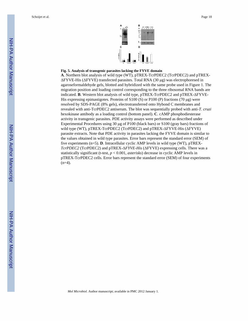
Fig. 5. Analysis of transgenic parasites lacking the FYVE domainA. Northern blot analysis of wild type (WT), pTREX-TcrPDEC2 (TcrPDEC2) and pTREX-ΔFYVE-His (ΔFYVE) transfected parasites. Total RNA (30 μg) was electrophoresed inagaroseformaldehyde gels, blotted and hybridized with the same probe used in Figure 1. Themigration position and loading control corresponding to the three ribosomal RNA bands areindicated. B. Western blot analysis of wild type, pTREX-TcrPDEC2 and pTREX-ΔFYVE-His expressing epimastigotes. Proteins of S100 (S) or P100 (P) fractions (70 μg) wereresolved by SDS-PAGE (8% gels), electrotransferred onto Hybond C membranes andrevealed with anti-TcrPDEC2 antiserum. The blot was sequentially probed with anti-T. cruzihexokinase antibody as a loading control (bottom panel). C. cAMP phosphodiesteraseactivity in transgenic parasites. PDE activity assays were performed as described underExperimental Procedures using 30 μg of P100 (black bars) or S100 (gray bars) fractions ofwild type (WT), pTREX-TcrPDEC2 (TcrPDEC2) and pTREX-ΔFYVE-His (ΔFYVE)parasite extracts. Note that PDE activity in parasites lacking the FYVE domain is similar tothe values obtained in wild type parasites. Error bars represent the standard error (SEM) offive experiments (n=5). D. Intracellular cyclic AMP levels in wild type (WT), pTREX-TcrPDEC2 (TcrPDEC2) and pTREX-ΔFYVE-His (ΔFYVE) expressing cells. There was astatistically significant (t-test, p < 0.001, asterisks) decrease in cyclic AMP levels inpTREX-TcrPDEC2 cells. Error bars represent the standard error (SEM) of four experiments(n=4).
Schoijet et al. Page 18
Mol Microbiol. Author manuscript; available in PMC 2012 January 1.
NIH
-PA Author Manuscript
NIH
-PA Author Manuscript
NIH
-PA Author Manuscript
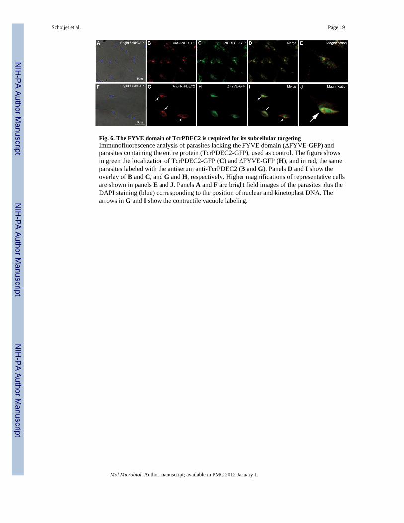
Fig. 6. The FYVE domain of TcrPDEC2 is required for its subcellular targetingImmunofluorescence analysis of parasites lacking the FYVE domain (ΔFYVE-GFP) andparasites containing the entire protein (TcrPDEC2-GFP), used as control. The figure showsin green the localization of TcrPDEC2-GFP (C) and ΔFYVE-GFP (H), and in red, the sameparasites labeled with the antiserum anti-TcrPDEC2 (B and G). Panels D and I show theoverlay of B and C, and G and H, respectively. Higher magnifications of representative cellsare shown in panels E and J. Panels A and F are bright field images of the parasites plus theDAPI staining (blue) corresponding to the position of nuclear and kinetoplast DNA. Thearrows in G and I show the contractile vacuole labeling.
Schoijet et al. Page 19
Mol Microbiol. Author manuscript; available in PMC 2012 January 1.
NIH
-PA Author Manuscript
NIH
-PA Author Manuscript
NIH
-PA Author Manuscript
Copyright © 2022 FDOKUMEN