Phosphodiesterase Type 5 Inhibitors Increase Herceptin Transport and Treatment Efficacy in Mouse...
10
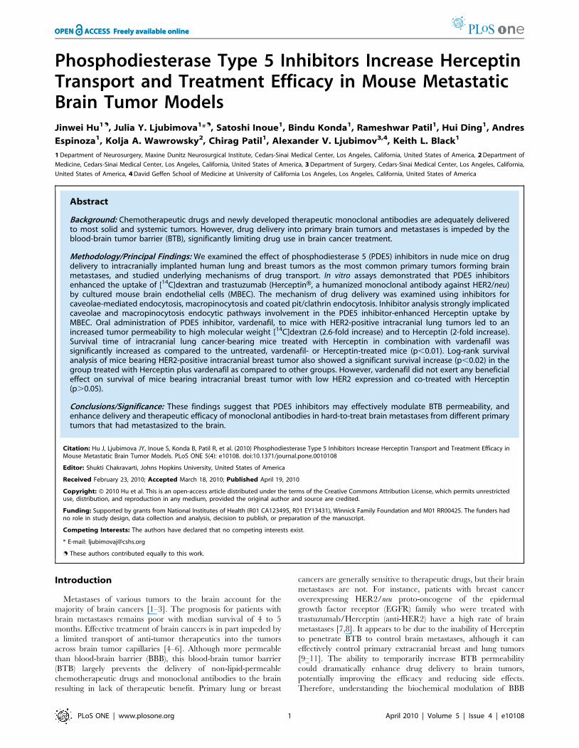
Phosphodiesterase Type 5 Inhibitors Increase Herceptin Transport and Treatment Efficacy in Mouse Metastatic Brain Tumor Models Jinwei Hu 1. , Julia Y. Ljubimova 1 * . , Satoshi Inoue 1 , Bindu Konda 1 , Rameshwar Patil 1 , Hui Ding 1 , Andres Espinoza 1 , Kolja A. Wawrowsky 2 , Chirag Patil 1 , Alexander V. Ljubimov 3,4 , Keith L. Black 1 1 Department of Neurosurgery, Maxine Dunitz Neurosurgical Institute, Cedars-Sinai Medical Center, Los Angeles, California, United States of America, 2 Department of Medicine, Cedars-Sinai Medical Center, Los Angeles, California, United States of America, 3 Department of Surgery, Cedars-Sinai Medical Center, Los Angeles, California, United States of America, 4 David Geffen School of Medicine at University of California Los Angeles, Los Angeles, California, United States of America Abstract Background: Chemotherapeutic drugs and newly developed therapeutic monoclonal antibodies are adequately delivered to most solid and systemic tumors. However, drug delivery into primary brain tumors and metastases is impeded by the blood-brain tumor barrier (BTB), significantly limiting drug use in brain cancer treatment. Methodology/Principal Findings: We examined the effect of phosphodiesterase 5 (PDE5) inhibitors in nude mice on drug delivery to intracranially implanted human lung and breast tumors as the most common primary tumors forming brain metastases, and studied underlying mechanisms of drug transport. In vitro assays demonstrated that PDE5 inhibitors enhanced the uptake of [ 14 C]dextran and trastuzumab (HerceptinH, a humanized monoclonal antibody against HER2/neu) by cultured mouse brain endothelial cells (MBEC). The mechanism of drug delivery was examined using inhibitors for caveolae-mediated endocytosis, macropinocytosis and coated pit/clathrin endocytosis. Inhibitor analysis strongly implicated caveolae and macropinocytosis endocytic pathways involvement in the PDE5 inhibitor-enhanced Herceptin uptake by MBEC. Oral administration of PDE5 inhibitor, vardenafil, to mice with HER2-positive intracranial lung tumors led to an increased tumor permeability to high molecular weight [ 14 C]dextran (2.6-fold increase) and to Herceptin (2-fold increase). Survival time of intracranial lung cancer-bearing mice treated with Herceptin in combination with vardenafil was significantly increased as compared to the untreated, vardenafil- or Herceptin-treated mice (p,0.01). Log-rank survival analysis of mice bearing HER2-positive intracranial breast tumor also showed a significant survival increase (p,0.02) in the group treated with Herceptin plus vardenafil as compared to other groups. However, vardenafil did not exert any beneficial effect on survival of mice bearing intracranial breast tumor with low HER2 expression and co-treated with Herceptin (p.0.05). Conclusions/Significance: These findings suggest that PDE5 inhibitors may effectively modulate BTB permeability, and enhance delivery and therapeutic efficacy of monoclonal antibodies in hard-to-treat brain metastases from different primary tumors that had metastasized to the brain. Citation: Hu J, Ljubimova JY, Inoue S, Konda B, Patil R, et al. (2010) Phosphodiesterase Type 5 Inhibitors Increase Herceptin Transport and Treatment Efficacy in Mouse Metastatic Brain Tumor Models. PLoS ONE 5(4): e10108. doi:10.1371/journal.pone.0010108 Editor: Shukti Chakravarti, Johns Hopkins University, United States of America Received February 23, 2010; Accepted March 18, 2010; Published April 19, 2010 Copyright: ß 2010 Hu et al. This is an open-access article distributed under the terms of the Creative Commons Attribution License, which permits unrestricted use, distribution, and reproduction in any medium, provided the original author and source are credited. Funding: Supported by grants from National Institutes of Health (R01 CA123495, R01 EY13431), Winnick Family Foundation and M01 RR00425. The funders had no role in study design, data collection and analysis, decision to publish, or preparation of the manuscript. Competing Interests: The authors have declared that no competing interests exist. * E-mail: [email protected] . These authors contributed equally to this work. Introduction Metastases of various tumors to the brain account for the majority of brain cancers [1–3]. The prognosis for patients with brain metastases remains poor with median survival of 4 to 5 months. Effective treatment of brain cancers is in part impeded by a limited transport of anti-tumor therapeutics into the tumors across brain tumor capillaries [4–6]. Although more permeable than blood-brain barrier (BBB), this blood-brain tumor barrier (BTB) largely prevents the delivery of non-lipid-permeable chemotherapeutic drugs and monoclonal antibodies to the brain resulting in lack of therapeutic benefit. Primary lung or breast cancers are generally sensitive to therapeutic drugs, but their brain metastases are not. For instance, patients with breast cancer overexpressing HER2/neu proto-oncogene of the epidermal growth factor receptor (EGFR) family who were treated with trastuzumab/Herceptin (anti-HER2) have a high rate of brain metastases [7,8]. It appears to be due to the inability of Herceptin to penetrate BTB to control brain metastases, although it can effectively control primary extracranial breast and lung tumors [9–11]. The ability to temporarily increase BTB permeability could dramatically enhance drug delivery to brain tumors, potentially improving the efficacy and reducing side effects. Therefore, understanding the biochemical modulation of BBB PLoS ONE | www.plosone.org 1 April 2010 | Volume 5 | Issue 4 | e10108
-
Upload
independent -
Category
Documents
-
view
3 -
download
0
Transcript of Phosphodiesterase Type 5 Inhibitors Increase Herceptin Transport and Treatment Efficacy in Mouse...
Phosphodiesterase Type 5 Inhibitors Increase HerceptinTransport and Treatment Efficacy in Mouse MetastaticBrain Tumor ModelsJinwei Hu1., Julia Y. Ljubimova1*., Satoshi Inoue1, Bindu Konda1, Rameshwar Patil1, Hui Ding1, Andres
Espinoza1, Kolja A. Wawrowsky2, Chirag Patil1, Alexander V. Ljubimov3,4, Keith L. Black1
1 Department of Neurosurgery, Maxine Dunitz Neurosurgical Institute, Cedars-Sinai Medical Center, Los Angeles, California, United States of America, 2 Department of
Medicine, Cedars-Sinai Medical Center, Los Angeles, California, United States of America, 3 Department of Surgery, Cedars-Sinai Medical Center, Los Angeles, California,
United States of America, 4 David Geffen School of Medicine at University of California Los Angeles, Los Angeles, California, United States of America
Abstract
Background: Chemotherapeutic drugs and newly developed therapeutic monoclonal antibodies are adequately deliveredto most solid and systemic tumors. However, drug delivery into primary brain tumors and metastases is impeded by theblood-brain tumor barrier (BTB), significantly limiting drug use in brain cancer treatment.
Methodology/Principal Findings: We examined the effect of phosphodiesterase 5 (PDE5) inhibitors in nude mice on drugdelivery to intracranially implanted human lung and breast tumors as the most common primary tumors forming brainmetastases, and studied underlying mechanisms of drug transport. In vitro assays demonstrated that PDE5 inhibitorsenhanced the uptake of [14C]dextran and trastuzumab (HerceptinH, a humanized monoclonal antibody against HER2/neu)by cultured mouse brain endothelial cells (MBEC). The mechanism of drug delivery was examined using inhibitors forcaveolae-mediated endocytosis, macropinocytosis and coated pit/clathrin endocytosis. Inhibitor analysis strongly implicatedcaveolae and macropinocytosis endocytic pathways involvement in the PDE5 inhibitor-enhanced Herceptin uptake byMBEC. Oral administration of PDE5 inhibitor, vardenafil, to mice with HER2-positive intracranial lung tumors led to anincreased tumor permeability to high molecular weight [14C]dextran (2.6-fold increase) and to Herceptin (2-fold increase).Survival time of intracranial lung cancer-bearing mice treated with Herceptin in combination with vardenafil wassignificantly increased as compared to the untreated, vardenafil- or Herceptin-treated mice (p,0.01). Log-rank survivalanalysis of mice bearing HER2-positive intracranial breast tumor also showed a significant survival increase (p,0.02) in thegroup treated with Herceptin plus vardenafil as compared to other groups. However, vardenafil did not exert any beneficialeffect on survival of mice bearing intracranial breast tumor with low HER2 expression and co-treated with Herceptin(p.0.05).
Conclusions/Significance: These findings suggest that PDE5 inhibitors may effectively modulate BTB permeability, andenhance delivery and therapeutic efficacy of monoclonal antibodies in hard-to-treat brain metastases from different primarytumors that had metastasized to the brain.
Citation: Hu J, Ljubimova JY, Inoue S, Konda B, Patil R, et al. (2010) Phosphodiesterase Type 5 Inhibitors Increase Herceptin Transport and Treatment Efficacy inMouse Metastatic Brain Tumor Models. PLoS ONE 5(4): e10108. doi:10.1371/journal.pone.0010108
Editor: Shukti Chakravarti, Johns Hopkins University, United States of America
Received February 23, 2010; Accepted March 18, 2010; Published April 19, 2010
Copyright: � 2010 Hu et al. This is an open-access article distributed under the terms of the Creative Commons Attribution License, which permits unrestricteduse, distribution, and reproduction in any medium, provided the original author and source are credited.
Funding: Supported by grants from National Institutes of Health (R01 CA123495, R01 EY13431), Winnick Family Foundation and M01 RR00425. The funders hadno role in study design, data collection and analysis, decision to publish, or preparation of the manuscript.
Competing Interests: The authors have declared that no competing interests exist.
* E-mail: [email protected]
. These authors contributed equally to this work.
Introduction
Metastases of various tumors to the brain account for the
majority of brain cancers [1–3]. The prognosis for patients with
brain metastases remains poor with median survival of 4 to 5
months. Effective treatment of brain cancers is in part impeded by
a limited transport of anti-tumor therapeutics into the tumors
across brain tumor capillaries [4–6]. Although more permeable
than blood-brain barrier (BBB), this blood-brain tumor barrier
(BTB) largely prevents the delivery of non-lipid-permeable
chemotherapeutic drugs and monoclonal antibodies to the brain
resulting in lack of therapeutic benefit. Primary lung or breast
cancers are generally sensitive to therapeutic drugs, but their brain
metastases are not. For instance, patients with breast cancer
overexpressing HER2/neu proto-oncogene of the epidermal
growth factor receptor (EGFR) family who were treated with
trastuzumab/Herceptin (anti-HER2) have a high rate of brain
metastases [7,8]. It appears to be due to the inability of Herceptin
to penetrate BTB to control brain metastases, although it can
effectively control primary extracranial breast and lung tumors
[9–11]. The ability to temporarily increase BTB permeability
could dramatically enhance drug delivery to brain tumors,
potentially improving the efficacy and reducing side effects.
Therefore, understanding the biochemical modulation of BBB
PLoS ONE | www.plosone.org 1 April 2010 | Volume 5 | Issue 4 | e10108
and BTB is critical for developing novel safe and effective means of
opening barriers for specific drug delivery to brain tumors.
Phosphodiesterase type 5 (PDE5) is a cGMP-specific phospho-
diesterase, which selectively inhibits cGMP degradation [12].
PDE5 inhibitors, sildenafil (ViagraH) and vardenafil (LevitraH), are
widely used, FDA-approved, oral medicines to treat erectile
dysfunction in men [13,14]. The cyclic guanosine monophosphate
(cGMP) is an important intracellular second messenger that has
been implicated in the regulation of vascular tone and permeabil-
ity [15]. Modulation of PDE activity, which can subsequently lead
to the intracellular cGMP accumulation, may result in increased
permeability of capillaries, including microvessels in brain tumors
[16]. Our previous publication showed that orally administered
vardenafil and sildenafil not only selectively increased BTB
permeability through KCa channel in gliosarcoma animal models,
but enhanced anti-tumor efficacy of a chemotherapeutic agent,
doxorubicin [17]. Here we sought to determine whether these
marketed PDE5 inhibitors could increase BTB permeability and
thereby improve the efficacy of monoclonal antibody treatment of
metastatic lung and breast brain tumors positive for HER2/neu
expression.
Materials and Methods
Cell CultureMouse brain endothelial cells (MBEC), CRL-5904 cells (human
non-small cell lung cancer from a brain metastasis), BT-474 cells
(breast cancer cell line) and MDA-MB-435 (breast cancer cell line)
were obtained from the American Type Culture Collection
(ATCC, Manassas, VA), and were maintained in standard tissue
culture conditions. CRL-5904 and BT-474 tumor cell lines are
positive for HER2 expression, while MDA-MB-435 has low
HER2 expression [18].
Tumor ImplantationAll animal studies were approved by Cedars-Sinai Medical
Center Institutional Animal Care and Usage Committee (IACUC)
and were conducted in strict accordance to the IACUC protocol
#2044.
The metastatic brain tumor xenograft models were established
using athymic nude mice (Charles River Laboratories Interna-
tional, Inc., Wilmington, MA). CRL-5904 cells (56104), BT-474
cells (16105) or MDA-MB-435 (56104) in 2 ml of 1.2%
methylcellulose/saline were implanted into the striatum respec-
tively with the coordinates of 2.3 mm lateral to bregma and
3.0 mm deep from dura. To establish breast metastatic brain
tumor model, estrogen pellets (Innovative Research of America,
Sarasota, FL) were implanted subcutaneously in nude mice one
week before intracranial tumor implantation.
In Vivo BTB Permeability StudyFourteen days after tumor implantation, mice were treated with
vardenafil (LevitraH, GlaxoSmithKline, Research Triangle Park,
NC) at an oral dose of 10 mg/kg for 60 min for the maximal effect
[17], bradykinin (1.8 mg/kg; Sigma-Aldrich, St. Louis, MO) for
15 min [19,20], or saline as control. Mice were then subjected to
the regional permeability studies by tail vein injection of
radiotracer [14C]dextran (molecular weight 70,000 D; 50 mCi/
kg; Sigma-Aldrich, St. Louis, MO). The method used to determine
the unilateral transport constant Ki (ml/g/min), the initial rate for
blood-to-brain transfer of radiotracer, has been described in our
previous publication with minor modification [17]. In brief, the Ki
was determined by radiotracer [14C]dextran in the tumor core and
contralateral brain tissue using quantitative autoradiography
(QAR). Quantitative analysis of the regional radioactivity was
performed using ImageJ software (National Institutes of Health,
Bethesda, MD).
Herceptin-Alexa Fluor 680 Uptake in Metastatic BrainTumor Model
Trastuzumab (HerceptinH, Genentech, Inc., San Francisco, CA)
was labeled with an Alexa FluorH680 fluorescent dye (Invitrogen,
Carlsbad, CA) using Xenofluor labeling kit (Caliper, Alameda,
CA). Tumor-bearing mice were treated with oral vardenafil
(10 mg/kg) or saline as control, and then mice were subjected to
the permeability studies by tail vein injection of 5 mg/kg
Herceptin-Alexa Fluor 680. Xenogen IVIS 200 whole animal
fluorescent imager (Caliper Life Sciences, Hopkinton, MA) was
used for assessment of drug distribution and localization in
isoflurane-anesthetized nude mice at different time points (before
drug administration; 1 h, 3 h, 6 h after drug administration). Six
hours after drug administration, mice were anesthetized with i.p.
ketamine and xylazine and subjected to transcardial perfusion to
eliminate the circulating drug in blood vessels. The brains were
harvested to detect the fluorescent signal. Signal intensities in the
tumor were analyzed by Xenogen Living ImageH, Version 2.50
(Caliper Life Sciences, Hopkinton, MA). The brains were snap-
frozen in liquid nitrogen and sectioned on a cryostat (Leica
Microsystems, Mannheim, Germany). Herceptin-Alexa Fluor 680
accumulation in the tumor cells was further studied by confocal
microscopy using Leica confocal laser scanning microscope TCS
SP5 (Leica Microsystems, Germany).
Survival Study in Brain Metastasis Models: Lung andBreast Cancers
For the survival experiments, CRL-5904, BT-474 or MDA-
MB-435 tumor-bearing mice were randomly divided into four
groups treated as follows: (1) control saline; (2) vardenafil (10 mg/
kg, orally, five times per week; dose was based on previous data
[17]); (3) Herceptin (10 mg/kg, intravenously via tail vein, twice
per week; dose was based on our preliminary data and a previous
publication [21]; and (4) Herceptin (10 mg/kg, intravenously,
twice per week) plus vardenafil (10 mg/kg, orally, five times per
week). Mice received their treatments beginning at day 4 after
tumor implantation. CRL-5904 cell implantation and survival
experiment was repeated two times with six animals per group.
The survival data were very similar and were thus combined
together. BT-474 cell line is growing longer and the experiment
duration is estimated at about three months. This experiment was
performed once with a higher number of animals per group, n = 8.
MDA-MB-435 tumor-bearing survival study had 5 to 8 mice per
group. Mice were monitored carefully for clinical signs attributable
to brain tumor growth or until death. The efficacy of therapy was
estimated by the median survival time of the animals.
Western Blot Analysis of Apoptotic Effect on Brain Tissuesof Treated Mice
Normal and tumor mice brain tissues were collected after
treatment with saline, vardenafil, Herceptin, and Herceptin plus
vardenafil. Samples from five tumors in each group were pooled.
Total protein was extracted and concentrations were determined
using a BCA assay kit (Bio-Rad Laboratories, Hercules, CA).
Equal amounts of protein were loaded on 10% SDS-polyacryl-
amide gel and transferred to nitrocellulose membranes. For
apoptosis detection, the membranes were probed with primary
antibodies to cleaved Poly-ADP-Ribose-Polymerase (PARP), an
89-kDa PARP fragment that is considered as a marker of
Levitra in Brain Drug Delivery
PLoS ONE | www.plosone.org 2 April 2010 | Volume 5 | Issue 4 | e10108
apoptosis, and to an internal control, glyceraldehyde 3-phosphate
dehydrogenase (GAPDH; BD Pharmingen, San Diego, CA),
respectively. Horseradish peroxidase-conjugated secondary anti-
bodies were used for detection followed by enhanced chemilumi-
nescence development (Bio-Rad Laboratories, Hercules, CA).
In Vitro BBB Permeability ModelTo mimic BBB condition in vitro, transwell system (Corning,
Inc., Corning, NY) was used for cellular drug transport study.
MBEC were seeded into the inserts of transwell system at 26104/
well and cultured for 2 to 3 days until confluence. Washed cells
were incubated at 37uC for 30 min in culture media without
serum as control or in media with 10 mg/ml vardenafil. Then,
these media were removed and replaced with [14C]dextran at
0.2 mCi/ml in the inserts. After 5 or 30 min incubation, the media
from the low chambers were collected and subjected to
radioactivity measuring by liquid scintillation counting using a
Beckman LS 6000 Scintillation Counter (Beckman Coulter,
Fullerton, CA). Triplicate wells were run for each group.
Drug Uptake by Endothelial CellsThe same numbers of MBEC (26104/well) were seeded in flat
bottom 96-well plates and cultured until confluence. To verify
which endocytosis pathways were involved in PDE5 inhibitor-
induced drug uptake, several inhibitors were examined by first
incubating cultured MBEC for 30 min in serum-free medium as
follows: Filipin (1 to 5 mg/ml) and methyl-b-cyclodextrin (2.5 to
10 mM) to inhibit caveolae-mediated endocytosis; amiloride (25 to
50 mm) to inhibit macropinocytosis; chlorpromazine (2.5 to 10 mg/
ml) and phenylarsine oxide (7.5 to 30 mM) to inhibit coated pit/
clathrin endocytosis pathway [22,23]. All inhibitors were pur-
chased from Sigma-Aldrich (St. Louis, MO). The medium was
replaced with the one containing pathway inhibitors and PDE5
inhibitors (50 mg/ml sildenafil, ViagraH, Pfizer, NY; or 10 mg/ml
vardenafil), and cells were incubated at 37uC for 30 min. Then the
medium was removed and replaced with Herceptin-Alexa Fluor
680 (25 mg/ml) for 0.5 or 3 hrs. After washing, cells were lysed in
100 ml of 1% (w/v) Triton X-100. Two repeats of this experiment
were run, with similar results. Uptake data are expressed as
percent of control. Uptake of labeled Herceptin in cultures without
endocytosis inhibitors was also tested. A standard curve was
obtained in untreated cells using different dilutions of Herceptin-
Alexa Fluor 680 in lysis buffer. Fluorescent signals were measured
using excitation at 680 nm and emission at 720 nm in a
microplate reader (Molecular Devices, Sunnyvale, CA). Triplicate
wells were run for each group. Data are expressed as mg Herceptin
per one ml of lysis buffer.
Herceptin-Alexa Fluor 680 Uptake by Cultured MBECMBEC were seeded in Lab-TekTM chamber slides (Thermo
Fisher Scientific, Rochester, NY). Washed cells were pretreated
with filipin (5 mg/ml) for 30 min in serum-free media followed by
incubation at 37uC for 30 min in culture media as control or
media with a PDE 5 inhibitor (10 mg/ml vardenafil). Then, this
medium was removed and replaced with the one containing
Herceptin-Alexa Fluor 680 (25 mg/ml) for 3 hrs. Cells were
washed with PBS, fixed with 4% paraformaldehyde for 20 min,
and incubated with anti-caveolin-1 (1:100; BD Biosciences, San
Jose, CA) antibodies at 4uC overnight. The signals were detected
with Texas Red-conjugated secondary antibodies (1:200; Jackson
ImmunoResearch, West Grove, PA). To detect HER2 expression
on cells, they were stained with anti-HER2 (1:100; Upstate,
Temecula, CA) by indirect immunofluorescence. The cells were
counterstained with 49, 6-diamidino-2-phenylindole (DAPI; Vec-
tor Laboratories, Burlingame, CA) and cover-slipped. The slides
were examined under a Leica confocal microscope. Negative
control experiments included omission of primary antibodies and
were routinely performed.
StatisticsStatistical analyses were performed using the Student’s t-test
(Prism4, GraphPad Software, San Diego, CA). Animal survival
was analyzed by the Kaplan-Meier method (log-rank test). Data
were expressed as mean 6 standard error of mean (SEM). p,0.05
was considered significant.
Results
Vardenafil Effects on Cell Membrane Permeability in Vitroand in Vivo
In the in vitro setting, a Corning transwell system was used for
cellular drug transport study as a model of BBB. Mouse brain
endothelial cells were seeded into the transwell inserts for the
examination of transcytosis of [14C]dextran (MW 70,000 D this
molecular weight was selected to compare with big molecular
drugs, therapeutic monoclonal antibody in our case). At different
intervals after dextran addition, the medium in the low chamber
was collected to detect the transported dextran by liquid
scintillation counting. The results showed that vardenafil
significantly increased the transcytosis of [14C]dextran through
MBEC compared to medium-treated cells (Figure 1a) both at
5 min (110.6767.37 vs. 70.6763.06 cpm; p,0.01) and 30 min
(136.3364.54 vs. 64.00610.58 cpm; p,0.01).
In the in vivo study, vardenafil (n = 5) was given at an oral dose of
10 mg/kg for the maximal effect according to previous publication
[17]. Its effects on the initial blood to brain or blood to tumor
transport, Ki, were studied and compared with that of bradykinin
(positive control), which increases BTB transport [17,24], after
infusing with a radiotracer-high molecular weight [14C]dextran.
Vardenafil treatment resulted in a 2.6-fold increase in BTB
permeability (38.49614.23 nCi/g vs. 15.0966.09 nCi/g in saline-
treated controls; p,0.01). Consistent with previous publications
[17,24], administration of bradykinin (1.8 mg/kg, n = 6) similarly
increased the Ki in the brain tumors (40.06611.13 vs.
15.0966.09 nCi/g; p,0.01) as compared to the saline-treated
controls (n = 5). Transport of [14C]dextran in normal brain was
not affected by the treatments (Figure 1b).
To examine in vitro uptake of Herceptin-Alexa Fluor 680,
endothelial cells were incubated with labeled drug with or without
preincubation with vardenafil. Vardenafil-treated MBEC showed
significantly increased Herceptin-Alexa Fluor 680 uptake
(Figure 2a) compared to medium only control (1.1960.03
vs. 0.6560.07 mg/ml, p,0.01 at 30 min; 1.7960.07 vs.
0.9560.05 mg/ml, p,0.01 at 3 hrs). It should be noted that
MBEC did not express surface HER2 in contrast to CRL-5904
and BT-474 HER2-postive tumor cell lines used (Figure 2b).
Mechanisms of Antibody Delivery Through EndothelialSystem in Vitro
To explore the underlying mechanisms of PDE5 inhibitors-
induced BTB permeability increase, we have examined the role of
three major pathways for substance endocytosis, such as clathrin-
and caveolae-mediated transport, as well as macropinocytosis. We
further investigated whether increased transport induced by PDE5
inhibitors was associated with any of these major mechanisms
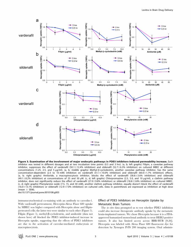
(Figures 3, 4). Caveolae-mediated endocytosis was inhibited by
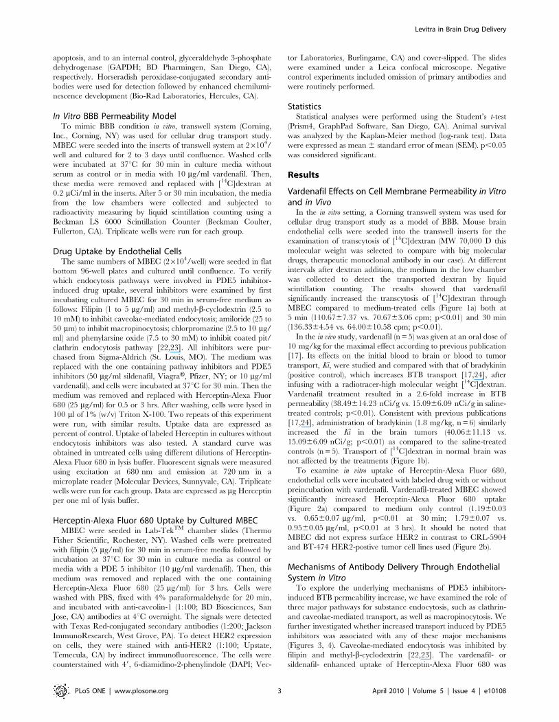
filipin and methyl-b-cyclodextrin [22,23]. The vardenafil- or
sildenafil- enhanced uptake of Herceptin-Alexa Fluor 680 was
Levitra in Brain Drug Delivery
PLoS ONE | www.plosone.org 3 April 2010 | Volume 5 | Issue 4 | e10108

inhibited by 50-60% by the pretreatment of the cells with filipin or
methyl-b-cyclodextrin (Figure 3 a, b; left and middle graphs;
Figure 4, p,0.001 vs. vardenafil/sildenafil-stimulated control).
Because vardenafil and sildenafil increased Herceptin uptake in
vitro about two-fold (Figure 2a), caveolae pathway inhibitors agents
essentially brought Herceptin uptake down to control levels
without PDE5 inhibitors. Next, we examined the role of
macropinocytosis by using its inhibitor, amiloride [22,25]. The
uptake of Herceptin-Alexa Fluor 680 increased by vardenafil or
sildenafil was also similarly inhibited by amiloride pretreatment
(Figure 3 a, b; right graphs; Figure 4; p,0.001 vs. vardenafil/
sildenafil-stimulated control). However, clathrin pathway inhibi-
tors, chlorpromazine and phenylarsine oxide, did not significantly
interfere with the effects of vardenafil or sildenafil (Figure 3 c, d;
Figure 4; p.0.05 vs. vardenafil/sildenafil-stimulated control),
possibly due to lack of HER2 receptor expression on MBEC
(Figure 2b). Therefore, stimulation of caveolae and macropinocy-
tosis pathways may play a major role in PDE5 inhibitor effect on
Herceptin uptake.
To further examine the effect of PDE5 inhibitors on drug
uptake, we detected Herceptin-Alexa Fluor 680 in cultured MBEC
using confocal microscopy. Caveolae expression was monitored by
Figure 1. PDE5 inhibitor, vardenafil, increases permeability in an in vitro BBB model and in metastatic brain tumors in mice. (a)Vardenafil significantly increases the uptake of [14C]dextran (MW 70,000 D) by MBEC compared to medium treated-cells at 5 min (p,0.01) and 30minutes (p,0.01) respectively in an in vitro BBB model. (b) Effects of oral PDE5 inhibitor on the rate of radioactive dextran transport, Ki, into themetastatic brain tumors. Vardenafil (10 mg/kg) was administered orally followed by permeability determination as in a previous publication [17].Intravenous administration of short-term vascular modulator, bradykinin (1.8 mg/kg) served as a positive control [31,43]. Oral administration ofvardenafil (n = 5) significantly increases Ki in the tumor center compared with saline control (n = 5). Intravenous infusion of bradykinin (n = 6) alsosignificantly increases Ki in the tumor core compared with control. No permeability increase in the contralateral normal brains. **, p,0.01.doi:10.1371/journal.pone.0010108.g001
Figure 2. PDE5 inhibitor induces permeability increase of therapeutic antibody, Herceptin. (a) PDE5 inhibitor vardenafil significantlyincreases Herceptin-Alexa Fluor 680 uptake by cultured MBEC compared to medium treated-cells at 0.5 and 3 hrs (close to 2-fold; vardenafil vs.control). **, p,0.01. (b) HER2 expression on different cell lines. BT-474 breast cells have the highest HER2 expression, followed by CRL-5904 cells.HER2 expression on MBEC is not detected. Lack of HER2 on MBEC suggests that Herceptin could not be internalized by receptor-mediated clathrin-dependent endocytosis and may account for absence of effect observed with the corresponding inhibitors, chlorpromazine and phenylarsine oxide(Figure 4). Nuclei are counterstained with DAPI. Scale bar = 25 mm.doi:10.1371/journal.pone.0010108.g002
Levitra in Brain Drug Delivery
PLoS ONE | www.plosone.org 4 April 2010 | Volume 5 | Issue 4 | e10108
immunocytochemical co-staining with an antibody to caveolin-1.
With vardenafil pretreatment, Herceptin-Alexa Fluor 680 uptake
by MBEC was higher compared with Herceptin alone and filipin-
pretreated cells; the latter two were similar to each other (Figure 5).
Filipin (Figure 5), methyl-b-cyclodextrin, and amiloride (data not
shown here) all blocked the PDE5 inhibitor-induced increase in
Herceptin uptake, suggesting that the effects of PDE5 inhibitors
are due to the activation of caveolae-mediated endocytosis or
macropinocytosis.
Effect of PDE5 Inhibitors on Herceptin Uptake byMetastatic Brain Tumors
The in vitro data prompted us to test whether PDE5 inhibition
could also increase therapeutic antibody uptake by the metastatic
brain-implanted tumors. We chose Herceptin because it is a FDA-
approved humanized monoclonal antibody to treat HER2-positive
tumors. It also has limited access across BBB/BTB [9,26].
Herceptin was labeled with Alexa Fluor 680 fluorescent dye for
detection by Xenogen IVIS 200 imaging system. Oral adminis-
Figure 3. Examination of the involvement of major endocytic pathways in PDE5 inhibitors-induced permeability increase. Eachinhibitor was tested in different dosages and at two incubation time points (0.5 and 3 hrs). (a, b; left graphs) Filipin, a caveolae pathwayinhibitor, suppresses the effect of vardenafil (50.1612.4% inhibition) and sildenafil (52.766.3% inhibition) on cultured MBEC at differentconcentrations (1.25, 2.5, and 5 mg/ml). (a, b; middle graphs) Methyl-b-cyclodextrin, another caveolae pathway inhibitor, has the sameconcentration-dependent (2.5 to 10 mM) inhibition on vardenafil (51.1610.0% inhibition) and sildenafil (60.061.7% inhibition) effects.(a, b; right graphs) Amiloride, a macropinocytosis inhibitor, blocks the effect of vardenafil (58.863.6% inhibition) and sildenafil(49.168.2% inhibition) at concentrations of 25 and 50 mM. (c, d; left graphs) Chlorpromazine (2.5, 5.0, and 10 mg/ml), a clathrin pathwayinhibitor, does not significantly reduce the effect of vardenafil (27.069.8% inhibition) or sildenafil (18.863.8% inhibition) on cultured MBEC.(c, d; right graphs) Phenylarsine oxide (7.5, 15, and 30 mM), another clathrin pathway inhibitor, equally doesn’t block the effect of vardenafil(16.0613.1% inhibition) or sildenafil (12.967.0% inhibition) on cultured cells. Data in parentheses are expressed as inhibition at high dose(mean 6 SEM).doi:10.1371/journal.pone.0010108.g003
Levitra in Brain Drug Delivery
PLoS ONE | www.plosone.org 5 April 2010 | Volume 5 | Issue 4 | e10108
tration of vardenafil (10 mg/kg, n = 5) resulted in significant 2-fold
increase of Herceptin-Alexa Fluor 680 transport in brain tumor
compared with saline treated-mice (Figure 6 a-c; n = 5; p,0.05).
Herceptin-Alexa Fluor 680 signal was not detected in the
contralateral normal brains (Figure 6a, b). The accumulation of
Herceptin was further observed on brain tumor cryostat sections
by confocal microscopy. Sections of vardenafil-treated tumors had
markedly higher drug accumulation compared with Herceptin
alone (Figure 6d) or saline-treated controls. No fluorescence signal
was detected in the contralateral normal brain tissue.
Treatment of Lung and Breast Intracranial TumorsTo determine if the permeability increase in metastatic brain
tumors by the PDE5 inhibitors can be translated into improved
efficacy of tumor therapy, Herceptin was administered to mice
with brain-implanted lung metastatic CRL-5904 and breast
cancer BT-474 tumors positive for HER2/neu. CRL-5904 bearing
mice were treated with Herceptin (10 mg/kg, twice per week,
intravenously) [21] beginning at day 4 after tumor implantation,
with oral administration of vardenafil (10 mg/kg, 5 times per
week). Log-rank analysis of the Kaplan–Meier survival curves
showed a significant increase (p,0.01) in survival of mice treated
with Herceptin plus vardenafil as compared to the saline-treated,
vardenafil alone- or Herceptin alone-treated mice (Figure 7a). The
mean survival time for mice treated with Herceptin plus vardenafil
combination increased by about 30% and was significantly longer
(3568 days) compared to those of saline- (2262 days), Herceptin
alone- (2562 days), and vardenafil alone-treated mice (2562
days). In mice bearing intracranial BT-474 breast tumors, log-rank
analysis of the Kaplan–Meier survival curves also showed a
significant survival increase (p,0.02) in the group treated with
Herceptin plus vardenafil as compared to the saline-treated,
Herceptin alone- or vardenafil alone-treated mice (Figure 7b). The
mean survival time for mice treated with Herceptin plus vardenafil
was significantly longer (72618 days) compared to those of saline-
(5767 days), Herceptin alone- (5969 days), and vardenafil alone-
treated mice (50611 days). Survival with Herceptin plus
vardenafil was thus about 20% longer than with Herceptin alone.
However, there was no beneficial effect of vardenafil on Herceptin
treatment of mice bearing low HER2 expressing MDA-MB-435
tumor (p.0.05; Figure 7c).
Herceptin binds to the extracellular domain of HER2 and
inhibits proliferation and survival of HER2-dependent tumors by
promoting DNA fragmentation associated with apoptotic cell
death [27]. In addition to monitoring survival, we also detected the
apoptotic effect on the brain tissue after the treatments. The brain
tumor tissues and contralateral normal brain tissues were
collected, pooled at five per group, and subjected to apoptotic
assay by Western blot for cleaved 89-kDa fragment of poly-ADP-
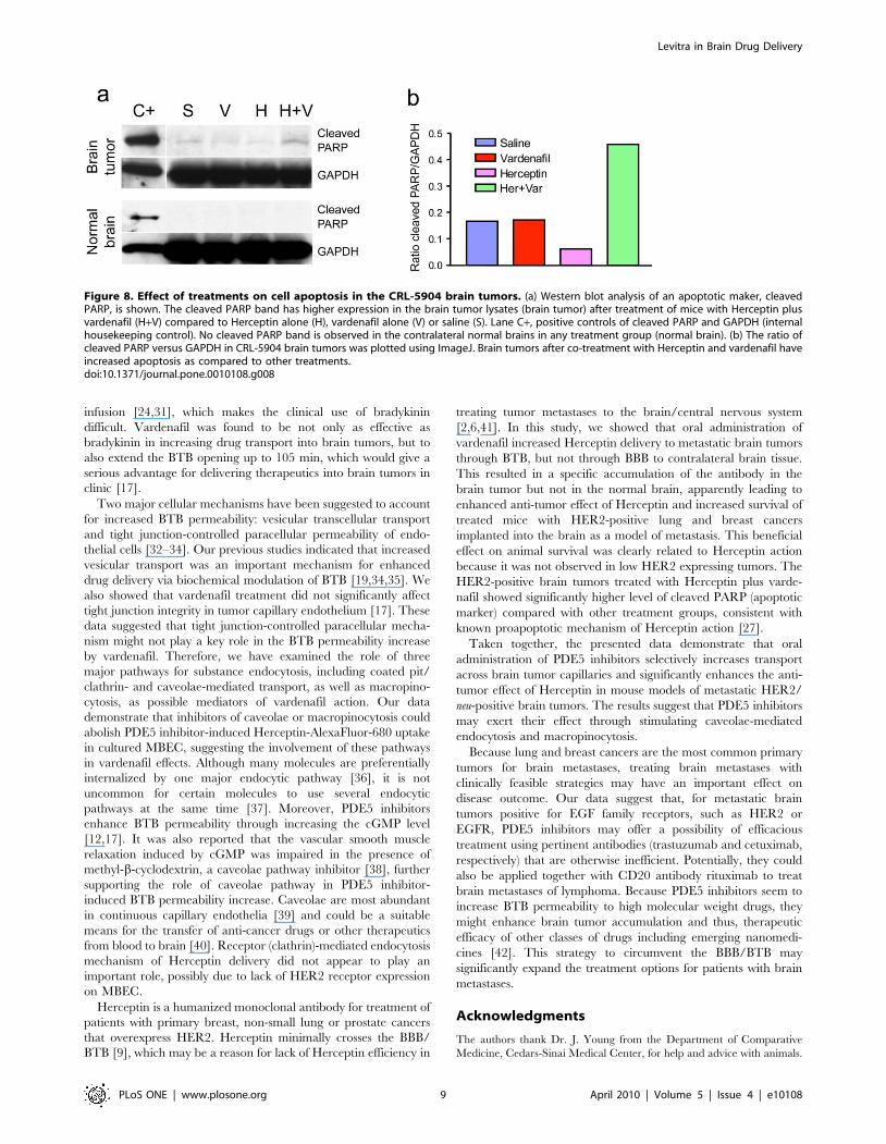
ribose-polymerase (PARP) as a marker of apoptosis. CRL-5904
brain tumors treated by Herceptin plus vardenafil showed about
two-fold higher level of cleaved PARP compared with untreated,
vardenafil alone- or Herceptin alone-treated brain tumors
(Figure 8a, top). The ratio of cleaved PARP relative to
housekeeping protein GAPDH showed marked increase in the
Herceptin plus vardenafil group compared to all other groups
(Figure 8b). We did not detect any cleaved PARP in the
contralateral normal brain tissues with different treatments
(Figure 8a, bottom). The enhanced apoptosis in the Herceptin
plus vardenafil treated brain tumor tissue indicated that vardenafil
Figure 4. Effect of inhibitors of three major endocytic pathways on reversing vardenafil and sildenafil enhancement of Herceptinuptake. Bar graph represents high dosage results in Fig. 3, averaged for both 0.5 and 3 hrs of incubation (mean 6 SEM). Control, Herceptin uptakeby cells incubated with PDE5 inhibitors (data on vardenafil and sildenafil were similar, so they were combined and taken as 100%). Both inhibitors ofcaveolae endocytic pathway (filipin and methyl-b-cyclodextrin) significantly reduced PDE5 inhibitor effect compared to vardenafil/sildenafil-stimulated control, bringing Herceptin uptake to baseline level. The same was true for macropinocytosis inhibitor, amiloride. The effect of eitherinhibitor of coated pit/clathrin pathway (chlorpromazine or phenylarsine oxide) on Herceptin uptake was non-significant (N.S.). V, vardenafil; S,sildenafil; MPC, macropinocytosis. ***, p,0.001.doi:10.1371/journal.pone.0010108.g004
Levitra in Brain Drug Delivery
PLoS ONE | www.plosone.org 6 April 2010 | Volume 5 | Issue 4 | e10108
increased uptake and efficacy of Herceptin in the brain tumors. In
BT-474 tumors we did not detect a significant increase in cleaved
PARP in vardenafil plus Herceptin treatment compared with
other groups, maybe due to the longer survival time (up to 3
months) in this animal model.
Discussion
In this study, we sought to determine whether cGMP-specific
PDE5 inhibitors could increase the rate of delivery of large
macromolecular drugs exemplified by therapeutic antibodies
across the BTB in order to improve efficacy of drug therapy for
metastatic brain tumors. We have also examined possible
mechanisms underlying PDE5 inhibitor action, related to changes
in vesicular transport. It is shown here that oral administration of
PDE5 inhibitor vardenafil selectively increased the rate of brain
tumor accumulation of intravenously administered [14C] high
molecular weight dextran and a monoclonal antibody trastuzu-
mab/Herceptin against HER/neu. Importantly, Herceptin in
combination with vardenafil significantly improved the survival
of metastatic brain tumor-bearing mice with increased tumor
apoptosis. Inhibitors of caveolae endocytosis and macropinocytosis
pathway could block PDE5 inhibitor-induced Herceptin uptake by
cultured mouse brain endothelial cells, suggesting that stimulation
of these pathways was a likely mechanism of PDE5 inhibitor
action. Collectively, these findings suggest that PDE5 may serve as
an effective target for pharmacological modulation of BTB
permeability to enhance delivery and therapeutic effect of
otherwise inefficient drugs in the metastatic brain tumors.
Vardenafil and sildenafil are oral drugs that are currently used
to treat erectile dysfunction in men. They selectively inhibit the
activity of cGMP-specific PDE5, and subsequently increase the
cGMP level [12]. Our previous study showed that oral
administration of vardenafil and sildenafil selectively increased
BTB permeability (1.8-fold and 2.7-fold, respectively) compared
with control groups. Transport across the BTB into tumor tissues
reached the maximum at 60 to 75 min after administration of
sildenafil and vardenafil [17]. In the present study, we chose the
optimal dose of vardenafil at 10 mg/kg to treat metastatic brain
tumor-bearing mice. Orally administered vardenafil selectively
increased permeability of metastatic brain tumors, but not the
contralateral normal brain to high molecular weight [14C]
dextran. Bradykinin that is known to transiently open up BTB
was included in this study as a positive control. As expected,
bradykinin resulted in a significant increase in BTB permeability,
which is consistent with our previous observations [16,28,29].
However, bradykinin has a short half-life [30], and its effect on
transport into tumors is diminished within 15–20 min after
Figure 5. Co-localization of Herceptin-Alexa Fluor 680 with caveolae in cultured MBEC. Cultured cells have low uptake of Herceptin (Her),which is markedly increased when co-administered with vardenafil (Var). Note pronounced co-localization of Herceptin with caveolin-1. Theenhancing effect of vardenafil is virtually abolished by filipin (Fil). Vardenafil was used at 10 mg/ml. Nuclei were counterstained with DAPI. Scale bar= 25 mm.doi:10.1371/journal.pone.0010108.g005
Levitra in Brain Drug Delivery
PLoS ONE | www.plosone.org 7 April 2010 | Volume 5 | Issue 4 | e10108
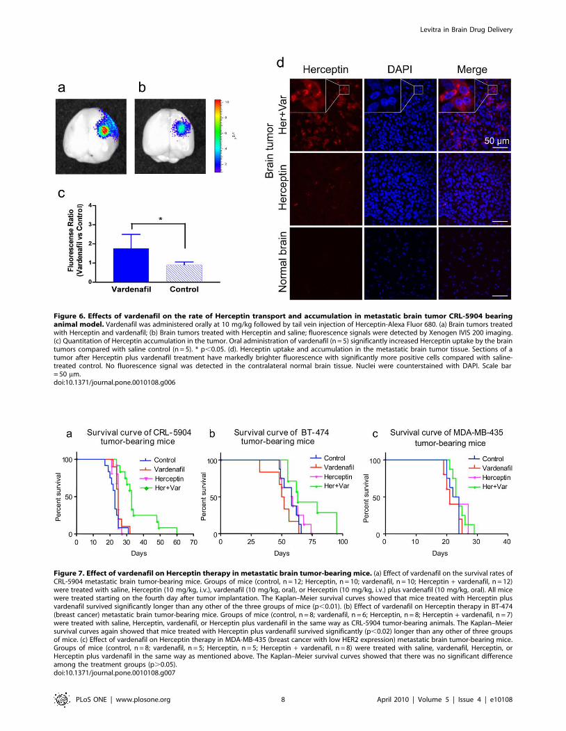
Figure 6. Effects of vardenafil on the rate of Herceptin transport and accumulation in metastatic brain tumor CRL-5904 bearinganimal model. Vardenafil was administered orally at 10 mg/kg followed by tail vein injection of Herceptin-Alexa Fluor 680. (a) Brain tumors treatedwith Herceptin and vardenafil; (b) Brain tumors treated with Herceptin and saline; fluorescence signals were detected by Xenogen IVIS 200 imaging.(c) Quantitation of Herceptin accumulation in the tumor. Oral administration of vardenafil (n = 5) significantly increased Herceptin uptake by the braintumors compared with saline control (n = 5). * p,0.05. (d). Herceptin uptake and accumulation in the metastatic brain tumor tissue. Sections of atumor after Herceptin plus vardenafil treatment have markedly brighter fluorescence with significantly more positive cells compared with saline-treated control. No fluorescence signal was detected in the contralateral normal brain tissue. Nuclei were counterstained with DAPI. Scale bar= 50 mm.doi:10.1371/journal.pone.0010108.g006
Figure 7. Effect of vardenafil on Herceptin therapy in metastatic brain tumor-bearing mice. (a) Effect of vardenafil on the survival rates ofCRL-5904 metastatic brain tumor-bearing mice. Groups of mice (control, n = 12; Herceptin, n = 10; vardenafil, n = 10; Herceptin + vardenafil, n = 12)were treated with saline, Herceptin (10 mg/kg, i.v.), vardenafil (10 mg/kg, oral), or Herceptin (10 mg/kg, i.v.) plus vardenafil (10 mg/kg, oral). All micewere treated starting on the fourth day after tumor implantation. The Kaplan–Meier survival curves showed that mice treated with Herceptin plusvardenafil survived significantly longer than any other of the three groups of mice (p,0.01). (b) Effect of vardenafil on Herceptin therapy in BT-474(breast cancer) metastatic brain tumor-bearing mice. Groups of mice (control, n = 8; vardenafil, n = 6; Herceptin, n = 8; Herceptin + vardenafil, n = 7)were treated with saline, Herceptin, vardenafil, or Herceptin plus vardenafil in the same way as CRL-5904 tumor-bearing animals. The Kaplan–Meiersurvival curves again showed that mice treated with Herceptin plus vardenafil survived significantly (p,0.02) longer than any other of three groupsof mice. (c) Effect of vardenafil on Herceptin therapy in MDA-MB-435 (breast cancer with low HER2 expression) metastatic brain tumor-bearing mice.Groups of mice (control, n = 8; vardenafil, n = 5; Herceptin, n = 5; Herceptin + vardenafil, n = 8) were treated with saline, vardenafil, Herceptin, orHerceptin plus vardenafil in the same way as mentioned above. The Kaplan–Meier survival curves showed that there was no significant differenceamong the treatment groups (p.0.05).doi:10.1371/journal.pone.0010108.g007
Levitra in Brain Drug Delivery
PLoS ONE | www.plosone.org 8 April 2010 | Volume 5 | Issue 4 | e10108
infusion [24,31], which makes the clinical use of bradykinin
difficult. Vardenafil was found to be not only as effective as
bradykinin in increasing drug transport into brain tumors, but to
also extend the BTB opening up to 105 min, which would give a
serious advantage for delivering therapeutics into brain tumors in
clinic [17].
Two major cellular mechanisms have been suggested to account
for increased BTB permeability: vesicular transcellular transport
and tight junction-controlled paracellular permeability of endo-
thelial cells [32–34]. Our previous studies indicated that increased
vesicular transport was an important mechanism for enhanced
drug delivery via biochemical modulation of BTB [19,34,35]. We
also showed that vardenafil treatment did not significantly affect
tight junction integrity in tumor capillary endothelium [17]. These
data suggested that tight junction-controlled paracellular mecha-
nism might not play a key role in the BTB permeability increase
by vardenafil. Therefore, we have examined the role of three
major pathways for substance endocytosis, including coated pit/
clathrin- and caveolae-mediated transport, as well as macropino-
cytosis, as possible mediators of vardenafil action. Our data
demonstrate that inhibitors of caveolae or macropinocytosis could
abolish PDE5 inhibitor-induced Herceptin-AlexaFluor-680 uptake
in cultured MBEC, suggesting the involvement of these pathways
in vardenafil effects. Although many molecules are preferentially
internalized by one major endocytic pathway [36], it is not
uncommon for certain molecules to use several endocytic
pathways at the same time [37]. Moreover, PDE5 inhibitors
enhance BTB permeability through increasing the cGMP level
[12,17]. It was also reported that the vascular smooth muscle
relaxation induced by cGMP was impaired in the presence of
methyl-b-cyclodextrin, a caveolae pathway inhibitor [38], further
supporting the role of caveolae pathway in PDE5 inhibitor-
induced BTB permeability increase. Caveolae are most abundant
in continuous capillary endothelia [39] and could be a suitable
means for the transfer of anti-cancer drugs or other therapeutics
from blood to brain [40]. Receptor (clathrin)-mediated endocytosis
mechanism of Herceptin delivery did not appear to play an
important role, possibly due to lack of HER2 receptor expression
on MBEC.
Herceptin is a humanized monoclonal antibody for treatment of
patients with primary breast, non-small lung or prostate cancers
that overexpress HER2. Herceptin minimally crosses the BBB/
BTB [9], which may be a reason for lack of Herceptin efficiency in
treating tumor metastases to the brain/central nervous system
[2,6,41]. In this study, we showed that oral administration of
vardenafil increased Herceptin delivery to metastatic brain tumors
through BTB, but not through BBB to contralateral brain tissue.
This resulted in a specific accumulation of the antibody in the
brain tumor but not in the normal brain, apparently leading to
enhanced anti-tumor effect of Herceptin and increased survival of
treated mice with HER2-positive lung and breast cancers
implanted into the brain as a model of metastasis. This beneficial
effect on animal survival was clearly related to Herceptin action
because it was not observed in low HER2 expressing tumors. The
HER2-positive brain tumors treated with Herceptin plus varde-
nafil showed significantly higher level of cleaved PARP (apoptotic
marker) compared with other treatment groups, consistent with
known proapoptotic mechanism of Herceptin action [27].
Taken together, the presented data demonstrate that oral
administration of PDE5 inhibitors selectively increases transport
across brain tumor capillaries and significantly enhances the anti-
tumor effect of Herceptin in mouse models of metastatic HER2/
neu-positive brain tumors. The results suggest that PDE5 inhibitors
may exert their effect through stimulating caveolae-mediated
endocytosis and macropinocytosis.
Because lung and breast cancers are the most common primary
tumors for brain metastases, treating brain metastases with
clinically feasible strategies may have an important effect on
disease outcome. Our data suggest that, for metastatic brain
tumors positive for EGF family receptors, such as HER2 or
EGFR, PDE5 inhibitors may offer a possibility of efficacious
treatment using pertinent antibodies (trastuzumab and cetuximab,
respectively) that are otherwise inefficient. Potentially, they could
also be applied together with CD20 antibody rituximab to treat
brain metastases of lymphoma. Because PDE5 inhibitors seem to
increase BTB permeability to high molecular weight drugs, they
might enhance brain tumor accumulation and thus, therapeutic
efficacy of other classes of drugs including emerging nanomedi-
cines [42]. This strategy to circumvent the BBB/BTB may
significantly expand the treatment options for patients with brain
metastases.
Acknowledgments
The authors thank Dr. J. Young from the Department of Comparative
Medicine, Cedars-Sinai Medical Center, for help and advice with animals.
Figure 8. Effect of treatments on cell apoptosis in the CRL-5904 brain tumors. (a) Western blot analysis of an apoptotic maker, cleavedPARP, is shown. The cleaved PARP band has higher expression in the brain tumor lysates (brain tumor) after treatment of mice with Herceptin plusvardenafil (H+V) compared to Herceptin alone (H), vardenafil alone (V) or saline (S). Lane C+, positive controls of cleaved PARP and GAPDH (internalhousekeeping control). No cleaved PARP band is observed in the contralateral normal brains in any treatment group (normal brain). (b) The ratio ofcleaved PARP versus GAPDH in CRL-5904 brain tumors was plotted using ImageJ. Brain tumors after co-treatment with Herceptin and vardenafil haveincreased apoptosis as compared to other treatments.doi:10.1371/journal.pone.0010108.g008
Levitra in Brain Drug Delivery
PLoS ONE | www.plosone.org 9 April 2010 | Volume 5 | Issue 4 | e10108
Author Contributions
Conceived and designed the experiments: JYL SI AL KLB. Performed the
experiments: JH JYL SI BK RP HD AE KW. Analyzed the data: JH JYL
RP HD CP AL KLB. Contributed reagents/materials/analysis tools: JH
JYL SI RP HD AL. Wrote the paper: JH JYL AL KLB.
References
1. Rizzi A, Tondini M, Rocco G, Rossi G, Robustellini M, et al. (1990) Lung
cancer with a single brain metastasis: therapeutic options. Tumori 76: 579–581.
2. Lin NU, Bellon JR, Winer EP (2004) CNS metastases in breast cancer. J Clin
Oncol 22: 3608–3617.
3. Lu J, Steeg PS, Price JE, Krishnamurthy S, Mani SA, et al. (2009) Breast cancer
metastasis: challenges and opportunities. Cancer Res 69: 4951–4953.
4. Fine RL, Chen J, Balmaceda C, Bruce JN, Huang M, et al. (2006) Randomized
study of paclitaxel and tamoxifen deposition into human brain tumors:
implications for the treatment of metastatic brain tumors. Clin Cancer Res
12: 5770–5776.
5. Hau P, Fabel K, Baumgart U, Rummele P, Grauer O, et al. (2004) Pegylated
liposomal doxorubicin-efficacy in patients with recurrent high-grade glioma.
Cancer 100: 1199–1207.
6. Doolittle ND, Peereboom DM, Christoforidis GA, Hall WA, Palmieri D, et al.
(2007) Delivery of chemotherapy and antibodies across the blood-brain barrier
and the role of chemoprotection, in primary and metastatic brain tumors: report
of the Eleventh Annual Blood-Brain Barrier Consortium meeting. J Neurooncol
81: 81–91.
7. Ono M, Ando M, Yunokawa M, Nakano E, Yonemori K, et al. (2009) Brain
metastases in patients who receive trastuzumab-containing chemotherapy for
HER2-overexpressing metastatic breast cancer. Int J Clin Oncol 14: 48–52.
8. Yau T, Swanton C, Chua S, Sue A, Walsh G, et al. (2006) Incidence, pattern
and timing of brain metastases among patients with advanced breast cancer
treated with trastuzumab. Acta Oncol 45: 196–201.
9. Pestalozzi BC, Brignoli S (2000) Trastuzumab in CSF. J Clin Oncol 18:
2349–2351.
10. Ningaraj NS, Sankpal UT, Khaitan D, Meister EA, Vats T (2009) Activation of
KATP channels increases anticancer drug delivery to brain tumors and survival.
Eur J Pharmacol 602: 188–193.
11. Sun M, Behrens C, Feng L, Ozburn N, Tang X, et al. (2009) HER family
receptor abnormalities in lung cancer brain metastases and corresponding
primary tumors. Clin Cancer Res 15: 4829–4837.
12. Juilfs DM, Soderling S, Burns F, Beavo JA (1999) Cyclic GMP as substrate and
regulator of cyclic nucleotide phosphodiesterases (PDEs). Rev Physiol Biochem
Pharmacol 135: 67–104.
13. Boolell M, Allen MJ, Ballard SA, Gepi-Attee S, Muirhead GJ, et al. (1996)
Sildenafil: an orally active type 5 cyclic GMP-specific phosphodiesterase
inhibitor for the treatment of penile erectile dysfunction. Int J Impot Res 8:
47–52.
14. Stark S, Sachse R, Liedl T, Hensen J, Rohde G, et al. (2001) Vardenafil
increases penile rigidity and tumescence in men with erectile dysfunction after a
single oral dose. Eur Urol 40: 181-188; discussion 189–190.
15. Michel CC (1998) Capillaries, caveolae, calcium and cyclic nucleotides: a new
look at microvascular permeability. J Mol Cell Cardiol 30: 2541–2546.
16. Sugita M, Black KL (1998) Cyclic GMP-specific phosphodiesterase inhibition
and intracarotid bradykinin infusion enhances permeability into brain tumors.
Cancer Res 58: 914–920.
17. Black KL, Yin D, Ong JM, Hu J, Konda BM, et al. (2008) PDE5 inhibitors
enhance tumor permeability and efficacy of chemotherapy in a rat brain tumor
model. Brain Res 1230: 290–302.
18. Tan M, Lan KH, Yao J, Lu CH, Sun M, et al. (2006) Selective inhibition of
ErbB2-overexpressing breast cancer in vivo by a novel TAT-based ErbB2-
targeting signal transducers and activators of transcription 3-blocking peptide.
Cancer Res 66: 3764–3772.
19. Ningaraj NS, Rao MK, Black KL (2003) Adenosine 59-triphosphate-sensitive
potassium channel-mediated blood-brain tumor barrier permeability increase in
a rat brain tumor model. Cancer Res 63: 8899–8911.
20. Asotra K, Ningaraj N, Black KL (2003) Measurement of blood-brain and blood-
tumor barrier permeabilities with [14C]-labeled tracers. Methods Mol Med 89:
177–190.
21. Arpino G, Gutierrez C, Weiss H, Rimawi M, Massarweh S, et al. (2007)
Treatment of human epidermal growth factor receptor 2-overexpressing breast
cancer xenografts with multiagent HER-targeted therapy. J Natl Cancer Inst 99:
694–705.
22. Nam HY, Kwon SM, Chung H, Lee SY, Kwon SH, et al. (2009) Cellular uptake
mechanism and intracellular fate of hydrophobically modified glycol chitosan
nanoparticles. J Control Release 135: 259–267.23. Yumoto R, Nishikawa H, Okamoto M, Katayama H, Nagai J, et al. (2006)
Clathrin-mediated endocytosis of FITC-albumin in alveolar type II epithelial cellline RLE-6TN. Am J Physiol Lung Cell Mol Physiol 290: L946–955.
24. Inamura T, Black KL (1994) Bradykinin selectively opens blood-tumor barrier inexperimental brain tumors. J Cereb Blood Flow Metab 14: 862–870.
25. Mercer J, Helenius A (2009) Virus entry by macropinocytosis. Nat Cell Biol 11:
510–520.26. Slamon DJ, Leyland-Jones B, Shak S, Fuchs H, Paton V, et al. (2001) Use of
chemotherapy plus a monoclonal antibody against HER2 for metastatic breastcancer that overexpresses HER2. N Engl J Med 344: 783–792.
27. Menendez JA, Vellon L, Colomer R, Lupu R (2005) Oleic acid, the main
monounsaturated fatty acid of olive oil, suppresses Her-2/neu (erbB-2)expression and synergistically enhances the growth inhibitory effects of
trastuzumab (Herceptin) in breast cancer cells with Her-2/neu oncogeneamplification. Ann Oncol 16: 359–371.
28. Hu J, Yuan X, Ko MK, Yin D, Sacapano MR, et al. (2007) Calcium-activatedpotassium channels mediated blood-brain tumor barrier opening in a rat
metastatic brain tumor model. Mol Cancer 6: 22.
29. Matsukado K, Inamura T, Nakano S, Fukui M, Bartus RT, et al. (1996)Enhanced tumor uptake of carboplatin and survival in glioma-bearing rats by
intracarotid infusion of bradykinin analog, RMP-7. Neurosurgery 39: 125-133;discussion 133–124.
30. Nakano S, Matsukado K, Black KL (1996) Increased brain tumor microvessel
permeability after intracarotid bradykinin infusion is mediated by nitric oxide.Cancer Res 56: 4027–4031.
31. Ningaraj NS, Rao M, Hashizume K, Asotra K, Black KL (2002) Regulation ofblood-brain tumor barrier permeability by calcium-activated potassium
channels. J Pharmacol Exp Ther 301: 838–851.32. Stewart PA, Hayakawa K, Farrell CL (1994) Quantitation of blood-brain barrier
ultrastructure. Microsc Res Tech 27: 516–527.
33. Laquintana V, Trapani A, Denora N, Wang F, Gallo JM, et al. (2009) Newstrategies to deliver anticancer drugs to brain tumors. Expert Opin Drug Deliv 6:
1017–1032.34. Hashizume K, Black KL (2002) Increased endothelial vesicular transport
correlates with increased blood-tumor barrier permeability induced by
bradykinin and leukotriene C4. J Neuropathol Exp Neurol 61: 725–735.35. Ningaraj NS, Rao M, Black KL (2003) Calcium-dependent potassium channels
as a target protein for modulation of the blood-brain tumor barrier. Drug NewsPerspect 16: 291–298.
36. Doherty GJ, McMahon HT (2009) Mechanisms of endocytosis. Annu RevBiochem 78: 857–902.
37. Meijering BD, Juffermans LJ, van Wamel A, Henning RH, Zuhorn IS, et al.
(2009) Ultrasound and microbubble-targeted delivery of macromolecules isregulated by induction of endocytosis and pore formation. Circ Res 104:
679–687.38. Linder AE, McCluskey LP, Cole KR, 3rd, Lanning KM, Webb RC (2005)
Dynamic association of nitric oxide downstream signaling molecules with
endothelial caveolin-1 in rat aorta. J Pharmacol Exp Ther 314: 9–15.39. Schnitzer JE, Oh P (1994) Albondin-mediated capillary permeability to albumin.
Differential role of receptors in endothelial transcytosis and endocytosis of nativeand modified albumins. J Biol Chem 269: 6072–6082.
40. Cornford EM, Hyman S (2005) Localization of brain endothelial luminal and
abluminal transporters with immunogold electron microscopy. NeuroRx 2:27–43.
41. Contessa JN, Hamstra DA (2008) Revoking the privilege: targeting HER2 in thecentral nervous system. Mol Pharmacol 73: 271–273.
42. Fujita M, Khazenzon NM, Ljubimov AV, Lee BS, Virtanen I, et al. (2006)Inhibition of laminin-8 in vivo using a novel poly(malic acid)-based carrier
reduces glioma angiogenesis. Angiogenesis 9: 183–191.
43. Black KL, Cloughesy T, Huang SC, Gobin YP, Zhou Y, et al. (1997)Intracarotid infusion of RMP-7, a bradykinin analog, and transport of gallium-
68 ethylenediamine tetraacetic acid into human gliomas. J Neurosurg 86:603–609.
Levitra in Brain Drug Delivery
PLoS ONE | www.plosone.org 10 April 2010 | Volume 5 | Issue 4 | e10108