Defence LK - Freshwater Sponges, Hydroids & Polyzoa
201
The Project Gutenberg eBook, Freshwater Sponges, Hydroids & Polyzoa, by Nelson Annandale This eBook is for the use of anyone anywhere at no cost and with almost no restrictions whatsoever. You may copy it, give it away or re-use it under the terms of the Project Gutenberg License included with this eBook or online at www.gutenberg.org Title: Freshwater Sponges, Hydroids & Polyzoa Author: Nelson Annandale Release Date: June 24, 2011 [eBook #36504] Language: English ***START OF THE PROJECT GUTENBERG EBOOK FRESHWATER SPONGES, HYDROIDS & POLYZOA*** E-text prepared by Bryan Ness, Carol Brown, Sharon Joiner, and the Online Distributed Proofreading Team (http://www.pgdp.net) from page images generously made available by Internet Archive (http://www.archive.org) Note: Project Gutenberg also has an HTML version of this file which includes the original illustrations. See 36504-h.htm or 36504-h.zip: (http://www.gutenberg.org/files/36504/36504-h/36504-h.htm) or (http://www.gutenberg.org/files/36504/36504-h.zip) Images of the original pages are available through Internet Archive. See http://www.archive.org/details/freshwatersponge00anna The Fauna of British India, Including Ceylon and Burma. Published Under the Authority of the Secretary of State for India in Council. Edited by A. E. Shipley, M.A., Sc.D., HON. D.Sc., F.R.S. FRESHWATER SPONGES, HYDROIDS & POLYZOA. by
-
Upload
khangminh22 -
Category
Documents
-
view
1 -
download
0
Transcript of Defence LK - Freshwater Sponges, Hydroids & Polyzoa

The Project Gutenberg eBook, Freshwater Sponges, Hydroids & Polyzoa, byNelson Annandale
This eBook is for the use of anyone anywhere at no cost and withalmost no restrictions whatsoever. You may copy it, give it away orre-use it under the terms of the Project Gutenberg License includedwith this eBook or online at www.gutenberg.org
Title: Freshwater Sponges, Hydroids & Polyzoa
Author: Nelson Annandale
Release Date: June 24, 2011 [eBook #36504]
Language: English
***START OF THE PROJECT GUTENBERG EBOOK FRESHWATER SPONGES, HYDROIDS &POLYZOA***
E-text prepared by Bryan Ness, Carol Brown, Sharon Joiner, andthe Online Distributed Proofreading Team (http://www.pgdp.net)from page images generously made available by Internet Archive(http://www.archive.org)
Note: Project Gutenberg also has an HTML version of this file which includes the original illustrations. See 36504-h.htm or 36504-h.zip: (http://www.gutenberg.org/files/36504/36504-h/36504-h.htm) or (http://www.gutenberg.org/files/36504/36504-h.zip)
Images of the original pages are available through Internet Archive. See http://www.archive.org/details/freshwatersponge00anna
The Fauna of British India, Including Ceylon and Burma.
Published Under the Authority of the Secretary ofState for India in Council.
Edited by A. E. Shipley, M.A., Sc.D., HON. D.Sc., F.R.S.
FRESHWATER SPONGES, HYDROIDS & POLYZOA.
by

N. ANNANDALE, D.SC.,
Superintendent and Trustee (_Ex Officio_) of the Indian Museum,Fellow of the Asiatic Society of Bengal and of the Calcutta University.
London:Taylor and Francis, Red Lion Court, Fleet Street.
Calcutta:Thacker, Spink, & Co.
Bombay:Thacker & Co., Limited.
Berlin:R. Friedländer & Sohn, 11 Carlstrasse.
August, 1911.
Printed at Today & Tomorrow's Printers & Publishers, Faridabad
CONTENTS.
Page
EDITOR'S PREFACE v
SYSTEMATIC INDEX vii
GENERAL INTRODUCTION 1 Biological Peculiarities 2 Geographical Distribution 5 Geographical List 7 Special Localities 13 Nomenclature and Terminology 17 Material 20
INTRODUCTION TO PART I. (_Spongillidæ_) 27 The Phylum Porifera 27 General Structure 29 Skeleton and Spicules 33 Colour and Odour 35 External Form and Consistency 37 Variation 39 Nutrition 41 Reproduction 41 Development 45 Habitat 47 Animals and Plants commonly associated with Freshwater Sponges 49 Freshwater Sponges in relation to Man 50 Indian Spongillidæ compared with those of other Countries 51 Fossil Spongillidæ 52 Oriental Spongillidæ not yet found in India 52 History of the Study of Freshwater Sponges 54 Literature 55

GLOSSARY OF TECHNICAL TERMS USED IN PART I. 61
SYSTEMATIC LIST OF THE INDIAN SPONGILLIDÆ 63
INTRODUCTION TO PART II. (_Hydrida_) 129 The Phylum Coelenterata and the Class Hydrozoa 129 Structure of Hydra 130 Capture and Ingestion of Prey: Digestion 133 Colour 134 Behaviour 135 Reproduction 136 Development of the Egg 139 Enemies 139 Coelenterates of Brackish Water 139 Freshwater Coelenterates other than Hydra 141 History of the Study of Hydra 142 Bibliography of Hydra 143
GLOSSARY OF TECHNICAL TERMS USED IN PART II. 145
LIST OF THE INDIAN HYDRIDA 146
INTRODUCTION TO PART III. (_Ctenostomata_ and _Phylactolæmata_) 163 Status and Structure of the Polyzoa 163 Capture and Digestion of Food: Elimination of Waste Products 166 Reproduction: Budding 168 Development 170 Movements 172 Distribution of the Freshwater Polyzoa 173 Polyzoa of Brackish Water 174 History of the Study of Freshwater Polyzoa 177 Bibliography of the Freshwater Polyzoa 178
GLOSSARY OF TECHNICAL TERMS USED IN PART III. 181
SYNOPSIS OF THE CLASSIFICATION OF THE POLYZOA 183
SYNOPSIS OF THE SUBCLASSES, ORDERS, AND SUBORDERS 183
SYNOPSIS OF THE LEADING CHARACTERS OF THE DIVISIONS OF THE SUBORDER CTENOSTOMATA 185
SYSTEMATIC LIST OF THE INDIAN FRESHWATER POLYZOA 187
APPENDIX TO THE VOLUME 239 Hints on the Preparation of Specimens 239
ADDENDA 242 Part I. 242 Part II. 245 Part III. 245
ALPHABETICAL INDEX 249
EXPLANATION OF PLATES.
EDITOR'S PREFACE.
Dr. N. Annandale's volume on the Freshwater SPONGES, POLYZOA, and

HYDRIDA contains an account of three of the chief groups of freshwaterorganisms. Although he deals mainly with Indian forms the book containsan unusually full account of the life-history and bionomics offreshwater Sponges, Polyzoa, and Hydrozoa.
I have to thank Dr. Annandale for the great care he has taken in thepreparation of his manuscript for the press, and also the Trustees ofthe Indian Museum, Calcutta, for their kindness in placing material atthe disposal of the Author.
A. E. SHIPLEY. Christ's College, Cambridge, March 1911.
SYSTEMATIC INDEX.
Page PORIFERA.
Order HALICHONDRINA 65
Fam. 1. SPONGILLIDÆ 65
1. Spongilla, _Lamarck_ 67 1A. Euspongilla, _Vejdovsky_ 69 1. lacustris, _auct._ 69 1_a_. reticulata, _Annandale_ 71, 241 2. proliferens, _Annandale_ 72 3. alba, _Carter_ 76 3_a_. cerebellata, _Bowerbank_ 76 3_b_. bengalensis, _Annandale_ 77 4. cinerea, _Carter_ 79, 241 5. travancorica, _Annandale_ 81 6. hemephydatia, _Annandale_ 82 7. crateriformis (_Potts_) 83 1B. Eunapius, _J. E. Gray_ 86 8. carteri, _Carter_ 87, 241 8_a_. mollis, _Annandale_ 88 8_b_. cava, _Annandale_ 88 9. fragilis, _Leidy_ 95 9_a_. calcuttana, _Annandale_ 96 9_b_. decipiens, _Weber_ 97 10. gemina, _Annandale _ 97 11. crassissima, _Annandale_ 98 11_a_. crassior, _Annandale_ 98 1C. Stratospongilla, _Annandale_ 100 12. indica, _Annandale_ 100 13. bombayensis, _Carter_ 102, 241 13_a_. pneumatica, _Annandale_ 241 14. ultima, _Annandale_ 104 2. Pectispongilla, _Annandale_ 106 15. aurea, _Annandale_ 106 15 _a_. subspinosa, _Annandale_ 107 3. Ephydatia, _Lamouroux_ 108 16. meyeni (_Carter_) 108 fluviatilis, _auct._ 242 4. Dosilia, _Gray_ 110 17. plumosa (_Carter_) 111 5. Trochospongilla, _Vejdovsky_ 113 18. latouchiana, _Annandale_ 115

19. phillottiana, _Annandale_ 117 20. pennsylvanica (_Potts_) 118 6. Tubella, _Carter_ 120 21. vesparioides, _Annandale_ 120 7. Corvospongilla, _Annandale_ 122 22. burmanica (_Kirkpatrick_) 123 caunteri, _Annandale_ 243 23. lapidosa (_Annandale_) 124
HYDROZOA.
Order ELEUTHEROBLASTEA 147
Fam. 1. HYDRIDÆ 147
1. Hydra, _Linné_ 147 24. vulgaris, _Pallas_ 148 25. oligactis, _Pallas_ 158, 245
POLYZOA.
Order CTENOSTOMATA 189
Div. 1. Vesicularina 189
Fam. 1. VESICULARIDÆ 189
1. Bowerbankia, _Farre_ 189 caudata, _Hincks_ 189 bengalensis, _Annandale_ 189
Div. 2. Paludicellina 190
Fam. 1. PALUDICELLIDÆ 191
1. Paludicella, _Gervais_ 192 2. Victorella, _Kent_ 194 26. bengalensis, _Annandale_ 195
Fam. 2. HISLOPIIDÆ 199
1. Hislopia, _Carter_ 199 27. lacustris, _Carter_ 202 27 _a_. moniliformis, _Annandale_ 204
Order PHYLACTOLÆMATA 206
Div. 1. Plumatellina 206
Fam. 1. FREDERICELLIDÆ 208
1. Fredericella, _Gervais_ 208 28. indica, _Annandale_ 210, 245
Fam. 2. PLUMATELLIDÆ 211
Subfam. A. _Plumatellinæ_ 212
1. Plumatella, _Lamarck_ 212 29. fruticosa, _Allman_ 217 30. emarginata, _Allman_ 220, 245 31. javanica, _Kraepelin_ 221 32. diffusa, _Leidy_ 223, 245 33. allmani, _Hancock_ 224, 246

34. tanganyikæ, _Rousselet_ 225, 246 35. punctata, _Hancock_ 227 2. Stolella, _Annandale_ 229 36. indica, _Annandale_ 229 himalayana, _Annandale_ 246
Subfam. B. _Lophopinæ_ 231
1. Lophopodella, _Rousselet_ 231 37. carteri (_Hyatt_) 232 37 _a_. himalayana (_Annandale_) 233 2. Pectinatella, _Leidy_ 235 38. burmanica, _Annandale_ 235
GENERAL INTRODUCTION TO THE VOLUME.
Although some zoologists have recently revived the old belief that thesponges and the coelenterates are closely allied, no one in recent timeshas suggested that there is any morphological relationship betweeneither of these groups and the polyzoa. Personally I do not think thatany one of the three groups is allied to any other so far as anatomy isconcerned; but for biological reasons it is convenient to describe thefreshwater representatives of the three groups in one volume of the"Fauna."
Indeed, I originally proposed to the Editor that this volume shouldinclude an account not only of the freshwater species, but of all thosethat have been found in stagnant water of any kind. It is oftendifficult to draw a line between the fauna of brackish ponds and marshesand that of pure fresh water or that of the sea, and this isparticularly the case as regards the estuarine tracts of India andBurma.
Pelseneer[A] has expressed the opinion that the Black Sea and theSouth-east of Asia are the two districts in the world most favourablefor the study of the origin of a freshwater fauna from a marine one. Thetransition in particular from the Bay of Bengal, which is much less saltthan most seas, to the lower reaches of the Ganges or the Brahmaputra ispeculiarly easy, and we find many molluscs and other animals of marineorigin in the waters of these rivers far above tidal influence.Conditions are unfavourable in the rivers themselves for the developmentand multiplication of organisms of many groups, chiefly because of theenormous amount of silt held in suspension in the water and constantlybeing deposited on the bottom, and a much richer fauna exists in pondsand lakes in the neighbourhood of the rivers and estuaries than inrunning water. I have only found three species of polyzoa and three ofsponges in running water in India, and of these six species, five havealso been found in ponds or lakes. I have, on the other hand, foundthree coelenterates in an estuary, and all three species are essentiallymarine forms, but two have established themselves in ponds of brackishwater, one (the sea-anemone _Sagartia schilleriana_) undergoing in sodoing modifications of a very peculiar and interesting nature. It is notuncommon for animals that have established themselves in pools ofbrackish water to be found occasionally in ponds of fresh water; but Ihave not been able to discover a single instance of an estuarine speciesthat is found in the latter and not in the former.
[Footnote A: "L'origine des animaux d'eau douce," Bull. de l'Acad. roy. de Belgique (Classe des Sciences), No. 12, 1905, p. 724.]

For these reasons I intended, as I have said, to include in this volumedescriptions of all the coelenterates and polyzoa known to occur inpools of brackish water in the estuary of the Ganges and elsewhere inIndia, but as my manuscript grew I began to realize that this would beimpossible without including also an amount of general introductorymatter not justified either by the scope of the volume or by specialknowledge on the part of its author. I have, however, given in theintroduction to each part a list of the species found in stagnantbrackish water with a few notes and references to descriptions.
BIOLOGICAL PECULIARITIES OF THE SPONGES, COELENTERATES, AND POLYZOA OFFRESH WATER.
There is often an external resemblance between the representatives ofthe sponges, coelenterates, and polyzoa that causes them to be classedtogether in popular phraseology as "zoophytes"; and this resemblance isnot merely a superficial one, for it is based on a similarity in habitsas well as of habitat, and is correlated with biological phenomena thatlie deeper than what are ordinarily called habits. These phenomena areof peculiar interest with regard to difficult questions of nutrition andreproduction that perhaps can only be solved by a close study of animalsliving together in identical conditions and exhibiting, apparently inconsequence of so living, similar but by no means identical tendencies,either anatomical or physiological, in certain directions.
One of the most important problems on which the study of the sponges,coelenterates, and polyzoa of stagnant water throws light is that of theproduction of resting buds and similar reproductive bodies adapted towithstand unfavourable conditions in a quiescent state and to respond tothe renewal of favourable conditions by a renewed growth and activity.
Every autumn, in an English pond or lake, a crisis takes place in theaffairs of the less highly organized inhabitants, and preparations aremade to withstand the unfavourable conditions due directly or indirectlyto the low winter temperature of the water: the individual must perishbut the race may be preserved. At this season _Hydra_, which has beenreproducing its kind by means of buds throughout the summer, developseggs with a hard shell that will lie dormant in the mud until nextspring; the phylactolæmatous polyzoa produce statoblasts, thectenostomatous polyzoa resting-buds ("hibernacula"), and the spongesgemmules. Statoblasts, hibernacula, and gemmules are alike producedasexually, but they resemble the eggs of _Hydra_ in being provided witha hard, resistant shell, and in having the capacity to lie dormant untilfavourable conditions return.
In an Indian pond or lake a similar crisis takes place in the case ofmost species, but it does not take place at the same time of year in thecase of all species. Unfortunately the phenomena of periodicphysiological change have been little studied in the freshwater fauna ofmost parts of the country, and as yet we know very little indeed of thebiology of the Himalayan lakes and tarns, the conditions in whichresemble those to be found in similar masses of water in Europe muchmore closely than they do those that occur in ponds and lakes in atropical plain. In Bengal, however, I have been able to devoteconsiderable attention to the subject, and can state definitely thatsome species flourish chiefly in winter and enter the quiescent stage atthe beginning of the hot weather (that is to say about March), whileothers reach their maximum development during the "rains" (July toSeptember) and as a rule die down during winter, which is the driest aswell as the coolest time of year.
The following is a list of the forms that in Bengal are definitely known

to produce hard-shelled eggs, gemmules, resting-buds, or statoblastsonly or most profusely at the approach of the hot weather and toflourish during winter:--
_Spongilla carteri._ _Sponging alba._ _Spongilla alba_ var. _bengalensis_. _Spongilla crassissima._ _Hydra vulgaris._ _Victorella bengalensis._ _Plumatella fruticosa._ _Plumatella emarginata._ _Plumatella javanica._
The following forms flourish mainly during the "rains":--
_Spongilla lacustris_ subsp. _reticulata_. _Trochospongilla latouchiana._ _Trochospongilla phillottiana._ _Stolella indica._
The following flourish throughout the year:--
_Spongilla proliferens._ _Hislopia lacustris._
It is particularly interesting to note that three of the species thatflourish in the mild winter of Bengal, namely _Hydra vulgaris_,_Plumatella emarginata_, and _P. fruticosa_, are identical with speciesthat in Europe perish in winter. There is evidence, moreover, that thestatoblasts of the genus to which two of them belong burst more readily,and thus give rise to new colonies, after being subjected to aconsiderable amount of cold. In Bengal they only burst after beingsubjected to the heat of the hot weather. Does extreme heat have asimilar effect on aquatic organisms as extreme cold? There is someevidence that it has.
The species that flourish in India during the rains are all forms whichhabitually live near the surface or the edge of ponds or puddles, andare therefore liable to undergo desiccation as soon as the rains ceaseand the cold weather supervenes.
The two species that flourish all the year round do not, properlyspeaking, belong to one category, for whereas _Hislopia lacustris_produces no form of resting reproductive body but bears eggs andspermatozoa at all seasons, _Spongilla proliferens_ is a short-livedorganism that undergoes a biological crisis every few weeks; that is tosay, it begins to develop gemmules as soon as it is fully formed, andapparently dies down as soon as the gemmules have attained maturity. Thegemmules apparently lie dormant for some little time, but incessantreproduction is carried on by means of external buds, a very rare methodof reproduction among the freshwater sponges.
The facts just stated prove that considerable specific idiosyncrasyexists as regards the biology of the sponges, hydroids, and polyzoa ofstagnant water in Bengal; but an even more striking instance of thisphenomenon is afforded by the sponges _Spongilla bombayensis_ and_Corvospongilla lapidosa_ in Bombay. These two sponges resemble oneanother considerably as regards their mode of growth, and are foundtogether on the lower surface of stones. In the month of November,however, _C. lapidosa_ is in full vegetative vigour, while _C.bombayensis_, in absolutely identical conditions, is already reduced toa mass of gemmules, having flourished during the "rains." It is thusclear that the effect of environment is not identical in different

species. This is more evident as regards the groups of animals underconsideration in India (and therefore probably in other tropicalcountries) than it is in Europe. The subject is one well worthy of studyelsewhere than in India, for it is significant that specimens of _S.bombayensis_ taken in November in S. Africa were in a state of activity,thus contrasting strongly with specimens taken at the same time of year(though not at the same season from a climatic point of view) in theBombay Presidency.
GEOGRAPHICAL DISTRIBUTION OF THE INDIAN SPECIES.
The geographical distribution of the lower invertebrates of fresh and ofstagnant water is often an extremely wide one, probably because theindividual of many species exists at certain seasons or in certaincircumstances in a form that is not only resistant to unfavourableenvironment, but also eminently capable of being transported by wind orcurrents. We therefore find that some genera and even species arepractically cosmopolitan in their range, while others, so far as ourknowledge goes, appear to have an extraordinarily discontinuousdistribution. The latter phenomenon may be due solely to our ignoranceof the occurrence of obscure genera or species in localities in whichthey have not been properly sought for, or it may have some realsignificance as indicating that certain forms cannot always increase andmultiply even in those localities that appear most suitable for them. Asan example of universally distributed species we may take the Europeanpolyzoa of the genus _Plumatella_ that occur in India, while of specieswhose range is apparently discontinuous better examples could not befound than the sponges _Trochospongilla pennsylvanica_ and _Spongillacrateriformis_, both of which are only known from N. America, theBritish Isles, and India.
My geographical list of the species of sponges, coelenterates, andpolyzoa as yet found in fresh water in India is modelled on Col.Alcock's recently published list of the freshwater crabs (Potamonidæ) ofthe Indian Empire[B]. I follow him in accepting, with slightmodifications of my own, Blanford's physiographical rather than hiszoogeographical regions, not because I think that the latter have beenor ought to be superseded so far as the vertebrates are concerned, butrather because the limits of the geographical distribution of aquaticinvertebrates appear to depend on different factors from those thataffect terrestrial animals or even aquatic vertebrates.
[Footnote B: Cat. Ind. Dec. Crust. Coll. Ind. Mus., part i, fasc. ii (Potamonidæ), 1910.]
"Varieties" are ignored in this list, because they are not considered tohave a geographical significance. The parts of India that are leastknown as regards the freshwater representatives of the groups underconsideration are the valley of the Indus, the lakes of Kashmir andother parts of the Himalayas, the centre of the Peninsula, and the basinof the Brahmaputra. Those that are best known are the districts roundBombay, Calcutta, Madras and Bangalore, Travancore and NorthernTenasserim. Little is known as regards Ceylon, and almost nothing asregards the countries that surround the Indian Empire, a few speciesonly having been recorded from Yunnan and the Malay Peninsula, none fromPersia, Afghanistan, or Eastern Turkestan, and only one from Tibet.Professor Max Weber's researches have, however, taught us something asregards Sumatra and Java, while the results of various expeditions toTropical Africa are beginning to cast light on the lower invertebratesof the great lakes in the centre of that continent and of the basin ofthe Nile.
It is not known to what altitude the three groups range in the Himalayas

and the hills of Southern India. No sponge has been found in Indianterritory at an altitude higher than that of Bhim Tal in Kumaon (4,500feet), and _Hydra_ is only known from the plains; but a variety of _H.oligactis_ was taken by Capt. F. H. Stewart in Tibet at an altitude ofabout 15,000 feet. _Plumatella diffusa_ flourishes at Gangtok in Sikhim(6,100 feet), and I have found statoblasts of _P. fruticosa_ in theneighbourhood of Simla on the surface of a pond situated at an altitudeof about 8,000 feet; Mr. R. Kirkpatrick obtained specimens of the genusin the Botanical Gardens at Darjiling (6,900 feet), and two species havebeen found at Kurseong (4,500-5,000 feet) in the same district.
GEOGRAPHICAL LIST OF THE FRESHWATER SPONGES, HYDROIDS, AND POLYZOA OFINDIA, BURMA, AND CEYLON.
[A * indicates that a species or subspecies has only been found in onephysiographical region or subregion so far as the Indian Empire isconcerned; a ! that the species has also been found in Europe, a $ inNorth America, a + in Africa, and a @ in the Malay Archipelago.]
1. Western Frontier Territory[C].
(Baluchistan, the Punjab, and the N.W. Frontier Province.)
[Footnote C: I include Baluchistan in this territory largely for climatic reasons.]
SPONGES:-- 1. _Spongilla_ (_Eunapius_) _carteri_!@ (Lahore).
HYDROIDS:-- 1. _Hydra oligactis_!$ (Lahore).
POLYZOA:-- 1. _Plumatella fruticosa_!$ (Lahore). 2. _Plumatella diffusa_!$ (Lahore).
2. Western Himalayan Territory.
(Himalayas from Hazara eastwards as far as Nepal.)
SPONGES:-- 1. _Spongilla_ (_Eunapius_) _carteri_!@ (Bhim Tal). 2. _Ephydatia meyeni_@ (Bhim Tal).
HYDROIDS:--None known (_Hydra oligactis_ recorded from Tibet).
POLYZOA:-- 1. _Plumatella allmani_! (Bhim Tal). 2. _Plumatella fruticosa_!$ (Simla). 3. _Lophopodella carteri_+ (Bhim Tal).
3. North-Eastern Frontier Territory.
(Sikhim, Darjiling and Bhutan, and the Lower BrahmaputraDrainage-System.)
SPONGES:-- _Spongilla proliferens_@ (Assam).
HYDROIDS:--None known.

POLYZOA:-- 1. _Plumatella fruticosa_! (Kurseong and Assam). 2. _Plumatella diffusa_!$ (Sikhim). 3. _Plumatella javanica_@ (Kurseong).
4. Burma Territory.
(Upper Burma, Arrakan, Pegu, Tenasserim.)
SPONGES:-- 1. _Spongilla_ (_Euspongilla_) _proliferens_@ (Upper Burma, Pegu). 2. _Spongilla_ (_Euspongilla_) _crateriformis_!$ (Tenasserim). 3. _Spongilla_ (_Eunapius_) _carteri_!@ (Upper Burma, Pegu, Tenasserim). 4. _Trochospongilla latouchiana_ (Tenasserim). 5. _Trochospongilla phillottiana_ (Tenasserim). 6. _Tubella vesparioides_* (Tenasserim). 7. _Corvospongilla burmanica_* (Pegu).
HYDROIDS:-- 1. _Hydra vulgaris_!$ (Upper Burma and Tenasserim).
POLYZOA:-- 1. _Plumatella emarginata_!$ (Pegu, Upper Burma). 2. _Plumatella allmani_! (Tenasserim). 3. _Pectinatella burmanica_ (Tenasserim). 4. _Hislopia lacustris_ (Pegu).
5 _a._ Peninsular Province--Main Area.
(The Peninsula east of the Western Ghats.)
SPONGES:-- 1. _Spongilla_ (_Euspongilla_) _lacustris_ subsp. _reticulata_ (Orissa, Madras). 2. _Spongilla_ (_Euspongilla_) _proliferens_@ (Madras). 3. _Spongilla_ (_Euspongilla_) _alba_+ (N. Madras, Orissa, Hyderabad). 4. _Spongilla_ (_Euspongilla_) _hemephydatia_* (Orissa). 5. _Spongilla_ (_Euspongilla_) _crateriformis_!$. 6. _Spongilla_ (_Eunapius_) _carteri_!@. 7. _Spongilla_ (_Eunapius_) _gemina_* (Bangalore). 8. _Spongilla_ (_Stratospongilla_) _bombayensis_+ (Mysore). 9. _Dosilia plumosa_ (N. Madras).
HYDROIDS:-- 1. _Hydra vulgaris_!$.
POLYZOA:-- 1. _Plumatella fruticosa_! (Madras, Bangalore). 2. _Lophopus_ (?_Lophopodella_), sp. (Madras). 3. _Pectinatella burmanica_ (Orissa). 4. _Victorella bengalensis_ (Madras). 5. _Hislopia lacustris_ (Nagpur).
5b. Peninsular Province--Malabar Zone.
(Western Ghats from Tapti R. to Cape Comorin and eastwardsto the sea.)
SPONGES:-- 1. _Spongilla_ (_Euspongilla_) _lacustris_ subsp. _reticulata_
(W. Ghats). 2. _Spongilla_ (_Euspongilla_) _proliferens_@ (Cochin). 3. _Spongilla_ (_Euspongilla_) _alba_+. 4. _Spongilla_ (_Euspongilla_) _cinerea_*. 5. _Spongilla_ (_Euspongilla_) _travancorica_* (Travancore). 6. _Spongilla_ (_Euspongilla_) _crateriformis_!$ (Cochin). 7. _Spongilla_ (_Eunapius_) _carteri_!@. 8. _Spongilla_ (_Stratospongilla_) _indica_* (W. Ghats). 9. _Spongilla _ (_Stratospongilla_) _bombayensis_+ (Bombay, W. Ghats). 10. _Spongilla_ (_Stratospongilla_) _ultima_* (Travancore). 11. _Pectispongilla aurea_* (Travancore, Cochin). 12. _Ephydatia meyeni_@ (Bombay, Travancore). 13. _Dosilia plumosa_ (Bombay). 14. _Trochospongilla pennsylvanica_*!$ (Travancore). 15. _Corvospongilla lapidosa_* (W. Ghats).
HYDROIDS:--None recorded.
POLYZOA:-- 1. _Fredericella indica_* (W. Ghats and Travancore). 2. _Plumatella fruticosa_! (Bombay). 3. _Plumatella javanica_@ (Travancore). 4. _Plumatella tanganyikæ_*+ (W. Ghats). 5. _Lophopodella carteri_+ (Bombay, W. Ghats).
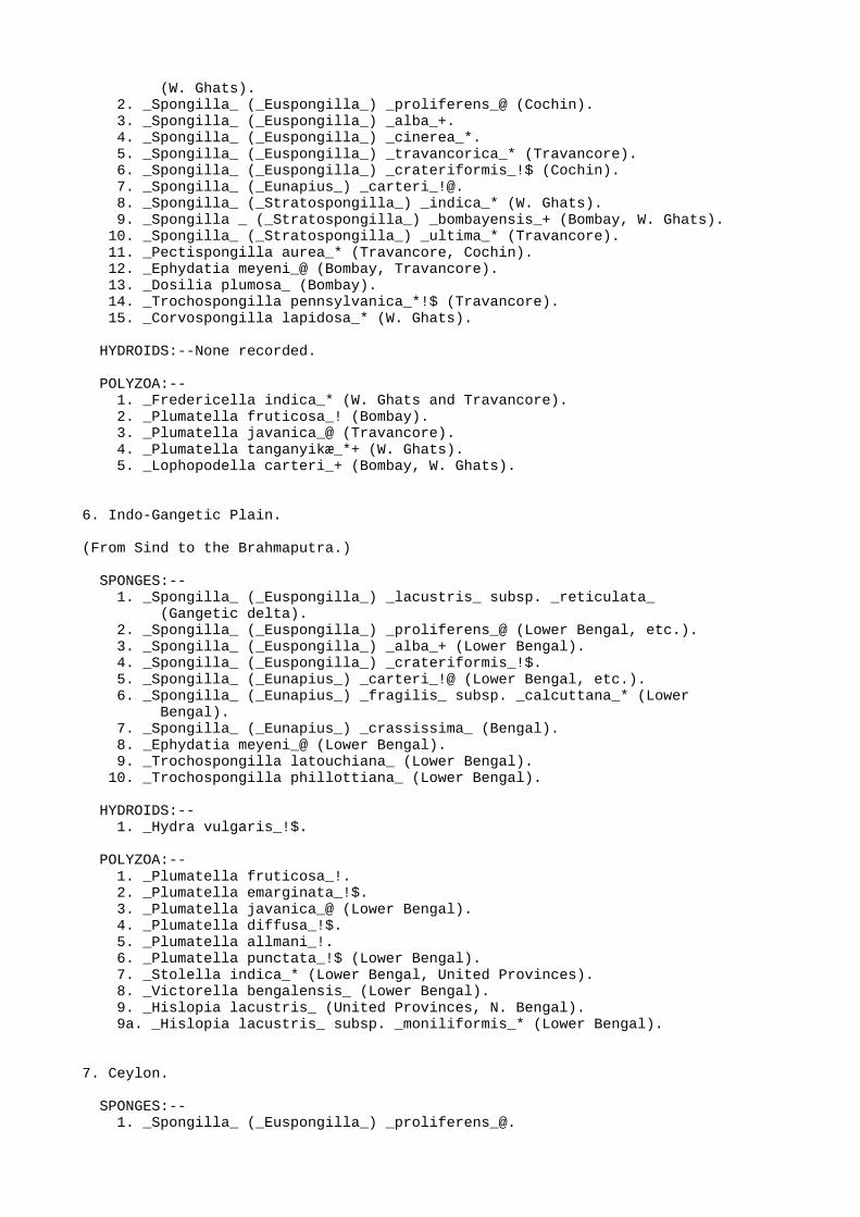
6. Indo-Gangetic Plain.
(From Sind to the Brahmaputra.)
SPONGES:-- 1. _Spongilla_ (_Euspongilla_) _lacustris_ subsp. _reticulata_ (Gangetic delta). 2. _Spongilla_ (_Euspongilla_) _proliferens_@ (Lower Bengal, etc.). 3. _Spongilla_ (_Euspongilla_) _alba_+ (Lower Bengal). 4. _Spongilla_ (_Euspongilla_) _crateriformis_!$. 5. _Spongilla_ (_Eunapius_) _carteri_!@ (Lower Bengal, etc.). 6. _Spongilla_ (_Eunapius_) _fragilis_ subsp. _calcuttana_* (Lower Bengal). 7. _Spongilla_ (_Eunapius_) _crassissima_ (Bengal). 8. _Ephydatia meyeni_@ (Lower Bengal). 9. _Trochospongilla latouchiana_ (Lower Bengal). 10. _Trochospongilla phillottiana_ (Lower Bengal).
HYDROIDS:-- 1. _Hydra vulgaris_!$.
POLYZOA:-- 1. _Plumatella fruticosa_!. 2. _Plumatella emarginata_!$. 3. _Plumatella javanica_@ (Lower Bengal). 4. _Plumatella diffusa_!$. 5. _Plumatella allmani_!. 6. _Plumatella punctata_!$ (Lower Bengal). 7. _Stolella indica_* (Lower Bengal, United Provinces). 8. _Victorella bengalensis_ (Lower Bengal). 9. _Hislopia lacustris_ (United Provinces, N. Bengal). 9a. _Hislopia lacustris_ subsp. _moniliformis_* (Lower Bengal).
7. Ceylon.
SPONGES:-- 1. _Spongilla_ (_Euspongilla_) _proliferens_@.

2. _Spongilla_ (_Eunapius_) _carteri_!@.
HYDROIDS:-- 1. _Hydra vulgaris_!$.
POLYZOA:-- 1. ? _Plumatella emarginata_!$. 2. _Pectinatella burmanica._
The most striking feature of this list is the evidence it affords as tothe distinct character of the fauna of the Malabar Zone, a feature thatis also remarkably clear as regards the Potamonidæ, one genus of which(_Gecarcinucus_) is peculiar, so far as India is concerned, to thatzone. As regards the sponges we may note the occurrence of no less thanthree species of the subgenus _Stratospongilla_, which has not beenfound elsewhere in India except on one occasion in Mysore, and of aspecies of the genus _Corvospongilla_, which is unknown from the rest ofPeninsular India and from the Himalayas. The genus _Pectispongilla_ isonly known from the Malabar Zone. Among the polyzoa the genus_Fredericella_[D] appears to be confined, so far as the Indian andBurmese fauna is concerned, to the Malabar Zone, and the same is true asregards the group of species to which _Plumatella tanganyikæ_, anAfrican form, belongs.
[Footnote D: Mr. S. W. Kemp recently obtained at Mangaldai, near the Bhutan frontier of Assam, a single specimen of what may be a species of _Fredericella_.]
A further examination of the list of Malabar species and a considerationof allied forms shows that the majority of the forms restricted to theMalabar Zone are either African or else closely allied to African forms.The genus _Corvospongilla_, except for one Burmese species, is otherwisepeculiar to Tropical Africa; while _Stratospongilla_, although notconfined to Africa, is more prolific in species in that continent thanin any other. _Spongilla (Stratospongilla) bombayensis_ has only beenfound in Bombay, the Western Ghats, Mysore, and Natal, and _Plumatellatanganyikæ_ only in the Western Ghats and Central Africa. The genus_Fredericella_ (which also occurs in Europe, N. America, and Australia)is apparently of wide distribution in Africa, while _Lophopodella_(which in India is not confined to the Malabar Zone) is, except for aJapanese race of the Indian species, restricted outside India, so far aswe know, to East Africa.
A less definite relationship between the sponges and polyzoa of theMalabar Zone and those of countries to the east of India is suggested bythe following facts:--
(1) The occurrence of the genus _Corvospongilla_ in Burma;
(2) the occurrence of the subgenus _Stratospongilla_ in Sumatra, China, and the Philippines;
(3) the occurrence of a race of _Lophopodella carteri_ in Japan;
(4) the occurrence of a species allied to _Plumatella tanganyikæ_ in the Philippines.
It will be noted that in each of these instances the relationshipextends to Africa as well as to the Eastern countries, and is moremarked in the former direction. The species of _Stratospongilla_,moreover, that occurs in Sumatra (_S. sumatrensis_) also occurs inAfrica, while those that have been found in China and the Philippinesare aberrant forms.

At first sight it might appear that these extra-Indian relationshipsmight be explained by supposing that gemmules and statoblasts werebrought to the Malabar Coast from Africa by the aërial currents of themonsoon or by marine currents and carried from India eastwards by thesame agency, this agency being insufficient to transport them to theinterior and the eastern parts of the Peninsula. The work of LaTouche[E] on wind-borne foraminifera in Rajputana is very suggestive inthis direction; but that the peculiar sponge and polyzoon fauna ofMalabar is due to the agency either of wind or of marine currents may bedenied with confidence, for it is a striking fact that most of thecharacteristic genera and subgenera of the Zone have restingreproductive bodies that are either fixed to solid objects or else aredevoid of special apparatus to render them light. The former is the caseas regards all species of _Corvospongilla_ and all Indian and most otherspecies of _Stratospongilla_, the gemmules of which not only areunusually heavy but also adhere firmly; while the statoblasts of_Fredericella_ have no trace of the air-cells that render the freestatoblasts of all other genera of phylactolæmatous polyzoa peculiarlylight and therefore peculiarly liable to be transported by wind.
[Footnote E: See Mem. Geol. Surv. Ind. XXXV (1), p. 39 (1902).]
A true geographical or geological explanation must therefore be soughtfor the relationship between the sponges and polyzoa of Malabar, ofAfrica, and of the Eastern countries--a relationship that is well knownto exist as regards other groups of animals. No more satisfactoryexplanation has as yet been put forward than that of a former landconnection between Africa and the Malaysia through Malabar at a period(probably late Cretaceous) when the Western Ghats were much higher thanthey now are[F].
[Footnote F: See Ortmann, "The Geographical Distribution of Freshwater Decapods and its bearing upon Ancient Geography," Proc. Amer. Phil. Soc. xli, p. 380, fig. 6 (1902); also Suess, "The Face of the Earth" (English ed.) i, p. 416 (1904).]
There is little to be said as regards the distribution of the sponges,hydroids, and polyzoa of fresh water in other parts of India. It may benoted, however, that the species known from the Punjab are all widelydistributed Palæarctic forms, and that the genus _Stolella_ isapparently confined to the Indo-Gangetic Plain. Two species of spongeare peculiar to Lower Burma, one of them (_Corvospongilla burmanica_)representing the geographical alliance already discussed as regards theMalabar Zone, the other (_Tubella vesparioides_) closely related to aMalaysian species (_T. vesparium_ from Borneo) and perhaps representingthe northern limit of the Malaysian element well known in the fauna ofLower Burma. Of the sponges and polyzoa of Ceylon we know as yet toolittle to make it profitable to discuss their affinities. All that haveas yet been discovered occur also in Peninsular India; nor do theyafford any evidence of a connection with the Malabar Zone.
The question of the geographical range of the sponges, hydroids, andpolyzoa of brackish water may be considered briefly, for it is ofimportance in considering that of those which are confined to freshwater. Some of these species from brackish water (e. g., _Membraniporalacroixii_) are identical with others (e. g., _Victorella bengalensis_and _Bowerbankia caudata_ subsp. _bengalensis_) closely related toEuropean forms. Others again (e. g., _Loxosomatoides colonialis_ and_Sagartia schilleriana_) are known as yet from the Ganges delta only. Inour ignorance of the Indian representatives of the groups to which theybelong, it is impossible to assert that their distribution is actually

so restricted as it seems.
SOME SPECIAL LOCALITIES.
In order to avoid constant repetition as regards the conditions thatprevail at the places most frequently mentioned in this volume, a fewdetails as regards them may be conveniently stated here.
_Lower Bengal._
CALCUTTA is situated on the River Hughli at a point about 90 miles fromthe open sea. The water of the river is practically fresh, but isstrongly affected by the tides; it is always turbid and of a brownishcolour. The river, however, is not a good collecting ground for sponges,coelenterates, and polyzoa, and none of the species described in thisvolume have been obtained from it. It is in the Calcutta "tanks" thatmost of my investigations have been made. These tanks are ponds, mostlyof artificial origin, very numerous, of varying size but never verylarge or deep. Most of them contain few solid objects to which sedentaryorganisms can fix themselves, and such ponds are of course poor insponges and polyzoa. Others, however, support a prolific growth of weedssuch as _Pistia stratiotes_, _Lemna_, and _Limnanthemum_, and a few havebrickwork or artificial stonework at their sides. In those parts of thetown that approach the Salt Lakes (large lagoons and swamps of brackishwater connected with the sea by the Mutlah River) the water of the pondsis slightly brackish and permits few plants except algæ to flourish. Fewof the bigger tanks ever dry up. The best of the tanks from thesponge-collector's point of view, so far as I have been able todiscover, is the one in the compound of the Indian Museum. It enjoys allthe advantages of light and shade, solid supports, prolific aquaticvegetation, considerable depth, and the vicinity of human dwellings thatseem to be favourable to the growth of sponges, no less than ninespecies of which, representing three genera and two subgenera, growabundantly in it. _Hydra_ also flourishes in this pond, but for somereasons there are few polyzoa. The phylactolæmatous species of thelatter group, however, are extraordinarily abundant in one of the tanksin the Zoological Gardens at Alipore. In this tank, which unlike theMuseum tank is directly connected with the river, no less than sixspecies and varieties of the genus _Plumatella_ have been found growingtogether on sticks, floating seeds, and water-plants. Except _Hislopia_,which is common on _Vallisneria_ in one tank on the Maidan (opposite theBengal Club), the ctenostomes of stagnant water are only found in thetanks near the Salt Lakes.
PORT CANNING is situated on the Mutlah River about 30 miles fromCalcutta and about 60 from the open sea. The Mutlah is really a tidalcreek rather than a river, in spite of the fact that it runs for aconsiderable number of miles, and its waters are distinctly brackish.Water taken from the edge at Port Canning in March was found to contain25.46 per thousand of saline residue. The interesting feature of PortCanning, however, is from a zoological point of view not the Mutlah butcertain ponds of brackish water now completely separated from it, exceptoccasionally when the river is in flood, but communicating regularlywith it in the memory of living persons. These ponds, which wereapparently not in existence in 1855, have on an average an area of abouthalf an acre each, and were evidently formed by the excavation of earthfor the construction of an embankment along the Mutlah. They are veryshallow and lie exposed to the sun. The salinity differs considerably indifferent ponds, although the fauna seems to be identical; the water ofone pond was found to contain 22.88 per thousand of saline residue inMay, 20.22 per thousand in March, and 12.13 in December. A second pondin the neighbourhood of the first and apparently similar to it in everyway contained only 9.82 per thousand in July, after the rains had

broken. The fauna of these ponds includes not only a freshwater sponge(_Spongilla alba_ var. _bengalensis_) but also many aquatic insects(_e. g._, larvæ of mosquitos and of _Chironomus_ and several species ofbeetles and Rhynchota); while on the other hand essentially marinecoelenterates (_Irene ceylonensis_, etc.) and worms (_e. g._, thegephyrean _Physcosoma lurco_[G]) form a part of it, together with formsof intermediate habitat such as _Bowerbankia caudata_ subsp._bengalensis_, _Victorella bengalensis_, and several fish and crustaceacommon in brackish water.
[Footnote G: I am indebted to Mr. W. F. Lanchester for the identification of this species.]
_Orissa._
Orissa may be described in general terms as consisting of the coastalarea of Bengal south of the Gangetic delta. It extends in inland,however, for a considerable distance and includes hilly tracts. There isno geographical boundary between it and the north-eastern part of theMadras Presidency or the eastern part of the Central Provinces.
CHILKA LAKE.--This marine lake is a shallow lagoon measuring about 40miles in length and 10 miles in breadth, and formed in geologicallyrecent times by the growth of a narrow sand-bank across the mouth of awide bay. At its northern end it communicates with the sea by a narrowchannel, and throughout its length it is strongly affected by the tides.At its south end, which is actually situated in the Ganjam district ofMadras, the water is distinctly brackish and is said to be nearly freshat certain times of year. At this end there are numerous smallartificial pools of brackish water somewhat resembling those of PortCanning as regards their fauna.
SUR (or SAR) LAKE.--A shallow, freshwater lake of very variable sizesituated a few miles north of Puri on the Orissa coast. In origin itprobably resembled the Chilka Lake, but it is now separated from the seaby about 3 miles of barren sand dunes, among which numerous little poolsof rain-water are formed during the rains. These dry up completely inwinter, and even the lake itself is said sometimes almost to disappear,although when it is full it is several miles in length. The fauna isessentially a freshwater one, but includes certain Mysidæ and othercrustacea usually found in brackish water.
_Bombay Presidency._
BOMBAY.--The town of Bombay, built on an island near the mainland, issituated close to swamps and creeks of brackish water not unlike thosethat surround Calcutta. Its "tanks," however, differ from those ofCalcutta in having rocky bottoms and, in many cases, in drying upcompletely in the hot weather. Of the fauna of the swamps extremelylittle is known, but so far as the sponges and polyzoa of the tanks areconcerned the work undertaken by Carter was probably exhaustive.
IGATPURI.--Igatpuri is situated at an altitude of about 2000 feet, 60miles north-east of Bombay. Above the town there is a lake of severalsquare miles in area whence the water-supply of several stations in theneighbourhood is obtained. The water is therefore kept free fromcontamination. The bottom is composed of small stones and slopesgradually up at the edges. During the dry weather its level sinksconsiderably. Several interesting sponges and polyzoa have been found inthis lake, most of them also occurring in a small pond in theneighbourhood in which clothes are washed and the water is often full ofsoap-suds.
_Southern India._

MADRAS.--The city of Madras is built by the sea, straggling over a largearea of the sandy soil characteristic of the greater part of the eastcoast of India. In wet weather this soil retains many temporary pools ofrain-water, and there are numerous permanent tanks of no great size inthe neighbourhood of the town. The so-called Cooum River, which flowsthrough the town, is little more than a tidal creek, resembling theMutlah River of Lower Bengal on a much smaller scale. The sponges andpolyzoa as yet found in the environs of Madras are identical with thosefound in the environs of Calcutta.
BANGALORE.--Bangalore (Mysore State) is situated near the centre of theMadras Presidency on a plateau about 3000 feet above sea-level. Thesurrounding country is formed of laterite rock which decomposes readilyand forms a fine reddish silt in the tanks. These tanks are numerous,often of large size, and as a rule at least partly of artificial origin.Their water supports few phanerogamic plants and is, as my friend Dr.Morris Travers informs me, remarkably free from salts in solution. Thesponge fauna of the neighbourhood of Bangalore appears to beintermediate between that of Madras and that of Travancore.
THE BACKWATERS OF COCHIN AND TRAVANCORE.--The "backwaters" of Cochin andTravancore were originally a series of shallow lagoons stretching alongthe coast of the southern part of the west coast of India for a distanceof considerably over a hundred miles. They have now been joined togetherby means of canals and tunnels to form a tidal waterway, whichcommunicates at many points directly with the sea. The salinity of thewater differs greatly at different places and in different seasons, andat some places there is an arrangement to keep out sea-water while therice-fields are being irrigated. The fauna is mainly marine, but in theless saline parts of the canals and lakes many freshwater species arefound.
_Shasthancottah._--There are two villages of this name, one situated onthe backwater near Quilon (coast of Travancore), the other about threemiles inland on a large freshwater lake. This lake, which does notcommunicate with the backwater, occupies a narrow winding rift severalmiles in length at a considerable depth below the surrounding country.Its bottom is muddy and it contains few water-plants, although in someplaces the water-plants that do exist are matted together to formfloating islands on which trees and bushes grow. The fauna, at any rateas regards mollusca and microscopic organisms, is remarkably poor, buttwo species of polyzoa (_Fredericella indica_ and _Plumatellafruticosa_) and one of sponge (_Trochospongilla pennsylvanica_) grow inconsiderable abundance although not in great luxuriance.
_The Himalayas._
BHIM TAL[H] is a lake situated at an altitude of 4500 feet in that partof the Western Himalayas known as Kumaon, near the plains. It has asuperficial area of several square miles, and is deep in the middle. Itsbottom and banks are for the most part muddy. Little is known of itsfauna, but two polyzoa (_Plumatella allmani_ and _Lophopodella carteri_)and the gemmules of two sponges (_Spongilla carteri_ and _Ephydatiameyeni_) have been found in it.
[Footnote H: The fauna of this lake and of others in the neighbourhood has recently been investigated by Mr. S. W. Kemp. See the addenda at the end of this volume.--_June 1911._]
* * * * *
NOMENCLATURE AND TERMINOLOGY.

The subject of nomenclature may be considered under four heads:--(I.)the general terminology of the various kinds of groups of individualsinto which organisms must be divided; (II.) the general nomenclature ofspecimens belonging to particular categories, such as types, co-types,etc.; (III.) the nomenclature that depends on such questions as that of"priority"; and (IV.) the special terminology peculiar to the differentgroups. The special terminology peculiar to the different groups isdealt with in the separate introductions to each of the three parts ofthis volume.
(I.)
No group of animals offers greater difficulty than the sponges,hydroids, and polyzoa (and especially the freshwater representatives ofthese three groups) as regards the question "What is a species?" and thekindred questions, "What is a subspecies?" "What is a variety?" and"What is a phase?" Genera can often be left to look after themselves,but the specific and kindred questions are answered in so many differentways, if they are even considered, by different systematists, especiallyas regards the groups described in this volume, that I feel it necessaryto state concisely my own answers to these questions, not for theguidance of other zoologists but merely to render intelligible thesystem of classification here adopted. The following definitions shouldtherefore be considered in estimating the value of "species," etc.,referred to in the following pages.
_Species._--A group of individuals differing in constant characters of adefinite nature and of systematic[I] importance from all others in thesame genus.
[Footnote I: "What characters are of systematic importance?" is a question to which different answers must be given in the case of different groups.]
_Subspecies._--An isolated or local race, the individuals of whichdiffer from others included in the same species in characters that areconstant but either somewhat indefinite or else of little systematicimportance.
_Variety._--A group of individuals not isolated geographically fromothers of the same species but nevertheless exhibiting slight, notaltogether constant, or indefinite differences from the typical form ofthe species (_i. e._, the form first described).
_Phase._--A peculiar form assumed by the individuals of a species whichare exposed to peculiarities in environment and differ from normalindividuals as a direct result.
There are cases in which imperfection of information renders itdifficult or impossible to distinguish between a variety and asubspecies. In such cases it is best to call the form a variety, forthis term does not imply any special knowledge as regards itsdistribution or the conditions in which it is found.
I use the term "form" in a general sense of which the meaning ormeanings are clear without explanation.
(II.)
The question of type specimens must be considered briefly. There are twoschools of systematists, those who assert that one specimen and one only

must be the type of a species, and those who are willing to acceptseveral specimens as types. From the theoretical point of view it seemsimpossible to set up any one individual as the ideal type of a species,but those who possess collections or are in charge of museums prefer,with the natural instinct of the collector, to have a definite singletype (of which no one else can possibly possess a duplicate) in theirpossession or care, and there is always the difficulty that a zoologistin describing a species, if he recognizes more than one type, mayinclude as types specimens that really belong to more than one species.These difficulties are met by some zoologists by the recognition ofseveral specimens as paratypes, all of equal value; but this, after all,is merely a terminological means of escaping from the difficulty,calculated to salve the conscience of a collector who feels unwilling togive up the unique type of a species represented by other specimens inhis collection. The difficulty as regards the confounding of specimensof two or more species as the types of one can always be adjusted if theauthor who discovers the mistake redescribes one of the species underthe original name and regards the specimen that agrees with hisdescription as the type, at the same time describing a new species withanother of the specimens as its type. Personally I always desire toregard the whole material that forms the basis of an originaldescription of a species as the type, but museum rules often render thisimpossible, and the best that can be done is to pick out one specimenthat seems particularly characteristic and to call it the type, the restof the material being termed co-types. A peculiar difficulty arises,however, as regards many of the sponges, coelenterates, and polyzoa,owing to the fact that they are often either compound animals, eachspecimen consisting of more than one individual, or are easily divisibleinto equivalent fragments. If the single type theory were driven to itslogical conclusion, it would be necessary to select one particular polypin a hydroid colony, or even the part of a sponge that surrounded aparticular osculum as the type of the species to which the hydroid orthe sponge belonged. Either by accident or by design specimens ofSpongillidæ, especially if kept dry, are usually broken into severalpieces. There is, as a matter of fact, no reason to attribute thepeculiarly sacrosanct nature of a type to one piece more than another.In such cases the biggest piece may be called the type, while thesmaller pieces may be designated by the term "schizotype."
The more precise definition of such terms as topotype, genotype, _ethujus generis omnis_ is nowadays a science (or at any rate a form oftechnical industry) by itself and need not be discussed here.
(III.)
In 1908 an influential committee of British zoologists drew up astrenuous protest against the unearthing of obsolete zoological names(see 'Nature,' Aug. 1908, p. 395). To no group does this protest applywith greater force than to the three discussed in this volume. It isdifficult, however, to adopt any one work as a standard of nomenclaturefor the whole of any one of them. As regards the Spongillidæ it isimpossible to accept any monograph earlier than Potts's "Fresh-WaterSponges" (P. Ac. Philad., 1887), for Bowerbank's and Carter's earliermonographs contained descriptions of comparatively few species. EvenPotts's monograph I have been unable to follow without divergence, forit seems to me necessary to recognize several genera and subgenera thathe ignored. The freshwater polyzoa, however, were dealt with in socomprehensive a manner by Allman in his "Fresh-Water Polyzoa" (London,1856) that no difficulty is experienced in ignoring, so far asnomenclature is concerned, any earlier work on the group; while asregards other divisions of the polyzoa I have followed Hincks's "BritishMarine Polyzoa" (1880), so far as recent researches permit. In mostcases I have not attempted to work out an elaborate synonymy of species

described earlier than the publication of the works just cited, for todo so is a mere waste of time in the case of animals that call for amost precise definition of species and genera and yet were oftendescribed, so far as they were known earlier than the dates in question,in quite general terms. I have been confirmed in adopting this course bythe fact that few of the types of the earlier species are now inexistence, and that a large proportion of the Indian forms have onlybeen described within the last few years.
MATERIAL.
The descriptions in this volume are based on specimens in the collectionof the Indian Museum, the Trustees of which, by the liberal manner inwhich they have permitted me to travel in India and Burma on behalf ofthe Museum, have made it possible not only to obtain material for studyand exchange but also to observe the different species in their naturalenvironment. This does not mean to say that specimens from othercollections have been ignored, for many institutions and individualshave met us generously in the matter of gifts and exchanges, and ourcollection now includes specimens of all the Indian forms, named innearly all cases by the author of the species, except in those ofspecies described long ago of which no authentic original specimens cannow be traced. Pieces of the types of all of the Indian Spongillidædescribed by Carter have been obtained from the British Museum throughthe kind offices of Mr. R. Kirkpatrick. The Smithsonian Institution hassent us from the collection of the United States National Museumspecimens named by Potts, and the Berlin Museum specimens named byWeltner, while to the Imperial Academy of Sciences of St. Petersburg weowe many unnamed but interesting sponges. Dr. K. Kraepelin and Dr. W.Michaelsen have presented us with specimens of most of the species andvarieties of freshwater polyzoa described by the former in his greatmonograph and elsewhere. We owe to Dr. S. F. Harmer, formerly of theCambridge University Museum and now Keeper in Zoology at the BritishMuseum, to Professor Max Weber of Amsterdam, Professor Oka of Tokyo, andseveral other zoologists much valuable material. I would speciallymention the exquisite preparations presented by Mr. C. Rousselet.Several naturalists in India have also done good service to the Museumby presenting specimens of the three groups described in this volume,especially Major H. J. Walton, I.M.S., Major J. Stephenson, I.M.S., Dr.J. R. Henderson and Mr. G. Matthai of Madras, and Mr. R. ShunkaraNarayana Pillay of Trivandrum.
The following list shows where the types of the various species,subspecies, and varieties are preserved, so far as it has been possibleto trace them. I have included in this list the names of all speciesthat have been found in stagnant water, whether fresh or brackish, butthose of species not yet found in fresh water are enclosed in squarebrackets.
+-----------------------------------------------------------------------+| INDIAN SPONGILLIDÆ. || |+---------------------------------+----------------------+--------------+| NAME. | TYPE IN COLL. | MATERIAL || | | EXAMINED. |+---------------------------------+----------------------+--------------+| _Spongilla lacustris_ subsp. | Ind. Mus. | Type. || _reticulata_ | | |+---------------------------------+----------------------+--------------+| _Spongilla proliferens_ | " " | " |+---------------------------------+----------------------+--------------+| _Spongilla alba_ | Brit. and Ind. Mus. | Schizotype. |+---------------------------------+----------------------+--------------+

| [_Spongilla alba_ var. | Ind. Mus. | Type || _bengalensis_] | | |+---------------------------------+----------------------+--------------+| _Spongilla alba_ var. | Brit. Mus. | {Specimens || _cerebellata_ | | {compared || | | {with type. |+---------------------------------+----------------------+--------------+| _Spongilla cinerea_ | Brit. and Ind. Mus. | Schizotype. |+---------------------------------+----------------------+--------------+| [_Spongilla travancorica_] | Ind. Mus. | Type. |+---------------------------------+----------------------+--------------+| _Spongilla hemephydatia_ | " " | " |+---------------------------------+----------------------+--------------+| _Spongilla crateriformis_ | U.S. Nat. Mus. | Co-type. |+---------------------------------+----------------------+--------------+| _Spongilla carteri_ | Brit. and Ind. Mus. | Schizotype. |+---------------------------------+----------------------+--------------+| _Spongilla carteri_ var. | Ind. Mus. | Type. || _mollis_ | | |+---------------------------------+----------------------+--------------+| _Spongilla carteri_ var. | " " | " || _cava_ | | |+---------------------------------+----------------------+--------------+| _Spongilla carteri_ var. | " " | " || _lobosa_ | | |+---------------------------------+----------------------+--------------+| _Spongilla fragilis_ subsp. | " " | " || _calcuttana_ | | |+---------------------------------+----------------------+--------------+| _Spongilla fragilis_ subsp. | Amsterdam Mus. | Co-type. || _decipiens_ | | |+---------------------------------+----------------------+--------------+| _Spongilla gemina_ | Ind. Mus. | Type. |+---------------------------------+----------------------+--------------+| _Spongilla crassissima_ | " " | " |+---------------------------------+----------------------+--------------+| _Spongilla crassissima_ var. | " " | " || _crassior_ | | |+---------------------------------+----------------------+--------------+| _Spongilla bombayensis_ | Brit. and Ind. Mus. | Schizotype. |+---------------------------------+----------------------+--------------+| _Spongilla indica_ | Ind. Mus. | Type. |+---------------------------------+----------------------+--------------+| _Spongilla ultima_ | " " | " |+---------------------------------+----------------------+--------------+| _Pectispongilla aurea_ | " " | " |+---------------------------------+----------------------+--------------+| _Ephydatia meyeni_ | Brit. and Ind. Mus. | Schizotype. |+---------------------------------+----------------------+--------------+| _Dosilia plumosa_ | " " " " | " |+---------------------------------+----------------------+--------------+| _Trochospongilla latouchiana_ | Ind. Mus. | Type. |+---------------------------------+----------------------+--------------+| _Trochospongilla phillottiana_ | " " | " |+---------------------------------+----------------------+--------------+| _Trochospongilla pennsylvanica_ | U.S. Nat. Mus. | Co-type. |+---------------------------------+----------------------+--------------+| _Tubella vesparioides_ | Ind. Mus. | Type. |+---------------------------------+----------------------+--------------+| _Corvospongilla burmanica_ | Brit. and Ind. Mus. | Schizotype. |+---------------------------------+----------------------+--------------+| _Corvospongilla lapidosa_ | Ind. Mus. | Type. |+---------------------------------+----------------------+--------------+| INDIAN COELENTERATES OF STAGNANT WATER. |

+---------------------------------+----------------------+--------------+| HYDROZOA. | | |+---------------------------------+----------------------+--------------+| _Hydra oligactis_ | Not in existence. | |+---------------------------------+----------------------+--------------+| _Hydra vulgaris_ | " " | |+---------------------------------+----------------------+--------------+| [_Syncoryne filamentata_] | Ind. Mus. | Type. |+---------------------------------+----------------------+--------------+| [_Bimeria vestita_] | ? Not in existence. | |+---------------------------------+----------------------+--------------+| [_Irene ceylonensis_] | {Hydroid in Ind.} | Hydroid type || | {Mus., Medusa} | || | {in Brit. Mus.} | |+---------------------------------+----------------------+--------------+| ACTINIARIA. | | |+---------------------------------+----------------------+--------------+| [_Sagartia schilleriana_] | Ind. Mus. | Types. |+---------------------------------+----------------------+--------------+| [_Sagartia schilleriana_ | " " | " || subsp. _exul_] | | |+---------------------------------+----------------------+--------------+| INDIAN POLYZOA OF STAGNANT WATER. |+---------------------------------+----------------------+--------------+| ENTOPROCTA. | | |+---------------------------------+----------------------+--------------+| [_Loxosomatoides colonialis_] | Ind. Mus. | Types. |+---------------------------------+----------------------+--------------+| ECTOPROCTA CHEILOSTOMATA. | | |+---------------------------------+----------------------+--------------+| [_Membranipora lacroixii_] | ? Paris Mus. | |+---------------------------------+----------------------+--------------+| [_Membranipora bengalensis_] | Ind. Mus. | Types. |+---------------------------------+----------------------+--------------+| ECTOPROCTA STENOSTOMATA. | | |+---------------------------------+----------------------+--------------+| [_Bowerbankia caudata_ subsp. | Ind. Mus. | Types. || _bengalensis_] | | |+---------------------------------+----------------------+--------------+| _Victorella bengalensis_ | " " | " |+---------------------------------+----------------------+--------------+| _Hislopia lacustris_ | ? Not in existence. | |+---------------------------------+----------------------+--------------+| _Hislopia lacustris_ subsp. | Ind. Mus. | " || _moniliformis_ | | |+---------------------------------+----------------------+--------------+| ECTOPROCTA PHYLACTOLÆMATA. | | |+---------------------------------+----------------------+--------------+| _Fredericella indica_ | Ind. Mus. | Type. |+---------------------------------+----------------------+--------------+| _Plumatella fruticosa_ | Not in existence. | |+---------------------------------+----------------------+--------------+| _Plumatella diffusa_ |?Philadelphia Acad.[J]| |+---------------------------------+----------------------+--------------+| _Plumatella allmani_ | Not in existence. | |+---------------------------------+----------------------+--------------+| _Plumatella emarginata_ | " " | |+---------------------------------+----------------------+--------------+| | {Hamburg and} | One of the || _Plumatella javanica_ | {Ind. Mus. } | types. |+---------------------------------+----------------------+--------------+| | {Brit. and Ind.} | One of the || _Plumatella tanganyikæ_ | {Mus. } | types. |+---------------------------------+----------------------+--------------+

| _Stolella indica_ | Ind. Mus. | Type. |+---------------------------------+----------------------+--------------+| _Lophopodella carteri_ | Brit. Mus. | " |+---------------------------------+----------------------+--------------+| _Lophopodella carteri_ var. | Ind. Mus. | " || _himalayana_ | | |+---------------------------------+----------------------+--------------+| _Pectinatella burmanica_ | Ind. Mus. | " |+---------------------------------+----------------------+--------------+
[Footnote J: I have failed to obtain from the Philadelphia Academy of Science a statement that the type of this species is still in existence.]
The literature dealing with the various groups described in the volumeis discussed in the introductions to the three parts. Throughout thevolume I have, so far as possible, referred to works that can beconsulted in Calcutta in the libraries of the Indian Museum, theGeological Survey of India, or the Asiatic Society of Bengal. The namesof works that are not to be found in India are marked with a *. Therarity with which this mark occurs says much for the fortunate positionin which zoologists stationed in Calcutta find themselves as regardszoological literature, for I do not think that anything essential hasbeen omitted.
It remains for me to express my gratitude to those who have assisted mein the preparation of this volume. The names of those who havecontributed specimens for examination have already been mentioned. Ihave to thank the Trustees of the Indian Museum not only for theirliberal interpretation of my duties as an officer of the Museum but alsofor the use of all the drawings and photographs and some of the blocksfrom which this volume is illustrated. Several of the latter havealready been used in the "Records of the Indian Museum." From the Editorof the "Fauna" I have received valuable suggestions, and I am indebtedto Dr. Weltner of the Berlin Museum for no less valuable references toliterature. Mr. F. H. Gravely, Assistant Superintendent in the IndianMuseum, has saved me from several errors by his criticism.
The majority of the figures have been drawn by the draftsmen of theIndian Museum, Babu Abhoya Charan Chowdhary, and of the Marine Survey ofIndia, Babu Shib Chandra Mondul, to both of whom I am much indebted fortheir accuracy of delineation.
No work dealing with the sponges of India would be complete without atribute to the memory of H. J. Carter, pioneer in the East of the studyof lower invertebrates, whose work persists as a guide and anencouragement to all of us who are of the opinion that biologicalresearch on Indian animals can only be undertaken in India, and thateven systematic zoological work can be carried out in that country withsuccess. I can only hope that this, the first volume in the officialFauna of the Indian Empire to be written entirely in India, may provenot unworthy of his example.
Indian Museum, Calcutta Oct. 23rd, 1910.
PART I.
FRESHWATER SPONGES
(SPONGILLIDÆ).
INTRODUCTION TO PART I.
I.
THE PHYLUM PORIFERA.
The phylum Porifera or Spongiæ includes the simplest of the Metazoa ormulticellular animals. From the compound Protozoa its members aredistinguished by the fact that the cells of which they are composedexhibit considerable differentiation both in structure and in function,and are associated together in a definite manner, although they are notcombined to form organs and systems of organs as in the higher Metazoa.Digestion, for instance, is performed in the sponges entirely byindividual cells, into the substance of which the food is taken, and theproducts of digestion are handed on to other cells without theintervention of an alimentary canal or a vascular system, while there isno structure in any way comparable to the nervous system of more highlyorganized animals.
The simplest form of sponge, which is known as an olynthus, is a hollowvase-like body fixed at one end to some solid object, and with anopening called the osculum at the other. The walls are perforated bysmall holes, the pores, from which the name Porifera is derived.
Externally the surface is protected by a delicate membrane formed offlattened cells and pierced by the pores, while the interior of the vaseis covered with curious cells characteristic of the sponges, and knownas choanocytes or collar-cells. They consist of minute oval orpear-shaped bodies, one end of which is provided with a rim or collar ofapparently structureless membrane, while a flagellum or whip-like lashprojects from the centre of the surface surrounded by the collar. Thesecollar-cells are practically identical with those of which the Protozoaknown as Choanoflagellata consist; but it is only in the sponges[K] thatthey are found constantly associated with other cells unlike themselves.
[Footnote K: Except in "_Proterospongia_," an organism of doubtful affinities but not a sponge. It consists of a mass of jelly containing ordinary cells, with collar-cells _outside_.]
In addition to the collar-cells, which form what is called the gastrallayer, and the external membrane (the derma or dermal membrane), thesponge contains cells of various kinds embedded in a structurelessgelatinous substance, through which they have the power of freemovement. Most of these cells have also the power of changing their formin an "amoeboid" manner; that is to say, by projecting and withdrawingfrom their margin mobile processes of a more or less finger-like form,but unstable in shape or direction. The protoplasm of which some of thecells are formed is granular, while that of others is clear andtranslucent. Some cells, which (for the time being at any rate) do notexhibit amoeboid movements, are glandular in function, while othersagain give rise in various ways to the bodies by means of which thesponge reproduces its kind. There is evidence, however, that any onekind of cell, even those of the membrane and the gastral layer, canchange its function and its form in case of necessity.
Most sponges possess a supporting framework or skeleton. In some it isformed entirely of a horny substance called spongin (as in thebath-sponge), in others it consists of spicules of inorganic matter

(either calcareous or siliceous) secreted by special cells, or of suchspicules bound together by spongin. Extraneous objects, such assand-grains, are frequently included in the skeleton. The spongin issecreted like the spicules by special cells, but its chemical structureis much more complicated than that of the spicules, and it is notsecreted (at any rate in most cases) in such a way as to form bodies ofa definite shape. In the so-called horny sponges it resembles the chitinin which insects and other arthropods are clothed.
* * * * *
In no adult sponge do the collar-cells completely cover the whole of theinternal surface, the olynthus being a larval form, and by no means acommon larval form. It is only found in certain sponges with calcareousspicules. As the structure of the sponge becomes more complicated thecollar-cells are tucked away into special pockets or chambers known asciliated chambers, and finally the approach to these chambers, both fromthe external surface and from the inner or gastral cavity, takes theform of narrow tubes or canals instead of mere pores. With furthercomplexity the simple internal cavity tends to disappear, and the spongeproliferates in such a way that more than one osculum is formed. In theclass Demospongiæ, to which the sponges described in this volume belong,the whole system is extremely complicated.
The skeleton of sponges, when it is not composed wholly of spongin,consists of, or at any rate contains, spicules that have a definitechemical composition and definite shapes in accordance with the class,order, family, genus, and species of the sponge. Formerly sponges wereseparated into calcareous, siliceous, and horny sponges by the nature oftheir skeleton; and although the system of classification now adoptedhas developed into a much more complex one and a few sponges are knownthat have both calcareous and siliceous spicules, the question whetherthe spicules are formed of salts of lime or of silica (strictly speakingof opal) is very important. All Demospongiæ that have spicules at allhave them of the latter substance, and the grade Monaxonida, in whichthe freshwater sponges constitute the family Spongillidæ, ischaracterized by the possession of spicules that have typically the formof a needle pointed at both ends. Although spicules of this simple formmay be absent in species that belong to the grade, the larger spicules,which are called megascleres, have not normally more than one main axisand are always more or less rod-like in outline. They are usuallyarranged so as to form a reticulate skeleton. Frequently, however, themegascleres or skeleton-spicules are not the only spicules present, forwe find smaller spicules (microscleres) of one or more kinds lying loosein the substance of the sponge and in the external membrane, or, in theSpongillidæ only, forming a special armature for the reproductive bodiesknown as gemmules.
All sponges obtain their food in the same way, namely by means of thecurrents of water set up by the flagella of the collar-cells. Theseflagella, although apparently there is little concerted action amongthem, cause by their rapid movements changes of pressure in the watercontained in the cavities of the sponge. The water from outsidetherefore flows in at the pores and finally makes its way out of theoscula. With the water minute particles of organic matter are broughtinto the sponge, the collar-cells of which, and probably other cells,have the power of selecting and engulfing suitable particles. Inside thecells these particles undergo certain chemical changes, and are at leastpartially digested. The resulting substances are then handed on directlyto other cells, or, as some assert, are discharged into the commonjelly, whence they are taken up by other cells.
Sponges reproduce their kind in more ways than one, _viz._, by means ofeggs (which are fertilized as in other animals by spermatozoa), by means

of buds, and by means of the peculiar bodies called gemmules thestructure and origin of which is discussed below (p. 42). They are ofgreat importance in the classification of the Spongillidæ. Sponges canalso be propagated artificially by means of fission, and it is probablethat this method of reproduction occurs accidentally, if not normally,in natural circumstances.
GENERAL STRUCTURE OF THE SPONGILLIDÆ.
It would be impracticable in this introduction to give a full account ofthe structure of the Spongillidæ, which in some respects is stillimperfectly known. Students who desire further information shouldconsult Professor Minchin's account of the sponges in Lankester's'Treatise on Zoology,' part ii, or, if a less technical description isdesired, Miss Sollas's contribution to the 'Cambridge Natural History,'vol. i, in which special attention is paid to _Spongilla_.
The diagram reproduced in fig. 1 gives a schematic view of a verticalsection through a living freshwater sponge. Although it represents thestructure of the organism as being very much simpler than is actuallythe case, and entirely omits the skeleton, it will be found useful asindicating the main features of the anatomy.
[Illustration: Fig. 1.--Diagram of a vertical section through afreshwater sponge (_modified from Kükenthal_).
A=pores; B=subdermal cavity; C=inhalent canal; D=ciliated chamber;E=exhalent canal; F=osculum; G=dermal membrane; H=eggs; J=gemmule.]
It will be noted that the diagram represents an individual with a singleosculum or exhalent aperture. As a rule adult Demospongiæ have severalor many oscula, but even in the Spongillidæ sponges occur in which thereis only one. New oscula are formed by a kind of proliferation thatrenders the structure still more complex than it is when only oneexhalent aperture is present.
The little arrows in the figure indicate the direction of the currentsof water that pass through the sponge. It enters through small holes inthe derma into a subdermal cavity, which separates the membrane from thebulk of the sponge. This space differs greatly in extent in differentspecies. From the subdermal space the water is forced by the action ofthe flagella into narrow tubular canals that carry it into the ciliatedchambers. Thence it passes into other canals, which communicate withwhat remains of the central cavity, and so out of the oscula.
The ciliated chambers are very minute, and the collar-cells excessivelyso. It is very difficult to examine them owing to their small size anddelicate structure. Fig. 2 D represents a collar-cell of a sponge seenunder a very high power of the microscope in ideal conditions.
[Illustration: Fig. 2.--Sponge cells.
A=bubble-cells of _Ephydatia mülleri_, × 350 (_after Weltner_).B=gemmule-cell of _Spongilla lacustris_ containing green corpuscles(shaded dark), × 800 (_after Weltner_). C=gemmule-cell of _Ephydatiablembingia_ showing "tabloids" of food-material, × 1150 (_after Evans_).D=collar-cell of _Esperella ægagrophila_, × 1600 (_after Vosmaer andPekelharing_). E=three stages in the development of a gemmule-spicule of_E. blembingia_ (_after Evans_), × 665. F=outline of porocytes of _S.proliferens_, × ca. 1290: _e_=dermal cell; _n_=nucleus; _p_=pore;_p.c._=pore-cell.]
The nature of the inhalent apertures in the external membrane has been

much discussed as regards the Demospongiæ, but the truth seems to bethat their structure differs considerably even in closely alliedspecies. At any rate this is the case as regards the Indian _Spongillæ_.In all species the membrane is composed of flattened cells of irregularshape fitted together like the pieces of a puzzle-picture. In somespecies (e. g., _Spongilla carteri_) the apertures in the membraneconsist merely of spaces between adjacent cells, which may be a littlemore crowded together than is usual. But in others (e. g., _Spongillaproliferens_ and _Spongilla crassissima_) in which the pores areextremely small, each pore normally pierces the middle of a flat,ring-shaped cell or porocyte. Occasionally, however, a pore may be foundthat is enclosed by two narrow, crescent-shaped cells joined together attheir tips to form a ring. The porocytes of sponges like _Spongillacarteri_ are probably not actually missing, but instead of being in theexternal membrane are situated below the derma at the external entranceto the canals that carry water to the flagellated chambers or even atthe entrance to the chambers themselves[L]. Some authors object ontheoretical grounds to the statement that porocytes exist in theDemospongia, and it is possible that these cells have in this gradeneither the same origin as, nor a precisely similar function to, theporocytes of other sponges. When they occur in the dermal membrane nogreat difficulty is experienced in seeing them under a sufficiently highpower of the microscope, if the material is well preserved and mountedand stained in a suitable manner[M]. In most sponges the porocytes cancontract in such a way that the aperture in their centre is practicallyclosed, but this power appears to be possessed by the porocytes of_Spongilla_ only to a very limited extent, although they closelyresemble the porocytes of other sponges in appearance.
[Footnote L: _Cf._ Weltner, "Spongillidenstudien, V," Arch. Naturg. Berlin, lxxiii (i), p. 273 (1907).]
[Footnote M: It is difficult to see any trace of them in thin microtome sections. A fragment of the membrane must be mounted whole.]
The external membrane in many Spongillidæ is prolonged round and abovethe oscula so as to form an oscular collar. This structure is highlycontractile, but cannot close together. As a rule it is much moreconspicuous in living sponges than in preserved specimens.
It is not necessary to deal here with most of the cells thatoccur in the parenchyma or gelatinous part of the sponge. A fulllist of the kinds that are found is given by Dr. Weltner in his"Spongillidenstudien, V," p. 276 (Arch. Naturg. Berlin, lxxiii (i),1907). One kind must, however, be briefly noticed as being of somesystematic importance, namely the "bubble-cells" (fig. 2 A) that arecharacteristic of some species of _Ephydatia_ and other genera. Thesecells are comparatively large, spherical in form; each of them containsa globule of liquid which not only occupies the greater part of thecell, but forces the protoplasm to assume the form of a delicate filmlining the cell-wall and covering the globule. In optical section"bubble-cells" have a certain resemblance to porocytes, but the cell isof course imperforate and not flattened.
SKELETON AND SPICULES.
[Illustration: Fig. 3.--Radial sections of fragments of the skeletons of_Spongillæ_.
A, _S. crassissima_ var. _crassior_ (from Rajshahi); B, _S. carteri_(from Calcutta); _a_=transverse, _b_=radiating fibres; _e_=externalsurface of the sponge.]

In the Spongillidæ the spicules and the skeleton are more important asregards the recognition of genera and species than the soft parts. Theskeleton is usually reticulate, but sometimes consists of a mass ofspicules almost without arrangement. The amount of spongin present isalso different in different species. The spicules in a reticulateskeleton are arranged so as to form fibres of two kinds--radiatingfibres, which radiate outwards from the centre of the sponge andfrequently penetrate the external membrane, and transverse fibres, whichrun across from one radiating fibre to another. The fibres are composedof relatively large spicules (megascleres) arranged parallel to oneanother, overlapping at the ends, and bound together by means of a moreor less profuse secretion of spongin. In some species they are actuallyenclosed in a sheath of this substance. The radiating fibres are usuallymore distinct and stouter than the transverse ones, which are oftenrepresented by single spicules but are sometimes splayed out at the endsso as to assume in outline the form of an hour-glass (fig. 3 B). Theradiating fibres frequently raise up the membrane at their freeextremities just as a tent-pole does a tent.
Normal spicules of the skeleton are always rod-like or needle-like, andeither blunt or pointed at both ends; they are either smooth, granular,or covered with small spines. Sometimes spicules of the same type form amore or less irregular transverse network at the base or on the surfaceof the sponge.
[Illustration: Fig. 4.--Part of an oscular collar of _Spongillalacustris_ subsp. _reticulata_, showing arrangement of microscleres inthe derma (magnified).]
From the systematist's point of view, the structure of the free spiculesfound scattered in the substance and membrane of the sponge, andespecially of those that form the armature of the gemmules, is of moreimportance than that of the skeleton-spicules. Free spicules are absentin many species; when present they are usually needle-like and pointedat the tips. In a few species, however, they are of variable orirregular form, or consist of several or many shafts meeting in a commoncentral nodule. In one genus (_Corvospongilla_) they resemble a doublegrappling-iron in form, having a circle of strongly recurved hooks atboth ends. The free microscleres, or flesh-spicules as they are oftencalled, are either smooth, granular, or spiny.
Gemmule-spicules, which form a characteristic feature of theSpongillidæ, are very seldom absent when the gemmules are mature. Theyare of the greatest importance in distinguishing the genera. In theirsimplest form they closely resemble the free microscleres, but inseveral genera they bear, either at or near one end or at or near bothends, transverse disks which are either smooth or indented round theedge. In one genus (_Pectispongilla_) they are provided at both ends notwith disks but with vertically parallel rows of spines resembling combsin appearance.
The simpler spicules of the Spongillidæ are formed in single cells (seefig. 2 E), but those of more complicated shape are produced by severalcells acting in concert. Each spicule, although it is formed mainly ofhydrated silica (opal), contains a slender organic filament runningalong its main axis inside the silica. This filament, or rather the tubein which it is contained, is often quite conspicuous, and in somespecies (e. g., _Spongilla crassissima_) its termination is marked atboth ends of the megasclere by a minute conical protuberance in thesilica.
Unless sponges are alchemists and can transmute one element intoanother, the material of which the spicules are made must ultimately

come from the water in which the sponges live, or the rocks or otherbodies to or near which they are attached. The amount of water that mustpass through a large specimen of such a sponge as _Spongilla carteri_ inorder that it may obtain materials for its skeleton must be enormous,for silica is an insoluble substance. I have noticed, however, that thissponge is particularly abundant and grows with special luxuriance inponds in which clothes are washed with soap, and my friend Mr. G. H.Tipper has suggested to me that possibly the alkali contained in thesoap-suds may assist the sponge in dissolving out the silica containedin the mud at the bottom of the ponds. The question of how the mineralmatter of the skeleton is obtained is, however, one about which we knownothing definite.
The spongin that binds the skeleton-spicules together takes the form ofa colourless or yellowish transparent membrane, which is oftenpractically invisible. When very abundant it sometimes extends acrossthe nodes of the skeleton as a delicate veil. In some sponges it alsoforms a basal membrane in contact with the object to which the sponge isattached, and in some such cases the spongin of the radiating fibres isin direct continuity with that of the basal membrane.
COLOUR AND ODOUR.
Most freshwater sponges have a bad odour, which is more marked in somespecies than in others. This odour is not peculiar to the Spongillidæ,for it is practically identical with that given out by the common marinesponge _Halichondria panicea_. Its function is probably protective, buthow it is produced we do not know.
The coloration of freshwater sponges is usually dull and uniform, but_Pectispongilla aurea_ is of the brilliant yellow indicated by its name,while many species are of the bright green shade characteristic ofchlorophyll, the colouring matter of the leaves of plants. Many speciesare brown or grey, and some are almost white.
These colours are due to one of three causes, or to a combination ofmore than one of them, viz.:--(1) the inhalation of solid inorganicparticles, which are engulfed by the cells; (2) the presence in thecells of coloured substances, solid or liquid, produced by the vitalactivities of the sponge; and (3) the presence in the cells of peculiarorganized living bodies known as "green corpuscles."
Sponges living in muddy water are often nearly black. This is becausethe cells of their parenchyma are gorged with very minute solidparticles of silt. If a sponge of the kind is kept in clean water for afew days, it often becomes almost white. An interesting experiment iseasily performed to illustrate the absorption and final elimination ofsolid colouring matter by placing a living sponge (small specimens of_Spongilla carteri_ are suitable) in a glass of clean water, andsprinkling finely powdered carmine in the water. In a few hours thesponge will be of a bright pink colour, but if only a little carmine isused at first and no more added, it will regain its normal greyish huein a few days.
The colouring matter produced by the sponge itself is of twokinds--pigment, which is probably a waste product, and the substancesproduced directly by the ingestion of food or in the process of itsdigestion. When pigment is produced it takes the form of minute granuleslying in the cells of the parenchyma, the dermal membrane being as arule colourless. Very little is known about the pigments of freshwatersponges, and even less about the direct products of metabolism. It isapparently the latter, however, that give many otherwise colourlesssponges a slight pinkish or yellowish tinge directly due to the presence

in cells of the parenchyma of minute liquid globules. In one form of_Spongilla carteri_ these globules turn of a dark brown colour iftreated with alcohol. The brilliant colour of _Pectispongilla aurea_ isdue not to solid granules but to a liquid or semi-liquid substancecontained in the cells.
The green corpuscles of the Spongillidæ are not present in all species.There is every reason to think that they represent a stage in thelife-history of an alga, and that they enter the sponge in an activecondition (see p. 49).
A fourth cause for the coloration of freshwater sponges may be notedbriefly. It is not a normal one, but occurs commonly in certain forms(e. g., _Spongilla alba_ var. _bengalensis_). This cause is the growthin the canals and substance of the sponge of parasitic algaæ, which turnthe whole organism of a dull green colour. They do not do so, however,until they have reduced it to a dying state. The commonest parasite ofthe kind is a filamentous species particularly common in brackish waterin the Ganges delta.
EXTERNAL FORM AND CONSISTENCY.
[Illustration: Fig. 5.--Part of a type-specimen of _Spongilla lacustris_subsp. _reticulata_ (nat. size).]
The external form of sponges is very variable, but each species,subspecies, or variety of the Spongillidæ has normally a characteristicappearance. The European race of _Spongilla lacustris_, for example,consists in favourable circumstances of a flattened basal part fromwhich long cylindrical branches grow out; while in the Indian race ofthe species these branches are flattened instead of being cylindrical,and anastomose freely. The structure of the branches is identical withthat of the basal part. Many other species (for instance, _Spongillabombayensis_ and _S. ultima_) never produce branches but always consistof lichenoid or cushion-shaped masses. The appearance of _Spongillacrateriformis_, when it is growing on a flattened surface which allowsit to develop its natural form, is very characteristic, for it consistsof little flattened masses that seem to be running out towards oneanother, just as though the sponge had been dropped, spoonful byspoonful, in a viscous condition from a teaspoon. Some species, such as_Trochospongilla phillottiana_, cover large areas with a thin film ofuniform thickness, while others (e. g., _Spongilla alba_ and _Ephydatiameyeni_) consist of irregular masses, the surface of which bearsnumerous irregular ridges or conical, subquadrate, or digitateprocesses. In a few forms (e. g., _Corvospongilla burmanica_) thesurface is covered with small turret-like projections of considerableregularity, and some (e. g., _Spongilla crassissima_) naturally assume aspherical or oval shape with an absolutely smooth surface.
The production of long branches is apparently rare in tropicalfreshwater sponges.
The form of the oscula is characteristic in many cases. No other Indianspecies has them so large, or with such well-defined margins as_Spongilla carteri_ (Pl. II, fig. 1). In many species (Pl. II, fig. 3)they have a stellate appearance owing to the fact that grooves in thesubstance of the sponge radiate round them beneath the externalmembrane. In other species they are quite inconspicuous and very small.
[Illustration: Fig. 6.--Radial section through part of a dried sponge of_Spongilla crassissima_ (from Calcutta), × 5.]
Spongillidæ differ greatly in consistency. _Spongilla crassissima_ and

_Corvospongilla lapidosa_ are almost stony, although the former isextremely light, more like pumice than true stone. Other species (e. g.,_Trochospongilla latouchiana_) are hard but brittle, while others againare soft and easily compressed, as _Spongilla lacustris_, the variety_mollis_ of _S. carteri_, and _S. crateriformis_. The consistency of asponge depends on two factors--the number of spicules present, and theamount of spongin. In _Corvospongilla lapidosa_ the number of spiculesis very large indeed. They are not arranged so as to form a reticulateskeleton but interlock in all directions, and there is hardly anyspongin associated with them. In _Spongilla crassissima_, on the otherhand, the number of spicules although large is not unusually so; butthey form a very definitely reticulate skeleton, and are bound togetherby an unusually profuse secretion of spongin. In _S. carteri_ var._mollis_ both spicules and spongin are reduced to a minimum, and theparenchyma is relatively more bulky than usual.
VARIATION.
Sponges are very variable organisms, and even a slight change in theenvironment of the freshwater species often produces a considerablechange in form and structure. Some species vary in accordance with theseason, and others without apparent cause. Not only have many given riseto subspecies and "varieties" that possess a certain stability, but mostif not all are liable to smaller changes that apparently affect both theindividual and the breed, at any rate for a period.
(a) _Seasonal Variation._
Weltner has shown in a recent paper (Arch. Natg. Berlin, lxxiii (i), p.276, 1907) that in Europe those individuals of _Ephydatia_ which arefound (exceptionally) in an active condition in winter differconsiderably both as regards the number of their cells and their anatomyfrom those found in summer. In Calcutta the majority of the individualsof _Spongilla carteri_ that are found in summer have their externalsurface unusually smooth and rounded, and contain in their parenchymanumerous cells the protoplasm of which is gorged with liquid. Thesecells give the whole sponge a faint pinkish tinge during life; but if itis plunged in spirit, both the liquid in the cells and the spirit turnrapidly of a dark brown colour. Specimens of _Spongilla crateriformis_taken in a certain tank in Calcutta during the cold weather had themajority of the skeleton-spicules blunt, while the extremities of thegemmule-spicules were distinctly differentiated. Specimens of the samespecies taken from the same tank in July had the skeleton-spiculespointed, while the extremities of the gemmule-spicules were much lessclearly differentiated. I have been unable to confirm this byobservations made on sponges from other tanks, but it would certainlysuggest that at any rate the breed of sponges in the tank firstinvestigated was liable to seasonal variation.
(b) _Variation due directly to Environment._
The characteristic external form of freshwater sponges is liable in mostcases to be altered as a direct result of changes in the environment.The following are two characteristic instances of this phenomenon.
Certain shrubs with slender stems grow in the water at the edge ofIgatpuri Lake. The stems of these shrubs support many large examples of_Spongilla carteri_, which are kept in almost constant motion owing tothe action of the wind on those parts of the shrubs that are not underwater. The surface of the sponges is so affected by the currents ofwater thus set up against it that it is covered with deep grooves andhigh irregular ridges like cockscombs. Less than a hundred yards fromthe lake there is a small pond in which _Spongilla carteri_ is also

abundant. Here it grows on stones at the bottom and has thecharacteristic and almost smooth form of the species.
My second instance also refers in part to Igatpuri Lake. _Corvospongillalapidosa_ is common in the lake on the lower surface of stones, and alsooccurs at Nasik, about thirty miles away, on the walls of a conduit ofdirty water. In the latter situation it has the form of large sheets ofa blackish colour, with the surface corrugated and the osculainconspicuous, while in the clear waters of the lake it is of a paleyellowish colour, occurs in small lichenoid patches, and has its oscularendered conspicuous, in spite of their minute size, by being raised onlittle conical eminences in such a way that they resemble the craters ofvolcanoes in miniature.
Both the European and the Indian races of _Spongilla lacustris_ fail todevelop branches if growing in unfavourable conditions. In specimensobtained from the River Spree near Berlin these structures are sometimesmany inches in length; while in mature specimens taken under stones inLoch Baa in the Island of Mull the whole organism consisted of a minutecushion-shaped mass less than an inch in diameter, and was alsodeficient in spicules. Both these breeds belong to the same species, andprobably differ as a direct result of differences in environment.
(c) _Variation without apparent cause._
Plate I in this volume illustrates an excellent example of variation inexternal form to which it is impossible to assign a cause with anydegree of confidence. The three specimens figured were all taken in thesame pond, and at the same season, but in different years. It ispossible that the change in form, which was not peculiar to a fewindividuals but to all those in several adjacent ponds, was due to adifference in the salinity of the water brought about by a more or lessabundant rainfall; but of this I have been able to obtain no evidence insucceeding years.
Many Spongillidæ vary without apparent cause as regards the shape, size,and proportions of their spicules. This is the case as regards mostspecies of _Euspongilla_ and _Ephydatia_, and is a fact to which carefulconsideration has to be given in separating the species.
NUTRITION.
Very little is known about the natural food of freshwater sponges,except that it must be of an organic nature and must be either in a veryfinely divided or in a liquid condition. The cells of the sponge seem tohave the power of selecting suitable food from the water that flows pastthem, and it is known that they will absorb milk. The fact that theyengulf minute particles of silt does not prove that they lack the powerof selection, for extraneous matter is taken up by them not only as foodbut in order that it may be eliminated. Silt would soon block up thecanals and so put a stop to the vital activity of the sponge, if it werenot got rid of, and presumably it is only taken into the cells in orderthat they may pass it on and finally disgorge it in such a way or insuch a position that it may be carried out of the oscula. The siliceouspart of it may be used in forming spicules.
It is generally believed that the green corpuscles play an importantpart in the nutrition of those sponges in which they occur, and therecan be no doubt that these bodies have the power peculiar to allorganisms that produce chlorophyll of obtaining nutritive substancesdirect from water and carbonic oxide through the action of sunlight.Possibly they hand on some of the nourishment thus obtained to thesponges in which they live, or benefit them by the free oxygen given out

in the process, but many Spongillidæ do well without them, even whenliving in identical conditions with species in which they abound.
REPRODUCTION.
Both eggs and buds are produced by freshwater sponges (the latter rarelyexcept by one species), while their gemmules attain an elaboration ofstructure not observed in any other family of sponges.
Probably all Spongillidæ are potentially monoecious, that is to say,able to produce both eggs and spermatozoa. In one Indian species,however, in which budding is unusually common (viz. _Spongillaproliferens_), sexual reproduction takes place very seldom, if ever. Itis not known whether the eggs of sponges are fertilized by spermatozoafrom the individual that produces the egg or by those of otherindividuals, but not improbably both methods of fertilization occur.
The egg of a freshwater sponge does not differ materially from that ofother animals. When mature it is a relatively large spherical cellcontaining abundant food-material and situated in some natural cavity ofthe sponge. In the earlier stages of its growth, however, it exhibitsamoeboid movements, and makes its way through the common jelly. As itapproaches maturity it is surrounded by other cells which containgranules of food-material. The food-material is apparently transferredby them in a slightly altered form to the egg. The egg has no shell, butin some species (e. g. _Ephydatia blembingia_[N]) it is surrounded,after fertilization, by gland-cells belonging to the parent sponge,which secrete round it a membrane of spongin. Development goes on withinthe chamber thus formed until the larva is ready to assume a free life.
[Footnote N: Rec. Ind. Mus. i, p. 269 (1907).]
The spermatozoon is also like that of other animals, consisting of arounded head and a lash-like tail, the movements of which enable it tomove rapidly through the water. Spermatozoa are produced in _Spongilla_from spherical cells not unlike the eggs in general appearance. Thecontents of these cells divide and subdivide in such a way that theyfinally consist of a mass of spermatozoa surrounded by a single coveringcell, which they finally rupture, and so escape.
[Illustration: Fig. 7.--Diagram of a vertical section through thegemmule of _Spongilla proliferens_.
A=cellular contents; B=internal chitinous layer; C=external chitinouslayer; D=pneumatic coat; E=gemmule-spicule; F=external membrane;G=foraminal tubule.]
Gemmules are asexual reproductive bodies peculiar to the sponges, butnot to the Spongillidæ. They resemble the statoblasts of thephylactolæmatous polyzoa in general structure as well as in function,which is mainly that of preserving the race from destruction by suchagencies as drought, starvation, and temperatures that are either toohigh or too low for its activities. This function they are enabled toperform by the facts that they are provided with coverings not only veryhard but also fitted to resist the unfavourable agencies to which thegemmules are likely to be exposed, and that they contain abundantfood-material of which use can be made as soon as favourable conditionsoccur again.
Internally the gemmule consists of a mass of cells containingfood-material in what may be called a tabloid form, for it consists ofminutely granular plate-like bodies. These cells are enclosed in aflask-like receptacle, the walls of which consist of two chitinous

layers, a delicate inner membrane and an outer one of considerablestoutness. The mouth of the flask is closed by an extension of the innermembrane, and in some species is surrounded by a tubular extension ofthe external membrane known as the foraminal tubule. Externally thegemmule is usually covered by what is called a "pneumatic coat," also of"chitin" (spongin), but usually of great relative thickness andhoneycombed by spaces which contain air, rendering the structurebuoyant. The pneumatic coat also contains the microsclerescharacteristic of the species; it is often limited externally by a thirdchitinous membrane, on which more gemmule-spicules sometimes lieparallel to the surface.
The cells from which those of the gemmules are derived are akin inorigin to those that give rise to eggs and spermatozoa. Some zoologistsare therefore of the opinion that the development of the gemmule is aninstance of parthenogenesis--that is to say of an organism arising froman egg that has not been fertilized. But some of the collar-cells,although most of them originate from the external ciliated cells of thelarva, have a similar origin. The building-up of the gemmule affords anexcellent instance of the active co-operation that exists between thecells of sponges, and of their mobility, for the food-material that hasto be stored up is brought by cells from all parts of the sponge, andthese cells retire after discharging their load into those of the younggemmule.
The formation of the gemmule of _Ephydatia blembingia_, a Malayanspecies not yet found in India, is described in detail by Dr. R. Evans(Q. J. Microsc. Sci. London, xliv, p. 81, 1901).
Gemmules are produced by the freshwater sponges of Europe, N. Americaand Japan at the approach of winter, but in the tropical parts of Indiathey are formed more frequently at the approach of the hot weather (p.4). After they are fully formed the sponge that has produced them dies,and as a rule disintegrates more or less completely. In some species,however, the greater part of the skeleton remains intact, if it is notdisturbed, and retains some of the gemmules in its meshwork, where theyfinally germinate. Other gemmules are set free. Some of them float onthe surface of the water; others sink to the bottom. In any case all ofthem undergo a period of quiescence before germinating. It has beenfound that they can be kept dry for two years without dying.
The function of the special spicules with which the gemmules of theSpongillidæ are provided appears to be not only to protect them but moreespecially to weight them to the extent suitable to the habits of eachspecies. Species that inhabit running water, for example, in some caseshave heavier gemmule-spicules than those that live in stagnant water,and their gemmules are the less easily carried away by the currents ofthe river. The gemmules of sponges growing in lakes are sometimesdeficient in spicules. This is the case as regards the form of_Spongilla lacustris_ found in Lake Baa, Isle of Mull, as regards _S.helvetica_ from the Lake of Geneva, _S. moorei_ from Lake Tanganyika,and _S. coggini_ from Tali-Fu in Yunnan; also as regards the species of_Spongilla_ and _Ephydatia_ found in Lake Baikal, many of the sponges ofwhich are said never to produce gemmules.
Except in the genus _Corvospongilla_ and the subgenus _Stratospongilla_,in both of which the air-spaces of the gemmules are usually no more thancavities between different chitinous membranes, the pneumatic coat iseither "granular" or "cellular." Neither of these terms, however, mustbe understood in a physiological sense, for what appear to be granulesin a granular coat are actually minute bubbles of air contained inlittle cavities in a foam-like mass of chitin (or rather spongin), whilethe cells in a cellular one are only larger and more regular air-spaceswith thin polygonal walls and flat horizontal partitions. The walls of
these spaces are said in some cases to contain a considerable amount ofsilica.
The gemmules with their various coverings are usually spherical inshape, but in some species they are oval or depressed in outline. Theylie as a rule free in the substance of the sponge, but in some speciesadhere at its base to the object to which it is attached. In somespecies they are joined together in groups, but in most they are quitefree one from another.
Reproductive buds[O] are produced, so far as is known, by very fewSpongillidæ, although they are common enough in some other groups ofsponges. In the only freshwater species in which they have been found toform a habitual means of reproduction, namely in _Spongillaproliferens_, they have much the appearance of abortive branches, and itis possible that they have been overlooked for this reason in otherspecies, for they were noticed by Laurent in _Spongilla lacustris_ aslong ago as 1840 (CR. Sé. Acad. Sci. Paris, xi, p. 478). The budsnoticed by Laurent, however, were only produced by very young sponges,and were of a different nature from those of _S. proliferens_, perhapsrepresenting a form of fission rather than true budding (see 'Voyage dela Bonite: Zoophytologie,' Spongiaires, pl. i (Paris, 1844)).
[Footnote O: Proliferation whereby more than one osculum is produced is really a form of budding, but in most sponges this has become no longer a mode of reproduction but the normal method by which size is increased, and must therefore be considered merely as a vegetative process.]
In _Spongilla proliferens_, a common Indian species, the buds arise asthickenings of the strands of cells accompanying the radiatingspicule-fibres of the skeleton, which project outwards from the surfaceof the sponge. The thickenings originate beneath the surface andcontain, at the earliest stage at which I have as yet examined them, allthe elements of the adult organism (_i. e._ flesh-spicules, ciliatedchambers, efferent and afferent canals, parenchyma-cells of varioussorts) except skeleton fibres, gemmules, and a dermal membrane. Asection at this period closely resembles one of an adult sponge, exceptthat the structure is more compact, the parenchyma being relativelybulky and the canals of small diameter.
Laurent observed reproduction by splitting in young individuals of_Spongilla_, but I have not been able to obtain evidence myself thatthis method of reproduction occurs normally in Indian species. Ininjured specimens of _Spongilla carteri_, however, I have observed aphenomenon that seems to be rather an abnormal form of budding, littlerounded masses of cells making their way to the ends of the radiatingskeleton fibres and becoming transformed into young sponges, which breakloose and so start an independent existence. Possibly the buds observedby Laurent in _S. lacustris_ were of a similar nature.
DEVELOPMENT.
(a) _From the Egg._
After fertilization, the egg, lying in its cavity in the sponge,undergoes a complete segmentation; that is to say, becomes divided intoa number of cells without any residuum remaining. The segmentation,however, is not equal, for it results in the formation of cells of twodistinct types, one larger and less numerous than the other. As theprocess continues a pear-shaped body is produced, solid at the broaderend, which consists of the larger cells, but hollow at the other.

Further changes result in the whole of the external surface becomingciliated or covered with fine protoplasmic lashes, each of which arisesfrom a single small cell; considerable differentiation now takes placeamong the cells, and spicules begin to appear. At this stage or earlier(for there seem to be differences in different species and individualsas to the stage at which the young sponge escapes) the larva makes itsway out of the parent sponge. After a brief period of free life, inwhich it swims rapidly through the water by means of its cilia, it fixesitself by the broad end to some solid object (from which it can nevermove again) and undergoes a final metamorphosis. During this process theciliated cells of the external layer make their way, either by afolding-in of the whole layer or in groups of cells, into the interior,there change into collar-cells and arrange themselves in specialcavities--the ciliated chambers of the adult. Finally an osculum, pores,&c., are formed, and the sponge is complete.
This, of course, is the merest outline of what occurs; other changesthat take place during the metamorphosis are of great theoreticalinterest, but cannot be discussed here. The student may refer to Dr. R.Evans's account of the larval development of _Spongilla lacustris_ inthe Q. J. Microsc. Sci. London, xlii, p. 363 (1899).
(b) _From the Gemmule._
The period for which the gemmule lies dormant probably depends to someextent upon environment and to some extent on the species to which itbelongs. Carter found that if he cleaned gemmules with a handkerchiefand placed them in water exposed to sunlight, they germinated in a fewdays; but in Calcutta gemmules of _Spongilla alba_ var. _bengalensis_treated in this way and placed in my aquarium at the beginning of thehot weather, did not germinate until well on in the "rains." Even then,after about five months, only a few of them did so. Zykoff found that inEurope gemmules kept for two years were still alive and able togerminate.
Germination consists in the cellular contents of the gemmule burstingthe membrane or membranes in which they are enclosed, and making theirway out of the gemmule in the form of a delicate whitish mass, whichsometimes issues through the natural aperture in the outer chitinouscoat and sometimes through an actual rent in this coat. In the lattercase the development of the young sponge is more advanced than in theformer.
The fullest account of development from the gemmule as yet published isby Zykoff, and refers to _Ephydatia_ in Europe (Biol. Centralbl. Berlin,xii, p. 713, 1892).
His investigations show that the bursting of the gemmule is not merely amechanical effect of moisture or any such agency but is due todevelopment of the cellular contents, which at the time they escape haveat least undergone differentiation into two layers. Of the moreimportant soft structures in the sponge the osculum is the first toappear, the ciliated chambers being formed later. This is the oppositeof what occurs in the case of the bud, but in both cases the apertureappears to be produced by the pressure of water in the organism. Themanner and order in which the different kinds of cells originate in thesponge derived from a gemmule give support to the view that theprimitive cell-layers on which morphologists lay great stress are not ofany great importance so far as sponges are concerned.
(c) _Development of the Bud._
As the bud of _Spongilla proliferens_ grows it makes its way up theskeleton-fibre to which it was originally attached, pushing the dermal
membrane, which expands with its growth, before it. The skeleton-fibredoes not, however, continue to grow in the bud, in which a number offiner fibres make their appearance, radiating from a point approximatelyat the centre of the mass. As the bud projects more and more from thesurface of the sponge the dermal membrane contracts at its base, so asfinally to separate it from its parent. Further details are given on p.74.
HABITAT.
Mr. Edward Potts[P], writing on the freshwater sponges of North America,says:--"These organisms have occasionally been discovered growing inwater unfit for domestic uses; but as a rule they prefer pure water, andin my experience the finest specimens have always been found where theyare subjected to the most rapid currents." True as this is of theSpongillidæ of temperate climates, it is hardly applicable to those oftropical India, for in this country we find many species growing mostluxuriantly and commonly in water that would certainly be consideredunfit for domestic purposes in a country in which sanitation was treatedas a science. Some species, indeed, are only found in ponds of waterpolluted by human agency, and such ponds, provided that other conditionsare favourable, are perhaps the best collecting grounds. Otherfavourable conditions consist in a due mixture of light and shade, alack of disturbance such as that caused by cleaning out the pond, andabove all in the presence of objects suitable for the support ofsponges.
[Footnote P: P. Ac. Philad. 1887, p. 162.]
I do not know exactly why light and shade must be mixed in a habitatfavourable for the growth of sponges, for most species prefer shade, ifit be not too dense; but it is certainly the case that, with a fewexceptions, Indian Spongillidæ flourish best in water shaded at theedges by trees and exposed to sunlight elsewhere. One of the exceptionsto this rule is the Indian race of _Spongilla lucustris_, which is foundin small pools of water in sand-dunes without a particle of shade.Several species are only found on the lower surface of stones and rootsin circumstances which do not suggest that their position merelyprotects them from mud, which, as Mr. Potts points out, is their "greatenemy." A notable instance is _Trochospongilla pennsylvanica_, which isfound hiding away from light in America and Europe as well as in India.
It is curious that it should be easy to exterminate the sponges in apond by cleaning it out, for one would have thought that sufficientgemmules would have remained at the edge, or would have been broughtrapidly from elsewhere, to restock the water. Mr. Green has, however,noted that _Spongilla carteri_ has disappeared for some years from asmall lake at Peradeniya in which it was formerly abundant, owing to thelake having been cleaned out, and I have made similar observations onseveral occasions in Calcutta.
The question of the objects to which sponges attach themselves is oneintimately connected with that of the injury done them by mud. The deltaof the Ganges is one of the muddiest districts on earth. There are nostones or rocks in the rivers and ponds, but mud everywhere. If a spongesettles in the mud its canals are rapidly choked, its vital processescease, and it dies. In this part of India, therefore, most sponges arefound fixed either to floating objects such as logs of wood, to verticalobjects such as the stems of bulrushes and other aquatic plants, or tothe tips of branches that overhang the water and become submerged duringthe "rains." In Calcutta man has unwittingly come to the assistance ofthe sponges, not only by digging tanks but also by building"bathing-ghats" of brick at the edge, and constructing, with æsthetic

intentions if not results, masses of artificial concrete rocks in orsurrounding the water. There are at least two sponges (the typical formof _Spongilla alba_ and _Ephydatia meyeni_) which in Calcutta are onlyfound attached to such objects. The form of _S. alba_, however, that isfound in ponds of brackish water in the Gangetic delta has not derivedthis artificial assistance from man, except in the few places wherebrick bridges have been built, and attaches itself to the stem and rootsof a kind of grass that grows at the edge of brackish water. This spongeseems to have become immune even to mud, the particles of which areswallowed by its cells and finally got rid of without blocking up thecanals.
Several Indian sponges are only found adhering to stones and rocks.Among these species _Corvospongilla lapidosa_ and our representatives ofthe subgenus _Stratospongilla_ are noteworthy. Some forms (e. g._Spongilla carteri_ and _S. crateriformis_) seem, however, to be just asmuch at home in muddy as in rocky localities, although they avoid themud itself.
There is much indirect evidence that the larvæ of freshwater spongesexercise a power of selection as regards the objects to which they affixthemselves on settling down for life.
Few Spongillidæ are found in salt or brackish water, but _Spongillaalba_ var. _bengalensis_ has been found in both, and is abundant in thelatter; indeed, it has not been found in pure fresh water. _Spongillatravancorica_ has only been found in slightly brackish water, while _S.lacustris_ subsp. _reticulata_ and _Dosilia plumosa_ occur in both freshand brackish water, although rarely in the latter. The Spongillidæ areessentially a freshwater family, and those forms that are found in anybut pure fresh water must be regarded as aberrant or unusually tolerantin their habits, not as primitive marine forms that still linger halfwayto the sea.
ANIMALS AND PLANTS COMMONLY ASSOCIATED WITHFRESHWATER SPONGES.
(a) _Enemies._
Freshwater sponges have few living enemies. Indeed, it is difficult tosay exactly what is an enemy of a creature so loosely organized as asponge. There can be little doubt, in any case, that the neuropteroidlarva (_Sisyra indica_) which sucks the cells of several species shouldbe classed in this category, and it is noteworthy that several speciesof the same genus also occur in Europe and N. America which also attacksponges. Other animals that may be enemies are a midge larva (_Tanypus_sp.) and certain worms that bore through the parenchyma (p. 93), but Iknow of no animal that devours sponges bodily, so long as they areuninjured. If their external membrane is destroyed, they are immediatelyattacked by various little fish and also by snails of the genera_Limnæa_ and _Planorbis_, and prawns of the genus _Palæmon_.
Their most active and obvious enemy is a plant, not an animal,--to wit,a filamentous alga that blocks up their canals by its rapid growth (p.79).
(b) _Beneficial Organisms._
The most abundant and possibly the most important organisms that may beconsidered as benefactors to the Spongillidæ are the green corpusclesthat live in the cells of certain species (fig. 2, p. 31), notably_Spongilla lacustris_, _S. proliferens_, and _Dosilia plumosa_. I have

already said that these bodies are in all probability algæ which livefree in the water and move actively at one stage of their existence, butsome of them are handed on directly from a sponge to its descendants inthe cells of the gemmule. In their quiescent stage they have beenstudied by several zoologists, notably by Sir Ray Lankester[Q] and Dr.W. Weltner[R], but the strongest light that has been cast on theirorigin is given by the researches of Dr. F. W. Gamble and Mr. F. Keeble(Q. J. Microsc. Sci. London, xlvii, p. 363, 1904, and li, p. 167, 1907).These researches do not refer directly to the Spongillidæ but to alittle flat-worm that lives in the sea, _Convoluta roscoffiensis_. Thegreen corpuscles of this worm so closely resemble those of _Spongilla_that we are justified in supposing a similarity of origin. It has beenshown by the authors cited that the green corpuscles of the worm are atone stage minute free-living organisms provided at one end with fourflagella and at the other with a red pigment spot. The investigators areof the opinion that these organisms exhibit the essential characters ofthe algæ known as Chlamydomonadæ, and that after they have entered theworm they play for it the part of an excretory system.
[Footnote Q: Q. J. Microsc. Sci. London, xxii. p. 229 (1882).]
[Footnote R: Arch. Naturg. Berlin, lix (i), p. 260 (1893).]
As they exist in the cells of _Spongilla_ the corpuscles are minute ovalbodies of a bright green colour and each containing a highly refractilecolourless granule. A considerable number may be present in a singlecell. It is found in European sponges that they lose their green colourif the sponge is not exposed to bright sunlight. In India, however,where the light is stronger, this is not always the case. Even when thecolour goes, the corpuscles can still be distinguished as pale images oftheir green embodiment. They are called _Chlorella_ by botanists, whohave studied their life-history but have not yet discovered the fullcycle. See Beyerinck in the Botan. Zeitung for 1890 (vol. xlviii, p.730, pl. vii; Leipzig), and for further references West's 'BritishFreshwater Algæ,' p. 230 (1904).
The list of beneficent organisms less commonly present than the greencorpuscles includes a _Chironomus_ larva that builds parchment-liketubes in the substance of _Spongilla carteri_ and so assists insupporting the sponge, and of a peculiar little worm (_Chætogasterspongillæ_[S]) that appears to assist in cleaning up the skeleton of thesame sponge at the approach of the hot weather and in setting free thegemmules (p. 93).
[Footnote S: Journ. As. Soc. Beng. n. s. ii, 1906, p. 189.]
(c) _Organisms that take shelter in the Sponge or adhere to itexternally._
There are many animals which take shelter in the cavities of the spongewithout apparently assisting it in any way. Among these are the littlefish _Gobius alcockii_, which lays its eggs inside the oscula of _S.carteri_, thus ensuring not only protection but also a proper supply ofoxygen for them (p. 94); the molluscs (_Corbula_, spp.) found inside _S.alba_ var. _bengalensis_ (p. 78); and the Isopod (_Tachæaspongillicola_) that makes its way into the oscula of _Spongillacarteri_ and _S. crateriformis_ (pp. 86, 94).
In Europe a peculiar ciliated Protozoon (_Trichodina spongillæ_) isfound attached to the external surface of freshwater sponges. I havenoticed a similar species at Igatpuri on _Spongilla crateriformis_, butit has not yet been identified. It probably has no effect, good or bad,on the sponge.

FRESHWATER SPONGES IN RELATION TO MAN.
In dealing with _Spongilla carteri_ I have suggested that sponges may beof some hygienic importance in absorbing putrid organic matter fromwater used both for ablutionary and for drinking purposes, as is socommonly the case with regard to ponds in India. Their bad odour hascaused some species of Spongillidæ to be regarded as capable ofpolluting water, but a mere bad odour does not necessarily imply thatthey are insanitary.
Unless my suggestion that sponges purify water used for drinkingpurposes by absorbing putrid matter should prove to be supported byfact, the Spongillidæ cannot be said to be of any practical benefit toman. The only harm that has been imputed to them is that of pollutingwater[T], of blocking up water-pipes by their growth--a very rareoccurrence,--and of causing irritation to the human skin by means oftheir spicules--a still rarer one. At least one instance is, however,reported in which men digging in a place where a pond had once been wereattacked by a troublesome rash probably due to the presence ofsponge-spicules in the earth, and students of the freshwater spongesshould be careful not to rub their eyes after handling dried specimens.
[Footnote T: See Potts, Proc. Ac. Philad. 1884, p. 28.]
INDIAN SPONGILLIDÆ COMPARED WITH THOSE OF OTHER COUNTRIES.
In Weltner's catalogue of the freshwater sponges (1895) seventy-sixrecent species of Spongillidæ (excluding _Lubosmirskia_) are enumerated,and the number now known is well over a hundred. In India we havetwenty-nine species, subspecies, and varieties, while from the whole ofEurope only about a dozen are known. In the neighbourhood of Calcuttanine species, representing three genera and a subgenus, have been found;all of them occur in the Museum tank. The only other region of similarextent that can compare with India as regards the richness of itsfreshwater sponge fauna is that of the Amazon, from which about twentyspecies are known. From the whole of North America, which has probablybeen better explored than any other continent so far as Spongillidæ areconcerned, only twenty-seven or twenty-eight species have been recorded.
The Indian species fall into seven genera, one of which (_Spongilla_)consists of three subgenera. With one exception (that of_Pectispongilla_, which has only been found in Southern India) thesegenera have a wide distribution over the earth's surface, and this isalso the case as regards the subgenera of Spongilla. Four genera(_Heteromeyenia_, _Acalle_, _Parmula_, and _Uruguaya_) that have not yetbeen found in India are known to exist elsewhere.
Five of the Indian species are known to occur in Europe, viz.,_Spongilla lacustris_, _S. crateriformis_, _S. carteri_, _S. fragilis_,_Trochospongilla pennsylvanica_; while _Ephydatia meyeni_ isintermediate between the two commonest representatives of its genus inthe Holarctic Zone, _Ephydatia fluviatilis_ and _E. mülleri_. Of thespecies that occur both in India and in Europe, two (_Spongillalacustris_ and _S. fragilis_) are found in this country in formssufficiently distinct to be regarded as subspecies or local races.Perhaps this course should also be taken as regards the Indian forms of_S. carteri_, of which, however, the commonest of the Indian races wouldbe the typical one; but _S. crateriformis_ and _T. pennsylvanica_ seemto preserve their specific characters free from modification, whetherthey are found in Europe, Asia, or America.

The freshwater sponges of Africa have been comparatively little studied,but two Indian species have been discovered, _S. bombayensis_ in Nataland _S. alba_ var. _cerebellata_ in Egypt. Several of the species fromthe Malabar Zone are, moreover, closely allied to African forms (p. 11).
FOSSIL SPONGILLIDÆ.
The Spongillidæ are an ancient family. Young described a species(_Spongilla purbeckensis_) from the Upper Jurassic of Dorset (Geol. Mag.London (new series) v, p. 220 (1878)), while spicules, assigned byEhrenberg to various genera but actually those of _Spongilla lacustris_or allied forms, have been found in the Miocene of Bohemia (seeEhrenberg's 'Atlas für Micro-Geologie,' pl. xi (Leipzig, 1854), andTraxler in Földt. Közl., Budapest, 1895, p. 211). _Ephydatia_ is alsoknown in a fossil condition, but is probably less ancient than_Spongilla_.
Ehrenberg found many sponge spicules in earth from various parts of theIndian Empire (including Baluchistan, Mangalore, Calcutta, the Nicobarsand Nepal) and elsewhere, and it might be possible to guess at theidentity of some of the more conspicuous species figured in his 'Atlas.'The identification of sponges from isolated spicules is, however, alwaysa matter of doubt, and in some cases Ehrenberg probably assignedspicules belonging to entirely different families or even orders to thesame genus, while he frequently attributed the different spicules of thesame species to different genera. Among his fossil (or supposed fossil)genera that may be assigned to the Spongillidæ wholly or in part are_Aphidiscus_, _Spongolithis_, _Lithastericus_ and _Lithosphæridium_,many of the species of these "genera" certainly belonging to _Spongilla_and _Ephydatia_.
ORIENTAL SPONGILLIDÆ NOT YET FOUND IN INDIA.
Few freshwater sponges that have not been found in India are as yetknown from the Oriental Region, and there is positive as well asnegative evidence that Spongillidæ are less abundant in Malaysia than inthis country. The following list includes the names of those that havebeen found, with notes regarding each species. It is quite possible thatany one of them may be found at any time within the geographicalboundaries laid down for this 'Fauna.' I have examined types or co-typesin all cases except that of _Ephydatia fortis_, Weltner.
I. _Spongilla_ (_Euspongilla_) _microsclerifera_*, Annandale(Philippines). P. U.S. Mus. xxxvii, p. 131 (1909).
This sponge is closely related to _S. lacustris_, but apparently doesnot produce branches. It is remarkable for the enormous number ofmicroscleres in its parenchyma.
II. _S._ (_Euspongilla_) _philippinensis_*, Annandale (Philippines). P.U.S. Mus. xxxvi, p. 629 (1909).
Related to _S. alba_ and still more closely to _S. sceptrioides_ ofAustralia. From the former it is readily distinguished by havingminutely spined megascleres, green corpuscles, slender gemmule-spiculeswith short spines and no free microscleres.
III. _S._ (? _Euspongilla_) _yunnanensis_*, Annandale (W. China). Rec.Ind. Mus. v, p. 197 (1910).
Apparently allied to _S. philippinensis_ but with smoothskeleton-spicules and a more delicate skeleton.
IV. _S._ (_Stratospongilla_) _sinensis_*, Annandale (Foochow, China). P.U.S. Mus. xxxviii, p. 183 (1910).
This species and _S. clementis_ are referred to _Stratospongilla_ withsome doubt. Their gemmules are intermediate in structure between thoseof that subgenus and those of _Euspongilla_. In _S. sinensis_ thegemmules are packed together in groups at the base of the sponge, andtheir spicules are smooth, stout, and gradually pointed.
V. _S._ (_Stratospongilla_) _clementis_*, Annandale (Philippines). P.U.S. Mus. xxxvi, p. 631 (1909).
The gemmules are single and closely adherent at the base of the sponge.Their spicules are very slender and minutely spined.
VI. _S._ (? _Stratospongilla_) _coggini_*, Annandale (W. China). Rec.Ind. Mus. v, p. 198 (1910).
The gemmules apparently lack microscleres. They resemble those of _S.clementis_, to which the species is probably related, in other respects.The skeleton-spicules are spiny and rather stout, the species beingstrongly developed at the two ends.
VII. _S._ (_Stratospongilla_) _sumatrana_*, Weber (Malay Archipelago).Zool. Ergebnisse einer Reise in Niederländisch Ost-Indien, i. p. 38(1890).
Closely allied to _S. indica_ (p. 100) but with pointedskeleton-spicules.
VIII. _Ephydatia fortis_, Weltner (Philippines). Arch. Naturgesch.lxi(i), p. 141 (1895).
This species is remarkable for the great development of the spines onthe shaft of the gemmule-spicules.
IX. _Ephydatia bogorensis_*, Weber (Malay Archipelago). Zool. Ergebnisseeiner Reise in Niederländisch Ost-Indien, i, p. 33 (1890).
The gemmule-spicules have rather narrow flattish disks, the edge ofwhich is feebly but closely serrated.
X. _E. blembingia_*, Evans (Malay Peninsula). Q. J. Microsc. Sci.London, xliv, p. 81 (1901).
The gemmules resemble those of _Dosilia plumosa_ but are spherical.There are no free microscleres.
XI. _Tubella vesparium_*, v. Martens (Borneo). Arch. Naturg.
Berlin, xxxiv, p. 62 (1868).
Closely related to _T. vesparioides_ (p. 189), but with spinymegascleres.
As regards _Spongilla decipiens_*, Weber, from the Malay Archipelago,see p. 97.
II.
HISTORY OF THE STUDY OF FRESHWATER SPONGES.

The bath-sponge was known to the Greeks at an early date, and Homerrefers to it as being used for cleansing furniture, for expungingwriting, and for ablutionary purposes. He also mentions its peculiarstructure, "with many holes." "Many things besides," wrote the Englishnaturalist Ray in his 'Historia Plantarum' (1686), "regarding the powersand uses of sponges have the Ancients: to them refer." Ray himselfdescribes at least one freshwater species, which had been found in anEnglish river, and refers to what may be another as having been broughtfrom America. In the eighteenth century Linné, Pallas and other authorsdescribed the commoner European Spongillidæ in general terms, sometimesas plants and sometimes as animals, more usually as zoophytes or"plant-animals" partaking of the nature of both kingdoms. The gemmuleswere noted and referred to as seeds. The early naturalists of theLinnæan Epoch, however, added little to the general knowledge of theSpongillidæ, being occupied with theory in which theological disputeswere involved rather than actual observation, and, notwithstanding thefact that the animal nature of sponges was clearly demonstrated byEllis[U] in 1765, it was not until the nineteenth century was welladvanced that zoologists could regard sponges in anything like animpartial manner.
[Footnote U: Phil. Trans. Roy. Soc. lv, p. 280.]
One of the pioneers in the scientific study of the freshwater forms wasthe late Dr. H. J. Carter, who commenced his investigations, and carriedout a great part of them, in Bombay with little of the apparatus nowconsidered necessary, and with a microscope that must have been grosslydefective according to modern ideas. His long series of papers(1848-1887) published in the 'Annals and Magazine of Natural History' isan enduring monument to Indian zoology, and forms the best possibleintroduction to the study of the Spongillidæ. Even his earlier mistakesare instructive, for they are due not so much to actual errors inobservation as to a faithful transcription of what was observed withfaulty apparatus.
Contemporary with Carter were two authors whose monographs on thefreshwater sponges did much to advance the study of the group, namely,J. S. Bowerbank, whose account of the species known at the time waspublished in the 'Proceedings of the Zoological Society of London' in1882, and the veteran American naturalist Mr. Edward Potts, whose studyof the freshwater sponges culminated in his monograph published in the'Proceedings of the Academy of Natural Sciences of Philadelphia' in1887. Carter's own revision of the group was published in the 'Annalsand Magazine of Natural History' in 1881. The names of Vejdovsky, whoprefaced Potts's monograph with an account of the European species, andof Dybowsky, who published several important papers on classification,should also be mentioned, while Weltner's catalogue of the known species(1895) is of the greatest possible value to students of the group.
Many authors have dealt with the physiology, reproduction anddevelopment of the Spongillidæ, especially in recent years; Dr. R.Evans's description of the larva of _Spongilla lacustris_ (1899), andhis account of the development of the gemmule in _Ephydatia blembingia_(1901), Zykoff's account of the development of the gemmule and of thesponge from the gemmule (1892), and Weltner's observations on colour andother points (1893, 1907), may be mentioned in particular. Laurent'sobservations on development (1844), which were published in the 'Voyagede la Bonite,' and especially the exquisite plates which accompany them,have not received the notice they deserve, probably on account of theirmethod of publication.
LITERATURE.

The fullest account of the literature on the Spongillidæ as yetpublished will be found in the first of Weltner's 'Spongillidenstudien'(Archiv für Naturgeschichte, lix (i), p. 209, 1893). Unfortunately itcontains no references of later date than 1892. The following list isnot a complete bibliography, but merely a list of books and papers thatshould prove of use to students of the Oriental Spongillidæ.
(a) _Works of Reference._
1863. BOWERBANK, "A Monograph of the Spongillidæ," P. Zool. Soc. London,1863, pp. 440-472, pl. xxxviii.
1867. GRAY, J. E., "Notes on the arrangement of Sponges, with thedescription of some new genera." _ibid._ 1867, pp. 492-558.
1881. CARTER, "History and classification of the known species of_Spongilla_," Ann. Nat. Hist. (5) vii, pp. 77-107, pls. v, vi.
1883. VEJDOVSKY, "Die Süsswasserschwämme Böhmens," Abh. Kön. Böhm. Ges.Wiss. (math.-natur. Classe), xii, pp. 1-43, pls. i-iii.
1887. VOSMAER, "Spongien (Porifera)," in Bronn's Thier-Reichs.
1887. POTTS, "Contributions towards a synopsis of the American forms ofFresh-Water Sponges, with descriptions of those named by other authorsand from all parts of the world," P. Ac. Philad. pp. 158-279, pls.v-xii.
1887. VEJDOVSKY, "Diagnosis of the European Spongillidæ," _ibid._ pp.172-180.
1888. WIERZEJSKI, "Beitrag zur Kenntnis der Süsswasserschwämme," Verh.k.-k. zool.-bot. Ges. Wien, xxxviii, pp. 529-536, pl. xii.
1891. WELTNER, in Zacharias's Die Tier- und Pflanzenwelt desSüsswassers: I, Die Süsswasserschwämme.
1895. WELTNER, "Spongillidenstudien, III," Arch. Naturg. Berlin, lxi(i), pp. 114-144.
1895. KORSCHELT and HEIDER, Text-book of the Embryology ofInvertebrates: English edition, prepared by E. L. Mark and W. McM.Woodworth, Vol. I, chap. i.
1900. MINCHIN, Sponges--Phylum Porifera in Lankester's "Treatise onZoology," ii.
1905. KÜKENTHAL, W., Leitfaden für das Zoologische Praktikum (3rd Ed.,Jena), 2. Kursus: Porifera, Schwämme, p. 31.
1906. SOLLAS, I. B. J., Cambridge Natural History--I. Porifera(Sponges).
1909. WELTNER, "Spongillidæ, Süsswasserschwämme," in Brauer's "DieSüsswasserfauna Deutschlands," Heft xix, pp. 177-190.
1910. LLOYD, An Introduction to Biology for Students in India.
(b) _Special Memoirs on Anatomy, Physiology, and Development._
1844. LAURENT, "Recherches sur l'Hydre et l'Eponge d'eau douce," Voyagede la Bonite, ii, pp. 113-276.
1854. CARTER, "Zoosperms in _Spongilla_," Ann. Nat. Hist. (2) xiv, pp.

334-336, pl. xi, figs. 1-6.
1857. CARTER, "On the ultimate structure of _Spongilla_, and additionalnotes on Freshwater Infusoria," Ann. Nat. Hist. (2) xx, pp. 21-41, pl.i, figs. 1-11.
1859. CARTER, "On the identity in structure and composition of theso-called 'seed-like body' of _Spongilla_ with the winter-egg of theBryozoa, and the presence of starch-granules in each," Ann. Nat. Hist.(3) iii, pp. 331-343, pl. viii.
1859. LIEBERKÜHN, "Neue Beiträge zur Anatomie der Spongien," Arch. Anat.Phys. J. Müller, pp. 374-375, 526-528.
1871. CARTER, "Discovery of the animal of the Spongiadæ confirmed," Ann.Nat. Hist. (4) vii, p. 445.
1871. HAECKEL, "Ueber die sexuelle Fortpflanzung und das natürlicheSystem der Schwämme," Jenaische Zeitschr. f. Naturw. vi, pp. 643, 645.
1874. CARTER, "On the nature of the seed-like body of _Spongilla_; onthe origin of the mother-cell of the spicule; and on the presence ofspermatozoa in the _Spongida_," Ann. Nat. Hist. (4) xiv, pp. 97-111.
1874. LANKESTER, E. RAY, "The mode of occurrence of chlorophyll in_Spongilla_," Q. J. Micr. Sci. xiv, pp. 400-401.
1875. SORBY, H., "On the Chromatological relations of _Spongillafluviatilis_," Q. J. Micr. Sci. xv, pp. 47-52.
1878. GANIN, "Zur Entwickelung der _Spongilla fluviatilis_," Zool. Anz.I, pp. 195-199.
1882. CARTER, "Spermatozoa, polygonal cell-structure, and the greencolour in _Spongilla_, together with a new species," Ann. Nat. Hist. (5)x, pp. 362-372, pl. 16.
1882. GEDDES, "Further researches on animals containing chlorophyll,"Nature, xxv, pp. 303-305, 361-362.
1882. LANKESTER, E. RAY, "On the chlorophyll-corpuscles and amyloiddeposits of _Spongilla_ and _Hydra_," Q. J. Micr. Sci. xxii (n. s.), pp.229-254, pl. xx.
1883. MARSHALL, W., "Einige vorläutige Bemerkungen über die Gemmulä derSüsswasserschwämme," Zool. Anz. vi, pp. 630-634, 648-652.
1884. CARTER, "The branched and unbranched forms of the FreshwaterSponges considered generally," Ann. Nat. Hist. (5) xiii, pp. 269-273.
1884. MARSHALL, W., "Vorläutige Bemerkungen über dieFortpflanzungsverhältnisse von _Spongilla lacustris_," Ber. Naturf. Ges.Leipzig,* pp. 22-29.
1884. POTTS, "Freshwater Sponges as improbable causes of the pollutionof river-water," P. Ac. Philad. pp. 28-30.
1885. SCHULZE, F. E., "Über das Verhältniss der Spongien zu denChoanoflagellaten," SB. preuss. Akad. Wiss. Berlin, pp. 179-191.
1886. GOETTE, Untersuchungen zur Entwickelungsgeschichte von _Spongillafluviatilis_*, Hamburg und Leipzig (5 plates).
1886. WIERZEJSKI, "Le développement des Gemmules des Eponges d'eau douce

d'Europe," Arch. Slaves Biologie, i, pp. 26-47 (1 plate).
1887. CARTER, "On the reproductive elements of the _Spongida_," Ann.Nat. Hist. (5) xix, pp. 350-360.
1889. MAAS, "Zur Metamorphose der Spongillalarve," Zool. Anz. xii, pp.483-487.
1890. MAAS, "Ueber die Entwickelung des Süsswasserschwämmes," Zeitschr.Wiss. Zool. 1, pp. 527-554, pls. xxii, xxiii.
1890. WEBER, M. et Mme. A., "Quelques nouveau cas de Symbiose," Zool.Ergebn. einer Reise Niederländ. Ost-Indien, i, pp. 48-72, pl. v.
1892. ZYKOFF, "Die Entwicklung der Gemmulä der _Ephydatia fluviatilis_auct.," Zool. Anz. xv, pp. 95-96.
1892. ZYKOFF, "Die Bildung der Gemmulä bei _Ephydatia Fluviatilis_," RevueSc. Nat. Soc. St. Pétersbourg,* pp. 342-344.
1892. ZYKOFF, "Die Entwicklung der Gemmulä bei _Ephydatia fluviatilis_auct.," Bull. Soc. Imp. Natur. Moscou, n. s. vi, pp. 1-16, pl. i, ii.
1892. ZYKOFF, "Entwickelungsgeschichte von _Ephydatia mülleri_, Liebk.aus den Gemmulæ," Biol. Centralbl. xii, pp. 713-716.
1893. WELTNER, "Spongillidenstudien, II," Arch. Naturg. Berlin, lix (1),pp. 245-282, pls. viii, ix.
1899. EVANS, R., "The structure and metamorphosis of the larva of_Spongilla lacustris_," Q. J. Micr. Sci. xlii, pp. 363-476, pls.xxxv-xli.
1901. EVANS, R., "A description of _Ephydatia blembingia_, with anaccount of the formation and structure of the gemmule," Q. J. Micr. Sci.xliv, pp. 71-109, pls. i-iv.
1907. WELTNER, "Spongillideustudien, V.: Zur Biologie von _Ephydatiafluviatilis_ and die Bedeutung der Amöbocyten für die Spongilliden,"Arch. Naturg. Berlin, lxxiii (i), pp. 273-286.
1907. ANNANDALE, "The buds of _Spongilla proliferens_, Annand.," Rec.Ind. Mus. i, pp. 267, 268.
1907. ANNANDALE, "Embryos of _Ephydatia blembingia_, Evans," _ibid._ p.269.
1907. ANNANDALE, "The nature of the pores in _Spongilla_," _ibid._ pp.270-271.
(c) _Descriptions of Asiatic Species[V] and of Animals associated withthem._
[Footnote V: Descriptions of Siberian sponges are not included in these references.]
1847-1848. CARTER, "Notes on the species, structure, and animality ofthe Freshwater Sponges in the tanks of Bombay (Genus _Spongilla_),"Trans. Bombay Med. & Phys. Soc., 1847, and Ann. Nat. Hist. (2) i, pp.303-311, 1848.
1849. CARTER, "A descriptive account of the Freshwater Sponges (Genus_Spongilla_) in the Island of Bombay, with observations on theirstructure and development," Ann. Nat. Hist. (2) iv, pp. 81-100, pls.

iii-v.
1868. MARTENS, E. VON, "Ueber einige östasiatische Süsswasserthiere,"Arch. Naturg. Berlin, xxxiv, pp. 1-67: IV., Ein Süsswasserschwamm ausBorneo, pp. 61-64, pl. i, fig. 1.
1881. CARTER, "On _Spongilla cinerea_," Ann. Nat. Hist. (5) vii, p. 263.
1890. WEBER, M., "Zoologische Ergebnisse einer Reise in NiederländischOst-Indien," i, pp. 30-47, pl. iv.
1901. EVANS, R., "A description of _Ephydatia blembingia_, with anaccount of the formation and structure of the gemmule," Q. J. Micr. Sci.xliv, pp. 71-109, pls. i-iv.
1901. WELTNER, "Süsswasserspongien von Celebes (Spongillidenstudien,IV.)," Arch. Naturg. Berlin, lxvii (1) (Special Number), pp. 187-204,pls. vi, vii.
1906. ANNANDALE, "A variety of _Spongilla lacustris_ from brackish waterin Bengal," J. As. Soc. Bengal, (n. s.) ii, pp. 55-58.
1906. ANNANDALE, "Some animals found associated with _Spongilla carteri_in Calcutta," _ibid._ pp. 187-196.
1907. WILLEY, "Freshwater Sponge and Hydra in Ceylon," Spolia Zeylanica,iv, pp. 184-185.
1907. ANNANDALE, "On Freshwater Sponges from Calcutta and theHimalayas," J. As. Soc. Bengal, (n. s.) iii, pp. 15-26.
1907. ANNANDALE, "Gemmules of _Trochospongilla phillottiana_, Annand.,"Rec. Ind. Mus. i, p. 269.
1907. ANNANDALE, "Description of two new Freshwater Sponges from EasternBengal, with remarks on allied forms," _ibid._ pp. 387-392.
1908. ANNANDALE, "Preliminary notice of a collection of Sponges from W.India, with descriptions of two new species," Rec. Ind. Mus. ii, pp.25-28.
1908. KIRKPATRICK, "Description of a new variety of _Spongillaloricata_, Weltner," _ibid._ pp. 97-99.
1908. ANNANDALE, "Preliminary notice of a collection of Sponges fromBurma, with the description of a new species of _Tubella_," _ibid._ pp.157-158.
1909. ANNANDALE, "Report on a small collection of Sponges fromTravancore," Rec. Ind. Mus. iii, pp. 101-104, pl. xii.
1909. NEEDHAM, "Notes on the Neuroptera in the collection of the IndianMuseum," _ibid._ pp. 206-207.
1909. ANNANDALE, "Description of a new species of _Spongilla_ fromOrissa," _ibid._ p. 275.
1909. ANNANDALE, "Beiträge zur Kenntnis der Fauna von Süd-Afrika: IX.Freshwater Sponges," Zool. Jahrb. (Syst.) xxvii, pp. 559-568.
1909. ANNANDALE, "Report on a collection of Freshwater Sponges fromJapan," Annot. Zool. Japon, vii, pp. 105-112, pl. ii.
1909. ANNANDALE, "Freshwater Sponges in the collection of the United

States National Museum: Part I. Specimens from the Philippines andAustralia," P. U.S. Mus. xxxvi, pp. 627-632.
1909. ANNANDALE, "Freshwater Sponges collected in the Philippines by the'Albatross' Expedition," _ibid._ xxxvii, pp. 131-132.
1909. ANNANDALE, "Freshwater Sponges in the collection of the UnitedStates National Museum: Part II. Specimens from North and SouthAmerica," _ibid._ pp. 401-406.
1910. ANNANDALE, "Freshwater Sponges in the collection of the UnitedStates National Museum: Part III. Description of a new species of_Spongilla_ from China," _ibid._ xxxviii, p. 183.
1910. ANNANDALE, "Description of a new species of Sponge from CapeComorin," Rec. Ind. Mus. v, p. 31.
1910. STEPHENSON, "On some aquatic Oligochæte worms commensal in_Spongilla carteri_," _ibid._ pp. 233-240.
1910. ANNANDALE, "Note on a Freshwater Sponge and Polyzoon from Ceylon,"Spolia Zeylanica, vii. p. 63, pl. i.
GLOSSARY OF TECHNICAL TERMS USED IN PART I.
_Amphioxi_ (adj. Rod-like spicules sharp at both ends. _amphioxous_)
_Amphistrongyli_ (adj. Rod-like spicules blunt at both ends. _amphistrongylous_)
_Basal membrane_ A horny, structureless membrane found at the base of some sponges.
_Birotulate_ (subst. or adj.) Spicule with a transverse disk at both ends.
_Bubble-cells_ Spherical cells of the parenchyma the contents of which consist of a drop of liquid covered by a thin film of protoplasm.
_Ciliated_ (or _flagellated_) A cavity lined with collar-cells. _chamber_
_Collar-cell_ (_choanocyte_) Cell provided at one end with a membranous collar and a vibratile lash or flagellum that springs from within the collar.
_Derma_ or _ectodermal layer_ A layer of flat cells arranged like a pavement on the surface of the sponge.
_Exhalent_ (or _efferent_) A tubular canal through which water _canal_ passes from a ciliated chamber towards the osculum.
_Fibres_ (skeleton) Thread-like structures that compose the skeleton of the sponge and are formed (in the Spongillidæ) mainly of

overlapping spicules.
_Flesh-spicules_ Microscleres (_q. v._) that lie free in the parenchyma and the derma.
_Foramen_ An orifice of the gemmule.
_Foraminal tubule_ A horny tube that surrounds the foramina of some gemmules.
_Gemmule_ A mass of cells packed with food-material, surrounded by at least one horny coat, capable of retaining vitality in unfavourable conditions and finally of giving origin to a new sponge.
_Green corpuscles_ Minute green bodies found inside cells of sponges and other animals and representing a stage in the life-history of an alga (_Chlorella_).
_Inhalent_ (or _afferent_) A tubular canal through which water canal passes from the exterior towards a ciliated chamber.
_Megascleres_ The larger spicules that (in the Spongillidæ) form the basis of the skeleton of the sponge.
_Microscleres_ Smaller spicules that lie free in the substance or the derma of the sponge, or are associated with the gemmule.
_Monaxon_ (Of spicules) having a single main axis; (of sponges) possessing skeleton spicules of this type.
_Osculum_ An aperture through which water is ejected from the sponge.
_Oscular collar_ A ring-shaped membrane formed by an extension of the derma round an osculum.
_Parenchyma_ The gelatinous part of the sponge.
_Pavement layer_ Adherent gemmules arranged close together in a single layer at the base of a sponge.
_Pneumatic coat_ A horny or chitinous layer on the surface of the gemmule containing air-spaces. If these spaces are of regular form and arrangement it is said to be _cellular_; if they are minute and irregular it is called _granular_.
_Pore_ A minute hole through which water is taken into the sponge.
_Pore-cell_ (_porocyte_) A cell pierced by a pore.
_Radiating fibres_ Fibres in the skeleton of a sponge that are vertical or radiate from its centre.
_Rotula_ A transverse disk borne by a microsclere.

_Rotulate_ (subst. or adj.) Spicule bearing one or two transverse disks.
_Spicule_ A minute mineral body of regular and definite shape due not to the forces of crystallization but to the activity of the living cell or cells in which it is formed.
_Spongin_ The horny substance found in the skeletal framework and the coverings of gemmules of sponges. Structures formed of this substance are often referred to as _chitinous_.
_Subdermal cavity_ A cavity immediately below the derma (_q. v._).
_Transverse fibres_ Fibres in the skeleton of a sponge that run across between the radiating fibres.
_Tubelliform_ (of spicule) Having a straight shaft with a transverse disk at one end and a comparatively small knob-like projection at the other.
SYSTEMATIC LIST OF THE INDIAN SPONGILLIDÆ.
[Types, schizotypes, or cotypes have been examined in the case of allspecies, &c., whose names are marked thus, *.]
Genus 1. SPONGILLA, Lamarck (1816). Subgenus A. EUSPONGILLA, Vejdovsky (1883). 1. ? _S. lacustris_, auct. (perhaps in N.W. India). 1_a_. _S. lacustris_ subsp. _reticulata_*, Annandale (1907). 2. _S. proliferens_*, Annandale (1907). 3. _S. alba_*, Carter (1849). 3_a_. _S. alba_ var. _cerebellata_, Bowerbank (1863). 3_b_. _S. alba_ var. _bengalensis_*, Annandale (1906). 4. _S. cinerea_*, Carter (1849). 5. _S. travancorica_*, Annandale (1909). 6. _S. hemephydatia_*, Annandale (1909). 7. _S. crateriformis_* (Potts) (1882). Subgenus B. EUNAPIUS, J. E. Gray (1867). 8_a_. _S. carteri_ var. _mollis_*, nov. 8_b_. _S. carteri_ var. _cava_*, nov. 8_c_. _S. carteri_ var. _lobosa_*, nov. 9_a_. _S. fragilis_ subsp. _calcuttana_*, nov. 9_b_. _S. fragilis_ var. _decipiens_, Weber (probably Malaysian, not Indian). 10. _S. gemina_*, sp. nov. 11. _S. crassissima_*, Annandale (1907). 11_a_. _S. crassissima_ var. _crassior_*, Annandale (1907). Subgenus C. STRATOSPONGILLA, Annandale (1909). 12. _S. indica_*, Annandale (1908). 13. _S. bombayensis_*, Carter (1882). 14. _S. ultima_*, Annandale (1910).
Genus 2. PECTISPONGILLA, Annandale (1909). 15. _P. aurea_*, Annandale (1909).

15_a_. _P. aurea_ var. _subspinosa_*, nov.
Genus 3. EPHYDATIA, Lamouroux (1816). 16. _E. meyeni_* (Carter) (1849).
Genus 4. DOSILIA, J. E. Gray (1867). 17. _D. plumosa_* (Carter) (1849).
Genus 5. TROCHOSPONGILLA, Vejdovsky (1883). 18. _T. latouchiana_*, Annandale (1907). 19. _T. phillottiana_*, Annandale (1907). 20. _T. pennsylvanica_* (Potts) (1882).
Genus 6. TUBELLA, Carter (1881). 21. _T. vesparioides_*, Annandale (1908).
Genus 7. CORVOSPONGILLA, nov. 22. _C. burmanica_* (Kirkpatrick) (1908). 23. _C. lapidosa_* (Annandale) (1908).
Order HALICHONDRINA.
Siliceous monaxon sponges in which the horny skeleton is much reduced orabsent and the spicular skeleton is more or less definitely reticulate.The microscleres are usually rod-like and rarely have more than one mainaxis.
Family SPONGILLIDÆ.
SPONGILLADÆ, J. E. Gray, P. Zool. Soc. London, 1867, p. 550.
Freshwater Halichondrina which at certain seasons produce gemmules armedwith peculiar microscleres. Two distinct kinds of microsclere are oftenpresent, that associated with the gemmule sometimes consisting of avertical shaft at the ends of which transverse disks or rotulæ areborne. There is always at least a trace of a subdermal cavity.
Many authors divide the Spongillidæ into two subfamilies:--Spongillinæ(or Euspongillinæ), in which the gemmule-spicules have no transverserotulæ, and Meyeninæ (or Ephydatiinæ), in which they have rotules at oneor both ends. So gradual, however, is the transition that I find itdifficult to decide in one instance to which of two genera, typicalrespectively of the two "subfamilies," a species should be assigned.Minchin in his account of the Porifera in Lankester's "Treatise onZoology" (1900) regards the Spongillidæ merely as a subfamily of theHeterorrhaphidæ, and there certainly are few differences of a definitenature between them and the marine family (or subfamily) Remeridæ.
_Key to the Indian Genera of_ Spongillidæ.
I. Microscleres without transverse disks. A. Microscleres of the parenchyma similar in general structure to those or the gemmule; the latter without comb-like vertical rows of spines at the ends SPONGILLA, p. 67. B. Microscleres of the gemmule with comb-like vertical rows of spines at both ends PECTISPONGILLA, p. 106.
II. Some or all of the microscleres birotulate. (Birotulate microscleres of one kind only.)

A. Microscleres of the gemmule birotulate, the rotules with serrated or strongly sinuous edges; parenchyma spicules usually absent, never of complicated structure EPHYDATIA, p. 108. B. Microscleres of the gemmule as in _Ephydatia_; microscleres of the parenchyma consisting of numerous shafts meeting in different planes in a central nodule DOSILIA, p. 110. C. Microscleres as in _Ephydatia_ except that the rotulæ of the gemmule-spicules have smooth edges TROCHOSPONGILLA, p. 113. D. Microscleres of the gemmule without a trace of rotules, those of the parenchyma birotulate CORVOSPONGILLA, nov., p. 122.
III. Microscleres of the gemmule with a well-developed basal rotule and a vertical shaft ending above in a mere knob. TUBELLA, p. 120.
The most distinct genus of Spongillidæ not yet found in India is_Heteromeyenia_, Potts. It is easily distinguished from all others bythe fact that the birotulate spicules of the gemmule are of two quitedistinct kinds, which occur together on every mature gemmule._Heteromeyenia_ is represented by several American species, one of whichhas been found in Europe. _Acalle_, J. E. Gray, which is represented bya single South American species (_Spongilla recurvata_, Bowerbank), isrelated to _Heteromeyenia_ but has one kind of gemmule-spiculetubelliform, the other birotulate. Probably _Uraguaya_, Carter, shouldbe regarded as a subgenus of _Trochospongilla_ with an unusually solidskeleton; it is peculiar to S. America. _Parmula_, Carter (=_Drulia_,Gray) includes South American forms allied to _Tubella_, but with theshaft of the gemmule-spicule degenerate and consisting of a mereprojection in the centre of a shield-like body, which represents thelower rotule. The status of _Potamolepis_, Marshall, originallydescribed from the Lake of Galilee, is very doubtful; possibly some orall of its species belong to the subgenus of _Spongilla_ here called_Stratospongilla_ (p. 100); but they are stated never to producegemmules. The same is the case as regards _Pachydictyum_, Weltner, whichconsists of a single species from Celebes.
The sponges from Lake Baikal assigned by Weltner (Arch. Naturg. lxi (i)p. 131) to the subfamily Lubomirskinæ are of doubtful position and neednot be considered here; while _Lessepsia_, Keller, from one of the saltlakes on the Suez Canal, certainly does not belong to the family,although it is assigned to it by von Lendenfeld (Mon. Horny Sponges, p.904 (1889)) and subsequently by Minchin (Porifera, p. 152, inLankester's Treatise on Zoology, part ii (1900)).
Genus 1. SPONGILLA, _Lamarck_ (Carter _emend._).
_Spongilla_, Lamarck, Histoire des Animaux sans Vertèbres, ii, p. 111 (1836). _Spongilla_, Carter, Ann. Nat. Hist. (5) vii, p. 86 (1881). _Euspongilla_, Vejdovsky, Abh. Böhm. Ges. xii, p. 15 (1883). _Spongilla_, Potts, P. Ac. Philad. 1887, p. 182.
TYPE, _Spongilla lacustris_, auctorum.
Spongillidæ in which the gemmules have (normally) cylindrical orsubcylindrical spicules that are sharp or blunt at the ends, without adistinct transverse disk or disks and without comb-like vertical rows ofspines.

The skeleton is variable in structure, sometimes being almost amorphous,sometimes having well-defined radiating and transverse fibres firmlycompacted with spongin. The skeleton-spicules are either sharp or bluntat the ends. Flesh-spicules are often absent; when present they areneedle-like and resemble the gemmule-spicules in general structure; theyhave not even rudimentary rotules at their ends. The gemmules either liefree in the substance of the sponge or are attached to its support;sometimes they adhere together in free or attached groups.
_Spongilla_ is undoubtedly the most primitive genus of the Spongillidæ,its spicules showing less sign of specialization than those of any othergenus included in the family. As a fossil it goes back at any rate tothe Upper Jurassic (p. 52).
GEOGRAPHICAL DISTRIBUTION.--Cosmopolitan. In most countries the majorityof the freshwater sponges belong to this genus, but in Japan _Ephydatia_seems to predominate.
_Key to the Indian Species of_ Spongilla.
I. Gemmule provided with a thick, apparently granular pneumatic coat in which the gemmule-spicules are arranged tangentially or vertically. (Subgenus _Euspongilla_, p. 69.) A. No foraminal tubule. _a._ Sponge bright green, soft and compressiblewhen fresh, very fragile dry _lacustris_, p. 69. _a'._ Sponge white or grey, hard both fresh and dry _alba_, p. 76. B. A foraminal tubule present. _b._ Skeleton-spicules smooth. beta. Gemmules free; gemmule-spicules arranged tangentially and horizontally _proliferens_, p. 72. beta'. Gemmules free; gemmule-spicules arranged vertically or nearly so in a single series _hemephydatia_, p. 82. beta''. Gemmules firmly fixed to the support of the sponge; gemmule-spicules almost vertical, irregularly arranged, as a rule in more than one series _travancorica_, p. 81. _b'._ Skeleton-spicules spiny or irregular in outline. beta'''. Gemmule-spicules tangential and horizontal, without rudimentary rotules _cinerea_, p. 79. beta''''. Gemmule-spicules vertical or nearly so, often with rudimentary rotules at the tips _crateriformis_, p. 83.
II. Gemmules surrounded in several layers by distinct polygonal air-spaces with chitinous walls. (Subgenus _Eunapius_, p. 86.) A. Gemmules single. Skeleton- and gemmule-spicules smooth, pointed, not very stout _carteri_, p. 87. B. Gemmules bound together in pairs. Skeleton friable; skeleton-spicules

slender _gemina_, nov., p. 97. C. Gemmules bound together in free groups of more than two or forming a "pavement-layer" at the base of the sponge. _c._ Skeleton friable; skeleton-spicules slender _fragilis_, p. 95. _c'._ Skeleton very hard and resistant; skeleton-spicules stout _crassissima_, p. 98.
III. Gemmules without or with irregular pneumatic coat, covered by a chitinous membrane or membranes in which the gemmule-spicules lie parallel to the surface. (Subgenus _Stratospongilla_, p. 100.)
A. Skeleton spicules spiny or irregular in outline.
_a._ Skeleton-spicules blunt; gemmules covered by a single chitinous membrane _indica_, p. 100.
_a'._ Skeleton-spicules sharp; gemmules covered by two chitinous membranes _bombayensis_, p. 102.
B. Skeleton-spicules smooth. Skeleton-spicules sharp; gemmule spicules very irregular in form _ultima_, p. 104.
Subgenus A. EUSPONGILLA, _Vejdovsky_.
_Euspongilla_, Vejdovsky, Abh. Böhm. Ges. xii, p. 15 (1883). _Euspongilla_, _id._, in Potts's "Fresh-Water Sponges," P. Ac. Philad. 1887, p. 172. _Euspongilla_, Weltner, in Zacharias's Tier- und Pflanzenwelt des Süsswassers, i, p. 210 (1891).
TYPE, _Spongilla lacustris_, auctorum.
Spongillæ in which the gemmules are covered with a thick, apparentlygranular pneumatic coat. A delicate membrane often occurs outside thiscoat, but it is never thick or horny. The gemmules usually lie free inthe sponge but sometimes adhere to its support; rarely they are fastenedtogether in groups (_e. g._ in _S. aspinosa_, Potts). Theskeleton-spicules are never very stout and the skeleton is alwaysdelicate.
The species in this subgenus are closely allied and must bedistinguished rather by the sum of their peculiarities than by any onecharacter. They occur in all countries in which Spongillidæ are found.Seven Indian species may be recognized.
1. Spongilla lacustris, _auctorum_.
_Spongilla lacustris_, Bowerbank, P. Zool. Soc. London, 1863, p. 441, pl. xxxviii, fig. 14. _Spongilla lacustris_, Carter, Ann. Nat. Hist. (5) vii, p. 87 (1881). _Euspongilla lacustris_, Vejdovsky, in Potts's "Fresh-Water Sponges," P. Ac. Philad. 1887, p. 172. _Spongilla lacustris_, Potts, _ibid._, p. 186, pl. v, fig. 1, pl. vii,

figs. 1-6. _Euspongilla lacustris_, Weltner, in Zacharias's Tier- und Pflanzenwelt des Süsswassers, i, p. 211, figs. 36-38 (1891). _Spongilla lacustris_, _id._, Arch. Naturg. lxi (i), pp. 118, 133-135 (1895). _Spongilla lacustris_, Annandale, J. Linn. Soc., Zool., xxx, p. 245 (1908).
[I have not attempted to give a detailed synonymy of this common species. There is no means of telling whether many of the earlier names given to forms or allies of _S. lacustris_ are actual synonyms, and it would serve no useful purpose, so far as the fauna of India is concerned, to complicate matters by referring to obscure descriptions or possible descriptions of a species only represented in India, so far as we know, by a specialized local race, to which separate references are given.]
_Sponge_ soft and easily compressed, very brittle when dry, usuallyconsisting of a flat or rounded basal portion of no great depth and oflong free cylindrical branches, which droop when removed from the water;branches occasionally absent. Colour bright green when the sponge isgrowing in a strong light, dirty flesh-colour when it is growing in theshade. (Even in the latter case traces of the "green corpuscles" can bedetected in the cells of the parenchyma.) Oscula star-shaped, ofmoderate size, as a rule rendered conspicuous by the furrows thatradiate from them over the outer surface of the parenchyma below theexternal membrane; oscular collars well developed.
_Skeleton_ reticulate, loose, with definite radiating and transversefibres held together by a small quantity of spongin; the fibres slenderbut not extremely so.
_Spicules._ Skeleton-spicules smooth, sharply pointed, long, slender.Flesh-spicules slender, covered with small spines, sharply pointed,nearly straight. Gemmule-spicules resembling the flesh-spicules butshorter and as a rule more strongly curved, sometimes bent so as to formsemicircular figures, usually pointed somewhat abruptly; their spinesrelatively longer than those of the flesh-spicules, often curvedbackwards, especially near the ends of the spicules, at which pointsthey are often longer than elsewhere.
_Gemmules_ usually numerous in autumn, lying free in the sponge,spherical, variable in size but usually rather large, as a rule coveredwith a thick granular coat in which the spicules are arrangedtangentially; a horizontal layer of spicules often present in theexternal membrane; the granular coat and its spicules occasionallydeficient. No foraminal tubule; its place sometimes taken by an open,bowl-shaped chitinous structure the base of which is in continuity withthe inner chitinous coat of the gemmule.
_S. lacustris_ is an extremely variable species, varying in the size,proportions and shape of its spicules, in its external form and in thesize and structure of the gemmule. A considerable number of varietieshave been described from different parts of Europe and N. America, butsome of these may represent distinct but closely-allied species;descriptions of most of them will be found in Potts's "Fresh-WaterSponges." The embryology and the earlier stages of the development fromthe egg have been described in great detail by Evans (Quart. J. Micr.Sci. (n. s.) xlii, p. 363 (1899)), while the anatomy and physiology arediscussed by most authors who have written on these features in theSpongillidæ.
TYPE.--It is impossible to say who was the first authority to use the

name _Spongilla lacustris_ in the sense in which it is used by recentauthors. No type can therefore be recognized.
GEOGRAPHICAL DISTRIBUTION.--_S. lacustris_ occurs all over Europe and N.America and is probably the commonest species in most parts of bothcontinents. It has also been found in Northern Asia and may occur in theHimalayan lakes and in the north-west of India.
1 _a._ Subspecies reticulata*, _Annandale_.
_Spongilla reticulata_, Annandale, Rec. Ind. Mus. i, p. 387, pl. xiv, fig. 1 (1907).
_Spongilla lacustris_ subspecies _reticulata_, _id._, P. U.S. Mus. xxxvii, p. 401 (1909).
This race differs from the typical _S. lacustris_ in the followingparticulars:--
(1) The branches are always compressed and anastomose freely when well developed (fig. 5, p. 37);
(2) the skeleton-fibres are finer;
(3) the skeleton-spicules are longer;
(4) the gemmule-spicules are longer and more slender and are never strongly bent.
[Illustration: Fig. 8.
A=gemmule-spicules of _Spongilla lacustris_ subsp. _reticulata_ (fromtype); B=gemmule-spicules of _S. alba_ from Calcutta: both highlymagnified.]
As regards the form of the skeleton- and gemmule-spicules and also thatof the branches the subspecies _reticulata_ resembles _S. alba_ ratherthan _S. lacustris_, but owing to the fact that it agrees with _S.lacustris_ in its profuse production of branches, in possessing greencorpuscles and in its fragility, I think it should be associated withthat species.
The branches are sometimes broad (fig. 5, p. 37), sometimes veryslender. In the latter condition they resemble blades of grass growingin the water.
TYPE in the Indian Museum; a co-type in the British Museum.
GEOGRAPHICAL DISTRIBUTION.--All over Eastern India and Burma; also inthe Bombay Presidency. _Localities:_--BENGAL, Port Canning, Gangesdelta; Rajshahi (Rampur Bhulia) on the Ganges, 150 miles N. of Calcutta(_Annandale_); Puri district, Orissa (_Annandale_); R. Jharai, Siripur,Saran district, Tirhut (_M. Mackenzie_): MADRAS PRESIDENCY, Madras(town) (_J. R. Henderson_): BOMBAY PRESIDENCY, Igatpuri, W. Ghats(_Annandale_).
BIOLOGY.--This subspecies is usually found in small masses of water,especially in pools of rain-water, but Mr. Mackenzie found it growingluxuriantly in the Jharai at a time of flood in September. It is veryabundant in small pools among the sand-dunes that skirt the greater partof the east coast of India. Here it grows with great rapidity during the"rains," and often becomes desiccated even more rapidly as soon as therain ceases. As early in the autumn as October I have seen masses of the

sponge attached, perfectly dry, to grass growing in the sand near theSur Lake in Orissa. They were, of course, dead but preserved a life-likeappearance. Some of them measured about six inches in diameter. At PortCanning the sponge grows during the rains on the brickwork of bridgesover ditches of brackish water that dry up at the beginning of winter,while at Rajshahi and at Igatpuri I found it at the edges of smallponds, at the latter place in November, at the former in February.Specimens taken at Madras by Dr. Henderson during the rains in smallponds in the sand contained no gemmules, but these structures are verynumerous in sponges examined in autumn or winter.
Numerous larvæ of _Sisyra indica_ (p. 92) were found in this sponge atRajshahi. Unlike those obtained from _S. alba_, they had a green colourowing to the green matter sucked from the sponge in their stomachs. The_coralloides_ phase of _Plumatella fruticosa_ (p. 219) was also found in_S. lacustris_ subsp. _reticulata_ at Rajshahi.
So far as my experience goes, this subspecies has always a bright greencolour due to the presence of "green corpuscles," even when it isgrowing in a pond heavily shaded by trees or under the arch of a smallbridge. Probably the more intense light of India enables the corpusclesto flourish in situations in which in Europe they would lose theirchlorophyll.
2. Spongilla proliferens*, _Annandale_.
_Spongilla cinerea_, Weber (_nec_ Carter), Zool. Ergeb. Niederl. Ost-Ind. vol. i, pp. 35, 46 (1890).
_Spongilla proliferens_, Annandale, J. Asiat. Soc. Bengal, 1907, p. 15, fig. 1.
_Spongilla proliferens_, _id._, Rec. Ind. Mus. i, pp. 267, 271 (1907).
_Sponge_ forming soft, shallow cushions rarely more than 10 cm. indiameter on the leaves of water-plants, or small irregular masses ontheir roots and stems. Colour bright green. Oscula moderate, flat,surrounded by deep, cone-shaped collars; radiating furrows and canals inthe parenchyma surrounding them often deep. External pores containednormally in single cells. The surface frequently covered by smallrounded buds; true branches if present more or less flattened orconical, always short, as a rule absent.
_Skeleton_ loose, feebly reticulate at the base of the sponge;transverse fibres slender in the upper part of the sponge, oftenscarcely recognizable at its base. Very little spongin present.
_Spicules._ Skeleton-spicules long, smooth, sharply pointed; the lengthon an average at least 20 times the greatest breadth, often more.Flesh-spicules slender, gradually pointed, nearly straight, covered withminute straight or nearly straight spines. Gemmule-spicules verysimilar, but usually a little stouter and often blunt at the ends; theirspines rather longer than those on the flesh-spicules, usually morenumerous near the ends than in the middle of the spicule, slightlyretroverted, those at the extreme tips often so arranged as to suggest arudimentary rotule.
[Illustration: Fig. 9.--Gemmule of _Spongilla proliferens_ as seen inoptical section (from Calcutta), × 140.]
_Gemmules_ usually numerous, lying free near the base of the sponge,very variable in size, spherical, surrounded by a thick granular layer

in which the spicules, which are always very numerous, are arrangedtangentially, their position being more near the vertical than thehorizontal; a few horizontal spicules usually present on the externalsurface of the gemmule, which frequently has a ragged appearance owingto some of the tangential spicules protruding further than others.Foraminal tubule stout, cylindrical, usually somewhat contorted; itsorifice irregular in outline. Sometimes more than one foramen present.
_S. proliferens_ can be distinguished from all forms of _S. lacustris_and _S. alba_ by the fact that its gemmules possess a foraminal tubule;from _S. cinerea_ it can be distinguished by its colour and its smoothskeleton-spicules, and from _S. travancorica_ by its free gemmules. Ihave been enabled by the kindness of Prof. Max Weber to examinespecimens from Celebes and Java identified by him as _S. cinerea_,Carter, and have no doubt that they belong to my species.
TYPE in the Indian Museum; a co-type in the British Museum.
GEOGRAPHICAL DISTRIBUTION.--All over Eastern India and Burma; also inCochin on the west coast; Ceylon; W. China; Java, Flores and Celebes._Localities_:--BENGAL, Calcutta and neighbourhood (_Annandale_);Berhampore, Murshidabad district (_R. E. Lloyd_): ASSAM, Mangal-dai nearthe Bhutan frontier (_S. W. Kemp_): MADRAS PRESIDENCY, Madras (town) andneighbourhood (_J. R. Henderson_); Rambha, Ganjam district(_Annandale_); Bangalore, Mysore (alt. _ca._ 3000 ft.) (_Annandale_);Ernakulam and Trichur, Cochin (_G. Mathai_): BURMA, Rangoon(_Annandale_, _J. Coggin Brown_); Prome, Upper Burma (_J. CogginBrown_); Kawkareik, Amherst district, Tenasserim (_Annandale_): CEYLON,between Maradankawela and Galapita-Gala, North Central Province(_Willey_). Mr. J. Coggin Brown has recently brought back specimens fromYunnan.
BIOLOGY.--_S. proliferens_ is usually found in ponds which never dry up;Prof. Max Weber found it in small streams in Malaysia. It is common inIndia on the leaves of _Vallisneria_ and _Limnanthemum_, on the roots of_Pistia stratiotes_ and on the stems of rushes and grass. So far as Ihave been able to discover, the life of the individual sponge is short,only lasting a few weeks.
Sexual reproduction occurs seldom or never, but reproduction by means ofbuds and gemmules continues throughout the year. The former is a raremethod of reproduction in most Spongillidæ but in this species occursnormally and constantly, the buds being often very numerous on theexternal surface. They arise a short distance below the surface asthickenings in the strands of cells that accompany the radiating fibresof the skeleton. As they grow they push their way up the fibres, forcingthe external membrane outwards. The membrane contracts gradually roundtheir bases, cuts off communication between them and the parent spongeand finally sets them adrift. No hole remains when this takes place, forthe membrane closes up both round the base of the bud and over theaperture whence it has emerged.
The newly liberated bud already possesses numerous minute pores, but asyet no osculum; its shape exhibits considerable variation, but the endthat was farthest from the parent-sponge before liberation is alwaysmore or less rounded, while the other end is flat. The size also variesconsiderably. Some of the buds float, others sink. Those that float doso either owing to their shape, which depends on the degree ofdevelopment they have reached before liberation, or to the fact that abubble of gas is produced in their interior. The latter phenomenon onlyoccurs when the sun is shining on the sponge at the moment they are setfree, and is due to the action of the chlorophyll of the green bodies soabundant in certain of the parenchyma cells of this species. If theliberation of the bud is delayed rather longer than usual, numbers of

flesh-spicules are produced towards the ends of the primaryskeleton-fibres and spread out in one plane so as to have a fan-likeoutline; in such buds the form is more flattened and the distal end lessrounded than in others, and the superficial area is relatively great, sothat they float more readily. Those buds that sink usually fall in sucha way that their proximal, flattened end comes in contact with thebottom or some suspended object, to which it adheres. Sometimes,however, owing to irregularity of outline in the distal end, theproximal end is uppermost. In this case it is the distal end thatadheres. Whichever end is uppermost, it is in the uppermost end, or asit may now be called, the upper surface, that the osculum is formed.Water is drawn into the young sponge through the pores and, finding nooutlet, accumulates under the external membrane, the subdermal cavitybeing at this stage even larger than it is in the adult sponge.Immediately after adhesion the young sponge flattens itself out. Thisprocess apparently presses together the water in the subdermal cavityand causes a large part of it to accumulate at one point, which isusually situated near the centre of the upper surface. A transparentconical projection formed of the external membrane arises at this point,and at the tip of the cone a white spot appears. What is the exact causeof this spot I have not yet been able to ascertain, but it marks thepoint at which the imprisoned water breaks through the expandedmembrane, thus forming the first osculum. Before the aperture is formed,it is already possible to distinguish on the surface of the parenchymanumerous channels radiating from the point at which the osculum will beformed to the periphery of the young sponge. These channels as a rulepersist in the adult organism and result from the fact that the inhalentapertures are situated at the periphery, being absent from both theproximal and the distal ends of the bud. In the case of floating budsthe course of development is the same, except that the osculum, as inthe case of development from the gemmule in other species (see Zykoff,Biol. Centrbl. xii, p. 713, 1892), is usually formed before adhesiontakes place.
The sponge of _S. proliferens_ is usually too small to afford shelter toother animals, and I have not found in it any of those commonlyassociated with _S. carteri_ and _S. alba_.
Owing to its small size _S. proliferens_ is more easily kept alive in anaquarium than most species, and its production of buds can be studied incaptivity. In captivity a curious phenomenon is manifested, viz. theproduction of extra oscula, often in large numbers. This is due eitherto a feebleness in the currents of the sponge which makes it difficultto get rid of waste substances or to the fact that the canals getblocked. The effluent water collects in patches under the externalmembrane instead of making its way out of the existing oscula, and newoscula are formed over these patches in much the same way as the firstosculum is formed in the bud.
3. Spongilla alba*, _Carter_.
_Spongilla alba_, Carter, J. Bombay Asiat. Soc. iii, p. 32, pl. i, fig. 4 & Ann. Nat. Hist. (2) iv, p. 83, pl. iii, fig. 4 (1849)
_Spongilla alba_, Bowerbank, P. Zool. Soc. London, 1863, p. 463 pl. xxxviii, fig. 15.
_Spongilla alba_, Carter, Ann. Nat. Hist. (5) vii, p. 88 (1881).
_Spongilla alba_, Petr, Rozp. Ceske Ak. Praze, Trída, ii, pl. i, figs. 3-6 (1899) (text in Czech).
_Spongilla alba_, Annandale, Rec. Ind. Mus. i, p. 388, pl. xiv, fig. 2 (1907).
_Sponge_ forming masses of considerable area, but never of more thanmoderate depth or thickness. Surface smooth and undulating or withirregular or conical projections; sponge hard but brittle; colour whiteor whitish; oscula of moderate or large size, never very conspicuous;radiating furrows absent or very short; external membrane adhering tothe substance of the sponge.
_Skeleton_ forming a moderately dense network of slender radiating andtransverse fibres feebly held together; little spongin present; themeshes much smaller than in _S. lacustris_ or _S. proliferens_.
_Spicules._ Skeleton-spicules smooth, sharply pointed, slender, feeblycurved. Gemmule-spicules (fig. 8, p. 71) slender, cylindrical, blunt orabruptly pointed at the ends, feebly curved, bearing relatively longbackwardly directed spines, which are usually more numerous at the endsthan near the middle of the shaft. Flesh-spicules very numerous in theparenchyma and especially the external membrane, as a rule considerablymore slender and more sharply pointed than the gemmule-spicules, coveredwith straight spines which are often longer at the middle of the shaftthan at the ends.
_Gemmules_ usually of large size, with a moderately thick granularlayer; spicules never very numerous, often lying horizontally on theexternal surface of the gemmule as well as tangentially in the granularlayer; no foraminal tubule; a foraminal cup sometimes present.
3_a_. Var. cerebellata, _Bowerbank_.
_Spongilla cerebellata_, Bowerbank, P. Zool. Soc. London, 1863, p. 465, pl. xxxviii, fig. 16.
_Spongilla alba_ var. _cerebellata_, Carter, Ann. Nat. Hist. (5) vii, p. 88 (1881).
_Spongilla cerebellata_, Weltner, Arch. Naturg. lxi (i), p. 117 (1895).
_Spongilla cerebellata_, Kirkpatrick, Ann. Nat. Hist. (7) xx, p. 523 (1907).
This variety is distinguished from the typical form by the total absenceof flesh-spicules. The gemmule-spicules are also more numerous and crossone another more regularly.
3_b_. Var. bengalensis*, _Annandale_. (Plate I, figs. 1-3.)
_Spongilla lacustris_ var. _bengalensis_, Annandale, J. Asiat. Soc. Bengal, 1906, p. 56.
_Spongilla alba_ var. _marina_, _id._, Rec. Ind. Mus. i, p. 389 (1907).
The sponge is either devoid of branches or produces irregular,compressed, and often digitate processes, sometimes of considerablelength and delicacy. Flesh-spicules are usually present throughout thesponge, but are sometimes absent from one part of a specimen and presentin others. Some of the gemmules are often much smaller than the others.Perhaps this form should be regarded as a phase rather than a true

variety (see p. 18).
All forms of _S. alba_ can be distinguished from all forms of _S.lacustris_ by the much closer network of the skeleton and by theconsequent hardness of the sponge; also by the complete absence of greencorpuscles.
TYPES. The types of the species and of the var. _cerebellata_ are in theBritish Museum, with fragments of the former in the Indian Museum; thatof var. _bengalensis_ is in the Indian Museum, with a co-type in London.
GEOGRAPHICAL DISTRIBUTION.--India And Egypt. _Localities_:--BOMBAYPRESIDENCY, island of Bombay (_Carter_); Igatpuri, W. Ghats(_Annandale_): BENGAL, Calcutta; Port Canning, Ganges delta (var._bengalensis_) (_Annandale_); Garia, Salt Lakes, nr. Calcutta (var._bengalensis_) (_B. L. Chaudhuri_); Chilka Lake, Orissa (var._bengalensis_) (_Gopal Chunder Chatterjee_): MADRAS PRESIDENCY, Rambha,Ganjam district (_Annandale_): NIZAM'S TERRITORY, Aurangabad(_Bowerbank_, var. _cerebellata_). The var. _cerebellata_ has also beentaken near Cairo.
BIOLOGY.--The typical form of the species is usually found growing onrocks or bricks at the edges of ponds, while the variety _bengalensis_abounds on grass-roots in pools and swamps of brackish water in theGanges delta and has been found on mussel-shells (_Modiola jenkinsi_,Preston) in practically salt water in the Chilka Lake. Carter procuredthe typical form at Bombay on stones which were only covered for sixmonths in the year, and "temporarily on floating objects." In Calcuttathis form flourishes in the cold weather on artificial stonework in the"tanks" together with _S. carteri_, _S. fragilis_, _Ephydatia meyeni_,and _Trochospongilla latouchiana_.
The variety _bengalensis_ is best known to me as it occurs in certainponds of brackish water at Port Canning on the Mutlah River, whichconnects the Salt Lakes near Calcutta with the sea. It appears in theseponds in great luxuriance every year at the beginning of the coldweather and often coats the whole edge for a space of several hundredfeet, growing in irregular masses which are more or less fused togetheron the roots and stems of a species of grass that flourishes in suchsituations. Apparently the tendency for the sponges to form branches ismuch more marked in some years than in others (see Pl. I, figs. 1-3).The gemmules germinate towards the end of the "rains," and large massesof sponge are not formed much before December. At this season, however,the level of the water in the ponds sinks considerably and many of thesponges become dry. If high winds occur, the dry sponges are broken upand often carried for considerable distances over the flat surroundingcountry. In January the gemmules floating on the surface of the pondsform a regular scum. _S. alba_ var. _bengalensis_ is the only spongethat occurs in these ponds at Port Canning, but _S. lacustris_, subsp._reticulata_, is occasionally found with it on brickwork in the ditchesthat drain off the water from the neighbouring fields into the Mutlahestuary. The latter sponge, however, perishes as these ditches dry up,at an earlier period than that at which _S. alba_ reaches its maximumdevelopment.
The larvæ of _Sisyra indica_ are commonly found in the oscula of thetypical form of _S. alba_ as well as in those of _S. lacustris_ subsp._reticulata_, and _S. carteri_; but the compact structure of the spongerenders it a less suitable residence for other _incolæ_ than _S.carteri_.
In the variety _bengalensis_, as it grows in the ponds at Port Canning,a large number of arthropods, molluscs and other small animals takeshelter. Apart from protozoa and rotifers, which have as yet been little

studied, the following are some of the more abundant inhabitants of thesponge:--The sea-anemone, _Sagartia schilleriana_ subsp. _exul_ (see p.140), which frequently occurs in very large numbers in the broadercanals; the free-living nematode, _Oncholaimus indicus_[W], which makesits way in and out of the oscula; molluscs belonging to several speciesof the genus _Corbula_, which conceal themselves in the canals but aresometimes engulfed in the growing sponge and so perish; youngindividuals of the crab _Varuna litterata_, which hide among thebranches and ramifications of the larger sponges together with severalsmall species of prawns and the schizopod _Macropsis orientalis_[X]; thepeculiar amphipod _Quadrivisio bengalensis_[Y], only known from theponds at Port Canning, which breeds in little communities inside thesponge; a small isopod[Z], allied to _Sphæroma walkeri_, Stebbing; thelarva of a may-fly, and those of at least two midges (Chironomidæ).
[Footnote W: O. von Linstow, Rec. Ind. Mus. i, p. 45 (1907).]
[Footnote X: W. M. Tattersall, _ibid._, ii, p. 236 (1908).]
[Footnote Y: T. R. R. Stebbing, _ibid._, i, p. 160 (1907); and N. Annandale, _ibid._, ii, p. 107 (1908).]
[Footnote Z: Mr. Stebbing has been kind enough to examine specimens of this isopod, which he will shortly describe in the Records of the Indian Museum. _S. walkeri_, its nearest ally, was originally described from the Gulf of Manaar, where it was taken in a tow-net gathering (see Stebbing in Herdman's Report on the Ceylon Pearl Fisheries, pt. iv, p. 31 (1905)).]
The peculiarly mixed nature (marine and lacustrine) of the faunaassociated with _S. alba_ in the ponds at Port Canning is wellillustrated by this list, and it only remains to be stated that littlefish (_Gobius alcockii_, _Barbus stigma_, _Haplochilus melanostigma_,_H. panchax_, etc.) are very common and feed readily on injured sponges.They are apparently unable to attack a sponge so long as its externalmembrane is intact, but if this membrane is broken, they swarm round thesponge and devour the parenchyma greedily. In fresh water one of thesefishes (_Gobius alcockii_, see p. 94) lays its eggs in sponges.
The chief enemy of the sponges at Port Canning is, however, not ananimal but a plant, viz., a green filamentous alga which grows insidethe sponge, penetrating its substance, blocking up its canals and socausing it to die. Similar algæ have been described as being beneficialto the sponges in which they grow[AA], but my experience is that theyare deadly enemies, for the growth of such algæ is one of thedifficulties which must be fought in keeping sponges alive in anaquarium. The alga that grows in _S. alba_ often gives it a dark greencolour, which is, however, quite different from the bright green causedby the presence of green corpuscles. The colour of healthy specimens ofthe variety _bengalensis_ is a rather dark grey, which appears to be dueto minute inorganic particles taken into the cells of the parenchymafrom the exceedingly muddy water in which this sponge usually grows. Ifthe sponge is found in clean water, to whichever variety of the speciesit belongs, it is nearly white with a slight yellowish tinge. Even whenthe typical form is growing in close proximity to _S. proliferens_, asis often the case, no trace of green corpuscles is found in its cells.
[Footnote AA: See M. and A. Weber in M. Weber's Zool. Ergeb. Niederl. Ost-Ind. vol. i, p. 48, pl. v (1890).]
4. Spongilla cinerea*, _Carter_.

_Spongilla cinerea_, Carter, J. Bombay Soc. iii, p. 30, pl. i, fig. 5, & Ann. Nat. Hist. (2) iv, p. 82, pl. iii, fig. 5 (1849).
_Spongilla cinerea_, Bowerbank, P. Zool. Soc. London, 1863, p. 468, pl. xxxviii, fig. 19.
_Spongilla cinerea_, Carter, Ann. Nat. Hist. (5) vii, p. 263 (1881).
_Sponge_ forming large, flat sheets, never more than a few millimetresin thickness, without a trace of branches, compact but very friable, ofa dark greyish colour; oscula small and inconspicuous or moderatelylarge, never prominent; membrane adhering closely to the sponge.
_Skeleton_ with well-defined but slender radiating fibres, which containvery little spongin; transverse fibres close together but consisting forthe most part of one or two spicules only.
_Spicules._ Skeleton-spicules short, slender, sharply pointed, minutelyserrated or irregular in outline, almost straight. Gemmule-spicules verysmall, rather stout, cylindrical, pointed, covered with relatively longand stout spines which are either straight or directed towards the endsof the spicule. Flesh-spicules fairly numerous in the external membranebut by no means abundant in the parenchyma, very slender, graduallypointed, covered uniformly with minute but distinct spines.
_Gemmules_ very small, only visible to the naked eye as minute specks,as a rule numerous, free in the substance of the sponge, each providedwith a slender foraminal tubule and covered with a thick granular coatin which the gemmule-spicules are arranged almost horizontally; ahorizontal layer of spicules also present on the external surface of thegemmule; gemmule-spicules very numerous.
[Illustration: Fig. 10.--Gemmules and fragment of the skeleton of_Spongilla cinerea_ (from type specimen), × 35.]
This sponge is easily distinguished from its Indian allies by the formof its skeleton-spicules, which are, as Bowerbank expresses it,"subspined"; that it to say, under a high power of the microscope theiroutline appears to be very minutely serrated, although under a low powerthey seem to be quite smooth. The spicules also are smaller than thoseof _S. alba_, the only species with which _S. cinerea_ is likely to beconfused, and the gemmule has a well-developed foraminal tubule; theskeleton is much closer than in _S. proliferens_.
TYPE in the British Museum; a piece in the Indian Museum.
GEOGRAPHICAL DISTRIBUTION.--_S. cinerea_ is only known from the BombayPresidency. Carter obtained the original specimens at Bombay and theonly ones I have found were collected at Nasik, which is situated on theeastern slopes of the Western Ghats, about 90 miles to the north-east.
BIOLOGY.--Carter's specimens were growing on gravel, rocks and stones atthe edge of "tanks," and were seldom covered for more than six months inthe year. Mine were on the sides of a stone conduit built to facilitatebathing by conveying a part of the water of the Godaveri River under abridge. They were accompanied by _Spongilla indica_ and _Corvospongillalapidosa_ (the only other sponges I have found in running water inIndia) and in the month of November appeared to be in active growth.
5. Spongilla travancorica*, _Annandale_.

_Spongilla travancorica_, Annandale, Rec. Ind. Mus. iii, p. 101, pl. xii, fig. 1 (1909).
_Sponge_ small, encrusting, without branches, hard but brittle; itsstructure somewhat loose; colour dirty white. Dermal membrane in closecontact with the skeleton; pores and oscula inconspicuous. Surfaceminutely hispid, smooth and rounded as a whole.
_Skeleton_ consisting of moderately stout and coherent radiating fibresand well-defined transverse ones; a number of horizontal megasclerespresent at the base and surface, but not arranged in any definite order.No basal membrane.
[Illustration: Fig. 11.--Microscleres of _Spongilla travancorica_.
A=Gemmule-spicules; B=flesh-spicules (from type specimen), × 240.]
_Spicules._ Skeleton-spicules smooth, pointed at either end, moderatelystout, straight or curved, sometimes angularly bent; curvature usuallyslight. Free microscleres abundant in the dermal membrane, slender,nearly straight, gradually and sharply pointed, profusely ornamentedwith short straight spines, which are much more numerous and longer atthe middle than near the ends. Gemmule-spicules stouter and ratherlonger, cylindrical, terminating at each end in a sharp spine,ornamented with shorter spines, which are more numerous and longer atthe ends than at the middle; at the ends they are sometimes directedbackwards, without, however, being curved.
_Gemmules_ firmly adherent to the support of the sponge, at the base ofwhich they form a layer one gemmule thick; each provided with at leastone foraminal tubule, which is straight and conical: two tubules, one atthe top and one at one side, usually present. Granular layer welldeveloped. Spicules arranged irregularly in this layer, as a rule beingmore nearly vertical than horizontal but pointing in all directions, notconfined externally by a membrane; no external layer of horizontalspicules.
_Measurements of Spicules and Gemmules._
Length of skeleton-spicules 0.289-0.374 mm. Greatest diameter of skeleton-spicules 0.012-0.016 " Length of free microscleres 0.08-0.096 " Greatest diameter of free microscleres 0.002 mm. Length of gemmule-spicules 0.1-0.116 " Diameter of gemmule-spicule 0.008 mm. " " gemmule 0.272-0.374 "
This species is easily distinguished from its allies of the subgenus_Euspongilla_ by its adherent gemmules with their (usually) multipleapertures and rough external surface.
TYPE in the collection of the Indian Museum.
HABITAT. Backwater near Shasthancottah, Travancore, in slightly brackishwater; on the roots of shrubs growing at the edge; November, 1908(_Annandale_).
The specimens were dead when found.
6. Spongilla hemephydatia*, _Annandale_.
_Spongilla hemephydatia_, Annandale, Rec. Ind. Mus. iii, p.

275 (1909).
[Illustration: Fig. 12.--Gemmule and spicules of _Spongillahemephydatia_ (from type specimen).]
_Sponge_ soft, fragile, amorphous, of a dirty yellow colour, with largeoscula, which are not conspicuously raised above the surface but openinto very wide horizontal channels in the substance of the sponge. Theoscular collars are fairly well developed, but the subepidermal space isnot extensive.
_Skeleton_ diffuse, consisting of very fine radiating fibres, which arecrossed at wide and irregular intervals by still finer transverse ones;very little chitinoid substance present.
_Spicules._ Skeleton-spicules smooth, slender, sharply pointed at bothends, nearly straight. No true flesh-spicules. Gemmule-spicules straightor nearly so, cylindrical, or constricted in the middle, obscurelypointed or blunt, clothed with short, sharp, straight spines, which arevery numerous but not markedly longer at the two ends; these spiculesfrequently found free in the parenchyma.
_Gemmules_ numerous, small, free, spherical, yellow, with awell-developed granular coat (in which the spicules are arranged almosthorizontally) and external to it a fine membrane which in preservedspecimens becomes puckered owing to unequal contraction; each gemmulewith a single aperture provided with a straight, rather wide, but verydelicate foraminal tubule.
_Measurements of Spicules and Gemmules._
Length of skeleton-spicule 0.313 mm. Breadth of skeleton-spicule 0.012 " Length of gemmule-spicule 0.062 " Breadth of gemmule-spicule 0.004 " Diameter of gemmule 0.313-0.365 mm.
This sponge in its general structure bears a very close resemblance to_Spongilla crateriformis_.
TYPE in the collection of the Indian Museum.
HABITAT. Growing on weeds at the edge of the Sur Lake, Orissa, October1908. Only one specimen was taken, together with many examples of _S.lacustris_ subsp. _reticulata_, _S. carteri_ and _S. crassissima_.
7. Spongilla crateriformis* (_Potts_).
_Meyenia crateriforma_, Potts, P. Ac. Philad. 1882, p. 12.
_Meyenia crateriformis, id., ibid._ 1887, p. 228, pl. v, fig. 6, pl. x, fig. 5.
? _Ephydatia crateriformis_, Hanitsch, Nature, ii, p. 511 (1895).
_Ephydatia crateriformis_, Weltner, Arch. Naturg. lxi (i), pp. 122, 134 (1895).
? _Ephydatia crateriformis_, Hanitsch, Irish Natural. iv, p. 125, pl. iv, fig. 5 (1895).
_Ephydatia indica_, Annandale, J. Asiat. Soc. Bengal, 1907,

p. 20 (figures poor).
_Ephydatia indica, id._, Rec. Ind. Mus. i, pp. 272, 279, 388, 391 (1907).
_Ephydatia crateriformis_, Scharff, European Animals, p. 34 (1907).
_Ephydatia crateriformis_, Annandale, P. U.S. Mus. xxxvii, p. 402, fig. 1 (1909).
_Sponge_ very fragile, forming soft irregular masses on the roots andstems of water-plants, between which it is sometimes stretched as adelicate film, or thin layers or cushions on flat surfaces. Osculalarge, flat, circular, or of irregular shape, opening into broadhorizontal canals, which at their distal end are superficial and oftencovered by the external membrane only. Colour white, yellowish, greyish,or blackish.
_Skeleton_ very delicate; radiating fibres rarely consisting of morethan two parallel spicules; transverse fibres far apart, frequentlyconsisting of single spicules; very little spongin present.
[Illustration: Fig 13.--Spicules of _Spongilla crateriformis_.
A. From specimen taken in July in a tank on the Calcutta maidan. B. Fromtype specimen of _Ephydatia indica_ taken in the Indian Museum tank inwinter. Both figures × 240.]
_Spicules._ Skeleton-spicules feebly curved, slender, as a ruleirregular in outline, sometimes almost smooth; the ends as a rulesharply pointed, often constricted off and expanded so as to resemblespear-heads, occasionally blunt. No true flesh-spicules.Gemmule-spicules often free in the parenchyma, cylindrical, slender,very variable in length in different sponges, straight or nearly so, asa rule with an irregular circle of strong straight or recurved spines ateither end resembling a rudimentary rotule, and with shorter straightspines scattered on the shaft, sometimes without the rudimentary rotule,either truncate at the ends or terminating in a sharp spine.
_Gemmules_ small, free, each surrounded by a thick granular layer inwhich the spicules stand upright or nearly so, and covered externally bya delicate but very distinct chitinous membrane; no horizontal spicules;foramen situated at the base of a crater-like depression in the granularcoat, which is sometimes raised round it so as to form a conspicuousrampart; a short, straight foraminal tubule.
The shape of the spicules is extremely variable, and sponges in whichthey are very different occur in the same localities and even in thesame ponds. It is possible that the differences are directly due toslight changes in the environment, for in one pond in Calcutta a formwith _Spongilla_-like gemmule-spicules appears to replace the typicalform, which is common in winter, during the hot weather and "rains." Ihave not, however, found this to be the case in other ponds. Perhaps _S.hemephydatia_ will ultimately prove to be a variety of this veryvariable species, but its smooth and regular skeleton-spicules andshort-spined gemmule-spicules afford a ready method of distinguishing itfrom _S. crateriformis_. The two sponges are easily distinguished fromall others in the subgenus _Euspongilla_ by the upright and regulararrangement of their gemmule-spicules, for although in _S. proliferens_and _S. travancorica_ some of the gemmule-spicules are nearly vertical,their arrangement is always irregular, a large proportion of thespicules make an acute angle with the inner coat of the gemmule and afew as a rule lie parallel to it. The systematic position of _S.

crateriformis_ is almost exactly intermediate between _Euspongilla_ and_Ephydatia_, to which genus it has hitherto been assigned. I think,however, that taking into consideration its close relationship to _S.hemephydatia_, it is best to assign it to _Spongilla_, as itsrudimentary rotules never form distinct disks. I have examined some ofPotts's original specimens from different American localities and candetect no constant difference between them and Indian specimens.
TYPES in the United States National Museum; co-types in Calcutta.
GEOGRAPHICAL DISTRIBUTION.--This sponge was originally described fromNorth America (in which continent it is widely distributed) and has beenrecorded from the west of Ireland with some doubt. In India and Burma itis widely distributed. BENGAL, Calcutta and neighbourhood (_Annandale_);Sonarpur, Gangetic delta (_Annandale_); BOMBAY PRESIDENCY, IgatpuriLake, W. Ghats (altitude _ca._ 2,000 feet) (_Annandale_); MADRASPRESIDENCY, neighbourhood of Madras town (_J. R. Henderson_); Museumcompound, Egmore (Madras town) (_Annandale_); near Bangalore (alt. _ca._3,000 ft.), Mysore State (Annandale); Ernakulam, Cochin (_G. Mathai_):BURMA, Kawkareik, interior of Amherst district, Tenasserim, and theMoulmein waterworks in the same district (_Annandale_).[AB]
[Footnote AB: Mr. C. A. Paiva, Assistant in the Indian Museum, has lately (March 31st, 1911) obtained specimens of _S. crateriformis_ in a small pond of fresh water on Ross Island in the Andaman group. The existence of this widely distributed species on an oceanic island is noteworthy.]
BIOLOGY.--_S. crateriformis_ flourishes in Calcutta throughout the year.Here it is usually found adhering to the roots of water-plants,especially _Pistia_ and _Limnanthemum_. In the case of the former itoccurs at the surface, in that of the latter at the bottom. When growingnear the surface or even if attached to a stone at the bottom in clearwater, it is invariably of a pale yellowish or greyish colour. Whengrowing on the roots of _Limnanthemum_ in the mud of the Gangeticalluvium, however, it is almost black, and when growing in the reddishmuddy waters of the tanks round Bangalore of a reddish-brown colour.This appears to be due entirely to the absorption of minute particles ofinorganic matter by the cells of the parenchyma. If black sponges of thespecies are kept alive in clean water, they turn pure white in less thana week, apparently because these particles are eliminated. When growingon stones the sponge, as found in India, often conforms exactly withPotts's description: "a filmy grey sponge, branching off here and there... yet with a curious lack of continuity...."
The wide efferent canals of this sponge afford a convenient shelter tosmall crustacea, and the isopod _Tachæa spongillicola_, Stebbing (see p.94), is found in them more abundantly than in those of any other sponge.This is especially the case when the sponge is growing at the bottom. Onthe surface of the sponge I have found a peculiar protozoon whichresembles the European _Trichodina spongillæ_ in general structure butbelongs, I think, to a distinct species, if not to a distinct genus.
Subgenus B. EUNAPIUS, _J. E. Gray_.
_Eunapius_, J. E. Gray (_partim_), P. Zool. Soc. London, 1867, p. 552.
_Spongilla_ (_s. str._), Vejdovsky, in Potts's "Fresh-Water Sponges," P. Ac. Philad. 1887, p. 172.
_Spongilla_ (_s. str._), Weltner, in Zacharias's Tier- und Pflanzenwelt des Süsswassers, i, p. 214 (1891).

_Spongilla_ (_s. str._), Annandale, Zool. Jahrb., Syst. xxvii, p. 559 (1909).
TYPE, _Spongilla carteri_, Carter.
Spongillæ in which the gemmules are covered with layers of distinctpolygonal air-spaces with chitinous walls.
The gemmules are usually fastened together in groups, which may eitherbe free in the sponge or adhere to its support as a "pavement layer";sometimes, however, they are not arranged in this manner, but are quiteindependent of one another. The skeleton is usually delicate, sometimesvery stout (_e. g._, in _S. nitens_, Carter).
The term _Eunapius_ here used is not quite in the original sense, forGray included under it Bowerbank's _Spongilla paupercula_ which is nowregarded as a form of _S. lacustris_. His description, nevertheless,fits the group of species here associated except in one particular,viz., the smoothness of the gemmule-spicules to which he refers, forthis character, though a feature of _S. carteri_, is not found incertain closely allied forms. The use of "_Spongilla_" in a double sensemay be avoided by the adoption of Gray's name.
The subgenus _Eunapius_ is, like _Euspongilla_, cosmopolitan. It is not,however, nearly so prolific in species. Four can be recognized in India,two of which range, in slightly different forms, as far north as Europe,one of them also being found in North America, Northern Asia, andAustralia.
8. Spongilla carteri* _Carter_ (_Bowerbank_, in litt.). (Plate II. fig.1.)
_Spongilla friabilis_?, Carter (_nec_ Lamarck), J. Bombay Asiat. Soc. iii, p. 31, pl. i, fig. 3 (1849), & Ann. Nat. Hist. (2) iv, p. 83, pl. ii. fig. 3 (1849).
_Spongilla carteri_, Carter, Ann. Nat. Hist. (3) iii, p. 334, pl. viii, figs. 1-7 (1859).
_Spongilla carteri_, Bowerbank, P. Zool. Soc. London, 1863, p. 469, pl. xxxviii, fig. 20.
_Eunapius carteri_, J. E. Gray, _ibid._ 1867, p. 552.
_Spongilla carteri_, Carter, Ann. Nat. Hist. (5) vii, p. 86 (1881).
_Spongilla carteri_, _id._, _ibid._ x, p. 369 (1882).
_Spongilla carteri_, Potts, P. Ac. Philad. 1887, p. 194.
_Spongilla carteri_, Weltner, Arch. Naturg. lxi (i), pp. 117, 134 (1895).
_Spongilla carteri_, Kirkpatrick, P. Zool. Soc. London, 1906 (i), p. 219, pl. xv, figs. 3, 4 (? figs. 1, 2).
_Spongilla carteri_, Annandale, J. Asiat. Soc. Bengal, 1906, p. 188, pl. i, fig. 1.
_Spongilla carteri_, Willey, Spolia Zeyl. iv, p. 184 (1907).

_Spongilla carteri_, Annandale, _ibid._ vii, p. 63, pl. 1, fig. 1 (1910).
[Illustration: Fig. 14.--Gemmule of _Spongilla carteri_ (from Calcutta),as seen in optical section, × 140.]
_Sponge_ massive, as a rule with the surface smooth and rounded,occasionally bearing irregular ridges, which may even take the form ofcockscombs; the oscula large, rounded, conspicuous but not raised abovethe surface of the sponge, leading into broad vertical canals; thelateral canals, except in the immediate vicinity of the central verticalones, not very broad; the oscular collars extending for a considerabledistance over the oscula in living or well-preserved specimens, neverstanding out from the surface; the oscula never surrounded by radiatingfurrows. The inhalent pores surrounded externally by unmodified cells ofthe external membrane. Colour greyish, sometimes with a flush of greenon the external surface.
The sponge has a peculiarly strong and offensive smell.
_Skeleton_ fairly compact, with well-developed radiating fibres; thetransverse fibres splayed out at either end so that they sometimesresemble a pair of fans joined together by the handles (fig. 3, p. 33).A moderate amount of spongin present.
_Spicules._ Skeleton-spicules smooth, pointed, nearly straight, neververy stout but somewhat variable in exact proportions. Gemmule-spiculessimilar but much smaller. (There are no true flesh-spicules, butimmature skeleton-spicules may easily be mistaken for them.)
_Gemmules_ as a rule numerous, spherical or flattened at the base,variable in size, each covered by a thick coat consisting of severallayers of relatively large polygonal air-spaces. A single aperturesurrounded by a crater-like depression in the cellular coat and providedwith a foraminal tubule resembling an inverted bottle in shape. (Thistubule, which does not extend beyond the surface of the cellular coat,is liable to be broken off in dried specimens.) The spicules variable inquantity, arranged irregularly among the spaces of the cellular coat andusually forming a sparse horizontal layer on its external surface. Eachgemmule contained in a cage of skeleton-spicules, by the pressure ofwhich it is frequently distorted.
8_a._ Var. mollis*, nov.
This variety is characterized by a paucity of skeleton-spicules. Thesponge is therefore soft and so fragile that it usually breaks in piecesif lifted from the water by means of its support. Owing to the paucityof skeleton-spicules, which resemble those of the typical formindividually, the radiating and transverse fibres are extremelydelicate.
Common in Calcutta.
8_b._ Var. cava*, nov.
This variety is characterized by the fact that the oscula open intobroad horizontal canals, the roof of which is formed by a thin layer ofparenchyma and skeleton or, in places, of the external membrane only.The skeleton is loose and fragile, and the living sponge has a peculiarglassy appearance. In spirit the colour is yellowish, during life it isgreenish or white.

Taken at Bombay; November, 1907.
8_c._ Var. lobosa*, nov.
The greater part of the sponge in this variety consists of a number ofcompressed but pointed vertical lobes, which arise from a relativelyshallow, rounded base, in which the oscula occur. The dried sponge has ayellowish colour.
Apparently common in Travancore.
* * * * *
I cannot distinguish these three "varieties"[AC] from the typical formas distinct species; indeed, their status as varieties is a littledoubtful in two cases out of the three. Var. _cava_ appears to be avariety in the strict sense of the word (see p. 18), for it was found onthe island of Bombay, the original locality of the species, growing sideby side with the typical form. Var. _lobosa_, however, should perhaps beregarded as a subspecies rather than a variety, for I have receivedspecimens from two localities in the extreme south-west of India andhave no evidence that the typical form occurs in that part of thecountry. Evidence, however, is rather scanty as regards the occurrenceof freshwater sponges in S. India. Var. _mollis_, again, may be a phasedirectly due to environment. It is the common form in the ponds ofcertain parts (_e. g._ in the neighbourhood of the Maidan and atAlipore) of the Calcutta municipal area, but in ponds in other parts(_e. g._ about Belgatchia) of the same area, only the typical form isfound. It is possible that the water in the former ponds may bedeficient in silica or may possess some other peculiarity that rendersthe production of spicules difficult for _S. carteri_; but this seemshardly probable, for _S. crassissima_, a species with a rather densesiliceous skeleton, flourishes in the same ponds. I have noticed that inponds in which the aquatic vegetation is luxuriant and such genera ofplants as _Pistia_ and _Limnanthemum_ flourish, there is always atendency for _S. carteri_ to be softer than in ponds in which thevegetation is mostly cryptogamic, and in Calcutta those parts of thetown in which sponges of this species produce most spicules are those inwhich a slight infiltration of brackish water into the ponds may besuspected; but in the interior of India, in places where the water isabsolutely fresh, hard specimens seem to be the rule rather than theexception.
[Footnote AC: The only complete European specimen of the species I have seen differs considerably in outward form from any Indian variety, consisting of a flat basal area from which short, cylindrical turret-like branches arise. This specimen is from Lake Balaton in Hungary and was sent me by Prof. von Daday de Dees of Buda-Pesth.]
_S. carteri_ is closely related to _S. nitens_, Carter (Africa, andpossibly S. America), but differs from that species in its comparativelyslender, sharp skeleton-spicules and smooth gemmule-spicules. It mayreadily be distinguished from all other Indian freshwater sponges by itslarge, deep, round oscula, but this feature is not so marked in var._lobosa_ as in the other forms. The typical form and var. _mollis_ growto a larger size than is recorded for any other species of the family. Ipossess a specimen of the typical form from the neighbourhood ofCalcutta which measures 30 × 27 cm. in diameter and 19.5 cm. in depth,and weighs (dry) 24-3/4 oz. The base of this specimen, which is solidthroughout, is nearly circular, and the general form is mound-shaped.Another large specimen from Calcutta is in the form of an irregularwreath, the greatest diameter of which is 34 cm. This specimen weighs

(dry) 16-1/4 oz. Both these specimens probably represent the growth ofseveral years.
TYPES.--The types of the varieties _mollis_, _cava_ and _lobosa_ are inthe collection of the Indian Museum. I regard as the type of the speciesthe specimen sent by Carter to Bowerbank and by him named _S. carteri_,although, owing to some confusion, Carter's description under this nameappeared some years before Bowerbank's. This specimen is in the BritishMuseum, with a fragment in the Indian Museum.
GEOGRAPHICAL DISTRIBUTION.--The range of the species extends westwardsto Hungary, southwards to Mauritius and eastwards to the island ofMadura in the Malay Archipelago; a specimen from Lake Victoria Nyanza inCentral Africa has been referred to it by Kirkpatrick (P. Zool. Soc.London, 1906 (i), p. 219), but I doubt whether the identification iscorrect. In India _S. carteri_ is by far the most universallydistributed and usually much the commonest freshwater sponge; it is oneof the only two species as yet found in Ceylon. Specimens are known fromthe following localities:--PUNJAB, Lahore (_J. Stephenson_): BOMBAYPRESIDENCY, island of Bombay (_Carter_, _Kirkpatrick_, _Annandale_);Igatpuri, W. Ghats (alt. _ca._ 2,000 ft.) (_Annandale_): UNITEDPROVINCES (plains), Agra (_Kirkpatrick_); Lucknow: HIMALAYAS, Bhim Tal,Kumaon (alt. 4,500 ft.) (_Annandale_); Tribeni, Nepal (_Hodgart_):BENGAL, Calcutta and neighbourhood; Rajshahi (Rampur Bhulia) on the R.Ganges about 150 miles N. of Calcutta (_Annandale_); Berhampur,Murshidabad district (_R. E. Lloyd_); Pusa, Darbbhanga district(_Bainbrigge Fletcher_); Siripur, Saran district, Tirhut (_M.Mackenzie_); Puri and the Sur Lake, Orissa (_Annandale_): MADRASPRESIDENCY, near Madras town (_J. R. Henderson_); Madura district (_R.Bruce Foote_); Bangalore (_Annandale_) and Worgaum, Mysore State(2,500-3,000 ft.); Ernakulam and Trichur, Cochin (_G. Mathai_);Trivandrum and the neighbourhood of C. Comorin, Travancore (var._lobosa_) (_R. S. N. Pillay_): BURMA, Kawkareik, interior of Amherstdistrict, Tenasserim (_Annandale_); Rangoon (_Annandale_); Bhamo, UpperBurma (_J. Coggin Brown_): CEYLON, Peradeniya (_E. E. Green_); outlet ofthe Maha Rambaikulam between Vavuniya and Mamadu, Northern Province(_Willey_); Horowapotanana, between Trincomalee and Anuradihapura,North-Central Province (_Willey_).
BIOLOGY.--_S. carteri_ usually grows in ponds and lakes; I have neverseen it in running water. Mr. Mackenzie found it on the walls of oldindigo wells in Tirhut.
The exact form of the sponge depends to some extent on the forces actingon it during life. At Igatpuri, for instance, I found that specimensattached to the stems of shrubs growing in the lake and constantlyswayed by the wind had their surface irregularly reticulated with highundulating ridges, while those growing on stones at the bottom of aneighbouring pond were smooth and rounded.
Sponges of this species do not shun the light.
In Calcutta _S. carteri_ flourishes during the cold weather (November toMarch). By the end of March many specimens that have attached themselvesto delicate stems such as those of the leaves of _Limnanthemum_, or tothe roots of _Pistia stratiotes_, have grown too heavy for their supportand have sunk down into the mud at the bottom of the ponds, in whichthey are quickly smothered. Others fixed to the end of branchesoverhanging the water or to bricks at the edge have completely dried up.A large proportion, however, still remain under water; but even thesebegin to show signs of decay at this period. Their cells migrate to theextremities of the sponge, leaving a mass of gemmules in the centre, andfinally perish.

Few sponges exist in an active condition throughout the hot weather. Themajority of those that do so exhibit a curious phenomenon. Their surfacebecomes smoothly rounded and they have a slightly pinkish colour; themajority of the cells of their parenchyma, if viewed under a high powerof the microscope, can be seen to be gorged with very minute drops ofliquid. This liquid is colourless in its natural condition, but if thesponge is plunged into alcohol the liquid turns of a dark brown colourwhich stains both the alcohol and the sponge almost instantaneously.Probably the liquid represents some kind of reserve food-material. Evenin the hot weather a few living sponges of the species may be found thathave not this peculiarity, but, in some ponds at any rate, the majoritythat survive assume the peculiar summer form, which I have also found atLucknow.
Reproduction takes place in _S. carteri_ in three distinct ways, two ofwhich may be regarded as normal, while the third is apparently theresult of accident. If a healthy sponge is torn into small pieces andthese pieces are kept in a bowl of water, little masses of cellscongregate at the tips of the radiating fibres of the skeleton andassume a globular form. At first these cells are homogeneous, havingclear protoplasm full of minute globules of liquid. The masses differconsiderably in size but never exceed a few millimetres in diameter. Inabout two days differentiation commences among the cells; then spiculesare secreted, a central cavity and an external membrane formed, and anaperture, the first osculum, appears in the membrane. In about ten daysa complete young sponge is produced, but the details of development havenot been worked out.
The most common normal form of reproduction is by means of gemmules,which are produced in great numbers towards the end of the cold weather.If small sponges are kept alive in an aquarium even at the beginning ofthe cold weather, they begin to produce gemmules almost immediately, butthese gemmules although otherwise perfect, possess few or nogemmule-spicules. If the sponge becomes desiccated at the end of thecold weather and is protected in a sheltered place, some or all of thegemmules contained in the meshes of its skeleton germinate _in situ_ assoon as the water reaches it again during the "rains." It is by acontinuous or rather periodical growth of this kind, reassumed seasonafter season, that large masses of sponge are formed. In such masses itis often possible to distinguish the growth of the several years, but asa rule the layers become more or less intimately fused together, for nolimiting membrane separates them. A large proportion of the gemmulesare, however, set free and either float on the surface of the water thatremains in the ponds or are dried up and carried about by the wind. Inthese circumstances they do not germinate until the succeeding coldweather, even if circumstances other than temperature are favourable;but as soon as the cold weather commences they begin to produce newsponges with great energy.
Sexual reproduction, the second normal form, takes place in _S. carteri_mainly if not only at the approach of a change of season, that is to sayabout March, just before the hot weather commences, and about November,just as the average temperature begins to sink to a temperate level. Atthese seasons healthy sponges may often be found full of eggs andembryos, which lie in the natural cavities of the sponge withoutprotecting membrane.
In the ponds of Calcutta a large number of animals are found associatedin a more or less definite manner with _Spongilla carteri_. Only one,however, can be described with any degree of certainty as being innormal circumstances an enemy, namely the larva of _Sisyra indica_,[AD]and even in the case of this little insect it is doubtful how far itsattacks are actually injurious to the sponge. The larva is often foundin considerable numbers clinging to the oscula and wide efferent canals

of _S. carteri_, its proboscis inserted into the substance of thesponge. If the sponge dies and the water becomes foul the larvæ swim orcrawl away. If the sponge dries up, they leave its interior (in which,however, they sometimes remain for some days after it has become dry)and pupate in a silken cocoon on its surface. Hence they emerge asperfect insects after about a week.
[Footnote AD: Needham. Rec. Ind. Mus. iii, p. 206 (1909).]
An animal that may be an enemy of _S. carteri_ is a flat-worm (anundescribed species of _Planaria_) common in its larger canals andremarkable for the small size of its pharynx. The same worm, however, isalso found at the base of the leaves of bulrushes and in other likesituations, and there is no evidence that it actually feeds on thesponge. Injured sponges are eaten by the prawn _Palæmon lamarrei_,which, however, only attacks them when the dermal membrane is broken. A_Tanypus_ larva (Chironomid Diptera) that makes its way though thesubstance of the sponge may also be an enemy; it is commoner in decayingthan in vigorous sponges.
The presence of another Chironomid larva (_Chironomus_, sp.) appears tobe actually beneficial. In many cases it is clear that this larva andthe sponge grow up together, and the larva is commoner in vigorous thanin decayed sponges. Unlike the _Tanypus_ larva, it builds parchment-liketubes, in which it lives, on the surface of the sponge. The sponge,however, often grows very rapidly and the larva is soon in danger ofbeing engulfed in its substance. The tube is therefore lengthened in avertical direction to prevent this catastrophe and to maintaincommunication with the exterior. The process may continue until it isover an inch in length, the older part becoming closed up owing to thepressure of the growing sponge that surrounds it. Should the sponge die,the larva lives on in its tubes without suffering, and the ends of tubescontaining larvæ may sometimes be found projecting from the worn surfaceof dead sponges. The larva does not eat the sponge but captures smallinsects by means of a pair of legs on the first segment of its thorax.In so doing it thrusts the anterior part of its body out of the tube, tothe inner surface of which it adheres by means of the pair of false legsat the tip of the abdomen. This insect, which is usually found in thevariety _mollis_, appears to do good to the sponge in two ways--bycapturing other insects that might injure it and by giving support toits very feeble skeleton.
A precisely similar function, so far as the support of the sponge isconcerned, is fulfilled by the tubular zooecia of a phase of thepolyzoon _Plumatella fruticosa_ (see p. 218) which in India is morecommonly found embedded in the substance of _S. carteri_ than in that ofany other species, although in Great Britain it is generally found inthat of _S. lacustris_, which is there the commonest species offreshwater sponge.
Another animal that appears to play an active part in the oeconomy ofthe sponge is a peculiar little worm (_Chætogaster spongillæ_) alsofound among the zooecia of _Plumatella_ and belonging to a widelydistributed genus of which several species are found in association withpond-snails. _Chætogaster spongillæ_ often occurs in enormous numbers indead or dying sponges of _S. carteri_, apparently feeding on thedecaying organic matter of the sponge and assisting by its movements inreleasing numerous gemmules. In so doing it undoubtedly assists in thedissemination of the species.
Major J. Stephenson (Rec. Ind. Mus. v, p. 233) has recently found twoother species of oligochætes inhabiting _S. carteri_ var. _lobosa_ fromTravancore. Both these species, unlike _Chætogaster spongillæ_, belongto a genus that is vegetarian in habits. One of them, _Nais pectinata_,

has not yet been found elsewhere, while the other, _Nais communis_, hasa very wide distribution. The latter, however, occurs in the sponge intwo forms--one with eyes, the other totally blind. The blind form (_N.communis_ var. _cæca_) has only been found in this situation, but theother (var. _punjabensis_) lives free as well as in association with thesponge, in which the blind form was the commoner of the two.
The majority of the animals found in association with _S. carteri_ gainshelter without evident assistance to the sponge. This is the case asregards the little fish (_Gobius alcockii_), one of the smallest of thevertebrates (length about 1/2 inch), which lays its eggs in the patentoscula, thus securing for them a situation peculiarly favourable totheir development owing to the constant current of water that passesover them. In the absence of sponges, however, this fish attaches itseggs to the floating roots of the water-plant _Pistia stratiotes_.Numerous small crustacea[AE] also take temporary or permanent refuge inthe cavities of _S. carteri_, the most noteworthy among them being theIsopod _Tachæa spongillicola_[AF], the adults of which are found in thecanal of this and other sponges, while the young cling to the externalsurface of the carapace of _Palæmon lamarrei_ and other small prawns.Many worms and insects of different kinds also enter the canals of _S.carteri_, especially when the sponge is becoming desiccated; fromhalf-dry sponges numerous beetles and flies may be bred, notably themoth-fly _Psychoda nigripennis_[AG] of which enormous numbers sometimeshatch out from such sponges.
[Footnote AE: According to the late Rai Bahadur R. B. Sanyal, freshwater sponges are called in Bengali "shrimps' nests." From his description it is evident that he refers mainly to _S. carteri_ (see Hours with Nature, p. 46; Calcutta 1896).]
[Footnote AF: Stebbing, J. Linn. Soc. xxx, p. 40; Annandale, Rec. Ind. Mus. i, p. 279.]
[Footnote AG: Brunetti, Rec. Ind. Mus. ii, p. 376 (1908).]
As the sponge grows it frequently attaches itself to small molluscs suchas the young of _Vivipara bengalensis_, which finally become buried inits substance and thus perish. Possibly their decaying bodies may affordit nourishment, but of the natural food of sponges we know little. _S.carteri_ flourishes best and reaches its largest size in ponds used fordomestic purposes by natives of India, and thrives in water thick withsoap-suds. It is possible, though direct proof is lacking, that thesponge does good in purifying water used for washing the clothes,utensils, and persons of those who drink the same water, by absorbingdecaying animal and vegetable matter from it.
Various minute algæ are found associated with _S. carteri_, but of theselittle is yet known. The green flush sometimes seen on the surface ofthe typical form is due to the fact that the superficial cells of theparenchyma contain green corpuscles. These, however, are never verynumerous and are not found in the inner parts of the sponge, perhapsowing to its massive form. It is noteworthy that these green bodiesflourish in large numbers throughout the substance of sponges of _S.proliferens_, a species always far from massive, growing in the sameponds as _S. carteri_.
9. Spongilla fragilis, _Leidy_.
_Spongilla fragilis_, Leidy, P. Ac. Philad. 1851, p. 278.
_Spongilla lordii_, Bowerbank, P. Zool. Soc. London, 1863,

p. 466, pl. xxxviii, fig. 17.
_Spongilla contecta_, Noll, Zool. Garten*, 1870, p. 173.
_Spongilla ottavænsis_, Dawson, Canad. Nat.* (new series) viii, p. 5 (1878).
_Spongilla sibirica_, Dybowski, Zool. Anz., Jahr. i, p. 53 (1878).
_Spongilla morgiana_, Potts, P. Ac. Philad. 1880, p. 330.
_Spongilla lordii_, Carter, Ann. Nat. Hist. (5) vii, p. 89, pl. vi, fig. 13 (1881).
_Spongilla sibirica_, Dybowski, Mém. Ac. St. Pétersb. (7) xxx, no. x, p. 10, fig. 12.
_Spongilla glomerata_, Noll, Zool. Anz., Jahr. ix, p. 682 (1886).
_Spongilla fragilis_, Vejdovsky, P. Ac. Philad. 1887, p. 176.
_Spongilla fragilis_, Potts, _ibid._ p. 197, pl. v, fig. 2; pl. viii, figs. 1-4.
_Spongilla fragilis_, Weltner, Arch. Naturg. lix (1), p. 266, pl. ix, figs. 18-20 (1893).
_Spongilla fragilis_, _id._, Arch. Naturg. lxi (i), p. 117 (1895).
_Spongilla fragilis_, _id._, in Semon's Zool. Forsch. in Austral. u. d. Malay. Arch. v, part v, p. 523.
_Spongilla fragilis_, Annandale, P. U.S. Mus. xxxvii, p. 402 (1909).
_Spongilla fragilis_, _id._, Annot. Zool. Japon. vii, part ii, p. 106, pl. ii, fig. 1 (1909).
_Sponge_ flat, lichenoid, never of great thickness, devoid of branches,dense in texture but very friable; colour brown, green, or whitish;oscula numerous, small, flat, distinctly star-shaped.
_Skeleton_ with well defined radiating and transverse fibres, which arenever strong but form a fairly dense network with a small amount ofspongin.
_Spicules._ Skeleton-spicules smooth, sharply pointed, moderately stout,as a rule nearly straight. No flesh-spicules. Gemmule-spiculescylindrical, blunt or abruptly pointed, nearly straight, covered withrelatively stout, straight, irregular spines, which are equallydistributed all over the spicule.
_Gemmules_ bound together in free groups of varying numbers and forminga flat layer at the base of the sponge; each gemmule small in size,surrounded by a thick cellular coat of several layers; with a relativelylong and stout foraminal tubule, which projects outwards through thecellular coat at the sides of the group or at the top of the basal layerof gemmules, is usually curved, and is not thickened at the tip; morethan one foraminal tubule sometimes present on a single gemmule;gemmule-spicules arranged horizontally or at the base of the cellular

coat.
The species as a species is easily distinguished from all others, itsnearest ally being the N. American _S. ingloriformis_ with sparselyspined skeleton-spicules which are very few in number, and gemmulegroups in which the foraminal tubules all open downwards.
Several varieties of _S. fragilis_ have been described in Europe andAmerica.
TYPE.--Potts refers to the type as being in the Academy of NaturalSciences at Philadelphia.
GEOGRAPHICAL DISTRIBUTION.--All over Europe and N. America; also inSiberia, Australia, and S. America. The species is included in this workin order that its Asiatic local races may be fitly described.
9 _a._ Subsp. calcuttana*, nov.
? _Spongilla decipiens_, Weltner (_partim_), Arch. Naturg. lxi (i), pp. 117, 134 (1895).
_Spongilla decipiens_, Annandale, Journ. As. Soc. Beng. 1906, p. 57.
_Spongilla fragilis_, _id._, Rec. Ind. Mus. i, p. 390 (1907).
[Illustration: Fig. 15.--_Spongilla fragilis_ subsp. _calcuttana_.A=group of gemmules, × 70; B=spicules, × 240. From type specimen.]
This local race, which is common in Calcutta, is distinguished from thetypical form mainly by the shape of its skeleton-spicules, most of whichare abruptly pointed or almost rounded at the tips, sometimes bearing aminute conical projection at each end. The gemmule-spicules, which areusually numerous, are slender. The foraminal tubules are usually longand bent, but are sometimes very short and quite straight. The colour isusually greyish, occasionally brown.
I have not found this race except in Calcutta, in the ponds of which itgrows on bricks or, very commonly, on the stems of bulrushes, oftencovering a considerable area.
TYPE in the Indian Museum.
9 _b._ Subsp. decipiens*, _Weber_.
_Spongilla decipiens_, Weber, Zool. Ergeb. Niederländ. Ost-Ind. i, p. 40, pl. iv, figs. 1-5 (1890).
This (?) local race is distinguished by the fact that the foraminaltubules are invariably short and straight and thickened at the tips, andthat gemmule-spicules do not occur on the external surface of thecellular coat of the gemmules.
I include Weber's _Spongilla decipiens_ in the Indian fauna on theauthority of Weltner, who identified specimens from the Museum "tank,"Calcutta, as belonging to this form. All, however, that I have examinedfrom our "tank" belong to the subspecies _calcuttana_, most of theskeleton-spicules of which are much less sharp than those of_decipiens_. By the kindness of Prof. Max Weber I have been able toexamine a co-type of his species, which is probably a local race

peculiar to the Malay Archipelago.
TYPE in the Amsterdam Museum; a co-type in Calcutta.
Perhaps the Japanese form, which has spindle-shaped gemmule-spiculeswith comparatively short and regular spines, should be regarded as athird subspecies, and the Siberian form as a fourth.
10. Spongilla gemina*, sp. nov.
_Sponge_ forming small, shallow, slightly dome-shaped patches of a moreor less circular or oval outline, minutely hispid on the surface,friable but moderately hard. Oscula numerous but minute andinconspicuous, never star-shaped. Dermal membrane adhering closely tothe sponge. Colour grey or brown.
_Skeleton_ forming a close and regular network at the base of thesponge, becoming rather more diffuse towards the external surface; theradiating and the transverse fibres both well developed, of almost equaldiameter. Little spongin present.
_Spicules._ Skeleton-spicules slender, smooth, sharply pointed. Noflesh-spicules. Gemmule-spicules long, slender, cylindrical, blunt orbluntly pointed, somewhat irregularly covered with minute straightspines.
_Gemmules_ small, bound together in pairs, as a rule free in theparenchyma but sometimes lightly attached at the base of the sponge.Each gemmule flattened on the surface by which it is attached to itstwin, covered with a thin coat of polygonal air-spaces which containstwo layers of gemmule-spicules crossing one another irregularly in ahorizontal plane. One or two foraminal tubules present on the surfaceopposite the flat one, bending towards the latter, often of considerablelength, cylindrical and moderately stout.
TYPE in the Indian Museum.
This species is closely allied to _S. fragilis_, from which it may bedistinguished by the curious twinned arrangement of its gemmules. Italso differs from _S. fragilis_ in having extremely small andinconspicuous oscula.
_Locality._ I only know this sponge from the neighbourhood of Bangalore,where Dr. Morris Travers and I found it in October, 1910 growing onstones and on the leaves of branches that dipped into the water at theedge of a large tank.
11. Spongilla crassissima*, _Annandale_.
_Spongilla crassissima_, Annandale, J. Asiat. Soc. Bengal, 1907, p. 17, figs. 2, 3.
_Spongilla crassissima_, _id._, _ibid._ p. 88.
_Spongilla crassissima_, _id._, Rec. Ind. Mus. i. p. 390, pl. xiv, fig. 4 (1907).
_Sponge_ very hard and strong, nearly black in colour, sometimes with agreenish tinge, forming spherical, spindle-shaped or irregular masseswithout branches but often several inches in diameter. Oscula circularor star-shaped, usually surrounded by radiating furrows; pores normallycontained in single cells. External membrane closely adherent to the

sponge except immediately round the oscula.
_Skeleton_ dense, compact and only to be broken by the exercise ofconsiderable force; radiating and transverse fibres not very stout butfirmly bound together by spongin (fig. 6, p. 38), which occasionallyextends between them as a delicate film; their network close and almostregular.
_Spicules._ Skeleton-spicules smooth, feebly curved, sausage-shaped butby no means short, as a rule bearing at each end a minute conicalprojection which contains the extremity of the axial filament. Noflesh-spicules. Gemmule-spicules closely resembling those of _S.fragilis_ subsp. _calcuttana_, but as a rule even more obtuse at theends.
_Gemmules_ as in _S. fragilis_ subsp. _calcuttana_; a basal layer ofgemmules rarely formed.
11 _a._ Var. crassior*, _Annandale_.
_Spongilla crassior_, Annandale, Rec. Ind. Mus. i, p. 389, pl. xiv, fig. 3 (1907).
This variety differs from the typical form chiefly in its even strongerskeleton (fig. 3, p. 33) and its stouter skeleton-spicules, which do notso often possess a terminal projection. The sponge is of a brownishcolour and forms flat masses of little thickness but of considerablearea on sticks and on the stems of water-plants.
TYPES.--The types of both forms are in the Indian Museum. Co-types havebeen sent to London.
GEOGRAPHICAL DISTRIBUTION.--This sponge is only known from Bengal. Thevariety _crassior_ was found at Rajshahi (Rampur Bhulia) on the Ganges,about 150 miles N. of Calcutta, while the typical form is fairly commonin the "tanks" of Calcutta and very abundant in the Sur Lake near Puriin Orissa.
[Illustration: Fig. 16.--Spicules of _Spongilla crassissima_ var._crassior_ (from type specimen), × 240.]
BIOLOGY.--_S. crassissima_ is usually found near the surface in shallowwater. Attached to the roots of the floating water-plant _Pistiastratiotes_ it assumes a spherical form, while on sticks or like objectsit is spindle-shaped. Sometimes it is found growing on the same stick orreed-stem as _S. carteri_, the two species being in close contact and_S. carteri_ always overlapping _S. crassissima_. The dark colour is dueto minute masses of blackish pigment in the cells of the parenchyma. Thedense structure of the sponge is not favourable to the presence of_incolæ_, but young colonies of the polyzoon _Plumatella fruticosa_ aresometimes overgrown by it. Although they may persist for a time byelongating their tubular zooecia through the substance of the sponge,they do not in these circumstances reach the same development as whenthey are overgrown by the much softer _S. carteri_.
_S. crassissima_ is found during the "rains" and the cold weather. InCalcutta it attains its maximum size towards the end of the latterseason. In spite of its hard and compact skeleton, the sponge does notpersist from one cold weather to another.
A curious phenomenon has been noticed in this species, but only in thecase of sponges living in an aquarium, viz. the cessation during theheat of the day of the currents produced by its flagella.

Subgenus C. STRATOSPONGILLA, _Annandale_.
_Stratospongilla_, Annandale, Zool. Jahrb., Syst. xxvii, p. 561 (1909).
TYPE, _Spongilla bombayensis_, Carter.
Spongillæ in the gemmules of which the pneumatic layer is absent orirregularly developed, its place being sometimes taken by air-spacesbetween the stout chitinous membranes that cover the gemmule. At leastone of these membranes is always present.
The gemmule-spicules lie in the membrane or membranes parallel to thesurface of the gemmule, and are often so arranged as to resemble amosaic. The gemmules themselves are usually adherent to the support ofthe sponge. The chitinous membrane or membranes are often in continuitywith a membrane that underlies the base of the sponge. The skeleton isusually stout, though often almost amorphous, and the skeleton-spiculesare sometimes sausage-shaped.
Sponges of this subgenus form crusts or sheets on solid submergedobjects.
_Stratospongilla_ is essentially a tropical subgenus, having itshead-quarters in Central Africa and Western India. One of its species,however, (_S. sumatrana_*, Weber) occurs both in Africa and the MalayArchipelago, while another has only been found in S. America (_S.navicella_, Carter).
Aberrant species occur in China (_S. sinensis_*, _S. coggini_*) and thePhilippines (_S. clementis_*). Three species have been found in theBombay Presidency and Travancore, one of which (_S. bombayensis_*)extends its range eastwards to Mysore and westwards across the IndianOcean to Natal.
12. Spongilla indica*, _Annandale_.
_Spongilla indica_, Annandale, Rec. Ind. Mus. ii, p. 25, figs. 1, 2 (1908).
_Sponge_ forming a very thin layer, of a bright green or pale greycolour; surface smooth, minutely hispid; pores and oscula inconspicuous,the latter approached in some instances by radiating furrows; subdermalcavity small; texture compact, rather hard.
_Skeleton_ incoherent, somewhat massive owing to the large number ofspicules present. Spicules forming triangular meshes and occasionallyarranged in vertical lines several spicules broad but without spongin.
_Spicules._ Skeleton-spicules straight or nearly straight, slender,cylindrical, amphistrongylous, uniformly covered with minute, sharpspines; flesh-spicules slender, sharply pointed, straight or curved,irregularly covered with relatively long, straight sharp spines,abundant in the dermal membrane, scarce in the substance of the sponge.Gemmule-spicules short, stout, sausage-shaped, covered with minutespines, which are sometimes absent from the extremities.
_Gemmules_ spherical, somewhat variable in size, with a single aperture,which is provided with a trumpet-shaped foraminal tubule and is situatedat one side of the gemmule in its natural position; the inner chitinouscoat devoid of spicules, closely covered by an outer coat composed of a

darkly coloured chitinoid substance in which the gemmule-spicules areembedded, lying parallel or almost parallel to the inner coat. The outercoat forms a kind of mantle by means of the skirts of which the gemmuleis fastened to the support of the sponge. This coat is pierced by theforaminal tubule. The gemmules are distinct from one another.
[Illustration: Fig. 17.--Gemmule of _Spongilla indica_ seen from theside (from type specimen), magnified.]
Average length of skeleton-spicules 0.2046 mm. " breadth of skeleton-spicules 0.0172 " " length of flesh-spicules 0.053 " " breadth of flesh-spicules 0.0053 " " length of gemmule-spicules 0.044 " " breadth of gemmule-spicules 0.0079 "
_S. indica_ is closely allied to _S. sumatrana_*, Weber, which has beenfound both in the Malay Archipelago and in East Africa. It may bedistinguished by its blunt, almost truncated megascleres andcomparatively slender gemmule-spicules.
TYPE in the Indian Museum.
HABITAT, etc.--Growing, together with _S. cinerea_ and _Corvospongillalapidosa_, on the stone sides of an artificial conduit in the R.Godaveri at Nasik on the eastern side of the Western Ghats in the BombayPresidency. The water was extremely dirty and was used for bathingpurposes. The sponge was green where the light fell upon it, grey whereit was in the shadow of the bridge under which the conduit ran. The onlyspecimens I have seen were taken in November, 1907.
13. Spongilla bombayensis*, _Carter_. (Plate II, fig. 2.)
_Spongilla bombayensis_, Carter, Ann. Nat. Hist. (5) x, p. 369, pl. xvi, figs. 1-6 (1882).
_Spongilla bombayensis_, Annandale, Zool. Jahrb., Syst. xxvii, p. 562, figs. B, C (1909).
_Sponge_ hard but friable, forming thin layers or cushions; its surfaceoften irregular but without a trace of branches; its area never verygreat; oscula inconspicuous; external membrane adhering closely to thesponge; colour brownish or greyish.
[Illustration: Fig. 18.--Gemmule of _Spongilla bombayensis_ as seen fromabove (from type specimen), magnified.]
_Skeleton_ almost amorphous, very dense, consisting of large numbers ofspicules arranged irregularly; radiating fibres occasionally visible insections, but almost devoid of spongin; a more or less definitereticulation of horizontal spicules lying immediately under the externalmembrane.
_Spicules._ Skeleton-spicules slender, pointed, feebly curved,irregularly roughened or minutely spined all over the surface.Flesh-spicules straight, narrowly rhomboidal in outline, sharplypointed, slender, covered with minute, irregular, straight spines,scanty in the parenchyma, abundant in the external membrane.Gemmule-spicules sausage-shaped or bluntly pointed, variable in lengthbut usually rather stout, covered with minute spines, as a ruledistinctly curved.
_Gemmules_ round or oval, firmly adherent[AH] to the base of the sponge,

as a rule rather shallowly dome-shaped, covered by two thick chitinousmembranes, in each of which there is a dense horizontal layer ofspicules; no granular or cellular covering; the two chitinous coatsseparated by an empty space; the aperture or apertures on the side ofthe gemmule in its natural position, provided with foraminal tubules,which may be either straight or curved, project through the outerchitinous membrane and often bend down towards the base of the gemmule.The spicules of the outer layer often more irregular in outline and lessblunt than those of the inner layer.
[Footnote AH: The outer covering by means of which the gemmule is fixed is not formed until the other structures are complete. In young sponges, therefore, free gemmules may often be found.]
This sponge is allied to _S. indica_, but is distinguished among othercharacters by its sharp skeleton-spicules and by the fact that thegemmule is covered by two chitinous membranes instead of one.
TYPE in the British Museum; a fragment in the Indian Museum.
GEOGRAPHICAL DISTRIBUTION.--S. and W. India and S. Africa. Carter's typewas found in the island of Bombay, my own specimens in Igatpuri Lake inthe Western Ghats. I have recently (October 1910) found sponges and baregemmules attached to stones at the end of a tank about 10 miles fromBangalore (Mysore State) in the centre of the Madras Presidency. Prof.Max Weber obtained specimens in Natal.
BIOLOGY.--The specimens collected by Prof. Weber in Natal and thosecollected by myself in the Bombay Presidency were both obtained in themonth of November. It is therefore very interesting to compare them froma biological point of view. In so doing, it must be remembered thatwhile in S. Africa November is near the beginning of summer, in India itis at the beginning of the "cold weather," that is to say, both thecoolest and the driest season of the year. The lake in which myspecimens were obtained had, at the time when they were collected,already sunk some inches below its highest level, leaving bare a gentlysloping bank of small stones. Adhering to the lower surface of thesestones I found many small patches of _Spongilla bombayensis_, quite drybut complete so far as their harder parts were concerned and with thegemmules fully formed at their base. From the shallow water at the edgeof the lake I took many similar stones which still remained submerged.It was evident that the sponge had been just as abundant on their lowersurface as on that of the stones which were now dry; but only thegemmules remained, sometimes with a few skeleton-spicules adhering tothem (Pl. II, fig. 2). The bulk of the skeleton had fallen away and theparenchyma had wholly perished. In a few instances a small sponge, oneor two millimetres in diameter, had already been formed among thegemmules; but these young sponges appeared to belong to some otherspecies, possibly _Spongilla indica_, which was also common in the lake.
Carter's specimen of _S. bombayensis_, which was evidently in much thesame condition as those I found still submerged a month later, was takenin October in a disused quarry. It was surrounded by a mass of _S.carteri_ three inches in diameter, and was attached to a herbaceousannual. The point on the edge of the quarry at which this plant grew wasnot reached by the water until July. It is therefore necessary to assumethat the gemmules of _S. bombayensis_ had been formed between July andOctober. Probably the larva of the sponge had settled down on the plantduring the "rains"--which commence in Bombay about the beginning ofJune--and had grown rapidly. The production of gemmules may have beenbrought about owing to the sponge being choked by the more vigorousgrowth of _S. carteri_, a species which grows to a considerable size ina comparatively short time, while _S. bombayensis_ apparently never

reaches a thickness of more than a few millimetres.
The manner in which the gemmules of _S. bombayensis_ are fastened to thesolid support of the sponge must be particularly useful in enabling themto sprout in a convenient environment as soon as the water reaches them.The fact that the gemmules remained fixed without support renders itunnecessary for the skeleton to persist as a cage containing them (or atany rate a proportion of them) during the period of rest.
Prof. Weber's specimens of _S. bombayensis_ were collected in a river,apparently on stones or rocks, towards the beginning of the S. Africansummer. They contain comparatively few gemmules and were evidently in avigorous condition as regards vegetative growth. Unfortunately we knownothing of the seasonal changes which take place in freshwater spongesin S. Africa, but the difference between these changes in Europe and inIndia shows that they are dependent on environment as well as theidiosyncrasy of the species. It is very interesting, therefore, to seethat the condition of sponges taken in S. Africa differs so widely fromthat of other individuals of the same species taken in India at the sameseason.
In Prof. Weber's specimens I have found numerous small tubules ofinorganic débris. These appear to be the work of Chironomid larvæ, ofwhich there are several specimens loose in the bottle containing thesponges. Other tubules of a very similar appearance but with a delicatechitinoid foundation appear to be the remains of a species of_Plumatella_ of which they occasionally contain a statoblast.
14. Spongilla ultima*, _Annandale_. (Plate II, fig. 3.)
_Spongilla ultima_, Annandale, Rec. Ind. Mus. v, p. 31 (1910).
_Sponge_ hard and strong, forming a thin layer on solid objects, of apale green colour (dry); the oscula small but rendered conspicuous bythe deep radiating furrows that surround them; external surface of thesponge rough but not spiny.
_Skeleton_ forming a compact but somewhat irregular reticulation inwhich the radiating fibres are not very much more distinct than thetransverse ones; a considerable amount of almost colourless sponginpresent.
_Spicules._ Skeleton-spicules smooth, stout, amphioxous, as a rulestraight or nearly straight, not infrequently inflated in the middle orotherwise irregular. No flesh-spicules. Gemmule-spicules variable insize, belonging to practically every type and exhibiting practicallyevery abnormality possible in the genus, the majority being more or lesssausage-shaped and having a roughened surface, but others beingcruciform, spherical, subspherical, rosette-like, needle-like, bifid oreven trifid at one extremity.
[Illustration: Fig. 19.--Spicules of _Spongilla ultima_ (from typespecimen), × 120.]
_Gemmules_ adherent, spherical, large, each covered by two distinctlayers of horizontal spicules; the outer layer intermixed withskeleton-spicules and often containing relatively large siliceousspheres, a large proportion of the spicules being irregular in shape;the spicules of the inner layer much more regular and as a rulesausage-shaped. The outer layer is contained in a chitinous membranewhich spreads out over the base of the sponge. The foraminal tubules areshort and straight.

This sponge is allied to _S. bombayensis_, from which it isdistinguished not only by the abnormal characters of itsgemmule-spicules and the absence of flesh-spicules, but also by the formof its skeleton-spicules and the structure of its skeleton. I haveexamined several specimens dry and in spirit; but _S. ultima_ is theonly Indian freshwater sponge, except _Corvospongilla burmanica_, I havenot seen in a fresh condition.
TYPES in the Indian Museum; co-types at Trivandrum.
HABITAT. Discovered by Mr. R. Shunkara Narayana Pillay, of theTrivandrum Museum, in a tank near Cape Comorin, the southernmost pointof the Indian Peninsula.
Genus 2. PECTISPONGILLA, _Annandale_.
_Pectispongilla_, Annandale, Rec. Ind. Mus. iii, p. 103 (1909).
TYPE, _Pectispongilla aurea_, Annandale.
The structure of the sponge resembling that of _Euspongilla_ or_Ephydatia_; but the gemmule-spicules bear at either end, at one sideonly, a double vertical row of spines, so that they appear when viewedin profile like a couple of combs joined together by a smooth bar.
[Illustration: Fig. 20.--Gemmule and spicules of _Pectispongilla aurea_(type specimen). _a_, Skeleton-spicules; _b_, gemmule-spicules; _b'_, asingle gemmule-spicule more highly magnified.]
GEOGRAPHICAL DISTRIBUTION.--The genus is monotypic and is only knownfrom Travancore and Cochin in the south-west of the Indian Peninsula.
15. Pectispongilla aurea*, _Annandale_.
_Pectispongilla aurea_, Annandale, _op. cit._, p. 103, pl. xii, fig. 2.
_Sponge_ forming minute, soft, cushion-like masses of a deep goldencolour (dull yellow in spirit); the surface smooth, minutely hispid. Onerelatively large depressed osculum usually present in each sponge; poresinconspicuous; dermal membrane in close contact with the parenchyma.
_Skeleton_ consisting of slender and feebly coherent radiating fibres asa rule two or three spicules thick, with single spicules or ill-definedtransverse fibres running horizontally. Towards the external surfacetransverse spicules are numerous, but they do not form any very regularstructure.
_Spicules._ Skeleton-spicules smooth, sharply pointed, straight ornearly so. Gemmule-spicules minute, with the stem smooth andcylindrical, relatively stout and much longer than the comb at eitherend; the two combs equal, with a number of minute, irregularly scatteredspines between the two outer rows of stouter ones. No free microscleres.
_Gemmules_ minute, spherical, with a single aperture, which is providedwith a very short foraminal tubule; the granular coat well developed;the spicules arranged in a slanting position, but more nearly verticallythan horizontally, with the combs pointing in all directions; noexternal chitinous membrane.
Length of skeleton-spicule 0.2859 mm. Greatest diameter of skeleton-spicule 0.014 "

Length of gemmule-spicule 0.032-0.036 mm. Length of comb of gemmule-spicule 0.008 mm. Greatest diameter of shaft of gemmule-spicule 0.004 " Diameter of gemmule 0.204-0.221 mm.
The gemmule-spicules first appear as minute, smooth, needle-like bodies,which later become roughened on one side at either end and so finallyassume the mature form. There are no bubble-cells in the parenchyma.
15_a._ Var. subspinosa*, nov.
This variety differs from the typical form in having its skeletonspicules covered with minute irregular spines or conical projections.
TYPES of both the typical form and the variety in the Indian Museum;co-types of the typical form in the Trivandrum Museum.
GEOGRAPHICAL DISTRIBUTION.--The same as that of the genus._Localities_:--Tenmalai, at the base of the western slopes of the W.Ghats in Travancore (typical form) (_Annandale_); Ernakulam and Trichurin Cochin (var. _subspinosa_) (_G. Mathai_).
BIOLOGY.--My specimens, which were taken in November, were growing onthe roots of trees at the edge of an artificial pool by the roadside.They were in rather dense shade, but their brilliant golden colour madethem conspicuous objects in spite of their small size. Mr. Mathai'sspecimens from Cochin were attached to water-weeds and to the husk of acocoanut that had fallen or been thrown into the water.
Genus 3. EPHYDATIA, _Lamouroux_.
_Ephydatia_, Lamouroux, Hist. des Polyp. corall. flex.* p. 6 (_fide_ Weltner) (1816).
_Ephydatia_, J. E. Gray, P. Zool. Soc. London. 1867, p. 550.
_Trachyspongilla_, Dybowsky (_partim_), Zool. Anz. i, p. 53 (1874).
_Meyenia_, Carter (_partim_), Ann. Nat. Hist. (5) vii, p. 90 (1881).
_Carterella_, Potts & Mills (_partim_), P. Ac. Philad. 1881, p. 150.
_Ephydatia_, Vejdovsky, Abh. Böhm. Ges. xii, p. 23 (1883).
_Meyenia_, Potts (_partim_), _ibid._ 1887, p. 210.
_Carterella_, _id._ (_partim_), _ibid._ 1887, p. 260.
_Ephydatia_, Weltner (_partim_), Arch. Naturg. lxi (i), p. 121 (1895).
_Ephydatia_, Annandale, P. U.S. Mus. xxxvii, p. 404 (1909).
TYPE, (?) _Spongilla fluviatilis_, auctorum.
This genus is separated from _Spongilla_ by the structure of thegemmule-spicules, which bear at either end a transverse disk withserrated or deeply notched edges, or at any rate with edges that aredistinctly undulated. The disks are equal and similar. True

flesh-spicules are usually absent, but more or less perfect birotulatesexactly similar to those associated with the gemmules are often foundfree in the parenchyma. The skeleton is never very stout and theskeleton-spicules are usually slender.
As has been already stated, some authors consider _Ephydatia_ as thetype-genus of a subfamily distinguished from the subfamily of which_Spongilla_ is the type-genus by having rotulate gemmule-spicules. Thetransition between the two genera, however, is a very easy one. Manyspecies of the subgenus _Euspongilla_, the typical subgenus of_Spongilla_ (including _S. lacustris_, the type-species of the genus),have the spines at the ends of the gemmule-spicules arranged in such away as to suggest rudimentary rotules, while in the typical form of _S.crateriformis_ this formation is so distinct that the species hashitherto been placed in the genus _Ephydatia_ (_Meyenia_), although insome sponges that agree otherwise with the typical form of the speciesthe gemmule-spicules are certainly not rotulate and in none do thesespicules bear definite disks.
GEOGRAPHICAL DISTRIBUTION.--_Ephydatia_, except _Spongilla_, is the mostgenerally distributed genus of the Spongillidæ, but in most countries itis not prolific in species. In Japan, however, it appears to predominateover _Spongilla_. Only one species is known from India, but another (_E.blembingia_*, Evans) has been described from the Malay Peninsula, whileWeber found both the Indian species and a third (_E. bogorensis_*) inthe Malay Archipelago.
16. Ephydatia meyeni* (_Carter_).
_Spongilla meyeni_, Carter, J. Bomb. Asiat. Soc. iii, p. 33, pl. i, fig. 1, & Ann. Nat. Hist. (2) iv, p. 84, pl. iii, fig. 1 (1849).
_Spongilla meyeni_, Bowerbank, P. Zool. Soc. London, 1863, p. 448, pl. xxxviii, fig. 4.
_Spongilla meyeni_, Carter, Ann. Nat. Hist. (5) vii, p. 93 (1881).
_Ephydatia fluviatilis_, Weber, Zool. Ergeb. Niederländ. Ost-Ind. i. pp. 32, 46 (1890).
_Ephydatia mülleri_, Weltner (_partim_), Arch. Naturg. lxi (i), p. 125 (1895).
_Ephydatia robusta_, Annandale, J. Asiat. Soc. Bengal, 1907, p. 24, fig. 7.
_Ephydatia mülleri_ subsp. _meyeni_, _id._, Rec. Ind. Mus. ii, p. 306 (1908).
_Sponge_ hard and firm but easily torn, usually of a clear white,sometimes tinged with green, forming irregular sheets or masses never ofgreat thickness, without branches but often with stout subquadrateprojections, the summits of which are marked with radiating grooves; thewhole surface often irregularly nodulose and deeply pitted; the osculainconspicuous; the membrane adhering closely to the parenchyma. _Theparenchyma contains numerous bubble-cells_ (see p. 31, fig. 2).
_Skeleton_ dense but by no means regular; the radiating fibres distinctand containing a considerable amount of spongin, at any rate in theouter part of the sponge; transverse fibres hardly distinguishable,single spicules and irregular bundles of spicules taking their place.

[Illustration: Fig. 21.--Gemmule and spicules of _Ephydatia meyeni_(from Calcutta). _a_, Skeleton-spicules; _b_, gemmule-spicules.]
_Spicules._ Skeleton-spicules entirely smooth, moderately stout, feeblycurved, sharply pointed. No flesh-spicules. Gemmule-spicules with theshaft as a rule moderately stout, much longer than the diameter of onedisk, smooth or with a few stout, straight horizontal spines, which arefrequently bifid or trifid; the disks flat, of considerable size, withtheir margins cleanly and deeply divided into a comparatively smallnumber of deep, slender, triangular processes of different sizes; theshaft extending not at all or very little beyond the disks.
_Gemmules_ spherical, usually numerous and of rather large size; eachcovered by a thick layer of minute air-spaces, among which thegemmule-spicules are arranged vertically, often in two or even threeconcentric series; a single short foraminal tubule; the pneumatic coatconfined externally by a delicate membrane, with small funnel-shapedpits over the spicules of the outer series.
I think that the gemmules found by me in Bhim Tal and assigned toPotts's _Meyenia robusta_ belong to this species, but some of thespicules are barely as long as the diameter of the disks. In any casePotts's description is so short that the status of his species isdoubtful. His specimens were from N. America.
_E. meyeni_ is closely related to the two commonest Holarctic species ofthe genus, _E. fluviatilis_ and _E. mülleri_, which have been confusedby several authors including Potts. From _E. fluviatilis_ it isdistinguished by the possession of bubble-cells in the parenchyma, andfrom _E. mülleri_ by its invariably smooth skeleton-spicules and therelatively long shafts of its gemmule-spicules. The latter character isa marked feature of the specimens from the Malay Archipelago assigned byProf. Max Weber to _E. fluviatilis_; I am indebted to his kindness foran opportunity of examining some of them.
TYPE in the British Museum; a fragment in the Indian Museum.
GEOGRAPHICAL DISTRIBUTION.--India and Sumatra. _Localities_:--BENGAL,Calcutta and neighbourhood (_Annandale_); MADRAS PRESIDENCY, CapeComorin, Travancore (_Trivandrum Mus._): BOMBAY PRESIDENCY, Island ofBombay (_Carter_): HIMALAYAS, Bhim Tal, Kumaon (alt. 4,500 feet)(_Annandale_).
BIOLOGY.--My experience agrees with Carter's, that this species is neverfound on floating objects but always on stones or brickwork. It grows inthe Calcutta "tanks" on artificial stonework at the edge of the water,together with _Spongilla carteri_, _S. alba_, _S. fragilis_ subsp._calcuttana_, and _Trochospongilla latouchiana_. It flourishes duringthe cold weather and often occupies the same position in succeedingyears. In this event the sponge usually consists of a dead base, whichis of a dark brownish colour and contains no cells, and a living upperlayer of a whitish colour.
The larva of _Sisyra indica_ is sometimes found in the canals, but theclose texture of the sponge does not encourage the visits of other_incolæ_.
Genus 4. DOSILIA, _Gray_.
_Dosilia_, J. E. Gray, P. Zool. Soc. London, 1867, p. 550.
TYPE, _Spongilla plumosa_, Carter.

This genus is distinguished from _Ephydatia_ by the nature of the freemicroscleres, the microscleres of the gemmule being similar in the twogenera. The free microscleres consist as a rule of several or manyshafts meeting together in several or many planes at a common centre,which is usually nodular. The free ends of these shafts often possessrudimentary rotulæ. Occasionally a free microsclere may be found that isa true monaxon and sometimes such spicules are more or less distinctlybirotulate. The skeleton is also characteristic. It consists mainly ofradiating fibres which bifurcate frequently in such a way that abush-like structure is produced. Transverse fibres are very feeblydeveloped and are invisible to the naked eye. Owing to the structure ofthe skeleton the sponge has a feathery appearance.
Gray originally applied the name _Dosilia_ to this species and to_"Spongilla" baileyi_, Bowerbank. It is doubtful how far his genericdescription applies to the latter, which I have not seen; but althoughthe position of _"Spongilla" baileyi_ need not be discussed here, I maysay that I do not regard it as a congener of _Dosilia plumosa_, the freemicroscleres of which are of a nature rare but not unique in the family.With _Dosilia plumosa_ we must, in any case, associate in one genus thetwo forms that have been described as varieties, viz., _palmeri_*, Pottsfrom Texas and Mexico, and _brouini_*, Kirkpatrick from the White Nile.By the kindness of the authorities of the Smithsonian Institution andthe British Museum I have been able to examine specimens of all threeforms, in each case identified by the author of the name, and I aminclined to regard them as three very closely allied but distinctspecies. Species with free microscleres similar to those of these threeforms but with heterogeneous or tubelliform gemmule-spicules willprobably need the creation of a new genus or new genera for theirreception.
GEOGRAPHICAL DISTRIBUTION.--The typical species occurs in Bombay andMadras; _D. palmeri_ has probably an extensive range in the drier partsof Mexico and the neighbouring States, while _D. brouini_ has only beenfound on the banks of the White Nile above Khartoum, in Tropical Africa.
17. Dosilia plumosa* (_Carter_).
_Spongilla plumosa_, Carter, J. Bomb. Asiat. Soc. iii, p. 34, pl. i, fig. 2, & Ann. Nat. Hist. (2) iv, p. 85, pl. iii, fig. 2 (1849).
_Spongilla plumosa_, Bowerbank, P. Zool. Soc. London, 1863, p. 449, pl. xxxviii, fig. 5.
_Dosilia plumosa_, J. E. Gray, _ibid._ 1867, p. 551.
_Meyenia plumosa_, Carter, Ann. Nat. Hist. (5) vii, p. 94, pl. v, fig. 6 (1881).
_Meyenia plumosa_, Potts, P. Ac. Philad. 1887, p. 233.
_Ephydatia plumosa_, Weltner, Arch. Naturg. lxi (i), p. 126 (1895).
_Ephydatia plumosa_, Petr, Rozp. Ceske Ak. Praze, Trída ii, pl. ii, figs. 29, 30 (text in Czech) (1899).
_Sponge_ forming soft irregular masses which are sometimes as much as 14cm. in diameter, of a pale brown or brilliant green colour; no branchesdeveloped but the surface covered with irregular projections usually ofa lobe-like nature.

_Skeleton_ delicate, with the branches diverging widely, exhibiting thecharacteristic structure of the genus in a marked degree, containing aconsiderable amount of chitin, which renders it resistant in spite ofits delicacy.
_Spicules._ Skeleton-spicules smooth, sharply pointed, nearly straight,moderately slender, about twenty times as long as their greatesttransverse diameter. Flesh-spicules occasionally amphioxous orbirotulate and with a single shaft, more frequently consisting of manyshafts meeting in a distinct central nodule, which is itself smooth; theshafts irregularly spiny, usually more or less nodular at the tip, whichoften bears a distinct circle of recurved spines that give it a rotulateappearance. Gemmule-spicules with long, slender, straight shafts, whichbear short, slender, straight, horizontal spines sparsely andirregularly scattered over their surface; the rotulæ distinctly convexwhen seen in profile; their edge irregularly and by no means deeplynotched; the shafts not extending beyond their surface but clearly seenfrom above as circular umbones.
[Illustration: Fig. 22.--_Dosilia plumosa._
A=microscleres, × 240; B=gemmule as seen in optical section frombelow, × 75. (From Rambha.)]
_Gemmules._ Somewhat depressed, covered with a thick granular pneumaticcoat, in which the spicules stand erect; the single aperture depressed.Each gemmule surrounded more or less distinctly by a circle or severalcircles of flesh-spicules.
TYPE in the British Museum; some fragments in the Indian Museum.
GEOGRAPHICAL DISTRIBUTION.--Bombay and Madras. Carter's specimens weretaken in the island of Bombay, mine at Rambha in the north-east of theMadras Presidency. I have been unable to discover this species in theneighbourhood of Calcutta, but it is apparently rare wherever it occurs.
BIOLOGY.--Carter writes as regards this species:--"This is the coarsestand most resistant of all the species. As yet I have only found three orfour specimens of it, and these only in two tanks. I have never seen itfixed on any solid body, but always floating on the surface of thewater, about a month after the first heavy rains of the S.W. monsoonhave fallen. Having made its appearance in that position, and havingremained there for upwards of a month, it then sinks to the bottom. Thatit grows like the rest, adherent to the sides of the tank, must beinferred from the first specimen which I found (which exceeds two feetin circumference) having had a free and a fixed surface, the lattercoloured by the red gravel on which it had grown. I have noticed itgrowing, for two successive years in the month of July, on the surfaceof the water of one of the two tanks in which I have found it, and wouldaccount for its temporary appearance in that position, in the followingway, viz., that soon after the first rains have fallen, and the tankshave become filled, all the sponges in them appear to undergo a partialstate of putrescency, during which gas is generated in them, andaccumulates in globules in their structure, through which it must burst,or tear them from their attachments and force them to the surface of thewater. Since then the coarse structure of _plumosa_ would appear tooffer greater resistance to the escape of this air, than that of any ofthe other species, it is probable that this is the reason of my havinghitherto only found it in the position mentioned."
It seems to me more probable that the sponges are actually broken awayfrom their supports by the violence of the rain and retain airmechanically in their cavities. The only specimens of _D. plumosa_ that

I have seen alive were attached very loosely to their support. Inwriting of the "coarse structure" of this species, Carter evidentlyalludes to the wide interspaces between the component branches of theskeleton.
My specimens were attached to the stem of a water-lily growing in a poolof slightly brackish water and were of a brilliant green colour. Imistook them at first for specimens of _S. lacustris_ subsp._reticulata_ in which the branches had not developed normally. They weretaken in March and were full of gemmules. The pool in which they weregrowing had already begun to dry up.
Genus 5. TROCHOSPONGILLA, _Vejdovsky_.
_Trochospongilla_, Vejdovsky, Abh. K. Böhm. Ges. Wiss. xii, p. 31 (1883).
_Trochospongilla_, Wierzejski, Arch. Slaves de Biologie, i, p. 44 (1886).
_Trochospongilla_, Vejdovsky, P. Ac. Philad. 1887, p. 176.
_Meyenia_, Potts (_partim_), _ibid._ p. 210.
_Tubella_, _id._ (_partim_), _ibid._, p. 248.
_Meyenia_, Carter (_partim_), Ann. Nat. Hist. (5) vii, p. 90 (1881).
_Trochospongilla_, Weltner, in Zacharias's Tier- und Pflanzenwelt, i, p. 215 (1891).
_Trochospongilla_, _id._, Arch. Naturg. lxi (i), p. 120 (1895).
_Tubella_, _id._ (_partim_), _ibid._ p. 128.
TYPE, _Spongilla erinaceus_, Ehrenberg.
The characteristic feature of this genus is that the rotulæ of thegemmule-spicules, which are homogeneous, have smooth instead of serratededges. Their stem is always short and they are usually embedded in agranular pneumatic coat. The sponge is small in most of the species asyet known; in some species microscleres without rotulæ are associatedwith the gemmules.
[Illustration: Fig. 23.--A=skeleton-spicule of _Trochospongillalatouchiana_; A'=gemmule-spicule of the same species; B=gemmule of _T.phillottiana_ as seen in optical section from above; B'=skeleton-spiculeof same species: A, A', B' × 240; B × 75. All specimens from Calcutta.]
I think it best to include in this genus, as the original diagnosiswould suggest, all those species in which all the gemmule-spicules aredefinitely birotulate and have smooth edges to their disks, confiningthe name _Tubella_ to those in which the upper rotula is reduced to amere knob. Even in those species in which the two disks are normallyequal, individual spicules may be found in which the equality is onlyapproximate, while, on the other hand, it is by no means uncommon forindividual spicules in such species as _"Tubella" pennsylvanica_, whichis here included in _Trochospongilla_, to have the two disks nearlyequal, although normally the upper one is much smaller than the lower.There is very rarely any difficulty, however, in seeing at a glancewhether the edge of the disk is smooth or serrated, the only species in

which this difficulty would arise being, so far as I am aware, theAustralian _Ephydatia capewelli_* (Haswell), the disks of which areundulated and nodulose rather than serrated.
GEOGRAPHICAL DISTRIBUTION.--The genus includes so large a proportion ofsmall, inconspicuous species that its distribution is probably known butimperfectly. It would seem to have its headquarters in N. America butalso occurs in Europe and Asia. In India three species have been found,one of which (_T. pennsylvanica_) has an extraordinarily wide andapparently discontinuous range, being common in N. America, and havingbeen found in the west of Ireland, the Inner Hebrides, and near the westcoast of S. India. The other two Indian species are apparently of notuncommon occurrence in eastern India and Burma.
_Key to the Indian Species of_ Trochospongilla.
I. Rotules of the gemmule-spicules equal or nearly so. A. Skeleton-spicules smooth, usually pointed _latouchiana_, p. 115. B. Skeleton-spicules spiny, blunt _phillottiana_, p. 117. II. Upper rotule of the gemmule-spicules distinctly smaller than the lower. Skeleton-spicules spiny, pointed _pennsylvanica_, p. 118.
18. Trochospongilla latouchiana*, _Annandale_.
_Trochospongilla latouchiana_, Annandale, J. Asiat. Soc. Bengal, 1907, p. 21, fig. 5.
_Trochospongilla latouchiana_, _id._, Rec. Ind. Mus. ii, p. 157 (1908).
_Trochospongilla leidyi_, _id._ (_nec_ Bowerbank), _ibid._ iii, p. 103 (1909).
[Illustration: Fig. 24.--_Trochospongilla latouchiana._
Vertical section of part of skeleton with gemmules _in situ_, × 30; alsoa single gemmule, × 70. (From Calcutta).]
_Sponge_ forming cushion-shaped masses rarely more than a fewcentimetres in diameter or thickness and of a brown or yellow colour,hard but rather brittle; surface evenly rounded, minutely hispid; osculainconspicuous, small, circular, depressed, very few in number; externalmembrane adhering closely to the parenchyma; a chitinous membrane at thebase of the sponge. Larger sponges divided into several layers bysimilar membranes.
_Skeleton_ dense, forming a close reticulation; radiating fibres slenderbut quite distinct, running up right through the sponge, crossed atfrequent intervals by single spicules or groups of spicules.
_Spicules._ Skeleton-spicules smooth, about twenty times as long as thegreatest transverse diameter, as a rule sharply pointed; smoothamphistrongyli, which are often inflated in the middle, sometimes mixedwith them but never in large numbers. No flesh-spicules.Gemmule-spicules with the rotulæ circular or slightly asymmetrical, flator nearly flat, marked with a distinct double circle as seen from above,sometimes not quite equal; the shaft not projecting beyond them; thediameter of the rotule 4-1/2 to 5 times that of the shaft, which isabout 2-2/3 times as long as broad.

_Gemmules_ small (0.2 × 0.18 mm.), as a rule very numerous and scatteredthroughout the sponge, flask-shaped, clothed when mature with a thinmicrocell coat in which the birotulates are arranged with overlappingrotulæ, their outer rotulæ level with the surface; foraminal aperturecircular, situated on an eminence.
_Average Measurements._
Diameter of gemmule 0.2 × 0.18 mm. Length of skeleton-spicule 0.28 " Length of birotulate-spicule 0.175 " Diameter of rotula 0.02 "
_T. latouchiana_ is closely related to _T. leidyi_ (Bowerbank) from N.America, but is distinguished by its much more slenderskeleton-spicules, by the fact that the gemmules are not enclosed incages of megascleres or confined to the base of the sponge, and bydifferences in the structure of the skeleton.
TYPE in the Indian Museum.
GEOGRAPHICAL DISTRIBUTION.--Lower Bengal and Lower Burma._Localities_:--BENGAL, Calcutta and neighbourhood (_Annandale_): BURMA,Kawkareik, Amherst district, Tenasserim (_Annandale_).
BIOLOGY.--This species, which is common in the Museum tank, Calcutta, isapparently one of those that can grow at any time of year, provided thatit is well covered with water. Like _T. leidyi_ it is capable ofproducing fresh layers of living sponge on the top of old ones, fromwhich they are separated by a chitinous membrane. These layers are not,however, necessarily produced in different seasons, for it is oftenclear from the nature of the object to which the sponge is attached thatthey must all have been produced in a short space of time. What appearsto happen in most cases is this:--A young sponge grows on a brick, thestem of a reed or some other object at or near the edge of a pond, thewater in which commences to dry up. As the sponge becomes desiccated itscells perish. Its gemmules are, however, retained in the close-meshedskeleton, which persists without change of form. A heavy shower of rainthen falls, and the water rises again over the dried sponge. Thegemmules germinate immediately and their contents spread out over theold skeleton, secrete a chitinous membrane and begin to build up a newsponge. The process may be repeated several times at the change of theseasons or even during the hot weather, or after a "break in the rains."If, however, the dried sponge remains exposed to wind and rain for morethan a few months, it begins to disintegrate and its gemmules arecarried away to other places. Owing to their thin pneumatic coat andrelatively heavy spicules they are not very buoyant. Even in the mostfavourable circumstances the sponge of _T. latouchiana_ never formssheets of great area. In spite of its rapid growth it is frequentlyovergrown by _Spongilla carteri_.
19. Trochospongilla phillottiana*, _Annandale_.
_Trochospongilla phillottiana_, Annandale, J. Asiat. Soc. Bengal, 1907, p. 22, fig. 6.
_Trochospongilla phillottiana_, _id._, Rec. Ind. Mus. i, p. 269 (1907).
_Trochospongilla phillottiana_, _id._, _ibid._ ii, p. 157 (1908).

_Sponge_ hard but friable, forming sheets or patches often of greatextent but never more than about 5 mm. thick; the surface minutelyhispid, flat; colour pale yellow, the golden-yellow gemmules shiningthrough the sponge in a very conspicuous manner; oscula inconspicuous;external membrane adherent; no basal chitinous membrane.
_Skeleton_ dense but by no means strong; the reticulation close butproduced mainly by single spicules, which form triangular meshes;radiating fibres never very distinct, only persisting for a shortdistance in a vertical direction; each gemmule enclosed in an open,irregular cage of skeleton-spicules.
_Spicules._ Skeleton-spicules short, slender, blunt, more or lessregularly and strongly spiny, straight or feebly curved. Noflesh-spicules. Gemmule-spicules with the rotulæ circular, very wide ascompared with the shaft, concave on the surface, with the shaftprojecting as an umbo on the surface; the lower rotula often a littlelarger than the upper.
_Gemmules_ numerous, situated at the base of the sponge in irregular,one-layered patches, small (0.32 × 0.264 mm.), of a brilliant goldencolour, distinctly wider than high, with a single aperture situated onan eminence on the apex, each clothed (when mature) with a pneumaticcoat that contains relatively large but irregular air-spaces among whichthe spicules stand with the rotulæ overlapping alternately, afunnel-shaped pit in the coat descending from the surface to the upperrotula of each of them; the surface of the gemmule covered withirregular projections.
Diameter of gemmule 0.32 × 0.264 mm. Length of skeleton-spicule 0.177 " Length of gemmule-spicule 0.015 " Diameter of rotule 0.022 "
This species appears to be related to _T. pennsylvanica_, from which itdiffers mainly in the form of its gemmule-spicules and the structure ofits gemmule. My original description was based on specimens in which thegemmule-spicules were not quite mature.
TYPE in the Indian Museum.
GEOGRAPHICAL DISTRIBUTION.--Lower Bengal and Lower Burma._Localities_:--BENGAL, Calcutta (_Annandale_): BURMA, jungle pool nearKawkareik, Amherst district, Tenasserim (_Annandale_).
BIOLOGY.--This species covers a brick wall at the edge of the Museumtank in Calcutta every year during the "rains." In the cold weather thewall is left dry, but it is usually submerged to a depth of several feetbefore the middle of July. It is then rapidly covered by a thin layer ofthe sponge, which dies down as soon as the water begins to sink when the"rains" are over. For some months the gemmules adhere to the wall onaccount of the cage of spicules in which each of them is enclosed, butlong before the water rises again the cages disintegrate and thegemmules are set free. Many of them fall or are carried by the wind intothe water, on the surface of which, owing to their thick pneumatic coat,they float buoyantly. Others are lodged in cavities in the wall. On thewater the force of gravity attracts them to one another and to the edgeof the pond, and as the water rises they are carried against the walland germinate. In thick jungle at the base of the Dawna Hills nearKawkareik[AI] in the interior of Tenasserim, I found the leaves ofshrubs which grew round a small pool, covered with little dry patches ofthe sponge, which had evidently grown upon them when the bushes weresubmerged. This was in March, during an unusually severe drought.

[Footnote AI: This locality is often referred to in zoological literature as Kawkare_et_ or Kawkari_t_, or even K_o_kari_t_.]
20. Trochospongilla pennsylvanica* (_Potts_).
_Tubella pennsylvanica_, Potts, P. Ac. Philad. 1882, p. 14.
_Tubella pennsylvanica_, _id._, _ibid._ 1887, p. 251, pl. vi, fig. 2, pl. xii, figs. 1-3.
_Tubella pennsylvanica_, Mackay, Trans. Roy. Soc. Canada, 1889, Sec. iv, p. 95.
_Tubella pennsylvanica_, Hanitsch, Nature, li, p. 511 (1895).
_Tubella pennsylvanica_, Weltner, Arch. Naturg. lxi (i), p. 128 (1895).
_Tubella pennsylvanica_, Hanitsch, Irish Natural. iv, p. 129 (1895).
_Tubella pennsylvanica_, Annandale, J. Linn. Soc., Zool., xxx, p. 248 (1908).
_Tubella pennsylvanica_, _id._, Rec. Ind. Mus. iii, p. 102 (1909).
_Tubella_ _pennsylvanica_, _id._, P. U.S. Mus. xxxvii, p. 403, fig. 2 (1909).
_Sponge_ soft, fragile, forming small cushion-shaped masses, grey orgreen; oscula few in number, often raised on sloping eminencessurrounded by radiating furrows below the external membrane; externalmembrane adhering to the parenchyma.
_Skeleton_ close, almost structureless. "Surface of mature specimensoften found covered with parallel skeleton spicules, not yet arranged toform cell-like interspaces" (_Potts_).
_Spicules._ Skeleton-spicules slender, cylindrical, almost straight,sharp or blunt, minutely, uniformly or almost uniformly spined; spinessometimes absent at the tips. No flesh-spicules. Gemmule-spicules withthe lower rotula invariably larger than the upper; both rotulæ flat orsomewhat sinuous in profile, usually circular but sometimes asymmetricalor subquadrate in outline, varying considerably in size.
_Gemmules_ small, numerous or altogether absent, covered with a granularpneumatic coat of variable thickness; the rotulæ of the gemmule-spiculesoverlapping and sometimes projecting out of the granular coat.
The measurements of the spicules and gemmules of an Indian specimen andof one from Lehigh Gap, Pennsylvania, are given for comparison:--
Travancore. Pennsylvania. Length of skeleton-spicules 0.189-0.242 mm. 0.16-0.21 mm. (average 0.205 mm.) (average 0.195 mm.) Breadth " " 0.0084-0.0155 mm. 0.0084 mm. Length of birotulate 0.0126 " 0.0099 " Diameter of upper rotula 0.0084 " 0.0084 " " lower " 0.0169 " 0.0168 "

" gemmule 0.243-0.348 mm. 0.174-0.435 mm.
The spicules of the Travancore specimen are, therefore, a trifle largerthan those of the American one, but the proportions are closely similar.
The difference between the gemmule-spicules of this species and those ofsuch a form as _T. phillottiana_ is merely one of degree and can hardlybe regarded as a sufficient justification for placing the two species indifferent genera. If, as I have proposed, we confine the generic name_Tubella_ to those species in which the gemmule-spicules are really like"little trumpets," the arrangement is a much more natural one, for thesespecies have much in common apart from the gemmule-spicules. _T.pennsylvanica_ does not appear to be very closely related to any otherknown species except _T. phillottiana_.
TYPE in the U.S. National Museum, from which specimens that appear to beco-types have been sent to the Indian Museum.
GEOGRAPHICAL DISTRIBUTION.--Very wide and apparently discontinuous:--N.America (widely distributed), Ireland (_Hanitsch_), Hebrides of Scotland(_Annandale_), Travancore, S. India (_Annandale_). The only Indianlocality whence I have obtained specimens is Shasthancottah Lake nearQuilon in Travancore.
BIOLOGY.--In Shasthancottah Lake _T. pennsylvanica_ is found on theroots of water-plants that are matted together to form floating islands.It appears to avoid light and can only be obtained from roots that havebeen pulled out from under the islands. In Scotland I found it on thelower surface of stones near the edge of Loch Baa, Isle of Mull. In suchcircumstances the sponge is of a greyish colour, but specimens of thevariety _minima_ taken by Potts on rocks and boulders in Bear Lake,Pennsylvania, were of a bright green.
Sponges taken in Travancore in November were full of gemmules; in myScottish specimens (taken in October) I can find no traces of thesebodies, but embryos are numerous.
Genus 6. TUBELLA, _Carter_.
_Tubella_, Carter, Ann. Nat. Hist. (5) vii, p. 96 (1881).
_Tubella_, Potts (_partim_), P. Ac. Philad. 1887, p. 248.
_Tubella_, Weltner (_partim_), Arch. Naturg. lxi (i), p. 128 (1895).
TYPE, _Spongilla paulula_, Bowerbank.
This genus is distinguished from _Ephydatia_ and _Trochospongilla_ bythe fact that the two ends of the gemmule-spicules are unlike not onlyin size but also in form. It sometimes happens that this unlikeness isnot so marked in some spicules as in others, but in some if not in allthe upper end of the shaft (that is to say the end furthest removed fromthe inner coat of the gemmule in the natural position) is reduced to arounded knob, while the lower end expands into a flat transverse diskwith a smooth or denticulated edge. The spicule thus resembles a littletrumpet resting on its mouth. The shaft of the spicule is generallyslender and of considerable length. The skeleton of the sponge is as arule distinctly reticulate and often hard; the skeleton-spicules areeither slender or stout and sometimes change considerably in proportionsand outline as they approach the gemmules.
GEOGRAPHICAL DISTRIBUTION.--The genus is widely distributed in the

tropics of both Hemispheres, its headquarters apparently being in S.America; but it is nowhere rich in species. Only two are known from theOriental Region, namely _T. vesparium_* from Borneo, and _T.vesparioides_* from Burma.
21. Tubella vesparioides*, _Annandale_. (Plate II, fig. 4.)
_Tubella vesparioides_, Annandale, Rec. Ind. Mus. ii, p. 157 (1908).
_Sponge_ forming rather thick sheets of considerable size, hard butbrittle, almost black in colour; oscula inconspicuous; external membranesupported on a reticulate horizontal skeleton.
_Skeleton._ The surface covered with a network of stout spicule-fibres,the interstices of which are more or less deeply sunk, with sharp fibresprojecting vertically upwards at the nodes; the whole mass pervaded by asimilar network, which is composed of a considerable number of spiculeslying parallel to one another, overlapping at the ends and boundtogether by a profuse secretion of spongin.
[Illustration: Fig. 25.--Spicules of _Tubella vesparioides_ (from typespecimen). × 240.]
_Spicules._ Skeleton-spicules slender, smooth, amphioxous, bent in awide arc or, not infrequently, at an angle. No true flesh-spicules.Gemmule-spicules terminating above in a rounded, knob-like structure andbelow in a relatively broad, flat rotula, which is very deeply andirregularly indented round the edge when mature, the spicules at anearlier stage of development having the form of a sharp pin with a roundhead; shaft of adult spicules projecting slightly below the rotula,long, slender, generally armed with a few stout conical spines, whichstand out at right angles to it.
_Gemmules_ numerous throughout the sponge, spherical, provided with ashort, straight foraminal tubule, surrounded by one row of spicules,which are embedded in a rather thin granular coat.
Average length of skeleton-spicule 0.316 mm. " breadth of skeleton-spicule 0.0135 " " length of gemmule-spicule 0.046 " " diameter of rotula 0.0162 " " " gemmule 0.446 "
This sponge is closely related to _Tubella vesparium_ (v. Martens) fromBorneo, from which it may be distinguished by its smoothskeleton-spicules and the deeply indented disk of its gemmule-spicules.The skeleton-fibres are also rather less stout. By the kindness of Dr.Weltner, I have been able to compare types of the two species.
TYPE in the Indian Museum.
HABITAT.--Taken at the edge of the Kanghyi ("great pond") at Mudon nearMoulmein in the Amherst district of Tenasserim. The specimens wereobtained in March in a dry state and had grown on logs and brancheswhich had evidently been submerged earlier in the year. The name_vesparium_ given to the allied species on account of its resemblance toa wasps' nest applies with almost equal force to this Burmese form.
Genus 7. CORVOSPONGILLA, nov.
TYPE[AJ], _Spongilla loricata_, Weltner.

[Footnote AJ: Potts's _Spongilla novæ-terræ_ from Newfoundland and N. America cannot belong to this genus although it has similar flesh-spicules, for, as Weltner has pointed out (_op. cit. supra_ p. 126), the gemmule-spicules are abortive rotulæ. This is shown very clearly in the figure published by Petr (Rozp. Ceske Ak. Praze, Trída, ii, pl. ii, figs. 27, 28, 1899), who assigns the species to _Heteromeyenia_. Weltner places it in _Ephydatia_, and it seems to be a connecting link between the two genera. It has been suggested that it is a hybrid (Traxler, Termes. Fuzetek, xxi, p. 314, 1898).]
Spongillidæ in which the gemmule-spicules are without a trace of rotulæand the flesh-spicules have slender cylindrical shafts that bear at ornear either end a circle of strong recurved spines. The gemmule-spiculesare usually stout and sausage-shaped, and the gemmules resemble those of_Stratospongilla_ in structure. The skeleton is strong and theskeleton-spicules stout, both resembling those of the "genus"_Potamolepis_, Marshall.
As in all other genera of Spongillidæ the structure of the skeleton issomewhat variable, the spicule-fibres of which it is composed being muchmore distinct in some species than in others. The skeleton-spicules areoften very numerous and in some cases the skeleton is so compact andrigid that the sponge may be described as stony. The flesh-spiculesclosely resemble the gemmule-spicules of some species of _Ephydatia_ and_Heteromeyenia_.
GEOGRAPHICAL DISTRIBUTION.--The species of this genus are probablyconfined to Africa (whence at least four are known) and the OrientalRegion. One has been recorded from Burma and another from the BombayPresidency.
_Key to the Indian Species of_ Corvospongilla.
I. Gemmule with two layers of gemmule-spicules; those of the inner layer not markedly smaller than those of the outer. _burmanica_, p. 123.
II. Gemmule with two layers of gemmule-spicules, the outer of which contains spicules of much greater size than the inner. _lapidosa_, p. 124.
22. Corvospongilla burmanica* (_Kirkpatrick_). (Plate II, fig. 5.)
_Spongilla loricata_ var. _burmanica_, Kirkpatrick, Rec. Ind. Mus. ii, p. 97, pl. ix (1908).
_Sponge_ forming a shallow sheet, hard, not very strong, of a palebrownish colour; the surface irregularly spiny; the oscula small butconspicuous, circular, raised on little turret-like eminences; theexternal membrane adhering closely to the sponge.
_Skeleton_ dense but by no means regular; the network composed largelyof single spines; thick radiating fibres distinguishable in the upperpart of the sponge.
_Spicules._ Skeleton-spicules smooth, not very stout, amphistrongylous,occasionally a little swollen at the ends, often with one or morefusiform swellings, measuring on an average about 0.27 × 0.0195 mm.

Flesh-spicules with distinct rotules, the recurved spines numbering 4 to6, measuring about 1/7 the length of the spicules; the shaft by no meansstrongly curved; their length from 0.03-0.045 mm. Gemmule-spiculesamphioxous, as a rule distinctly curved, sometimes swollen at the ends,covered regularly but somewhat sparsely with fine spines, not measuringmore than 0.49 × 0.078 mm.
_Gemmules_ strongly adherent, arranged in small groups, either single ordouble; when single spherical, when double oval; each gemmule or pair ofgemmules covered by two layers of gemmule-spicules bound together inchitinous substance; the inner layer on the inner coat of the gemmule,the outer one separated from it by a space and in contact with the outercage of skeleton-spicules; the size of the gemmule-spicules variable inboth layers; external to the outer layer a dense cage ofskeleton-spicules; foraminal tubule short, cylindrical.
This sponge is closely related to _S. loricata_, Weltner, of whichKirkpatrick regards it as a variety. "The main difference," he writes,"between the typical African form and the Burmese variety consists inthe former having much larger microstrongyles (83 × 15.7 µ [0.83 × 0.157mm.]) with larger and coarser spines;... Judging from Prof. Weltner'ssections of gemmules, these bodies lack the definite outer shell ofsmooth macrostrongyles [blunt skeleton-spicules], though this may notimprobably be due to the breaking down and removal of this layer. Afurther difference consists in the presence, in the African specimen, ofslender, finely spined strongyles [amphistrongyli], these being absentin the Burmese form, though perhaps this fact is not of muchimportance."
TYPE in the British Museum; a piece in the Indian Museum.
HABITAT.--Myitkyo, head of the Pegu-Sittang canal, Lower Burma (_E. W.Oates_).
BIOLOGY.--The sponge had grown over a sheet of the polyzoon _Hislopialacustris_, Carter (see p. 204), remains of which can be detected on itslower surface.
"Mr. E. W. Oates, who collected and presented the sponge, writes thatthe specimen was found encrusting the vertical and horizontal surfacesof the bottom beam of a lock gate, where it covered an area of sixsquare feet. The beam had been tarred several times before the spongewas discovered. The portion of the gate on which the sponge was growingwas submerged from November to May for eight hours a day at springtides, but was entirely dry during the six days of neap tides. From Mayto October it was constantly submerged. The sponge was found in April.Although the canal is subject to the tides, the water at the lock isalways fresh. The colour of the sponge during life was the same as inits present condition."
23. Corvospongilla lapidosa* (_Annandale_).
_Spongilla lapidosa_ Annandale, Rec. Ind. Mus. ii, pp. 25, 26, figs. 3, 4, 5 (1908).
The _sponge_ forms a thin but extremely hard and resistant crust thesurface of which is either level, slightly concave, or distinctlycorrugated; occasional groups of spicules project from it, but theirarrangement is neither so regular nor so close as is the case in _C.burmanica_. The dermal membrane adheres closely to the sponge. Theoscula are small; some of them are raised above the general surface butnot on regular turret-shaped eminences. The colour is grey or black.There is a thick chitinous membrane at the base of the sponge.
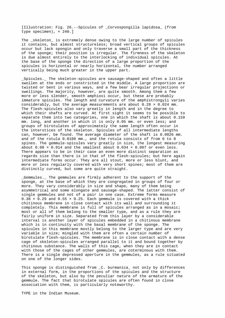
[Illustration: Fig. 26.--Spicules of _Corvospongilla lapidosa_ (fromtype specimen), × 240.]
The _skeleton_ is extremely dense owing to the large number of spiculesit contains, but almost structureless; broad vertical groups of spiculesoccur but lack spongin and only traverse a small part of the thicknessof the sponge; their position is irregular. The firmness of the skeletonis due almost entirely to the interlocking of individual spicules. Atthe base of the sponge the direction of a large proportion of thespicules is horizontal or nearly horizontal, the number arrangedvertically being much greater in the upper part.
_Spicules._ The skeleton-spicules are sausage-shaped and often a littleswollen at the ends or constricted in the middle. A large proportion aretwisted or bent in various ways, and a few bear irregular projections orswellings. The majority, however, are quite smooth. Among them a fewmore or less slender, smooth amphioxi occur, but these are probablyimmature spicules. The length and curvature of the amphistrongyli variesconsiderably, but the average measurements are about 0.28 × 0.024 mm.The flesh-spicules also vary greatly in length and in the degree towhich their shafts are curved. At first sight it seems to be possible toseparate them into two categories, one in which the shaft is about 0.159mm. long, and another in which it is only 0.05 mm. or even less; andgroups of birotulates of approximately the same length often occur inthe interstices of the skeleton. Spicules of all intermediate lengthscan, however, be found. The average diameter of the shaft is 0.0026 mm.and of the rotula 0.0106 mm., and the rotula consists of from 6 to 8spines. The gemmule-spicules vary greatly in size, the longest measuringabout 0.08 × 0.014 and the smallest about 0.034 × 0.007 or even less.There appears to be in their case an even more distinct separation asregards size than there is in that of the flesh-spicules; but here againintermediate forms occur. They are all stout, more or less blunt, andmore or less regularly covered with very short spines; most of them aredistinctly curved, but some are quite straight.
_Gemmules._ The gemmules are firmly adherent to the support of thesponge, at the base of which they are congregated in groups of four ormore. They vary considerably in size and shape, many of them beingasymmetrical and some elongate and sausage-shaped. The latter consist ofsingle gemmules and not of a pair in one case. Extreme forms measure0.38 × 0.29 and 0.55 × 0.25. Each gemmule is covered with a thickchitinous membrane in close contact with its wall and surrounding itcompletely. This membrane is full of spicules arranged as in a mosaic;most or all of them belong to the smaller type, and as a rule they arefairly uniform in size. Separated from this layer by a considerableinterval is another layer of spicules embedded in a chitinous membranewhich is in continuity with the basal membrane of the sponge. Thespicules in this membrane mostly belong to the larger type and are veryvariable in size; mingled with them are often a certain number ofbirotulate flesh-spicules. The membrane is in close contact with a densecage of skeleton-spicules arranged parallel to it and bound together bychitinous substance. The walls of this cage, when they are in contactwith those of the cages of other gemmules, are coterminous with them.There is a single depressed aperture in the gemmules, as a rule situatedon one of the longer sides.
This sponge is distinguished from _C. burmanica_ not only by differencesin external form, in the proportions of the spicules and the structureof the skeleton, but also by the peculiar nature of the armature of thegemmule. The fact that birotulate spicules are often found in closeassociation with them, is particularly noteworthy.
TYPE in the Indian Museum.

GEOGRAPHICAL DISTRIBUTION.--This sponge has only been found in theWestern Ghats of the Bombay Presidency. _Localities_:--Igatpuri Lake andthe R. Godaveri at Nasik.
BIOLOGY.--There is a remarkable difference in external form between thespecimens taken in Igatpuri and those from Nasik, and this difference isapparently due directly to environment. In the lake, the waters of whichare free from mud, the sponges were growing on the lower surface ofstones near the edge. They formed small crusts not more than about 5 cm.(2 inches) in diameter and of a pale greyish colour. Their surface wasflat or undulated gently, except round the oscula where it was raisedinto sharply conical eminences with furrowed sides. The specimens fromNasik, which is about 30 miles from Igatpuri, were attached, togetherwith specimens of _Spongilla cinerea_ and _S. indica_, to the sides of astone conduit full of very muddy running water. They were black incolour, formed broad sheets and were markedly corrugated on the surface.Their oscula were not raised on conical eminences and were altogethermost inconspicuous. The skeleton was also harder than that of spongesfrom the lake.
In the lake _C. lapidosa_ was accompanied by the gemmules of _Spongillabombayensis_, but it is interesting that whereas the latter sponge wasentirely in a resting condition, the former was in full vegetativevigour, a fact which proves, if proof were necessary, that the similarconditions of environment do not invariably have the same effect ondifferent species of Spongillidæ.
APPENDIX TO PART I.
FORM OF UNCERTAIN POSITION.
(Plate I, fig. 4.)
On more than one occasion I have found in my aquarium in Calcutta smallsponges of a peculiar type which I am unable to refer with certainty toany of the species described above. Fig. 4, pl. I, represents one ofthese sponges. They are never more than about a quarter of an inch indiameter and never possess more than one osculum. They arecushion-shaped, colourless and soft. The skeleton-spicules are smooth,sharply pointed, moderately slender and relatively large. They arearranged in definite vertical groups, which project through the dermalmembrane, and in irregular transverse formation. Small sphericalgemmules are present but have only a thin chitinous covering withoutspicules or foramen.
These sponges probably represent an abnormal form of some well-knownspecies, possibly of _Spongilla carteri_. I have seen nothing like themin natural conditions.
PART II.
FRESHWATER POLYPS
(HYDRIDA).
INTRODUCTION TO PART II.

I.
THE PHYLUM COELENTERATA AND THE CLASS HYDROZOA.
The second of the great groups or phyla into which the metazoa aredivided is the Coelenterata, in which are included most of the animalscommonly known as zoophytes, and also the corals, sea-anemones andjelly-fish. These animals are distinguished from the sponges on the onehand and from the worms, molluscs, arthropods, vertebrates, etc., on theother by possessing a central cavity (the coelenteron or "hollowinside") the walls of which are the walls of the body and consist of_two_ layers of cells separated by a structureless, or apparentlystructureless, jelly. This cavity has as a main function that of adigestive cavity.
An ideally simple coelenterate would not differ much in generalappearance from an olynthus (p. 27), but it would have no pores in thebody-wall and its upper orifice would probably be surrounded byprolongations of the body-wall in the form of tentacles. There would beno collar-cells, and the cells of the body generally would have a muchmore fixed and definite position and more regular functions than thoseof any sponge. The most characteristic of them would be the so-calledcnidoblasts. Each of these cells contains a capsule[AK] from which along thread-like body can be suddenly uncoiled and shot out.
[Footnote AK: Similar capsules are found in the tissues of certain worms and molluscs, but there is the strongest evidence that these animals, which habitually devour coelenterates, are able to swallow the capsules uninjured and to use them as weapons of defence (see Martin, Q. J. Micro. Sci. London, lii, p. 261, 1908, and Grosvenor, Proc. Roy. Soc. London, lxxii, p. 462, 1903). The "trichocysts" of certain protozoa bear a certain resemblance to the nettle-cells of coelenterates and probably have similar functions.]
The simplest in structure of the coelenterates are those that constitutethe class Hydrozoa. In this class the primitive central cavity is notdivided up by muscular partitions and there is no folding in of theanterior part of the body to form an oesophagus or stomatodæum such asis found in the sea-anemones and coral polyps. In many species andgenera the life-history is complex, illustrating what is called thealternation of generations. That is to say, only alternate generationsattain sexual maturity, those that do so being produced as buds from asexless generation, which itself arises from the fertilized eggs of aprevious sexual generation. The sexual forms as a rule differconsiderably in structure from the sexless ones; many medusæ are thesexual individuals in a life-cycle in which those of the sexlessgeneration are sedentary.
An excellent general account of the coelenterates will be found in theCambridge Natural History, vol. i (by Prof. Hickson).
STRUCTURE OF HYDRA.
_Hydra_, the freshwater polyp, is one of the simplest of the Hydrozoaboth as regards structure and as regards life-history. Indeed, itdiffers little as regards structure from the ideally simple coelenteratesketched in a former paragraph, while its descent is direct from onepolyp to another, every generation laying its own eggs[AL]. The animal

may be described as consisting of the following parts:--(1) an upright(or potentially upright) column or body, (2) a circle of contractiletentacles at the upper extremity of the column, (3) an oral disk orperistome surrounding the mouth and surrounded by the tentacles, and (4)a basal or aboral disk at the opposite extremity. The whole animal issoft and naked. The column, when the animal is at rest, is almostcylindrical in some forms but in others has the basal part distinctlynarrower than the upper part. It is highly contractile and whencontracted sometimes assumes an annulate appearance; but as a rule theexternal surface is smooth.
[Footnote AL: The statement is not strictly accurate as regards the Calcutta phase of _H. vulgaris_, for the summer brood apparently does not lay eggs but reproduces its species by means of buds only. This state of affairs, however, is probably an abnormality directly due to environment.]
The tentacles vary in number, but are never very numerous. They aredisposed in a single circle round the oral disk and are hollow, eachcontaining a prolongation of the central cavity of the column. Like thecolumn but to an even greater degree they are contractile, and in someforms they are capable of great elongation. They cannot seize any objectbetween them, but are able to move in all directions.
The disk that surrounds the mouth, which is a circular aperture, isnarrow and can to some extent assume the form of a conical proboscis,although this feature is never so marked as it is in some hydroids. Thebasal disk is even narrower and is not splayed out round the edges.
[Illustration: Fig. 27.--Nettle-cells of _Hydra_.
A=capsules from nettle-cells of a single specimen of the summer phase of_H. vulgaris_ from Calcutta, × 480: figures marked with a dash representcapsules with barbed threads. B=a capsule with the thread discharged,from the same specimen, × 480. C=capsule with barbed thread, from aspecimen of _H. oligactis_ from Lahore. D=undischarged nettle-cell of_H. vulgaris_ from Europe (after Nussbaum, highly magnified).E=discharged capsule of the same (after the same author)._a_=cnidoblast; _b_=capsule; _c_=thread; _d_=cnidocil. Only the base ofthe thread is shown in E.]
A section through the body-wall shows it to consist of the three typicallayers of the coelenterates, viz., (i) an outer cellular layer ofcomparatively small cells, the ectoderm; (ii) an intermediate,structureless or apparently structureless layer, the mesogloea or"central jelly"; and (iii) an internal layer or endoderm consisting ofrelatively large cells. The cells of the ectoderm are not homogeneous.Some of them possess at their base narrow and highly contractileprolongations that exercise the functions of muscles. Others aregland-cells and secrete mucus; others have round their margins delicateramifying prolongations and act as nerve-cells. Sense-cells, each ofwhich bears on its external surface a minute projecting bristle, arefound in connection with the nerve-cells, and also nettle-cells of morethan one type.
The mesogloea is very thin.
The endoderm consists mainly of comparatively large cells with polygonalbases which can be seen from the external surface of the column incolourless individuals. Their inner surface is amoeboid and in certainconditions bears one or more vibratile cilia or protoplasmic lashes.Nettle-cells are occasionally found in the endoderm, but apparently donot originate in this layer.

The walls of the tentacles do not differ in general structure from thoseof the column, but the cells of the endoderm are smaller and thenematocysts of the ectoderm more numerous, and there are other minordifferences.
A more detailed account of the anatomy of _Hydra_ will be found in anybiological text-book, for instance in Parker's Elementary Biology; butit is necessary here to say something more as regards the nettle-cells,which are of great biological and systematic importance.
A nettle-cell of the most perfect type and the structures necessary toit consist of the following parts:--
(1) A true cell (the cnidoblast), which contains-- (2) a delicate capsule full of liquid; (3) a long thread coiled up in the capsule; and (4) a cnidocil or sensory bristle, which projects from the external surface of the cnidoblast.
A nerve-cell is associated with each cnidoblast.
In _Hydra_ the nettle-cells are of two distinct types, in one of whichthe thread is barbed at the base, whereas in the other it is simple.Both types have often two or more varieties and intermediate formsoccur, but generally speaking the capsules with simple threads are muchsmaller than those with barbed ones. The arrangement of the nettle-cellsis not the same in all species of _Hydra_, but as a rule they are muchmore numerous in the tentacles than elsewhere on the body, each largecell being surrounded by several small ones. The latter are always muchmore numerous than the former.
CAPTURE AND INGESTION OF PREY: DIGESTION.
The usual food of _Hydra_ consists of small insect larvæ, worms, andcrustacea, but the eggs of fish are also devoured. The method in whichprey is captured and ingested has been much disputed, but the followingfacts appear to be well established.
If a small animal comes in contact with the tentacles of the polyp, itinstantly becomes paralysed. If it adheres to the tentacle, it perishes;but if, as is often the case, it does not do so, it soon recovers thepower of movement. Animals which do not adhere are generally those (suchas ostracod crustacea) which have a hard integument without weak spots.Nematocysts of both kinds shoot out their threads against prey withconsiderable violence, the discharge being effected, apparently inresponse to a chemical stimulus, by the sudden uncoiling of the threadand its eversion from the capsule. Apparently the two kinds of threadshave different functions to perform, for whereas there is no doubt thatthe barbed threads penetrate the more tender parts of the body againstwhich they are hurled, there is evidence that the simple threads do notdo so but wrap themselves round the more slender parts. Nussbaum (Arch.mikr. Anat. xxix, pl. xx, fig. 108) figures the tail of a _Cyclops_attacked by _Hydra vulgaris_ and shows several simple threads wrappedround the hairs and a single barbed thread that has penetrated theintegument. Sometimes the cyst adheres to the thread and remainsattached to its cnidoblast and to the polyp, but sometimes the threadbreaks loose. Owing to the large mass of threads that sometimescongregate at the weaker spots in the external covering of an animalattacked (_e. g._, at the little sensory pits in the integument of thedorsal surface of certain water-mites) it is often difficult to traceout the whole length of any one thread, and as a thread still attachedto its capsule is frequently buried in the body of the prey, right up to

the barbs, while another thread that has broken loose from its capsuleappears immediately behind the fixed one, it seems as though the barbs,which naturally point towards the capsule, had become reversed. Thisappearance, however, is deceptive. The barbs are probably connected withthe discharge of the thread and do not function at all in the same wayas those on a spear- or arrow-head, never penetrating the object againstwhich the projectile is hurled. Indeed, their position as regards thethread resembles that of the feathers on the shaft of an arrow ratherthan that of the barb of the head.
Adhesion between the tentacles and the prey is effected partly by thegummy secretion of the glands of the ectoderm, which is perhapspoisonous as well as adhesive, and partly by the threads. Once the preyis fast and has ceased to struggle, it is brought to the mouth, whichopens wide to receive it, by the contraction and the contortions of thetentacles, the column, and the peristome. At the same time a mass oftransparent mucus from the gastral cavity envelops it and assists indragging it in. There is some dispute as to the part played by thetentacles in conveying food into the mouth. My own observations lead meto think that, at any rate so far as _H. vulgaris_ is concerned, they donot push it in, but sometimes in their contortions they even enter thecavity accidentally.
When the food has once been engulfed some digestive fluid is apparentlypoured out upon it. In _H. vulgaris_ it is retained in the upper part ofthe cavity and the soluble parts are here dissolved out, the insolubleparts such as the chitin of insect larvæ or crustacea being ejected fromthe mouth. Digestion is, however, to a considerable extentintracellular, for the cells of the endoderm have the power of thrustingout from their surface lobular masses of their cell-substance in whichminute nutritive particles are enveloped and dissolved. The movements ofthe cilia which can also be thrust out from and retracted into thesecells, keep the food in the gastral cavity in motion and probably turnit round so as to expose all parts in turn to digestive action. Completedigestion, at any rate in the Calcutta form, takes several days toaccomplish, and after the process is finished a flocculent mass ofcolourless excreta is emitted from the mouth.
COLOUR.
In _Hydra viridis_, a species that has not yet been found in India, thegreen colour is due to the presence in the cells of green corpuscleswhich closely resemble those of the cells of certain freshwater sponges.They represent a stage in the life-cycle of _Chlorella vulgaris_,Beyerinck[AM], an alga which has been cultivated independently.
[Footnote AM: Bot. Zeitung, xlviii (1890): see p. 49, _antea_.]
In other species of the genus colour is largely dependent on food,although minute corpuscles of a _dark_ green shade are sometimes foundin the cells of _H. oligactis_. In the Calcutta phase of _H. vulgaris_colour is due entirely to amorphous particles situated mainly in thecells of the endoderm. If the polyp is starved or exposed to a hightemperature, these particles disappear and it becomes practicallycolourless. They probably form, therefore, some kind of food-reserve,and it is noteworthy that a polyp kept in the unnatural conditions thatprevail in a small aquarium invariably becomes pale, and that itsexcreta are not white and flocculent but contain dark granulesapparently identical with those found in the cells of colouredindividuals (p. 154).
Berninger[AN] has just published observations on the effect oflong-continued starvation on _Hydra_ carried out in Germany. He finds

that the tentacles, mouth, and central jelly disappear, and that aclosed "bladder" consisting of two cellular layers remains; but, tojudge from his figures, the colour does not disappear in thesecircumstances.
[Footnote AN: Zool. Anz. xxxvi, pp. 271-279, figs., Oct. 1910.]
BEHAVIOUR.
_Hydra viridis_ is a more sluggish animal than the other species of itsgenus and does not possess the same power of elongating its column andtentacles. It is, nevertheless, obliged to feed more frequently. Wagner(Quart. J. Micr. Sci. xlviii, p. 586, 1905) found it impossible to usethis species in his physiological experiments because it died ofstarvation more rapidly than other forms. This fact is interesting inview of the theory that the green corpuscles in the cells of _H.viridis_ elaborate nutritive substances for its benefit. _H. vulgaris_,at any rate in Calcutta, does not ordinarily capture prey more oftenthan about once in three days.
All _Hydræ_ (except possibly the problematical _H. rubra_ of Roux, p.160) spend the greater part of their time attached by the basal disk tosome solid object, but, especially in early life, _H. vulgaris_ is oftenfound floating free in the water, and all the species possess powers ofprogression. They do not, however, all move in the same way. _H.viridis_ progresses by "looping" like a geometrid caterpillar. Duringeach forward movement the column is arched downwards so that theperistome is in contact with the surface along which the animal ismoving. The basal disk is then detached and the column is twisted rounduntil the basal disk again comes in contact with the surface at a pointsome distance in advance of its previous point of attachment. Themanoeuvre is then repeated. _H. vulgaris_, when about to move, bendsdown its column so that it lies almost prone, stretches out itstentacles, which adhere near the tips to the surface (p. 153), detachesits basal disk, and then contracts the tentacles. The column is draggedforward, still lying almost prone, the basal disk is bent downwards andagain attached, and the whole movement is repeated. Probably _H.oligactis_ moves in the same way.
When _H. viridis_ is at rest the tentacles and column, according toWagner, exhibit rhythmical contractions in which those of the buds actin sympathy with those of the parent. In _H. vulgaris_ no such movementshave been observed. This species, however, when it is waiting for prey(p. 154) changes the direction of its tentacles about once in half anhour.
All species of _Hydra_ react to chemical and physical stimuli bycontraction and by movements of the column and tentacles, but if thestimuli are constantly repeated, they lose the power to some extent. Allspecies are attracted by light and move towards the point whence itreaches them. _H. vulgaris_, however, at any rate in India, is morestrongly repelled by heat. Consequently, if it is placed in a glassvessel of water, on one side of which the sun is shining directly, itmoves away from the source of the light[AO]. But if the vessel beprotected from the direct rays of the sun and only a subdued light fallson one side of it, the polyp moves towards that side. No species of thegenus is able to move in a straight line. Wilson (Amer. Natural. xxv, p.426, 1891) and Wagner (_op. cit. supra_) have published charts showingthe elaborately erratic course pursued by a polyp in moving from onepoint to another and the effect of light as regards its movements.
[Footnote AO: Mr. F. H. Gravely tells me that this is also

the case as regards _H. viridis_ in England, at any rate if freshly captured specimens are placed overnight in a bottle in a window in such a position that the early morning sunlight falls upon one side of the bottle.]
If an individual of _H. vulgaris_ that contains half digested food inits gastral cavity is violently removed from its natural surroundingsand placed in a glass of water, the column and tentacles contractstrongly for a few minutes. The body then becomes greatly elongated andthe tentacles moderately so; the tentacles writhe in all directions(their tips being sometimes thrust into the mouth), and the food isejected.
REPRODUCTION.
Reproduction takes place in _Hydra_ (i) by means of buds, (ii) by meansof eggs, and (iii) occasionally by fission.
(a) _Sexual Reproduction._
The sexual organs consist of ovaries (female) and spermaries (male).Sometimes the two kinds of organs are borne by the same individualeither simultaneously or in succession, but some individuals or racesappear to be exclusively of one sex. There is much evidence that inunfavourable conditions the larger proportion of individuals developonly male organs.
In temperate climates most forms of _Hydra_ breed at the approach ofwinter, but starvation undoubtedly induces a precocious sexual activity,and the same is probably the case as regards other unfavourableconditions such as lack of oxygen in the water and either too high ortoo low a temperature.
Downing states that in N. America (Chicago) _H. vulgaris_ breeds inspring and sometimes as late as December; in Calcutta it has only beenfound breeding in February and March. Except during the breeding-seasonsexual organs are absent; they do not appear in the same position on thecolumn in all species.
The spermaries take the form of small mound-shaped projections on thesurface of the column. Each consists of a mass of sperm-mother cells, inwhich the spermatozoa originate in large numbers. The spermatozoaresemble those of other animals, each possessing a head, which is shapedlike an acorn, and a long vibratile tail by means of which it movesthrough the water. In the cells of the spermary the spermatozoa areclosely packed together, with their heads pointing outwards towards thesummit of the mound through which they finally make their way into thewater. The aperture is formed by their own movements. Downing (Zool.Jahrb. (Anat.) xxi, p. 379, 1905) and other authors have studied theorigin of the spermatozoa in great detail.
[Illustration: Fig. 28.--Eggs of _Hydra_ (magnified).
A=egg of _H. vulgaris_ (after Chun). B=vertical section through egg of_H. oligactis_, form A (after Brauer). C=vertical section through egg of_H. oligactis_, form B (after Brauer).]
The ovaries consist of rounded masses of cells lying at the base of theectoderm. One of these cells, the future egg, grows more rapidly thanthe others, some or all of which it finally absorbs by means of lobosepseudopodia extruded from its margin. It then makes its way by amoeboidmovements between the cells of the ectoderm until it reaches the

surface. In _H. vulgaris_ (Mem. Asiat. Soc. Beng. i, p. 350, 1906) theegg is first visible with the aid of a lens as a minute star-shaped bodyof an intense white colour lying at the base of the ectoderm cells. Itincreases in size rapidly, gradually draws in its pseudopodia (the raysof the star) and makes its way through the ectoderm to the exterior. Theprocess occupies not more than two hours. The issuing ovum does notdestroy the ectoderm cells as it passes out, but squeezes them togetherround the aperture it makes. Owing to the pressure it exerts upon them,they become much elongated and form a cup, in which the embryo rests onthe surface of the parent. By the time that the egg has become globular,organic connection has ceased to exist. The embryo is held in positionpartly by means of the cup of elongated ectoderm cells and partly by adelicate film of mucus secreted by the parent. The most recent accountof the oogenesis ("ovogenesis") is by Downing (Zool. Jahrb. (Anat.)xxvii, p. 295, 1909).
(b) _Budding._
The buds of _Hydra_ arise as hollow outgrowths from the wall of thecolumn, probably in a definite order and position in each species. Thetentacles are formed on the buds much as the buds themselves arise onthe column. There is much dispute as to the order in which thesestructures appear on the bud, and Haacke (Jenaische Zeitschr. Naturwiss.xiv, p. 133, 1880) has proposed to distinguish two species, _H.trembleyi_ and _H. roeselii_, in accordance with the manner in which thephenomenon is manifested. It seems probable, however, that the number oftentacles that are developed in the first instance is due, at any rateto some extent, to circumstances, for in the summer brood of _H.vulgaris_ in Calcutta five usually appear simultaneously, while in thewinter brood of the same form four as a rule do so. Sometimes budsremain attached to their parents sufficiently long to develop budsthemselves, so that temporary colonies of some complexity arise, but Ihave not known this to occur in the case of Indian individuals.
(c) _Fission._
Reproduction by fission occurs naturally but not habitually in allspecies of _Hydra_. It may take place either by a horizontal or by avertical division of the column. In the latter case it may be eitherequal or unequal. If equal, it usually commences by an elongation in onedirection of the circumoral disk, which assumes a narrowly oval form;the tentacles increase in number, and a notch appears at either side ofthe disk and finally separates the column into two equal halves, each ofwhich is a complete polyp. The division sometimes commences at the baseof the column, but this is very rare. Transverse fission can be inducedartificially and is said to occur sometimes in natural conditions. Itcommences by a constriction of the column which finally separates theanimal into two parts, the lower of which develops tentacles and amouth, while the upper part develops a basal disk. Unequal verticaldivision occurs when the column is divided vertically in such a way thatthe two resulting polyps are unequal in size. It is apparently notaccompanied by any great increase in the number of the tentacles, butprobably starts by one of the tentacles becoming forked and finallysplitting down the middle.
The question of the regeneration of lost parts in _Hydra_ cannot well beseparated from that of reproduction by fission. Over a hundred and fiftyyears ago Trembley found that if a polyp were cut into several pieces,each piece produced those structures necessary to render it a perfectpolyp. He also believed that he had induced a polyp that had been turnedinside out to adapt itself to circumstances and to reverse the functionsand structure of the two cellular layers of its body. In this, however,

he was probably mistaken, for there can be little doubt that his polypturned right side out while not under his immediate observation. Manyinvestigators have repeated some of his other experiments with successin Europe, but the Calcutta _Hydra_ is too delicate an animal to survivevivisection and invariably dies if lacerated. It appears that, even infavourable circumstances, for a fresh polyp to be formed by artificialfission it is necessary for the piece to contain cells of bothcell-layers.
DEVELOPMENT OF THE EGG.
The egg of _Hydra_ is said to be fertilized as it lies at the base ofthe ectoderm, through which the fertilizing spermatozoon bores its way.As soon as the egg has emerged from the cells of its parent it begins tosplit up in such a manner as to form a hollow mass of comparativelylarge equal cells. Smaller cells are separated off from these and soonfill the central cavity. Before segmentation begins a delicate film ofmucus is secreted over the egg, and within this film the larger cellssecrete first a thick chitinous or horny egg-shell and within it adelicate membrane. Development in some cases is delayed for aconsiderable period, but sooner or later, by repeated division of thecells, an oval hollow embryo is formed and escapes into the water by thedisintegration of the egg-shell and the subsequent rupture of the innermembrane. Tentacles soon sprout out from one end of the embryo's bodyand a mouth is formed; the column becomes more slender and attachesitself by the aboral pole to some solid object.
ENEMIES.
_Hydra_ seems to have few natural enemies. Martin (Q. J. Micr. Sci.London, lii, p. 261, 1908) has, however, described how the minute worm_Microstoma lineare_ attacks _Hydra "rubra"_ in Scottish lochs, whilethe larva of a midge devours _H. vulgaris_ in considerable numbers inCalcutta tanks (p. 156).
COELENTERATES OF BRACKISH WATER.
Marine coelenterates of different orders not infrequently make their wayor are carried by the tide up the estuaries of rivers into brackishwater, and several species have been found living in isolated lagoonsand pools of which the water was distinctly salt or brackish. Among themost remarkable instances of such isolation is the occurrence in LakeQurun in the Fayûm of Egypt of _Cordylophora lacustris_ and of thepeculiar little hydroid recently described by Mr. C. L. Boulenger as_Moerisia lyonsi_ (Q. J. Micr. Sci. London, lii, p. 357, pls. xxii,xxiii, 1908). In the delta of the Ganges there are numerous ponds whichhave at one time been connected with estuaries or creeks of brackishwater and have become isolated either naturally or by the hand of manwithout the marine element in their fauna by any means disappearing (p.14). The following species have been found in such ponds:--
(_a_) _Hydrozoa._
(1) _Bimeria vestita_, Wright (1859).
Hincks, Hist. Brit. Hydr. Zooph. p. 103, pl. xv, fig. 2 (1868);Annandale, Rec. Ind. Mus. i, p. 141, fig. 3 (1907).
This is a European species which has also been found off S. America. Itoccurs not uncommonly in the creeks that penetrate into the Ganges deltaand has been found in pools of brackish water at Port Canning. The

Indian form is perhaps sufficiently distinct to be regarded as asubspecies. The medusoid generation is suppressed in this genus.
(2) _Syncoryne filamentata_, Annandale (1907).
Annandale, Rec. Ind. Mus. i, p. 139, figs. 1, 2 (1907).
Both hydroid and medusæ were found in a small pool of brackish water atPort Canning. The specific name refers to the fact that the ends of therhizomes from which the polyps arise are frequently free and elongate,for the young polyp at the tip apparently takes some time to assume itsadult form.
(3) _Irene ceylonensis_, Browne (1905).
Browne, in Herdman's Report on the Pearl Fisheries of Ceylon, iv, p. 140, pl. iii, figs. 9-11 (1905); Annandale, Rec. Ind. Mus. i, p. 142, fig. 4 (1907).
The medusa was originally taken off the coast of Ceylon, while thehydroid was discovered in ponds of brackish water at Port Canning. It isalmost microscopic in size.
The first two of these species belong to the order Gymnoblastea(Anthomedusæ) and the third to the Calyptoblastea (Leptomedusæ).
(b) _Actinozoa._
(4) _Sagartia schilleriana_, Stoliczka (1869).
_S. schilleriana_, Stoliczka, Journ. As. Soc. Beng. (2) xxxviii, p. 28, pls. x, xi (1869); _Metridium schillerianum_, Annandale, Rec. Ind. Mus. i, p. 47, pl. iii (1907).
This sea-anemone, which has only been found in the delta of the Ganges,offers a most remarkable instance of what appears to be rapid adaptationof a species to its environment. The typical form, which was describedin 1869 by Stoliczka from specimens taken in tidal creeks and estuariesin the Gangetic area and in the ponds at Port Canning, is found attachedto solid objects by its basal disk. The race (subsp. _exul_), however,that is now found in the same ponds has become elongate in form and hasadopted a burrowing habit, apparently owing to the fact that the bottomof the ponds in which it lives is soft and muddy.
In addition to these four species a minute hydroid belonging to theorder Gymnoblastea and now being described by Mr. J. Ritchie has beentaken in the ponds at Port Canning. It is a very aberrant form.
FRESHWATER COELENTERATES OTHER THAN HYDRA.
_Hydra_ is the only genus of coelenterates as yet found in fresh waterin India, but several others have been discovered in other countries.They are:--
(1) _Cordylophora lacustris_, Allman (1843).
Hincks, Hist. Brit. Hydr. Zooph. p. 16, pl. iii, fig. 2 (1868).
This is a branching hydroid that does not produce free medusæ. It formsbushy masses somewhat resembling those formed by a luxuriant growth of_Plumatella fruticosa_ (pl. iii, fig. 1) in general appearance. _C.

lacustris_ is abundant in canals, rivers, and estuaries in many parts ofEurope and has recently been found in the isolated salt lakeBirket-el-Qurun in the Fayûm of Egypt.
(2) _Cordylophora whiteleggei_, v. Lendenfeld (1887).
Zool. Jahrb. ii, p. 97 (1887).
A species or race of much feebler growth; as yet imperfectly known andonly recorded from fresh water in Australia.
_Cordylophora_ is a normal genus of the class Hydrozoa and the orderGymnoblastea; the next four genera are certainly Hydrozoa, but theiraffinities are very doubtful.
(3) _Microhydra ryderi_, Potts (1885).
Potts, Q. J. Micr. Sci. London, l, p. 623, pls. xxxv, xxxvi; Browne, _ibid._ p. 635, pl. xxxvii (1906).
This animal, which has been found in N. America and in Germany,possesses both an asexual hydroid and a sexual medusoid generation. Theformer reproduces its species by direct budding as well as by givingrise, also by a form of budding, to medusæ that become sexually mature.The hydroid has no tentacles.
(4) _Limnocodium sowerbii_, Lankester (1880).
Lankester, Q. J. Micr. Sci. London, xx, p. 351, pls. xxx, xxxi (1880); Fowler, _ibid._ xxx, p. 507, pl. xxxii (1890).
There is some doubt as to the different stages in the life-cycle of thisspecies. The medusa has been found in tanks in hot-houses in England,France and Germany, and a minute hydroid closely resembling that of_Microhydra ryderi_ has been associated with it provisionally.
(5) _Limnocodium kawaii_, Oka (1907).
Oka, Annot. Zool. Japon. vi, p. 219, pl. viii (1907).
Only the medusa, which was taken in the R. Yang-tze-kiang, is as yetknown.
(6) _Limnocnida tanganyikæ_, Bohm (1889).
R. T. Günther, Ann. Nat. Hist. (6) xi, p. 269, pls. xiii, xiv (1893).
Only the medusa, which is found in Lake Tanganyika, Lake Victoria Nyanzaand the R. Niger, has been found and it is doubtful whether a hydroidgeneration exists.
(7) _Polypodium hydriforme_, Ussow (1885).
Morph. Jahrb. xii, p. 137 (1887).
Two stages in this peculiar hydroid, which is found in the R. Volga, areknown, (_a_) a spiral ribbon-like form parasitic on the eggs of thesterlet (_Acipenser ruthenus_), and (_b_) a small _Hydra_-like form withboth filamentous and club-shaped tentacles. The life-history has not yetbeen worked out[AP].
[Footnote AP: Since this was written, Lippen has described a third stage in the life-history of _Polypodium_ (Zool. Anz.
Leipzig, xxxvii, Nr. 5, p. 97 (1911)).]
II.
HISTORY OF THE STUDY OF HYDRA.
Hydra was discovered by Leeuwenhoek at the beginning of the eighteenthcentury and had attracted the attention of several skilful and accurateobservers before that century was half accomplished. Among them thechief was Trembley, whose "Mémoires pour servir à l'histoire d'un genrede Polype d'eau douce"* was published at Paris 1744, and is remarkablenot only for the extent and accuracy of the observations it enshrinesbut also for the beauty of its plates. Baker in his work entitled "Anattempt towards a natural history of the Polyp"* (London, 1743) andRösel von Rosenhof in the third part of his "Insecten-Belustigung"(Nurenberg, 1755) also made important contributions to the study of thephysiology and structure of _Hydra_ about the same period. Linnéinvented the name _Hydra_, and in his "Fauna Sueica" and in the variouseditions of his "Systema Naturæ" described several forms in a mannerthat permits some of them to be recognized; but Linné did notdistinguish between the true _Hydra_ and other soft sessileCoelenterates, and it is to Pallas ("Elenchus Zoophytorum," 1766) thatthe credit properly belongs of reducing the genus to order. It is atribute to his insight that three of the four species he described arestill accepted as "good" by practically all students of theCoelenterates, while the fourth was a form that he had not himself seen.
In the nineteenth century the freshwater polyp became a favourite objectof biological observation and was watched and examined by a host ofobservers, among the more noteworthy of whom were Kleinenberg, Nussbaum,and Brauer, who has since the beginning of the present century made animportant contribution to the taxonomy of the genus.
BIBLIOGRAPHY OF HYDRA.
_Hydra_ has been examined by thousands of students in biologicallaboratories all over the civilized world, and the literature upon it ishardly surpassed in magnitude by that on any other genus but _Homo_. Thefollowing is a list of a few of the more important general memoirs andof the papers that refer directly to Asiatic material. A systematicbibliography is given by Bedot in his "Matériaux pour servir al'Histoire des Hydroïdes," Rev. Suisse Zool. xviii, fasc. 2 (1910).
(a) _General._
1743. BAKER, "An attempt towards a natural history of the Polyp"*(London).
1744. TREMBLEY, "Mémoires pour servir à l'histoire d'un genre de polypesd'eau douce"* (Paris).
1755. RÖSEL VON ROSENHOF, "Insecten-Belustigung: iii, Hist. Polyporum."
1766. PALLAS, "Elenchus Zoophytorum."
1844. LAURENT, "Rech. sur l'Hydre et l'Eponge d'eau douce" ("Voy. de laBonite, Zoophytologie").
1847. JOHNSTON, "A History of the British Zoophytes" (2nd edition).
1868. HINCKS, "History of British Hydroid Zoophytes."

1872. KLEINENBERG, "Hydra. Eine Anatomisch EntwicklungsgeschichtlicheUntersuchung."
1882. JICKELI, "Der Bau der Hydroidpolypen," Morph. Jahrb. viii, p. 373.
1887. NUSSBAUM, "Ueber die Theilbarkeit der lebendigen Materie. II.Mittheilung. Beiträge zur Naturgeschichte des Genus Hydra," Arch. mikr.Anat. Bonn, xxix, p. 265.
1891. BRAUER, "Über die Entwicklung von Hydra," Zeitschr. wiss. Zool.Leipzig, lii, p. 169.
1892. CHUN, "Coelenterata (Hohlthiere)," in Bronn's Thier-Reichs II (2).
1905. DOWNING, "The spermatogenesis of Hydra," Zool. Jahrb. (Anat.) xxi,p. 379.
1908. BRAUER, "Die Benennung und Unterscheidung der Hydra-Arten," Zool.Ann. xxxiii, p. 790.
1909. FRISCHHOLZ, "Biologie und Systematik im Genus Hydra," Braun'sAnnal. Zool. (Würzburg) iii, p. 105.
1910. BERNINGER, "Über Einwirkung des Hungers auf Hydra," Zool. Anz.xxxvi, p. 271.
(b) _Asiatic References._
1894. RICHARD, "Sur quelques Animaux inférieurs des eaux douces duTonkin (Protozoaires, Rotifères, Entomostracés)," Mém. Soc. zool.France, vii, p. 237.
1904. VON DADAY, "Mikroskopische Süsswasserthiere aus Turkestan," Zool.Jahrb. (Syst.) xix, p. 469.
1906. ANNANDALE, "Notes on the Freshwater Fauna of India. No. IV. _Hydraorientalis_ and its bionomical relations with other Invertebrates," J.Asiat. Soc. Bengal (new series), ii, p. 109.
1906. ANNANDALE, "The Common _Hydra_ of Bengal: its Systematic Positionand Life History," Mem. As. Soc. Bengal, i, p. 339.
1907. ANNANDALE, "Notes on the Freshwater Fauna of India. No. X. _Hydraorientalis_ during the Rains," J. Asiat. Soc. Bengal (new series), iii,p. 27.
1907. ANNANDALE, "Notes on the Freshwater Fauna of India. No. XI.Preliminary Note on the occurrence of a Medusa (_Irene ceylonensis_,Browne) in a brackish pool in the Ganges Delta and on the Hydroid Stageof the species," J. Asiat. Soc. Bengal (new series), iii, p. 79.
1907. WILLEY, "Freshwater Sponge and Hydra in Ceylon," Spolia Zeylan.Colombo, iv, p. 184.
1908. ANNANDALE, "Observations on specimens of _Hydra_ from Tibet, withnotes on the distribution of the genus in Asia," Rec. Ind. Mus. ii, p.311.
1910. POWELL, "Lessons in Practical Biology for Indian Students"(Bombay).
1910. LLOYD, "An Introduction to Biology for Students in India"(London).
GLOSSARY OF TECHNICAL TERMS USED IN PART II.
_Aboral_ (or _basal_) The disk by means of which a free polyp _disk_ attaches itself to external objects.
_Cnidoblast_ The living cell of the nematocyst or nettle-cell (_q. v._).
_Cnidocil_ A minute bristle that projects on the surface in connection with a nettle-cell (_q. v._).
_Column_ The upright or potentially upright part of a polyp (_q. v._).
_Ectoderm_ The external cell-layer of the body-wall.
_Endoderm_ The internal cell-layer of the body-wall.
_Green (chlorophyll) Minute green bodies contained in cells corpuscles_ of polyps or other animals and representing a stage in the life-history of an alga (_Chlorella_).
_Mesogloea_ The intermediate, gelatinous layer of the body-wall.
_Nettle-cell (nematocyst)_ A cell capsule full of liquid in which an eversible thread is coiled up.
_Oral disk_ The eminence that surrounds the mouth and is surrounded by tentacles.
_Peristome_ See "oral disk."
_Polyp_ An individual coelenterate of simple structure that is fixed temporarily or permanently by one end of a more or less cylindrical body and possesses a mouth at the other end.
_Tentacles_ Filamentous outgrowths (in _Hydra_ hollow) of the body-wall round the mouth.
LIST OF THE INDIAN HYDRIDA.
Class HYDROZOA.
Order ELEUTHEROBLASTEA.
Family HYDRIDÆ.
Genus HYDRA, _Linné_ (1746).
24. _H. vulgaris_, Pallas (1766).

25. _H. oligactis_, Pallas (1766).
Order ELEUTHEROBLASTEA.
Naked hydrozoa which reproduce their kind by means of buds or eggs, orby fission, without exhibiting the phenomena of alternation ofgenerations.
Family HYDRIDÆ.
HYDRAIDÆ, Johnston, Hist. Brit. Zooph. (ed. 2) i, p. 120 (1847). HYDRIDÆ, Hincks, Hist. Brit. Hydroid. Zooph. p. 309 (1868).
Small Eleutheroblastea in which the mouth is surrounded by hollowtentacles. Permanent colonies are not formed, but reproduction bybudding commonly takes place.
Genus HYDRA, _Linné_.
TYPE, _Hydra viridis_, Linné.
Freshwater polyps which produce eggs with hard chitinous shells.Although habitually anchored by the end of the body furthest from themouth to extraneous objects, they possess considerable powers oflocomotion. They are extremely contractile and change greatly from timeto time in both form and size.
Only three well-established species of the genus, which is universallydistributed and occurs only in fresh or brackish[AQ] water, can berecognized, namely, _H. viridis_, Linné (=_H. viridissima_, Pallas), _H.vulgaris_, Pallas (=_H. grisea_, Linné), and _H. oligactis_, Pallas(=_H. fusca_, Linné). The two latter occur in India, but _H. viridis_does not appear to have been found as yet anywhere in the OrientalRegion, although it is common all over Europe and N. America and also inJapan. The distribution of _H. vulgaris_ is probably cosmopolitan, butthere is some evidence that _H. oligactis_ avoids tropical districts,although, under the name _Hydra fusca_, it has been doubtfully recordedas occurring in Tonquin[AR].
[Footnote AQ: A small form of _H. viridis_ (var. _bakeri_, Marshall) is found in brackish water in England.]
[Footnote AR: Richard, Mém. Soc. zool. France, vii, p. 237 (1894).]
The three species may be distinguished from one another by the followingkey:--
[I. Colour leaf-green; the cells contain green (chlorophyll) corpuscles of definite form. A. Tentacles comparatively stout, habitually shorter than the column, which is cylindrical. Egg-shell without spines, ornamented with a reticulate pattern _viridis_.] II. Colour never leaf-green; no chlorophyll corpuscles present in the cells. A. Tentacles capable of great elongation but when the animal is at rest never very much longer than the column, which is cylindrical

when the gastral cavity is empty. Largest nettle-cells almost as broad as long. Egg-shell bearing long spines most of which are divided at the tips _vulgaris_, p. 148. B. Tentacles, even when the animal is at rest, much longer than the column, the basal part of which, even when the gastral cavity is empty, is constricted. Largest nettle-cells considerably longer than broad. Egg-shell smooth or bearing short, simple spines _oligactis_, p. 158.
24. Hydra vulgaris, _Pallas_.
Polypes de la seconde espèce, Trembley, Mém. pour servir à l'histoire d'un genre de polypes d'eau douce*, pl. i, figs. 2, 5; pl. vi, figs. 2, 8; pl. viii, figs. 1-7; pl. xi, figs. 11-13 (1744).
Rösel von Rosenhof, Insecten-Belustigung, iii, Hist. Polyporum, pls. lxxvi, lxxvii, lxxix-lxxxiii (1755).
? _Hydra polypus_, Linné, Fauna Suecica, p. 542 (1761).
_Hydra vulgaris_, Pallas, Elenchus Zoophytorum, p. 30 (1766).
? _Hydra attenuata_, _id_., _ibid_. p. 32.
_Hydra grisea_, Linné (Gmelin), Systema Naturæ (ed. 13), p. 3870 (1782).
_Hydra pallens_, _id_., _ibid_. p. 3871.
_Hydra vulgaris_, Ehrenberg, Abhandl. Akad. Wiss. Berlin, 1836, p. 134, taf. ii.
_Hydra brunnea_, Templeton, London's Mag. Nat. Hist. ix, p. 417 (1836).
_Hydra vulgaris_, Laurent, Rech. sur l'Hydre at l'Éponge d'eau douce (Voy. de la Bonite, Zoophytologie), p. 11, pl. i, pl. ii, figs. 2, 2'' (1844).
_Hydra vulgaris_, Johnston, Hist. British Zoophytes (ed. 2), i, p. 122, pl. xxix, fig. 2 (1847).
_Hydra vulgaris_, Hincks, Hist. British Hydroid Zoophytes, i, p. 314, fig. 41 (1868).
_Hydra aurantiaca_, Kleinenberg, Hydra, p. 70, pl. i, fig. 1, pl. iii, fig. 10 (1872).
_Hydra trembleyi_, Haacke, Zool. Anz. Leipzig, ii, p. 622 (1879).
_Hydra grisea_, Jickeli, Morph. Jahrb. viii, p. 391, pl. xviii, fig. 2 (1883).
_Hydra grisea_, Nussbaum, Arch. mikr. Anat. Bonn, xxix, p. 272, pl. xiii, pl. xiv, figs. 33, 37, 47 (1887).
? _Hydra hexactinella_, v. Lendenfeld, Zool. Jahrb. Jena,

ii, p. 96, pl. vi, figs. 13, 14 (1887).
? _Hydra hexactinella_, _id_., Proc. Linn. Soc. N. S. Wales, x, p. 678, p. xlviii, figs. 1-4 (1887).
_Hydra grisea_, Brauer, Zeit. wiss. Zool. Leipzig, lii, p. 169 (1891).
_Hydra grisea_, Chun, in Brönn's Thier-Reichs, ii (2), pl. ii, figs. 2_b_, 2_c_, 5 (1892).
_Hydra grisea_, Downing, Zool. Jahrb. (Anat.) Jena, xxi, p. 381 (1905).
_Hydra orientalis_, Annandale, J. Asiat. Soc. Bengal, (new series) i, 1905, p. 72.
_Hydra orientalis_, _id._, _ibid._ (new series) ii, 1906, p. 109.
_Hydra orientalis_, _id._, Mem. Asiat. Soc. Bengal, i, p. 340 (1906).
? _Hydra orientalis_, Willey, Spol. Zeylan. Colombo, iv, p. 185 (1907).
_Hydra grisea_, Weltner, Arch. Naturg. Berlin, lxxiii, i, p. 475 (1907).
_Hydra vulgaris_, Brauer, Zool. Anz. xxxiii, p. 792, fig. 1 (1908).
_Hydra orientalis_, Annandale, Rec. Ind. Mus. ii, p. 312 (1908).
_Hydra grisea_, Frischholz, Braun's Zool. Annal. (Würzburg), iii, pp. 107, 134, &c., figs. 1 and 10-17 (1909).
_Hydra grisea_, _id._, Biol. Centralbl. Berlin, xxix, p. 184 (1909).
_Hydra vulgaris_, Brauer, Die Süsswasserfauna Deutschlands, xix, p. 192, figs. 336-338 (1909).
_Hydra pentactinella_, Powell, Lessons in Practical Biology for Indian Students, p. 24 (Bombay, 1910).
Phase _orientalis*_, Annandale.
_Colour_ variable; in summer usually pale, in winter either deep orange,dull brown, or dark green. The cells do not contain spherical or ovalcoloured bodies.
[Illustration: Fig. 29.--_Hydra vulgaris_, from Calcutta (phase_orientalis_).
A=winter brood; B=summer brood, the same individual in an expanded and acontracted condition. B is more highly magnified than A.]
_Column_ slender and capable of great elongation, normally almostcylindrical, but when containing food often shaped like a wine-glass.The surface is thickly set with nettle-cells the cnidocils of which giveit an almost hirsute appearance under the microscope. When extended to

the utmost the column is sometimes nearly 30 mm. (1-1/5 inches) long,but more commonly it is about half that length or even shorter.
_Tentacles_ usually 4-6, occasionally 8. They are always slender exceptwhen they are contracted, then becoming swollen at the base and slightlyglobular at the tip. If the animal is at rest they are not very muchlonger than the body, but if it is hungry or about to move from oneplace to another they are capable of very great extension, oftenbecoming like a string of minute beads (the groups of nettle-cells)strung on an invisible wire.
_Nettle-cells._ The capsules with barbed threads (fig. 27, p. 131) arevery variable in size, but they are invariably broad in proportion totheir length and as a rule nearly spherical. In a _Hydra_ taken inCalcutta during the winter the largest capsules measured (unexploded)0.0189 mm. in breadth and 0.019 in length, but in summer they aresmaller (about 0.012 mm. in breadth). Smaller capsules with barbedthreads always occur. The barbed threads are very long and slender. Attheir base they bear a circle of stout and prominent spines, usually 4in number; above these there are a number of very small spines, but thesmall spines are usually obscure. Malformed corpuscles are common. Thecapsules with unbarbed threads are very nearly as broad at the distal asat the proximal end; they are broadly oval with rounded ends.
_Reproductive organs._ The reproductive organs are confined to the upperpart of the body. In India eggs (fig. 28, p. 137) are seldom produced.They sometimes appear, however, at the beginning of the hot weather. Inform they are spherical, and their shell bears relatively long spines,which are expanded, flattened and more or less divided at the tip. Thepart of the egg that is in contact with the parent-polyp is bare.Spermaries are produced more readily than ovaries; they are mammillatein form and number from 4 to 24. Ovaries and spermaries have not beenfound on the same individual.
_Buds_ are confined to a narrow zone nearer the base than the apex ofthe column. Rarely more than 2 are produced at a time, and I have neverseen an attached bud budding. In winter 5 tentacles are as a ruleproduced simultaneously, and in summer 4. In the former case a fifthoften makes its appearance before the bud is liberated.
In Calcutta two broods can be distinguished, a cold-weather brood, whichis larger, stouter, and more deeply coloured, produces buds more freely,has larger nematocysts, and as a rule possesses 6 tentacles; and ahot-weather brood, which is smaller, more slender and paler, producesbuds very sparingly, has smaller nematocysts, and as a rule possessesonly 4 or 5 tentacles. Only the cold-weather form is known to becomesexually mature. There is evidence, however, that in those parts ofIndia which enjoy a more uniform tropical climate than Lower Bengal,polyps found at all times of year resemble those found in the hotweather in Calcutta, and sometimes produce spermatozoa or eggs.
I have recently had an opportunity of comparing specimens of theCalcutta hot-weather form with well-preserved examples of _H. vulgaris_,Pallas (=_H. grisea_, Linn.), from England. They differ from thesepolyps in very much the same way as, but to a greater degree than theydo from the winter phase of their own race, and I have therefore nodoubt that _H. orientalis_ is merely a tropical phase of Pallas'sspecies. My description is based on Indian specimens, which seem todiffer, so far as anatomy is concerned, from European ones in thefollowing points:--
(1) The sexes are invariably distinct; (2) the nematocysts are invariably smaller.

I have seen in Burma an abnormal individual with no tentacles. Its buds,however, possessed these organs.
TYPE. None of the older types of _Hydra_ are now in existence. That of_H. orientalis_ is, however, in the collection of the Indian Museum.
GEOGRAPHICAL DISTRIBUTION.--_H. vulgaris_ is common in Europe and N.America and is probably found all over tropical Asia. The following areIndian and Ceylon localities:--BENGAL, Calcutta and neighbourhood(_Annandale_, _Lloyd_); Adra, Manbhum district (_Paiva_), Rampur Bhuliaon the R. Ganges (_Annandale_); Chakradharpur, Chota Nagpur(_Annandale_); Pusa, Bihar (_Annandale_); Puri, Orissa (_Annandale_):MADRAS, sea-beach near Madras town (_Henderson_): BOMBAY, island ofBombay (_Powell_): BURMA, Mandalay, Upper Burma, and Moulmein, N.Tenasserim (_Annandale_): CEYLON, Colombo and Peradeniya (_Willey_,_Green_). Dr. A. D. Imms tells me that he has obtained specimens thatprobably belong to this species in the Jumna at Allahabad.
BIOLOGY.--In India _H. vulgaris_ is usually found, so far as myexperience goes, in stagnant water. In Calcutta it is most abundant inponds containing plenty of aquatic vegetation, and seems to beespecially partial to the plant _Limnanthemum_, which has floatingleaves attached to thin stalks that spring up from the bottom, and to_Lemna_ (duckweed). Dr. Henderson, however, found specimens in a pool ofrain-water on the sea-shore near Madras.
There is evidence that each of the two broods which occur in LowerBengal represents at least one generation; probably it represents morethan one, for tentacles are rarely if ever produced after the animal hasobtained its full size, and never (or only owing to accident) decreasein number after they have once appeared. The winter form is foundchiefly near the surface of the water, especially on the roots ofduckweed and on the lower surface of the leaves of _Limnanthemum_; butthe summer form affects deeper water in shady places, and as a ruleattaches itself to wholly submerged plants. The latter form is to be metwith between March and October, the cold-weather form between Octoberand March, both being sometimes found together at the periods oftransition. In the unnatural environment of an aquarium, however,individuals of the winter form lose their colour and become attenuated,in these features resembling the summer form, even in the cooler months.Buds produced in these conditions rarely have more than five tentaclesor themselves produce buds freely after liberation.
The buds appear in a fixed order and position, at any rate onindividuals examined in winter; in specimens of the summer form theposition is fixed, but the order is irregular. Each quadrant of thecolumn has apparently the power of producing, in a definite zone nearerthe aboral pole than the mouth, a single bud; but the buds of thedifferent quadrants are not produced simultaneously. If we imagine thatthe quadrants face north, south, east, and west, and that the first budis produced in the north quadrant, the second will be produced in theeast quadrant, the third in the south, and the fourth in the west. It isdoubtful whether more than four buds are produced in the lifetime of anindividual, and apparently attached buds never bud in this race. Thesecond bud usually appears before the first is liberated, and this isalso the case occasionally as regards the third, but it is exceptionalfor four buds to be present at one time. About three weeks usuallyelapse between the date at which the bud first appears as a minuteconical projection on the surface of the parent and that at which itliberates itself. This it does by bending down, fixing itself to somesolid object by means of the tips of its tentacles, the gland-cells ofwhich secrete a gummy fluid, and then tearing itself free.
Although it is rare for more than two buds to be produced

simultaneously, budding is apparently a more usual form of reproductionthan sexual reproduction. Individuals that bear eggs have not yet beenfound in India in natural conditions, although males with functionalspermaries are not uncommon at the approach of the hot weather. The feweggs that I have seen were produced in my aquarium towards the end ofthe cold weather. Starvation, lack of oxygen, and too high a temperature(perhaps also lack of light) appear to stimulate the growth of the maleorgans in ordinary cases, but perhaps they induce the development ofovaries in the case of individuals that are unusually well nourished.
The spines that cover the egg retain débris of various kinds upon itssurface, so that it becomes more or less completely concealed by acovering of fragments of dead leaves and the like even before it isseparated from the polyp. Its separation is brought about by its fallingoff the column of the parent. Nothing is known of its subsequent fate,but probably it lies dormant in the mud through the hot weather. Eggsare sometimes produced that have no shells. This is probably due to thefact that they have not been fertilized.
Reproduction by fission occurs rarely in the Indian _Hydra_, but bothequal and unequal vertical fission have been observed. In the case ofequal fission the circumoral area lengthens in a horizontal direction,and as many extra tentacles as those the polyp already possesses maketheir appearance. The mouth then becomes constricted in the middle andnotches corresponding to its constriction appear at either side of theupper part of the column. Finally the whole animal divides into twoequal halves in a vertical direction. I have only seen one instance ofwhat appeared to be unequal vertical fission--that of a polyp consistingof two individuals still joined together by the basal disk, but oneabout half the size of the other. Each had three well-developedtentacles, and in addition a minute fourth tentacle. This was situatedon the side opposed to that of the other individual which bore a similartentacle. Transverse fission has not been observed. The Indian _Hydra_is a very delicate animal as compared with such a form as _H. viridis_,and all attempts to produce artificial fission without killing the polyphave as yet failed.
Young individuals are often, and adults occasionally, found floatingfree in the water, either with the mouth uppermost and the tentaclesextended so as to cover as large an area as possible or with the aboralpole at the surface. In the former case they float in mid-water, beingof nearly the same specific gravity as the water, and are carried aboutby any movement set up in it. In the latter case, however, the base ofthe column is actually attached to some small object such as the castskin of a water-flea or to a minute drop of mucus originally given outby the polyp's own mouth; the tentacles either hang downwards or arespread out round the mouth, and the animal is carried about by wind orother agencies acting on the surface.
In addition to this passive method of progression the polyp can crawlwith considerable rapidity. In doing so it bends its column down to theobject along which it is about to move in such a way that it lies almostparallel to the surface, the basal disk, however, being still attached.The tentacles are then extended and attach themselves near the tips tothe surface a considerable distance away. Attachment is effected by thesecretion of minute drops of adhesive substance from gland-cells. Thebasal disk is liberated and the tentacles contract, dragging the column,which still lies prone, along as they do so. The basal disk againaffixes itself, the tentacles wrench themselves free, the surface oftheir cells being often drawn out in the process into pseudopodia-likeprojections, which of course are not true pseudopodia[AS] but merelyprojections produced by the mechanical strain. The whole action is thenrepeated. The polyp can also pull itself across a space such as thatbetween two stems or leaves by stretching out one of its tentacles,

fixing the tip to the object it desires to reach, pulling itself freefrom its former point of attachment, and dragging itself across bycontracting the fixed tentacle. The basal disk is then turned round andfixed to the new support.
[Footnote AS: See Zykoff, Biol. Centralbl. xviii, p. 272 (1898), and Annandale, Rec. Ind. Mus. i, p. 67 (1907).]
The Indian polyp, like all its congeners, is attracted by light, but itis more strongly repelled by heat. Probably it never moves in a straightline, but if direct sunlight falls on one side of a glass aquarium, thepolyps move away from that side in a much less erratic course than isusually the case. If conditions are favourable, they often remain in onespot for weeks at a time, their buds congregating round them as they areset free. In a natural environment it seems that regular migrations takeplace in accordance with changes in temperature, for whereas in coolweather many individuals are found adhering to the lower surface of thefloating leaves of _Limnanthemum_, few are found in this positionimmediately after a rise in the thermometer. If the rise is only a smallone, they merely crawl down the stems to the end of which the leaves areattached, but as soon as the hot weather begins in earnest, the few thatsurvive make their way to the deepest and most shady part of the pond.In captivity the polyps seek the bottom of any vessel in which they arecontained, if sunlight falls on the surface of the water.
The chief function of the tentacles is that of capturing prey. TheIndian polyp feeds as a rule in the early morning, before the day hasbecome hot. In an aquarium at any rate, the tentacles are never morethan moderately extended during the night. If the polyp is hungry, theyare extended to their greatest length in the early morning, and if preyis not captured, they sometimes remain in this condition throughout theday. In these circumstances they hang down or stand up in the waterclosely parallel to one another, and often curved in the middle as if acurrent were directed against them. Prey that comes in contact with oneof them has little chance of escape, for nematocysts from all thetentacles can be readily discharged against it. Approximately once inhalf an hour the direction of the tentacles is changed, but I have beenunable to observe any regular rhythmical movements of the tentacles orany correlation between those of a parent polyp and the buds stillattached to it.
The prey consists chiefly of the young larvæ of midges (Chironomidæ) andmay-flies, but small copepod and phyllopod crustacea are also captured.
As soon as the prey adheres firmly to the tentacles and has becomeparalysed it is brought to the mouth by their contracting strongly andis involved in a mass of colourless mucus extruded from the digestivecavity. Partly by the contraction of muscle-fibres in the body-wall andpartly by movements of the mouth itself assisted by the mucus, whichapparently remains attached to the walls of the cavity, the food isbrought into the mouth. If it is at all bulky, it remains in the upperpart of the cavity, the gland-cells pouring out a digestive fluid uponit and so dissolving out soluble substances. A large share of thesubstances thus prepared falls down to the bottom of the cavity and arethere digested by the endoderm cells. The insoluble parts of the foodare, however, ejected from the mouth without ever reaching the base ofthe cavity.
The colour of the polyp appears to be due mainly to the results ofdigestion. Brown or orange individuals recently captured in a pond andkept in favourable conditions take three or four days to digest theirfood, and the excreta ejected from the mouth then take the form of awhite flocculent mass. If, however, the same individuals are kept forlong in a glass aquarium, they lose their colour, even though they feed

readily. Digestion is then a much more rapid process, and the excretacontain minute, irregular, coloured granules, which appear to beidentical with those contained in the endoderm cells of individuals thathave recently digested a meal fully. Starved individuals are alwaysnearly colourless. It seems, therefore, that in this species colour isdue directly to the products of digestion, and that digestion does nottake place so fully in unfavourable conditions or at a high temperatureas it does in more healthy circumstances. The dark green colour of somepolyps is, however, less easily explained. I have noticed that all theindividuals which have produced eggs in my aquarium have been of thiscolour, which they have retained in spite of captivity; whereasindividuals that produced spermatozoa often lost their colour completelybefore doing so, sometimes becoming of a milky white owing to theaccumulation of minute drops of liquid in their endoderm cells. Even ingreen individuals there is never any trace in the cells of colouredbodies of a definite form.
The Indian polyp, unlike European representatives of its species, is avery delicate little animal. In captivity at any rate, threecircumstances are most inimical to its life: firstly, a sudden rise inthe temperature, which may either kill the polyp directly or cause it tohasten its decease by becoming sexually mature; secondly, the lack of afree current of air on the surface of the aquarium; and thirdly, thegrowth of a bacterium, which forms a scum on the top of the water andclogs up the interstices between the leaves and stems of thewater-plants, soon killing them. If adult polyps are kept even in ashallow opaque vessel which is shut up in a room with closed shuttersthey generally die in a single night; indeed, they rarely survive formore than a few days unless the vessel is placed in such a position thatair is moving almost continuously over its surface. The bacterium towhich I allude often almost seals up the aquarium, especially in Marchand April, in which months its growth is very rapid. Strands of slimeproduced by it surround the polyp and even enter its mouth. In thisevent the polyp retracts its tentacles until they become mereprominences on its disk, and shrinks greatly in size. The colouringmatter in its body becomes broken up into irregular patches owing todegeneracy of the endoderm cells, and it dies within a few hours.
_Hydra_ in Calcutta is often devoured by the larva of a small midge(_Chironomus fasciatipennis_, Kieffer) common in the tanks from Novemberto February. In the early stages of its larval life this insect wandersfree among communities of protozoa (_Vorticella_, _Epistylis_, &c.) androtifers on which it feeds, but as maturity approaches begins to buildfor itself a temporary shelter of one of two kinds, either a delicatesilken tunnel the base of which is formed by some smooth naturalsurface, or a regular tube the base of which is fixed by a stalksituated near the middle of its length to some solid object, while thewhole surface is covered with little projections. The nature of thecovering appears to depend partly on that of the food-supply and partlyon whether the larva is about to change its skin.
I had frequently noticed that tunnels brought from the tank on the undersurface of _Limnanthemum_ leaves had a _Hydra_ fixed to them. Thisoccurred in about a third of the occupied shelters examined. The _Hydra_was always in a contracted condition and often more or less mutilated.By keeping a larva together with a free polyp in a glass of clean water,I have been able to observe the manner in which the polyp is capturedand entangled. The larva settles down near the base of its column andcommences to spin a tunnel. When this is partially completed, it passesa thread round the polyp's body to which it gives a sharp bite. Thiscauses the polyp to bend down its tentacles, which the larva entangleswith threads of silk, doing so by means of rapid, darting movements; forthe nettle-cells would prove fatal should they be shot out against itsbody, which is soft. Its head is probably too thickly coated with chitin

to excite their discharge. Indeed, small larvæ of this very species formno inconsiderable part of the food of the polyp, and, so far as myobservations go, a larva is always attacked in the body and swallowed ina doubled-up position.
When the _Hydra_ has been firmly built into the wall of the shelters andits tentacles fastened down by their bases on the roof, the larvaproceeds, sometimes after an interval of some hours, to eat the body,which it does very rapidly, leaving the tentacles attached to itsshelter. The meal only lasts for a few minutes; after it the larvaenjoys several hours' repose, protected by remains of its victim, whichretain a kind of vitality for some time. During this period it remainsstill, except for certain undulatory movements of the posterior part ofthe body which probably aid in respiration. Then it leaves the shelterand goes in search of further prey. Its food, even when living in atunnel, does not consist entirely of _Hydra_. I have watched a larvabuilding its shelter near a number of rotifers, some of which itdevoured and some of which it plastered on to its tunnel.
The tubular shelters occasionally found are very much stouter structuresthan the tunnels, but are apparently made fundamentally of the samematerials; and structures intermediate between them and the tunnels aresometimes produced. The larva as a rule fastens to them branchesdetached from living colonies of Vorticellid protozoa such as_Epistylis_[AT].
[Footnote AT: Further particulars regarding the life-history of this larva will be found on pp. 114 and 115, J. Asiat. Soc. Bengal, ii (n. s.) 1906.]
Of animals living in more or less intimate relations with the polyp, Ihave found two very distinct species of protozoa, neither of which isidentical with either of the two commonly found in association with_Hydra_ in Europe, _Trichodina pediculus_ and _Kerona polyporum_. On twooccasions, one in January and the other at the beginning of February, Ihave seen a minute colourless flagellate on the tentacles of theCalcutta polyp. On the first occasion the tentacles were completelycovered with this protozoon, so that they appeared at first sight asthough encased in flagellated epithelium. The minute organism wascolourless, transparent, considerably larger than the spermatozoa of_Hydra_, slightly constricted in the middle and rounded at each end. Itbore a long flagellum at the end furthest from its point of attachment,the method of which I could not ascertain. When separated from the polyplittle groups clung together in rosettes and gyrated in the water. Onthe other occasion only a few individuals were observed. Possibly thisflagellate was a parasite rather than a commensal, as the individual onwhich it swarmed was unusually emaciated and colourless, and boreneither gonads nor buds. The larger stinging cells were completelycovered by groups of the organism, and possibly this may have interferedwith the discharge of stinging threads.
The other protozoon was _Vorticella monilata_, Tatem, which has beenfound, not in association with _Hydra_, in Europe and S. America. InCalcutta I have only seen it attached to the column of the polyp, butprobably it would also be found, if carefully looked for, attached towater-weeds.
Especially in the four-rayed stage, the polyp not infrequently attachesitself to shells of _Vivipara_, and, more rarely, to those of othermolluscs. It is doubtful whether this temporary association between_Hydra_ and the mollusc is of any importance to the latter. Even whenthe polyp settles on its body and not on its shell (as is sometimes thecase) the _Vivipara_ appears to suffer no inconvenience, and makes noattempt to get rid of its burden. It is possible, on the other hand,

that the _Hydra_ may protect it by devouring would-be parasites; but ofthis there is no evidence[AU].
[Footnote AU: In the Calcutta tanks operculate molluscs such as _Vivipara_ are certainly more free from visible attack than non-operculate species. This is the case for instance, as regards the common aquatic glowworm (_Luciola_ sp.), which destroys large numbers of individuals of _Limnophysa_, _Limnæus_, &c. If it has been starved for several days in an aquarium it will attack an operculate form, but rarely with success. Similarly _Chætogaster bengalensis_ attaches itself exclusively to non-operculate forms. In the one case the polyp could do very little against an adversary with so stout an integument as the insect, while, in the other, it is doubtful whether the worm does any harm to its host. The polyp would afford very little protection against the snail's vertebrate enemies or against what appears to be its chief foe, namely, drought. As the water sinks in the tank non-operculate species migrate to the deeper parts, but _Vivipara_ and _Ampullaria_ close their shells, remain where they are, and so often perish, being left high and dry, exposed to the heat of the sun.]
The association, however, is undoubtedly useful to _Hydra_. The mud onthe shells of _Vivipara_ taken on floating objects shows that in coolweather the snail comes up from the bottom to the surface, and itprobably goes in the opposite direction in hot weather. Moreover, thecommon Calcutta species (_V. bengalensis_) feeds very largely, if notexclusively, on minute green algæ. It therefore naturally moves towardsspots where smaller forms of animal and vegetable life abound andconditions are favourable for the polyp. The polyp's means ofprogression are limited, and the use of a beast of burden is mostadvantageous to it, for it can detach itself when it arrives at afavourable habitat. If specimens are kept in water which is allowed tobecome foul, a very large proportion of them will attach themselves toany snails confined with them. Under natural conditions they would thusin all probability be rapidly conveyed to a more suitable environment.In the tanks it is far commoner to find young four-rayed polyps on_Vivipara_ than individuals with five or six rays; but the adults of thespecies are far less prone to change their position than are the young.
The Calcutta _Hydra_, especially in spring, exhibits a distinct tendencyto frequent the neighbourhood of sponges and polyzoa, such as _Spongillacarteri_ and the denser forms of _Plumatella_. Possibly this is owing tothe shade these organisms provide.
25. Hydra oligactis, _Pallas_.
Polypes de la troisième espèce, Trembley, Mém. hist. Polypes,* pl. i, figs. 3, 4, 6; pl. ii, figs. 1-4; pl. iii, fig. 11; pl. v, figs. 1-4; pl. vi, figs. 3-7, 9, 10; pl. viii, figs. 8, 11; pl. ix (1744).
Rösel von Rosenhof, Insekt.-Belustigung, iii, Hist. Polyp., pls. lxxxiv-lxxxvi (1755).
_Hydra socialis_, Linné, Fauna Sueica, p. 542 (1761).
_Hydra oligactis_, Pallas, Elench. Zooph. p. 29 (1766).
? _Hydra attenuata_, _id._, _ibid._ p. 32.
_Hydra fusca_, Linné, Syst. Nat. (ed. 13), p. 3870 (1782).

_Hydra oligactis_, Johnston, Brit. Zooph. i, p. 124, fig. 27 (p. 120) (1847).
_Hydra oligactis_, Hincks, Hist. Brit. Hydr. Zooph. i, p. 315, fig. 42 (1868).
_Hydra roeselii_, Haacke, Jena Zeitschr. Naturwiss. xiv, p. 135 (1880).
? _Hydra rhætica_, Asper, Zool. Anz. 1880, p. 204, figs. 1-3.
_Hydra vulgaris_, Jickeli (_nec_ Pallas), Morph. Jahrb. viii, p. 391, pl. xviii, fig. 3 (1882).
_Hydra fusca_, Nussbaum, Arch. mikr. Anat. Bonn, xxix, p. 273, pl. xiv, figs. 34-36, pl. xv, figs. 48-51, &c. (1887).
_Hydra fusca_, Brauer, Zeit. wiss. Zool. Leipzig, lii, p. 177, pl. xi, figs. 2, 5, 6; pl. xii, fig. 6 (1891).
_Hydra_ sp. ? _id._, _ibid._ pl. xi, figs. 3, 3a, 4, 7, 8; pl. xii, figs. 1, 2, 5-13.
_Hydra fusca_, Chun in Brönn's Thier-Reichs, ii (2), pl. ii, figs. 2(_a_), 4, 6 (1892).
_Hydra monoecia_, Downing, Science* (5) xii, p. 228.
_Hydra fusca_, _id._, Zool. Jahrb. (Anat.) xxi, p. 382 (1905).
_Hydra dioecia_, _id._, _ibid._ pl. xxiii, figs. 6, 7, &c.
_Hydra fusca_, Hertwig, Biol. Centralbl. xxvi, p. 489 (1906).
_Hydra oligactis_, Brauer, Zool. Anz. xxxiii, p. 792, fig. 2 (1908).
_Hydra polypus_, _id._, _ibid._
_Hydra fusca_, Frischholz, Ann. Zool. (Würzburg), iii, p. 114, figs. 2-9 (1909).
_Hydra oligactis_, Brauer, Süsswasserfauna Deutschl. xix, p. 193, figs. 339-341 (1909).
_Hydra polypus_, _id._, _ibid._ figs. 342-344.
This species differs from _H. vulgaris_ in the following characters:--
(1) Even when the gastral cavity is empty, the basal part of the column is distinctly more slender than the upper part; (2) even when the animal is at rest, the tentacles are much longer than the column; (3) the nettle-cells of both types are usually smaller and more uniform in size than in the other species; those with barbed threads (fig. 27, p. 131) are always flask-shaped and somewhat narrower in proportion to their length, while those with simple threads are pointed or almost pointed at their distal end; (4) the stinging threads of the more complex form are

comparatively stout and short; (5) there are comparatively few nettle-cells in the column; (6) the egg-shell is nearly smooth or covered more or less completely with short, simple spines (fig. 28, p. 137).
_H. oligactis_ is usually a more vigorous form than _H. vulgaris_ and,in spite of its name, has often a considerable number of tentacles. Thefew Indian specimens examined have, however, been small and have not hadmore than six tentacles. I have not seen an Indian specimen with morethan two buds, but European specimens sometimes produce a great many,and as the daughter buds do not always separate from the parent untilthey have themselves produced buds, temporary colonies of somecomplexity arise; Chun figures a specimen with nineteen daughter andgranddaughter buds[AV].
[Footnote AV: Pallas writes as regards this "pulcherrime vegetantem varietatem" with his usual critical insight, "Vix tamen peculiaris speciei nomine salutanda videtur." It is probably the _Hydra socialis_ of Linné.]
In Europe and N. America there appear to be two races or phases of thespecies. To avoid ambiguity they may be called form A and form B anddescribed as follows:--
Form A is of vigorous growth. It is as a rule dioecious, and its reproductive organs may be borne practically at any level on the surface of the column. Its eggs are spherical and as a rule covered almost uniformly with spines.
Form B is smaller and has smaller and more variable nettle-cells. Its reproductive organs are borne only on the distal third or at the base of its column and it is often monoecious. The lower surface of its egg is flattened, adherent, and devoid of spines.
The larger form (A) was originally named _Hydra monoecia_ by Downing,who in 1904 expressed a wish to substitute for the specific name, whichhad been given through inadvertence, the more appropriate one _dioecia_.As, however, it appears to be the commoner of the two in northernEurope, we may regard it as probably being the one named _Hydraoligactis_ by Pallas and therefore may accept it as the _forma typica_of that species. According to Brauer (1908) the smaller form is Linné's_Hydra polypus_; but the original description of the "species" hardlybears out this view. As reproductive organs have not yet been found inIndian specimens, it is impossible to say to which of the two forms theybelong.
A red form of _H. oligactis_ occurs in Tibet in the lake Rham-tso, at analtitude of about 15,000 feet and has been reported from various smalllakes in mountainous parts of Europe. It is probably the form called_Hydra rhætica_ by Asper, but his figures are lacking in detail andappear to have been drawn from specimens in a state of partialcontraction. _H. rubra_, Lewes (Ann. Mag. Nat. Hist. (3) v, p. 71,1860), may also be identical with this form. Roux, indeed, states that_H. rubra_ is only found living unattached at considerable depths (Ann.Biol. lacustre ii, p. 266, 1907); but this statement does not accordwith the fact that Lewes's specimens were found in ponds on WimbledonCommon.
TYPE not in existence.
GEOGRAPHICAL DISTRIBUTION.--_H. oligactis_ is widely distributed inEurope and N. America, but in India has only been found in and near thecity of Lahore in the Punjab.

BIOLOGY.--This species was found by Major J. Stephenson, I.M.S., in thebasin of a fountain at Lahore and in an ornamental canal in the ShalimarGardens on the outskirts of the same city. Nothing is known as regardsits habits in this country. In N. America, according to Downing, form Bbreeds in September and October and form A from October to December. Theeggs of form B remain attached to the parent until the two cellularlayers are formed and then drop off, whereas those of form A are fixedby the parent to some extraneous object, its column contracting untilthey are in a favourable position for attachment.
The colour of Indian examples of _H. oligactis_ apparently resemblesthat of the Calcutta winter brood of _H. vulgaris_ so far as visualeffect is concerned, but I have noticed in specimens from Lahore and theneighbourhood that very minute spherical bodies of a dark green colourare present in the endoderm cells.
PART III.
FRESHWATER POLYZOA
(CTENOSTOMATA & PHYLACTOLÆMATA).
INTRODUCTION TO PART III.
I.
STATUS AND STRUCTURE OF THE POLYZOA.
The Polyzoa constitute a class in the third great division of the animalkingdom, the so-called Triploblastea. In this division are included alsothe worms, molluscs, insects, crustacea, spiders, vertebrates, etc.; forheterogeneous as its elements appear, all these animals may beconsidered to have essential features in common, in particular a bodyconsisting primarily of three cellular layers. Most of them also possessa body cavity distinct from the alimentary canal. Some authors regardthe position of the polyzoa as near that of the higher worms, but thegroup is an isolated one.
In considering the anatomy of simple forms of animal life such as thesponges it is necessary to pay attention mainly to individual cells, butin discussing more complicated forms our notice is first attracted totissues and organs, for the cells of which these tissues and organs arecomposed have each a definite position, a definite structure, and adefinite function. The most characteristic feature of the polyzoa,considered from this point of view, is the fact that most of theirorgans fall into one of two categories and are connected either withwhat is called the "zooecium" or with what is known as the "polypide."The zooecium is a cage in which the polypide is enclosed, but it is aliving cage, differing from the shell of a snail or the tubes in whichmany worms encase themselves in being part of the animal itself. Thepolypide consists mainly of the organs connected directly and indirectlywith nutrition and of part of the muscular system; its name is derivedfrom the fact that it bears a superficial resemblance to a polyp such as_Hydra_.
The shape and structure of the zooecium differs greatly in different

groups of polyzoa. In its simplest form it is merely a cylindrical tubeof living matter which secretes an outer horny or gelatinous covering.It is open at the end furthest from its base, at which it is attachedeither to another zooecium or to some kind of supporting structure.Certain parts of the polypide can always be extruded from the aperture,which is known technically as the "orifice," or withdrawn through itinto the zooecium. When the polypide is retracted it draws in with it aportion of the zooecium. The dead outer layer or ectocyst lines part ofthe portion thus invaginated and forms the walls of a cavity within theorifice. The base of this cavity consists in many forms of a transversepartition pierced in the middle by a circular hole and known as the"diaphragm." The diaphragm, however, does not constitute the limit ofthe invaginated portion of the zooecium, for the living inner wall orendocyst is dragged in still further and forms a sheath round theretracted tentacles. When the tentacles are protruded they emergethrough the hole in the diaphragm, carrying with them their sheath ofendocyst. The invagination above the diaphragm, consisting of bothendocyst and ectocyst, is then everted.
The tentacles are a characteristic feature of the polypide. Togetherwith the base to which they are attached they are known as the"lophophore"; they surround the mouth, usually in a circle. They differwidely from the tentacles of _Hydra_ in both structure and function,although they too serve as organs for the capture of prey; they are nothighly contractile and are not provided with nettle-cells but arecovered with cilia, which are in constant motion. When extruded theyform a conspicuous calix-like crown to the zooecium, but in theretracted condition they are closely pressed together and lie parallelto one another. They are capable individually of motion in alldirections but, although they usually move in concert, they cannot as arule seize objects between them.
The mouth is a hole situated in the midst of the tentacles. It leadsdirectly into a funnel-shaped oesophagus, the upper part of which islined with cilia and is sometimes distinguished as the "pharynx," whilethe lower part, the oesophagus proper, is a thin-walled tube thatconnects the pharynx with the stomach, which it enters on the dorsalside. The stomach is a bulky organ that differs markedly in form andstructure in different groups of polyzoa. It is lined internally withglandular cells and the inner wall is sometimes thrown into folds or"rugæ." The part with which the oesophagus communicates is known as the"cardiac" portion, while the part whence the intestine originates iscalled the "pylorus" or "pyloric" portion. The intestine commences onthe ventral side opposite the entrance of the oesophagus and nearly on alevel with it, the bulk of the stomach depending between the two tubes.This part of the stomach is often produced into a blind tube, the fundusor cæcum. The alimentary canal may therefore be described as distinctlyY-shaped. The proximal part of the intestine is in some forms lined withcilia, and the tube as a whole is usually divided into two parts--theintestine proper, which is nearest the stomach, and the rectum, whichopens by the anus not far from the mouth.
The nervous system consists of a central ganglion or brain, which issituated at the base of the tentacles on the side nearest the anus andgives out radiating nerves in all directions. Close to the brain andproviding a communication between the cavity of the zooecium and thecavity in which the tentacles are contained (or, in the case of anexpanded polyp, the external world) is a ciliated tube known as the"intertentacular organ." Apparently it acts as a passage through whichthe genital products are expelled; but contradictory statements havebeen made regarding it, and perhaps it is present only at certainseasons or in certain conditions of the polypide.
[Illustration: Fig. 30.--Vertical section through a polypide of

_Alcyonidium_ with the polypide retracted (after Prouho).
A=orifice; B=contracted collar; C=diaphragm; D=parieto-vaginal muscles;E=tentacles; F=pharynx; G=oesophagus; H=stomach; J=intestine; K=rectum;L=intertentacular organ; M=retractor muscle; N=testes; O=ovary;P=funiculus; Q=parietal muscles; R=ectocyst; S=endocyst.]
The muscular system is often of a complicated nature, but three sets ofmuscles may be distinguished as being of peculiar importance, viz., (i)the retractor muscles, which are fixed to the base of the lophophore atone end and to the base of the zooecium at the other, and by contractingpull the former back into the zooecium; (ii) the parieto-vaginalmuscles, which connect the upper part of the invaginated portion of thezooecium with the main wall thereof; and (iii) the parietal muscles,which run round the inner wall of the zooecium and compress the zooeciumas a whole. The parietal muscles are not developed in thePhylactolæmata, the most highly specialized group of freshwater polyzoa.
The cavity between the polypide and the zooecium contains a reticulatetissue of cells known as the "funicular" tissue, and this tissue isusually concentrated to form a hollow strand or strands ("funiculi")that connect the outer wall of the alimentary canal with the endocyst.
This rapid sketch of the general anatomy of a simple polyzoon will bethe best understood by comparing it with fig. 30, which represents, in asomewhat diagrammatic fashion, a vertical section through a singlezooecium and polypide of the order Ctenostomata, to which some of thefreshwater species belong. The polypide is represented in a retractedcondition in which the Y-shaped disposition of the alimentary canal issomewhat obscured.
In the great majority of cases the polyzoa form permanent colonies orpolyparia, each of which consists of a number of individual zooecia andpolypides connected together by threads of living tissue. These coloniesare formed by budding, not by independent individuals becomingassociated together. In a few cases compound colonies are formed owingto the fact that separate simple colonies congregate and secrete acommon investment; but in these cases there is no organic connectionbetween the constituent colonies. It is only in the small subclassEntoprocta, the polypides and zooecia of which are not nearly sodistinct from one another as they are in other polyzoa (the Ectoprocta),that mature solitary individuals occur.
As representatives of both subclasses of polyzoa and of more than oneorder of Ectoprocta occur in fresh water, I have prefaced my descriptionof the Indian species with a synopsis of the more conspicuous charactersof the different groups (pp. 183-186).
CAPTURE AND DIGESTION OF FOOD: ELIMINATION OF WASTE PRODUCTS.
The food of all polyzoa consists of minute living organisms, but itsexact nature has been little studied as regards individual species andgenera. In _Victorella bengalensis_ it consists largely of diatoms,while the species of _Hislopia_ and _Arachnoidea_ possess an alimentarycanal modified for the purpose of retaining flagellate organisms untilthey become encysted. Similar organisms form a large part of the food ofthe phylactolæmata.
Although the tentacles may be correctly described as organs used incapturing prey, they do not themselves seize it but waft it by means ofthe currents set up by their cilia to the mouth, into which it is sweptby the currents produced by the cilia lining the pharynx. The tentaclesare also able in some species to interlace themselves in order to

prevent the escape of prey. Apparently they have the power of rejectingunsuitable food, for they may often be observed to bend backwards andforwards and thrust particles that have approached them away, and if thewater contains anything of a noxious nature in solution the lophophoreis immediately retracted, unless it has been completely paralysed. Inthe phylactolæmata the peculiar organ known as the epistome is capableof closing the mouth completely, and probably acts as an additionalsafeguard in preventing the ingestion of anything of an injuriousnature.
In many genera and larger groups the food commonly passes down thepharynx into the stomach without interruption, although it is probablethat in all species the oesophagus can be closed off from the stomach bya valve at its base. In some forms, however, a "gizzard" is interposedbetween the oesophagus and the stomach. This gizzard has not the samefunction in all cases, for whereas in some forms (_e. g._, in_Bowerbankia_) it is lined with horny projections and is a powerfulcrushing organ, in others (_e. g._, in _Hislopia_ or _Victorella_) itacts as an antechamber in which food can be preserved without beingcrushed until it is required for digestion, or rough indigestibleparticles can be retained which would injure the delicate walls of thestomach.
Digestion takes place mainly in the stomach, the walls of which are of aglandular nature. The excreta are formed into oval masses in the rectumand are extruded from the anus in this condition.
Although the gross non-nutritious parts of the food are passed _peranum_, the waste products of the vital processes are not eliminated soeasily, and a remarkable process known as the formation of brown bodiesfrequently takes place. This process cannot be described more clearlyand succinctly than by quoting Dr. Harmer's description of it from pp.471 and 472 of vol. ii. of the Cambridge Natural History, a volume towhich I have been much indebted in the preparation of this introduction.The description is based very largely on Dr. Harmer's ownobservations[AW].
[Footnote AW: Q. J. Micr. Sci. xxxiii, p. 123 (1892).]
"The tentacles, alimentary canal, and nervous system break down, and thetentacles cease to be capable of being protruded. The degeneratingorgans become compacted into a rounded mass, known from its colour asthe 'brown body.' This structure may readily be seen in a largeproportion of the zooecia of transparent species. In active parts of thecolony of the body-wall next develops an internal bud-like structure,which rapidly acquires the form of a new polypide. This takes the placeoriginally occupied by the old polypide, while the latter may eitherremain in the zooecium in the permanent form of a 'brown body,' or passto the exterior. In _Flustra_ the young polypide-bud becomes connectedwith the 'brown body' by a funiculus. The apex of the blind pouch or'cæcum' of the young stomach is guided by this strand to the 'brownbody,' which it partially surrounds. The 'brown body' then breaks up,and its fragments pass into the cavity of the stomach, from which theyreach the exterior by means of the anus."
Brown bodies are rarely if ever found in the phylactolæmata, in whichthe life of the colony is always short; but they are not uncommon in_Hislopia_ and _Victorella_, although in the case of the former they mayeasily escape notice on account of the fact that they are much paler incolour than is usually the case. When they are found in a ctenostome thecollar-like membrane characteristic of the suborder is extruded from theorifice (which then disappears) and remains as a conspicuous externaladdition to the zooecium, the ectocyst of which, at any rate in_Bowerbankia_ and _Victorella_, sometimes becomes thickened and dark in
colour.
It is noteworthy that the colouring matter of the brown bodies ispractically the only colouring matter found in the polypides of mostpolyzoa. Young polypides are practically colourless in almost all cases.
REPRODUCTION: BUDDING.
Polyzoa reproduce their species in three ways--(i) by means of eggs,(ii) by budding, and (iii) by means of bodies developed asexually andcapable of lying dormant in unfavourable conditions without losing theirvitality.
Most, if not all species are hermaphrodite, eggs and spermatozoa beingproduced either simultaneously or in succession by each individual, orby certain individuals in each zoarium. The reproductive organs areborne on the inner surface of the endocyst, as a rule in a definiteposition, and often in connection with the funiculus or funiculi. It isdoubtful to what extent eggs are habitually fertilized by spermatozoa ofthe individual that has borne them, but in some cases this ispractically impossible and spermatozoa from other individuals must beintroduced into the zooecium.
Budding as a rule does not result in the formation of independentorganisms, but is rather comparable to the proliferation that has becomethe normal method of growth in sponges, except of course thatindividuality is much more marked in the component parts of a polyzooncolony than it is in a sponge. In the genera described in this volumebudding takes place by the outgrowth of a part of the body-wall and theformation therein of a new polypide, but the order in which the budsappear and their arrangement in reference to the parent zooecium isdifferent in the different groups. In the freshwater ctenostomes threebuds are typically produced from each zooecium, one at the anterior endand one at either side, the two latter being exactly opposite oneanother. The parent zooecium in this formation arises from anotherzooecium situated immediately behind it, so that each zooecium, exceptat the extremities of the zoarium, is connected with four other zooecia,the five together forming a cross. The two lateral buds are, however,frequently suppressed, or only one of them is developed, and a linearseries of zooecia with occasional lateral branches is formed instead ofa series of crosses. In the phylactolæmata, on the other hand, thelinear method of budding is the typical one, but granddaughter-buds areproduced long before the daughter-buds are mature, so that the zooeciaare frequently pressed together, and lateral buds are producedirregularly. In _Victorella_ additional adventitious buds are producedfreely near the tip of the zooecium.
Reproduction by spontaneous fission sometimes occurs, especially in theLophopinæ, but the process differs from that which takes place when a_Hydra_ divides into two, for there is no division of individual zooeciaor polypides but merely one of the whole zoarium.
The production of reproductive bodies analogous to the gemmules ofsponges appears to be confined in the polyzoa to the species thatinhabit fresh or brackish water, nor does it occur in all of these.
All the phylactolæmata produce, within their zooecia, the bodies knownas statoblasts. These bodies consist essentially of masses of cellscontaining abundant food-material and enclosed in a capsule with thickhorny walls. In many cases the capsule is surrounded by a "swim-ring"composed of a mass of horny-walled chambers filled with air, whichrenders the statoblast extremely light and enables it to float on thesurface of the water; while in some genera the margin of the swim-ring

bears peculiar hooked processes, the function of which is obscure. Thewhole structure first becomes visible as a mass of cells (the origin ofall of which is not the same) formed in connection with the funiculus,and the statoblast may be regarded as an internal bud. Its origin anddevelopment in different genera has been studied by several authors,notably by Oka[AX] in _Pectinatella_, and by Braem[AY] in _Cristatella_.
[Footnote AX: Journ. Coll. Sci. Tokyo, iv, p. 124 (1891).]
[Footnote AY: Bibliotheca Zoologica, ii, pt. 6, p. 17 (1890).]
The external form of the statoblasts is very important in theclassification of the phylactolæmata, to which these structures areconfined. In all the genera that occur in India they are flattened andhave an oval, circular, or approximately oval outline.
In temperate climates statoblasts are produced in great profusion at theapproach of winter, but in India they occur, in most species, ingreatest numbers at the approach of the hot weather.
[Illustration: Fig. 31.--Part of the zoarium of _Victorella bengalensis_entirely transformed into resting buds, × 25. (From an aquarium inCalcutta.)]
In the family Paludicellidæ (ctenostomata) external buds which resemblethe statoblasts in many respects are produced at the approach ofunfavourable climatic conditions, but no such buds are known in thefamily Hislopiidæ, the zoaria of which appear to be practicallyperennial. The buds consist of masses of cells formed at the points atwhich ordinary buds would naturally be produced, but packed withfood-material and protected like statoblasts by a thick horny coat. Itseems also that old zooecia and polypides are sometimes transformed intobuds of the kind (fig. 31), and it is possible that there is someconnection between the formation of brown bodies and their production.Like the statoblasts of the phylactolæmata the resting buds of thePaludicellidæ are produced in Europe at the approach of winter, and inIndia at that of the hot weather.
DEVELOPMENT.
(a) _From the Egg._
Some polyzoa are oviparous, while in others a larva is formed within thezooecium and does not escape until it has attained some complexity ofstructure. Both the ctenostomatous genera that are found in fresh waterin India are oviparous, but whereas in _Victorella_ the egg is small andappears to be extruded soon after its fertilization, in _Hislopia_ itremains in the zooecium for a considerable time, increases to arelatively large size, and in some unknown manner accumulates aconsiderable amount of food-material before escaping. Unfortunately thedevelopment is unknown in both genera.
In the phylactolæmata the life-history is much better known, having beenstudied by several authors, notably by Allman, by Kraepelin, and byBraem (1908). The egg is contained in a thin membrane, and while stillenclosed in the zooecium, forms by regular division a hollow spherecomposed of similar cells. This sphere then assumes an ovoid form,becomes covered with cilia externally, and breaks its way through theegg-membrane into the cavity of the zooecium. Inside the embryo, by aprocess analogous to budding, a polypide or a pair of polypides isformed. Meanwhile the embryo has become distinctly pear-shaped, thepolypide or polypides being situated at its narrow end, in which a pore

makes its appearance. The walls are hollow in the region occupied by thepolypide, the cavity contained in them being bridged by slender threadsof tissue. The larva thus composed makes its way out of the zooecium,according to Kraepelin through the orifice of a degenerate bud formedfor its reception, and swims about for a short time by means of thecilia with which it is covered. Its broad end then affixes itself tosome solid object, the polypide is everted through the pore at thenarrow end and the whole of that part of the larva which formerlyenclosed it is turned completely inside out. A zoarium with its includedpolypides is finally produced from the young polypide by the rapiddevelopment of buds.
(b) _From the Statoblast and Resting Buds._
There is little information available as regards the development of theyoung polyzoon in the resting buds of the freshwater ctenostomes. In_Paludicella_ and _Pottsiella_ the capsule of the bud splitslongitudinally into two valves and the polypide emerges between them;but in _Victorella bengalensis_ one of the projections on the margin ofthe bud appears to be transformed directly into the tip of a newzooecium and the capsule is gradually absorbed.
Contradictory statements have been made as regards several importantpoints in the development of the statoblast and it is probable thatconsiderable differences exist in different species. The following factsappear to be of general application. The cellular contents of thecapsule consist mainly of a mass of cells packed with food-material in agranular form, the whole enclosed in a delicate membrane formed of flatcells. When conditions become favourable for development a cavityappears near one end of the mass and the cells that form its wallsassume a columnar form in vertical section. The cavity increases rapidlyin size, and, as it does so, a young polypide is budded off from itswalls. Another bud may then appear in a similar fashion, and thezooecium of the first bud assumes its characteristic features. Thecapsule then splits longitudinally into two disk-like valves and theyoung polypide, in some cases already possessing a daughter bud, emergesin its zooecium, adheres by its base to some external object andproduces a new polyparium by budding. The two valves of the statoblastoften remain attached to the zoarium that has emerged from between themuntil it attains considerable dimensions (see Plate IV, fig. 3 _a_).
What conditions favour development is a question that cannot yet beanswered in a satisfactory manner. Statoblasts can lie dormant formonths and even for years without losing their power of germinating, andit is known that in Europe they germinate more readily after beingsubjected to a low temperature. In tropical India this is, of course, animpossible condition, but perhaps an abnormally high temperature has thesame effect. At any rate it is an established fact that whereas thegemmules of most species germinate in Europe in spring, in Bengal theygerminate either at the beginning of the "rains" or at that of our mildIndian winter.
MOVEMENTS.
[Illustration: Fig. 32.--Zoarium of _Lophopodella carteri_ moving alongthe stem of a water plant, × 4. (From Igatpuri Lake.)]
In the vast majority of the polyzoa, marine as well as freshwater,movement is practically confined to the polypide, the external walls ofthe zooecium being rigid, the zooecia being closely linked together andthe whole zoarium permanently fixed to some extraneous object. In a fewfreshwater species belonging to the genera _Cristatella_, _Lophopus_,_Lophopodella_ and _Pectinatella_, the whole zoarium has the power of

progression. This power is best developed in _Cristatella_, which glidesalong with considerable rapidity on a highly specialized "sole" providedwith abundant mucus and representing all that remains of the ectocyst.It is by no means clear how the zoaria of the other genera move from oneplace to another, for the base is not modified, so far as can be seen,for the purpose, and the motion is extremely slow. It is probable,however, that progression is effected by alternate expansions andcontractions of the base, and in _Lophopodella_ (fig. 32), which movesrather less slowly than its allies, the anterior part of the base israised at times from the surface along which it is moving. The wholezoarium can be released in this way and occasionally drops through thewater, and is perhaps carried by currents from one place to another inso doing.
So far as the polypides are concerned, the most important movements arethose which enable the lophophore and the adjacent parts to be extrudedfrom and withdrawn into the zooecium. The latter movement is executed bymeans of the retractor muscles, which by contracting drag the extrudedparts back towards the posterior end of the endocyst, but it is not byany means certain how the extrusion of the lophophore is brought about.In most ctenostomes the action of the parietal muscles doubtless assistsin squeezing it out when the retractor and parieto-vaginal musclesrelax, but Oka states that protrusion can be effected in thephylactolæmata even after the zooecium has been cut open. Possibly somehydrostatic action takes place, however, and allowance must always bemade for the natural resilience of the inverted portion of the ectocyst.
Even when the polypide is retracted, muscular action does not cease, forfrequent movements, in some cases apparently rhythmical, of thealimentary canal may be observed, and in _Hislopia_ contraction of thegizzard takes place at irregular intervals.
When the lophophore is expanded, the tentacles in favourablecircumstances remain almost still, except for the movements of theircilia; but if a particle of matter too large for the mouth to swallow orotherwise unsuitable is brought by the currents of the cilia towards it,individual tentacles can be bent down to wave it away and similarmovements are often observed without apparent cause.
In the cheilostomes certain individuals of each zoarium are oftenprofoundly modified in shape and function and exhibit almost constantrhythmical or convulsive movements, some ("avicularia") being shapedlike a bird's beak and snapping together, others ("vibracula") beingmore or less thread-like and having a waving motion.
DISTRIBUTION OF THE FRESHWATER POLYZOA.
Fifteen genera of freshwater Polyzoa are now recognized, oneentoproctous and fourteen ectoproctous; five of the latter arectenostomatous and nine phylactolæmatous. Of the fourteen ectoproctousgenera seven are known to occur in India, viz., _Victorella_,_Hislopia_, _Fredericella_, _Plumatella_, _Stolella_, _Lophopodella_,and _Pectinatella_. Except _Stolella_, which is only known from northernIndia, these genera have an extremely wide geographical range;_Victorella_ occurs in Europe, India, Africa, and Australia; _Hislopia_in India, Indo-China, China, and Siberia; _Fredericella_ in Europe, N.America, Africa, India, and Australia; _Plumatella_ in all geographicalregions; _Lophopodella_ in E. and S. Africa, India, and Japan;_Pectinatella_ in Europe, N. America, Japan, and India.
Two genera, _Paludicella_ and _Lophopus_, have been stated oninsufficient grounds to occur in India. The former is known from Europeand N. America, and is said to have been found in Australia, while the

latter is common in Europe and N. America and also occurs in Brazil.
Of the genera that have not been found in this country the mostremarkable are _Urnatella_ and _Cristatella_. The former is the onlyrepresentative in fresh water of the Entoprocta and has only been foundin N. America. Each individual is borne upon a segmented stalk thesegments of which are enclosed in strong horny coverings and arebelieved to act as resting buds. _Cristatella_, which is common inEurope and N. America, is a phylactolæmatous genus of highly specializedstructure. It possesses a creeping "sole" or organ of progression at thebase of the zoarium.
The other phylactolæmatous genera that do not occur in India appear tobe of limited distribution, for _Australella_ is only known from N. S.Wales, and _Stephanella_ from Japan. The ctenostomatous _Arachnoidea_has only been reported from Lake Tanganyika, and _Pottsiella_ only froma single locality in N. America.
As regards the exotic distribution of the Indian species little need besaid. The majority of the _Plumatellæ_ are identical with Europeanspecies, while the only species of _Fredericella_ that has beendiscovered is closely allied to the European one. The Indian species of_Lophopodella_ occurs also in E. Africa and Japan, while that of_Pectinatella_ is apparently confined to India, Burma and Ceylon, but isclosely allied to a Japanese form.
POLYZOA OF BRACKISH WATER.
With the exception of _Victorella_, which occurs more commonly inbrackish than in fresh water and has been found in the sea, the generathat occur in fresh water are confined or practically confined to thatmedium; but certain marine ctenostomes and cheilostomes not uncommonlymake their way, both in Europe and in India, into brackish water, and inthe delta of the Ganges an entoproctous genus also does so. Thectenostomatous genera that are found occasionally in brackish waterbelong to two divisions of the suborder, the Vesicularina and theAlcyonellea. To the former division belongs _Bowerbankia_, a form ofwhich (_B. caudata_ subsp. _bengalensis_, p. 187) is often found in theGanges delta with _Victorella bengalensis_. No species of Alcyonelleahas, however, as yet been found in Indian brackish waters. The twoIndian cheilostomes of brackish water belong to a genus (_Membranipora_)also found in similar situations in Europe. One of them (_M.lacroixii_[AZ]) is, indeed, identical with a European form that occursin England both in the sea and in ditches of brackish water. I havefound it in the Cochin backwaters, in ponds of brackish water at thesouth end of the Chilka Lake (Ganjam, Madras), on the shore at Puri inOrissa, and in the Mutlah River at Port Canning. The second species (_M.bengalensis_, Stoliczka) is peculiar to the delta of the Ganges[BA] andhas not as yet been found in the open sea. The two species are easilyrecognized from one another, for whereas the lip of _M. bengalensis_(fig. 33) bears a pair of long forked spines, there are no suchstructures on that of _M. lacroixii_, the dorsal surface of which isremarkably transparent. _M. lacroixii_ forms a flat zoarium, the onlypart visible to the naked eye being often the beaded margin of thezooecia, which appears as a delicate reticulation on bricks, logs ofwood, the stems of rushes and of hydroids, etc.; but the zoarium of _M.bengalensis_ is as a rule distinctly foliaceous and has a peculiarsilvery lustre.
[Footnote AZ: There is some doubt as to the proper name of this species, which may not be the one originally described as _Membranipora lacroixii_ by Andouin. I follow Busk and Hincks in my identification (see Cat. Polyzoa Brit. Mus. ii,

p. 60, and Hist. Brit. Polyzoa, p. 129). Levinsen calls it _M. hippopus_, sp. nov. (see Morphological and Systematic Studies on the Cheilostomatous Bryozoa, p. 144; Copenhagen, 1909).]
[Footnote BA: Miss Thornely (Rec. Ind. Mus. i, p. 186, 1907) records it from Mergui, but this is an error due to an almost illegible label. The specimens she examined were the types of the species from Port Canning. Since this was written I have obtained specimens from Bombay--_April_, 1911.]
[Illustration: Fig. 33.--Outline of four zooecia of _Membraniporabengalensis_, Stoliczka (from type specimen, after Thornely). In theleft upper zooecium the lip is shown open.]
_Loxosomatoides_[BB] (fig. 34), the Indian entoproctous genus found inbrackish water, has not as yet been obtained from the open sea, but hasrecently been introduced, apparently from a tidal creek, into isolatedponds of brackish water at Port Canning. It is easily recognized by thechitinous shield attached to the ventral (posterior) surface.
[Footnote BB: Annandale, Rec. Ind. Mus. ii, p. 14 (1908).]
[Illustration: Fig. 34.--_Loxosomatoides colonialis_, Annandale.
A and B, a single individual of form A, as seen (A) in lateral, and (B)in ventral view; C, outline of a similar individual with the tentaclesretracted, as seen from in front (dorsal view); D, ventral view of anindividual and bud of form B. All the figures are from the typespecimens and are multiplied by about 70.]
II.
HISTORY OF THE STUDY OF THE FRESHWATER POLYZOA.
The naturalists of the eighteenth century were acquainted with more thanone species of freshwater polyzoon, but they did not distinguish thesespecies from the hydroids. Trembley discovered _Cristatella_, which hecalled "Polype à Panache," in 1741, and Linné described a species of_Plumatella_ under the name _Tubipora repens_ in 1758, while ten yearslater Pallas gave a much fuller description (under the name _Tubulariafungosa_) of the form now known as _Plumatella fungosa_ or _P. repens_var. _fungosa_. Although Trembley, Baker, and other early writers on thefauna of fresh water published valuable biological notes, the firstreally important work of a comprehensive nature was that of Dumortierand van Beneden, published in 1848. All previous memoirs were, however,superseded by Allman's Monograph of the Fresh-Water Polyzoa, which wasissued in 1857, and this memoir remains in certain respects the mostsatisfactory that has yet been produced. In 1885 Jullien published arevision of the phylactolæmata and freshwater ctenostomes which isunfortunately vitiated by some curious lapses in observation, but it isto Jullien that the recognition of the proper position of _Hislopia_ isdue. The next comprehensive monograph was that of Kraepelin, whichappeared in two parts (1887 and 1892) in the Abhandlungen des Naturwiss.Vereins of Hamburg. In its detailed information and carefully executedhistological plates this work is superior to any that preceded it or hassince appeared, but the system of classification adopted is perhaps lessliable to criticism than that followed by Braem in his "Untersuchungen,"published in the Bibliotheca Zoologica in 1888.
During the second half of the nineteenth century and the first decade ofthe twentieth several authors wrote important works on the embryology

and anatomy of the phylactolæmata, notably Kraepelin, Braem, and Oka;but as yet the ctenostomes of fresh water have received comparativelylittle attention from anything but a systematic point of view.
From all points of view both the phylactolæmata and the ctenostomes ofAsia have been generally neglected, except in the case of the Japanesephylactolæmata, which have been studied by Oka. Although Carter madesome important discoveries as regards the Indian forms, he did notdevote to them the same attention as he did to the sponges. In the caseof the only new genus he described he introduced a serious error intothe study of the two groups by placing _Hislopia_ among thecheilostomes, instead of in its true position as the type genus of ahighly specialized family of ctenostomes.
For fuller details as to the history of the study of the freshwaterPolyzoa the student may refer to Allman's and to Kraepelin's monographs.An excellent summary is given by Harmer in his chapter on the freshwaterPolyzoa in vol. ii. of the Cambridge Natural History; and Loppens hasrecently (1908) published in the Annales de Biologie lacustre a concisesurvey of the systematic work that has recently been undertaken.Unfortunately he perpetuates Carter's error as regards the position of_Hislopia_.
BIBLIOGRAPHY OF THE FRESHWATER POLYZOA.
A very full bibliography of the freshwater Polyzoa will be found in pt.i. of Kraepelin's "Die Deutschen Süsswasserbryozoen" (1887), whileLoppens, in his survey of the known species (Ann. Biol. lacustre, ii,1908), gives some recent references. The following list contains thetitles of some of the more important works of reference, of memoirs onspecial points such as reproduction and of papers that have a specialreference to Asiatic species. Only the last section is in any waycomplete.
(a) _Works of Reference._
1847. VAN BENEDEN, "Recherches sur les Bryozoaires fluviatiles deBelgique," Mém. Ac. Roy. Belgique, xxi.
1850. DUMORTIER and VAN BENEDEN, "Histoire Naturelle des Polypescomposés d'eau douce," 2^e partie, Mém. Ac. Roy. Bruxelles, xvi(complément).
1856. ALLMAN, "A Monograph of the Fresh-Water Polyzoa" (London).
1866-1868. HYATT, "Observations on Polyzoa, suborder Phylactolæmata,"Comm. Essex Inst. iv, p. 197, v, p. 97.
1880. HINCKS, "A History of the British Marine Polyzoa."
1885. JULLIEN, "Monographie des Bryozoaires d'eau douce," Bull. Soc.zool. France, x, p. 91.
1887 & 1892. KRAEPELIN, "Die deutschen Süsswasserbryozoen," Abhandl.Nat. Vereins Hamburg, x & xii.
1890. BRAEM, "Untersuchungen des Bryozoen des süssen Wassers," Bibl.Zool. ii, Heft 6 (Cassel).
1896. HARMER, Cambridge Natural History, ii, Polyzoa, chap. xviii.
1899. KORSCHELT and HEIDER, "Embryology of Invertebrates," vol. ii,chap. xvi. (English edition by Bernard and Woodward, 1899.)

1908. LOPPENS, "Les Bryozoaires d'eau douce," Ann. Biol. lacustre, iii.p. 141.
(b) _Special Works on Embryology, etc._
1875. NITSCHE, "Beiträge zur Kenntniss der Bryozoen," Zeitschr. f. wiss.Zool. xxv (supplement), p. 343.
1880. REINHARD, "Zur Kenntniss der Süsswasser-Bryozoen," Zool. Anz. iii,p. 208.
1888. BRAEM, "Untersuchungen über die Bryozoen des süssen Wassers,"Zool. Anz. xi, pp. 503, 533.
1891. OKA, "Observations on Freshwater Polyzoa," J. Coll. Sci. Tokyo,iv, p. 89.
1906. WILCOX, "Locomotion in young colonies of _Pectinatellamagnifica_," Biol. Bull. Wood's Hole, ii.
1908. BRAEM, "Die geschlechtliche Entwickelung von Fredericella sultananebst Beobachtungen über die weitere Lebensgeschichte der Kolonien,"Bibl. Zool. xx, Heft 52.
(c) _Papers that refer specifically to Asiatic species._
1851. LEIDY described _Plumatella diffusa_ in Proc. Ac. Philad. v, p.261 (1851).
1858. CARTER, "Description of a Lacustrine Bryozoon allied to_Flustra_," Ann. Nat. Hist. (3) i, p. 169.
1859. CARTER, "On the Identify in Structure and Composition of theso-called Seed-like Body of _Spongilla_ with the Winter-egg of theBryozoa: and the presence of Starch-granules in each," Ann. Nat. Hist.(3) iii, p. 331. (Statoblast of _Lophopodella_ described and figured.)
1862. MITCHELL, "Freshwater Polyzoa," Q. J. Micr. Sci. (new series) ii,p. 61. ("_Lophopus_" recorded from Madras.)
1866. HYATT, "Observations on Polyzoa, suborder Phylactolæmata," Comm.Essex Inst. iv, p. 197. ("_Pectinatella carteri_" named.)
1869. STOLICZKA, "On the Anatomy of _Sagartia schilleriana_ and_Membranipora bengalensis_, a new coral and a bryozoon living inbrackish water at Port Canning," J. As. Soc. Bengal, xxxviii, ii, p. 28.
1880. JULLIEN, "Description d'un nouveau genre de BryozoaireCheilostomien des eaux douces de la Chine et du Cambodge et de deuxespèces nouvelles," Bull. Soc. zool. France, v, p. 77. ("_Norodonia_"described.)
1885. JULLIEN, "Monographie des Bryozoaires d'eau douce," Bull. Soc.zool. France, x, p. 91. (_Hislopia_ assigned to the ctenostomes.)
1887. KRAEPELIN, "Die deutschen Süsswasserbryozoen," Abh. Ver. Hamburg,x. (_Plumatella philippinensis._)
1891. OKA, "Observations on Freshwater Polyzoa," J. Coll. Sci. Tokyo,iv, p. 89.
1898. MEISSNER, "Die Moosthiere Ost-Afrikas," in Mobius'sDeutsch-Ost-Afrika, iv. (_Lophopodella carteri_ recorded from E.

Africa.)
1901. KOROTNEFF, "Faunistische Studien am Baikalsee," Biol. Centrbl.xxi, p. 305. ("_Echinella_" described.)
1904-1906. ROUSSELET, "On a new Freshwater Polyzoon from Rhodesia,_Lophopodella thomasi_, gen. et sp. nov.", J. Quekett Club (2) ix, p.45. (Genus _Lophopodella_ described.)
1906. ANNANDALE, "Notes on the Freshwater Fauna of India. No. II. TheAffinities of _Hislopia_," J. As. Soc. Bengal (new series) ii, p. 59.
1906. KRAEPELIN, "Eine Süsswasser-bryozoë (_Plumatella_) aus Java,"Mitth. Mus. Hamburg, xxiii, p. 143.
1907. ANNANDALE, "Notes on the Freshwater Fauna of India. No. XII. ThePolyzoa occurring in Indian Fresh and Brackish Pools," J. As. Soc. Bengal(new series) iii, p. 83.
1907. ANNANDALE, "Statoblasts from the surface of a Himalayan Pond,"Rec. Ind. Mus. i, p. 177.
1907. ANNANDALE, "The Fauna of Brackish Ponds at Port Canning, LowerBengal: I.--Introduction and Preliminary Account of the Fauna," Rec.Ind. Mus. i, p. 35.
1907. ANNANDALE, "The Fauna of Brackish Ponds at Port Canning, LowerBengal: VI.--Observations on the Polyzoa, with further notes on thePonds," Rec. Ind. Mus. i, p. 197.
1907. ANNANDALE, "Further Note on a Polyzoon from the Himalayas," Rec.Ind. Mus. i, p. 145.
1907. ROUSSELET, "Zoological Results of the Third Tanganyika Expedition,conducted by Dr. W. A. Cunnington, 1904-1905.--Report on the Polyzoa,"P. Z. Soc. London, i, p. 250. (_Plumatella tanganyikæ._)
1907. OKA, "Eine dritte Art von _Pectinatella_ (_P. davenporti_, n.sp.)," Zool. Anz. xxxi, p. 716.
1907. APSTEIN, "Das Plancton im Colombo-See auf Ceylon," Zool. Jahrb.(Syst.) xxv, p. 201. (_Plumatella_ recorded.)
1907. WALTON, "Notes on _Hislopia lacustris_, Carter," Rec. Ind. Mus. i,p. 177.
1907-1908. OKA, "Zur Kenntnis der Süsswasser-Bryozoenfauna von Japan,"Annot. Zool. Japon, vi, p. 117.
1907-1908. OKA, "Ueber eine neue Gattung von Süsserwasserbryozoen,"Annot. Zool. Japon, vi, p. 277.
1908. ANNANDALE, "The Fauna of Brackish Ponds at Port Canning, LowerBengal: VII.--Further Observations on the Polyzoa with the descriptionof a new genus of Entoprocta," Rec. Ind. Mus. ii, p. 11.
1908. ANNANDALE, "Corrections as to the Identity of IndianPhylactolæmata," Rec. Ind. Mus. ii, p. 110.
1908. ANNANDALE, "Three Indian Phylactolæmata," Rec. Ind. Mus. ii, p.169.
1908. KIRKPATRICK, "Description of a new variety of _Spongillaloricata_, Weltner," Rec. Ind. Mus. ii, p. 97. (_Hislopia_ recorded from

Burma.)
1909. ANNANDALE, "Preliminary Note on a new genus of PhylactolæmatousPolyzoa," Rec. Ind. Mus. iii, p. 279.
1909. ANNANDALE, "A new species of _Fredericella_ from Indian Lakes,"Rec. Ind. Mus. iii. p. 373.
1909. WALTON, "Large Colonies of _Hislopia lacustris_," Rec. Ind. Mus.iii, p. 295.
1910. ANNANDALE, "Materials for a Revision of the PhylactolæmatousPolyzoa of India," Rec. Ind. Mus. v, p. 37.
1911. WEST and ANNANDALE, "Descriptions of Three Species of Algæassociated with Indian Freshwater Polyzoa," J. As. Soc. Bengal(_ined._).
GLOSSARY OF TECHNICAL TERMS USED IN PART III.
_Brown body_ A body formed in a zooecium by the degeneration of a polypide as a preparation for its regeneration.
_Cardiac portion_ (of That part which communicates with the the stomach). oesophagus.
_Collar_ A longitudinally pleated circular membrane capable of being thrust out of the orifice in advance of the lophophore and of closing together inside the zooecium above the tentacles when they are retracted.
_Dorsal surface_ (_Of zooecium_ or _polypide_) the surface nearest the mouth; (_of statoblast_) the surface furthest from that by which the statoblast is attached to the funiculus during development.
_Ectocyst_ The outer, structureless layer of the zooecium.
_Emarginate_ Having a thin or defective triangular area (of a zooecium) in the ectocyst at the tip.
_Endocyst_ The inner, living (cellular) layer of the zooecium.
_Epistome_ A leaf-like ciliated organ that projects upwards and forwards over the mouth between it and the anus.
_Funiculus_ A strand of tissue joining the alimentary canal to the endocyst.
_Furrowed_ Having a thin or defective longitudinal (of a zooecium) linear streak in the ectocyst on the dorsal surface.
_Gizzard_ A chamber of the alimentary canal situated at the cardiac end of the stomach and
provided internally with a structureless lining.
_Intertentacular organ_ A ciliated tube running between the cavity of the zooecium and the external base of the lophophore.
_Keeled_ Having a longitudinal ridge on the dorsal (of a zooecium) surface.
_Lophophore_ The tentacles with the base to which they are attached.
_Marginal processes_ Chitinous hooked processes on the margin (of statoblast). of the swim-ring (_q. v._).
_OEsophagus_ That part of the alimentary canal which joins the mouth to the stomach.
_Orifice_ The aperture through which the lophophore can be protruded from or retracted into the zooecium.
_Parietal muscles_ Transverse muscles running round the inner wall of the zooecium.
_Parieto-vaginal_ Muscles that surround the orifice, running _muscles_ between the folds of the zooecium in an oblique direction.
_Polyparium_ The whole body of zooecia and polypides which are in organic connection.
_Polypide_ The tentacular crown, alimentary canal, and retractor muscles of a polyzoon-individual.
_Pyloric portion_ That part which communicates with the (of the stomach). intestine.
_Resting bud_ An external bud provided with food-material in its cells, with a horny external coat and capable of lying dormant in unfavourable conditions.
_Retractor muscles_ The muscles by the action of which the lophophore can be pulled back into the zooecium.
_Statoblast_ An internal bud arising from the funiculus, containing food-material in its cells, covered with a horny coat and capable of lying dormant in unfavourable conditions.
_Swim-ring_ A ring of polygonal air-spaces surrounding the statoblast.
_Ventral surface_ (_Of zooecium_ or _polypide_) the surface nearest the anus; (_of statoblast_) the surface by which the statoblast is attached to the funiculus during development.
_Zoarium_ The whole body of zooecia which are in organic connection.

_Zooecium_ Those parts of the polyzoon-individual which constitute a case or "house" for the polypide.
SYNOPSIS OF THE CLASSIFICATION OF THE POLYZOA.
I.
SYNOPSIS OF THE SUBCLASSES, ORDERS, AND SUBORDERS.
Class POLYZOA.
Small coelomate animals, each individual of which consists of apolyp-like organism or polypide enclosed in a "house" or zooeciumcomposed partly of living tissues. The mouth is surrounded by a circleof ciliated tentacles that can be retracted within the zooecium; thealimentary canal, which is suspended in the zooecium, is Y-shaped andconsists of three parts, the oesophagus, the stomach, and the intestine.
Subclass ENTOPROCTA.
The anus as well as the mouth is enclosed in the circle of tentacles andthe zooecium is not very distinctly separated from the polypide. Someforms are solitary or form temporary colonies by budding.
Most Entoprocta are marine, but a freshwater genus (_Urnatella_) occursin N. America, while the Indian genus _Loxosomatoides_ (fig. 34, p. 176)is only known from brackish water.
Subclass ECTOPROCTA.
The anus is outside the circle of tentacles and the zooecium can alwaysbe distinguished from the polypide. All species form by buddingpermanent communities the individuals in which remain connected togetherby living tissue.
Order I. GYMNOLÆMATA.
Ectoproctous polyzoa the polypides of which have no epistome; thezooecia are in nearly all cases distinctly separated from one another bytransverse perforated plates.
Most of the Gymnolæmata are marine, but species belonging to two of thethree suborders into which they are divided often stray into brackishwater, while a few genera that belong to one of these two suborders arepractically confined to fresh water. The three suborders aredistinguished as follows:--
Suborder A. _CHEILOSTOMATA._
The zooecia are provided with a "lip" or lid hinged to the posteriormargin of the orifice (see fig. 33, p. 175). This lid closesautomatically outside the zooecium or in a special chamber on theexternal surface (the "peristome") when the polypide retracts and is
pushed open by the tentacles as they expand. The majority of the zooeciain each zoarium are more or less distinctly flattened, but some of themare often modified to form "vibracula" and "avicularia."
The Cheilostomata are essentially a marine group, but some species arefound in estuaries and even in pools and ditches of brackish water (fig.33).
Suborder B. _CTENOSTOMATA._
The zooecia are provided with a collar-like membrane which is pleatedvertically and closes together above the polypide inside the zooeciumwhen the former is retracted; it is thrust out of the zooecium andexpands into a ring-shaped form just before the tentacles are extruded.The zooecia are usually more or less tubular, but in some genera andspecies are flattened.
The majority of the Ctenostomata are marine, but some genera are foundin estuaries, while those of one section of the suborder live almostexclusively in fresh water.
Suborder C. _CYCLOSTOMATA._
The zooecia are provided neither with a lip nor with a collar-likemembrane. They are tubular and usually have circular orifices.
The Cyclostomata are exclusively marine.
Order II. PHYLACTOLÆMATA.
Ectoproctous polyzoa the polypides of which have a leaf-shaped organcalled an epistome projecting upwards and forwards within the circle oftentacles and between the mouth and the anus. The zooecia are notdistinct from one another, but in dendritic forms the zoarium is dividedirregularly by chitinous partitions.
The Phylactolæmata are, without exception, freshwater species.
II.
SYNOPSIS OF THE LEADING CHARACTERS OF THE DIVISIONS OF THE SUBORDERCTENOSTOMATA.
Suborder B. _CTENOSTOMATA._
The suborder has been subdivided in various ways by different authors.The system here adopted is essentially the same as that proposed in arecent paper by Waters (Journ. Linn. Soc. London, Zool. xxi, p. 231,1910), but I have thought it necessary to add a fourth division to thethree adopted by that author, namely, the Alcyonellea, Stolonifera, andVesicularina. This new division includes all the freshwater genera andmay be known as the Paludicellina. In none of these divisions are thetentacles webbed at the base.
The four divisions may be recognized from the following synopsis oftheir characteristic features:--

Division I. ALCYONELLEA.
The zooecia arise directly from one another in a fleshy or gelatinousmass. The polypide has no gizzard. The species are essentially marine,but a few are found in brackish water in estuaries.
Division II. STOLONIFERA.
The zooecia arise from expansions in a delicate creeping rhizome orroot-like structure, the order in which they are connected togetherbeing more or less irregular. As a rule (perhaps always) there is nogizzard. The species are marine.
Division III. VESICULARINA.
The zooecia grow directly from a tubular stem which is usually free andvertical, their arrangement being alternate, spiral or irregular. Thereis a stout gizzard which bears internal chitinous projections and istightly compressed when the polypide is retracted. The species areessentially marine, but a few are found in brackish water.
Division IV. PALUDICELLINA, nov.
The zooecia are arranged in a regular cruciform manner and arise eitherdirectly one from another or with the intervention of tubular processes.If the polypide has a gizzard it does not bear internal chitinousprojections. Most of the species are confined to fresh water, but a feware found in brackish water or even in the sea.
Although all true freshwater Ctenostomes belong to the fourth of thesedivisions, species of a genus (_Bowerbankia_) included in the third areso frequently found in brackish water and in association with onebelonging to the fourth, and are so easily confounded with the latter,that I think it necessary to include a brief description of the saidgenus and of the form that represents it in ponds of brackish water inIndia.
SYSTEMATIC LIST OF THE INDIAN FRESHWATER POLYZOA.
[The types have been examined in the case of all species, etc., whosenames are marked thus, *.]
Order I. GYMNOLÆMATA.
Suborder I. _CTENOSTOMATA._
[Division III. Vesicularina.]
[Genus BOWERBANKIA, Farre (1837).]
[_B. caudata_ subsp. _bengalensis_*, Annandale (1907). (Brackish water).]
Division IV. Paludicellina, nov.

Family I. PALUDICELLIDÆ.
Genus 1. PALUDICELLA, Gervais (1836).
? _Paludicella_ sp. (_fide_ Carter).
Genus 2. VICTORELLA, Kent (1870).
26._V. bengalensis_*, Annandale (1907).
Family II. HISLOPIIDÆ.
Genus HISLOPIA, Carter (1858).
27. _H. lacustris_, Carter (1858). 27 _a._ _H. lacustris_ subsp. _moniliformis_*, nov.
Order II. PHYLACTOLÆMATA.
Division I. Plumatellina.
Family 1. FREDERICELLIDÆ.
Genus FREDERICELLA, Gervais (1836).
28. _F. indica_*, Annandale (1909).
Family 2. PLUMATELLIDÆ.
Subfamily A. PLUMATELLINÆ.
Genus 1. PLUMATELLA, Lamarck (1816).
29. _P. fruticosa_, Allman (1844). 30. _P. emarginata_, Allman (1844). 31. _P. javanica_*, Kraepelin (1905). 32. _P. diffusa_, Leidy (1851). 33. _P. allmani_, Hancock (1850). 34. _P. tanganyikæ_*, Rousselet (1907). 35. _P. punctata_, Hancock (1850).
Genus 2. STOLELLA, Annandale (1909).
36. _S. indica_*, Annandale (1909).
Subfamily B. LOPHOPINÆ.
Genus 1. LOPHOPODELLA, Rousselet (1904).
37. _L. carteri_* (Hyatt) (1865). 37 _a._ _L. carteri_ var. _himalayana_* (Annandale) (1907).
Genus 2. PECTINATELLA, Leidy (1851).
38. _P. burmanica_*, Annandale (1908).
Order CTENOSTOMATA.
[Division VESICULARINA.

Family VESICULARIDÆ.
VESICULARIDÆ, Hincks, Brit. Marine Polyzoa, p. 512 (1880).
Zooecia constricted at the base, deciduous, attached to a stem that iseither recumbent or vertical.
Genus BOWERBANKIA, _Farre_.
_Bowerbankia_, Farre, Phil. Trans. Roy. Soc. cxxvii, p. 391 (1837).
_Bowerbankia_, Hincks, _op. cit._ p. 518.
_Zoarium_ vertical or recumbent. _Zooecia_ ovate or almost cylindrical,arranged on the stem singly, in clusters or in a subspiral line._Polypide_ with 8 or 10 tentacles.
Bowerbankia caudata, _Hincks_.
_Bowerbankia caudata_, Hincks, _op. cit._ p. 521, pl. lxxv, figs. 7, 8.
This species is easily distinguished from all others by the fact thatmature zooecia have always the appearance of being fixed to the sides ofa creeping, adherent stem and are produced, below the point at whichthey are thus fixed, into a pointed "tail."
Subsp. bengalensis, _Annandale_.
_Bowerbankia caudata_, Thornely, Rec. Ind. Mus. i, p. 196 (1907).
_Bowerbankia caudata_, Annandale, _ibid._ p. 203.
_Bowerbankia caudata_ race _bengalensis_, _id._, _ibid._ ii. p. 13 (1908).
The Indian race is only distinguished from the typical form by itsgreater luxuriance of growth and by the fact that the "tail" of thezooecia is often of relatively great length, sometimes equaling orexceeding the rest of the zooecium. The stem, which is divided atirregular intervals by partitions, often crosses and recrosses its owncourse and even anastomoses, and a fur-like structure is formed in whichthe zooecia representing the hairs become much elongated; but uprightbranches are never formed. The zoarium has a greenish or greyish tinge.
TYPE in the Indian Museum.
GEOGRAPHICAL DISTRIBUTION.--_B. caudata_ subsp. _bengalensis_ is commonin brackish water in the Ganges delta, where it often occurs in closeassociation with _Victorella bengalensis_, and also at the south end ofthe Chilka Lake in the north-east of the Madras Presidency. Although ithas not yet been found elsewhere, it probably occurs all round theIndian coasts.]
Division PALUDICELLINA, nov.
This division consists of two very distinct families, the species ofwhich are easily distinguished at a glance by the fact that in one (the

Paludicellidæ) the zooecia are tubular, while in the other (theHislopiidæ) they are broad and flattened. The anatomical andphysiological differences between the two families are important, andthey are associated together mainly on account of the method of buddingby means of which their zoaria are produced.
[Illustration: Fig. 35.--Single zooecia of _Victorella_ and _Hislopia_(magnified).
A, zooecium of _Victorella pavida_, Kent, with the polypide retracted(after Kraepelin).
B, zooecium of _Hislopia lacustris_, Carter (typical form from theUnited Provinces), with the collar completely and the tentacles partlyprotruded.
A=collar; B=orifice; C=tentacles; D=pharynx; E=oesophagus proper;F=gizzard; G=stomach; G'=cardiac portion of stomach; H=intestine;J=rectum; K=anus; L=young egg; M=green cysts in gizzard; N=testes;O=ovary; O'=funiculus.
The muscles are omitted except in fig. B.]
Family PALUDICELLIDÆ.
PALUDICELLIDÆ, Allman, Mon. Fresh-Water Polyzoa, p. 113 (1857).
HOMODIÆTIDÆ, Kent, Q. J. Micr. Sci. x, p. 35 (1870).
VICTORELLIDÆ, Hincks, Brit. Marine Polyzoa, p. 558 (1880).
PALUDICELLIDÉES, Jullien, Bull. Soc. zool. France, x, p. 174 (1885).
PALUDICELLIDES, Loppens, Ann. Biol. lacustre, iii, p. 170 (1908).
VICTORELLIDES, _id._, _ibid._ p. 171.
_Zoarium._ The zoarium is recumbent or erect, and is formed typicallyeither of zooecia arising directly in cruciform formation from oneanother, or of zooecia joined together in similar formation with theintervention of tubules arising from their own bases. Complicationsoften arise, however, either on account of the suppression of thelateral buds of a zooecium, so that the formation becomes linear insteadof cruciform, or by the production in an irregular manner of additionaltubules and buds from the upper part of the zooecia. A confused andtangled zoarium may thus be formed, the true nature of which can only berecognized by the examination of its terminal parts.
_Zooecia._ The zooecia are tubular and have a terminal or subterminalorifice, which is angulate or subangulate as seen from above. Owing tothis fact, to the stiff nature of the external ectocyst, to the actionof circular muscles that surround the tentacular sheath, and to thecylindrical form of the soft inverted part, the orifice, as seen fromabove, appears to form four flaps or valves, thus [illustration: sketch,similar to a cloverleaf inside a square with rounded corners].
_Polypide._ The alimentary canal is elongate and slender as a whole, theoesophagus (including the pharynx) being of considerable length. In_Paludicella_ and _Pottsiella_ the oesophagus opens directly into thecardiac limb of the stomach, which is distinctly constricted at its

base; but in _Victorella_ the base of the oesophagus is constricted offfrom the remainder to form an elongate oval sac the walls of which arelined with a delicate structureless membrane. _Victorella_ may thereforebe said to possess a gizzard, but the structure that must be sodesignated has not the function (that of crushing food) commonlyassociated with the name, acting merely as a chamber for the retentionof solid particles. In this genus the cardiac limb of the stomach isproduced and vertical but not constricted at the base. The tentacles inmost species number 8, but in _Paludicella_ there are 16.
_Resting buds._ The peculiar structures known in Europe as "hibernacula"are only found in this family. The name hibernacula, however, isinappropriate to the only known Indian species as they are formed inthis country at the approach of summer instead of, as in Europe and N.America, at that of winter. It is best, therefore, to call them "restingbuds." They consist of masses of cells congregated at the base of thezooecia, gorged with food material and covered with a resistant hornycovering.
The family Paludicellidæ consists of three genera which may bedistinguished as follows:--
I. Orifice terminal; main axis of the zooecium vertical; zooecia separated from one another by tubules. [A. Base of the zooecia not swollen; no adventitious buds POTTSIELLA.] B. Base of the zooecium swollen; adventitious buds produced near the tip VICTORELLA, p. 194. II. Orifice subterminal, distinctly on the dorsal surface; main axis of the zooecium horizontal (the zoarium being viewed from the dorsal surface); buds not produced at the tip of the zooecia PALUDICELLA, p. 192.
Of these three genera, _Pottsiella_ has not yet been found in India andis only known to occur in N. America. It consists of one species, _P.erecta_ (Potts) from the neighbourhood of Philadelphia in the UnitedStates.
_Victorella_ includes four species, _V. pavida_ known from England andGermany and said to occur in Australia, _V. mülleri_ from Germany(distinguished by possessing parietal muscles at the tip of thezooecia), _V. symbiotica_ from African lakes and _V. bengalensis_ fromIndia. These species are closely related.
_Paludicella_ is stated by Carter to have been found in Bombay, butprobably what he really found was the young stage of _V. bengalensis_. Asingle species is known in Europe and N. America, namely _P.ehrenbergi_, van Beneden (=_Alcyonella articulata_, Ehrenberg).
I have examined specimens of all the species of this family as yetknown.
Genus 1. PALUDICELLA, _Gervais_.
_Paludicella_, Gervais, Compt. Rend. iii, p. 797 (1836).
_Paludicella_, Allman, Mon. Fresh-Water Polyzoa, p. 113 (1857).
? _Paludicella_, Carter, Ann. Nat. Hist. (3) iii, p. 333 (1859).

_Paludicella_, Jullien, Bull. Soc. zool. France, x, p. 174 (1885).
_Paludicella_, Kraepelin, Deutsch. Süsswasserbryozoen, i, p. 96 (1887).
_Paludicella_, Loppens, Ann. Biol. lacustre, iv, p. 14 (1910).
_Zoarium._ The nature of the zoarium in this genus is well expressed byEhrenberg's specific name "_articulata_," although the name was givenunder a false impression. The zooecia arise directly from one another inlinear series with occasional side-branches. The side-branches are,however, often suppressed. The zoarium as a whole is either recumbentand adherent or at least partly vertical.
_Zooecia._ Although the zooecia are distinctly tubular as a whole, twolongitudinal axes may be distinguished in each, for the tip is bentupwards in a slanting direction, bearing the orifice at its extremity.The main axis is, however, at right angles to the dorso-ventral axis,and the dorsal surface, owing to the position of the aperture, canalways be readily distinguished from the ventral, even when the positionof the zooecium is vertical. Each zooecium tapers towards the posteriorextremity. Parietal muscles are always present.
[Illustration: Fig. 36.--Structure of _Paludicella ehrenbergi_ (A and Bafter Allman).
A=a single zooecium with the polypide retracted. B=the base of thelophophore as seen from above with the tentacles removed. C=the orificeof a polypide with the collar expanded and the tentacles partlyretracted. _a_=tentacles; _c_=collar; _d_=mouth; _e_=oesophagus;_f_=stomach; _g_=intestine; _k_=parieto-vaginal muscles; _p_=parietalmuscles; _o_=cardiac part of the stomach; _r_=retractor muscle;_s_=funiculus.]
_Polypide._ The most striking features of the polypide are the absenceof any trace of a gizzard and the highly specialized form assumed by thecardiac part of the stomach. There are two funiculi, both connecting thepyloric part of the stomach with the endocyst. The ovary develops at theend of the upper, the testis at that of the lower funiculus.
_Resting buds._ The resting buds are spindle-shaped.
Kraepelin recognized two species in the genus mainly by their method ofgrowth and the number of tentacles. In his _P. mülleri_ the zoarium isalways recumbent and the polypide has 8 tentacles, whereas in _P.articulata_ or _ehrenbergi_ the tentacles number 16 and upright branchesare usually developed. It is probable, however, that the former speciesshould be assigned to _Victorella_, for it is often difficult todistinguish _Paludicella_ from young specimens of _Victorella_ unlessthe latter bear adventitious terminal buds. The gizzard of _Victorella_can be detected in well-preserved material even under a fairly low powerof the microscope, and I have examined specimens of what I believe to bethe adult of _mülleri_ which certainly belong to that genus.
It is always difficult to see the collar of _Paludicella_, because ofits transparency and because of the fact that its pleats are apparentlynot strengthened by chitinous rods as is usually the case. Allmanneither mentions it in his description of the genus nor shows it in hisfigures, and Loppens denies its existence, but it is figured byKraepelin and can always be detected in well-preserved specimens, ifthey are examined carefully. If the collar were actually absent, its

absence would separate _Paludicella_ not only from _Victorella_ and_Pottsiella_, but also from all other ctenostomes. In any case,_Victorella_ is distinguished from _Paludicella_ and _Pottsiella_ byanatomical peculiarities (_e. g._, the possession of a gizzard and theabsence of a second funiculus) that may ultimately be consideredsufficiently great to justify its recognition as the type and only genusof a separate family or subfamily.
The description of _Paludicella_ is included here on account of Carter'sidentification of the specimens he found at Bombay; but its occurrencein India is very doubtful.
Genus 2. _VICTORELLA_, _Kent_.
_Victorella_, Kent, Q. J. Micr. Sci. x, p. 34 (1870).
_Victorella_, Hincks, Brit. Marine Polyzoa, p. 559 (1880).
_Victorella_, Kraepelin, Deutsch. Süsswasserbryozoen, i, p. 93 (1887).
TYPE, _Victorella pavida_, Kent.
_Zoarium._ The zoarium consists primarily of a number of erect orsemi-erect tubular zooecia joined together at the base in a cruciformmanner by slender tubules, but complications are introduced by the factthat adventitious buds and tubules are produced, often in large numbers,round the terminal region of the zooecia, and that these buds are oftenseparated from their parent zooecium by a tubule of considerable length,and take root among other zooecia at a distance from their point oforigin. A tangled mass may thus be formed in which it is difficult torecognize the regular arrangement of the zooecia that can be readilydetached at the growing points of the zoarium.
_Zooecia._ The zooecia when young closely resemble those of_Paludicella_, but as they grow the terminal upturned part increasesrapidly, while the horizontal basal part remains almost stationary andfinally appears as a mere swelling at the base of an almost verticaltube, in which by far the greater part, if not the whole, of thepolypide is contained. Round the terminal part of this tube adventitiousbuds and tubules are arranged more or less regularly. There are noparietal muscles.
_Polypide._ The polypide has 8 slender tentacles, which are thicklycovered with short hairs. The basal part of the oesophagus forms athin-walled sac (the "gizzard") constricted off from the upper portionand bearing internally a thin structureless membrane. Circular musclesexist in its wall but are not strongly developed on its upper part.There is a single funiculus, which connects the posterior end of thestomach with the base of the zooecium. The ovaries and testes are borneon the endocyst, not in connection with the funiculus.
_Resting buds._ The resting buds are flattened or resemble young zooeciain external form.
_Victorella_, although found in fresh water, occurs more commonly inbrackish water and is known to exist in the littoral zone of the sea.
26. Victorella bengalensis, _Annandale_.
_Victorella pavida_, Annandale (_nec_ Kent), Rec. Ind. Mus. i, p. 200, figs. 1-4 (1907).

_Victorella bengalensis_, _id._, _ibid._ ii, p. 12, fig. 1 (1908).
_Zoarium._ _The mature zoarium resembles a thick fur_, the hairs ofwhich are represented by elongate, erect, slender tubules (the zooecia),the arrangement of the whole being very complicated and irregular. Thebase of the zoarium often consists of an irregular membrane formed ofmatted tubules, which are sometimes agglutinated together by a gummysecretion. The zoarium as a whole has a faint yellowish tinge.
_Zooecia._ The zooecia when young are practically recumbent, each beingof an ovoid form and having a stout, distinctly quadrate orificialtubule projecting upwards and slightly forwards near the anterior marginof the dorsal surface. At this stage a single tubule, often of greatrelative length, is often given off near the orifice, bearing a bud atits free extremity. As the zooecium grows the tubular part becomes muchelongated as compared with the basal part and assumes a verticalposition. Its quadrate form sometimes persists but more oftendisappears, so that it becomes almost circular in cross-sectionthroughout its length. Buds are produced near the tip in considerableprofusion. As a rule, if they appear at this stage, the tubuleconnecting them with the parent zooecium is short or obsolete; sometimesthey are produced only on one side of the zooecium, sometimes on two.The buds themselves produce granddaughter and great-granddaughter buds,often connected together by short tubules, while still small andimperfectly developed. The swelling at the base of the zooecium, whenthe latter is fully formed, is small.
_Polypide._ The polypide has the features characteristic of the genus.The base of the gizzard is surrounded by a strong circular muscle.
[Illustration: Fig. 37.--_Victorella bengalensis_ (type specimens).
A=single zooecium without adventitious buds but with a young resting bud(_b_), × 70 (dorsal view); B=lateral view of a smaller zooecium withoutbuds, × 70; C=upper part of a zooecium with a single adventitious bud, ×70; D=outline of the upper part of a zooecium with adventitious buds ofseveral generations, × 35; E=remains of a zooecium with two resting buds(_b_) attached. All the specimens figured are from Port Canning and,except D, are represented as they appear when stained with borax carmineand mounted in canada balsam.]
_Resting buds._ The resting buds (fig. 31, p. 170) are somewhat variablein shape but are always flat with irregular cylindrical orsubcylindrical projections round the margin, on which the horny coat isthinner than it is on the upper surface. This surface is either smoothor longitudinally ridged.
TYPE in the Indian Museum.
This species differs from the European _V. pavida_ in very much the sameway as, but to a greater extent than, the Indian race of _Bowerbankiacaudata_ does from the typical English one (see p. 189). The growth ofthe zoarium is much more luxuriant, and the form of the resting buds isdifferent.
GEOGRAPHICAL DISTRIBUTION.--_V. bengalensis_ is abundant in pools ofbrackish water in the Ganges delta and in the Salt Lakes near Calcutta;it also occurs in ponds of fresh water near the latter. I have receivedspecimens from Madras from Dr. J. R. Henderson, and it is probable thatthe form from Bombay referred by Carter to _Paludicella_ belonged tothis species.

BIOLOGY.--In the Ganges delta _V. bengalensis_ is usually found coatingthe roots and stems of a species of grass that grows in and nearbrackish water, and on sticks that have fallen into the water. It alsospreads over the surface of bricks, and I have found a specimen on aliving shell of the common mollusc _Melania tuberculata_. Dr. Hendersonobtained specimens at Madras from the surface of a freshwater shrimp,_Palæmon malcolmsonii_. In the ponds at Port Canning the zoaria growside by side with, and even entangled with those of _Bowerbankiacaudata_ subsp. _bengalensis_, to the zooecia of which their zooeciabear a very strong external resemblance so far as their distal extremityis concerned. This resemblance, however, disappears in the case ofzooecia that bear terminal buds, for no such buds are borne by _B.caudata_; and the yellowish tint of the zoaria of _V. bengalensis_ ischaracteristic. Zoaria of the entoproct _Loxosomatoides colonialis_ andcolonies of the hydroid _Irene ceylonensis_ are also found entangledwith the zoaria of _V. bengalensis_, the zooecia of which are oftencovered with various species of Vorticellid protozoa and small rotifers.The growth of _V. bengalensis_ is more vigorous than that of the otherpolyzoa found with it, and patches of _B. caudata_ are frequentlysurrounded by large areas of _V. bengalensis_.
The food of _V. bengalensis_ consists largely of diatoms, the siliceousshells of which often form the greater part of its excreta. Minuteparticles of silt are sometimes retained in the gizzard, beingapparently swallowed by accident.
There are still many points to be elucidated as regards the productionand development of the resting buds in _V. bengalensis_, but two factsare now quite clear as regards them: firstly, that these buds areproduced at the approach of the hot weather and germinate in November orDecember; and secondly, that the whole zoarium may be transformed at theformer season into a layer of resting buds closely pressed together butsometimes exhibiting in their arrangement the typical cruciformformation. Resting buds may often be found in vigorous colonies as lateas the beginning of December; these buds have not been recently formedbut have persisted since the previous spring and have not yetgerminated. Sometimes only one or two buds are formed at the base of anexisting zooecium (fig. 37 _a_), but apparently it is possible not onlyfor a zooecium to be transformed into a resting bud but for it toproduce four other buds round its base before undergoing the change.Young polypides are formed inside the buds and a single zooecium sproutsout of each, as a rule by the growth of one of the basal projections,when conditions are favourable.
Polypides of _V. bengalensis_ are often transformed into brown bodies.When this occurs the orifice closes together, with the collar expandedoutside the zooecium. I have occasionally noticed that the ectocyst ofsuch zooecia was distinctly thicker and darker in colour than that ofnormal zooecia.
Eggs and spermatozoa are produced in great numbers, as a rulesimultaneously in the same zooecia, but individuals kept in captivityoften produce spermatozoa only. The eggs are small and are set free aseggs. Nothing is known as regards their development.
Polypides are as a rule found in an active condition only in the coldweather, but I have on one occasion seen them in this condition inAugust, in a small zoarium attached to a shell of _Melania tuberculata_taken in a canal of brackish water near Calcutta.
Family HISLOPIIDÆ.
HISLOPIDÉES, Jullien, Bull. Soc. zool. France, x, p. 180
(1885).
HISLOPIIDÆ, Annandale, Rec. Ind. Mus. i, p. 200 (1907).
_Zoarium_ recumbent, often forming an almost uniform layer on solidsubjects.
_Zooecia_ flattened, adherent; the orifice dorsal, either surrounded bya chitinous rim or situated at the tip of an erect chitinous tubule; noparietal muscles.
_Polypide_ with an ample gizzard which possesses a uniform chitinouslining and does not close together when the polypide is retracted.
_Resting bud_, not produced.
Only two genera can be recognized in this family, _Arachnoidea_, Moore,from Central Africa, and _Hislopia_, Carter, which is widely distributedin Eastern Asia. The former genus possesses an upright orificial tubuleand has zooecia separated by basal tubules. Its anatomy is imperfectlyknown, but it certainly possesses a gizzard of similar structure to thatof _Hislopia_, between which and _Victorella_ its zooecium isintermediate in form.
Genus HISLOPIA, _Carter_.
_Hislopia_, Carter, Ann. Nat. Hist. (3) i, p. 169 (1858).
_Hislopia_, Stolickza, J. As. Soc. Bengal, xxxviii (2), p. 61 (1869).
_Norodonia_, Jullien, Bull. Soc. zool. France, v, p. 77 (1880).
_Hislopia_, _id._, _ibid._ x, p. 183 (1885).
_Norodonia_, _id._, _ibid._ p. 180.
_Echinella_, Korotneff, Biol. Centrbl. xxi, p. 311 (1901).
_Hislopia_, Annandale, J. As. Soc. Bengal (new series) ii, p. 59 (1906).
_Hislopia_, Loppens, Ann. Biol. lacustre, iii, p. 175 (1908).
TYPE, _Hislopia lacustris_, Carter.
_Zoarium._ The zoarium consists primarily of a main axis running in astraight line, with lateral branches that point forwards and outwards.Further proliferation, however, often compacts the structure into analmost uniform flat area.
_Zooecia._ The zooecia (fig. 35 B, p. 190) are flat and have the orificesurrounded by a chitinous rim but not much raised above the dorsalsurface. They arise directly one from another.
_Polypide._ The polypide possesses from 12 to 20 tentacles. Itsfuniculus is rudimentary or absent. Neither the ovaries nor the testeshave any fixed position on the lateral walls of the zooecium to whichthey are confined.
The position of this genus has been misunderstood by several zoologists.

Carter originally described _Hislopia_ as a cheilostome allied to_Flustra_; in 1880 Jullien perpetuated the error in describing his_Norodonia_, which was founded on dried specimens of Carter's genus;while Loppens in 1908 still regarded the two "genera" as distinct andplaced them both among the cheilostomes. In 1885, however, Jullienretracted his statement that _Norodonia_ was a cheilostome and placedit, together with _Hislopia_, in a family of which he recognized thelatter as the eponymic genus. Carter's mistake arose from the fact thathe had only examined preserved specimens, in which the thickened rim ofthe orifice is strongly reminiscent of the "peristome" of certaincheilostomes, while the posterior of the four folds into which thetentacle sheath naturally falls (as in all ctenostomes, _cf._ thediagram on p. 191) is in certain conditions rather larger than the otherthree and suggests the "lip" characteristic of the cheilostomes. Ifliving specimens are examined, however, it is seen at once that theposterior fold, like the two lateral folds and the anterior one, changesits form and size from time to time and has no real resemblance to a"lip."
That there is a remarkable, if superficial, resemblance both as regardsthe form of the zooecium and as regards the method of growth between_Hislopia_ and certain cheilostomes cannot be denied, but the structureof the orifice and indeed of the whole organism is that of a ctenostomeand the resemblance must be regarded as an instance of convergencerather than of genetic relationship.
The most striking feature of the polypide of _Hislopia_ is its gizzard(fig. 38, p. 201) which is perhaps unique (except for that of_Arachnoidea_) both in structure and function. In structure itspeculiarities reside mainly in three particulars: (i), it is notconstricted off directly from the thin-walled oesophageal tube, butpossesses at its upper extremity a thick-walled tubular portion whichcan be entirely closed from the oesophagus at its upper end but alwaysremains in communication with the spherical part of the gizzard; (ii),this spherical part of the gizzard is uniformly lined with a thickchitinous or horny layer which in optical section has the appearance ofa pair of ridges; and (iii), there is a ring of long and very powerfulcilia round the passage from the gizzard to the stomach. The cardiaclimb of the stomach, which is large and heart-shaped, is obsolete. Thewall of the spherical part of the gizzard consists of two layers ofcells, an outer muscular layer consisting of powerful circular musclesand an inner glandular layer, which secretes the chitinous lining. Theinner walls of the tubular part consist of non-ciliated columnar cells,and when the polypide is retracted it lies almost at right angles to themain axis of the zooecium.
The spherical part of the gizzard invariably contains a number of greencells, which lie free in the liquid it holds and are kept in motion bythe cilia at its lower aperture. The majority of these cells can be seenwith the aid of a high power of the microscope to consist of a hardspherical coat or cyst containing green protoplasm in which a sphericalmass of denser substance (the nucleus) and a number of minutetransparent granules can sometimes be detected. The external surface ofmany of the cysts is covered with similar granules, but some are quiteclean.
There can be no doubt that these cysts represent a stage in thelife-history of some minute unicellular plant or animal. Indeed,although it has not yet been found possible to work out thislife-history in detail, I have been able to obtain much evidence thatthey are the resting stage of a flagellate organism allied to _Euglena_which is swallowed by the polyzoon and becomes encysted in its gizzard,extruding in so doing from its external surface a large proportion ofthe food-material that it has stored up within itself in the form of

transparent granules. It may also be stated that some of the organismsdie and disintegrate on being received into the gizzard, instead ofencysting themselves.
So long as the gizzard retains its spherical form the green cells andits other contents are prevented from entering the stomach by themovements of the cilia that surround its lower aperture, but every nowand then, at irregular intervals, the muscles that form its outer wallcontract. The chitinous lining although resilient and not inflexible istoo stiff to prevent the lumen of the gizzard being obliterated, but theaction of the muscles changes its contents from a spherical to an ovoidform and in so doing presses a considerable part of them down into thestomach, through the ring of the cilia.
[Illustration: Fig. 38.--Optical section of gizzard of _Hislopialacustris_, with contained green cysts, × 240.]
The contraction of the gizzard is momentary, and on its re-expansionsome of the green cysts that have entered the stomach are oftenregurgitated into it. Some, however, remain in the stomach, in whichthey are turned round and round by the action of the cilia at bothapertures. They are apparently able to retain their form for some hoursin these circumstances but finally disintegrate and disappear, beingdoubtless digested by the juices poured out upon them by the glandularlining of the stomach. In polypides kept under observation in cleantap-water all the cysts finally disappear, and the fæces assume a greencolour. In preserved specimens apparently unaltered cysts are sometimesfound in the rectum, but this is exceptional: I have observed nothing ofthe kind in living polypides. Cysts often remain for several daysunaltered in the gizzard.
Imperfect as these observations are, they throw considerable light onthe functions of the gizzard in _Hislopia_. Primarily it appears to actas a food-reservoir in which the green cysts and other minute organismscan be kept until they are required for digestion. When in the gizzardcertain organisms surrender a large proportion of the food-materialstored up for their own uses, and this food-material doubtless aids innourishing the polyzoon. Although the cysts in the gizzard arefrequently accompanied by diatoms, the latter are not invariablypresent. The cysts, moreover, are to be found in the zooecia ofpolypides that have formed brown bodies, often being actually enclosedin the substance of the brown body. The gizzards of the specimens of_Arachnoidea_ I have examined contain cysts that resemble those found inthe same position in _Hislopia_.
_Hislopia_ is widely distributed in the southern part of the OrientalRegion, and, if I am right in regarding _Echinella_, Korotneff as asynonym, extends its range northwards to Lake Baikal. It appears to be ahighly specialized form but is perhaps related, through _Arachnoidea_,to _Victorella_.
27. Hislopia lacustris, _Carter_.
_Hislopia lacustris_, Carter, Ann. Nat. Hist. (3) i, p. 170, pl. vii, figs. 1-3 (1858).
_Norodonia cambodgiensis_, Jullien, Bull. Soc. zool. France, v, p. 77, figs. 1-3 (1880).
_Norodonia sinensis_, _id._, _ibid._ p. 78, figs. 1-3.
_Norodonia cambodgiensis_, _id._, _ibid._ x, p. 181, figs. 244, 245 (1885).

_Norodonia sinensis_, _id._, _ibid._ p. 182, figs. 246, 247.
_Hislopia lacustris_, Annandale, J. As. Soc. Bengal (new series) iii, p. 85 (1907).
_Hislopia lacustris_, Walton, Rec. Ind. Mus. i, p. 177 (1907).
_Hislopia lacustris_, Kirkpatrick, _ibid._ ii, p. 98 (1908).
_Hislopia lacustris_, Walton, _ibid._ iii, p. 295 (1909).
_Zoarium._ The zoarium forms a flat, more or less solid layer and isclosely adherent to foreign objects. As a rule it covers a considerablearea, with radiating branches at the edges; but when growing on slendertwigs or the stems of water-plants it forms narrow, closely compressedmasses. One zooecium, however, never grows over another.
_Zooecia._ The zooecia are variable in shape. In zoaria which have spacefor free expansion they are as a rule irregularly oval, the posteriorextremity being often narrower than the anterior; but small triangularzooecia and others that are almost square may often be found. Whengrowing on a support of limited area the zooecia are smaller and as arule more elongate. The orifice is situated on a slight eminence nearerthe anterior than the posterior margin of the dorsal surface. It issurrounded by a strong chitinous rim, which is usually square orsubquadrate but not infrequently circular or subcircular. Sometimes aprominent spine is borne at each corner of the rim, but these spines areoften vestigial or absent; they are rarely as long as the transversediameter of the orifice. The zooecium is usually surrounded by achitinous margin, and outside this margin there is often a greater orless extent of adherent membrane. In some zooecia the margin is obsoleteor obsolescent. The dorsal surface is of a glassy transparency but by nomeans soft.
[Illustration: Fig. 39.--_Hislopia lacustris._
A=part of a zoarium of the subspecies _moniliformis_ (type specimen,from Calcutta), × 15; A=green cysts in gizzard; E=eggs.
B=outline of part of a zoarium of the typical form of the species fromthe United Provinces, showing variation in the form of the zooecia andof the orifice, × 15.]
_Polypide._ The polypide has from 12 to 20 tentacles, 16 being a commonnumber.
TYPE probably not in existence. It is not in the British Museum andProf. Dendy, who has been kind enough to examine the specimens fromCarter's collection now in his possession, tells me that there are noneof _Hislopia_ among them.
27 _a._ Subsp. moniliformis, nov.
_Hislopia lacustris_, Annandale, J. As. Soc. Bengal (new series) ii, p. 59, fig. 1 (1906).
In this race, which is common in Calcutta, the zooecia are almostcircular but truncate or concave anteriorly and posteriorly. They formlinear series with few lateral branches. I have found specimensoccasionally on the shell of _Vivipara bengalensis_, but they are muchmore common on the leaves of _Vallisneria spiralis_.

TYPE in the Indian Museum.
The exact status of the forms described by Jullien as _Norodoniacambodgiensis_ and _N. sinensis_ is doubtful, but I see no reason toregard them as specifically distinct from _H. lacustris_, Carter, ofwhich they may be provisionally regarded as varieties. The variety_cambodgiensis_ is very like my subspecies _moniliformis_ but has thezooecia constricted posteriorly, while var. _sinensis_, although thetypes were found on _Anodonta_ shells on which there was plenty of roomfor growth, resemble the confined phase of _H. lacustris_ so far as theform of their zooecia and of the orifice is concerned.
GEOGRAPHICAL DISTRIBUTION.--The typical form is common in northern Indiaand occurs also in Lower Burma; the subspecies _moniliformis_ appears tobe confined to Lower Bengal, while the varieties _cambodgiensis_ and_sinensis_ both occur in China, the former having been found also inCambodia and Siam. Indian and Burmese localities are:--BENGAL, Calcutta(subsp. _moniliformis_); Berhampur, Murshidabad district (_J. RobertsonMilne_): CENTRAL PROVINCES, Nagpur (_Carter_): UNITED PROVINCES,Bulandshahr (_H. J. Walton_): BURMA, Pegu-Sittang Canal (_Kirkpatrick_).
BIOLOGY.--Regarding the typical form of the species Major Walton writes(Rec. Ind. Mus. iii, p. 296):--"In volume i (page 177) of the Records ofthe Indian Museum, I described the two forms of colonies of _Hislopia_that I had found in the United Provinces (Bulandshahr). Of these, onewas a more or less linear arrangement of the zooecia on leaves andtwigs, and the other, and more common, form was an encrusting sheath onthe outer surface of the shells of _Paludina_. During the present'rains' (July 1908) I have found many examples of what may be considereda much exaggerated extension of the latter form. These colonies havebeen on bricks, tiles, and other submerged objects. The largest colonythat I have seen so far was on a tile; one side of the tile was exposedabove the mud of the bottom of the tank, and its area measured about 120square inches; the entire surface was almost completely covered by acontinuous growth of _Hislopia_. Another large colony was on a piece ofbark which measured 7 inches by 3 inches; both sides were practicallyeverywhere covered by _Hislopia_."
Major Walton also notes that in the United Provinces the growth of_Hislopia_ is at its maximum during "rains," and that at that time ofyear almost every adult _Paludina_ in a certain tank at Bulandshahr hadits shell covered with the zooecia. The Calcutta race flourishes all theyear round but never forms large or closely compacted zoaria, those onshells of _Vivipara_ exactly resembling those on leaves of_Vallisneria_.
In Calcutta both eggs and spermatozoa are produced at all times of theyear simultaneously in the same zooecia, but the eggs in one zooeciumoften vary greatly in size. When mature they reach relativelyconsiderable dimensions and contain a large amount of food material; butthey are set free from the zooecium as eggs. They lie loose in thezooecium at a comparatively small size and grow in this position.Nothing is known as regards the development of _Hislopia_.
Both forms of the species appear to be confined to water that is freefrom all traces of contamination with brine.
Order PHYLACTOLÆMATA.
The polypide in this order possesses a leaf-like ciliated organ (theepistome) which arises within the lophophore between the mouth and theanus and projects upwards and forwards over the mouth, which it can be

used to close. The zooecia are never distinct from one another, but indendritic forms such as _Plumatella_ the zoarium is divided at irregularintervals by chitinous partitions. The lophophore in most genera ishorseshoe-shaped instead of circular, the part opposite the anus beingdeeply indented. There are no parietal muscles. The orifice of thezooecium is always circular, and there is no trace of any structurecorresponding to the collar of the ctenostomes. The tentacles are alwayswebbed at the base.
All the phylactolæmata produce the peculiar reproductive bodies known asstatoblasts.
The phylactolæmata, which are probably descended from ctenostomatousancestors, are confined to fresh or slightly brackish water. Most of thegenera have a wide geographical distribution, but (with the exception ofa few statoblasts of almost recent date) only one fossil form(_Plumatellites_, Fric. from the chalk of Bohemia) has been referred tothe order, and that with some doubt.
It is convenient to recognize two main divisions of the phylactolæmata,but these divisions hardly merit the distinction of being regarded assuborders. They may be called Cristatellina and Plumatellina anddistinguished as follows:--
Division I, PLUMATELLINA, nov.--Ectocyst well developed; zoaria withouta special organ of progression; polypides contained in tubes.
Division II, CRISTATELLINA, nov.--Ectocyst absent except at the base ofthe zoarium which is modified to form a creeping "sole"; polypidesembedded in a common synoecium of reticulate structure.
The Cristatellina consist of a single genus and probably of a singlespecies (_Cristatella mucedo_, Cuvier), which is widely distributed inEurope and N. America, but has not been found in the Oriental Region.Eight genera of Plumatellina are known, and five (possibly six) of thesegenera occur in India.
Division PLUMATELLINA, nov.
The structure of the species included in this division is very uniformas regards the internal organs (see fig. 40 opposite and fig. 47 _a_, p.236). The alimentary canal is simpler than that of the Paludicellidæ. Ashort oesophagus leads directly into the stomach, the cardiac portion ofwhich is produced as a vertical limb almost cylindrical in form and notconstricted at the base. This limb is as a rule of greater length thanthe oesophagus. The pyloric part of the stomach is elongated and narrow,and the intestine short, straight, and of ovoid form. There are no ciliaat the pyloric opening. A single funiculus joins the posterior end ofthe stomach to the wall of the zooecium, bearing the statoblasts. Sexualorgans are often absent.
[Illustration: Fig. 40.--Structure of the Plumatellina (after Allman).
A=a zooecium of _Fredericella_ with the polypide extruded. B=thelophophore of _Lophopus_ (tentacles removed) as seen obliquely from theright side. C=larva of _Plumatella_ as seen in optical section._a_=tentacles; _b_=velum; _c_=epistome; _d_=mouth; _e_=oesophagus;_f_=stomach; _g_=intestine; _h_=anus; _j_=retractor muscle;_k_=parieto-vaginal muscles; _l_=funiculus.]
Two families may be recognized as constituting the division, _viz._,(_a_) the Fredericellidæ, which have a circular or oval lophophore andsimple statoblast without a swim-ring, and (_b_) the Plumatellidæ, in

which the lophophore is shaped like a horseshoe and some or all of thestatoblasts are provided with a ring of air-spaces.
Family 1. FREDERICELLIDÆ.
FREDERICELLIDÆ, Kraepelin, Deutsch. Süsswasserbryozoen, i, p. 168 (1887).
_Zoaria_ dendritic; _zooecia_ distinctly tubular, with the ectocyst welldeveloped; _statoblasts_ of one kind only, each surrounded by achitinous ring devoid of air-spaces; _polypides_ with the lophophorecircular or oval when expanded.
The Fredericellidæ consist of a single genus (_Fredericella_) whichincludes several closely-allied forms and has a wide geographicaldistribution.
Genus FREDERICELLA, _Gervais_ (1838).
_Fredericella_, Allman, Mon. Fresh-Water Polyzoa, p. 11 (1857).
_Plumatella_, ("arrêt de développement") Jullien, Bull. Soc. zool. France, x, p. 121 (1885).
_Fredericella_, Kraepelin, Deutsch. Süsswasserbryozoen, i, p. 99 (1887).
_Fredericella_, Goddard, Proc. Linn. Soc. N. S. Wales, xxxiv, p. 489 (1909).
This genus has the characters of the family. Its status has been muchdisputed, some authors regarding the shape of the lophophore as of greatmorphological importance, while Jullien believed that _Fredericella_ wasmerely an abnormal or monstrous form of _Plumatella_. The latter beliefwas doubtless due to the fact that the zoaria of the two genera bear avery close external resemblance to one another and are sometimes foundentangled together. The importance of the shape of the lophophore may,however, easily be exaggerated, for, as both Jullien and Goddard havepointed out, it assumes an emarginate form when retracted.
The best known species is the European and N. American _F. sultana_(Blumenbach), of which several varieties or phases have been describedas distinct. This form is stated to occur also in S. Africa. _F.australiensis_, Goddard[BC] from N. S. Wales is said to differ from thisspecies in having an oval instead of a circular lophophore and in othersmall anatomical characters; but it is doubtful how far these charactersare valid, for the lophophore appears to be capable of changing itsshape to some slight extent and has been stated by Jullien to behabitually oval in specimens from France. _F. cunningtoni_,Rousselet[BD] from Lake Tanganyika has stout zooecia encrusted withrelatively large sand-grains.
[Footnote BC: Proc. Linn. Soc. N. S. Wales, xxxiv, p. 489 (1909).]
[Footnote BD: Rousselet, Proc. Zool. Soc. London, 1907 (1), p. 254.]
The zoaria of _Fredericella_ are usually found attached to solid objectsin shallow water, but a form described as _F. duplessisi_, Ford has beenfound at a depth of 40 fathoms embedded in mud at the bottom of the Lake

of Geneva. _F. cunningtoni_ was dredged from depths of about 10 andabout 25 fathoms.
The statoblasts of this genus do not float and often germinate in theparent zooecium after its polypides have died. They are produced insmaller numbers than is usually the case in other genera of the order.The polypides sometimes undergo a process of regeneration, but withoutthe formation of brown bodies.
[Illustration: Fig. 41.--_Fredericella indica._
A=statoblast, × 120. B=outline of expanded lophophore and adjacentparts, × 75; a=anus, r=rectum. C=outline of zoarium on leaf ofwater-plant, × 3.
(A and B are from specimens from Igatpuri, C from specimen fromShasthancottah).]
28. Fredericella indica, _Annandale_.
_Fredericella indica_, Annandale, Rec. Ind. Mus. iii, p. 373, fig. (1909).
_Fredericella indica_, _id._, _ibid._ v, p. 39 (1910).
_Zoarium._ The zoarium is of delicate appearance and branches sparingly.It is often entirely recumbent but sometimes produces short, laxbranches that consist of two or three zooecia only.
_Zooecia._ The zooecia are very slender and almost cylindrical; they areslightly emarginate and furrowed, the keel in which the furrow runsbeing sometimes prominent. The external surface is minutely roughenedand apparently soft, for small grains of sand and other débris cling toit, but never thickly. The ectocyst is practically colourless but nottransparent.
_Statoblasts._ The statoblasts are variable in size and form but mostcommonly have a regular broad oval outline; sometimes they arekidney-shaped. The dorsal surface is covered with minute star-shapedprominences, which sometimes cover it almost uniformly and are sometimesmore numerous in the centre than towards the periphery. The ventralsurface is smooth.
_Polypide._ The lophophore bears about 20-25 tentacles, which are veryslender and of moderate length; the velum at their base is narrow; as arule the lophophore is accurately circular.
TYPE in the Indian Museum.
The most definite character in which this species differs from _F.sultana_ and _F. australiensis_ is the ornamentation of one surface ofthe statoblast, both surfaces of which are smooth in the two latterspecies. From _F. cunningtoni_, the statoblasts of which are unknown, itdiffers in having almost cylindrical instead of depressed zooecia and innot having the zooecia densely covered with sand-grains.
GEOGRAPHICAL DISTRIBUTION.--Western India (the Malabar Zone): IgatpuriLake, W. Ghats (alt. ca. 2,000 feet), Bombay Presidency, andShasthancottah Lake near Quilon, Travancore.
BIOLOGY.--In both the lakes in which the species has yet been found itwas collected in November. The specimens obtained in Travancore werefound to be undergoing a process of regeneration due at least partly to

the fact that most of the polypides had perished and that statoblastswere germinating in the old zooecia. Specimens from the BombayPresidency, which were obtained a little later in the month, were in amore vigorous condition, although even they contained many youngpolypides that were not yet fully formed. It seems, therefore, notimprobable that _F. indica_ dies down at the beginning of the hotweather and is regenerated by the germination of its statoblasts at thebeginning of the cold weather.
At Shasthancottah zoaria were found entangled with zoaria of a delicateform of _Plumatella fruticosa_ to which they bore a very close externalresemblance.
Family 2. PLUMATELLIDÆ.
PLUMATELLIDÆ, Allman (_partim_), Mon. Fresh-Water Polyzoa, pp. 76, 81 (1857).
Phylactolæmata which have horseshoe-shaped lophophores and awell-developed ectocyst not specialized to form an organ of progression.Some or all of the statoblasts are provided with a "swim-ring"consisting of symmetrically disposed, polygonal chitinous chamberscontaining air.
It is convenient to divide the Plumatellidæ as thus defined intosubfamilies (the Plumatellinæ and the Lophopinæ), which may be definedas follows:--
Subfamily A. PLUMATELLINÆ.
Zoarium dendritic or linear, firmly fixed to extraneous objects; zooeciatubular, not fused together to form a gelatinous mass.
Subfamily B. LOPHOPINÆ.
Zoarium forming a gelatinous mass in which the tubular nature of thezooecia almost disappears, capable to a limited extent of progressionalong a smooth surface.
Both these subfamilies are represented in the Indian fauna, thePlumatellinæ by two of the three genera known to exist, and theLophopinæ by two (or possibly three) of the four that have beendescribed. The following key includes all the known genera, but thenames of those that have not been recorded from India are enclosed insquare brackets.
_Key to the Genera of_ Plumatellidæ.
I. Statoblasts without marginal processes. A. Zooecia cylindrical, not embedded in a gelatinous investment (Plumatellinæ). _a_. Zooecia arising directly from one another; no stolon; free statoblast oval PLUMATELLA, p. 212. _a'_. Zooecia arising singly or in groups from an adherent stolon; free statoblasts oval. STOLELLA, p. 229. B. Zooecia cylindrical, embedded in a structureless gelatinous investment. Zooecia arising from a ramifying stolon; statoblasts circular [STEPHANELLA.] C. Polypides embedded in a hyaline synoecium

that conceals the cylindrical form of the zooecia (Lophopinæ). _c_. Polypides upright, their base far removed from that of the zoarium when they are expanded LOPHOPUS, p. 231. _c'_. Polypides recumbent for the greater part of their length at the base of the zoarium [AUSTRALELLA[BE].] II. Statoblasts armed (normally) with hooked processes (Lophopinæ). A. Processes confined to the extremities of the statoblast; zoaria remaining separate throughout life LOPHOPODELLA, p. 231. B. Processes entirely surrounding the statoblast; many zoaria embedded in a common gelatinous investment so as to form large compound colonies PECTINATELLA, p. 235.
[Footnote BE: See Rec. Ind. Mus. v, p. 40, footnote (1910).]
Subfamily A. PLUMATELLINÆ.
Of the two Indian genera of this subfamily, one (_Plumatella_) is almostuniversally distributed, while the other (_Stolella_) has only beenfound in the valley of the Ganges. The third genus of the subfamily(_Stephanella_) is only known from Japan.
It should be noted that zoaria of different species and genera of thissubfamily are often found in close proximity to one another and tozoaria of _Fredericella_, and that the branches of the different speciesare sometimes entangled together in such a way that they appear, unlesscarefully separated, to belong to the same zoarium.
Genus 1. PLUMATELLA, _Lamarck_.
_Plumatella_, Lamarck, Animaux sans Vert. (ed. 1re) ii, p. 106 (1816).
_Alcyonella_, _id_., _ibid_. p. 100.
_Plumatella_, Allman, Mon. Fresh-Water Polyzoa, p. 92 (1857).
_Alcyonella_, _id_., _ibid_. p. 86.
_Plumatella_, Hyatt, Comm. Essex Inst. iv, p. 207, pl. viii (1866).
_Plumatella_, Jullien (_partim_), Bull. Soc. zool. France, x, p. 100 (1885).
_Hyalinella_, _id_., _ibid_. p. 133.
_Plumatella_, Kraepelin, Deutsch. Süsswass. Bryozoen, i, p. 104 (1887).
_Plumatella_, Braem, Unter. ü. Bryozoen des süssen Wassers, p. 2 (Bibliotheca Zoologica, ii, 1890).
_Zoarium_ dendritic, recumbent, erect, or partly recumbent and partlyerect.
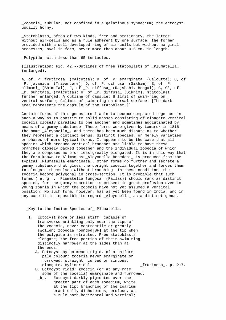
_Zooecia_ tubular, not confined in a gelatinous synoecium; the ectocystusually horny.
_Statoblasts_ often of two kinds, free and stationary, the latterwithout air-cells and as a rule adherent by one surface, the formerprovided with a well-developed ring of air-cells but without marginalprocesses, oval in form, never more than about 0.6 mm. in length.
_Polypide_ with less than 65 tentacles.
[Illustration: Fig. 42.--Outlines of free statoblasts of _Plumatella_(enlarged).
A, of _P. fruticosa_ (Calcutta); B, of _P. emarginata_ (Calcutta); C, of_P. javanica_ (Travancore); D, of _P. diffusa_ (Sikhim); E, of _P.allmani_ (Bhim Tal); F, of _P. diffusa_ (Rajshahi, Bengal); G, G', of_P. punctata_ (Calcutta); H, of _P. diffusa_ (Sikhim), statoblastfurther enlarged: A=outline of capsule; B=limit of swim-ring onventral surface; C=limit of swim-ring on dorsal surface. [The darkarea represents the capsule of the statoblast.]]
Certain forms of this genus are liable to become compacted together insuch a way as to constitute solid masses consisting of elongate verticalzooecia closely parallel to one another and sometimes agglutinated bymeans of a gummy substance. These forms were given by Lamarck in 1816the name _Alcyonella_, and there has been much dispute as to whetherthey represent a distinct genus, distinct species, or merely varietiesor phases of more typical forms. It appears to be the case that allspecies which produce vertical branches are liable to have thesebranches closely packed together and the individual zooecia of whichthey are composed more or less greatly elongated. It is in this way thatthe form known to Allman as _Alcyonella benedeni_ is produced from thetypical _Plumatella emarginata_. Other forms go further and secrete agummy substance that glues the upright zooecia together and forces themto elongate themselves without branching. In these conditions thezooecia become polygonal in cross-section. It is probable that suchforms (_e. g._, _Plumatella fungosa_ (Pallas)) should rank as distinctspecies, for the gummy secretion is present in great profusion even inyoung zoaria in which the zooecia have not yet assumed a verticalposition. No such form, however, has as yet been found in India, and inany case it is impossible to regard _Alcyonella_ as a distinct genus.
_Key to the Indian Species of_ Plumatella.
I. Ectocyst more or less stiff, capable of transverse wrinkling only near the tips of the zooecia, never contractile or greatly swollen; zooecia rounded[BF] at the tip when the polypide is retracted. Free statoblasts elongate; the free portion of their swim-ring distinctly narrower at the sides than at the ends. A. Ectocyst by no means rigid, of a uniform pale colour; zooecia never emarginate or furrowed, straight, curved or sinuous, elongate, cylindrical _fruticosa_, p. 217. B. Ectocyst rigid; zooecia (or at any rate some of the zooecia) emarginate and furrowed. _b_. Ectocyst darkly pigmented over the greater part of each zooecium, white at the tip; branching of the zoarium practically dichotomous, profuse, as a rule both horizontal and vertical;

zooecia straight or slightly curved or sinuous _emarginata_, p. 220. _b'_. Ectocyst colourless and hyaline; branching of the zoarium sparse, lateral, irregular, horizontal; zooecia nearly straight, strongly emarginate and furrowed _javanica_, p. 221. _b''_. The majority of the zooecia distinctly L-shaped, one limb being as a rule adherent; ectocyst never densely pigmented. beta. Zooecia cylindrical, their furrowed keel never prominent _diffusa_, p. 223. beta'. Zooecia (or at any rate some of the zooecia) constricted or tapering at the base, their emargination and furrow conspicuous _allmani_, p. 224.
II. Ectocyst stiff; zooecia truncated when the polypide is retracted. Surface of zooecia minutely roughened, distinctly annulate on the distal part _tanganyikæ_, p. 225. III. Ectocyst swollen and contractile, capable of transverse wrinkling all over the zooecium; zooecia never emarginate _punctata_, p. 227.
[Footnote BF: In specimens preserved in spirit they are apt to collapse and therefore to become somewhat concave.]
There has always been much difficulty in separating the species of_Plumatella_, and even now there is no general consensus of opinion asto the number that should be recognized. The difficulty, however, ismuch reduced if the following precautions are observed:--
(1) If the zoarium appears to be tangled, if the branches intertwine or overlap, or if the zooecia are closely pressed together, the whole mass should be carefully dissected out. This is necessary not only because zoaria belonging to different species are sometimes found entangled together but also because it is often difficult to recognize the characteristic method of branching and shape of the zooecia unless it is done.
(2) As large a part as possible of each zoarium should be examined, preferably with a binocular microscope, and allowance should be made for irregularities and abnormalities of all kinds. What must be observed is the rule rather than the exceptions.
(3) When the statoblasts are being examined, care must be taken that they lie flat and that their surface is parallel to that of the nose-piece of the microscope. If they are viewed obliquely it is impossible to see their true outlines and proportions.
(4) In order to see the relative proportions of the capsule and the swim-ring it is necessary that the statoblast should be rendered transparent. This is often difficult owing to the presence of air in the air-cells, but strong nitric acid applied judiciously will render it possible (p. 240).
In supervising the preparation of the plates that illustrate this genusI have impressed upon the artist the importance of representing what hesaw rather than what he thought he ought to see, and the figures are

very close copies of actual specimens. I have deliberately chosen forrepresentation specimens of _Plumatella_ preserved by the simple methodswhich are often the only ones that it is possible for a traveller toadopt, for the great majority of naturalists will probably have noopportunity of examining living specimens or specimens preserved byspecial methods, and the main object, I take it, of this series is toenable naturalists first to distinguish the species described and thento learn something of their habitat and habits.
GEOGRAPHICAL DISTRIBUTION.--Of the seven species included in this keyfive have been found in Europe (namely _P. fruticosa_, _P. emarginata_,_P. diffusa_, _P. allmani_, and _P. punctata_), while of these five allbut _P. allmani_ are known to occur in N. America also. _P. javanica_ isapparently peculiar to the Oriental Region, while _P. tanganyikæ_ hasonly been taken in Central Africa and in the Bombay Presidency.
TYPES.--Very few of the type-specimens of the older species of_Plumatella_ are in existence. Allman's are neither in Edinburgh nor inLondon, and Mr. E. Leonard Gill, who has been kind enough to go throughthe Hancock Collection at Newcastle-on-Tyne, tells me that he cannottrace Hancock's. Those of the forms described by Kraepelin are inHamburg and that of _P. tanganyikæ_ in the British Museum, and there areschizotypes or paratypes of this species and of _P. javanica_ inCalcutta. The types of Leidy's species were at one time in thecollection of the Philadelphia Academy of Science.
BIOLOGY.--The zoaria of the species of _Plumatella_ are found firmlyattached to stones, bricks, logs of wood, sticks, floating seeds, thestems and roots of water-plants, and occasionally to the shells ofmolluscs such as _Vivipara_ and _Unio_. Some species shun the light, butall are apparently confined to shallow water.
Various small oligochæte worms (e. g., _Chætogaster spongillæ_,[BG]_Nais obtusa_, _Nais elinguis_, _Slavina appendiculata_ and _Pristinalongiseta_[BH]), take shelter amongst them; dipterous larvæ of the genus_Chironomus_ often build their protective tubes at the base of thezoaria, and the surface of the zooecia commonly bears a more or lessprofuse growth of such protozoa as _Vorticella_ and _Epistylis_. I haveseen a worm of the genus _Chætogaster_ devouring the tentacles of apolypide that had been accidentally injured, but as a rule the movementsof the lophophore are too quick to permit attacks of the kind, and Iknow of no active enemy of the genus. The growth of sponges at the baseof the zoaria probably chokes some species, but one form (_F.fruticosa_) is able to surmount this difficulty by elongating itszooecia (p. 219). A small worm (_Aulophorus tonkinensis_) which iscommon in ponds in Burma and the east of India as far west as Lucknow,often builds the tube in which it lives mainly of the free statoblastsof this genus. It apparently makes no selection in so doing but merelygathers the commonest and lightest objects it can find, for small seedsand minute fragments of wood as well as sponge gemmules and statoblastsof other genera are also collected by it. I know of no better way ofobtaining a general idea as to what sponges and phylactolæmata arepresent in a pond than to examine the tubes of _Aulophorus tonkinensis_.
[Footnote BG: Annandale, J. As. Soc. Bengal (n. s.) ii, p. 188, pl. i (1906).]
[Footnote BH: See Michaelsen, Mem. Ind. Mus. i, pp. 131-135 (1908).]
I am indebted to Mr. F. H. Gravely, Assistant Superintendent in theIndian Museum, for an interesting note regarding the food of_Plumatella_. His observations, which were made in Northamptonshire,were unfortunately interrupted at a critical moment, but I have

reproduced them with his consent in order that other observers mayinvestigate the phenomena he saw. Mr. Gravely noted that a small greenflagellate which was abundant in water in which _Plumatella repens_ wasgrowing luxuriantly, was swallowed by the polypides, and that if thepolyparium was kept in a shallow dish of water, living flagellata of thesame species congregated in a little pile under the anus of eachpolypide. His preparations show very clearly that the flagellates werepassing through the alimentary canal without apparent change, but themethod of preservation does not permit the retractile granules, whichwere present in large numbers in the cell-substance of the flagellates,to be displayed and it is possible that these granules had disappearedfrom those flagellates which are present in the recta of his specimens.It is clear, therefore, either that certain flagellates must passthrough the alimentary canal of _Plumatella_ unchanged, or that thepolyzoon must have the power of absorbing the stored food material theflagellates contain without doing them any other injury.
The free statoblasts of _Plumatella_ are as a rule set free before thecells they contain become differentiated, and float on the surface ofthe water for some time before they germinate; but occasionally a smallpolypide is formed inside the capsule while it is still in its parentzooecium. I have, however, seen only one instance of this prematuredevelopment, in a single statoblast contained in a small zoarium of _P.fruticosa_ found in Lower Burma in March. The fixed statoblasts usuallyremain fixed to the support of the zoarium, even when theirparent-zooecium decays, and germinate _in situ_.
The larva (fig. 40 C, p. 207) that originates from the egg of_Plumatella_ is a minute pear-shaped, bladder-like body coveredexternally with fine vibratile threads (cilia) and having a pore at thenarrow end. At the period at which it is set free from the parentzooecium it already contains a fully formed polypide or pair ofpolypides with the tentacles directed towards the narrow end. After abrief period of active life, during which it moves through the water bymeans of its cilia, it settles down on its broad end, which becomesadhesive; the polypide or pair of polypides is everted through the poreat the narrow end, the whole of this end is turned inside out, and afresh polyparium is rapidly formed by budding.
29. Plumatella fruticosa, _Allman_. (Plate III, fig. 1; plate IV, fig.4; plate V, fig. 1.)
_Plumatella fruticosa_, Allman, Ann. Nat. Hist. xiii, p. 331 (1844).
_Plumatella repens_, van Beneden (? _nec_ Linné), Mém. Acad. Roy. Belg. 1847, p. 21, pl. i, figs. 1-4.
_Plumatella fruticosa_, Johnston, Brit. Zooph. (ed. 2), p. 404 (1847).
_Plumatella coralloides_, Allman, Rep. Brit. Assoc. 1850, p. 335.
_Plumatella stricta_, _id._, Mon. Fresh-Water Polyzoa, p. 99, fig. 14 (1857).
_Plumatella fruticosa_, _id._, _ibid._ p. 102, pl. vi, figs. 3-5.
_Plumatella coralloides_, _id._, _ibid._ p. 103, pl. vii, figs. 1-4.

_Plumatella repens_ and _P. stricta_, Carter, Ann. Nat. Hist. (3) iii, p. 341 (1859).
_Plumatella lucifuga_, Jullien (_partim_), Bull. Soc. zool. France, x, p. 114 (1885).
_Plumatella princeps_ var. _fruticosa_, Kraepelin, Deutsch. Süsswasserbryozoen, i, p. 120, pl. vii, fig. 148 (1887).
_Plumatella fruticosa_, Braem, Unter. ii. Bryozoen des süssen Wassers, p. 9, pl. i, fig. 15 (Bibl. Zool. ii) (1890).
_Plumatella repens_, Annandale, J. As. Soc. Bengal (new series) iii, 1907, p. 88.
_Plumatella emarginata_, Loppens (_partim_), Ann. Biol. lacustre, iii, p. 161 (1908).
_Plumatella fruticosa_, Annandale, Rec. Ind. Mus. v, p. 45 (1910).
_Zoarium._ The zoarium in the typical form has a loose appearance due tothe fact that the branches are far apart and the ectocyst by no meansrigid. When young the zoarium is adherent, but in well-grown polypariavertical branches, often an inch or more in length, are freely produced.As a rule they have not the strength to stand upright if removed fromthe water. Branching is ordinarily lateral and as a rule occurs chieflyon one side of a main branch or trunk. In certain circumstances uprightzooecia are pressed together and reach a great length without branching,and in this form (_P. coralloides_, Allman) daughter-zooecia are oftenproduced at the tip of an elongated mother-zooecium in fan-likeformation. A depauperated form (_P. stricta_, Allman), occurs in whichthe vertical branches are absent or very short. In all forms internalpartitions are numerous and stout.
_Zooecia._ The zooecia are cylindrical and bear a simple keel on theirdorsal surface. They are never emarginate or furrowed. In the typicalform their diameter is more than half a millimetre, and they are alwaysof considerable length. The ectocyst is thin and never very rigid ordeeply pigmented, the colour usually being an almost uniform palepinkish brown and fading little towards the tip of the zooecium.
_Statoblasts._ Both free and stationary statoblasts are formed, but thelatter are rare and do not always adhere. They resemble the freestatoblasts in general form but have a solid margin instead of aswim-ring and are often minutely serrated round the edge. The freestatoblasts are at least considerably, sometimes very elongate; in allzoaria it is possible to find specimens that are more than twice as longas broad. The capsule is relatively large and resembles the swim-ring inoutline, so that the free portion of the latter is not much narrower atthe sides than at the ends. The sides are distinctly convex and the endsrounded; the swim-ring encroaches little on the surface of the capsule.
_Polypide._ The tentacles number between 40 and 50 and are not festoonedat the base. The stomach is slender and elongate.
TYPE not in existence.
SYSTEMATIC REMARKS.--_P. fruticosa_ is closely allied to _P. repens_(European and N. American) but always has much longer statoblasts. Threephases of the species may be distinguished as follows:--
A. (_Forma typica_). Zooecia stout in form, not greatly

elongate; free branches produced in profusion.
B. (_P. stricta_, Allman, _P. repens_, van Beneden). Zooecia slender; free branches absent or consisting of two or three zooecia only.
C. (_P. coralloides_, Allman). Vertical zooecia pressed together and greatly elongated.
Indian specimens of the typical form agree well with German specimenslabelled by Prof. Kraepelin _P. princeps_ var. _fruticosa_, andspecimens of the _coralloides_ phase could hardly be distinguished fromsimilar specimens from Scotland.
GEOGRAPHICAL DISTRIBUTION.--_P. fruticosa_ is widely distributed inEurope and probably in N. America. I have seen Indian specimens from thePunjab (Lahore, _Stephenson_), from Bombay, from Travancore, fromCalcutta and other places in the Ganges delta, from Rajshahi (RampurBhoolia) on the R. Ganges, from Kurseong in the E. Himalayas (alt. 4,500feet), and from Kawkareik in Tenasserim. Statoblasts found on thesurface of a pond near Simla in the W. Himalayas (alt. _ca._ 8,000feet), probably belong to this species.
BIOLOGY.--Allman states that in England _P. fruticosa_ is fond of stilland slowly-running water. The typical form and the _coralloides_ phasegrow abundantly in the Calcutta tanks, the former often attaining anextraordinary luxuriance. I have found the var. _stricta_ only in waterin which there was reason to suspect a lack of minute life (andtherefore of food), viz. in Shasthancottah Lake in Travancore, in aswamp in Lower Burma, and in a small jungle stream near the base of theWestern Ghats in Travancore. The species is the only one that I haveseen in running water in India, and the specimens obtained in the junglestream in Travancore are the only specimens I have taken in thesecircumstances. _P. fruticosa_ always grows near the surface or near theedge of water; it is found attached to the stems of bulrushes and otheraquatic plants, to floating seeds and logs and (rarely) to stones andbricks. So far as my experience goes it is only found, at any rate inCalcutta, in the cold weather and does not make its appearance earlierthan October.
The form Allman called _P. coralloides_ was found by him, "attached tofloating logs of wood, together with _P. repens_ and _Cordylophoralacustris_, and generally immersed in masses of _Spongillafluviatilis_." I have always found it immersed in sponges (_S.lacustris_, _S. alba_, _S. carteri_, and _S. crassissima_), except whenthe sponge in which it had been immersed had decayed. Indeed, thepeculiar form it has assumed appears to be directly due to the pressureof the growing sponge exerted on the zooecia, for it is often possibleto find a zoarium that has been partially overgrown by a sponge and hasretained its typical form so long as it was free but has assumed the_coralloides_ form where immersed.[BI] In Shasthancottah Lake,Travancore, I found specimens of the _stricta_ phase embedded in thegelatinous mass formed by a social rotifer and to some extentassimilated to the _coralloides_ form.
[Footnote BI: Braem (_op. cit._, p. 3, pl. i, fig. 1), has described and figured under the name _P. fungosa_ var. _coralloides_, Allman, a dense form that somewhat resembles this phase of _P. fruticosa_ but has become compacted without external pressure. It is, however, probably a form of _P. repens_ rather than _P. fungosa_ and differs in its broad statoblasts from any form of _P. fruticosa_. I have examined specimens of the same form from England.]

30. Plumatella emarginata, _Allman_. (Plate III, fig. 2; plate IV, figs.1, 1 _a._)
_Plumatella emarginata_, Allman, Ann. Nat. Hist. xiii, p. 330 (1844).
_Plumatella emarginata_, Johnston, Brit. Zooph. (ed. 2), p. 404 (1847).
_Alcyonella benedeni_, Allman, Mon. Fresh-Water Polyzoa, p. 89, pl. iv, figs. 5-11 (1857).
_Plumatella emarginata_, _id._, _ibid._ p. 104, pl. vii, figs. 5-10.
_Plumatella lucifuga_, Jullien, Bull. Soc. zool. France, x, figs. 89, 90, p. 114 (1885).
_Plumatella princeps_ var. _emarginata_, Kraepelin (_partim_), Deutsch. Süsswasserbryoz. p. 120, pl. iv, fig. 108, pl. v, fig. 123 (1887).
_Plumatella emarginata_, Braem, Unter. ii. Bryoz. süssen Wassers, p. 9, pl. i, figs. 12, 14 (Bibl. Zool. ii) (1890).
_Plumatella emarginata_, Annandale (_partim_), J. As. Soc. Bengal, (new series) iii, 1907, p. 89.
_Plumatella princeps_, Loppens (_partim_), Ann. Biol. lacustre, iii, p. 162, fig. 7 (1908).
_Plumatella emarginata_, Annandale, Rec. Ind. Mus. v, p. 47 (1910).
_Zoarium._ The zoarium often covers a considerable area on flat surfacesand is sometimes entirely recumbent. More usually, however, the youngerpart is vertical. In either case the branching is practicallydichotomous, two young zooecia arising almost simultaneously at the tipof a mother-zooecium and diverging from one another at a small angle.When the zoarium becomes vertical, rigid branches of as much as an inchin length are sometimes produced in this way and, arising parallel toone another, are pressed together to form an almost solid mass(=_Alcyonella benedeni_, Allman). In such cases the basal zooecium or atany rate the basal part of each upright branch is considerablyelongated. In recumbent zooecia the main branches often radiate outwardsfrom a common centre.
_Zooecia._ The zooecia are of almost equal width throughout, slender,and moderately elongate when recumbent. Their ectocyst is stiff; theyare emarginate at the tip and more or less distinctly furrowed on thedorsal surface, the keel in which the furrow runs not being prominent.The orifice is often on the dorsal surface even in upright branches.Each zooecium is of a dark brown or almost black colour for the greaterpart of its length but has a conspicuous white tip which is extendeddown the dorsal surface in the form of a triangle, its limits beingrather more extensive than and parallel to those of the emargination.
_Statoblast._ The majority of the free statoblasts are elongate andtruncate or subtruncate at the extremities, the sides being as a rulestraight and parallel. In every polyparium specimens will be found thatare between twice and thrice as long as broad. The capsule is, however,relatively much broader than the swim-ring, often being nearly circular,and there is therefore at either end a considerable extent of free
air-cells, while the extent of these cells at the sides of the capsuleis small. The air-cells cover a considerable part of the dorsal surfaceof the capsule. Fixed statoblasts are usually found in old colonies,especially at the approach of the hot weather. They have an oval formand are surrounded by a membranous margin on which traces ofreticulation can often be detected. As a rule statoblasts of both typesare produced in considerable but not in excessive numbers.
_Polypide._ There are about 40 tentacles, the velum at the base of whichextends upwards for a considerable distance without being festooned. Thestomach is elongate and slender and narrowly rounded at the base.
The method of branching, the coloration of the zooecia and the form ofthe free statoblast are all characteristic. Luxuriant or closelycompressed zoaria of _P. diffusa_ often bear a superficial resemblanceto those of _P. emarginata_, but the resemblance disappears if they arecarefully dissected out. Indian specimens of _P. emarginata_ agreeclosely with European ones.
GEOGRAPHICAL DISTRIBUTION.--_P. emarginata_ is a common species inEurope, N. America, and southern Asia and probably also occurs in Africaand Australia. I have examined specimens from Calcutta, Rangoon, andMandalay in Indian territory, and also from Jalor in the Patani States(Malay Peninsula) and the Talé Noi, Lakon Sitamarat, Lower Siam.Gemmules found by Apstein (Zool. Jahrb. (Syst.) xxv, 1907, p. 201) inplankton from the Colombo lake may belong to this species or to any ofthe others included by Kraepelin in his _P. princeps_.
BIOLOGY.--In Ireland Allan found _P. emarginata_ in streams andrivulets, but it also occurs in European lakes. In India I have onlyfound it in ponds. It prefers to adhere to the surface of stones orbricks, but when these are not available is found on the stems ofwater-plants. In the latter position the form called _Alcyonellabenedeni_ by Allman is usually produced, owing to the fact that theupright branches are crowded together through lack of space, very muchin the same way (although owing to a different cause) as those of _P.fruticosa_ are crowded together in the _coralloides_ phase, to which the_benedeni_ phase of _P. emarginata_ is in many respects analogous.
Although it is essentially a cold-weather species in Calcutta, _P.emarginata_ is sometimes found in a living condition during the "rains."Zoaria examined at this season, however, contains few living polypides,the majority of the zooecia having rotted away and left fixedstatoblasts only to mark their former position.
31. Plumatella javanica, _Kraepelin_.
_Plumatella javanica_, Kraepelin, Mitt. Nat. Mus. Hamb. xxiii, p. 143, figs. 1-3 (1903).
_Plumatella emarginata_ var. _javanica_, Loppens, Ann. Biol. lacustre, iii, p. 162 (1908).
_Plumatella javanica_, Annandale, Rec. Ind. Mus. v, p. 50 (1910).
_Plumatella allmani_ var. _dumortieri_, _id._ (_partim_) (_nec_ Allman), _ibid._ p. 49.
This species is related to _P. emarginata_, from which it may bedistinguished by the following characters:--
_Zoarium._ The zoarium is always entirely recumbent and branches

sparingly; its method of branching does not approach the dichotomoustype but is lateral and irregular. Linear series of zooecia withoutlateral branches are often formed.
_Zooecia._ The zooecia are slender and often very long; they arestrongly emarginate and furrowed, and the keel that contains the furrowis conspicuous. The ectocyst is hyaline and as a rule absolutelycolourless.
_Statoblasts._ The free statoblasts are variable in length, sometimesdistinctly elongate, sometimes elongate only to a moderate degree; theyare rounded at the extremities and have the sides slightly or distinctlyconvex outwards. The capsule is relatively large, and the free portionof the swim-ring is not much broader at the ends than at the sides. Thefixed statoblasts are elongate and surrounded by an irregularly shapedchitinous membrane, which is often of considerable extent. The whole ofthe dorsal surface is covered with what appear to be rudimentaryair-spaces some of which even contain air.
The transparent glassy ectocyst and strong furrowed keel of this speciesare very characteristic, but the former character is apt to be obscuredby staining due to external causes, especially when the zoarium isattached to dead wood. The shape of the free statoblasts is too variableto be regarded as a good diagnostic character, but the fixedstatoblasts, when they are to be found, are very characteristic inappearance. _P. javanica_ appears to be closely related to Allman's _P.dumortieri_, with which stained zoaria are apt to be confused. Thecharacter of the ectocyst is, however, different, and the free part ofthe swim-ring is distinctly narrower at the sides of the freestatoblasts. Dr. Kraepelin has been kind enough to send me one of thetypes.
TYPES in the Hamburg and Indian Museums.
GEOGRAPHICAL DISTRIBUTION.--Java, Penang, India. Indian localitiesare:--BENGAL, Calcutta; Berhampore, Murshidabad; R. Jharai, Siripur,Saran district, Tirhut: E. HIMALAYAS, Kurseong, Darjiling district (alt.4,500 feet): MADRAS PRESIDENCY, canal near Srayikaad, Travancore. Mr. C.W. Beebe has recently sent me a specimen taken by him in the BotanicalGardens at Penang.
BIOLOGY.--Very little is known about the biology of this species.Kraepelin took it in Java on the leaves of water-lilies. It is notuncommon during the cold weather in the Calcutta Zoological Gardens onfloating seeds and sticks and on the stems of bulrushes; in Travancore Itook it in November on the submerged leaves of _Pandani_ growing at theedge of a canal of slightly brackish water. Mr. Hodgart, the collectorof the Indian Museum, found it in the R. Jharai on the stems ofwater-plants at a time of flood in the "rains." In Calcutta it is oftenfound entangled with _P. fruticosa_ and _P. emarginata_.
32. Plumatella diffusa, _Leidy_. (Plate IV, fig. 2.)
_Plumatella diffusa_, Leidy, P. Ac. Philad. v, p. 261 (1852).
_Plumatella diffusa_, Allman, Mon. Fresh-Water Polyzoa, p. 105 (1857).
_Plumatella diffusa_, Hyatt, Comm. Essex Inst. iv, pl. viii, figs. 11, 12 (1866).
_Plumatella diffusa_, _id._, _ibid._ v, p. 107, fig. 12

(1868).
_Plumatella repens_, Jullien, Bull. Soc. zool. France, x, fig. 37 (_lapsus_ for 73), p. 110 (1885).
_Plumatella diffusa_, _id._, _ibid._ figs. 155, 157, pp. 130, 131.
_Plumatella allmani_ var. _diffusa_, Annandale, Rec. Ind. Mus. v, p. 49 (1910).
_Zoarium._ The zoarium often covers a considerable area on flat surfacesand is sometimes found crowded together on the stems of plants. In thelatter case the arrangement of the main branches is distinctly radiate.Upright branches occur rarely and never consist of more than threezooecia. The characteristic method of branching is best represented bythe following diagram:--
[Illustration: Fig. 43.]
The partitions are stout and numerous.
_Zooecia._ The great majority of the zooecia in each zoarium aredistinctly L-shaped, the long limb being usually adherent. The vitalorgans of the polypide are contained in the vertical limb, while thehorizontal one, in mature polyparia, is packed full of free statoblasts.The zooecia are cylindrical and as a rule obscurely emarginate andfurrowed. The ectocyst is stiff; it is never deeply pigmented but isusually of a transparent horn-colour at the base of each zooecium andcolourless at the tip, the contrast between the two portions never beingvery strong. The basal portion is rough on the surface, the distalportion smooth.
_Statoblasts._ Free statoblasts are produced in very great profusion andfixed statoblasts are also to be found as a rule. The latter resemblethose of _P. emarginata_. The free statoblasts are never very large orrelatively broad, but they vary considerably as regards size andoutline. The capsule is large, the sides convex outwards and theextremity more or less broadly rounded. The air-cells are unusuallylarge and extend over a great part of the dorsal surface of thestatoblast.
_Polypide._ The polypide is shorter and stouter than that of _P.emarginata_ and as a rule has fewer tentacles.
The most characteristic feature of this species is the form of thezooecia, which differ greatly from those of any other Indian species but_P. allmani_. In the latter they are distinctly "keg-shaped" (_i. e._,constricted at the base and swollen in the middle), and the zoariumnever spreads out over large surfaces in the way in which that of _P.diffusa_ does.
TYPE--? in the Philadelphia Academy of Sciences.
GEOGRAPHICAL DISTRIBUTION.--This species was originally described fromNorth America (in which it is apparently common) and occurs also inEurope. I have seen Indian specimens from the followinglocalities:--BENGAL, Calcutta and neighbourhood; Rajshahi (RampurBhulia): E. HIMALAYAS, Gangtok, Native Sikhim (alt. 6,150 feet)(_Kirkpatrick_, _Stewart_): PUNJAB, Lahore (_Stephenson_).
BIOLOGY.--_P. diffusa_ in Lower Bengal is a cold-weather species. It isremarkable for the enormous number of gemmules it produces and isusually found either on floating objects such as the stems of certain

water-plants, or on stones or bricks at the edge of ponds.
33. Plumatella allmani, _Hancock_. (Plate IV, figs. 3, 3 _a_.)
_Plumatella allmani_, Hancock, Ann. Nat. Hist. (2) v, p. 200, pl. v, fig. 3-4, pl. iii, fig. 2-3 (1850).
_Plumatella allmani_, Allman, Mon. Fresh-Water Polyzoa, p. 106, fig. 16 (1857).
_Plumatella elegans_, _id._, _ibid._ p. 107, pl. viii, figs. 6-10.
_Plumatella lucifuga_ ("forme rampante") Jullien, Bull. Soc. zool. France, x, p. 114 (1885).
This species is closely allied to _P. diffusa_, from which it differs inthe following characters:--
(1) The zoarium never covers a large area and as a rule grows sparingly and mainly in two directions.
(2) The zooecia are more irregular in shape, not so distinctly elbowed, smaller; they have a much more prominently keeled ridge. The great majority of them are constricted at the base and taper towards the orifice. In young zoaria they are almost colourless but in older ones there is a band of not very dense pigment round the base of the vertical limb.
(3) The free statoblasts are comparatively large and usually show a tendency to taper at the extremities, often being almost rhomboidal in form. The swim-ring does not extend so far over the dorsal surface as it does in those of _P. diffusa_; the "cells" of which it is composed are small.
TYPE not in existence.
I have seen every gradation between this form and Allman's _P. elegans_.
GEOGRAPHICAL DISTRIBUTION.--_P. allmani_ is apparently a rare species towhich there are few references in literature. It was originallydescribed from England and is stated by Jullien to occur in France. Ihave found specimens only in the lake Bhim Tal (alt. 4,500 feet) in theW. Himalayas.
BIOLOGY.--The original specimens were found by Hancock on stones. My ownwere growing on the leaves of water-plants, usually on the under side.When the zooecia were forced to stretch across from one leaflet toanother they assumed the sinuous form characteristic of Allman's _P.elegans_.
34. Plumatella tanganyikæ, _Rousselet_.
_Plumatella tanganyikæ_, Rousselet, Proc. Zool. Soc. London, 1907 (i), p. 252, pl. xiv, figs. 1-4.
_Plumatella bombayensis_, Annandale, Rec. Ind. Mus. ii, p. 169, figs. 1, 2 (1908).
_Plumatella bombayensis_, _id._, _ibid._ v, p. 51 (1910).

_Zoarium._ The whole colony is recumbent but branches freely and atshort intervals in a horizontal plane, so that the zooecia becomecrowded together and the branches sometimes overlap one another. Thezoarium often covers a considerable area, but growth seems to be mainlyin two directions. When growing on the stems of water-plants thebranches are often parallel and closely pressed together but remainrecumbent in this position. A stout membrane sometimes extends betweenbranches and individual zooecia.
_Zooecia._ The walls of the zooecia are thick, stiff, and more or lessdarkly but not opaquely pigmented; the external surface, although notvery smooth, is always clean. The two most noteworthy characters of thezooecia are (i) their truncated appearance when the polypide isretracted, and (ii) the conspicuous, although often irregular externalannulation of their walls. The tip of each zooecium, owing to the factthat the invaginated part of the ectocyst is soft and sharply separatedfrom the stiffened wall of the tube, terminates abruptly and is notrounded off gradually as is the case in most species of the genus;sometimes it expands into a trumpet-like mouth. The annulation of theexternal surface is due to numerous thickened areas of the ectocystwhich take the form of slender rings surrounding the zooecium; they aremost conspicuous on its distal half. On the dorsal surface of the baseof each zooecium there is a conspicuous furrowed keel, which, however,does not usually extend to the distal end; the latter is oval incross-section. The zooecia are short and broad; their base is alwaysrecumbent, and, when the zoarium is attached to a stone or shell, oftenseems to be actually embedded in the support; the distal part turnsupwards and is free, so that the aperture is terminal; the zooecia ofthe older parts of the zoarium exhibit the specific characters much moreclearly than those at the growing points.
_Polypide._ The lophophore bears 20 to 30 tentacles, which are long andslender; the velum at their base extends up each tentacle in the form ofa sharply pointed projection, but these projections do not extend formore than one-fifth of the length of the tentacles. Both the velum andthe tentacular sheath bear numerous minute tubercles on the externalsurface. The base of the stomach is rounded, and the whole of thealimentary canal has a stout appearance.
[Illustration: Fig. 44.--_Plumatella tanganyikæ_ from Igatpuri Lake.
A=outline of part of zoarium from a stone, × 16; B=outline of the tip ofa single zooecium, × 70; C=free statoblast, × 70.]
_Statoblasts._ Both fixed and free statoblasts are produced, but not invery large numbers. The latter are broadly oval and are surrounded by astout chitinous ring, which often possesses irregular membranousprojections; the surface is smooth. The free statoblasts are small andmoderately elongate, the maximum breadth as a rule measuring about 2/3of the length; the capsule is relatively large and the ring of air-cellsis not very much broader at the ends than at the sides; the dorsalsurface of the central capsule is profusely tuberculate. The outline ofthe whole structure is often somewhat irregular.
In deference to Mr. Rousselet's opinion expressed in a letter I havehitherto regarded the Bombay form of this species as distinct from theAfrican one, and there certainly is a great difference in the appearanceof specimens taken on the lower surface of stones in Igatpuri Lake andof the types of _P. tanganyikæ_, one of which is now in the collectionof the Indian Museum. The dark colour of the former, however, and theirvigorous growth appear to be directly due to environment, for thesecharacters disappear to a large extent in specimens growing on the stemsof water-plants in the same lake. Indeed, such specimens are exactlyintermediate between the form "_bombayensis_" and the typical form of

the species. _P. tanganyikæ_ is closely allied to _P. philippinensis_,Kraepelin, from the island of Luzon, but the latter has a smooth andpolished ectocyst devoid of annulations, and zooecia of a more elongateand regular form.
TYPES of the species in the British and Indian Museums, those of _P.bombayensis_ in the latter collection.
GEOGRAPHICAL DISTRIBUTION.--_P. tanganyikæ_ is only known as yet from L.Tanganyika in Central Africa and from Igatpuri in the Bombay Presidency.
BIOLOGY.--In both localities the zoaria were found in shallow water. InL. Tanganyika they were encrusting stones and shells, while at Igatpurithey were fixed for the most part to the lower surface of stones butwere also found on the stems of water-plants. My specimens from theBombay Presidency were taken, on two separate occasions, at the end ofNovember. At that date the zoaria were already decaying and largeblanks, marked out by fixed statoblasts, were often observed on thestones. Probably, therefore, the species flourishes during the "rains."
35. Plumatella punctata, _Hancock_. (Plate IV, fig. 5.)
_Plumatella punctata_, Hancock, Ann. Nat. Hist. (2) v, p. 200, pl. iii, fig. 1, and pl. v, figs. 6, 7 (1850).
_Plumatella vesicularis_, Leidy, P. Ac. Philad. vii, p. 192 (1854).
_Plumatella vitrea_, Hyatt, Comm. Essex Inst. iv, pl. ix, figs. 1, 2 (1866).
_Plumatella punctata_, Allman, Mon. Fresh-Water Polyzoa, p. 100, fig. 15 (1857).
_Plumatella vesicularis_, _id._, _ibid._ p. 101.
_Plumatella vitrea_, Hyatt, Proc. Essex Inst. v, p. 225, figs. 18, 19 (1868).
_Plumatella vesicularis_, _id._, _ibid._ p. 225.
_Hyalinella vesicularis_, Jullien, Bull. Soc. zool. France, x, p. 133, figs. 165-172 (1885).
_Hyalinella vitrea_, _id._, _ibid._ p. 134, figs. 173-179.
_Plumatella punctata_, Kraepelin, Deutsch. Süsswasserbryozoen, i, p. 126, pl. iv, figs. 115, 116; pl. v, figs. 124, 125; pl. vii, figs. 153, 154 (1887).
_Plumatella vesicularis_, Braem, Unters. ü. Bryozoen süssen Wassers, p. 8, pl. i, fig. 8 (Bibl. Zool. ii) (1890).
_Hyalinella punctata_, Loppens, Ann. Biol. lacustre, iii, p. 163 (1908).
_Plumatella punctata_, Annandale, Rec. Ind. Mus. v, p. 52 (1910).
_Zoarium._ The zoarium is entirely recumbent and often appears to forman almost uniform flat layer instead of a dendritic body. Sometimes,however, it is distinctly linear, with lateral branches producedirregularly at considerable distances apart.

_Zooecia._ The zooecia differ from those of all other species in havinga greatly swollen, soft ectocyst which can be transversely wrinkled allover the zooecium by the action of the muscles of the polypide and isdistinctly contractile. It is mainly owing to the swollen and almostgelatinous nature of the ectocyst that the dendritic character of thezoarium is frequently concealed, for the method of branching isessentially the same as that of _P. diffusa_, although the zooecia arenot so distinctly elbowed. The ectocyst is colourless or faintly tintedwith brown; as a rule it is not quite hyaline and the external surfaceis minutely roughened or tuberculate. The zooecia are not emarginate orfurrowed.
_Statoblasts._ Stationary statoblasts are not found. The freestatoblasts are variable and often asymmetrical in outline, but the freeportion of the swim-ring is always of nearly equal diameter all roundthe periphery and the capsule relatively large. Some of the statoblastsare always broad in comparison with their length.
_Polypide._ The polypide is comparatively short and stout. Europeanspecimens are said to have from 30 to 40 tentacles, but Indian specimenshave only from 20 to 30.
Shrunken specimens of the less congested forms of this species closelyresemble specimens of _P. repens_, but the statoblasts are more variablein shape and the ectocyst, even in such specimens, is thicker. Living orwell-preserved specimens cannot be mistaken for those of any otherspecies. Jullien regarded _P. punctata_ as the type of a distinct genus(_Hyalinella_) but included in _Plumatella_ at least one form (P."_arethusa_") which probably belongs to this species. Kraepelindistinguishes as "varieties" two phases, a summer phase ("var._prostrata_") and an autumn phase ("var. _densa_"). The former oftenforms linear series of considerable length with only an occasionalside-branch, while in the autumn phase branching is so profuse and thebranches are so closely pressed together that the zoarium comes toresemble a uniform gelatinous patch rather than a dendritic growth. Aphase resembling the European autumn form is the commonest in Calcuttaand I have also found one intermediate between this and Kraepelin's"var. _prostrata_," neither having any seasonal significance in India.
GEOGRAPHICAL DISTRIBUTION.--_P. punctata_ is widely distributed inEurope and N. America, but in the Oriental Region it has only been foundin Calcutta and the neighbourhood.
BIOLOGY.--In this part of India _P. punctata_ flourishes both during the"rains" and in winter. I have found specimens in June and July and alsoin December and January. The majority of them were attached to bricks,but some were on the roots of duckweed, the stems of water-plants, andthe tips of creepers falling into water. The species is often foundtogether with _Stolella indica_ and also with other species of its owngenus. It is most common, in the neighbourhood of Calcutta, in that partof the town which is near the Salt Lakes, and occurs in ponds the waterof which is slightly brackish.
Genus 2. STOLELLA, _Annandale_.
_Stolella_, Annandale, Rec. Ind. Mus. iii, p. 279 (1909).
_Stolella_, _id._, _ibid._ v, p. 53 (1910).
TYPE, _Stolella indica_, Annandale.
_Zoarium_. The zoarium consists of groups of zooecia (or occasionally ofsingle zooecia) joined together by an adherent rhizome. There is no

gelatinous investment.
_Zooecia._ The adult zooecia resemble those of _Plumatella_ except inbeing sometimes more or less upright.
_Polypide_ and _Statoblasts._ The polypide and statoblasts resemblethose of _Plumatella_. Fixed as well as free statoblasts occur.
This genus is closely allied to _Plumatella_, from which it is probablyderived. The root-like tube from which the zooecia arise is formed bythe great elongation of the basal part of a zooecium, and the zoariaclosely resemble those of _P. punctata_, for it is not until severalzooecia have been produced that the characteristic mode of growthbecomes apparent.
_Stolella_ has only been found in India and is monotypic[BJ].
[Footnote BJ: But see p. 246 (addenda).]
36. Stolella indica, _Annandale_. (Plate V, figs. 3, 4.)
_Stolella indica_, Annandale, Rec. Ind. Mus. iii, p. 279, fig. (1909).
_Stolella indica_, _id._, _ibid._ v, p. 53 (1910).
_Zoarium._ The zoarium is adherent and linear, having neither lateralnor vertical branches.
_Zooecia._ The zooecia are short and slender, erect or nearly so,distinctly emarginate and furrowed. Their ectocyst is soft, colourlessand transparent but minutely roughened on the surface.
_Polypide._ The tentacles number from 30 to 35 and are rather short andstout, sometimes being slightly expanded at the tips. The stomach iscomparatively short and abruptly truncated posteriorly.
_Statoblasts._ Both free and fixed statoblasts are found, and both arevariable in form, the latter varying in outline from the circular to thebroadly oval. The free statoblasts resemble those of _Plumatellapunctata_, but are sometimes rather more elongate.
TYPE in the Indian Museum.
[Illustration: Fig. 45.--Zoarium of _Stolella indica_ on stem ofwater-plant (from Calcutta), × 6.]
GEOGRAPHICAL DISTRIBUTION.--So far as we know, this species is confinedto the Indo-Gangetic Plain. Major Walton found it at Bulandshahr in theUnited Provinces, and it is not uncommon in the neighbourhood ofCalcutta.
BIOLOGY.--The zoaria of _S. indica_ are usually fixed to the roots ofduckweed or to the stems of other plants. They are often found togetherwith those of _P. punctata_. A slight infusion of brackish water intothe ponds in which it lives does not seem to be inimical to thisspecies, but I have found it in ponds in which nothing of the kind waspossible. It flourishes during the "rains" and, to judge from specimenskept in an aquarium, is very short-lived. Major Walton found it growingover a zoarium of _Hislopia lacustris_.
Subfamily B. LOPHOPINÆ.

The zoaria of this subfamily are never dendritic but form gelatinousmasses which, except in _Australella_, are cushion-shaped or sack-like.With the possible exception of _Australella_, they possess to a limitedextent the power of moving along vertical or horizontal surfaces, but itis by no means clear how they do so (see p. 172). The statoblasts areremarkable for their large size, and it is noteworthy that_Australella_, which is intermediate in structure between thePlumatellinæ and the Lophopinæ, possesses statoblasts of intermediatesize. The swim-ring is always well developed, and fixed statoblasts areunknown.
Only two genera (_Lophopodella_ and _Pectinatella_) have been definitelyproved to occur in India, but a third (_Lophopus_[BK]) is stated to havebeen found in Madras. Should it be met with it will easily be recognizedby the upright position of its polypides when their tentacles areexpanded and by the fact that the statoblasts never bear marginalprocesses.
[Footnote BK: Only two species are known, _L. crystallinus_ (Pallas) from Europe and N. America, with oval statoblasts that are produced and pointed at the two ends, and _L. jheringi_, Meissner from Brazil, with irregularly polygonal or nearly circular statoblasts.]
Genus 3. LOPHOPODELLA, _Rousselet_.
_Lophopodella_, Rousselet, Journ. Quek. Micr. Club (2) ix, p. 45 (1904).
_Lophopodella_, Annandale, Rec. Ind. Mus. v, p. 54 (1910).
TYPE, _Pectinatella carteri_, Hyatt.
_Zoarium._ The zoarium consists of a circular or oval mass of no greatsize. Polyparia do not form compound colonies.
_Polypides._ The polypides lie semi-recumbent in the mass and neverstand upright in a vertical position.
_Statoblasts._ The statoblasts are of considerable size and normallybear at both ends a series of chitinous processes armed with double rowsof small curved spinules.
As a rule the genus is easily recognized by means of the statoblasts,but sometimes the processes at the ends of these structures are absentor abortive and it is then difficult to distinguish them from those of_Lophopus_. There is, however, no species of that genus known that hasstatoblasts shaped like those of the Indian species of _Lophopodella_.
Three species of _Lophopodella_, all of which occur in Africa, have beendescribed; _L. capensis_ from S. Africa, which has the ends of thestatoblast greatly produced, _L. thomasi_ from Rhodesia, in which theyare distinctly concave, and _L. carteri_ from E. Africa, India andJapan, in which they are convex or truncate.
The germination of the gemmule and the early stages in the developmentof the polyparium of _L. capensis_ have been described by Miss Sollas(Ann. Nat. Hist. (8) ii, p. 264, 1908).
37. Lophopodella carteri (_Hyatt_). (Plate III, figs. 4, 4_a_.)
_Lophopus_ sp., Carter, Ann. Nat. Hist. (3) iii, p. 335, pl.

viii, figs. 8-15 (1859).
? _Lophopus_ sp., Mitchell, Q. J. Micr. Sci. London (3) ii, p. 61 (1862).
_Pectinatella carteri_, Hyatt, Comm. Essex Inst. iv, p. 203 (footnote) (1866).
_Pectinatella carteri_, Meissner, Die Moosthiere Ost-Afrikas, p. 4 (in Mobius's Deutsch-Ost-Afrika, iv, 1898).
_Lophopodella carteri_, Rousselet, Journ. Quek. Micr. Club, (2) ix, p. 47, pl. iii, figs. 6, 7 (1904).
_Lophopus carteri_, Annandale, Rec. Ind. Mus. ii, p. 171, fig. 3 (1908).
_Lophopodella carteri_, _id._, _ibid._ v, p. 55 (1910).
_Zoarium._ The zoarium as a rule has one horizontal axis longer than theother so that it assumes an oval form when the polypides are expanded;when they are retracted its outline is distinctly lobular. Viewed fromthe side it is mound-shaped. The polypides radiate, as a rule in severalcircles, from a common centre. The ectocyst is much swollen, hyaline andcolourless.
_Polypide._ The polypide has normally about 60 tentacles, the velum atthe base of which is narrow and by no means strongly festooned. Thestomach is yellow or greenish in colour. The extended part of thepolypide measures when fully expanded rather less than 3 mm., and eachlimb of the lophophore about the same.
_Statoblast._ The statoblast is variable in shape and size but measureson an average about 0.85 × 0.56 mm. The ends are truncate orsubtruncate; the capsule is small as compared with the swim-ring and asa rule circular or nearly so. The processes at the two ends are variablein number; so also are their spinules, which are arranged in twoparallel rows, one row on each side of the process, and are neither verynumerous nor set close together; as a rule they curve round through thegreater part of a circle and are absent from the basal part of theprocess.
[Illustration: Fig. 46.--Lophopodella carteri (from Igatpuri Lake).
A=outline of a zoarium with the polypides expanded, as seen from belowthrough glass to which it was attached, × 4; B=outline of a zoarium withthe polypides highly contracted, as seen from above, × 4; C=statoblast,× 75.]
37 _a._ Var. himalayana.
_Lophopus lendenfeldi_, Annandale (_nec_ Ridley), J. As. Soc. Bengal, (n. s.) iii, 1907, p. 92, pl. ii, figs. 1-4 (1907).
_Lophopus lendenfeldi_ var. _himalayanus_, _id._, Rec. Ind. Mus. i, p. 147, figs. 1, 2 (1907).
_Lophopus himalayanus_, _id._, _ibid._ ii, p. 172, fig. 4 (1908).
This variety differs from the typical form in having fewer tentacles and

in the fact that the marginal processes of the statoblast are abortiveor absent.
_Pectinatella davenporti_, Oka[BL] from Japan is evidently a local raceof _L. carteri_, from the typical form of which it differs in having themarginal processes of the statoblast more numerous and better developed.The abortive structure of these processes in var. _himalayana_ points toan arrest of development, for they are the last part of the statoblastto be formed.
[Footnote BL: Zool. Anz. xxxi, p. 716 (1907), and Annot. Zool. Japon. vi, p. 117 (1907).]
TYPES. The statoblasts mounted in Canada balsam by Carter and now in theBritish Museum must be regarded as the types of the species named butnot seen by Hyatt. The types of the var. _himalayana_ are in the IndianMuseum and those of the subspecies _davenporti_ presumably in thepossession of Dr. Oka in Tokyo.
GEOGRAPHICAL DISTRIBUTION.--The typical form occurs in Bombay, the W.Himalayas and possibly Madras, and its statoblasts have been found in E.Africa; the var. _himalayana_ has only been taken in the W. Himalayasand the subspecies _davenporti_ in Japan. Indian localities are:--BOMBAYPRESIDENCY, Igatpuri Lake, W. Ghats (alt. _ca._ 2,000 feet); the Islandof Bombay (_Carter_): W. HIMALAYAS, Bhim Tal, Kumaon (alt. 4,500 feet).
BIOLOGY.--_L. carteri_ is found on the lower surface of stones and onthe stems and leaves of water-plants, usually in lakes or large ponds.Although the zoaria do not form compound colonies by secreting a commonmembrane or investment, they are markedly gregarious. The most closelycongregated and the largest zoaria I have seen were assembled amongst agelatinous green alga of the genus _Tolypothrix_[BM] (Myxophyceæ) thatgrows on the vertical stems of a plant at the edge of Igatpuri Lake; itis noteworthy that in this case the alga seemed to take the place of thecommon investment of _Pectinatella burmanica_, in which green cells arepresent in large numbers (p. 237). The zoaria of _L. carteri_ are ableto change their position, and I found that if a number of them wereplaced in a bottle of water they slowly came together at one spot, thusapparently forming temporary compound colonies. Before a movement of thewhole zoarium commences its base becomes detached from its support atthe anterior end (fig. 32, p. 172), but the whole action is extremelyslow and I have not been able to discover any facts that cast light onits exact method of production. At Igatpuri statoblasts are beingproduced in considerable numbers at the end of November, but many youngzoaria can be found in which none have as yet been formed.
[Footnote BM: Prof. W. West will shortly describe this alga, which represents a new species, in the Journ. Asiat. Soc. Bengal, under the name _Tolypothrix lophopodellophila_.--_April 1911_.]
The larva of a fly of the genus _Chironomus_ is often found inhabiting atube below zoaria of _L. carteri_. It is thus protected from its enemiesbut can protrude its head from beneath the zoarium and seize the smallanimals on which it preys.
Genus 4. PECTINATELLA, _Leidy_.
_Cristatella_, Leidy, P. Ac. Philad. v, p. 265 (1852).
_Pectinatella_, _id._, _ibid._, p. 320.
_Pectinatella_, Allman, Mon. Fresh-Water Polyzoa, p. 81

(1857).
_Pectinatella_, Hyatt, Proc. Essex Inst. v, p. 227, fig. 20 (1867).
_Pectinatella_, Kraepelin, Deutsch. Süsswasserbryozoen, i, p. 133 (1887).
_Pectinatella_, Oka, Journ. Coll. Sci. Tokyo, iv, p. 89 (1891).
TYPE, _Pectinatella magnifica_, Leidy.
This genus is closely allied to _Lophopodella_, from which it is oftendifficult to distinguish young specimens. Adult zoaria are, however,always embedded together in groups in a gelatinous investment which theyare thought to secrete in common[BN], and the statoblasts are entirelysurrounded by processes that bear curved spinules at their tips only.The polypides have the same semi-recumbent position as those of_Lophopodella_ but are larger than those of any species of_Lophopodella_ or _Lophopus_ yet known. The statoblasts are larger thanthose of any other Plumatellidæ.
[Footnote BN: It is now perhaps open to doubt whether the investment is actually secreted by the polyzoon, for Prof. W. West has discovered in it the cells of an alga belonging to a genus which habitually secretes a gelatinous investment of its own (see p. 238, _post._).--_April 1911._]
The type-species was originally found in N. America but has since beentaken in several localities in continental Europe. Except this and theIndian form only one species is known, namely _P. gelatinosa_ fromJapan. _P. magnifica_ has circular statoblasts with long marginalprocesses, while in _P. gelatinosa_ the statoblasts are subquadrate andin _P. burmanica_ almost circular, both Asiatic forms having very shortmarginal processes.
The compound colonies formed by _Pectinatella_ are often of great size.Those of _P. gelatinosa_ are sometimes over 2 metres in length, whilethose of _P. burmanica_ in the Sur Lake appeared to be only limited asregards their growth by the shallowness of the water in which the reedsto which they were attached were growing. Some were observed that wereover 2 feet long.
38. Pectinatella burmanica, _Annandale_. (Plate III, fig. 5.)
_Pectinatella burmanica_, Annandale, Rec. Ind. Mus. ii, p. 174, fig. 5 (1908).
_Pectinatella burmanica_, _id._, _ibid._ v, p. 56 (1910).
_Pectinatella burmanica_, _id._, Spol. Zeyl. vii, p. 63, pl. i, fig. 3 (1910).
_Zoarium._ The zoaria are circular or nearly so except when about toundergo division, in which case they are constricted in the middle. As arule they measure nearly an inch (2 cm.) in diameter. The polypides havea definite arrangement in each zoarium, being divided into four groups,each of which has a fan-like form. In the first place they are separatedinto two main divisions in a line running through the centre of thezoarium, and secondly each main division is separated into twosubordinate ones in a line running across the other at right angles. Thenumber of zoaria joined together in a single compound colony is very

variable; sometimes there are only about half a dozen and sometimesseveral hundreds. The common investment in living colonies is often asmuch as two inches thick and has a translucent dark greenish colour dueto the presence in it of green cells.
[Illustration: Fig. 47.--_Pectinatella burmanica._
A=polypide with the lophophore expanded, × 15; _a_=oesophagus;_b_=cardiac limb of stomach; _c_=stomach; _d_=rectum; _e_=anus;_f_=funiculus. [The muscles are omitted and the external tubercles areonly shown on part of the polypide. The specimen is from the Sur Lake,Orissa.] B=statoblast from Ceylon, × 35.]
_Polypide._ The polypide can be extruded for a distance of at least 5mm. Its whole external surface is covered with minute tubercles. Thereare about 90 tentacles, which are long and slender, the velum at theirbase being narrow and almost straight. The stomach is of considerablestoutness.
_Statoblast._ The statoblasts are of large size, measuring from 1 to1.75 mm. in diameter. In form they are almost circular, but one side isalways slightly flattened. The marginal processes are very short andbear a single pair of hooks at the tip. The capsule is circular andsmall as compared with the free part of the swim-ring.
TYPE in the Indian Museum.
_P. burmanica_ is evidently a near relation of _P. gelatinosa_, Oka,from Japan, differing from that species in the shape of the statoblastsand in having much longer tentacles. The arrangement of the polypides inthe zoarium and the general structure of the statoblasts are verysimilar in the two species.
GEOGRAPHICAL DISTRIBUTION.--_P. burmanica_ was originally described froma swamp at Kawkareik in the Amherst district of Tenasserim but has alsobeen found in the Sur Lake near Puri in Orissa. Dr. A. Willey obtainedspecimens from a pool by the roadside between Maradankadewela andGalapitagala, at the foot of Ritigala, N. Central Province, Ceylon.
BIOLOGY.--The first specimen obtained was a statoblast fixed to a tubeof the oligochæte worm _Aulophorus tonkinensis_ taken at Kawkareik inMarch. At the same time young zoaria, which did not yet possess a commoninvestment, were found on a leaf growing on a twig which drooped intothe water. Large compound colonies were taken in Orissa in October. Theycompletely encased the stems of reeds, thus forming hollow cylinders,but slipped from their supports when the reeds were pulled out of thewater. In life they resembled gelatinous algæ rather than animals andexhibited a striking similarity to masses of zoaria of _Lophopodellacarteri_ surrounded by such algæ. Some of the colonies were evidentlydying and contained few polypides in a living condition, but manystatoblasts; others were in a flourishing condition and were producinglarvæ and statoblasts simultaneously.
A piece of a colony full of larvæ was placed before midday in anaquarium, which was kept in a shady verandah. Large numbers of larvæwere set free almost immediately. They measured about 2 mm. in lengthand were distinctly pear-shaped; each contained a pair of polypides,which occupied a comparatively small part of the interior, the whole ofthe broader half being hollow. The larvæ swam slowly, broad-end-first,by means of the cilia with which their surface was covered, occasionallygyrating on their long axis and always adopting an erratic course.Towards evening they showed signs of settling down, frequently touchingthe glass of the aquarium with their broad ends and sometimes remainingstill in this position for some minutes. Many attempts were, however,

made before fixation was completed, and this did not occur until afternightfall. By next morning every larva was fixed to the glass and hadeverted its two polypides. Unfortunately I was not able to trace thedevelopment further, but young compound colonies were found in which thesecretion of the common investment had just commenced. The zoaria inthese colonies measured about 1 cm. in diameter and already containedmany polypides each.
Oka has described the development from the statoblast of the alliedJapanese species. He found that each statoblast produced in the firstinstance a single polypide, and that the statoblasts, which wereproduced in autumn, lay dormant through the winter and germinated inspring. As the Sur Lake begins to undergo desiccation as soon as the"rains" cease, the statoblasts in it probably do not germinate until thebreak of the next "rains" about the middle of June. I have had driedstatoblasts in my possession for over two years. Their cellular contentsappear to be in good condition, although the cells show no signs ofdevelopment; but they have not germinated in my aquarium, in which someof them have now been kept for more than six months.
The green cells of the common investment are peculiar bodies thatdeserve further study than it has yet been possible to devote to them.Each cell is of ovoid form, varying somewhat in size but as a rulemeasuring about 0.03 × 0.008 mm. There can be no doubt that these bodiesrepresent a stage in the life-history of an alga[BO]. Diatoms, bacilliand other minute plants are often present in the membrane as well as thecharacteristic green cells, but do not form a constant feature of it.
[Footnote BO: Professor W. West identifies this algæ as _Dactylococcopsis pectinatellophila_, new species. It will be described, before the publication of this book, in the Journ. As. Soc. Bengal (1911). Prof. West has found, associated more or less fortuitously with _P. burmanica_, another alga, namely _Microcystis orissica_, also a new species.--_April 1911._]
APPENDIX TO THE VOLUME.
HINTS ON THE PREPARATION OF SPECIMENS.
_To preserve Spongillidæ._--Spongillidæ must be preserved dry or in verystrong alcohol. Formalin should not be used.
_To clean siliceous sponge spicules._--Place small fragments of thedried sponge (if alcohol is present, the reaction is apt to be violent)in a test tube, cover them with strong nitric acid and boil over theflame of a Bunsen burner or small spirit lamp until the solid particlesdisappear. Add a large quantity of water to the acid and filter throughpure cellulose filter-paper, agitating the liquid repeatedly. Pass cleanwater in considerable quantities through the filter-paper and dry thelatter carefully; place it in a spirally coiled wire and ignite with amatch, holding the wire in such a way that the spicules released by theburning of the paper fall into a suitable receptacle. They may then bepicked up with a camel's-hair brush and mounted in Canada balsam.
_To examine the skeleton of a Spongillid._--Cut thin hand-sections witha sharp scalpel, dehydrate if necessary, and mount in Canada balsam.
_To prepare gemmules for examination._--Place the gemmules dry in awatch-glass with a few drops of strong nitric acid. When gas is given

off freely add water in considerable quantities. Remove the gemmuleswith a camel's-hair brush to clean water, then to 50%, 70%, 90% andabsolute alcohol in succession, leaving them for an hour in eachstrength of spirit. Clear with oil of cloves and mount in Canada balsam.
_To ascertain the presence of bubble-cells in the parenchyma of aSpongillid._--Tease up a small piece of the sponge with a pair ofneedles, mount under a thin cover-slip in strong spirit, and examineunder a high power of the microscope.
_To preserve Hydra in an expanded condition._--Place the polyp in awatch-glass of clean water and wait until its tentacles are expanded.Heat a few drops of commercial formaldehyde and squirt the liquid whilestill hot at the _Hydra_, which will be killed instantaneously. Removeit to a solution of formaldehyde and spirit of the following formula:--
Commercial formaldehyde 1 part. Absolute alcohol 3 parts. Distilled water 7 parts.
Then pass the _Hydra_ through 50% and 70% alcohol and keep in 90%.
_To examine the capsules of the nettle-cells._--Place a living _Hydra_in a small drop of water on a slide and press a thin cover-slip downupon it.
_To preserve freshwater polyzoa in an expanded condition._--Place thepolyzoa in a glass tube full of clean water and allow them to expandtheir tentacles. Drop on them gradually when they are fully expanded a2% aqueous solution of cocaine, two or three drops at a time, untilmovement ceases in the tentacles. Then pour commercial formaldehyde intothe tube in considerable quantities. Allow the whole to stand for halfan hour. If it is proposed to stain the specimens for anatomicalinvestigation, they should then be removed through 50% and 70% to 90%alcohol. If, on the other hand, it is desired to keep them in alife-like condition they may be kept permanently in a solution of onepart of commercial formaldehyde in four parts of water. Care must betaken that the process of paralyzing the polypides is not undulyprolonged, and it is always as well to preserve duplicate specimens inspirit or formalin with the lophophore retracted.
_To prepare statoblasts for examination._--Place the statoblasts for afew minutes in strong nitric acid. Then remove the acid with water, passthrough alcohol, clear with oil of cloves, and mount in a small quantityof Canada balsam under a cover-slip, taking care that the statoblastslie parallel to the latter.
ADDENDA.
The following addenda are due mainly to an expedition to the lakes ofKumaon in the W. Himalayas undertaken by Mr. S. W. Kemp in May, 1911.
PART I.
Genus SPONGILLA.
Subgenus EUSPONGILLA (p. 69).

1 a. Spongilla lacustris, subsp. reticulata (p. 71).
Specimens were taken in the lake Malwa Tal (alt. 3600 feet) in Kumaon,while others have recently been obtained from the Kalicheduirrigation-tank in the Pagnor _talug_ of the Nellore district, Madras(_G. H. Tipper_).
4. Spongilla cinerea (p. 79).
Specimens were taken in Naukuchia Tal (alt. 4200 feet) in Kumaon. Theyhave a pale yellow colour when dry. This sponge has not hitherto beenfound outside the Bombay Presidency.
Subgenus EUNAPIUS (p. 86).
8. Spongilla carteri (p. 87).
Specimens were taken in Bhim Tal (alt. 4450 feet) and Sat Tal (alt. 4500feet). Some of them approach the variety _cava_ in structure.
Subgenus STRATOSPONGILLA (p. 100).
12. Spongilla bombayensis (p. 102).
Add a new variety:--
13 a. Var. pneumatica, nov.
(i.) The sponge forms a flat layer of a pale brownish colour as a rule with short and very delicate vertical branches. In one specimen it takes the form of an elegant cup attached, only at the base, to a slender twig.
(ii.) The gemmules are covered, outside the spicules, by a thick pneumatic coat of irregular formation and with comparatively large air-spaces.
(iii.) The gemmule-spicules are regularly sausage-shaped.
TYPES in the Indian Museum.
HABITAT. Naukuchia Tal (alt. 4200 feet), Kumaon, W. Himalayas (_S. W.Kemp_).
Genus EPHYDATIA (p. 108).
After _Ephydatia meyeni_, p. 108, add:--
Ephydatia fluviatilis, _auct._
? _Ephydatia fluviatilis_, Lamouroux, Encyclop. Méthod. ii, p. 327 (1824).
_Spongilla fluviatilis_, Bowerbank (_partim_), Proc. Zool. Soc. London, 1863, p. 445, pl. xxxviii, fig. 1.
_Ephydatia fluviatilis_, J. E. Gray (_partim_), Proc. Zool. Soc. London, 1867, p. 550.

_Meyenia fluviatilis_, Carter (_partim_), Ann. Nat. Hist. (5) vii, p. 92, pl. vi, fig. 11 _a_, _b_ (1881).
_Ephydatia fluviatilis_, Vejdovsky, Abh. k. Böhm. Gesellschaft Wiss. xii, p. 24, pl. i, figs. 1, 2, 7, 10, 14, 19 (1883).
_Ephydatia fluviatilis_, _id._, P. Ac. Philad. 1887, p. 178.
_Meyenia fluviatilis_ var. _gracilis_, Potts, _ibid._, p. 224.
_Meyenia robusta_, _id._, _ibid._, p. 225, pl. ix, fig. 5.
_Ephydatia fluviatilis_, Weltner, Arch. Naturg. Berlin, 1895 (i) p. 122.
_Ephydatia robusta_, Annandale, Journ. As. Soc. Bengal, 1907, p. 24, fig. 7.
_Ephydatia fluviatilis_, Weltner, in Brauer's Süsswasserfauna Deutschlands xix, Süsswasserschwämme, p. 185, figs. 316, 317 (1909).
_Ephydatia fluviatilis_, Annandale, P. U. S. Mus. xxxviii, p. 649 (1910).
[Many more references to this common species might be cited, but thosegiven above will be sufficient.]
This species only differs from _E. meyeni_ in the followingcharacters:--
(i.) there are no bubble-cells in the parenchyma;
(ii.) there is less spongin in the skeleton, which is less compact;
(iii.) the gemmule-spicules are longer, the shafts being as a rule longer than the diameter of the rotulæ;
(iv.) the gemmules are armed with a single row of regularly arranged spicules embedded in pneumatic tissue with minute air-spaces.
The sponge is a variable one and several "varieties" have been describedfrom different parts of the world. My Indian specimens come nearest tothe form described by Potts as _Meyenia robusta_, but have rather moreslender skeleton-spicules and more elongate gemmule-spicules. The latteralso appear to be less frequently "monstrous."
TYPE ?
GEOGRAPHICAL DISTRIBUTION.--_E. fluviatilis_ is widely distributed inEurope and occurs in N. America,[BP] S. Africa (var. _capensis_,Kirkpatrick), Australia, and Japan. Specimens were obtained by Mr. Kempfrom several lakes in Kumaon, namely Naukuchia Tal (alt. 4200 feet),Bhim Tal (4450 feet), Sat Tal (4500 feet), and Naini Tal (6300 feet).The gemmules from Bhim Tal referred by me to _E. robusta_ (Potts) alsobelong to this species.
[Footnote BP: Most of the forms assigned by Potts to this species belong to the closely allied _E. mülleri_ (Lieberkühn).]

_Biology._ The external form of the sponge is due in great part to itsenvironment. Specimens on small stones from the bottom of the KumaonLakes consist of thin disk-like films, often not more than a fewcentimetres in diameter and a few millimetres thick: others, growing onthin twigs, are elevated and compressed, resembling a cockscomb inappearance, while others again form nodules and masses of irregular formamong the branches of delicate water-weeds. Some of these last arepenetrated by zoaria of _Fredericella indica_.
Weltner has published some very interesting observations on the seasonalvariation of minute structure in European representatives of the species(Arch. Naturg. Berlin, lxxiii (i), p. 273 1907) and has discussed theformation of the abnormal spicules that sometimes occur (_ibid._ lxvii(Special Number), p. 191, pls. vi, vii, figs. 27-59, 1901).
Genus CORVOSPONGILLA (p. 122).
After _Corvospongilla burmanica_, p. 123, add a new species:--
Corvospongilla caunteri, nov.
_Sponge_ forming thin films of considerable area not more than 3 or 4mm. thick, of a bright green colour, moderately hard but friable. Thesurface smooth; oscula inconspicuous, surrounded by shallow andill-defined radiating furrows; a very stout basal membrane present.
[Illustration: Fig. 48.--_Corvospongilla caunteri_ (type, from Lucknow).
A=Gemmule; B=gemmule-spicules; C=flesh-spicules; D=Skeleton-spicules.]
_Skeleton_ reticulate but almost devoid of spongin, the reticulationsclose but formed mainly by single spicules; skeleton-fibres barelydistinguishable. A close layer of spicules lying parallel to the basalmembrane.
_Spicules._ Skeleton-spicules variable in size and shape, almoststraight, as a rule smooth, moderately stout, blunt or abruptly pointed;sometimes roughened or spiny at the tips, often sharply pointed.Flesh-spicules minute, few in number, with smooth, slender shafts whichare variable in length, never very strongly curved; the terminal spinesrelatively short, not strongly recurved. Gemmule-spiculesamphistrongylous or amphioxous, irregularly spiny, slender, of variablelength.
_Gemmules_ free in the substance of the sponge, spherical or somewhatdepressed, very variable in size but never large, having a thickexternal pneumatic coat in which the air-spaces are extremely small and,inside this coat, a single rather sparse layer of spicules lyingparallel to the gemmule. A single depressed aperture present.
TYPE in the Indian Museum.
HABITAT. Hazratganj, Lucknow; on piers of bridge in running water (_J.Caunter_, 29-30. iv. 11).
The structure of the gemmules of this species differs considerably fromthat in any other known species of the genus, in which these structuresare usually adherent and devoid of a true pneumatic coat. In some of thegemmules before me this coat measures in thickness about 1/9 of thetotal diameter of the gemmule. _C. caunteri_ is the first species of_Corvospongilla_ to be found in the Indo-Gangetic plain.

PART II.
Genus HYDRA (p. 147).
25. Hydra oligactis (p. 158).
Mr. Kemp found this species common in Bhim Tal in May. His specimens,which were of a reddish-brown colour in life, appear to have been ofmore vigorous constitution than those taken by Major Stephenson inLahore. Some of them had four buds but none were sexually mature.
PART III.
Genus FREDERICELLA (p. 208).
28. Fredericella indica (p. 210).
This species is common in some of the Kumaon lakes, in which it grows,at any rate at the beginning of summer, much more luxuriantly than itdoes in the lakes of the Malabar Zone in autumn, forming dense bushymasses on the under surface of stones, on sticks, &c. The verticalbranches often consist of many zooecia. Mr. Kemp took specimens in MalwaTal, Sath Tal, and Naini Tal (alt. 3600-6300 feet).
Genus PLUMATELLA (p. 212).
30. Plumatella emarginata (p. 220).
Mr. Kemp took bushy masses of this species in Malwa Tal and Bhim Tal.
32. Plumatella diffusa (p. 223).
This species is common in Malwa Tal and Bhim Tal in May.
33. Plumatella allmani (p. 224).
Mr. Kemp only found this species in Malwa Tal, in which (at any rate inMay) it appears to be less abundant than it is in Bhim Tal in autumn.Mr. Kemp's specimens belong to the form called _P. elegans_ by Allman.
34. Plumatella tanganyikæ (p. 225).
Specimens taken by Mr. Kemp, somewhat sparingly, in Bhim Tal and SathTal in May exhibit a somewhat greater tendency towards uprightness ofthe zooecia than those I found in autumn in Igatpuri lake. The ectocystis, in the former specimens, of a deep but bright reddish-brown. Thezoaria are attached to twigs and small stones.
Genus STOLELLA (p. 229).
After Stolella indica, p. 229, add a new species:--

Stolella himalayana, nov.
This species may be distinguished from _S. indica_ by (i) its entirelyrecumbent zooecia, and (ii) the lateral branches of its zoarium.
[Illustration: Fig. 49.--_Stolella himalayana_ (types, from the Kumaonlakes).
A. The greater part of a young zoarium. B. Part of a much olderzoarium.]
_Zoarium_ entirely recumbent, consisting of zooecia joined together,often in groups of three, by slender, transparent, tubular processes.These processes are often of great relative length; they are formed by amodification of the posterior or proximal part of the zooecia, fromwhich they are not separated by a partition, and they increase in lengthup to a certain point more rapidly than the zooecia proper. A zooeciumoften gives rise first to an anterior daughter-zooecium, the proximalpart of which becomes elongate and attenuated in due course, and then toa pair of lateral daughter-zooecia situated one on either side. As aresult of this method of budding a zoarium with a close superficialresemblance to that of _Paludicella_ is at first produced, but as thecolony increases in age and complexity this resemblance largelydisappears, for the zooecia and their basal tubules grow over oneanother and often become strangely contorted (fig. 49).
_Zooecia_ elongate and slender, flattened on the ventral, stronglyconvex on the dorsal surface; rather deep in proportion to theirbreadth; the ectocyst colourless, not very transparent except on thestolon-like tubular part; dorsal keel and furrow as a rule absent;orifice unusually inconspicuous, situated on a tubercle on the dorsalsurface.
_Polypide_ stout and short; the tip of the fundus of the stomach capableof very complete constriction; the retractor muscles unusually short andstout.
_Statoblasts._ Only free statoblasts have been observed. They resemblethose of _S. indica_, but are perhaps a little longer and more elongate.
TYPES in the Indian Museum.
The discovery of this species makes it necessary to modify the diagnosisof the genus, the essential character of which, as distinguishing itfrom _Plumatella_, is the differentiation of the proximal part of someor all of the zooecia to form stolon-like tubules. From _Stephanella_,Oka, it is distinguished by the absence of a gelatinous covering, and bythe fact that all the zooecia are attached, at least at the base, tosome extraneous object.
HABITAT. Malwa Tal, Kumaon (alt. 3600 feet), W. Himalayas (_Kemp_, May1911).
BIOLOGY. Mr. Kemp took three specimens, all attached to the lowersurface of stones. They contained few statoblasts and were evidently ina condition of vigorous growth. Between the lateral branches newpolyparia were developing in several instances from free statoblasts,each of which appeared to contain two polypides.
ALPHABETICAL INDEX.

All names printed in italics are synonyms.
When more than one reference is given, the page on which the descriptionoccurs is indicated by thickened numerals.
alba (Euspongilla) (Spongilla), 8, 9. alba (Spongilla), 4, 22, 63, ~76~. alba _var._ bengalensis (Spongilla), 4, 22, 63, ~77~. alba _var_. cerebellata (Spongilla), 22, 63, ~76~. _alba_ var. _marina_ (_Spongilla_), ~77~. _Alcyonella_, 212. Alcyonellea, 185. allmani (Plumatella), 7, 8, 9, 23, 188, ~224~, 246. _allmani_ var. _diffusa_ (_Plumatella_), 223. _allmani_ var. _dumortieri_ (_Plumatella_), 222. _attenuata_ (_Hydra_), 148, 158. _aurantiaca_ (_Hydra_), 148. aurea (Pectispongilla), 9, 22, 63, ~106~. aurea _var._ subspinosa (Pectispongilla), 63, ~107~.
_benedeni_ (_Alcyonella_), 220. bengalensis (Bowerbankia), 189. bengalensis (Membranipora), 23. bengalensis (Spongilla), 77. bengalensis (Victorella), 4, 8, 9, 23, 187, ~195~. blembingia (Ephydatia), 54. bogorensis (Ephydatia), 54. _bombayensis_ (_Plumatella_), 225. bombayensis (Spongilla), 22, 63, 100, ~102~, 241. bombayensis (Stratospongilla) (Spongilla), 8, 9. Bowerbankia, 187, ~189~. _brunnea_ (_Hydra_), 148. burmanica (Corvospongilla), 8, 22, 64, ~122~. burmanica (Pectinatella), 8, 10, 23, 188, ~235~.
calcuttana (Spongilla), 96. _cambodgiensis_ (_Norodonia_), 202. _Carterella_, 108. carteri (Eunapius) (Spongilla), 7, 8, 9, 10. _carteri_ (_Eunapius_), 87. carteri (Lophopodella), 7, 8, 23, 188, ~232~, 233. _carteri_ (_Lophopus_), 232. _carteri_ (_Pectinatella_), 231, carteri (Spongilla), 4, 22, 63, 86, ~87~, 241. carteri _var._ cava (Spongilla), 22, 63. carteri _var._ himalayana (Lophopodella), 23, 188. carteri _var._ lobosa (Spongilla), 22, 63. carteri _var._ mollis (Spongilla), 22, 63. caudata (Bowerbankia), 189. caudata _subsp._ bengalensis (Bowerbankia), 23, 189. caunteri (Corvospongilla), 243. cava (Spongilla), 88. cerebellata (Spongilla), 76. ceylonensis (Irene), 22, 140. Cheilostomata, 184. Chlorella, 50. cinerea (Euspongilla) (Spongilla), 9. cinerea (Spongilla), 22, 63, 72, 79, 241. clementis (Stratospongilla) (Spongilla), 53. coggini (Stratospongilla) (Spongilla), 53. colonialis (Loxosomatoides), 23. _contecta_ (_Spongilla_), 95.

_coralloides_ (_Plumatella_), 217. Corvospongilla, 64, ~122~, 243. crassior (Spongilla), 98. crassissima (Eunapius) (Spongilla), 9. crassissima (Spongilla), 4, 22, 63, ~98~. crassissima _var._ crassior (Spongilla), 23, 63. _crateriformis_ (_Meyenia_), 83. _crateriformis_ (_Ephydatia_), 83, 84. crateriformis (Euspongilla) (Spongilla), 8, 9. _crateriformis_ (_Meyenia_), 83. crateriformis (Spongilla), 22, 63, ~83~. _Cristatella_, 235. Cristatellina, 206. Ctenostomata, 184, 185, 187, ~189~. Cyclostomata, 184.
decipiens (Spongilla), 54, 96, ~97~. diffusa (Plumatella), 7, 8, 9, 23, 188, ~223~, 245. _di[oe]cia_ (_Hydra_), 158. Dosilia, 64, ~110~.
_Echinella_, 199. _elegans_ (_Plumatella_), 224. Eleutheroblastea, 146, 147. emarginata (Plumatella), 4, 8, 9, 10, 23, 188, 218, ~220~, 245. _emarginata_ var. _javanica_ (_Plumatella_), 221. Entoprocta, 183. Ephydatia, 64, ~108~, 242. _erinaceus_ (_Spongilla_), 114. Eunapius, 63, ~86~, 241. Euspongilla, 63, 67, ~69~, 241.
filamentata (Syncoryne), 22, 140. fluviatilis (Ephydatia), 109, ~242~. _fluviatilis_ (_Meyenia_), 242. fluviatilis (Spongilla), 108, 242. _fluviatilis_ var. _gracilis_ (_Meyenia_), 242. fortis (Ephydatia), 52, 53. fragilis (Spongilla), ~95~, 96. fragilis _subsp._ calcuttana (Eunapius) (Spongilla), 9. fragilis _subsp._ calcuttana (Spongilla), 22, 63. fragilis _subsp._ decipiens (Spongilla), 22, 63. Fredericella, 188, ~208~, 245. FREDERICELLIDÆ, 188, ~208~. _friabilis_ (_Spongilla_), 87. fruticosa (Plumatella), 4, 7, 8, 9, 23, 188, ~217~, 218. _fusca_ (_Hydra_), 158, 159.
Gecarcinucus, 10. gemina (Eunapius) (Spongilla), ~8~. gemina (Spongilla), 22, 63, ~97~. _glomerata_ (_Spongilla_), 95. _grisea_ (_Hydra_), 148, 149. Gymnolæmata, 184, 187.
Halichondrina, 65. hemephydatia (Euspongilla) (Spongilla), 8. hemephydatia (Spongilla), 22, 63, ~82~. _hexactinella_ (_Hydra_), 148. himalayana (Lophopodella), 233. himalayana (Stolella), 246. _himalayanus_ (_Lophopus_), 233. Hislopia, 187, ~199~. Hislopidées, 199.

HISLOPIIDÆ, 187, ~199~. Homodiætidæ, 191. _Hyalinella_, 212. Hydra, 146, ~147~, 245. Hydraidæ, 147. HYDRIDÆ, 146, 147. hydriforme (Polypodium), 142. Hydrozoa, 146.
_indica_ (_Ephydatia_), 83. indica (Fredericella), 9, 23, 188, ~209~, 245. indica (Spongilla), 22, 63, ~100~. indica (Stolella), 4, 9, 23, 188, ~229~. indica (Stratospongilla), (Spongilla), 9.
javanica (Plumatella), 4, 8, 9, 23, 188, ~221~, 222.
kawaii (Limnocodium), 141.
lacroixii (Membranipora), 23. lacustris (Cordylophora), 141. _lacustris_ (_Euspongilla_), 69. lacustris (Hislopia), 4, 8, 9, 23, 187, 199, ~202~, 204. lacustris (Spongilla), 63, 67, ~69~. lacustris _subsp._ moniliformis (Hislopia), 9, 23, 187. lacustris _subsp._ reticulata (Spongilla), 4, 8, 9, 22, 63, ~71~, 241. _lacustris_ var. _bengalensis_ (_Spongilla_), 77. lapidosa (Corvospongilla), 9, 22, 64, ~124~. _lapidosa_ (_Spongilla_), 124. latouchiana (Trochospongilla), 4, 8, 9, 22, 64, ~115~. _leidyi_ (_Trochospongilla_), 115. _lendenfeldi_ (_Lophopus_), 233. _lendenfeldi_ var. _himalayanus_ (_Lophopus_), 233. lobosa (Spongilla), 89. LOPHOPINÆ, 188, 211, ~231~. Lophopodella, 8, 188, ~231~. _Lophopus_, 8, 232. _lordii_ (_Spongilla_), 95. loricata (Spongilla), ~122~. _loricata_ var. _burmanica_, (_Spongilla_), 122. _lucifuga_ (_Plumatella_), 217, 220, 224.
magnifica (Pectinatella), 235. meyeni (Ephydatia), 7, 9, 17, 22, 64, ~108~. _meyeni_ (_Spongilla_), 108. _Meyenia_, 108, 113. microsclerifera (Euspongilla) (Spongilla), 53. mollis (Spongilla), 88. moniliformis (Hislopia), 204. _mon[oe]cia_ (_Hydra_), 158. _morgiana_ (_Spongilla_), 95. _mülleri_ (_Ephydatia_), 109, 243. _mülleri_ subsp. _meyeni_ (_Ephydatia_), 109.
_Norodonia_, 199.
oligactis (Hydra), 7, 22, 146, ~158~, 159, 245. _orientalis_ (_Hydra_), 148, 149. _ottavænsis_ (_Spongilla_), 95.
_pallens_ (_Hydra_), 148. Paludicella, 187, ~192~. PALUDICELLIDÆ, 187, ~191~. Paludicellidées, 191.

Paludicellides, 191. Paludicellina, 186, 187, ~190~. paulula (Spongilla), 120. _pavida_ (_Victorella_), 194, 195. Pectinatella, 188, ~235~. pectinatellophila (Dactyloccopsis), 238. Pectispongilla, 63, ~106~. pennsylvanica (Trochospongilla), 9, 22, 64, ~118~. _pennsylvanica_ (_Tubella_), 118. _pentactinella_ (_Hydra_), 149. philippinensis (Euspongilla) (Spongilla), 53. phillottiana (Trochospongilla), 4, 8, 9, 22, 64, ~117~. Phylactolæmata, 185, 188, ~206~. Plumatella, 188, 208, ~212~, 245. PLUMATELLIDÆ, 188, ~211~. Plumatellina, 188, ~206~. PLUMATELLINÆ, 188, 211, ~212~. plumosa (Dosilia), 8, 9, 22, 64, ~111~. _plumosa_ (_Ephydatia_), 111. _plumosa_ (_Meyenia_), 111. _plumosa_ (_Spongilla_), 111. pneumatica (Stratospongilla) (Spongilla), 241. _polypus_ (_Hydra_), 148, 159. Polyzoa, 183. _princeps_ (_Plumatella_), 220. _princeps_ var. _emarginata_ (_Plumatella_), 220. _princeps_ var. _fruticosa_ (_Plumatella_), 217. proliferens (Euspongilla) (Spongilla), 8, 9, 10. proliferens (Spongilla), 4, 8, 22, 63, ~72~. Proterospongia, 27. _punctata_ (_Hyalinella_), 228. punctata (Plumatella), 9, 188, ~227~.
_repens_ (_Plumatella_), 217, 223. reticulata (Spongilla), 71. _rhætica_ (_Hydra_), 158. _robusta_ (_Ephydatia_), 109, 242. _robusta_ (_Meyenia_), 242. _roeselii_ (_Hydra_), 158. ryderi (Microhydra), 141.
schilleriana (Sagartia), 2, 22, 140. schilleriana _subsp_. exul (Sagartia), 22. _sibirica_ (_Spongilla_), 95. _sinensis_ (_Norodonia_), 202. sinensis (Stratospongilla) (Spongilla), 53. _socialis_ (_Hydra_), 158. sowerbii (Limnocodium), 141. Spongilla, 63, ~67~, 86, 241. Spongilladæ, 65. SPONGILLIDÆ, 65. Stolella, 188, ~229~, 246. Stolonifera, 185. Stratospongilla, 63, ~100~, 241. _stricta_ (_Plumatella_), 217. subspinosa (Pectispongilla), 107. sumatrana (Stratospongilla) (Spongilla), 53.
tanganyikæ (Limnocnida), 142. tanganyikæ (Plumatella), 9, 23, 188, ~225~, 246. Trachospongilla, 64, ~113~. _Trachyspongilla_, 108. travancorica (Euspongilla) (Spongilla), 9. travancorica (Spongilla), 22, 63, ~81~.

_trembleyi_ (_Hydra_), 148. Tubella, 64, 113, ~120~.
ultima (Spongilla), 22, 63, ~105~. ultima (Stratospongilla) (Spongilla), 9.
VESICULARIDÆ, 189. Vesicularina, 186, 187, ~189~. _vesicularis_ (_Hyalinella_), 228. _vesicularis_ (_Plumatella_), 227, 228. vesparioides (Tubella), 8, 22, 64, ~120~. vesparium (Tubella), 54. vestita (Bimeria), 22, 139. Victorella, 189, ~194~. Victorellidæ, 191. Victorellides, 191. viridis (Hydra), 147. _vitrea_ (_Hyalinella_), 228. _vitrea_ (_Plumatella_), 227, 228. vulgaris (Hydra), 4, 8, 9, 10, 22, 130, 146, ~148~, 149, 158.
whiteleggei (Cordylophora), 141.
yunnanensis (Euspongilla) (Spongilla), 53.
PLATE I.
SPECIMENS OF _Spongilla_ PRESERVED IN SPIRIT.
Figs. 1-3. _S. (Euspongilla) alba_ var. _bengalensis_ (nat. size) from ponds of brackish water at Port Canning in the delta of the Ganges. Fig. 1 represents the type-specimen of the variety, and was taken in the winter of 1905-6. Figs. 2 and 3 represent specimens taken in the same ponds in the winters of 1907 and 1908 respectively.
Fig. 4. _Spongilla_ sp. (? abnormal form of _S. (Eunapius carteri_)) from an aquarium in Calcutta (× 10).
[Illustration: Freshwater Sponges. Plate I. A. C. Chowdhary, del. SPONGILLA.]
PLATE II.
PHOTOGRAPHS OF DRIED SPECIMENS OF _Spongilla_, _Tubella_, AND _Corvospongilla_.
Fig. 1. Part of a large specimen of _S. (Eunapius) carteri_ from Calcutta, to show the conspicuous rounded oscula (reduced).
Fig. 2. Gemmules of _S. (Stratospongilla) bombayensis_ on a stone from the edge of Igatpuri Lake, Bombay Presidency (nat. size).
Fig. 3. Part of one of the type-specimens of _S.

(Stratospongilla) ultima_ from Cape Comorin, Travancore, to show the star-shaped oscula (slightly enlarged).
Fig. 4. Part of the type specimen of _T. vesparioides_ (external membrane destroyed), to show the reticulate skeleton and the numerous gemmules (nat. size).
Fig. 5. Part of a schizotype of _C. burmanica_ to show the elevated oscula (nat. size).
[Illustration: Freshwater Sponges. Plate II. Photo by A. Chowdhary. Spongilla, Tubella, Corvospongilla.]
PLATE III.
PHOTOGRAPHS OF SPECIMENS OF _Plumatella_, _Lophopodella_, AND _Pectinatella_.
Fig. 1. Specimen in spirit of _P. fruticosa_ (typical form) on the leaf of a bulrush from a pond in the Calcutta Zoological Gardens (nat. size).
Fig. 2. A small zoarium of the _benedeni_ phase of _P. emarginata_ from Rangoon (nat. size). Part of the mass has been removed at one end to show the structure. The specimen was preserved in spirit.
Fig. 3. Part of a large zoarium of _P. diffusa_ on a log of wood from Gangtok, Sikhim (nat. size). An enlarged figure of another part of the same specimen is given in fig. 2, Pl. IV. The specimen was preserved in spirit.
Figs. 4, 4 _a_. Specimens of _L. carteri_ from Igatpuri Lake, Bombay, preserved in formalin. Fig. 4 represents a mass of polyparia surrounded by a green gelatinous alga on the stem of a water-plant; fig. 4_a_ an isolated polyparium with the polypides fully expanded from the under surface of a stone in the same lake. Both figures are of natural size.
Fig. 5. Part of a compound colony of _P. burmanica_ on the stem of a reed from the Sur Lake, Orissa (nat. size, preserved in formalin).
[Illustration: Phylactolaematous Polyzoa. Plate III. Photo by A. Chowdhary. Plumatella, Lophopodella, Pectinatella.]
PLATE IV.
SPECIMENS OF _Plumatella_.
Fig. 1. Vertical branch of a polyparium of _P. emarginata_ from Calcutta, to show method of branching (× 8). The specimen was preserved in formalin, stained with hæmalum, and after dehydration and clearing, mounted in canada balsam.
Fig. 1 _a._ Part of a young, horizontal zoarium of _P. emarginata_ from Rangoon (× 4, preserved in spirit).

Fig. 2. Part of a zoarium of _P. diffusa_ from Gangtok, Sikhim (× 4). See Pl. III, fig. 3.
Figs. 3, 3 _a._ Specimens in spirit of _P. allmani_ from Bhim Tal (lake), W. Himalayas. Fig. 3 represents a mature polyparium; fig. 3 _a_ a young polyparium to which the valves of the statoblast (×) whence it had arisen are still attached.
Fig. 4. Part of a zoarium of the _coralloides_ phase of _P. fruticosa_ (from Calcutta) preserved in spirit, as seen on the surface of the sponge in which it is embedded (× 3).
Fig. 5. Part of the margin of a living polyparium of _P. punctata_ from Calcutta (× 8) with the polypides fully expanded.
[Illustration: Phylactolaematous Polyzoa. Plate IV. A. C. Chowdhary, del. PLUMATELLA.]
PLATE V.
SPECIMENS OF _Plumatella_, _Stolella_, AND _Pectinatella_.
Fig. 1. Part of a zoarium of the _coralloides_ phase of _P. fruticosa_ (× 10) from Calcutta. The specimen, which was preserved in spirit, had been removed from a sponge of _Spongilla carteri_.
Fig. 2. Terminal branch of a polyparium of _P. punctata_ from Calcutta (× 30). The specimen was preserved in formalin, stained with hæmatoxylin, and finally mounted in canada balsam.
Fig. 3. Part of an adult polyparium of _S. indica_ from the United Provinces (× 30). The specimen was preserved in formalin, stained with hæmalum, and finally mounted in canada balsam. The lower zooecium contains a mature free statoblast, the upper one a fixed one.
Fig. 4. The growing point of a young polyparium of the same species from Calcutta (× 30), to show the method of formation of the stolon that connects the different groups of zooecia. The specimen had been treated in the same way as that represented in fig. 3.
Figs. 5, 5 _a_. Zoaria from a compound colony of _P. burmanica_ from the Sur Lake, Orissa (× 2). The specimens, which were preserved in formalin, are represented as seen from the adherent surface of the colony.
[Illustration: Phylactolaematous Polyzoa. Plate V. A. C. Chowdhary, del. Plumatella, Stolella, Pectinatella.]
* * * * *

Transcriber's note:
In the Systematic Index, sub-family items were renumbered from 15. through 38., to correspond to the numbers used in the text of the book. Greek letters used as symbols are spelled out, e.g. alpha, beta, etc. Letters missing or mis-typeset were inserted, e.g. 'practica ly' to 'practically' Footnotes were moved after the paragraph to which they pertain. Raised dots were replaced with decimal points in numeric notations. Bold page numbers in the index are surrounded by tildes, e.g. ~76~. Punctuation was standardized. Added a description of a sketch contained within one line of text.
Other changes: 'recognzied' to 'recognized' ... be recognized.... 'benegalensis' to 'bengalensis' ... lacustris var. bengalensis,... 'pecular' to 'peculiar' ... the peculiar amphipod ... 'milar' to 'similar' ... similar in the two ... 'large' to 'larger' ... a little larger than the upper ... 'pennsylvania' to 'pennsylvanica' ...Tubella pennsylvanica... 'variely' to 'variety' ... specimens of the variety ... 'measurments' to 'measurements' ... the average measurements ... 'It' to 'Its' ... Its buds, however, possessed ... 'dispsition' to 'disposition' ... 'Y-shaped disposition of ... 'Wood's Holl' to 'Wood's Hole' ... Biol. Bull. Wood's Hole,... '1852' to '1851' at the end of the citation of Leidy's paper, to match date at the beginning of the citation paragraph. 'syoecium' to 'synoecium' ...in a gelatinous synoecium... 'Lacustre' to lower case ...Ann. Biol. lacustre,... 'Dactyloccopsis' to 'Dactylococcopsis' ... Dactylococcopsis pectinatellophila ... 'amphioxus' to 'amphioxous' ... amphistrongylous or amphioxous ... 'Trìda' and 'Trida' to 'Trída' for consistency ... Praze, Trída ...
***END OF THE PROJECT GUTENBERG EBOOK FRESHWATER SPONGES, HYDROIDS &POLYZOA***
******* This file should be named 36504-8.txt or 36504-8.zip *******
This and all associated files of various formats will be found in:http://www.gutenberg.org/dirs/3/6/5/0/36504
Updated editions will replace the previous one--the old editionswill be renamed.
Creating the works from public domain print editions means that noone owns a United States copyright in these works, so the Foundation(and you!) can copy and distribute it in the United States withoutpermission and without paying copyright royalties. Special rules,set forth in the General Terms of Use part of this license, apply tocopying and distributing Project Gutenberg-tm electronic works toprotect the PROJECT GUTENBERG-tm concept and trademark. ProjectGutenberg is a registered trademark, and may not be used if youcharge for the eBooks, unless you receive specific permission. If youdo not charge anything for copies of this eBook, complying with the

rules is very easy. You may use this eBook for nearly any purposesuch as creation of derivative works, reports, performances andresearch. They may be modified and printed and given away--you may dopractically ANYTHING with public domain eBooks. Redistribution issubject to the trademark license, especially commercialredistribution.
*** START: FULL LICENSE ***
THE FULL PROJECT GUTENBERG LICENSEPLEASE READ THIS BEFORE YOU DISTRIBUTE OR USE THIS WORK
To protect the Project Gutenberg-tm mission of promoting the freedistribution of electronic works, by using or distributing this work(or any other work associated in any way with the phrase "ProjectGutenberg"), you agree to comply with all the terms of the Full ProjectGutenberg-tm License (available with this file or online athttp://www.gutenberg.org/license).
Section 1. General Terms of Use and Redistributing Project Gutenberg-tmelectronic works
1.A. By reading or using any part of this Project Gutenberg-tmelectronic work, you indicate that you have read, understand, agree toand accept all the terms of this license and intellectual property(trademark/copyright) agreement. If you do not agree to abide by allthe terms of this agreement, you must cease using and return or destroyall copies of Project Gutenberg-tm electronic works in your possession.If you paid a fee for obtaining a copy of or access to a ProjectGutenberg-tm electronic work and you do not agree to be bound by theterms of this agreement, you may obtain a refund from the person orentity to whom you paid the fee as set forth in paragraph 1.E.8.
1.B. "Project Gutenberg" is a registered trademark. It may only beused on or associated in any way with an electronic work by people whoagree to be bound by the terms of this agreement. There are a fewthings that you can do with most Project Gutenberg-tm electronic workseven without complying with the full terms of this agreement. Seeparagraph 1.C below. There are a lot of things you can do with ProjectGutenberg-tm electronic works if you follow the terms of this agreementand help preserve free future access to Project Gutenberg-tm electronicworks. See paragraph 1.E below.
1.C. The Project Gutenberg Literary Archive Foundation ("the Foundation"or PGLAF), owns a compilation copyright in the collection of ProjectGutenberg-tm electronic works. Nearly all the individual works in thecollection are in the public domain in the United States. If anindividual work is in the public domain in the United States and you arelocated in the United States, we do not claim a right to prevent you fromcopying, distributing, performing, displaying or creating derivativeworks based on the work as long as all references to Project Gutenbergare removed. Of course, we hope that you will support the ProjectGutenberg-tm mission of promoting free access to electronic works byfreely sharing Project Gutenberg-tm works in compliance with the terms ofthis agreement for keeping the Project Gutenberg-tm name associated withthe work. You can easily comply with the terms of this agreement bykeeping this work in the same format with its attached full ProjectGutenberg-tm License when you share it without charge with others.
1.D. The copyright laws of the place where you are located also governwhat you can do with this work. Copyright laws in most countries are in

a constant state of change. If you are outside the United States, checkthe laws of your country in addition to the terms of this agreementbefore downloading, copying, displaying, performing, distributing orcreating derivative works based on this work or any other ProjectGutenberg-tm work. The Foundation makes no representations concerningthe copyright status of any work in any country outside the UnitedStates.
1.E. Unless you have removed all references to Project Gutenberg:
1.E.1. The following sentence, with active links to, or other immediateaccess to, the full Project Gutenberg-tm License must appear prominentlywhenever any copy of a Project Gutenberg-tm work (any work on which thephrase "Project Gutenberg" appears, or with which the phrase "ProjectGutenberg" is associated) is accessed, displayed, performed, viewed,copied or distributed:
This eBook is for the use of anyone anywhere at no cost and withalmost no restrictions whatsoever. You may copy it, give it away orre-use it under the terms of the Project Gutenberg License includedwith this eBook or online at www.gutenberg.org
1.E.2. If an individual Project Gutenberg-tm electronic work is derivedfrom the public domain (does not contain a notice indicating that it isposted with permission of the copyright holder), the work can be copiedand distributed to anyone in the United States without paying any feesor charges. If you are redistributing or providing access to a workwith the phrase "Project Gutenberg" associated with or appearing on thework, you must comply either with the requirements of paragraphs 1.E.1through 1.E.7 or obtain permission for the use of the work and theProject Gutenberg-tm trademark as set forth in paragraphs 1.E.8 or1.E.9.
1.E.3. If an individual Project Gutenberg-tm electronic work is postedwith the permission of the copyright holder, your use and distributionmust comply with both paragraphs 1.E.1 through 1.E.7 and any additionalterms imposed by the copyright holder. Additional terms will be linkedto the Project Gutenberg-tm License for all works posted with thepermission of the copyright holder found at the beginning of this work.
1.E.4. Do not unlink or detach or remove the full Project Gutenberg-tmLicense terms from this work, or any files containing a part of thiswork or any other work associated with Project Gutenberg-tm.
1.E.5. Do not copy, display, perform, distribute or redistribute thiselectronic work, or any part of this electronic work, withoutprominently displaying the sentence set forth in paragraph 1.E.1 withactive links or immediate access to the full terms of the ProjectGutenberg-tm License.
1.E.6. You may convert to and distribute this work in any binary,compressed, marked up, nonproprietary or proprietary form, including anyword processing or hypertext form. However, if you provide access to ordistribute copies of a Project Gutenberg-tm work in a format other than"Plain Vanilla ASCII" or other format used in the official versionposted on the official Project Gutenberg-tm web site (www.gutenberg.org),you must, at no additional cost, fee or expense to the user, provide acopy, a means of exporting a copy, or a means of obtaining a copy uponrequest, of the work in its original "Plain Vanilla ASCII" or otherform. Any alternate format must include the full Project Gutenberg-tmLicense as specified in paragraph 1.E.1.
1.E.7. Do not charge a fee for access to, viewing, displaying,performing, copying or distributing any Project Gutenberg-tm works

unless you comply with paragraph 1.E.8 or 1.E.9.
1.E.8. You may charge a reasonable fee for copies of or providingaccess to or distributing Project Gutenberg-tm electronic works providedthat
- You pay a royalty fee of 20% of the gross profits you derive from the use of Project Gutenberg-tm works calculated using the method you already use to calculate your applicable taxes. The fee is owed to the owner of the Project Gutenberg-tm trademark, but he has agreed to donate royalties under this paragraph to the Project Gutenberg Literary Archive Foundation. Royalty payments must be paid within 60 days following each date on which you prepare (or are legally required to prepare) your periodic tax returns. Royalty payments should be clearly marked as such and sent to the Project Gutenberg Literary Archive Foundation at the address specified in Section 4, "Information about donations to the Project Gutenberg Literary Archive Foundation."
- You provide a full refund of any money paid by a user who notifies you in writing (or by e-mail) within 30 days of receipt that s/he does not agree to the terms of the full Project Gutenberg-tm License. You must require such a user to return or destroy all copies of the works possessed in a physical medium and discontinue all use of and all access to other copies of Project Gutenberg-tm works.
- You provide, in accordance with paragraph 1.F.3, a full refund of any money paid for a work or a replacement copy, if a defect in the electronic work is discovered and reported to you within 90 days of receipt of the work.
- You comply with all other terms of this agreement for free distribution of Project Gutenberg-tm works.
1.E.9. If you wish to charge a fee or distribute a Project Gutenberg-tmelectronic work or group of works on different terms than are setforth in this agreement, you must obtain permission in writing fromboth the Project Gutenberg Literary Archive Foundation and MichaelHart, the owner of the Project Gutenberg-tm trademark. Contact theFoundation as set forth in Section 3 below.
1.F.
1.F.1. Project Gutenberg volunteers and employees expend considerableeffort to identify, do copyright research on, transcribe and proofreadpublic domain works in creating the Project Gutenberg-tmcollection. Despite these efforts, Project Gutenberg-tm electronicworks, and the medium on which they may be stored, may contain"Defects," such as, but not limited to, incomplete, inaccurate orcorrupt data, transcription errors, a copyright or other intellectualproperty infringement, a defective or damaged disk or other medium, acomputer virus, or computer codes that damage or cannot be read byyour equipment.
1.F.2. LIMITED WARRANTY, DISCLAIMER OF DAMAGES - Except for the "Rightof Replacement or Refund" described in paragraph 1.F.3, the ProjectGutenberg Literary Archive Foundation, the owner of the ProjectGutenberg-tm trademark, and any other party distributing a ProjectGutenberg-tm electronic work under this agreement, disclaim allliability to you for damages, costs and expenses, including legalfees. YOU AGREE THAT YOU HAVE NO REMEDIES FOR NEGLIGENCE, STRICTLIABILITY, BREACH OF WARRANTY OR BREACH OF CONTRACT EXCEPT THOSEPROVIDED IN PARAGRAPH 1.F.3. YOU AGREE THAT THE FOUNDATION, THE

TRADEMARK OWNER, AND ANY DISTRIBUTOR UNDER THIS AGREEMENT WILL NOT BELIABLE TO YOU FOR ACTUAL, DIRECT, INDIRECT, CONSEQUENTIAL, PUNITIVE ORINCIDENTAL DAMAGES EVEN IF YOU GIVE NOTICE OF THE POSSIBILITY OF SUCHDAMAGE.
1.F.3. LIMITED RIGHT OF REPLACEMENT OR REFUND - If you discover adefect in this electronic work within 90 days of receiving it, you canreceive a refund of the money (if any) you paid for it by sending awritten explanation to the person you received the work from. If youreceived the work on a physical medium, you must return the medium withyour written explanation. The person or entity that provided you withthe defective work may elect to provide a replacement copy in lieu of arefund. If you received the work electronically, the person or entityproviding it to you may choose to give you a second opportunity toreceive the work electronically in lieu of a refund. If the second copyis also defective, you may demand a refund in writing without furtheropportunities to fix the problem.
1.F.4. Except for the limited right of replacement or refund set forthin paragraph 1.F.3, this work is provided to you 'AS-IS', WITH NO OTHERWARRANTIES OF ANY KIND, EXPRESS OR IMPLIED, INCLUDING BUT NOT LIMITED TOWARRANTIES OF MERCHANTIBILITY OR FITNESS FOR ANY PURPOSE.
1.F.5. Some states do not allow disclaimers of certain impliedwarranties or the exclusion or limitation of certain types of damages.If any disclaimer or limitation set forth in this agreement violates thelaw of the state applicable to this agreement, the agreement shall beinterpreted to make the maximum disclaimer or limitation permitted bythe applicable state law. The invalidity or unenforceability of anyprovision of this agreement shall not void the remaining provisions.
1.F.6. INDEMNITY - You agree to indemnify and hold the Foundation, thetrademark owner, any agent or employee of the Foundation, anyoneproviding copies of Project Gutenberg-tm electronic works in accordancewith this agreement, and any volunteers associated with the production,promotion and distribution of Project Gutenberg-tm electronic works,harmless from all liability, costs and expenses, including legal fees,that arise directly or indirectly from any of the following which you door cause to occur: (a) distribution of this or any Project Gutenberg-tmwork, (b) alteration, modification, or additions or deletions to anyProject Gutenberg-tm work, and (c) any Defect you cause.
Section 2. Information about the Mission of Project Gutenberg-tm
Project Gutenberg-tm is synonymous with the free distribution ofelectronic works in formats readable by the widest variety of computersincluding obsolete, old, middle-aged and new computers. It existsbecause of the efforts of hundreds of volunteers and donations frompeople in all walks of life.
Volunteers and financial support to provide volunteers with theassistance they need are critical to reaching Project Gutenberg-tm'sgoals and ensuring that the Project Gutenberg-tm collection willremain freely available for generations to come. In 2001, the ProjectGutenberg Literary Archive Foundation was created to provide a secureand permanent future for Project Gutenberg-tm and future generations.To learn more about the Project Gutenberg Literary Archive Foundationand how your efforts and donations can help, see Sections 3 and 4and the Foundation web page at http://www.gutenberg.org/fundraising/pglaf.
Section 3. Information about the Project Gutenberg Literary ArchiveFoundation

The Project Gutenberg Literary Archive Foundation is a non profit501(c)(3) educational corporation organized under the laws of thestate of Mississippi and granted tax exempt status by the InternalRevenue Service. The Foundation's EIN or federal tax identificationnumber is 64-6221541. Contributions to the Project GutenbergLiterary Archive Foundation are tax deductible to the full extentpermitted by U.S. federal laws and your state's laws.
The Foundation's principal office is located at 4557 Melan Dr. S.Fairbanks, AK, 99712., but its volunteers and employees are scatteredthroughout numerous locations. Its business office is located at809 North 1500 West, Salt Lake City, UT 84116, (801) 596-1887, [email protected]. Email contact links and up to date contactinformation can be found at the Foundation's web site and officialpage at http://www.gutenberg.org/about/contact
For additional contact information: Dr. Gregory B. Newby Chief Executive and Director [email protected]
Section 4. Information about Donations to the Project GutenbergLiterary Archive Foundation
Project Gutenberg-tm depends upon and cannot survive without widespread public support and donations to carry out its mission ofincreasing the number of public domain and licensed works that can befreely distributed in machine readable form accessible by the widestarray of equipment including outdated equipment. Many small donations($1 to $5,000) are particularly important to maintaining tax exemptstatus with the IRS.
The Foundation is committed to complying with the laws regulatingcharities and charitable donations in all 50 states of the UnitedStates. Compliance requirements are not uniform and it takes aconsiderable effort, much paperwork and many fees to meet and keep upwith these requirements. We do not solicit donations in locationswhere we have not received written confirmation of compliance. ToSEND DONATIONS or determine the status of compliance for anyparticular state visit http://www.gutenberg.org/fundraising/donate
While we cannot and do not solicit contributions from states where wehave not met the solicitation requirements, we know of no prohibitionagainst accepting unsolicited donations from donors in such states whoapproach us with offers to donate.
International donations are gratefully accepted, but we cannot makeany statements concerning tax treatment of donations received fromoutside the United States. U.S. laws alone swamp our small staff.
Please check the Project Gutenberg Web pages for current donationmethods and addresses. Donations are accepted in a number of otherways including checks, online payments and credit card donations.To donate, please visit:http://www.gutenberg.org/fundraising/donate
Section 5. General Information About Project Gutenberg-tm electronicworks.
Professor Michael S. Hart is the originator of the Project Gutenberg-tmconcept of a library of electronic works that could be freely sharedwith anyone. For thirty years, he produced and distributed Project

Gutenberg-tm eBooks with only a loose network of volunteer support.
Project Gutenberg-tm eBooks are often created from several printededitions, all of which are confirmed as Public Domain in the U.S.unless a copyright notice is included. Thus, we do not necessarilykeep eBooks in compliance with any particular paper edition.
Most people start at our Web site which has the main PG search facility:
http://www.gutenberg.org
This Web site includes information about Project Gutenberg-tm,including how to make donations to the Project Gutenberg LiteraryArchive Foundation, how to help produce our new eBooks, and how tosubscribe to our email newsletter to hear about new eBooks.