Gastrulation in Calcareous Sponges: In ... - Oxford Academic
10
342 INTEGR.COMP.BIOL., 45:342–351 (2005) Gastrulation in Calcareous Sponges: In Search of Haeckel’s Gastraea 1 SALLY P. LEYS 2, * AND DAFNE EERKES-MEDRANO² *Department of Biological Sciences, CW405, University of Alberta, Edmonton, Alberta T6G 2E9, Canada ² Deparment of Biology, University of Victoria, Victoria, British Columbia V8W 3N5, Canada SYNOPSIS. Haeckel’s studies of development in calcareous sponges (1872) led him to develop the ‘‘Gastraea Theory,’’ which proposes that the ancestral mode of germ layer formation, or gastrulation, was by invagi- nation to produce a functional gut. His observations that gastrulation in the Calcarea occurs by invagination of a ciliated larva upon settlement and metamorphosis were supported by remarkable photomicrographs of the stage by Hammer in 1908. Although no later work found the same stage, these concepts are repeated in texts today. We have re-examined embryogenesis and metamorphosis in Sycon sp. cf. S. raphanus in order to understand when gastrulation occurs. Almost all larvae settle on their ciliated anterior pole and meta- morphose into a bilayered juvenile whose interior cells rapidly differentiate into choanocytes and other cells of the young sponge. After a four-year search we have found the transitory stage shown by Hammer in which the anterior cells invaginate into the posterior half of the larva. The hole closes and it is not until some days later that the sponge forms an osculum at its apical pole. To understand whether invagination comprises gastrulation and if the hole can be considered to be a blastopore we have carried out a review of the literature dealing with this brief moment in calcaronean sponge development. Despite the intrigue of this type of metamorphosis, we conclude that gastrulation occurs earlier, during formation of the two cellular regions of the larva, and that metamorphosis involves the reorganization of these already differentiated regions. Considering the pivotal position occupied by the Calcarea as the possible sister-group to all other Metazoa, these results call for a reassessment of germ layer formation and of the relationships of the primary germ layers among basal metazoan phyla. INTRODUCTION One of the principal features that distinguishes mul- ticellular animals from colonial protists is development through embryogenesis to form multiple cell layers. The process by which this occurs is gastrulation. The term is derived from the name given by Ernst Haeckel to a stage in the development of calcareous sponges, the gastrula, a ciliated egg-shaped larva with a mouth and a gut (Haeckel, 1872). According to Haeckel, the gastrula stage can be found in the development of all animals, and represents the recapitulation of the an- cestral metazoan, the Gastraea, a diploblastic animal with a ciliated gut (Haeckel, 1874). The Metazoa, he argued, was therefore monophyletic. Although Haeck- el’s proposal incited many other studies (e.g., Met- schnikoff, 1874; Schulze, 1875, 1878; Minchin, 1896; Hammer, 1908), only Hammer was able to capture a stage that represented Haeckel’s gastrula, a larva in- vaginating to form a ciliated gut. It is on these re- markable photomicrographs that we base our under- standing of the origin of gastrulation by invagination. Even though over a century of research on animal development has shown that embryogenesis is not so tidy (Richardson, 1995), and that modes of gastrula- tion are highly varied throughout the animal kingdom (Gilbert and Raunio, 1997), gastrulation by invagina- tion to form the endoderm, or gut, is still widely con- ceived to be the ancestral method of germ layer for- mation (Wolpert, 1992; Denis, 1997; Nielsen, 2001; 1 From the Symposium Sponges: New Views of Old Animals pre- sented at the Annual Meeting of the Society for integrative and Comparative Biology, 5–9 January 2004, at New Orleans, Louisiana. 2 E-mail: [email protected] Arendt, 2004). However, it has also long been argued that ingression, rather than invagination, is the more common mode of germ layer formation among basal metazoans (Metschnikoff, 1874; Lankester, 1877) and that the ancestral metazoan did not necessarily possess a gut (Price and Patel, 2004). We believe that the as- sociation of the ancestral mode of gastrulation with gut formation by invagination is largely due to Haeckel’s hypothesis and Hammer’s images of invagination in the calcaronean larva. Gastrulation in its broadest sense is the reorganiza- tion of the cells of the blastula to form a multilayered embryo, the gastrula (Brusca et al., 1997). The re- markable consistency in the fate of these embryonic germ layers during the development of animals is, as Haeckel implied, one of the principal unifying feature of the Metazoa (Price and Patel, 2004). In many ani- mals formation of the germ layers is concomitant with formation of the gut. But sponges are not generally considered to possess a gut in either the larval or adult stage. They are unusual among metazoans in that their tissues surround a series of canals and chambers through which water is filtered to feed. Nevertheless reorganization of the tissues by ingression or delami- nation to form multilayered larvae does occur during embryogenesis in many sponge groups, and is consid- ered to represent gastrulation (Efremova, 1997; Boury- Esnault et al., 1999; Leys and Degnan, 2002; reviewed in Leys, 2004). Two new developments have brought us to re- examine development in calcareous sponges. First, while molecular phylogenies agree that the Metazoa is monophyletic, recent comparison of rRNA sequences and of sequences of protein coding genes suggest that Downloaded from https://academic.oup.com/icb/article/45/2/342/778513 by guest on 19 April 2022
-
Upload
khangminh22 -
Category
Documents
-
view
7 -
download
0
Transcript of Gastrulation in Calcareous Sponges: In ... - Oxford Academic
342
INTEGR. COMP. BIOL., 45:342–351 (2005)
Gastrulation in Calcareous Sponges: In Search of Haeckel’s Gastraea1
SALLY P. LEYS2,* AND DAFNE EERKES-MEDRANO†*Department of Biological Sciences, CW405, University of Alberta, Edmonton, Alberta T6G 2E9, Canada
†Deparment of Biology, University of Victoria, Victoria, British Columbia V8W 3N5, Canada
SYNOPSIS. Haeckel’s studies of development in calcareous sponges (1872) led him to develop the ‘‘GastraeaTheory,’’ which proposes that the ancestral mode of germ layer formation, or gastrulation, was by invagi-nation to produce a functional gut. His observations that gastrulation in the Calcarea occurs by invaginationof a ciliated larva upon settlement and metamorphosis were supported by remarkable photomicrographs ofthe stage by Hammer in 1908. Although no later work found the same stage, these concepts are repeatedin texts today. We have re-examined embryogenesis and metamorphosis in Sycon sp. cf. S. raphanus in orderto understand when gastrulation occurs. Almost all larvae settle on their ciliated anterior pole and meta-morphose into a bilayered juvenile whose interior cells rapidly differentiate into choanocytes and other cellsof the young sponge. After a four-year search we have found the transitory stage shown by Hammer inwhich the anterior cells invaginate into the posterior half of the larva. The hole closes and it is not untilsome days later that the sponge forms an osculum at its apical pole. To understand whether invaginationcomprises gastrulation and if the hole can be considered to be a blastopore we have carried out a review ofthe literature dealing with this brief moment in calcaronean sponge development. Despite the intrigue ofthis type of metamorphosis, we conclude that gastrulation occurs earlier, during formation of the two cellularregions of the larva, and that metamorphosis involves the reorganization of these already differentiatedregions. Considering the pivotal position occupied by the Calcarea as the possible sister-group to all otherMetazoa, these results call for a reassessment of germ layer formation and of the relationships of the primarygerm layers among basal metazoan phyla.
INTRODUCTION
One of the principal features that distinguishes mul-ticellular animals from colonial protists is developmentthrough embryogenesis to form multiple cell layers.The process by which this occurs is gastrulation. Theterm is derived from the name given by Ernst Haeckelto a stage in the development of calcareous sponges,the gastrula, a ciliated egg-shaped larva with a mouthand a gut (Haeckel, 1872). According to Haeckel, thegastrula stage can be found in the development of allanimals, and represents the recapitulation of the an-cestral metazoan, the Gastraea, a diploblastic animalwith a ciliated gut (Haeckel, 1874). The Metazoa, heargued, was therefore monophyletic. Although Haeck-el’s proposal incited many other studies (e.g., Met-schnikoff, 1874; Schulze, 1875, 1878; Minchin, 1896;Hammer, 1908), only Hammer was able to capture astage that represented Haeckel’s gastrula, a larva in-vaginating to form a ciliated gut. It is on these re-markable photomicrographs that we base our under-standing of the origin of gastrulation by invagination.
Even though over a century of research on animaldevelopment has shown that embryogenesis is not sotidy (Richardson, 1995), and that modes of gastrula-tion are highly varied throughout the animal kingdom(Gilbert and Raunio, 1997), gastrulation by invagina-tion to form the endoderm, or gut, is still widely con-ceived to be the ancestral method of germ layer for-mation (Wolpert, 1992; Denis, 1997; Nielsen, 2001;
1 From the Symposium Sponges: New Views of Old Animals pre-sented at the Annual Meeting of the Society for integrative andComparative Biology, 5–9 January 2004, at New Orleans, Louisiana.
2 E-mail: [email protected]
Arendt, 2004). However, it has also long been arguedthat ingression, rather than invagination, is the morecommon mode of germ layer formation among basalmetazoans (Metschnikoff, 1874; Lankester, 1877) andthat the ancestral metazoan did not necessarily possessa gut (Price and Patel, 2004). We believe that the as-sociation of the ancestral mode of gastrulation with gutformation by invagination is largely due to Haeckel’shypothesis and Hammer’s images of invagination inthe calcaronean larva.
Gastrulation in its broadest sense is the reorganiza-tion of the cells of the blastula to form a multilayeredembryo, the gastrula (Brusca et al., 1997). The re-markable consistency in the fate of these embryonicgerm layers during the development of animals is, asHaeckel implied, one of the principal unifying featureof the Metazoa (Price and Patel, 2004). In many ani-mals formation of the germ layers is concomitant withformation of the gut. But sponges are not generallyconsidered to possess a gut in either the larval or adultstage. They are unusual among metazoans in that theirtissues surround a series of canals and chambersthrough which water is filtered to feed. Neverthelessreorganization of the tissues by ingression or delami-nation to form multilayered larvae does occur duringembryogenesis in many sponge groups, and is consid-ered to represent gastrulation (Efremova, 1997; Boury-Esnault et al., 1999; Leys and Degnan, 2002; reviewedin Leys, 2004).
Two new developments have brought us to re-examine development in calcareous sponges. First,while molecular phylogenies agree that the Metazoa ismonophyletic, recent comparison of rRNA sequencesand of sequences of protein coding genes suggest that
Dow
nloaded from https://academ
ic.oup.com/icb/article/45/2/342/778513 by guest on 19 April 2022

343GASTRULATION IN CALCAREOUS SPONGES
in fact calcareous sponges might be more closely re-lated to cnidarians, ctenophores, and other metazoans,than they are to other sponges (Kruse et al., 1998;Borchiellini et al., 2001; Medina et al., 2001). Theprospect of a paraphyletic Porifera indicates that, rath-er than sponges being a dead-end phylum, a sponge-like animal was indeed ancestral to all metazoans. Sec-ond, the finding that expression patterns for genemarkers of germ layers (e.g., Brachyury, twist, snail,Endo 16, b-catenin) are highly conserved across dis-parate phyla means that we can now use these tech-niques to re-examine germ layer formation in basalmetazoans; essentially, we can try to re-evaluateHaeckel’s hypothesis.
Despite the recent proposals of poriferan paraphyly,the Calcarea have long been considered the most prim-itive sponges because of their supposedly simple bodyforms. As a result, these sponges have featured in in-troductory invertebrate courses providing the ‘primer’to sponge biology and presenting the general idea thatthey are well-studied and well-understood (Wallaceand Taylor, 1997). But even a cursory look at what isknown of the morphology of calcareous spongesquickly reveals only a smattering of published photo-micrographs–most of various developmental stages,none presenting a complete series, and none of thetissues of adult sponges. Text and review materiallargely stems from drawings from a suite of papers byDuboscq and Tuzet in the 1930s and ’40s (Duboscqand Tuzet, 1933, 1935, 1937; Tuzet, 1947). Reviewson the Calcarea were compiled by Tuzet (1963, 1973),Brien (1967) and Borojevic (1969, 1970).
The Calcarea is divided into two subclasses, the Cal-cinea and the Calcaronea (Manuel et al., 2002). In theformer, cleavage gives rise to a hollow blastula, whichis filled in to a greater or lesser extent by the unipolarimmigration of cells, much as occurs in hydrozoan cni-darians. The free-swimming larva has many similari-ties to the parenchymella of demosponges: it has aciliated pseudostratified epithelium, most have somenon-ciliated cells at the posterior pole, and most havea central cavity containing few to many cells (Fell,1997; Amano and Hori, 2001). There are few studiesof embryogenesis and metamorphosis in this group(Borojevic, 1969; Johnson, 1979).
The abundance of sponges from the subclass Cal-caronea in littoral waters partially explains the bias ofresearch on their development. Embryogenesis is un-usual: cleavage leads to a hollow blastula with inter-nally facing cilia; this turns inside out to form the am-phiblastula larva, which has ciliated columnar cells onthe anterior half, granular, globular cells making up theposterior half and center, except for a small inner cav-ity at the base of the anterior ciliated cells that containsextracellular matrix and bacteria (Amano and Hori,1992; Leys and Eerkes-Medrano, submitted). It is thisgroup that is for the most part represented in textstoday, and this group that has historically been at thefocus of the question of gastrulation in the Porifera.
In an attempt to understand gastrulation in calcare-
ous sponges and determine whether there might be ho-mology of the germ layers between the Calcarea andother metazoans, we have studied the development,metamorphosis, structure and function of the calcare-ous sponge Sycon, a member of the Calcaronea, thesponges studied by Haeckel and by his followers.
After four years we have finally found the transitorystage shown by Hammer (1908). Metamorphosis incalcaronean sponges takes place very rapidly so theevents are difficult to capture. Because all of the workthat addresses this vital point in the development ofcalcareous sponges was published between 1866 and1908, we feel it is necessary to first re-examine thiswork. This paper, therefore, presents an historical re-view of the concepts of gastrulation that arose fromresearch spawned by the publication of Haeckel’smonograph, together with a precis of our current find-ings. We feel a review is also necessary because mostof the articles from that period, and many of the re-views on the subject (Brien, 1967; Borojevic, 1969),are in German or French. Full details of our work ap-pear in three other papers on the structure and func-tion, and embryogenesis and metamorphosis of cal-caronean sponges (Leys and Eerkes-Medrano, submit-ted; Eerkes-Medrano and Leys, submitted; Leys et al.,in preparation).
METHODS
Specimens of the calcareous sponge Sycon sp. cf. S.raphanus3 were collected at 10m depths from dockpilings and from ropes suspended off the docks at theBamfield Marine Sciences Center, Bamfield, B.C.,Canada, from May–August in each of 2001–4. Thesponge was identified using keys by Manuel et al.(2002) and Austin and Ott (1987); a detailed descrip-tion of the soft tissues of the sponge is given elsewhere(Eerkes-Medrano and Leys, submitted) and a specimenhas been deposited at the Royal British Columbia Mu-seum (RBCM 004-049-001). Briefly, the sponge is acream coloured tube 5–10 cm long and 0.5 to1 cm indiameter, that arises from a short stalk (0.2–0.5 cmlong). Choanocyte chambers radiate out from a centralcavity and are coalescent for most of their length. Themesohyl of the atrial cavity wall contains tetractswhose long rays are 150–250 mm and whose short ray(35 mm) projects into the atrial cavity. The walls ofthe choanocyte chambers are lined by triacts whoserays are 20–250 mm. Diactines (oxeas) 600–1,000 mmlong form a fringe of spicules at the osculum and tuftsat the distal tips of the choanocyte chambers.
Sponges were placed in bowls of sea water whichwas allowed to warm slightly on the counter top (from9–138C). After 2–3 hr numerous larvae swam outthrough the oscula of adult sponges to the surface ofthe water; these were collected by pipette and trans-ferred to petri dishes containing one new glass or plas-tic coverslip on the bottom and a glass coverslip float-ing on the surface. Live larvae and juveniles were ob-
3 Past and current species terminology is provided in Appendix.
Dow
nloaded from https://academ
ic.oup.com/icb/article/45/2/342/778513 by guest on 19 April 2022
344 S. P. LEYS AND D. EERKES-MEDRANO
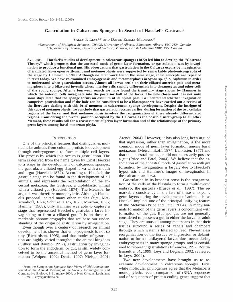
FIG. 1. Schematic of the possible gastrulation stages in the Calcaronea. A, Embryogenesis: (i–ii) eversion of the ‘‘stomoblastula,’’ consideredby all to be pseudogastrulation; (iii) ingression of cells into the larval cavity to form the posterior half of the amphiblastula larva (iv); (iii) isthe stage considered by Barrois (1876) and by Metschnikoff (1874) to be gastrulation. B–E Metamorphosis: B, Haeckel’s (i) morula, (ii)planula, (iii) gastrula, and (v) ascula (juvenile) with a syncytial outer layer. C, Schulze’s (1875) first study: gastrulation by invagination of theposterior cells (iii) to form a bilayered juvenile (v). D, Ingression (iii) and invagination (iv) of the anterior cells at metamorphosis accordingto Metschnikoff (1874). E, (iii) Invagination of the anterior cells, considered by Schulze (1878), Hammer (1908) and Duboscq and Tuzet(1937) to be gastrulation. (iv) Settlement of the invaginating larva according to Schulze (1878) and Hammer (1908).
served with an Olympus SZX12 stereomicroscopewith a 1.63 objective. Free-swimming and settled lar-vae were fixed at 0, 3, 6, 12, 24, and 48 hr after releasefrom the parent in a cocktail fixative of 1% OsO4, 2%glutaraldehyde in 0.45 M sodium acetate buffer, pH6.4, with 10 % sucrose in the final volume (see Leysand Degnan, 2002). As much sea water as possiblewas removed from the samples and 4–5 volumes ofcocktail fixative were added. Samples were fixed at48C for 2 hr. Specimens were then rinsed three timesin filtered sea water, dehydrated in ethanol and trans-ferred to the University of Alberta. Coverslips wereeither cut or broken into smaller pieces and both in-dividual larvae and coverslip pieces with settled larvaewere critical point dried and mounted on electron mi-croscope stubs using silver paint or nail polish; stubswere coated with gold and viewed in a JEOL 6301Ffield emission scanning electron microscope. Otherspecimens were embedded in epoxy (Taab 812) forthick and thin sections as described previously (Leysand Degnan, 2002). In 2004, large numbers of fixedadherent postlarvae were gently pried off the cover-slips using a microscalpel while still in 70% ethanol,
dehydrated and prepared for scanning electron micros-copy and thick and thin sections as described above.
EXISTING INFORMATION: GASTRULATION IN
THE CALCARONEA
During the development of calcaronean spongesthere are three moments when radical cell movementscause a dramatic change in the morphology of the em-bryo or larva; the first two occur during embryogen-esis (Gatenby, 1920; Duboscq and Tuzet, 1933; Gal-lissian, 1983; Franzen, 1988), the third, at metamor-phosis (Metschnikoff, 1874; Schulze, 1875; Barrois,1876; Schulze, 1878; Maas, 1894; Minchin, 1896;Hammer, 1908) (Fig. 1). The most difficult to observeare those at metamorphosis, and these cell movementsare what has long been considered gastrulation.
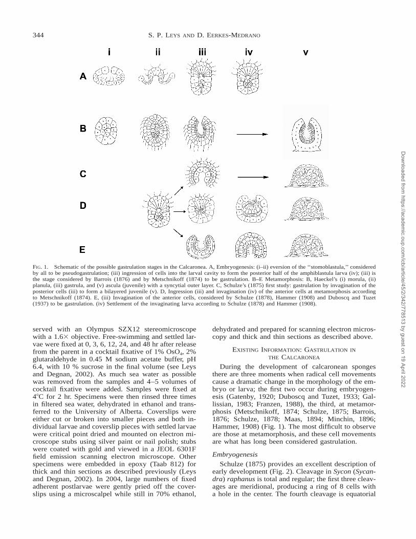
Embryogenesis
Schulze (1875) provides an excellent description ofearly development (Fig. 2). Cleavage in Sycon (Sycan-dra) raphanus is total and regular; the first three cleav-ages are meridional, producing a ring of 8 cells witha hole in the center. The fourth cleavage is equatorial
Dow
nloaded from https://academ
ic.oup.com/icb/article/45/2/342/778513 by guest on 19 April 2022
345GASTRULATION IN CALCAREOUS SPONGES
FIG. 2. Early embryogenesis in Sycon raphanus according toSchulze (1875). Images from plate 20. (a–f) 1–32 cell cleavage stag-es; (g) 64-cell stage blastula.
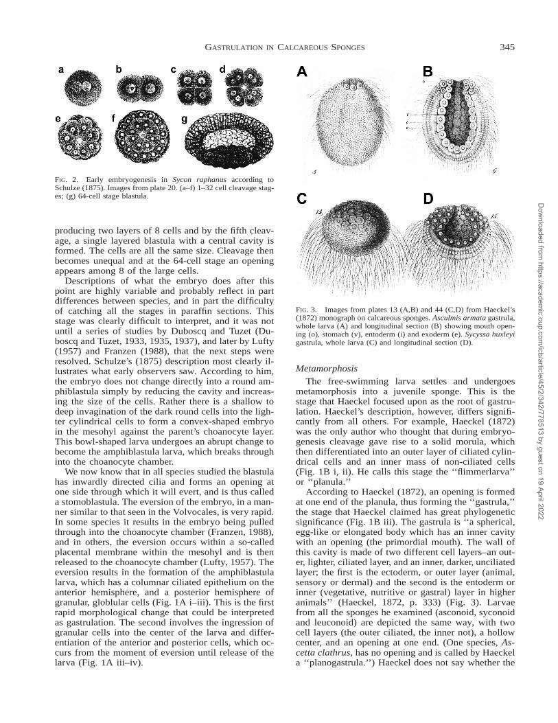
FIG. 3. Images from plates 13 (A,B) and 44 (C,D) from Haeckel’s(1872) monograph on calcareous sponges. Asculmis armata gastrula,whole larva (A) and longitudinal section (B) showing mouth open-ing (o), stomach (v), entoderm (i) and exoderm (e). Sycyssa huxleyigastrula, whole larva (C) and longitudinal section (D).
producing two layers of 8 cells and by the fifth cleav-age, a single layered blastula with a central cavity isformed. The cells are all the same size. Cleavage thenbecomes unequal and at the 64-cell stage an openingappears among 8 of the large cells.
Descriptions of what the embryo does after thispoint are highly variable and probably reflect in partdifferences between species, and in part the difficultyof catching all the stages in paraffin sections. Thisstage was clearly difficult to interpret, and it was notuntil a series of studies by Duboscq and Tuzet (Du-boscq and Tuzet, 1933, 1935, 1937), and later by Lufty(1957) and Franzen (1988), that the next steps wereresolved. Schulze’s (1875) description most clearly il-lustrates what early observers saw. According to him,the embryo does not change directly into a round am-phiblastula simply by reducing the cavity and increas-ing the size of the cells. Rather there is a shallow todeep invagination of the dark round cells into the ligh-ter cylindrical cells to form a convex-shaped embryoin the mesohyl against the parent’s choanocyte layer.This bowl-shaped larva undergoes an abrupt change tobecome the amphiblastula larva, which breaks throughinto the choanocyte chamber.
We now know that in all species studied the blastulahas inwardly directed cilia and forms an opening atone side through which it will evert, and is thus calleda stomoblastula. The eversion of the embryo, in a man-ner similar to that seen in the Volvocales, is very rapid.In some species it results in the embryo being pulledthrough into the choanocyte chamber (Franzen, 1988),and in others, the eversion occurs within a so-calledplacental membrane within the mesohyl and is thenreleased to the choanocyte chamber (Lufty, 1957). Theeversion results in the formation of the amphiblastulalarva, which has a columnar ciliated epithelium on theanterior hemisphere, and a posterior hemisphere ofgranular, globlular cells (Fig. 1A i–iii). This is the firstrapid morphological change that could be interpretedas gastrulation. The second involves the ingression ofgranular cells into the center of the larva and differ-entiation of the anterior and posterior cells, which oc-curs from the moment of eversion until release of thelarva (Fig. 1A iii–iv).
Metamorphosis
The free-swimming larva settles and undergoesmetamorphosis into a juvenile sponge. This is thestage that Haeckel focused upon as the root of gastru-lation. Haeckel’s description, however, differs signifi-cantly from all others. For example, Haeckel (1872)was the only author who thought that during embryo-genesis cleavage gave rise to a solid morula, whichthen differentiated into an outer layer of ciliated cylin-drical cells and an inner mass of non-ciliated cells(Fig. 1B i, ii). He calls this stage the ‘‘flimmerlarva’’or ‘‘planula.’’
According to Haeckel (1872), an opening is formedat one end of the planula, thus forming the ‘‘gastrula,’’the stage that Haeckel claimed has great phylogeneticsignificance (Fig. 1B iii). The gastrula is ‘‘a spherical,egg-like or elongated body which has an inner cavitywith an opening (the primordial mouth). The wall ofthis cavity is made of two different cell layers–an out-er, lighter, ciliated layer, and an inner, darker, unciliatedlayer; the first is the ectoderm, or outer layer (animal,sensory or dermal) and the second is the entoderm orinner (vegetative, nutritive or gastral) layer in higheranimals’’ (Haeckel, 1872, p. 333) (Fig. 3). Larvaefrom all the sponges he examined (asconoid, syconoidand leuconoid) are depicted the same way, with twocell layers (the outer ciliated, the inner not), a hollowcenter, and an opening at one end. (One species, As-cetta clathrus, has no opening and is called by Haeckela ‘‘planogastrula.’’) Haeckel does not say whether the
Dow
nloaded from https://academ
ic.oup.com/icb/article/45/2/342/778513 by guest on 19 April 2022
346 S. P. LEYS AND D. EERKES-MEDRANO
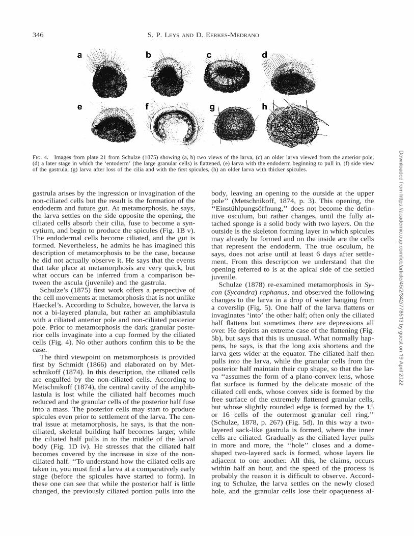
FIG. 4. Images from plate 21 from Schulze (1875) showing (a, b) two views of the larva, (c) an older larva viewed from the anterior pole,(d) a later stage in which the ‘entoderm’ (the large granular cells) is flattened, (e) larva with the endoderm beginning to pull in, (f) side viewof the gastrula, (g) larva after loss of the cilia and with the first spicules, (h) an older larva with thicker spicules.
gastrula arises by the ingression or invagination of thenon-ciliated cells but the result is the formation of theendoderm and future gut. At metamorphosis, he says,the larva settles on the side opposite the opening, theciliated cells absorb their cilia, fuse to become a syn-cytium, and begin to produce the spicules (Fig. 1B v).The endodermal cells become ciliated, and the gut isformed. Nevertheless, he admits he has imagined thisdescription of metamorphosis to be the case, becausehe did not actually observe it. He says that the eventsthat take place at metamorphosis are very quick, butwhat occurs can be inferred from a comparison be-tween the ascula (juvenile) and the gastrula.
Schulze’s (1875) first work offers a perspective ofthe cell movements at metamorphosis that is not unlikeHaeckel’s. According to Schulze, however, the larva isnot a bi-layered planula, but rather an amphiblastulawith a ciliated anterior pole and non-ciliated posteriorpole. Prior to metamorphosis the dark granular poste-rior cells invaginate into a cup formed by the ciliatedcells (Fig. 4). No other authors confirm this to be thecase.
The third viewpoint on metamorphosis is providedfirst by Schmidt (1866) and elaborated on by Met-schnikoff (1874). In this description, the ciliated cellsare engulfed by the non-ciliated cells. According toMetschnikoff (1874), the central cavity of the amphib-lastula is lost while the ciliated half becomes muchreduced and the granular cells of the posterior half fuseinto a mass. The posterior cells may start to producespicules even prior to settlement of the larva. The cen-tral issue at metamorphosis, he says, is that the non-ciliated, skeletal building half becomes larger, whilethe ciliated half pulls in to the middle of the larvalbody (Fig. 1D iv). He stresses that the ciliated halfbecomes covered by the increase in size of the non-ciliated half. ‘‘To understand how the ciliated cells aretaken in, you must find a larva at a comparatively earlystage (before the spicules have started to form). Inthese one can see that while the posterior half is littlechanged, the previously ciliated portion pulls into the
body, leaving an opening to the outside at the upperpole’’ (Metschnikoff, 1874, p. 3). This opening, the‘‘Einstuhlpungsoffnung,’’ does not become the defin-itive osculum, but rather changes, until the fully at-tached sponge is a solid body with two layers. On theoutside is the skeleton forming layer in which spiculesmay already be formed and on the inside are the cellsthat represent the endoderm. The true osculum, hesays, does not arise until at least 6 days after settle-ment. From this description we understand that theopening referred to is at the apical side of the settledjuvenile.
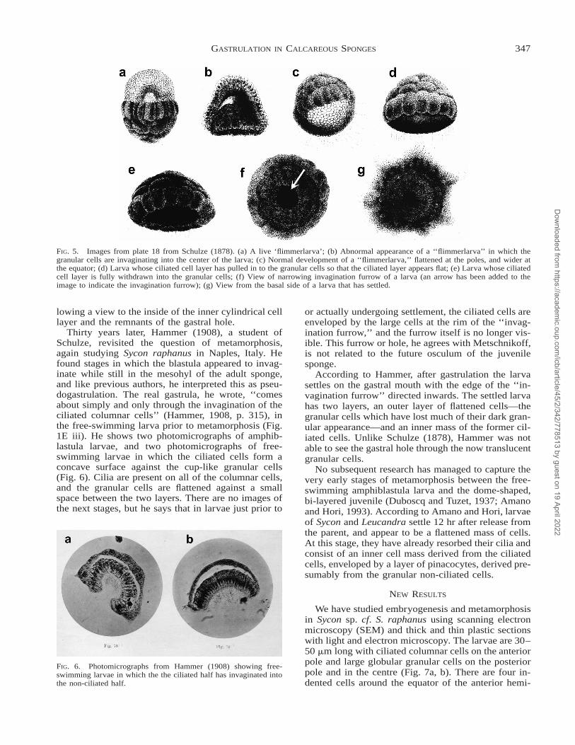
Schulze (1878) re-examined metamorphosis in Sy-con (Sycandra) raphanus, and observed the followingchanges to the larva in a drop of water hanging froma coverslip (Fig. 5). One half of the larva flattens orinvaginates ‘into’ the other half; often only the ciliatedhalf flattens but sometimes there are depressions allover. He depicts an extreme case of the flattening (Fig.5b), but says that this is unusual. What normally hap-pens, he says, is that the long axis shortens and thelarva gets wider at the equator. The ciliated half thenpulls into the larva, while the granular cells from theposterior half maintain their cup shape, so that the lar-va ‘‘assumes the form of a plano-convex lens, whoseflat surface is formed by the delicate mosaic of theciliated cell ends, whose convex side is formed by thefree surface of the extremely flattened granular cells,but whose slightly rounded edge is formed by the 15or 16 cells of the outermost granular cell ring.’’(Schulze, 1878, p. 267) (Fig. 5d). In this way a two-layered sack-like gastrula is formed, where the innercells are ciliated. Gradually as the ciliated layer pullsin more and more, the ‘‘hole’’ closes and a dome-shaped two-layered sack is formed, whose layers lieadjacent to one another. All this, he claims, occurswithin half an hour, and the speed of the process isprobably the reason it is difficult to observe. Accord-ing to Schulze, the larva settles on the newly closedhole, and the granular cells lose their opaqueness al-
Dow
nloaded from https://academ
ic.oup.com/icb/article/45/2/342/778513 by guest on 19 April 2022
347GASTRULATION IN CALCAREOUS SPONGES
FIG. 5. Images from plate 18 from Schulze (1878). (a) A live ‘flimmerlarva’; (b) Abnormal appearance of a ‘‘flimmerlarva’’ in which thegranular cells are invaginating into the center of the larva; (c) Normal development of a ‘‘flimmerlarva,’’ flattened at the poles, and wider atthe equator; (d) Larva whose ciliated cell layer has pulled in to the granular cells so that the ciliated layer appears flat; (e) Larva whose ciliatedcell layer is fully withdrawn into the granular cells; (f) View of narrowing invagination furrow of a larva (an arrow has been added to theimage to indicate the invagination furrow); (g) View from the basal side of a larva that has settled.
FIG. 6. Photomicrographs from Hammer (1908) showing free-swimming larvae in which the the ciliated half has invaginated intothe non-ciliated half.
lowing a view to the inside of the inner cylindrical celllayer and the remnants of the gastral hole.
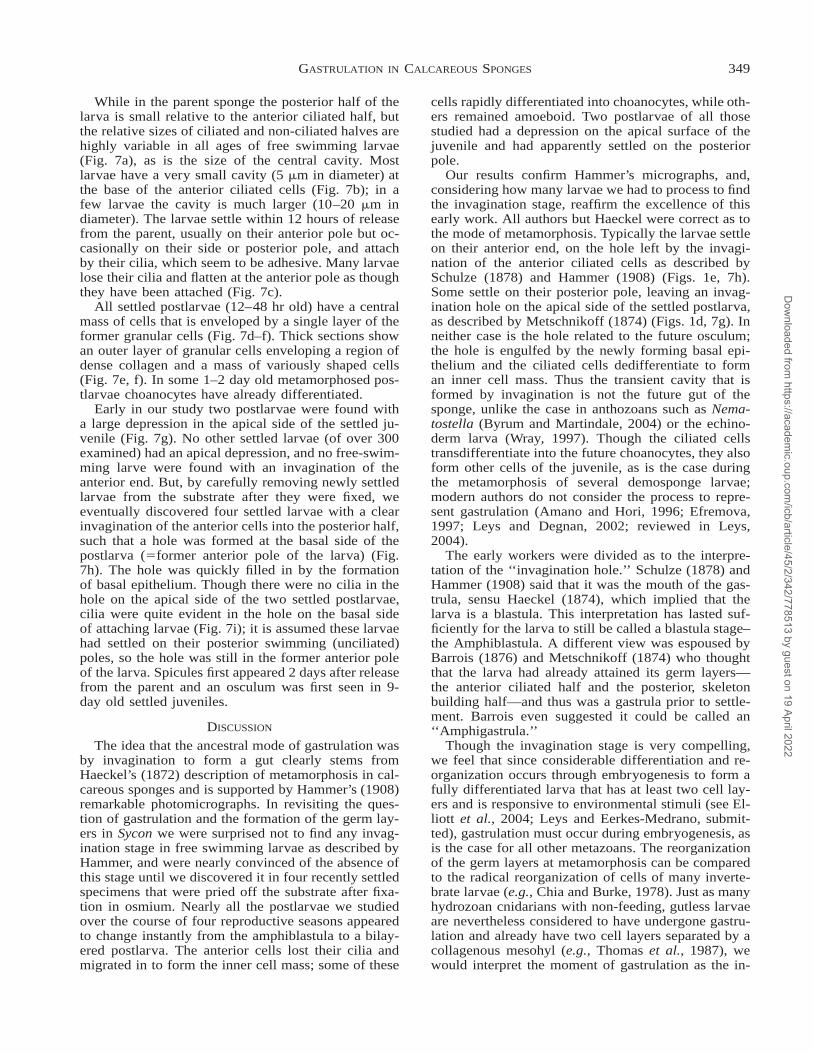
Thirty years later, Hammer (1908), a student ofSchulze, revisited the question of metamorphosis,again studying Sycon raphanus in Naples, Italy. Hefound stages in which the blastula appeared to invag-inate while still in the mesohyl of the adult sponge,and like previous authors, he interpreted this as pseu-dogastrulation. The real gastrula, he wrote, ‘‘comesabout simply and only through the invagination of theciliated columnar cells’’ (Hammer, 1908, p. 315), inthe free-swimming larva prior to metamorphosis (Fig.1E iii). He shows two photomicrographs of amphib-lastula larvae, and two photomicrographs of free-swimming larvae in which the ciliated cells form aconcave surface against the cup-like granular cells(Fig. 6). Cilia are present on all of the columnar cells,and the granular cells are flattened against a smallspace between the two layers. There are no images ofthe next stages, but he says that in larvae just prior to
or actually undergoing settlement, the ciliated cells areenveloped by the large cells at the rim of the ‘‘invag-ination furrow,’’ and the furrow itself is no longer vis-ible. This furrow or hole, he agrees with Metschnikoff,is not related to the future osculum of the juvenilesponge.
According to Hammer, after gastrulation the larvasettles on the gastral mouth with the edge of the ‘‘in-vagination furrow’’ directed inwards. The settled larvahas two layers, an outer layer of flattened cells—thegranular cells which have lost much of their dark gran-ular appearance—and an inner mass of the former cil-iated cells. Unlike Schulze (1878), Hammer was notable to see the gastral hole through the now translucentgranular cells.
No subsequent research has managed to capture thevery early stages of metamorphosis between the free-swimming amphiblastula larva and the dome-shaped,bi-layered juvenile (Duboscq and Tuzet, 1937; Amanoand Hori, 1993). According to Amano and Hori, larvaeof Sycon and Leucandra settle 12 hr after release fromthe parent, and appear to be a flattened mass of cells.At this stage, they have already resorbed their cilia andconsist of an inner cell mass derived from the ciliatedcells, enveloped by a layer of pinacocytes, derived pre-sumably from the granular non-ciliated cells.
NEW RESULTS
We have studied embryogenesis and metamorphosisin Sycon sp. cf. S. raphanus using scanning electronmicroscopy (SEM) and thick and thin plastic sectionswith light and electron microscopy. The larvae are 30–50 mm long with ciliated columnar cells on the anteriorpole and large globular granular cells on the posteriorpole and in the centre (Fig. 7a, b). There are four in-dented cells around the equator of the anterior hemi-
Dow
nloaded from https://academ
ic.oup.com/icb/article/45/2/342/778513 by guest on 19 April 2022

348 S. P. LEYS AND D. EERKES-MEDRANO
FIG. 7. Light and scanning electron micrographs of Sycon sp. cf. S. raphanus. (a) A 2-hr-old free-swimming larva showing the anteriorciliated hemisphere (ap) and large globular, granular cells of the posterior hemisphere (pp). One of the four ‘‘cross cells’’ on the anteriorhemisphere is shown by the arrow. (b) A longitudinal section through a plastic embedded free-swimming larva in the same orientation as (a);lc, larval cavity. (c) A scanning electron micrograph (SEM) of a 12-hr old larva that has lost the cilia and flattened (arrow) on the anteriorpole as though it had been attached to the substrate. (d) SEM of a newly adherent postlarva. (e) Light micrograph of a section horizontallythrough a plastic embedded newly adherent and metamorphosing larva (between 12 hours and 2 days after release from the parent). Thepostlarva has an outer layer of granular cells (arrow) surrounding a group of variously shaped cells in a collagenous extracellular matrix(arrowhead). (f) Longitudinal (vertical) section through a newly adherent postlarva at the same stage as (e). The outer epithelium is continuousaround the entire postlarva from apical (arrow) to basal (arrowhead) surfaces. (g) SEM of one of two postlarvae that settled with a depressionor ‘hole’ (arrow) in the apical side. (h) SEM of a postlarva that was detached from the substrate after fixation with osmium. The anteriorciliated cells have invaginated into the posterior cells forming a ‘hole’ in the basal side (arrow). (i) SEM providing a view into the invaginationhole of the adherent larva in (h) showing cilia (arrowhead). Scale bars: a–h:10 mm; i: 2 mm.
sphere that correspond to the cross cells of Duboscqand Tuzet (1941). The larvae swim by rotating in aright hand direction (clockwise as seen from the pos-terior pole) as described previously (Elliott et al.,2004). Several thousand larvae were observed live andof these, some 300 were observed as they underwent
metamorphosis in petri dishes. Larvae and postlarvaecollected during four different years (2001–4) werefixed for electron microscopy; we examined over athousand individual larvae and postlarvae at differentstages of settlement and metamorphosis and capturedimages of 380 individuals.
Dow
nloaded from https://academ
ic.oup.com/icb/article/45/2/342/778513 by guest on 19 April 2022
349GASTRULATION IN CALCAREOUS SPONGES
While in the parent sponge the posterior half of thelarva is small relative to the anterior ciliated half, butthe relative sizes of ciliated and non-ciliated halves arehighly variable in all ages of free swimming larvae(Fig. 7a), as is the size of the central cavity. Mostlarvae have a very small cavity (5 mm in diameter) atthe base of the anterior ciliated cells (Fig. 7b); in afew larvae the cavity is much larger (10–20 mm indiameter). The larvae settle within 12 hours of releasefrom the parent, usually on their anterior pole but oc-casionally on their side or posterior pole, and attachby their cilia, which seem to be adhesive. Many larvaelose their cilia and flatten at the anterior pole as thoughthey have been attached (Fig. 7c).
All settled postlarvae (12–48 hr old) have a centralmass of cells that is enveloped by a single layer of theformer granular cells (Fig. 7d–f). Thick sections showan outer layer of granular cells enveloping a region ofdense collagen and a mass of variously shaped cells(Fig. 7e, f). In some 1–2 day old metamorphosed pos-tlarvae choanocytes have already differentiated.
Early in our study two postlarvae were found witha large depression in the apical side of the settled ju-venile (Fig. 7g). No other settled larvae (of over 300examined) had an apical depression, and no free-swim-ming larve were found with an invagination of theanterior end. But, by carefully removing newly settledlarvae from the substrate after they were fixed, weeventually discovered four settled larvae with a clearinvagination of the anterior cells into the posterior half,such that a hole was formed at the basal side of thepostlarva (5former anterior pole of the larva) (Fig.7h). The hole was quickly filled in by the formationof basal epithelium. Though there were no cilia in thehole on the apical side of the two settled postlarvae,cilia were quite evident in the hole on the basal sideof attaching larvae (Fig. 7i); it is assumed these larvaehad settled on their posterior swimming (unciliated)poles, so the hole was still in the former anterior poleof the larva. Spicules first appeared 2 days after releasefrom the parent and an osculum was first seen in 9-day old settled juveniles.
DISCUSSION
The idea that the ancestral mode of gastrulation wasby invagination to form a gut clearly stems fromHaeckel’s (1872) description of metamorphosis in cal-careous sponges and is supported by Hammer’s (1908)remarkable photomicrographs. In revisiting the ques-tion of gastrulation and the formation of the germ lay-ers in Sycon we were surprised not to find any invag-ination stage in free swimming larvae as described byHammer, and were nearly convinced of the absence ofthis stage until we discovered it in four recently settledspecimens that were pried off the substrate after fixa-tion in osmium. Nearly all the postlarvae we studiedover the course of four reproductive seasons appearedto change instantly from the amphiblastula to a bilay-ered postlarva. The anterior cells lost their cilia andmigrated in to form the inner cell mass; some of these
cells rapidly differentiated into choanocytes, while oth-ers remained amoeboid. Two postlarvae of all thosestudied had a depression on the apical surface of thejuvenile and had apparently settled on the posteriorpole.
Our results confirm Hammer’s micrographs, and,considering how many larvae we had to process to findthe invagination stage, reaffirm the excellence of thisearly work. All authors but Haeckel were correct as tothe mode of metamorphosis. Typically the larvae settleon their anterior end, on the hole left by the invagi-nation of the anterior ciliated cells as described bySchulze (1878) and Hammer (1908) (Figs. 1e, 7h).Some settle on their posterior pole, leaving an invag-ination hole on the apical side of the settled postlarva,as described by Metschnikoff (1874) (Figs. 1d, 7g). Inneither case is the hole related to the future osculum;the hole is engulfed by the newly forming basal epi-thelium and the ciliated cells dedifferentiate to forman inner cell mass. Thus the transient cavity that isformed by invagination is not the future gut of thesponge, unlike the case in anthozoans such as Nema-tostella (Byrum and Martindale, 2004) or the echino-derm larva (Wray, 1997). Though the ciliated cellstransdifferentiate into the future choanocytes, they alsoform other cells of the juvenile, as is the case duringthe metamorphosis of several demosponge larvae;modern authors do not consider the process to repre-sent gastrulation (Amano and Hori, 1996; Efremova,1997; Leys and Degnan, 2002; reviewed in Leys,2004).
The early workers were divided as to the interpre-tation of the ‘‘invagination hole.’’ Schulze (1878) andHammer (1908) said that it was the mouth of the gas-trula, sensu Haeckel (1874), which implied that thelarva is a blastula. This interpretation has lasted suf-ficiently for the larva to still be called a blastula stage–the Amphiblastula. A different view was espoused byBarrois (1876) and Metschnikoff (1874) who thoughtthat the larva had already attained its germ layers—the anterior ciliated half and the posterior, skeletonbuilding half—and thus was a gastrula prior to settle-ment. Barrois even suggested it could be called an‘‘Amphigastrula.’’
Though the invagination stage is very compelling,we feel that since considerable differentiation and re-organization occurs through embryogenesis to form afully differentiated larva that has at least two cell lay-ers and is responsive to environmental stimuli (see El-liott et al., 2004; Leys and Eerkes-Medrano, submit-ted), gastrulation must occur during embryogenesis, asis the case for all other metazoans. The reorganizationof the germ layers at metamorphosis can be comparedto the radical reorganization of cells of many inverte-brate larvae (e.g., Chia and Burke, 1978). Just as manyhydrozoan cnidarians with non-feeding, gutless larvaeare nevertheless considered to have undergone gastru-lation and already have two cell layers separated by acollagenous mesohyl (e.g., Thomas et al., 1987), wewould interpret the moment of gastrulation as the in-
Dow
nloaded from https://academ
ic.oup.com/icb/article/45/2/342/778513 by guest on 19 April 2022

350 S. P. LEYS AND D. EERKES-MEDRANO
gression of cells into the larval cavity after eversionand prior to release from the parent, that is duringembryogenesis, not at metamorphosis. This is in ac-cordance with other modern interpretations of gastru-lation in the Porifera (Efremova, 1997; Boury-Esnaultet al., 1999; reviewed in Leys, 2004).
The problem with interpretation of the ‘invaginationhole’ today comes from the association of gastrulationwith gut formation, which stems directly from Haeck-el’s definition and descriptions of this pivotal stage insponge development. Though in many animals gastru-lation results in formation of the endoderm, in basalgroups it is frequently a two step process, first result-ing in the formation of germ layers, and only later inthe formation of the gut. Within our current under-standing of sponge structure, few authors would ho-mologize the choanocyte epithelium of sponges withthe lining of the gut in other animals.
Thus we suggest that the principal function of gas-trulation is formation of the germ layers and that for-mation of the gut has secondarily become part of theprocess. As a corollary we suggest that the primitivemode of gastrulation was by ingression or delamina-tion, not invagination. If gastrulation is not primarilyto do with gut formation, this may explain why wefind expression of conserved endomesodermal genesin animals which are not otherwise considered to havea third germ layer (Groger et al., 1999; Adell et al.,2003; Manuel et al., 2004). This said, the image ofinvagination of Sycon at metamorphosis shows a pro-cess intriguingly similar to the invagination that occursduring echinoderm gastrulation and begs interpreta-tion. A re-evaluation of the relationships between germlayers of basal and higher metazoan phyla including acomparison of gene expression patterns in poriferanmodels—when possible—may shed light on the pri-mary function of gastrulation.
ACKNOWLEDGMENTS
This research was funded by NSERC. We thank thedirector and staff of the Bamfield Marine SciencesCentre for use of facilities for carrying out this work,G. Elliott and E. Cheung for technical assistance, andScott Nichols and Gert Worheide for organizing thesymposium on Sponges: New Views of Old Animals.SL is grateful to the Society for Integrative and Com-parative Biology for funding portions of the trip toNew Orleans to deliver this paper at the 2004 meeting.
REFERENCES
Adell, T., V. A. Grebenjuk, M. Wiens, and W. E. G. Muller. 2003.Isolation and characterization of two T-box genes from sponges,the phylogenetically oldest metazoan taxon. 213:421–434.
Amano, S. and I. Hori. 1992. Metamorphosis of calcareous spongesI. Ultrastructure of free-swimming larvae. Invertebr. Reprod.Dev. 21:81–90.
Amano, S. and I. Hori. 1993. Metamorphosis of calcareous spongesII. Cell rearragement and differentiation in metamorphosis. In-vertebr. Reprod. Dev. 24:13–26.
Amano, S. and I. Hori. 1996. Transdifferentiation of larval flagel-lated cells to choanocytes in the metamorphosis of the demos-ponge Haliclona permollis. Biol. Bull. 190:161–172.
Amano, S. and I. Hori. 2001. Metamorphosis of coeloblastula per-formed by multipotential larval flagellated cells in the calcare-ous sponge Leucosolenia laxa. Biol. Bull. 200:20–32.
Arendt, D. 2004. Comparative aspects of gastrulation. In C. Stern(ed.), Gastrulation. From cells to embryos pp. 679–693. ColdSpring Harbor Press, Cold Spring Harbor.
Austin, W. C. and B. Ott. 1987. Porifera. In E. N. Kozloff (ed.),Marine invertebrates of the Pacific Northwest, pp 6–31. U. ofWashington Press, Seattle.
Barrois, M. C. 1876. L’embryologie de quelques eponges de la man-che. Annales des Sciences Naturelles, Zoologie et Paleontologie3:1–84.
Borchiellini, C., M. Manuel, E. Alivon, N. Boury-Esnault, J. Vace-let, and Y. Le Parco. 2001. Sponge paraphyly and the origin ofMetazoa. Evol. Biol. 14:171–179.
Borojevic, R. 1969. Etude du developpement et de la differenciationcellulaire d’eponges calcaires calcineennes (Genres Clathrina etAscandra). Annales d’embryologie et de morphogenese 2:15–36.
Borojevic, R. 1970. Differenciation cellulaire dans l’embyogeneseet la morphogenese chez les spongiaires. Zool. Soc. Lond. 25:467–490.
Boury-Esnault, N., S. Efremova, C. Bezac, and J. Vacelet. 1999.Reproduction of a hexactinellid sponge: First description of gas-trulation by cellular delamination in the Porifera. Invertebr. Re-prod. Dev. 35:187–201.
Brien, P. 1967. Les eponges. Leur nature metazoaire, leur gastrula-tion, leur etat colonial. Ann. Soc. Roy. Zool. Belg. 97:197–235.
Brusca, G. J., R. C. Brusca, and S. F. Gilbert. 1997. Characteristicsof metazoan development. In R. Gilbert and A. M. Raunio (ed.),Embryology. Constructing the organism, pp 3–19. Sinauer As-sociates, Inc., Sunderland, Massachusetts.
Byrum, C. A. and M. Q. Martindale. 2004. Gastrulation in the Cni-daria and Ctenophora. In C. Stern (ed.), Gastrulation. Fromcells to embryos pp. 33–50. Cold Spring Harbor Press, ColdSpring Harbor.
Chia, F.-S. and R. D. Burke. 1978. Echinoderm metamorphosis: Fateof larval structures. In F.-S. Chia and M. E. Rice (eds.), Settle-ment and metamorphosis of marine invertebrate larvae. pp.219–234. Elsevier North-Holland Inc., New York.
Denis, H. 1997. L’origine de la gastrulation. Medecine/sciences 13:1503–1515.
Duboscq, O. and O. Tuzet. 1933. Quelques structure des amphiblas-tules d’Eponges calcaires. C.R. Acad. Sc. Paris 197:561–563.
Duboscq, O. and O. Tuzet. 1935. Un nouveau stade du developpe-ment des Eponges calcaires. C.R. Acad. Sc. Paris 200:1788–1790.
Duboscq, O. and O. Tuzet. 1937. L’ovogenese, la fecondation et lespremiers stades du developpement des eponges calcaires. Arch.Zool. Exp. Gen. 79:157–316.
Efremova, S. M. 1997. Once more on the position among Metazoa—Gastrulation and germinal layers of sponges. Berliner Geowiss.Abh. 20:7–15.
Elliott, G. R. D., T. A. Macdonald, and S. P. Leys. 2004. Spongelarval phototaxis: A comparative study. In M. Pansini, R. Pron-zato, G. Bavestrello and R. Manconi (eds.). Boll. Mus. Ist. Biol.Univ. Genova. pp. 291–300.
Fell, P. E. 1997. Porifera, the Sponges. In S. F. Gilbert and A. M.Raunio (eds.), Embryology: Constructing the organism. pp. 39–54. Sinauer.
Franzen, W. 1988. Oogenesis and larval develoment of Scypha cil-iata (Porifera, Calcarea). Zoomorphology 107:349–357.
Gallissian, M. F. 1983. Etude ultrastructurale du developpement em-bryonnaire chez Grantia compressa F. (Porifera, Calcarea).Arch. Anat. Microsc. 72:59–75.
Gatenby, J. B. 1920. The germ-cells, fertilization, and early devel-opment of Grantia (Sycon) compressa. Linn. J. Zoology 34:261–297.
Gilbert, F. S. and A. M. Raunio. 1997. Embryology: Constructingthe organism. Sinauer Associates, Sunderland, Massachusetts.
Groger, H., P. Callaerts, W. J. Gehring, and V. Schmid. 1999. Geneduplication and recruitment of a specific tropomyosin into stri-
Dow
nloaded from https://academ
ic.oup.com/icb/article/45/2/342/778513 by guest on 19 April 2022
351GASTRULATION IN CALCAREOUS SPONGES
ated muscle cells in the jellyfish Podocoryne carnea. J. Exp.Zool. 285:378–386.
Haeckel, E. 1872. Die Kalkschwamme. Eine monographie. Verlagvon Georg Reimer, Berlin.
Haeckel, E. 1874. Die Gastraea-Theorie, die phylogenetische Clas-sification des Thierreichs und die Homologie der Keimblatter.J. Zeitschr. fur Naturwiss. 8:1–55.
Hammer, E. 1908. Neu Beitrage zur Kenntnis der Histologie undEntwicklung von Sycon raphanus. Archiv fur Biontologie 2:289–334.
Johnson, M. F. 1979. Gametogenesis and embryonic development inthe calcareous sponges Clathrina coriacea and C. blanca fromSanta Catalina Island, California. 78:183–191
Jorgensen, M. 1910. Beitrage zur Kenntnis der Eibildung, Reifung,Befuchtung und Furchung bei Schwammen (Syconen). Archivf. Zellforschung 4:164–242.
Kruse, M., S. P. Leys, I. M. Muller, and W. E. G. Muller. 1998.Phylogenetic postition of the hexactinellida within the phylumPorifera based on the amino acid sequnce of the Protein KinaseC from Rhabdocalyptus. J. Mol. Evol. 46:721–728.
Lankester, E. R. 1877. Notes on the embryology and classificationof the animal kingdom: Comprising a revision of speculationsrelative to the origin and significance of the germ-layers. Q. J.Micr. Sci. 17:399–441.
Leys, S. P. 2004. Gastrulation in sponges. In C. Stern (ed.), Gastru-lation: From cells to embryo. Cold Spring Harbor Press, pp.23–31.
Leys, S. P. and B. M. Degnan. 2002. Embryogenesis and metamor-phosis in a haplosclerid demosponge: Gastrulation and trans-differentiation of larval ciliated cells to choanocytes. Invertebr.Biol. 121:171–189.
Lufty, R. G. 1957. On the placental membrane of calcareous spong-es. 58:239–246.
Maas, O. 1894. Die embryonal-entwicklung und metamorphose dercornacuspongien. Zool. Jahrb. Anat. VII:331–448.
Manuel, M., C. Borchiellini, E. Alivon, Y. Le Parco, J. Vacelet, andN. Boury-Esnault. 2003. Phylogeny and evolution of calcareoussponges: Monophyly of calcinea and calcaronea, high level ofmorphological homoplasy, and the primitive nature of axialsymmetry. Syst. Biol. 3:311–333.
Manuel, M., R. Borojevic, N. Boury-Esnault, and J. Vacelet. 2002.Class Calcarea Bowerbank, 1864. In J. A. Hooper and R. W.M. Van Soest (eds.), Systema Porifera: A guide to the classifi-cation of sponges. pp. 1103–1110. Kluwer Academic/PlenumPublishers, New York.
Manuel, M., Y. Le Parco, and C. Borchiellini. 2004. Comparativeanalysis of Brachyury T-domains, with the characterization oftwo new sequences, from a hexactinellid and a calcisponge.340:291–301.
Medina, M., A. G. Collins, J. Silberman, and M. L. Sogin. 2001.Evaluating hypotheses of basal animal phylogeny using com-plete sequences of large and small subunit rRNA. Proc. Natl.Acad. Sci. U.S.A 98:9707–9712.
Metschnikoff, E. 1874. Zur Entwickelungsgeschichte der Kalksch-wamme. Zeitschr. f. Wiss. Zool. 24:1–14.
Minchin, E. A. 1896. Note on the larva and post larval developmentof Leucosolenia variabilis, H. sp., with remarks on the devel-opment of other Asconidae. Proc. R. Soc. Lond. B 40:42–52.
Nielsen, C. 2001. Animal evolution. Interrelationships of the livingphyla. Oxford University Press, Oxford.
Price, A. L. and N. H. Patel. 2004. The evolution of gastrulation:Cellular and molecular aspects. In C. Stern (ed.), Gastrulation.From cells to embryos. pp. 695–701. Cold Spring Harbor Press.
Richardson, M. K. 1995. Heterochrony and the phylotypic period.172:412–421.
Schmidt, O. 1866. Zweites Supplement der Spongien des adriatisch-en Meeres. Leipzig.
Schulze, F. E. 1875. Ueber den Bau und die Entwicklung von Sy-candra raphanus Haeckel. Zeitschr. f. wiss. Zool. 25:247–280.
Schulze, F. E. 1878. Untersuchungen uber den Bau und die Entwick-lung der Spongien. Die Metamorphose von Sycandra raphanus.Zeitschr. f. Wiss. Zool. 31:262–295.
Thomas, M. B., N. C. Edwards, and T. A. Norris. 1987. Gastrulationin Halocordyle disticha (Hydrozoa, Athecata). Invertebr. Re-prod. Dev. 12:91–102.
Tuzet, O. 1947. L’ovogenese et la fecondation de l’eponge calcaireLeucosolenia (Clathrina) coriacea Mont. et de l’eponge siliceu-se Reniera elegans Bow. 85:127–148.
Tuzet, O. 1963. The phylogeny of sponges according to embryolog-ical, histological, and seological data, and their affinities withthe Protozoa and the Cnidaria. In E. C. Dougherty (ed.), Thelower metazoa. Comparative biology and phylogeny, pp. 129–148. The University of California Press, Berkeley, CA.
Tuzet, O. 1973. Eponges calcaires. In P.-P. Grasse (ed.), Traite deZoologie, pp. 27–132. Masson et Cie.
Wallace, R. L. and W. K. Taylor. 1997. Invertebrate Zoology: Alaboratory manual. Prentice Hall, Upper Saddle River, New Jer-sey.
Wolpert, L. 1992. Gastrulation and the evolution of development. InDevelopment Supplement: 7–13.
Wray, G. A. 1997. Echinoderms. In R. Gilbert and A. M. Raunio(eds.), Embryology. Constructing the organism. pp. 309–329.Sinauer Associates, Sunderland, MA.
APPENDIXA note on species identifications:The taxonomy of the Calcarea has a long and convoluted history.
Haeckel proposed the first taxonomy choosing names for genera thatreflected body architecture; his system was not followed. Metschni-koff (1874) identified the species he studied, Sycon ciliatum, asequivalent to Haeckel’s Sycandra raphanus. The species referred toas Sycandra raphanus by Haeckel and Schulze has been synonom-ized under Sycon (see Manuel et al., 2002), and the species referredto as Sycon ciliatum by Duboscq and Tuzet (1937) was later iden-tified as Sycon raphanus. Scypha is now also included in Sycon.Gatenby (1920) also equated Grantia with Sycon. According to Ma-nuel et al. (2002), all genera but Scypha are now valid. Hence, forthe purposes of this paper, all workers investigated a syconoidsponge identified at some point as Sycon. The genus Sycon is cur-rently considered to be polyphyletic (Manuel et al. 2003). No revi-sion of the Pacific species has been carried out, but the spongestudied here was identified as Sycon according to the keys and de-scriptions of Manuel et al. (2002), and as most similar to S. ra-phanus according to the Keys of Austin and Ott (1987).
Table of current and past species names:
Current name* Past name Author
Sycon raphanus Sycandra raphanus Schmidt, 1866Haeckel, 1872Schulze, 1875, 1878Jorgensen, 1910
Sycon raphanus Sycon raphanus Hammer, 1908Sycon raphanus Sycon ciliatum Metschnikoff, 1874
Duboscq and Tuzet, 1937Sycon ciliatum Scypha ciliata Franzen, 1988Grantia compressa Grantia compressa
Sycon compressaGatenby, 1920Barrois, 1876
* From Manuel et al. (2002).
Dow
nloaded from https://academ
ic.oup.com/icb/article/45/2/342/778513 by guest on 19 April 2022