Emys orbicularis: An Evolutionary Perspective on - Gastrulation
10
Molecular Characterization of the Gastrula in the Turtle Emys orbicularis: An Evolutionary Perspective on Gastrulation Marion Coolen 1 , Delphine Nicolle 1 , Jean-Louis Plouhinec 1¤b , Aure ´ lie Gombault 1 , Tatjana Sauka- Spengler 1¤c,¤d , Arnaud Menuet 1 , Claude Pieau 2¤a , Sylvie Mazan 1 * 1 De ´veloppement et Evolution des verte ´bre ´s, UMR 6218, CNRS et Universite ´ d’Orle ´ ans, Orleans, France, 2 Institut Jacques Monod, UMR7592, CNRS et Universite ´ Pierre et Marie Curie-Paris6 and Paris7, Paris, France Abstract Due to the presence of a blastopore as in amphibians, the turtle has been suggested to exemplify a transition form from an amphibian- to an avian-type gastrulation pattern. In order to test this hypothesis and gain insight into the emergence of the unique characteristics of amniotes during gastrulation, we have performed the first molecular characterization of the gastrula in a reptile, the turtle Emys orbicularis. The study of Brachyury, Lim1, Otx2 and Otx5 expression patterns points to a highly conserved dynamic of expression with amniote model organisms and makes it possible to identify the site of mesoderm internalization, which is a long-standing issue in reptiles. Analysis of Brachyury expression also highlights the presence of two distinct phases, less easily recognizable in model organisms and respectively characterized by an early ring- shaped and a later bilateral symmetrical territory. Systematic comparisons with tetrapod model organisms lead to new insights into the relationships of the blastopore/blastoporal plate system shared by all reptiles, with the blastopore of amphibians and the primitive streak of birds and mammals. The biphasic Brachyury expression pattern is also consistent with recent models of emergence of bilateral symmetry, which raises the question of its evolutionary significance. Citation: Coolen M, Nicolle D, Plouhinec J-L, Gombault A, Sauka-Spengler T, et al. (2008) Molecular Characterization of the Gastrula in the Turtle Emys orbicularis: An Evolutionary Perspective on Gastrulation. PLoS ONE 3(7): e2676. doi:10.1371/journal.pone.0002676 Editor: Daphne Soares, University of Maryland, United States of America Received February 13, 2008; Accepted May 23, 2008; Published July 16, 2008 Copyright: ß 2008 Coolen et al. This is an open-access article distributed under the terms of the Creative Commons Attribution License, which permits unrestricted use, distribution, and reproduction in any medium, provided the original author and source are credited. Funding: This work was supported by funds from the Universite ´ Paris Sud, Universite ´ d’Orle ´ans and CNRS. J.L P. and T.S.S. were recipients of PhD fellowships from the Ministe `re de la Recherche and M.C from CNRS. Competing Interests: The authors have declared that no competing interests exist. * E-mail: [email protected] ¤a Current address: Franconville, France, ¤b Current address: Department of Biological Chemistry, University of California Los Angeles, Los Angeles, California, United States of America, ¤c Current address: Howard Hughes Medical Institute, University of California Los Angeles, Los Angeles, California, United States of America, ¤d Current address: Division of Biology, California Institute of Technology, Pasadena, California, United States of America Introduction Analyses focused on a very limited number of model organisms have led to major advances in our understanding of the molecular mechanisms controlling key developmental processes. These studies have deeply impacted our understanding of the unity of metazoans, by showing that as diverse organisms as the mouse, Drosophila and even Hydra use a relatively small set of related regulatory modules, repeatedly co-opted and adapted to different cellular contexts, to build their body plan. At the microevolution- ary scale, they have also paved the way for accurate identifications of genetic modifications responsible for behavioral or morpholog- ical diversifications, thus enlightening the genetic architecture underlying these evolutionary processes [1,2]. However, at the macroevolutionary scale, attempts to reconstruct an evolutionary pathway through comparisons between selected, often distantly related, model organisms or through analyses of mutant phenotypes interpreted as atavisms, often remain hazardous. The major obstacle to these approaches is that each model organism has diverged from ancestral patterns by an accumulation of taxa- or even species-specific changes. Developmental genetics, which mainly focus on phenotypes associated to a very limited number of often dramatic genetic changes such as gene inactivation or ectopic mis-expressions, are unlikely to reconstruct this succession of events. Such a problem is not easily resolved in the absence of extant transition forms but one way to alleviate the difficulty can be to first assess the generality of the mechanisms characterized in a given model organism within a taxon of relatively closely related species. Such comparisons at moderate evolutionary scale must help to infer the ancestral state of a given taxon, which can then be used for comparisons with more distantly related species. We have used this strategy in order to gain insight into the emergence of the amniote-like gastrulation pattern and better understand its link with the gastrulation patterns observed in amphibians, the sister-group of amniotes. Our current knowledge of this process in amniotes mainly relies on studies conducted in two model organisms, the mouse and the chick. During gastrulation, these two species share a number of unique features, never found in amphibians, despite the substantial variations observed in this taxon [3–5]. An obvious difference is that in avians as in mammals, mesendoderm internalization takes place PLoS ONE | www.plosone.org 1 July 2008 | Volume 3 | Issue 7 | e2676
-
Upload
khangminh22 -
Category
Documents
-
view
0 -
download
0
Transcript of Emys orbicularis: An Evolutionary Perspective on - Gastrulation
Molecular Characterization of the Gastrula in the TurtleEmys orbicularis: An Evolutionary Perspective onGastrulationMarion Coolen1, Delphine Nicolle1, Jean-Louis Plouhinec1¤b, Aurelie Gombault1, Tatjana Sauka-
Spengler1¤c,¤d, Arnaud Menuet1, Claude Pieau2¤a, Sylvie Mazan1*
1 Developpement et Evolution des vertebres, UMR 6218, CNRS et Universite d’Orleans, Orleans, France, 2 Institut Jacques Monod, UMR7592, CNRS et Universite Pierre et
Marie Curie-Paris6 and Paris7, Paris, France
Abstract
Due to the presence of a blastopore as in amphibians, the turtle has been suggested to exemplify a transition form from anamphibian- to an avian-type gastrulation pattern. In order to test this hypothesis and gain insight into the emergence of theunique characteristics of amniotes during gastrulation, we have performed the first molecular characterization of thegastrula in a reptile, the turtle Emys orbicularis. The study of Brachyury, Lim1, Otx2 and Otx5 expression patterns points to ahighly conserved dynamic of expression with amniote model organisms and makes it possible to identify the site ofmesoderm internalization, which is a long-standing issue in reptiles. Analysis of Brachyury expression also highlights thepresence of two distinct phases, less easily recognizable in model organisms and respectively characterized by an early ring-shaped and a later bilateral symmetrical territory. Systematic comparisons with tetrapod model organisms lead to newinsights into the relationships of the blastopore/blastoporal plate system shared by all reptiles, with the blastopore ofamphibians and the primitive streak of birds and mammals. The biphasic Brachyury expression pattern is also consistentwith recent models of emergence of bilateral symmetry, which raises the question of its evolutionary significance.
Citation: Coolen M, Nicolle D, Plouhinec J-L, Gombault A, Sauka-Spengler T, et al. (2008) Molecular Characterization of the Gastrula in the Turtle Emys orbicularis:An Evolutionary Perspective on Gastrulation. PLoS ONE 3(7): e2676. doi:10.1371/journal.pone.0002676
Editor: Daphne Soares, University of Maryland, United States of America
Received February 13, 2008; Accepted May 23, 2008; Published July 16, 2008
Copyright: � 2008 Coolen et al. This is an open-access article distributed under the terms of the Creative Commons Attribution License, which permitsunrestricted use, distribution, and reproduction in any medium, provided the original author and source are credited.
Funding: This work was supported by funds from the Universite Paris Sud, Universite d’Orleans and CNRS. J.L P. and T.S.S. were recipients of PhD fellowshipsfrom the Ministere de la Recherche and M.C from CNRS.
Competing Interests: The authors have declared that no competing interests exist.
* E-mail: [email protected]
¤a Current address: Franconville, France,¤b Current address: Department of Biological Chemistry, University of California Los Angeles, Los Angeles, California, United States of America,¤c Current address: Howard Hughes Medical Institute, University of California Los Angeles, Los Angeles, California, United States of America,¤d Current address: Division of Biology, California Institute of Technology, Pasadena, California, United States of America
Introduction
Analyses focused on a very limited number of model organisms
have led to major advances in our understanding of the molecular
mechanisms controlling key developmental processes. These
studies have deeply impacted our understanding of the unity of
metazoans, by showing that as diverse organisms as the mouse,
Drosophila and even Hydra use a relatively small set of related
regulatory modules, repeatedly co-opted and adapted to different
cellular contexts, to build their body plan. At the microevolution-
ary scale, they have also paved the way for accurate identifications
of genetic modifications responsible for behavioral or morpholog-
ical diversifications, thus enlightening the genetic architecture
underlying these evolutionary processes [1,2]. However, at the
macroevolutionary scale, attempts to reconstruct an evolutionary
pathway through comparisons between selected, often distantly
related, model organisms or through analyses of mutant
phenotypes interpreted as atavisms, often remain hazardous.
The major obstacle to these approaches is that each model
organism has diverged from ancestral patterns by an accumulation
of taxa- or even species-specific changes. Developmental genetics,
which mainly focus on phenotypes associated to a very limited
number of often dramatic genetic changes such as gene
inactivation or ectopic mis-expressions, are unlikely to reconstruct
this succession of events. Such a problem is not easily resolved in
the absence of extant transition forms but one way to alleviate the
difficulty can be to first assess the generality of the mechanisms
characterized in a given model organism within a taxon of
relatively closely related species. Such comparisons at moderate
evolutionary scale must help to infer the ancestral state of a given
taxon, which can then be used for comparisons with more distantly
related species.
We have used this strategy in order to gain insight into the
emergence of the amniote-like gastrulation pattern and better
understand its link with the gastrulation patterns observed in
amphibians, the sister-group of amniotes. Our current knowledge
of this process in amniotes mainly relies on studies conducted in
two model organisms, the mouse and the chick. During
gastrulation, these two species share a number of unique features,
never found in amphibians, despite the substantial variations
observed in this taxon [3–5]. An obvious difference is that in
avians as in mammals, mesendoderm internalization takes place
PLoS ONE | www.plosone.org 1 July 2008 | Volume 3 | Issue 7 | e2676
by ingression through an elongated posterior structure, the
primitive streak, while in most amphibians it involves involution
cell movements at the level of a round-shaped blastopore [6].
Another difference is the presence of an extraembryonic cell
population (extra-embryonic ectoderm in the mouse and area
opaca in the chick), which encircles embryonic territories and has
no equivalent in amphibians. Most studies aimed at understanding
the relationships between amniote- and amphibian-type gastrula-
tion patterns have therefore been focussed on the emergence of
these characteristics [7,8]. However, while the latter is shared by
all amniotes, the presence of a primitive streak is actually restricted
to mammals and avians. In all other species including turtles,
lepidosaurs and crocodiles, the closest relatives of birds, no such
elongated structure is observed. Instead, gastrulation involves the
formation of a blastopore and adjacent marked thickening termed
the blastoporal plate, both located close to the posterior part of the
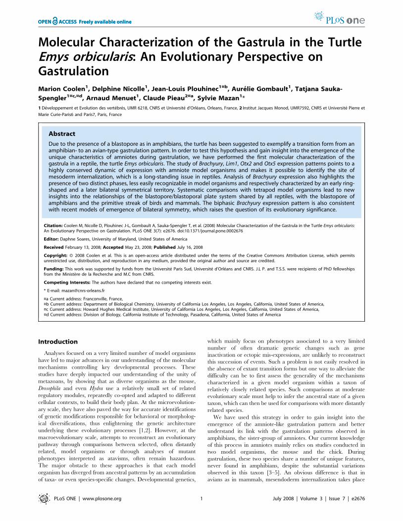
area pellucida (Fig. 1) [9]. Together with the presence of a
blastopore in amphibians, the phylogenetic distribution of the
blastopore/blastoporal plate pattern among amniotes (Fig. 1), has
been taken as evidence for its ancestral character in the taxon,
suggesting that the avian and mammalian primitive streaks may
represent independent modifications of this system [9]. A better
understanding of the relationships between these structures is
important to delineate the amniote ancestral state and the
specificities of their model organisms. However, no molecular
characterization of gastrulation has been thus far reported in a
reptile, which makes accurate comparisons difficult. In order to
better understand the relationships between the primitive streak of
birds and mammals, blastopore of amphibians and blastopore/
blastoporal plate system of reptiles, we have analyzed the
expression pattern of four regional markers of gastrulation in a
turtle, Emys orbicularis. Analysis has been more particularly focussed
on regional markers of internalizing mesoderm in amniote model
organisms, such as Brachyury, often considered as a general
primitive streak marker, Lim1, restricted to the anterior primitive
streak throughout gastrulation, and Otx2, which displays a
dynamic expression pattern in the early anterior primitive streak,
organizer and its derivatives, in addition to a prominent later
anterior neuroectoderm expression. These data lead to a
reassessment of current models of the emergence of amniote
characteristics during gastrulation and highlight an unexpected
parallel with recent hypotheses on the origin of the vertebrate
embryonic axes.
Materials and Methods
Turtle embryos and stagingPermission from the French Ministry of Ecology was given to C.
P. to capture a maximum of 60 gravid females, collect eggs and
incubate them in controlled laboratory conditions, one proportion
of them being kept for scientific research and the rest released
either on the site of capture or placed in a breeding unit (Ferme
aux crocodiles, Pierrelatte, France) for a program of reintroduction
of endangered species (arretes nu 2003-E-2504DDAF/457 and
nu2004-E-1161 DDAF/185). This permission was given following
advice of the Conseil National de la Protection de la Nature. Adult
animals were released on their site of capture following spawning.
A total of 150 eggs were used for these analyses. Eggs were
incubated at 30uC and dissected at gastrulation or early
neurulation stages. Experimental procedures on early embryos
comply with institutional regulations (code rural article R 214-90).
Stages were determined following the criteria described in Chelydra
serpentina and Testudo hermanni [10,11] from stage 2 onwards. At
earlier stages (stages 0–1), we carried out systematic histological
analyses in order to better understand the changes in morphology
at the earliest available stages in E. orbicularis.
NomenclaturePosterior-ventral, anterior-dorsal. The orientation of the
blastoporal canal changes between stages 0 and 1, being converted
from a vertical elongated cavity to an almost horizontal one. For
this reason, we refer to the blastopore rims as posterior or anterior
at the earliest stage studied (stage 0a), and ventral or dorsal starting
from stage 0b. This nomenclature thus refers to the future
polarities of the embryo proper when it becomes visible.
Reptilian. Even though reptiles (which comprise chelonians,
lepidosaurs and crocodilians) form a paraphyletic group, we refer
to their mode of gastrulation as ‘‘reptilian’’ for convenience
purposes. The ‘‘reptilian’’ gastrulation pattern, characterized by
the presence of a posterior blastopore and blastoporal plate, is very
similar in chelonians, lepidosaurs and crocodilians.
Probe amplificationOtx2, Otx5, Lim1 and Brachyury probes were obtained by
degenerate PCR starting from E. orbicularis genomic DNA or
stage 1 cDNA as described previously [12–14]. The degenerate
primers used and sequences of the resulting amplified products are
Figure 1. Gastrulation patterns of the major tetrapod groups. The phylogenetic relationships between the major amniote taxa are takenfrom [45]. Model species are indicated in italics. The mode of gastrulation in each group is shown on the right, as reviewed in [9].doi:10.1371/journal.pone.0002676.g001
Gastrulation in the Turtle
PLoS ONE | www.plosone.org 2 July 2008 | Volume 3 | Issue 7 | e2676
shown in Supplementary Figure S1. Their identity was confirmed
by systematic reverse Blast analyses against Genbank.
ISH and sectioningTurtle embryos were obtained as described in [15], fixed
overnight in paraformaldehyde 4% in PBS (PFA 4%), and stored
dehydrated in methanol 100%. Whole-mount in situ hybridizations
were conducted using in vitro synthesized digoxigenin-labeled
antisense RNAs, after the protocol described in [12] with the
following adaptation. The proteinase K treatment was carried out
at a concentration of 10 mg/ml for 10 minutes at room
temperature from stages 0 to 4 and extended to 20 minutes at
later stages. Histological analyses were systematically carried out
following in situ hybridizations, using cryostat sections as described
previously [12].
Results
Morphological aspects of gastrulation in the turtle Emysorbicularis
Developmental tables have been published for two turtle
species, Chelydra serpentina [10] and Testudo hermanni boettgeri [11]
with congruent stage definitions. These descriptions, mainly based
on external morphological changes, were also found to largely
apply to E. orbicularis. However, they do not address the
histological changes observed between stage 0 and stage 1,
respectively defined by the appearance of the blastopore and
cephalic enlargement. We therefore performed a detailed
histological description of Emys orbicularis embryos prior to stage
1. As previously described [reviewed in 9], the blastopore appears
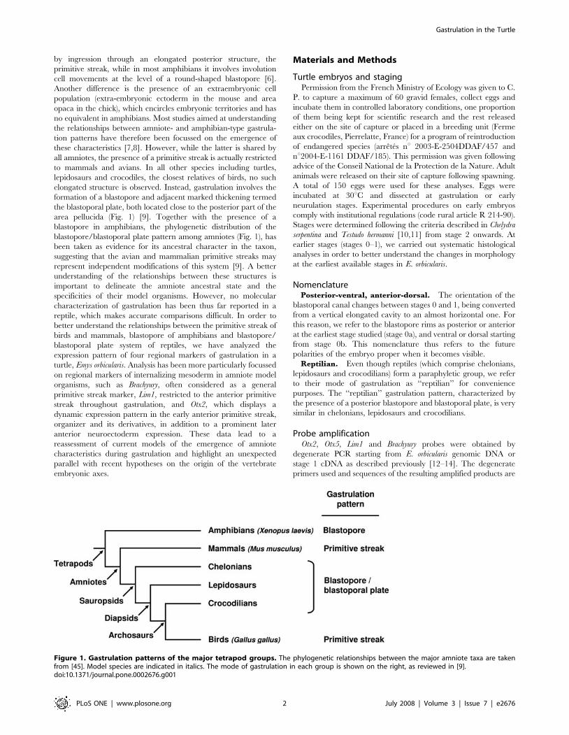
shortly after oviposition, as a transverse fold in the posterior third
of the blastoderm (dorsal view in Fig. 2). As visible in sections
(Fig. 2A) an elongated transverse cavity extends anteriorly and
ventrally towards the yolk. Three cell populations are observed at
this stage, a superficial epithelium, markedly thickened in the
major part of the blastoderm and consisting of a single-cell layer
peripherally, a thin lower epithelial cell sheet in direct contact with
the yolk, and intermediate mesenchymal cells (Fig. 2A–C).
Following anatomists, we refer to the superficial and lower cell
layers as epiblast and hypoblast respectively. The mesenchymal
cells are partitioned into two distinct populations, one that appears
dispersed in the blastocoel cavity in the anterior moiety of the
blastoderm, and the other more compact, around the elongating
blastopore and posterior to it, under the epithelial layer of the
blastoporal plate. The absence of a bilayered organization around
the blastopore rims suggests that delamination cell movements
from the epiblast prevail at this stage. We refer to this stage as
stage 0a, based on two criteria, an external one, the shape of the
dorsal blastopore lip, which consistently appeared horizontal in
these early embryos, and an histological one, the absence of a
bilayered organization indicative of involution cell movements at
this level. The ventral extension of the blastopore cavity could be
more or less pronounced in embryos displaying these character-
istics, a fusion between the blastocoel walls and hypoblast taking
place at late stage 0a. At stage 0b, the dorsal lip of the blastopore
changes shape, with a slight posterior extension. The blastoporal
cavity now shows a marked anterior to posterior orientation
(Fig. 2E–F) and a bilayered organisation, indicative of epiblast
involution movements, becomes obvious at the anterior and lateral
rims of the blastopore (Fig. 2D–F). In contrast, no indication of
Figure 2. Histological characteristics of stage 0 E. orbicularis embryos. Schematic dorsal views of turtle embryos at stages 0a to late 0b (0b+)are shown in the upper line, with the area opaca in blue and the area pellucida in white. The level and plane of sections shown in A–L are indicatedby dotted lines on the schematic views. The approximate location of the sections shown in G–L are also shown on a tranverse section (F) of a slightlyyounger embryo to help interpretation of the histologies. Red arrowheads in J points to a characteristic protrusion of the dorsal lip, which starts toform at stage 0b and appears as a narrow extension between the lateral lips at late stage 0b. Section J is a tangential section of this protrusion. Blackarrowheads indicate the blastoporal cavity or canal. A–C, D–F : sagittal sections of respectively stage 0a and 0b embryos; G–L : transverse sections of alate stage 0b embryo. bl, blastocoel; h, hypoblast; bp, blastoporal plate; e, epiblast. Scale bar : 500 mm.doi:10.1371/journal.pone.0002676.g002
Gastrulation in the Turtle
PLoS ONE | www.plosone.org 3 July 2008 | Volume 3 | Issue 7 | e2676
involution could be observed at the posterior/ventral lip level,
histological views rather suggesting a global delamination (Fig. 2E–
F). These broad characteristics are maintained at late stage 0b
(0b+ in Fig. 2G–L), with the following modifications. First, the
previously slit-shaped blastopore opening narrows, thus forming
the blastoporal canal (Fig. 2J). The posterior extension of the
dorsal lip, which started to form at stage 0b, becomes more
pronounced and now markedly protrudes between the lateral lips
(red arrowhead in Fig. 2J). This structure completely regresses at
stage 1, characterized by the appearance of two prominent
bilateral ventral bulges [10, 11 ; see Fig. 3 and 4, this study].
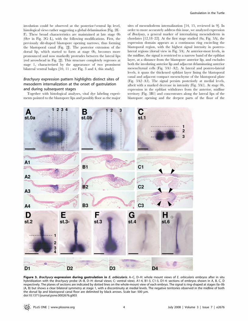
Brachyury expression pattern highlights distinct sites ofmesoderm internalization at the onset of gastrulationand during subsequent stages
Together with histological analyses, vital dye labeling experi-
ments pointed to the blastopore lips and possibly floor as the major
sites of mesendoderm internalization [14, 15, reviewed in 9]. In
order to more accurately address this issue, we analyzed expression
of Brachyury, a general marker of internalizing mesendoderm in
chordates [12,18–22]. At the first stage studied (0a, Fig. 3A), the
expression domain appears as a continuous ring encircling the
blastoporal region, with the highest signal intensity in postero-
lateral regions (dorsal view in Fig. 3A). At anterior-most levels, in
the midline, the signal is restricted to a narrow band of the epiblast
layer, at a distance from the blastopore anterior lip, and excludes
both the involuting anterior lip and adjacent delaminating anterior
mesenchymal cells (Fig. 3A1–A2). At lateral and postero-lateral
levels, it spans the thickened epiblast layer lining the blastoporal
canal and adjacent compact mesenchyme of the blastoporal plate
(Fig. 3A2–A3). The signal persists posteriorly at medial levels,
albeit with a marked decrease in intensity (Fig. 3A1). At stage 0b,
expression in the epiblast withdraws from the anterior, midline
territory (Fig. 3B1) and concentrates along the lateral lips of the
blastopore opening and the deepest parts of the floor of the
Figure 3. Brachyury expression during gastrulation in E. orbicularis. A–C, D–H: whole mount views of E. orbicularis embryos after in situhybridization with the Brachyury probe (A–B, D–H: dorsal views; C: ventral view). A1-4, B1-3, C1-3, D1-4: sections of embryos shown in A, B, C, Drespectively. The planes of sections are indicated by dotted lines on the whole-mount view of each embryo. The signal is ring-shaped at stages 0a–0b(A, B) but shows a clear bilateral symmetry at stage 1, with a discontinuity at medial levels. The negative territories observed in the midline of boththe dorsal lip and blastoporal canal floor are delimited by black arrows. Scale bar: 500 mm.doi:10.1371/journal.pone.0002676.g003
Gastrulation in the Turtle
PLoS ONE | www.plosone.org 4 July 2008 | Volume 3 | Issue 7 | e2676
blastoporal canal (Fig. 3B1–B2). Expression persists in the adjacent
mesenchyme, that posteriorly forms a stream of elongated cells
extending below the superficial epithelial layer and towards the
posterior limit of the blastoderm (Fig. 3B1–B2). At stage 1, the
broad characteristics of the labeled territory radically change.
Transcripts become restricted to two bilateral sites of expression,
comprising the involuting lateral lips, adjacent epiblast and
mesenchyme. The signal thus completely disappears from the
midline, both from the epiblast involuting at the dorsal rim of the
blastoporal canal opening and from the floor of the blastoporal
canal and underlying mesenchyme (Fig. 3C, C1–C3). These
characteristics of Brachyury expression around the blastopore are
maintained at later stages with two modifications. The bilateral
expression territories previously excluding the dorsal midline now
include the dorsal blastopore lip (Fig. 3D, 3D2–D4). In addition,
as in all chordates, the forming notochord, which extends anterior
to the Brachyury positive dorsal-most involution zone, becomes a
major expression site (Fig. 3D–H, 3D1).
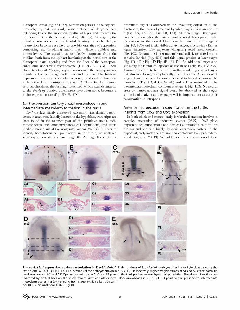
Lim1 expression territory : axial mesendoderm andintermediate mesoderm formation in the turtle
Lim1 displays highly conserved expression sites during gastru-
lation in amniotes. Initially located to the hypoblast, transcripts are
later found in the anterior part of the primitive streak, axial
mesendoderm including prechordal cell populations, and inter-
mediate mesoderm of the urogenital system [23–25]. In order to
identify homologous cell populations in the turtle, we analyzed
Lim1 expression starting from stage 0b. At stage 0b to 0b+, a
prominent signal is observed in the involuting dorsal lip of the
blastopore, the mesenchyme and hypoblast layers lying anterior to
it (Fig. 4A, 4A1–A3; Fig. 4B, 4B1). At these stages, the signal
completely excludes the lateral and ventral blastoporal plate.
Expression in the dorsal blastopore lip persists until stage 2
(Fig. 4C, 4C5) and is still visible at later stages, albeit with a fainter
signal intensity. The adjacent elongating axial mesendoderm
(Fig. 4C2–C4) and the looser mesenchymal cells lying anterior to it
are also labeled (Fig. 4C1) and this signal persists at later stages
(Fig. 4D, 4D1; Fig. 4E; Fig. 4F, 4F1–F4). An additional expression
site along the lateral lips appears at late stage 1 (Fig. 4C, 4C5–C6).
Transcripts are detected not only in the involuting epiblast layer
but also in cells ingressing laterally from this area. At subsequent
stages, Lim1 expression becomes localized to lateral regions of the
mesoderm (Fig. 4D, 4D1–D4; 4E) and is later restricted to the
intermediate mesoderm component (stage 4; Fig. 4F3). No neural
crest or neuroectoderm signal could be observed at the stages
studied and analyses at later stages will be important to assess their
conservation in tetrapods.
Anterior neuroectoderm specification in the turtle:insights from Otx2 and Otx5 expression
In both chick and mouse, early forebrain formation involves a
complex succession of inductive events [26,27]. Otx2 plays
important cell-autonomous and non cell-autonomous roles in this
process and shows a highly dynamic expression pattern in the
hypoblast, early node and anterior neuroectoderm from pre- to late-
streak stages [25,28–33]. We addressed the conservation of these
Figure 4. Lim1 expression during gastrulation in E. orbicularis. A–F: dorsal views of E. orbicularis embryos after in situ hybridization using theLim1 probe. A1-3, B1, C1-6, D1-4, F1-4: sections of the embryos shown in A, B, C, D, F respectively. Higher magnifications of A1 and A2 at the dorsal liplevel are shown in A1’ and A2’. Opened arrowheads in A1-2 and B1 point to the Lim1 positive mesenchymal cell population. The planes of sections areindicated by dotted lines on the whole-mount view of each embryo. Black arrowheads in C, D, E, F, F3 point to the prospective intermediatemesoderm expressing Lim1 starting from stage 1+. Scale bar: 500 mm.doi:10.1371/journal.pone.0002676.g004
Gastrulation in the Turtle
PLoS ONE | www.plosone.org 5 July 2008 | Volume 3 | Issue 7 | e2676
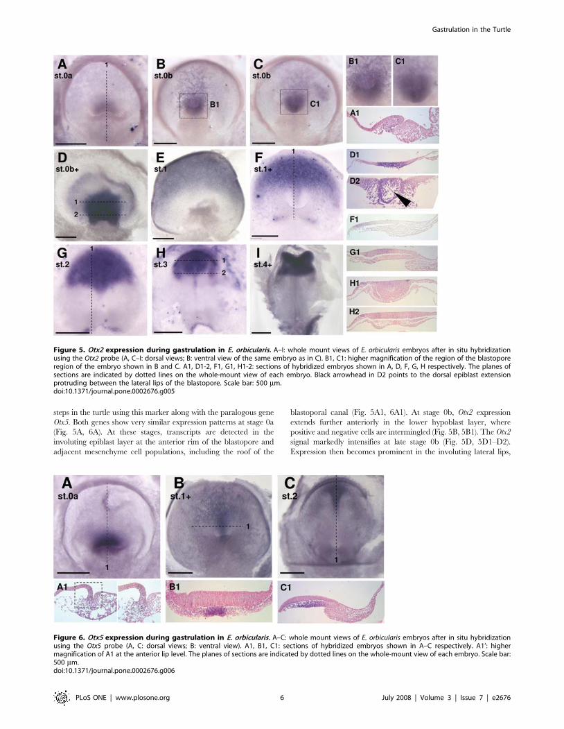
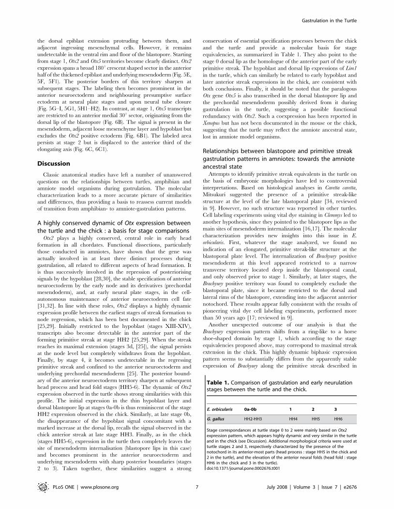
steps in the turtle using this marker along with the paralogous gene
Otx5. Both genes show very similar expression patterns at stage 0a
(Fig. 5A, 6A). At these stages, transcripts are detected in the
involuting epiblast layer at the anterior rim of the blastopore and
adjacent mesenchyme cell populations, including the roof of the
blastoporal canal (Fig. 5A1, 6A1). At stage 0b, Otx2 expression
extends further anteriorly in the lower hypoblast layer, where
positive and negative cells are intermingled (Fig. 5B, 5B1). The Otx2
signal markedly intensifies at late stage 0b (Fig. 5D, 5D1–D2).
Expression then becomes prominent in the involuting lateral lips,
Figure 5. Otx2 expression during gastrulation in E. orbicularis. A–I: whole mount views of E. orbicularis embryos after in situ hybridizationusing the Otx2 probe (A, C–I: dorsal views; B: ventral view of the same embryo as in C). B1, C1: higher magnification of the region of the blastoporeregion of the embryo shown in B and C. A1, D1-2, F1, G1, H1-2: sections of hybridized embryos shown in A, D, F, G, H respectively. The planes ofsections are indicated by dotted lines on the whole-mount view of each embryo. Black arrowhead in D2 points to the dorsal epiblast extensionprotruding between the lateral lips of the blastopore. Scale bar: 500 mm.doi:10.1371/journal.pone.0002676.g005
Figure 6. Otx5 expression during gastrulation in E. orbicularis. A–C: whole mount views of E. orbicularis embryos after in situ hybridizationusing the Otx5 probe (A, C: dorsal views; B: ventral view). A1, B1, C1: sections of hybridized embryos shown in A–C respectively. A1’: highermagnification of A1 at the anterior lip level. The planes of sections are indicated by dotted lines on the whole-mount view of each embryo. Scale bar:500 mm.doi:10.1371/journal.pone.0002676.g006
Gastrulation in the Turtle
PLoS ONE | www.plosone.org 6 July 2008 | Volume 3 | Issue 7 | e2676
the dorsal epiblast extension protruding between them, and
adjacent ingressing mesenchymal cells. However, it remains
undetectable in the ventral rim and floor of the blastopore. Starting
from stage 1, Otx2 and Otx5 territories become clearly distinct. Otx2
expression spans a broad 180u crescent shaped sector in the anterior
half of the thickened epiblast and underlying mesendoderm (Fig. 5E,
5F, 5F1). The posterior borders of this territory sharpen at
subsequent stages. The labeling then becomes prominent in the
anterior neuroectoderm and neighbouring presumptive surface
ectoderm at neural plate stages and upon neural tube closure
(Fig. 5G–I, 5G1, 5H1–H2). In contrast, at stage 1, Otx5 transcripts
are restricted to an anterior medial 30u sector, originating from the
dorsal lip of the blastopore (Fig. 6B). The signal is present in the
mesendoderm, adjacent loose mesenchyme layer and hypoblast but
excludes the Otx2 positive ectoderm (Fig. 6B1). The labeled area
persists at stage 2 but is displaced to the anterior third of the
elongating axis (Fig. 6C, 6C1).
Discussion
Classic anatomical studies have left a number of unanswered
questions on the relationships between turtles, amphibian and
amniote model organisms during gastrulation. The molecular
characterization leads to a more accurate picture of similarities
and differences, thus providing a basis to reassess current models
of transition from amphibian- to amniote-gastrulation patterns.
A highly conserved dynamic of Otx expression betweenthe turtle and the chick : a basis for stage comparisons
Otx2 plays a highly conserved, central role in early head
formation in all chordates. Functional dissections, particularly
those conducted in amniotes, have shown that the gene was
actually involved in at least three distinct processes during
gastrulation, all related to different aspects of head formation. It
is thus successively involved in the repression of posteriorising
signals by the hypoblast [28,30], the stable specification of anterior
neuroectoderm by the early node and its derivatives (prechordal
mesendoderm), and, at early neural plate stages, in the cell-
autonomous maintenance of anterior neuroectoderm cell fate
[31,32]. In line with these roles, Otx2 displays a highly dynamic
expression profile between the earliest stages of streak formation to
node regression, which has been best documented in the chick
[25,29]. Initially restricted to the hypoblast (stages XIII-XIV),
transcripts also become detectable in the anterior part of the
forming primitive streak at stage HH2 [25,29]. When the streak
reaches its maximal extension (stages 3d, [25]), the signal persists
at the node level but completely withdraws from the hypoblast.
Finally, by stage 4, it becomes undetectable in the regressing
primitive streak and confined to the anterior neuroectoderm and
underlying prechordal mesendoderm [25]. The posterior bound-
ary of the anterior neuroectoderm territory sharpen at subsequent
head process and head fold stages (HH5-6). The dynamic of Otx2
expression observed in the turtle shows strong similarities with this
profile. The initial expression in the thin hypoblast layer and
dorsal blastopore lip at stages 0a-0b is thus reminiscent of the stage
HH2 expression observed in the chick. Similarly, at late stage 0b,
the disappearance of the hypoblast signal concomitant with a
marked increase at the dorsal lip, recalls the signal observed in the
chick anterior streak at late stage HH3. Finally, as in the chick
(stages HH5-6), expression in the turtle then completely leaves the
site of mesendoderm internalisation (blastopore lips in this case)
and becomes prominent in the anterior neuroectoderm and
underlying mesendoderm with sharp posterior boundaries (stages
2 to 3). Taken together, these similarities suggest a strong
conservation of essential specification processes between the chick
and the turtle and provide a molecular basis for stage
equivalencies, as summarized in Table 1. They also point to the
stage 0 dorsal lip as the homologue of the anterior part of the early
primitive streak. The hypoblast and dorsal lip expressions of Lim1
in the turtle, which can similarly be related to early hypoblast and
later anterior streak expressions in the chick, are consistent with
both conclusions. Finally, it should be noted that the paralogous
Otx gene Otx5 is also transcribed in the dorsal blastopore lip and
the prechordal mesendoderm possibly derived from it during
gastrulation in the turtle, suggesting a possible functional
redundancy with Otx2. Such a coexpression has been reported in
Xenopus but has not been documented in the mouse or the chick,
suggesting that the turtle may reflect the amniote ancestral state,
lost in amniote model organisms.
Relationships between blastopore and primitive streakgastrulation patterns in amniotes: towards the amnioteancestral state
Attempts to identify primitive streak equivalents in the turtle on
the basis of embryonic morphologies have led to controversial
interpretations. Based on histological analyses in Caretta caretta,
Mitsukuri suggested the presence of a primitive streak-like
structure at the level of the late blastoporal plate [34, reviewed
in 9]. However, no such structure was reported in other turtles.
Cell labeling experiments using vital dye staining in Clemmys led to
another hypothesis, since they pointed to the blastopore lips as the
main sites of mesendoderm internalization [16,17]. The molecular
characterization provides new insights into this issue in E.
orbicularis. First, whatever the stage analyzed, we found no
indication of an elongated, primitive streak-like structure at the
blastoporal plate level. The internalization of Brachyury positive
mesendoderm at this level appeared restricted to a narrow
transverse territory located deep inside the blastoporal canal,
and only observed prior to stage 1. Similarly, at later stages, the
Brachyury positive territory was found to completely exclude the
blastoporal plate, since it became restricted to the dorsal and
lateral rims of the blastopore, extending into the adjacent anterior
notochord. These results appear fully consistent with the results of
pioneering vital dye cell labeling experiments, performed more
than 50 years ago [17; reviewed in 9].
Another unexpected outcome of our analysis is that the
Brachyury expression pattern shifts from a ring-like to a horse
shoe-shaped domain by stage 1, which according to the stage
equivalencies proposed above, may correspond to maximal streak
extension in the chick. This highly dynamic biphasic expression
pattern seems to substantially differs from the apparently stable
expression of Brachyury along the primitive streak described in
Table 1. Comparison of gastrulation and early neurulationstages between the turtle and the chick.
E. orbicularis 0a-0b 1 2 3
G. gallus HH2-HH3 HH4 HH5 HH6
Stage correspondances at turtle stage 0 to 2 were mainly based on Otx2expression pattern, which appears highly dynamic and very similar in the turtleand in the chick (see Dicussion). Additional morphological criteria were used atturtle stages 2 and 3, respectively characterized by the presence of thenotochord in its anterior-most parts (head process : stage HH5 in the chick and2 in the turtle), and the elevation of the anterior neural folds (head fold : stageHH6 in the chick and 3 in the turtle).doi:10.1371/journal.pone.0002676.t001
Gastrulation in the Turtle
PLoS ONE | www.plosone.org 7 July 2008 | Volume 3 | Issue 7 | e2676
amniote model organisms. However, despite the absence of overt
morphological changes of the primitive streak during gastrulation,
a very similar transition may also take place in birds and
mammals, between streak elongation and regression stages
(respectively HH2-HH4 and HH4-HH6, corresponding to stages
0–1 and 1–3 in the turtle). In support of this possibility, high
resolution analyses of cell movements recently reported in the
chick have shown that, shortly after stage XII, cell converge not
only to the ‘‘future primitive streak axis’’ (‘‘vertical’’ axis), aligned
along the future antero-posterior polarity of the embryo itself, but
also more unexpectedly to a transverse (‘‘horizontal’’) territory
located near the edge of the area pellucida and described ‘‘as an
arc parallel to the posterior marginal zone’’ [8]. This latter
territory is reminiscent of the early Brachyury positive transverse
domain observed at the floor of the blastoporal canal in the turtle.
It could thus be a remnant of the posterior rim of the ancestral
reptilian blastopore, whereas the other, ‘‘vertical’’ site of cell
convergence reported in the chick could be evolutionarily related
to its dorsal and lateral lips. Similarly, comparisons between the
turtle and the rabbit, which as the chick and actually most
mammals, develops as a flat blastoderm thus yielding excellent
spatial resolutions of expression patterns, point to another
noticeable similarity. In the latter, the anterior part of the
primitive streak is negative for Brachyury expression at stages 3–4,
prior to the onset of node regression and head process formation,
except for a transient, discontinuous node expression phase [35].
We could not detect a clear equivalent of this transient node
expression, which may have escaped detection in the turtle. In
contrast, the exclusion of Brachyury transcripts from anterior streak
in the rabbit is clearly reminiscent of the absence of expression
observed in the turtle at the Lim1 and Otx expressing dorsal
blastopore lip until stage 1, just prior to the onset of anterior
notochord elongation, which only becomes clearly visible at stage
2. This early internalized Lim1 and Otx expressing mesendoderm
cell population may thus correspond to prechordal mesendoderm,
negative for Brachyury in all vertebrates [12]. Cell fate analyses will
be essential to validate this interpretation. Taken together, these
data suggest that the two phases of Brachyury expression, which
appear conspicuous in the turtle, may be general among amniotes.
Comparisons between amphibian- and amniote-likegastrulation patterns : insights from the turtle
Xenopus- and avian-like gastrulation patterns differ by a number of
distinctive features, which in the latter are likely to correspond to
derived characteristics, such as the segregation and radial expansion
of territories that do not contribute to the embryo proper, as well as
a posterior restriction and characteristic elongation of the site of
mesendoderm internalization. More than ten years ago, Arendt and
Nubler-Jung proposed a hypothetical sequence of progressive
modifications to account for the emergence of these characteristics
[7]. This model relied on three crucial steps, (1) a progressive
withdrawal of the presumptive mesoderm from ventral territories,
(2) a fusion of the resulting posterior wings of internalizing
mesoderm, at the level of a structure proposed to be related to the
blastoporal plate of modern turtles, and (3) an elongation of this
structure thus converted into an avian-type primitive streak
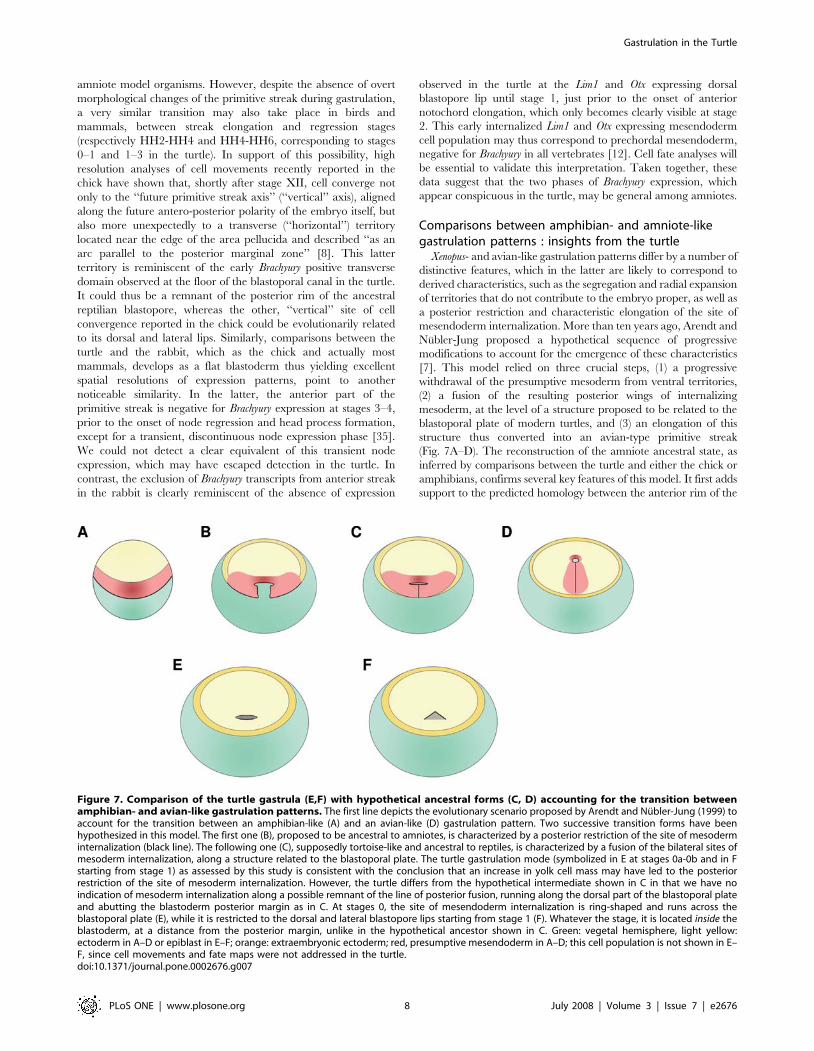
(Fig. 7A–D). The reconstruction of the amniote ancestral state, as
inferred by comparisons between the turtle and either the chick or
amphibians, confirms several key features of this model. It first adds
support to the predicted homology between the anterior rim of the
Figure 7. Comparison of the turtle gastrula (E,F) with hypothetical ancestral forms (C, D) accounting for the transition betweenamphibian- and avian-like gastrulation patterns. The first line depicts the evolutionary scenario proposed by Arendt and Nubler-Jung (1999) toaccount for the transition between an amphibian-like (A) and an avian-like (D) gastrulation pattern. Two successive transition forms have beenhypothesized in this model. The first one (B), proposed to be ancestral to amniotes, is characterized by a posterior restriction of the site of mesoderminternalization (black line). The following one (C), supposedly tortoise-like and ancestral to reptiles, is characterized by a fusion of the bilateral sites ofmesoderm internalization, along a structure related to the blastoporal plate. The turtle gastrulation mode (symbolized in E at stages 0a-0b and in Fstarting from stage 1) as assessed by this study is consistent with the conclusion that an increase in yolk cell mass may have led to the posteriorrestriction of the site of mesoderm internalization. However, the turtle differs from the hypothetical intermediate shown in C in that we have noindication of mesoderm internalization along a possible remnant of the line of posterior fusion, running along the dorsal part of the blastoporal plateand abutting the blastoderm posterior margin as in C. At stages 0, the site of mesendoderm internalization is ring-shaped and runs across theblastoporal plate (E), while it is restricted to the dorsal and lateral blastopore lips starting from stage 1 (F). Whatever the stage, it is located inside theblastoderm, at a distance from the posterior margin, unlike in the hypothetical ancestor shown in C. Green: vegetal hemisphere, light yellow:ectoderm in A–D or epiblast in E–F; orange: extraembryonic ectoderm; red, presumptive mesendoderm in A–D; this cell population is not shown in E–F, since cell movements and fate maps were not addressed in the turtle.doi:10.1371/journal.pone.0002676.g007
Gastrulation in the Turtle
PLoS ONE | www.plosone.org 8 July 2008 | Volume 3 | Issue 7 | e2676
turtle blastopore and the Xenopus dorsal lip (Fig. 7A and 7E). The
involution cell movement, likely corresponding to an ancestral
character retained by turtles, thus prevails at this level in both
species. Similarly, these territories share expression of organizer
markers. In particular, Lim1, as well as both Otx2 and Otx5 are
specifically expressed at the dorsal lip of the blastopore or Spemann
organizer in Xenopus, throughout gastrulation for the former and
during early stages (stage 10–10.5) for the latter two [36,37,38,39],
exactly as observed in the turtle. A very similar dynamic of Brachyury
expression, initially located at a short distance anterior to the dorsal
lip, is also observed in the turtle as in amphibians [40]. As also
predicted by the model, the characterization of the turtle gastrula is
consistent with a posterior restriction of presumptive ventral
mesoderm and its complete withdrawal from the blastoderm
margin, which as in the chick, never expresses Brachyury. While
such a restriction of mesendoderm internalization to one side of the
embryo, also observed in salamanders, gymnophionans and to a
large extent selacians [5,12], may be a general evolutionary
tendency, repeatedly associated with an increase of the yolk mass,
its complete exclusion from the blastoderm margin remains an
amniote specificity, shared by the turtle, as observed in [9]. Finally,
the occurrence of the last step of the model, hypothesized to account
for the conversion of the reptilian blastopore into the elongated
avian primitive streak, has also recently gained support from
analyses of the genetic mechanisms controlling streak extension at
pre-gastrulation stages. This morphogenetic process has been
shown to rely on the Wnt-PCP pathway, suggesting that the
recruitment of additional medial cell intercalation movements may
have been involved in the emergence of the primitive streak during
evolution [8]. The characterization of the turtle gastrulation pattern
fully supports this conclusion and since no such elongated structure
is observed in the turtle, suggests that this modification may have
taken place among amniotes rather than prior to their emergence.
Despite these congruencies, the turtle gastrula does not resemble
the ancestral, supposedly tortoise-like form, depicted by Arendt
and Nubler-Jung in their model (compare Fig. 7C and 7E). We
thus find no evidence of an elongated site of mesoderm
internalisation abutting the posterior blastoderm margin. This
does not preclude the possibility that the transition from an
amphibian-to an amniote-like gastrulation mode may have
involved the proposed transition form but our work shows that
extant turtles do not exemplify this hypothetical ancestral state.
Other evolutionary scenarios, involving for instance an earlier
radialization of the extra-embryonic ectoderm and concomitant
re-localization of mesoderm inducing factors towards the center of
the blastoderm remain equally opened.
An ancient origin for the gnathostome gastrulationpattern?
Theoretical analyses combining mathematical modeling and
comparisons at large evolutionary scale, between diploblasts and
bilaterians, have led to the suggestion that the construction of the
vertebrate body axis may involve the combined action of two
signaling centers, or organizers, of very different evolutionary origins
[41,42]. According to this model, the first one, located at the
blastopore margin and involved in brain antero-posterior patterning,
may be derived from an ancient organizer, already present in the last
common ancestor of diploblasts and bilaterians. In line with this
hypothesis, the blastoporal region acts as a genuine organizer in Hydra
or Nematostella vectensis, and the pattern which it generates along the
unique axis of the body column appears related to the antero-
posterior pattern of the brain of extant vertebrates [43,44]. The
second signaling system, of more recent origin, corresponds to the
Spemann organizer homologue, proposed to primarily control dorso-
ventral polarity during subsequent axis elongation [41,42]. The
action of this second organizer may have played a crucial part in the
emergence of the vertebrate body plan, by converting a radially to a
bilaterally symmetrical animal form. It is intriguing to note that the
gastrulation pattern observed in Emys orbicularis is strikingly similar to
the prototypical vertebrate pattern hypothesized in this model. In the
turtle, Brachyury expression thus shifts from a largely radial
symmetrical ring to a territory exhibiting a clear bilateral symmetry.
A similar shift between a radial and a bilaterally symmetrical
expression territory is also observed in the dogfish, despite the
extensive morphological divergence and evolutionary distance
between the turtle and the shark [12,13]. Detailed analyses of the
signaling systems controlling antero-posterior and dorso-ventral
patterning in vertebrates combined with systematic comparative
analyses aimed at reconstructing the ancestral state of the major
metazoan taxa, will be essential to assess the evolutionary significance
of this observation. Taking advantage of the characteristics of non-
model organisms such as the turtle, which in some cases lend
themselves to more straightforward interpretations than model
organisms, could be important in such comparative approaches.
Supporting Information
Figure S1 Partial E. orbicularis Lim1, Brachyury, Otx2 and
Otx5 sequences used as probes in this study.
Found at: doi:10.1371/journal.pone.0002676.s001 (0.03 MB
DOC)
Acknowledgments
We are grateful to Ferdinand Marletaz for its contribution to the
experiments on Brachyury expression pattern and to Hans Meinhardt for
critical reading and enlightening comments on the manuscript.
Author Contributions
Conceived and designed the experiments: MC SM. Performed the
experiments: MC DN JLP AG TSS. Analyzed the data: MC AM SM.
Contributed reagents/materials/analysis tools: CP. Wrote the paper: MC
SM. Obtained turtles’ embryos: CP.
References
1. Nachman MW (2005) The genetic basis of adaptation: lessons from concealing
coloration in pocket mice. Genetica 123: 125–136.
2. Peichel CL (2005) Fishing for the secrets of vertebrate evolution in threespine
sticklebacks. Dev Dyn 234: 815–823.
3. del Pino EM, Venegas-Ferrin M, Romero-Carvajal A, Montenegro-Larrea P,
Saenz-Ponce N, et al. (2007) A comparative analysis of frog early development.
Proc Natl Acad Sci U S A 104: 11882–11888.
4. Moya IM, Alarcon I, del Pino EM (2007) Gastrulation of Gastrotheca
riobambae in comparison with other frogs. Dev Biol 304: 467–478.
5. Keller R, Shook D (2004) Gastrulation in Amphibians. In: Stern CD, ed.
Gastrulation: from cells to embryo. Cold Spring Harbor, NY: Cold Spring
Harbor Press. pp 171–204.
6. Stern CD (2004) Gastrulation in the chick. In: Stern CD, ed. Gastrulation: from
cells to embryo. Cold Spring Harbor, NY: Cold Spring Harbor Press. pp
219–232.
7. Arendt D, Nubler-Jung K (1999) Rearranging gastrulation in the name of yolk:
evolution of gastrulation in yolk-rich amniote eggs. Mech Dev 81: 3–
22.
8. Voiculescu O, Bertocchini F, Wolpert L, Keller RE, Stern CD (2007) The
amniote primitive streak is defined by epithelial cell intercalation before
gastrulation. Nature 449: 1049–1052.
9. Gilland E, Burke A (2004) Gastrulation in Reptiles. In: Stern CD, ed.
Gastrulation: from cells to embryo. Cold Spring Harbor, NY: Cold Spring
Harbor Press. pp 205–217.
Gastrulation in the Turtle
PLoS ONE | www.plosone.org 9 July 2008 | Volume 3 | Issue 7 | e2676
10. Yntema CL (1968) A series of stages in the embryonic development of Chelydra
serpentina. J. Morphol. 125: 219–251.11. Guyot G, Pieau C, Renous S (1994) Developpement embryonnaire d’une tortue
terrestre, la tortue d’Hermann, Testudo hermanni Gmelin, 1789. Annales des
Sciences Naturelles, Zoologie, Paris 15: 115–137.12. Sauka-Spengler T, Baratte B, Lepage M, Mazan S (2003) Characterization of
Brachyury genes in the dogfish S. canicula and the lamprey L. fluviatilis. Insightsinto gastrulation in a chondrichthyan. Dev Biol 263: 296–307.
13. Coolen M, Sauka-Spengler T, Nicolle D, Le-Mentec C, Lallemand Y, et al.
(2007) Evolution of axis specification mechanisms in jawed vertebrates: insightsfrom a chondrichthyan. PLoS ONE 2: e374.
14. Plouhinec JL, Sauka-Spengler T, Germot A, Le Mentec C, Cabana T, et al.(2003) The mammalian Crx genes are highly divergent representatives of the
Otx5 gene family, a gnathostome orthology class of orthodenticle-relatedhomeogenes involved in the differentiation of retinal photoreceptors and
circadian entrainment. Mol Biol Evol 20: 513–521.
15. Dorizzi M, Richard-Mercier N, Pieau C (1996) The ovary retains male potentialafter the thermosensitive period for sex determination in the turtle Emys
orbicularis. Differentiation 60: 193–201.16. Chandrasekharan Nayar M (1966) In vitro vital staining of chelonian
blastoderms. Indian JExpBio. pp 131–134.
17. Pasteels J (1937) Etude sur la gastrulation des vertebres meroblastiques. II.Reptiles. Arch Biol. pp 105–184.
18. Holland PW, Koschorz B, Holland LZ, Herrmann BG (1995) Conservation ofBrachyury (T) genes in amphioxus and vertebrates: developmental and
evolutionary implications. Development 121: 4283–4291.19. Knezevic V, De Santo R, Mackem S (1997) Two novel chick T-box genes
related to mouse Brachyury are expressed in different, non-overlapping
mesodermal domains during gastrulation. Development 124: 411–419.20. Schulte-Merker S, Hammerschmidt M, Beuchle D, Cho KW, De Robertis EM,
et al. (1994) Expression of zebrafish goosecoid and no tail gene products in wild-type and mutant no tail embryos. Development 120: 843–852.
21. Smith JC, Price BM, Green JB, Weigel D, Herrmann BG (1991) Expression of a
Xenopus homolog of Brachyury (T) is an immediate-early response to mesoderminduction. Cell 67: 79–87.
22. Wilkinson DG, Bhatt S, Herrmann BG (1990) Expression pattern of the mouseT gene and its role in mesoderm formation. Nature 343: 657–659.
23. Shawlot W, Behringer RR (1995) Requirement for Lim1 in head-organizerfunction. Nature 374: 425–430.
24. Barnes JD, Crosby JL, Jones CM, Wright CV, Hogan BL (1994) Embryonic
expression of Lim-1, the mouse homolog of Xenopus Xlim-1, suggests a role inlateral mesoderm differentiation and neurogenesis. Dev Biol 161: 168–178.
25. Chapman SC, Schubert FR, Schoenwolf GC, Lumsden A (2002) Analysis ofspatial and temporal gene expression patterns in blastula and gastrula stage chick
embryos. Dev Biol 245: 187–199.
26. Levine AJ, Brivanlou AH (2007) Proposal of a model of mammalian neuralinduction. Dev Biol 308: 247–256.
27. Stern CD (2005) Neural induction: old problem, new findings, yet morequestions. Development 132: 2007–2021.
28. Acampora D, Mazan S, Lallemand Y, Avantaggiato V, Maury M, et al. (1995)Forebrain and midbrain regions are deleted in Otx2-/- mutants due to a
defective anterior neuroectoderm specification during gastrulation. Develop-
ment 121: 3279–3290.
29. Bally-Cuif L, Gulisano M, Broccoli V, Boncinelli E (1995) c-otx2 is expressed in
two different phases of gastrulation and is sensitive to retinoic acid treatment in
chick embryo. Mech Dev 49: 49–63.
30. Perea-Gomez A, Lawson KA, Rhinn M, Zakin L, Brulet P, et al. (2001) Otx2 is
required for visceral endoderm movement and for the restriction of posterior
signals in the epiblast of the mouse embryo. Development 128: 753–765.
31. Rhinn M, Dierich A, Le Meur M, Ang S (1999) Cell autonomous and non-cell
autonomous functions of Otx2 in patterning the rostral brain. Development 126:
4295–4304.
32. Rhinn M, Dierich A, Shawlot W, Behringer RR, Le Meur M, et al. (1998)
Sequential roles for Otx2 in visceral endoderm and neuroectoderm for forebrain
and midbrain induction and specification. Development 125: 845–856.
33. Simeone A, Acampora D, Mallamaci A, Stornaiuolo A, D’Apice MR, et al.
(1993) A vertebrate gene related to orthodenticle contains a homeodomain of the
bicoid class and demarcates anterior neuroectoderm in the gastrulating mouse
embryo. EMBO J 12: 2735–2747.
34. Mitsukuri (1896) On the fate of the blastopore, the relations of the primitive
streak, and the formation of the posterior end of the embryo in Chelonia,
together with remarks on the nature of meroblastic ova in vertebrates. J Coll Sci
Imp Univ Tokyo 10: 1–119.
35. Viebahn C, Stortz C, Mitchell SA, Blum M (2002) Low proliferative and high
migratory activity in the area of Brachyury expressing mesoderm progenitor cells
in the gastrulating rabbit embryo. Development 129: 2355–2365.
36. Taira M, Jamrich M, Good PJ, Dawid IB (1992) The LIM domain-containing
homeobox gene Xlim-1 is expressed specifically in the organizer region of
Xenopus gastrula embryos. Genes Dev 6: 356–366.
37. Taira M, Saint-Jeannet JP, Dawid IB (1997) Role of the Xlim-1 and Xbra genes
in anteroposterior patterning of neural tissue by the head and trunk organizer.
Proc Natl Acad Sci USA 94: 895–900.
38. Pannese M, Polo C, Andreazzoli M, Vignali R, Kablar B, Barsacchi G,
Boncinelli E (1995) The Xenopus homologue of Otx2 is a maternal homeobox
gene that demarcates and specifies anterior body regions. Development 121:
707–720.
39. Vignali R, Colombetti S, Lupo G, Zhang W, Stachel S, Harland RM,
Barsacchi G (2000) Xotx5b, a new member of the Otx gene family, may be
involved in anterior and eye development in Xenopus laevis. Mech Dev 96:
3–13.
40. Ibrahim H, Winklbauer R (2001) Mechanisms of mesendoderm internalization
in the Xenopus gastrula: lessons from the ventral side. Dev Biol 240: 108–122.
41. Meinhardt H (2004) Different strategies for midline formation in bilaterians. Nat
Rev Neurosci 5: 502–510.
42. Meinhardt H (2006) Primary body axes of vertebrates: generation of a near-
Cartesian coordinate system and the role of Spemann-type organizer. Dev Dyn
235: 2907–2919.
43. Kraus Y, Fritzenwanker JH, Genikhovich G, Technau U (2007) The blastoporal
organiser of a sea anemone. Curr Biol 17: R874–876.
44. Fritzenwanker JH, Genikhovich G, Kraus Y, Technau U (2007) Early
development and axis specification in the sea anemone Nematostella vectensis.
Dev Biol 310: 264–279.
45. Gauthier JA, Kluge AG, Rowe T (1988) The early evolution of the amniota. In:
Benton MJ, ed. The phylogeny and classification of the amniota. Oxford:
Clarendon press. pp 103–155.
Gastrulation in the Turtle
PLoS ONE | www.plosone.org 10 July 2008 | Volume 3 | Issue 7 | e2676