Decreased expression of EZH2 is associated with upregulation of ER and favorable outcome to...
27
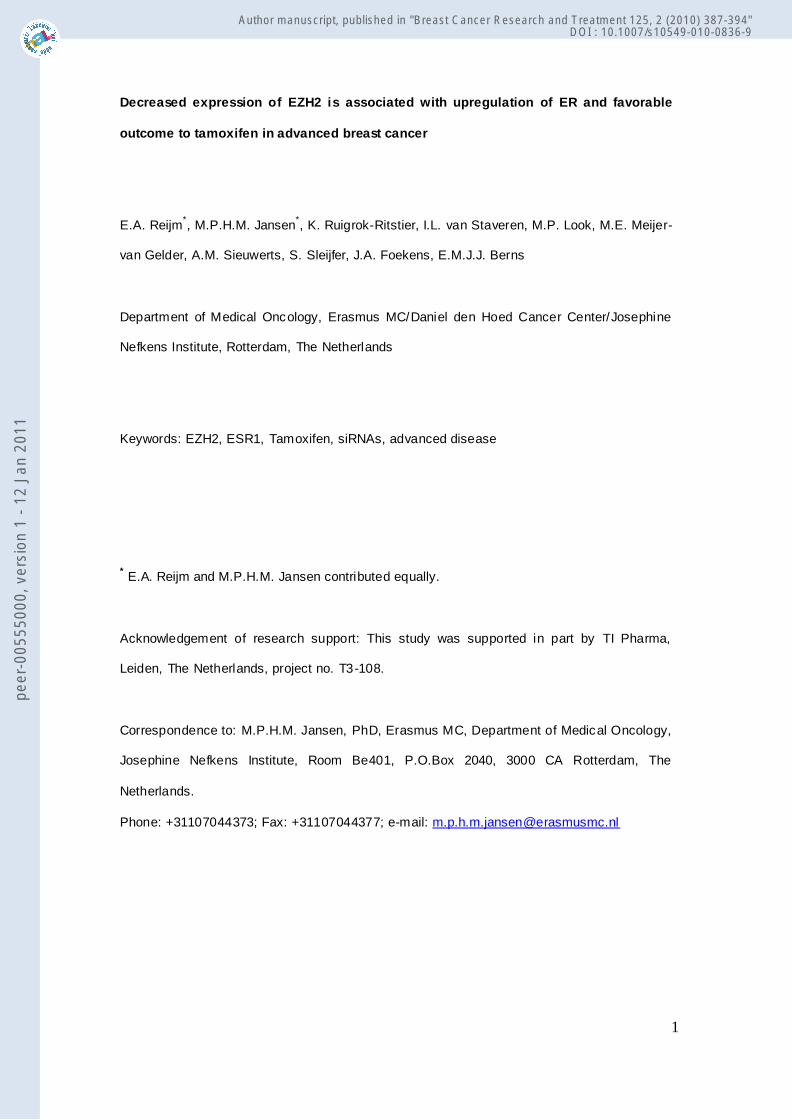
1 Decreased expression of EZH2 is associated with upregulation of ER and favorable outcome to tamoxifen in advanced breast cancer E.A. Reijm * , M.P.H.M. Jansen * , K. Ruigrok-Ritstier, I.L. van Staveren, M.P. Look, M.E. Meijer- van Gelder, A.M. Sieuwerts, S. Sleijfer, J.A. Foekens, E.M.J.J. Berns Department of Medical Oncology, Erasmus MC/Daniel den Hoed Cancer Center/Josephine Nefkens Institute, Rotterdam, The Netherlands Keywords: EZH2, ESR1, Tamoxifen, siRNAs, advanced disease * E.A. Reijm and M.P.H.M. Jansen contributed equally. Acknowledgement of research support: This study was supported in part by TI Pharma, Leiden, The Netherlands, project no. T3-108. Correspondence to: M.P.H.M. Jansen, PhD, Erasmus MC, Department of Medical Oncology, Josephine Nefkens Institute, Room Be401, P.O.Box 2040, 3000 CA Rotterdam, The Netherlands. Phone: +31107044373; Fax: +31107044377; e-mail: [email protected] peer-00555000, version 1 - 12 Jan 2011 Author manuscript, published in "Breast Cancer Research and Treatment 125, 2 (2010) 387-394" DOI : 10.1007/s10549-010-0836-9
-
Upload
independent -
Category
Documents
-
view
0 -
download
0
Transcript of Decreased expression of EZH2 is associated with upregulation of ER and favorable outcome to...
1
Decreased expression of EZH2 is associated with upregulation of ER and favorable
outcome to tamoxifen in advanced breast cancer
E.A. Reijm*, M.P.H.M. Jansen
*, K. Ruigrok-Ritstier, I.L. van Staveren, M.P. Look, M.E. Meijer-
van Gelder, A.M. Sieuwerts, S. Sleijfer, J.A. Foekens, E.M.J.J. Berns
Department of Medical Oncology, Erasmus MC/Daniel den Hoed Cancer Center/Josephine
Nefkens Institute, Rotterdam, The Netherlands
Keywords: EZH2, ESR1, Tamoxifen, siRNAs, advanced disease
* E.A. Reijm and M.P.H.M. Jansen contributed equally.
Acknowledgement of research support: This study was supported in part by TI Pharma,
Leiden, The Netherlands, project no. T3-108.
Correspondence to: M.P.H.M. Jansen, PhD, Erasmus MC, Department of Medical Oncology,
Josephine Nefkens Institute, Room Be401, P.O.Box 2040, 3000 CA Rotterdam, The
Netherlands.
Phone: +31107044373; Fax: +31107044377; e-mail: [email protected]
peer
-005
5500
0, v
ersi
on 1
- 12
Jan
201
1Author manuscript, published in "Breast Cancer Research and Treatment 125, 2 (2010) 387-394"
DOI : 10.1007/s10549-010-0836-9
2
Abstract
Purpose: To investigate EZH2 in a large series of breast cancer patients for its prognostic and
predictive value, and to evaluate its functional role in treatment response in vitro.
Experimental design: EZH2 levels were measured using quantitative Real-Time Polymerase
Chain Reaction (qRT-PCR) in primary breast cancer specimens and related to
clinicopathologic factors and disease outcome. EZH2 expression was downregulated with
siRNAs in MCF7, to assess expression alterations of putative EZH2 downstream genes and
to determine cell numbers after treatment with the anti-estrogen ICI 164.384.
Results: In 688 lymph node-negative patients who did not receive adjuvant systemic therapy,
EZH2 was not significantly correlated with metastasis-free survival (MFS). In 278 patients with
advanced disease treated with first-line tamoxifen monotherapy, the tertile with highest EZH2
levels was associated with the lowest clinical benefit (OR=0.48; P=0.02) and with a shorter
progression-free survival (PFS) in both univariate (HR=1.80; P<0.001) and multivariate
analysis, including traditional factors (HR=1.61; P=0.004).
In vitro, EZH2 silencing in MCF7 caused a 38% decrease in cell numbers (P<0.001) whereas
ICI 164.384 treatment resulted in a 25% decrease (P<0.001) compared to controls.
Combining EZH2 silencing with ICI-treatment reduced cell numbers with 67% (P<0.001)
compared to control conditions. EZH2 downregulation was associated with an almost 2-fold
upregulation of the estrogen receptor alpha (ER) (P=0.001).
Conclusion: EZH2 has no prognostic value in breast cancer. High levels of EZH2 are
associated with poor outcome to tamoxifen therapy in advanced breast cancer.
Downregulated EZH2 leads to upregulation of the ER and better response to anti-estrogens.
peer
-005
5500
0, v
ersi
on 1
- 12
Jan
201
1
3
Introduction
The anti-estrogen tamoxifen has been used for treatment of estrogen receptor alpha (ER)
positive breast cancer for more than 20 years both in the adjuvant and advanc ed setting.
Although the majority of breast tumors express the ER, approximately half of the patients with
ER-positive advanced disease does not respond to endocrine therapy or will eventually
develop resistance. As a consequence, there is a high need for markers to identify patients
likely to benefit from tamoxifen and to get a better insight into mechanisms conferring
resistance.
In a previous genome-wide profiling study in breast cancer patients with advanced disease,
we revealed an 81-gene signature for resistance to first-line tamoxifen treatment [1]. One of
the gene families from this profile is Enhancer of Zeste, consisting of Enhancer of Zeste
Homolog 1 (EZH1, OMIM 601674) and Enhancer of Zeste Homolog 2 (EZH2, OMIM 601573).
Both EZ homologs belong to Polycomb group (PcG) proteins, and are involved in
transcriptional control and epigenetic memory maintenance for preservation of cellular
characteristics [2]. EZH2 comprises the core of the Polycomb Repressor Complex 2 (PRC2)
[3-5], has histone lysine methyltransferase activity, and mediates di- and trimethylation on
histone 3 lysine residue 27 (H3K27) [2]. EZH1 can also be a part of PRC2, although with low
histone lysine methyltransferase activity [6]. We previously explored EZH1 with qRT-PCR, but
did not observe a significant correlation with clinical outcome in 229 ER-positive tumors of
patients with advanced disease treated with first-line tamoxifen monotherapy [7].
In contrast to EZH1, EZH2 has been extensively studied in malignancies. Increased
expression of EZH2 in breast, prostate and bladder cancer have been associated with a high
histological grade and increased tumor cell proli feration [8-10]. In addition, EZH2 was
identified as an adverse prognostic marker for breast and prostate cancer, but these studies
included only small series of patients [11, 12]. Its predictive value for outcome to tamoxifen in
advanced breast cancer has, however, not been studied yet.
The aims of the current study were (1) to assess the prognostic value of EZH2 in a large
series of patients, (2) to study its predictive value for outcome to tamoxifen treatment in
advanced breast cancer and (3) to explore its functional role in endocrine therapy resistance.
peer
-005
5500
0, v
ersi
on 1
- 12
Jan
201
1
4
Patients and methods
Patients
This retrospective study, in which coded tumor tissues were used, has been approved by the
medical ethics committee of the Erasmus MC Rotterdam, The Netherlands (MEC 02.953),
was performed in accordance with the Code of Conduct of the Federation of Medical
Scientific Societies in the Netherlands (http://www.fmwv.nl ), and reported following the
REMARK recommendations [13], wherever possible. Frozen breast tumor tissue specimens
of female patients with primary operable breast cancer who entered the clinic between 1979
and 1996 were analyzed. Follow-up, tumor staging, and response to therapy was defined by
standard International Union Against Cancer (Geneva, Switzerland) classification criteria [14].
Tumor protein expression levels of ER and progesterone receptor (PgR) were determined
[15], and 10 fmol/mg cytosolic protein was used as cut-off point to classify tumors as ER
and/or PgR-positive. The following criteria were applied to include tumor specimens from final
analysis: (1) sufficient frozen tumor material, (2) more than 30% epithelial tumor cells in
haematoxylin and eosin stained sections, (3) breast tumor tissue specimen of good RNA
quality according to predefined criteria [16]; and (4) EZH2 mRNA expression levels were
measured and reference mRNA levels were detectable. After applying the inclusion criteria,
tumor specimens and clinical data of 1,318 patients were available for analysis. From these
1,318 patients (for clinicopathologic details, see Supplementary Table S1) 580 patients (44%)
underwent breast conserving lumpectomy and 738 patients modified mastectomy (56%). The
median follow-up time of patients alive was 90 months, range 4-231 months. Eight hundred
eighty-nine patients did not receive adjuvant systemic therapy, while 429 patients (33%; all
lymph node-positive) did; 198 (15%) were treated with hormonal therapy, 216 (16%) with
chemotherapy (70 patients anthracycline-based (FAC/FEC) and 146 patients non-
anthracycline-based (CMF)) and 15 patients received both hormonal and chemotherapy.
Hormonal therapy of advanced disease
ER-positive tumors of 249 patients (out of the 1,318 M0 patients) who developed advanced
disease after treatment for primary breast cancer and who received first -line tamoxifen
therapy were included in this study. This set was completed with 29 tumors of patients with
peer
-005
5500
0, v
ersi
on 1
- 12
Jan
201
1
5
distant metastases at initial diagnosis (M1 patients). These 278 patients were divided
according to response to tamoxifen. Clinical benefit from tamoxifen, defined as a complete or
partial response according to standard International Union Against Cancer (Geneva,
Switzerland) classification criteria [14] or no change longer than 6 months after treatment
initiation (stable disease), was observed in 173 patients (62%); 11 patients showed complete
response, 38 a partial response and 124 patients had stable disease. No clinical benefit
occurred in 105 patients (38%).
RNA isolation and quantitative real-time PCR
Tissue processing, RNA isolation, cDNA synthesis, quantitative real -time polymerase chain
reaction (qRT-PCR) and expression data generation were performed as described previously
[16]. The qRT-PCR assays were carried out on an ABI Prism 7700 Sequence Detection
System (Applied Biosystems, Nieuwerkerk a/d IJssel, The Netherlands) or a MX3000P Real -
time PCR system (Stratagene, Amsterdam, The Netherlands). Assay -on-Demand kits
(Applied Biosystems) were used to measure mRNA levels of EZH2 (Hs00544830_m1) and
ER (Hs00174860_m1; measures ESR1). Primer sequences of the reference genes PGBD,
HPRT, B2M have been described [16]. Forty rounds of amplification were performed and
fluorescent signals of Taqman probes were used to generate Cycle threshold (Ct) values to
calculate mRNA expression levels. Expression levels of EZH2 and ER were normalized
against average expression levels of three reference genes in tumor samples and against
HPRT levels in cell lines [16].
Breast cancer cell line and RNA interference
MCF7, an estrogen sensitive ER-positive breast cancer cell line, was cultured in RPMI 1640
containing phenol red and 10% heat-inactivated fetal calf serum (Sigma-Aldrich Chemie). To
perform EZH2 knockdown experiments, small interfering RNA (siRNA) targeting EZH2 mRNA
(Qiagen, Venlo, The Netherlands) was used according recommendations and described
previously [17]. Two different siRNA duplexes for EZH2 were pooled with target sequences:
r(CCAUGUUUACAACUAUCAA)dTdT (sense) and r(UUGAUAGUUGUAAACAUGG)dTdT
(antisense) for the first duplex and r(GCAAAUUCUCGGUGUCAAA)dTdT (sense) and
peer
-005
5500
0, v
ersi
on 1
- 12
Jan
201
1
6
r(UUUGACACCGAGAAUUUGC)dTdT (antisense) for the second duplex. As control, MCF7
cells were transfected with non-specific silencing pool of siRNAs (Qiagen). siRNAs (5nM)
were int roduced via inverse transfection into MCF7, using HiPerfect transfection reagent
(Qiagen). Six experiments were independently performed at different time points in 24 (N=1)
or 96 wells plates (N=5). The 24 and 96 wells plate contained 330,000 cells and 6,000 cells
per well, respectively, and 2 and 8 wells per condition was used and pooled for further
analyses. Within three experiments in 96 wells plates, part of the cells was harvested after 96
hours for mRNA and protein analysis. The remaining part was again transfected with siRNAs,
and subsequently grown for 96 hours in standard culture medium supplemented with the pure
ER antagonist ICI 164.384 (100nM) or with ethanol vehicle alone as control. The pure ER
antagonist ICI was used to exclude the agonistic effects of tamoxifen. To assess the effect of
EZH2 silencing, cell numbers were counted with a coulter counter at day 4 and 8. To
determine response to 96 hours of ICI164.384 treatment cell numbers were counted at day 8.
Throughout an experiment, culture medium was renewed every 3 days and at the end cells
were lysed and RNA and protein isolated using the MirVana Paris kit (Ambion, Foster City,
CA, USA).
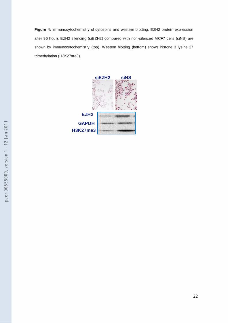
Immunocytochemistry and Western blotting
Cytospins were prepared from MCF7 cells of above experiments, fixed with 1% formaldehyde
and incubated with a monoclonal antibody against EZH2 (BD Biosciences, San Jose, CA,
(1:2000)) and a secondary peroxidase conjugated rabbit anti-mouse antibody. EZH2 protein
expression was visualized with a diaminobenzidine staining reaction. Western blotting of
protein samples were performed as described previously [17]. Antibodies against EZH2
(monoclonal (1:2000), BD Biosciences, 1:2000), H3K27 (polyclonal (1:5000), ABCAM,
Cambridge, MA, USA) and GAPDH (monoclonal (1:500), Chemicon Inc., Temecula, CA,
USA) were used and detected with horseradish peroxidase (HRP)-conjugated or HRP-
polymer (DAKO Real Envision, DAKO, Diagnostica GmbH, Hamburg, Germany) labeled
secondary antibodies and chemiluminescent reagents (ECL-kit, Pierce, Rockford, IL). The
Scanalytics One-D program (Alpha Innotech Ltd., Cannock, UK) was used for quantification.
peer
-005
5500
0, v
ersi
on 1
- 12
Jan
201
1
7
Data analysis and statistics
The relationship of EZH2 expression levels with patient and tumor characteristics was
investigated using nonparametric methods, i.e., Spearman rank correlations for continuous
variables and Wilcoxon rank-sum or Kruskal-Wallis test for ordered variables. For the
analyses with continuous variables, mRNA levels of EZH2 were log transformed and of ER
and PgR were box-cox transformed to reduce skewness of the distribution. Logistic
regression analysis was used to calculate the odds ratio (OR) that defines the relation
between expression levels and clinical benefit from therapy. The Cox proprotional hazards
model was used to compute the hazard ratio (HR) in the analysis of metastasis -free survival
(MFS), overall survival (OS) and progression-free survival (PFS). MFS and OS were
previously described [18]. PFS was defined as the time elapsed between initiation of
tamoxifen therapy and the first detection of disease progression. In multivariate analysis,
logistic and Cox regression analysis was applied to determine whether EZH2 had predictive
value and was independent when added to the base model of traditional factors. The Cox
proportional hazard assumption was not violated as verified by a test based on Schoenfeld
residuals. Both HR and OR were represented with their 95% confidence intervals (95% CI).
Survival curves were generated using the Kaplan and Meier method and a log rank test was
used to test for differences. Computations were done with the STATA statistical package,
release 10.1 (STATA Corp., College Station, TX). In the in vitro studies, a student t-test was
performed to test for significance for differences in cell counts and RNA levels. All P -values
were two-sided and P<0.05 was considered statistically significant.
peer
-005
5500
0, v
ersi
on 1
- 12
Jan
201
1
8
Results
Associations of EZH2 mRNA expression levels with clinicopathological factors
In this study, we determined the mRNA expression levels in 1,318 primary breast carcinomas.
Median expression levels of EZH2, its interquartile range, and its association with patient and
tumor characteristics are summarized in Supplementary Table S1. Briefly, high EZH2 mRNA
levels were significantly associated with younger age, premenopausal status, poor histologic
grade, larger tumor size, and status of ER, PgR, ERBB2 and EGFR. An inverse correlation
between EZH2 and ER was observed (P<0.001, rs=-0.33). Expression of EZH2 was higher in
ERBB2 positive samples compared with ERBB2 negative samples (P<0.001).
EZH2 levels and clinical outcome
The prognostic value of EZH2 was assessed in 688 lymph node -negative (LNN) patients who
did not receive any adjuvant systemic therapy. EZH2 levels, as continuous variable, were not
significantly correlated with metastasis-free survival (MFS) (HR=1.14, 95% CI: 0.98-1.32;
P=0.10). Considering overall survival (OS), EZH2 was significantly associated in LNN patients
(HR=1.25, 95% CI: 1.07-1.46; P=0.006).
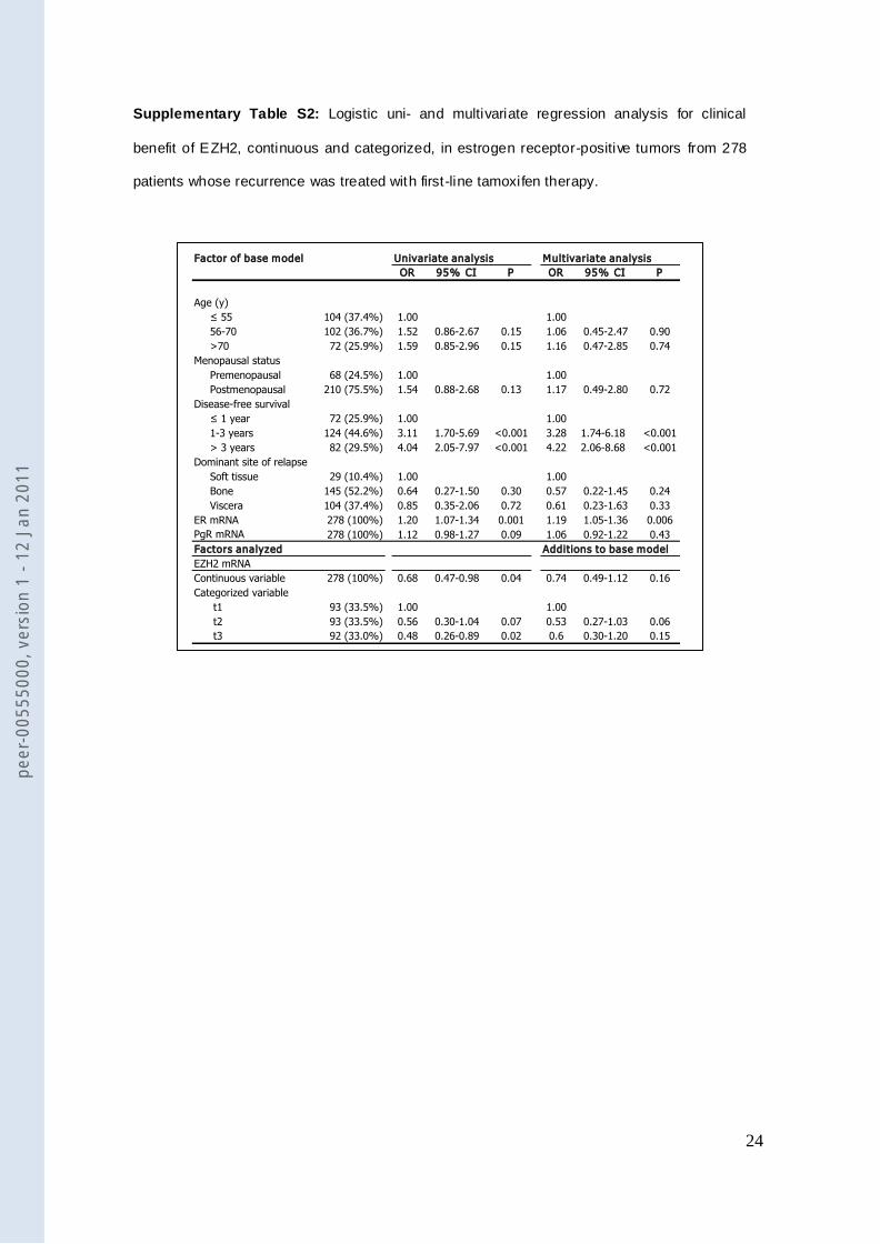
Association of EZH2 levels with clinical benefit and progression-f ree survival
In univariate analysis, increasing EZH2 expression levels as continuous variable were
significantly associated with a lower clinical benefit in patients with advanced breast cancer
treated with first-line tamoxifen monotherapy (n=278) (OR=0.68, 95% CI: 0.47 -0.98; P=0.04)
(Supplementary Table S2). In analogy, after categorizing EZH2 expression levels into tertiles,
the highest tertile was significantly associated with a lower clinical benefit to tamoxifen
therapy (OR=0.48, 95% CI: 0.26-0.89; P=0.02). In multivariate analysis, however, when
corrected for t raditional predictive factors including age, menopausal status, DSR, DFS, ER
and PgR levels, no significant association with clinical benefit was observed (Supplementary
Table S2). In univariate analysis, increasing EZH2 expression levels analyzed as continuous
variable were significantly associated with shorter PFS (HR=1.28, 95% CI: 1.08-1.51;
P=0.004) (Table 1). In univariate analysis, compared with tumors in the lowest tertile of EZH2
expression, those with the highest EZH2 levels were associated with a poor PFS (HR=1.80,
peer
-005
5500
0, v
ersi
on 1
- 12
Jan
201
1
9
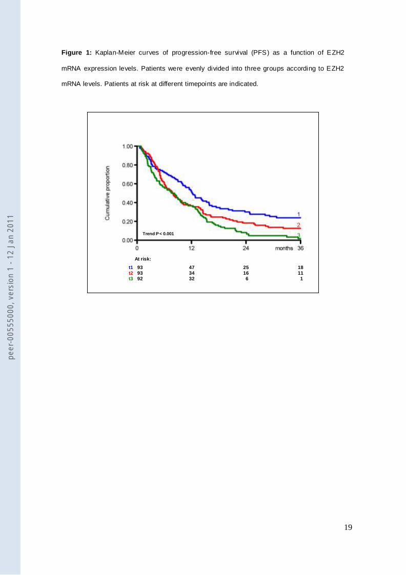
95%CI: 1.32-2.46; P<0.001) (Table 1, Figure 1). Remarkably, the intermediate and highest
expression groups have a similar PFS during the first 12 months while the curves diverge
thereafter. In multivariate analysis, compared with tumors with EZH2 levels in the lowest
tertile, those with highest EZH2 levels were significantly associated with poor PFS (HR=1.61,
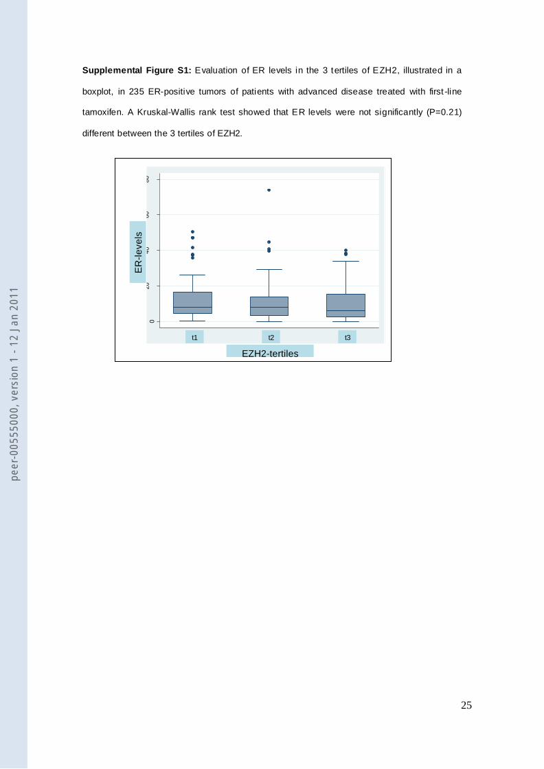
95% CI: 1.16-2.24; P=0.004). Moreover, ER levels were not significantly (P=0.21) different
between the 3 different tertiles of EZH2 (Supplemental Figure S1).
EZH2 and RNA interference
To investigate how EZH2 is functionally involved in response to anti-estrogens, we performed
in vitro studies in the ER-positive human breast cancer cell line MCF7. To this end MCF7
cells were treated with ICI164.384 combined with non-silencing (NS) or EZH2 silencing.
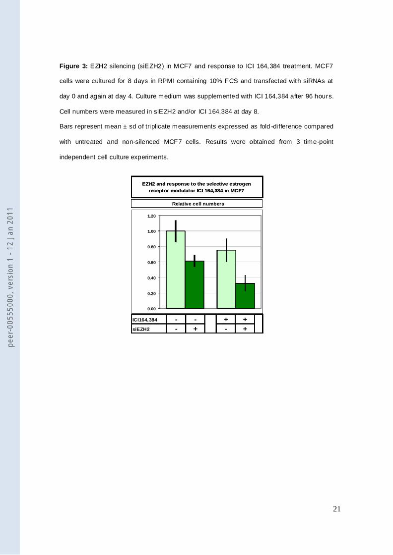
When cells transfected with siNS were exposed to 100nM ICI164.384 a decrease in cell
numbers of 25% (range 12-30%, N=3, P<0.001) after 96 hours was observed, confirming that
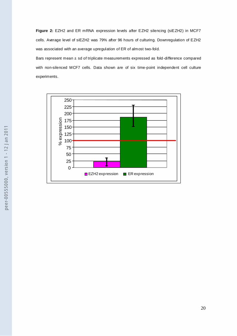
MCF7 is an anti-estrogen sensitive cell line. Knockdown experiments showed an average
silencing level of EZH2 of 79% (range 64%-93%, N=6, P<0.001) after 96 hours (Figure 2).
EZH2 silencing caused a significant decrease in cell numbers of 38% (range 17 -53%, N=3,
P<0.001) compared with controls (Figure 3). When EZH2 silencing and ICI-treatment were
combined, cell numbers were reduced with 67% (range 54-75%, N=3, P<0.001) compared to
non-silenced MCF7 cells. Both immunocytochemistry of cytospins and Western blotting
confirm knockdown of EZH2 on the protein level after 96 hours of EZH2 silencing (Figure 4).
To validate EZH2 silencing functionally we demonstrated (Figure 4) that methylation of lysine
residue 27 of histone 3 diminishes when silencing EZH2.
Association between EZH2 levels and ER expression
In view of the inverse correlation between EZH2 and ER mRNA expression levels observed in
our 1,318 breast cancer tumor specimens, we next studied the effect of EZH2 silencing on ER
expression in vitro. Downregulation of EZH2 in MCF7 cells (79% after 96 hours, range 64%-
93%, N=6) was associated with an average two-fold upregulation of ER (N=6, range 1.5-2.3,
P=0.001) (Figure 2), which is concordant with the observed inverse correlation between EZH2
and ER mRNA expression in clinical samples.
peer
-005
5500
0, v
ersi
on 1
- 12
Jan
201
1
10
Discussion
We demonstrate here for the first time that a significant association exists between high levels
of EZH2 with outcome in terms of PFS for ER-positive breast cancer patients treated with
first-line tamoxifen for advanced disease. Additionally, it was shown that EZH2 expression
impacts response to tamoxifen rather than reflecting tumor aggressiveness since no
prognostic value for EZH2 was revealed in LNN primary breast cancer who did not receive
adjuvant systemic therapy.
In contrast to our findings, EZH2 has previously been suggested to bear prognostic value in
breast cancer. Based on in silico analysis, on a relatively small set of 78 tumors of young LNN
patients (<55 years) with low grade tumors [19], Kleer et al. reported that mRNA expression
of EZH2 was significantly higher in invasive carcinomas that metastasize within 5 years of
primary diagnosis compared with invasive carcinomas that did not [11]. In another study
immunohistochemistry was used to demonstrate an association between EZH2 expression
and increased tumor cell proli feration in melanoma, prostate, endometrial, and breast cancer
[9]. Association of high EZH2 expression with unfavorable prognosis was revealed in all
investigated tumor types with the exception of breast cancer as survival data were not
available. We observed that EZH2 associates significantly with overall survival, however,
overall survival is not only dependent on tumor aggressiveness but also on treatment and
response of the patient in the adjuvant and/or advanced disease setting. That EZH2 in our
study predicts poor overall survival in LNN patients who did not obtain adjuvant systemic
therapy suggests an association with treatment response in the advanced disease setting, as
for example shown for first-line tamoxifen monotherapy in this study. Collett et al. also used
immunohistochemistry to assess the prognostic value of EZH2 in breast cancer [8]. In 190
tumors (100 EZH2-negative and 90 EZH2-positive), they demonstrated a significant
correlation between EZH2 positivity and high disease stage at time of diagnosis in terms of
locally advanced disease or metastatic disease. However, this group comprises only 6
patients with metastatic disease at initial diagnosis. Additionally, this set was heterogeneous
with both LNN and LN-positive tumors. And though treatment status was not discussed, it is
reasonable to assume that LNP patients have received systemic treatment thereby impacting
outcome and obscuring EZH2’s true prognostic value. However, in our study population
P=0.001
peer
-005
5500
0, v
ersi
on 1
- 12
Jan
201
1
11
comprising the largest series of patients studied so far, 688 LNN patients, who did not receive
any adjuvant systemic therapy enabling to assess the true prognostic value of EZH2, we
could not confirm its prognostic value. This discrepancy in findings remains to be clarified but
might be due to the methodology applied and the cohorts of patients studied.
Furthermore, we found an association between EZH2 and the type of response to therapy in
advanced disease with the highest EZH2 levels related to poor outcome. In order to elucidate
the potential underlying mechanisms for this association, we explored the association
between EZH2 expression and ER expression. In our 1,318 breast cancer tumor specimens,
we observed an inverse association between EZH2 and ER mRNA expression levels.
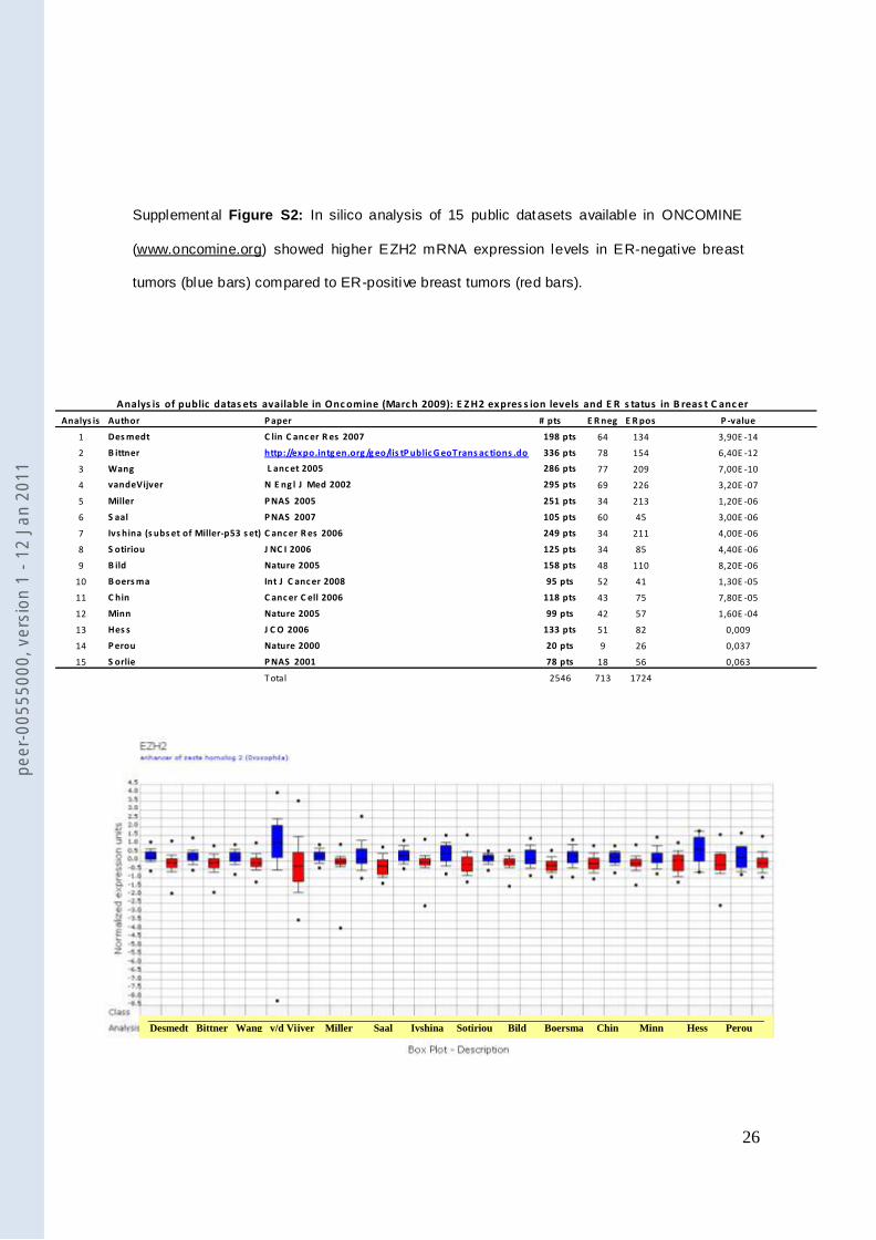
Accordingly, in silico analysis of 15 independent breast cancer datasets of the Oncomine
database (supplementary Figure S2), with in total 2,437 samples (713 ER-negative, 1724 ER-
positive), confirmed this finding. In addition, we performed functional studies in which EZH2
expression was silenced with siRNAs in the human estrogen sensitive breast cancer cell line
MCF7. The decrease in cell numbers following downregulating EZH2 expression in MCF7
suggests that EZH2 has an effect on cell proliferation, in agreement with previously performed
studies showing an association between EZH2 and cell proli feration [8, 11]. We have shown
that growth inhibition in the ICI+siEZH2 experiment (67% inhibition) adds up the effect of
EZH2 silencing and ICI164,384 treatment on MCF7 growth (38% and 25% inhibition,
respectively). These combined ICI+siEZH2 experiments demonstrate no overlap in growth
inhibition and indicates that the effect of EZH2 silencing on MCF7 growth is independent of
the effect of ER inhibition by ICI. Our observed upregulation of ER by silencing of EZH2 and
the inverse correlation between EZH2 and ER status (ER-negative vs ER-positive) in breast
cancer specimens (Supplemental Figure S2), however, suggest an EZH2 and ER interaction
which may also result in enhanced sensitivity to ICI164.384. Although the observed two-fold
upregulation of ER does not seem impressive at first glance, it is already observed after 96
hours of culturing. Moreover, MCF7 is a cell line with already one of the highest ER
expression levels in our panel of 39 breast cancer cell lines (data not shown). In that
perspective, an almost 2-fold upregulation can be regarded substantial. Furthermore, the
extent of ER upregulation by silencing EZH2 is in the range of what has been described for
other downstream factors. For example, Yu et al published a 1.6-fold upregulation of the
peer
-005
5500
0, v
ersi
on 1
- 12
Jan
201
1
12
Adrenergic Receptor when silencing EZH2 [20]. We hypothesize that the promoter region of
ER lacks DNA methylation in MCF7 and that the observed upregulation of ER is
predominantly due to decreased histone H3K27 trimethylation caused by EZH2 knockdown.
Further studies are needed to verify this hypothesis.
Given the presumed role of EZH2 in the regulation of ER expression, EZH2 might be an
interesting target for therapy. Recently, Varambally et al. discovered a physiological EZH2-
inhibitor, miRNA-101, which inhibits the expression and function of EZH2 in cancer cell lines
[21]. It has been shown that miRNA -101 expression diminishes during cancer progression,
resulting in an increased EZH2 expression and concomitant dysregulation of epigenetic
pathways. This cascade is thought to underlie progression of several types of cancers, e.g.,
prostate, brain and lung cancer [21]. In addition, also miRNA-26a has been reported to post-
transcriptionally repress EZH2 [22]. Recently, Kota et al. demonstrated the capacity of
miRNA-26a as an anti-tumor therapy in a mouse model of hepatocellular carcinomas, where it
resulted in inhibition of cancer cell proliferation and induction of tumor-specific apoptosis [23].
As a result, it would be interesting to further investigate EZH2 as a potential target for therapy,
and include miRNA-101 and miRNA-26a as ‘in vivo’ inhibitors.
In conclusion, EZH2 has no prognostic value in our large set of LNN adjuvant untreated
breast cancer patients. However, high EZH2 levels are associated with unfavorable outcome
to tamoxifen treatment in breast cancer patients with advanced disease, which suggests that
EZH2 can be used as a predictive marker. Moreover, downregulation of EZH2 caused
additional growth inhibition next to anti-estrogen therapy in vitro and resulted in ER
upregulation. If validated, EZH2 may be considered to serve as a potential target to increase
the anti-tumor activity of anti-estrogen therapies in breast cancer. Furthermore, its
assessment may contribute to a more appropriate selection of patients for tamoxifen therapy
and thereby a more tailored management of patients with breast cancer.
Disclosure of potential conflicts of interest
No potential conflicts of interest were disclosed.
peer
-005
5500
0, v
ersi
on 1
- 12
Jan
201
1
13
References
1. Jansen MP, Foekens JA, van Staveren IL et al (2005) Molecular classification of
tamoxifen-resistant breast carcinomas by gene expression profiling. J Clin Oncol
23:732-740.
2. Schuettengruber B, Chourrout D, Vervoort M et al (2007) Genome regulation by
polycomb and trithorax proteins. Cell 128:735-745.
3. Cao R, Wang L, Wang H et al (2002) Role of histone H3 lysine 27 methylation in
Polycomb-group silencing. Science 298:1039-1043.
4. Czermin B, Melfi R, McCabe D et al (2002) Drosophila enhancer of Zeste/ESC
complexes have a histone H3 methyltransferase activity that marks chromosomal
Polycomb sites. Cell 111:185-196.
5. Kuzmichev A, Nishioka K, Erdjument-Bromage H et al (2002) Histone
methyltransferase activity associated with a human multiprotein complex containing
the Enhancer of Zeste protein. Genes Dev 16:2893-2905.
6. Margueron R, Li G, Sarma K et al (2008) Ezh1 and Ezh2 maintain repressive
chromatin through different mechanisms. Mol Cell 32:503-518.
7. Jansen M, Foekens J, Ritstier K et al (2005) A miniPathway for tamoxifen therapy
resistance. Breast Cancer Res and Treat 94:S31-S31.
8. Collett K, Eide GE, Arnes J et al (2006) Expression of enhancer of zeste homologue
2 is significantly associated with increased tumor cell proli feration and is a marker of
aggressive breast cancer. Clin Cancer Res 12:1168-1174.
9. Bachmann IM, Halvorsen OJ, Collett K et al (2006) EZH2 expression is associated
with high proliferation rate and aggressive tumor subgroups in cutaneous melanoma
and cancers of the endometrium, prostate, and breast. J Clin Oncol 24:268 -273.
10. Raman JD, Mongan NP, Tickoo SK et al (2005) Increased expression of the
polycomb group gene, EZH2, in transitional cell carcinoma of the bladder. Clin
Cancer Res 11:8570-8576.
11. Kleer CG, Cao Q, Varambally S et al (2003) EZH2 is a marker of aggressive breast
cancer and promotes neoplastic transformation of breast epithelial cells. Proc Natl
Acad Sci USA 100:11606-11611.
peer
-005
5500
0, v
ersi
on 1
- 12
Jan
201
1
14
12. Varambally S, Dhanasekaran SM, Zhou M et al (2002) The polycomb group protein
EZH2 is involved in progression of prostate cancer. Nature 419:624 -629.
13. McShane LM, Altman DG, Sauerbrei W, et al (2006) REporting recommendations for
tumor MARKer prognostic studies (REMARK). Breast Cancer Res and Treat
100:229-235.
14. Hayward JL, Carbone PP, Heuson JC et al (1977) Assessment of response to
therapy in advanced breast cancer: a project of the Programme on Clinical Oncology
of the International Union Against Cancer, Geneva, Switzerland. Cancer 39:1289-
1294.
15. Foekens JA, Portengen H, van Putten WL et al (1989) Prognostic value of estrogen
and progesterone receptors measured by enzyme immunoassays in human breast
tumor cytosols. Cancer Res 49:5823-5828.
16. Sieuwerts AM, Meijer-van Gelder ME, Timmermans M et al (2005) How ADAM-9 and
ADAM-11 differentially from estrogen receptor predict response to tamoxifen
treatment in patients with recurrent breast cancer: a ret rospective study. Clin Cancer
Res 11:7311-7321.
17. Jansen MP, Ruigrok-Ritstier K, Dorssers LC et al (2009) Downregulation of SIAH2,
an ubiquitin E3 ligase, is associated with resistance to endocrine therapy in breast
cancer. Breast Cancer Res and Treat 116:263-271.
18. van Agthoven T, Sieuwerts AM, Veldscholte J et al (2009) CITED2 and NCOR2 in
anti-oestrogen resistance and progression of breast cancer. Br J Cancer 101:1824-
1832.
19. van 't Veer LJ, Dai H, van de Vijver MJ et al (2002) Gene expression profiling predicts
clinical outcome of breast cancer. Nature 415:530-536.
20. Yu J, Cao Q, Mehra R et al (2007) Integrative genomics analysis reveals silencing of
beta-adrenergic signaling by polycomb in prostate cancer. Cancer Cell 12:419-431.
21. Varambally S, Cao Q, Mani RS et al (2008) Genomic loss of microRNA-101 leads to
overexpression of histone methyltransferase EZH2 in cancer. Science 322:1695-
1699.
22. Wong CF, Tellam RL (2008) MicroRNA -26a targets the histone methyltransferase
peer
-005
5500
0, v
ersi
on 1
- 12
Jan
201
1

15
Enhancer of Zeste homolog 2 during myogenesis. J Biol Chem 283:9836-9843.
23. Kota J, Chivukula RR, O'Donnell KA et al (2009) Therapeutic microRNA delivery
suppresses tumorigenesis in a murine liver cancer model. Cell 137:1005 -1017.
peer
-005
5500
0, v
ersi
on 1
- 12
Jan
201
1
16
Legends
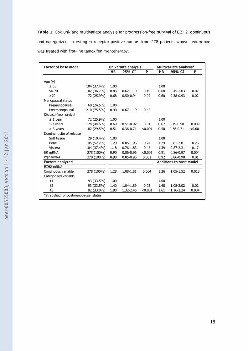
Table 1: Cox uni- and multivariate analysis for progression-free survival of EZH2, continuous
and categorized, in estrogen receptor-positive tumors from 278 patients whose recurrence
was treated with first-line tamoxifen monotherapy.
Figure 1: Kaplan-Meier curves of progression-free survival (PFS) as a function of EZH2
mRNA expression levels. Patients were evenly divided into three groups according to EZH2
mRNA levels. Patients at risk at different timepoints are indicated.
Figure 2: EZH2 and ER mRNA expression levels after EZH2 silencing (siEZH2) in MCF7
cells. Average level of siEZH2 was 79% after 96 hours of culturing. Downregulation of EZH2
was associated with an average upregulation of ER of almost two-fold.
Bars represent mean ± sd of triplicate measurements expressed as fold -difference compared
with non-silenced MCF7 cells. Data shown are of six time-point independent cell culture
experiments.
Figure 3: EZH2 silencing (siEZH2) in MCF7 and response to ICI 164,384 treatment. MCF7
cells were cultured for 8 days in RPMI containing 10% FCS and transfected with siRNAs at
day 0 and again at day 4. Culture medium was supplemented with ICI 164,384 after 96 hours.
Cell numbers were measured in siEZH2 and/or ICI 164,384 at day 8.
Bars represent mean ± sd of triplicate measurements expressed as fold -difference compared
with untreated and non-silenced MCF7 cells. Results were obtained from 3 time-point
independent cell culture experiments.
Figure 4: Immunocytochemistry of cytospins and western blotting. EZH2 protein expression
after 96 hours EZH2 silencing (siEZH2) compared with non -silenced MCF7 cells (siNS) are
shown by immunocytochemistry (top). Western blotting (bottom) shows histone 3 lysine 27
trimethylation (H3K27me3).
peer
-005
5500
0, v
ersi
on 1
- 12
Jan
201
1
17
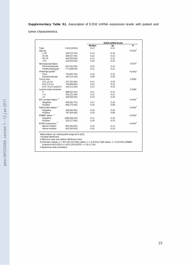
Supplementary Table S1. Association of EZH2 mRNA expression levels with patient and
tumor characteristics.
Supplementary Table S2: Logistic uni- and multivariate regression analysis for clinical
benefit of EZH2, continuous and categorized, in estrogen receptor-positive tumors from 278
patients whose recurrence was treated with first-line tamoxifen therapy.
Supplemental Figure S1: Evaluation of ER levels in the 3 tertiles of EZH2, illustrated in a
boxplot, in 235 ER-positive tumors of patients with advanced disease treated with first -line
tamoxifen. A Kruskal-Wallis rank test showed that ER levels were not significantly (P=0.21)
different between the 3 tertiles of EZH2.
Supplementary Figure S2: In silico analysis of 15 public datasets available in ONCOMINE
(www.oncomine.org) showed higher EZH2 mRNA expression levels in ER-negative breast
tumors (blue bars) compared to ER-positive breast tumors (red bars).
peer
-005
5500
0, v
ersi
on 1
- 12
Jan
201
1
18
Table 1: Cox uni- and multivariate analysis for progression-free survival of EZH2, continuous
and categorized, in estrogen receptor-positive tumors from 278 patients whose recurrence
was treated with first-line tamoxifen monotherapy.
Factor of base model Univariate analysis Multivariate analysis*
HR 95% CI P HR 95% CI P
Age (y)
≤ 55 104 (37.4%) 1.00 1.00
56-70 102 (36.7%) 0.83 0.62-1.10 0.19 0.68 0.45-1.03 0.07
>70 72 (25.9%) 0.68 0.50-0.94 0.02 0.60 0.38-0.93 0.02
Menopausal status
Premenopausal 68 (24.5%) 1.00
Postmenopausal 210 (75.5%) 0.90 0.67-1.19 0.45
Disease-free survival
≤ 1 year 72 (25.9%) 1.00 1.00
1-3 years 124 (44.6%) 0.69 0.51-0.92 0.01 0.67 0.49-0.90 0.009
> 3 years 82 (29.5%) 0.51 0.36-0.71 <0.001 0.50 0.36-0.71 <0.001
Dominant site of relapse
Soft tissue 29 (10.4%) 1.00 1.00
Bone 145 (52.2%) 1.29 0.85-1.96 0.24 1.29 0.81-2.01 0.26
Viscera 104 (37.4%) 1.18 0.76-1.83 0.45 1.39 0.87-2.21 0.17
ER mRNA 278 (100%) 0.90 0.86-0.96 <0.001 0.91 0.86-0.97 0.004
PgR mRNA 278 (100%) 0.90 0.85-0.96 0.001 0.92 0.86-0.98 0.01
Factors analyzed
EZH2 mRNA
Continuous variable 278 (100%) 1.28 1.08-1.51 0.004 1.26 1.05-1.52 0.015
Categorized variable
t1 93 (33.5%) 1.00 1.00
t2 93 (33.5%) 1.40 1.04-1.89 0.03 1.48 1.08-2.02 0.02
t3 92 (33.0%) 1.80 1.32-2.46 <0.001 1.61 1.16-2.24 0.004
*stratisfied for postmenopausal status
Additions to base model
peer
-005
5500
0, v
ersi
on 1
- 12
Jan
201
1
19
Figure 1: Kaplan-Meier curves of progression-free survival (PFS) as a function of EZH2
mRNA expression levels. Patients were evenly divided into three groups according to EZH2
mRNA levels. Patients at risk at different timepoints are indicated.
Trend P < 0.001
t1t2t3
939392
473432
25166
18111
At risk:
peer
-005
5500
0, v
ersi
on 1
- 12
Jan
201
1
20
Figure 2: EZH2 and ER mRNA expression levels after EZH2 silencing (siEZH2) in MCF7
cells. Average level of siEZH2 was 79% after 96 hours of culturing. Downregulation of EZH2
was associated with an average upregulation of ER of almost two-fold.
Bars represent mean ± sd of triplicate measurements expressed as fold -difference compared
with non-silenced MCF7 cells. Data shown are of six time-point independent cell culture
experiments.
P = 0.001
0
25
50
75
100
125
150
175
200
225
250
% e
xp
ressio
n
EZH2 expression ER expression
peer
-005
5500
0, v
ersi
on 1
- 12
Jan
201
1
21
Figure 3: EZH2 silencing (siEZH2) in MCF7 and response to ICI 164,384 treatment. MCF7
cells were cultured for 8 days in RPMI containing 10% FCS and transfected with siRNAs at
day 0 and again at day 4. Culture medium was supplemented with ICI 164,384 after 96 hours.
Cell numbers were measured in siEZH2 and/or ICI 164,384 at day 8.
Bars represent mean ± sd of triplicate measurements expressed as fold -difference compared
with untreated and non-silenced MCF7 cells. Results were obtained from 3 time-point
independent cell culture experiments.
ICI164,384 - - + +
siEZH2 - + - +
EZH2 and response to the selective estrogen
receptor modulator ICI 164,384 in MCF7
Relative cell numbers
0.00
0.20
0.40
0.60
0.80
1.00
1.20
ICI164,384 - - + +
siEZH2 - + - +
EZH2 and response to the selective estrogen
receptor modulator ICI 164,384 in MCF7
Relative cell numbers
0.00
0.20
0.40
0.60
0.80
1.00
1.20
peer
-005
5500
0, v
ersi
on 1
- 12
Jan
201
1
22
Figure 4: Immunocytochemistry of cytospins and western blotting. EZH2 protein expression
after 96 hours EZH2 silencing (siEZH2) compared with non -silenced MCF7 cells (siNS) are
shown by immunocytochemistry (top). Western blotting (bottom) shows histone 3 lysine 27
trimethylation (H3K27me3).
siEZH2 siNS
EZH2
GAPDH H3K27me3
peer
-005
5500
0, v
ersi
on 1
- 12
Jan
201
1
23
Supplementary Table S1. Association of EZH2 mRNA expression levels with patient and
tumor characteristics.
EZH2 mRNA levels
Median iqr PTotal 1318 (100%) 0.12 0.11Age (y) <0.001
< 40 163 (12.4%) 0.13 0.14
41-55 493 (37.4%) 0.12 0.1256-70 443 (33.6%) 0.11 0.11>70 219 (16.6%) 0.10 0.10
Menopausal status 0.019†
Premenopausal 547 (41.5%) 0.12 0.12Postmenopausal 771 (58.5%) 0.11 0.11
Histologic grade‡
<0.001*Poor 734 (55.7%) 0.13 0.12Good/moderate 187 (14.2%) 0.09 0.10
Tumor size 0.008*pT1, <2 cm 417 (31.6%) 0.11 0.10pT2, 2-5 cm 749 (56.8%) 0.12 0.12pT3, >5 cm and pT4 152 (11.5%) 0.12 0.10
Lymph nodes involved 0.338*0 688 (52.2%) 0.11 0.121-3 281 (21.3%) 0.11 0.11>3 349 (26.5%) 0.13 0.10
ER protein status ‡
<0.001
Negative 350 (26.7%) 0.17 0.16Positive 962 (73.3%) 0.10 0.09
PgR protein status ‡
<0.001
Negative 449 (36.0%) 0.16 0.16Positive 797 (64.0%) 0.10 0.08
ERBB2 status ‡
<0.001†
Negative 1080 (83.0%) 0.11 0.10Positive 222 (17.0%) 0.16 0.13
EGFR expression ‡
<0.001Below median 652 (50.0%) 0.10 0.11Above median 651 (50.0%) 0.14 0.14
Abbreviation: iqr, interquartile range (q75-q25).* Kruskal-Wallis test.† Wilcoxon rank-sum (Mann-Whitney) U test.
‡ Unknown: Grade, n = 397 (30.1%); ER status, n = 6 (0.5%); PgR status, n =72 (5.5%); ERBB2 (cutpoint=18.01355), n =16 (1.2%); EGFR, n =15 (1.1%).
# Spearman rank correlation.
#
#
#
#
peer
-005
5500
0, v
ersi
on 1
- 12
Jan
201
1
24
Supplementary Table S2: Logistic uni- and multivariate regression analysis for clinical
benefit of EZH2, continuous and categorized, in estrogen receptor-positive tumors from 278
patients whose recurrence was treated with first-line tamoxifen therapy.
Factor of base model Univariate analysis Multivariate analysis
OR 95% CI P OR 95% CI P
Age (y)
≤ 55 104 (37.4%) 1.00 1.00
56-70 102 (36.7%) 1.52 0.86-2.67 0.15 1.06 0.45-2.47 0.90
>70 72 (25.9%) 1.59 0.85-2.96 0.15 1.16 0.47-2.85 0.74
Menopausal status
Premenopausal 68 (24.5%) 1.00 1.00
Postmenopausal 210 (75.5%) 1.54 0.88-2.68 0.13 1.17 0.49-2.80 0.72
Disease-free survival
≤ 1 year 72 (25.9%) 1.00 1.00
1-3 years 124 (44.6%) 3.11 1.70-5.69 <0.001 3.28 1.74-6.18 <0.001
> 3 years 82 (29.5%) 4.04 2.05-7.97 <0.001 4.22 2.06-8.68 <0.001
Dominant site of relapse
Soft tissue 29 (10.4%) 1.00 1.00
Bone 145 (52.2%) 0.64 0.27-1.50 0.30 0.57 0.22-1.45 0.24
Viscera 104 (37.4%) 0.85 0.35-2.06 0.72 0.61 0.23-1.63 0.33
ER mRNA 278 (100%) 1.20 1.07-1.34 0.001 1.19 1.05-1.36 0.006
PgR mRNA 278 (100%) 1.12 0.98-1.27 0.09 1.06 0.92-1.22 0.43
Factors analyzed
EZH2 mRNA
Continuous variable 278 (100%) 0.68 0.47-0.98 0.04 0.74 0.49-1.12 0.16
Categorized variable
t1 93 (33.5%) 1.00 1.00
t2 93 (33.5%) 0.56 0.30-1.04 0.07 0.53 0.27-1.03 0.06
t3 92 (33.0%) 0.48 0.26-0.89 0.02 0.6 0.30-1.20 0.15
Additions to base model
peer
-005
5500
0, v
ersi
on 1
- 12
Jan
201
1
25
Supplemental Figure S1: Evaluation of ER levels in the 3 tertiles of EZH2, illustrated in a
boxplot, in 235 ER-positive tumors of patients with advanced disease treated with first -line
tamoxifen. A Kruskal-Wallis rank test showed that ER levels were not significantly (P=0.21)
different between the 3 tertiles of EZH2.
020
40
60
80
ER
X-4
1 2 3
EZH2-tertiles
t1 t2 t3
ER
-le
ve
ls
peer
-005
5500
0, v
ersi
on 1
- 12
Jan
201
1
26
Supplemental Figure S2: In silico analysis of 15 public datasets available in ONCOMINE
(www.oncomine.org) showed higher EZH2 mRNA expression levels in ER-negative breast
tumors (blue bars) compared to ER-positive breast tumors (red bars).
Analys is Author P aper # pts E R neg E R pos P -value
1 Des medt C lin C anc er R es 2007 198 pts 64 134 3,90E -14
2 B ittner http://expo.intg en.org /g eo/lis tP ublic G eoTrans ac tions .do 336 pts 78 154 6,40E -12
3 Wang L anc et 2005 286 pts 77 209 7,00E -10
4 vandeVijver N E ng l J Med 2002 295 pts 69 226 3,20E -07
5 Miller P NAS 2005 251 pts 34 213 1,20E -06
6 S aal P NAS 2007 105 pts 60 45 3,00E -06
7 Ivs hina (s ubs et of Miller-p53 s et) C anc er R es 2006 249 pts 34 211 4,00E -06
8 S otiriou J NC I 2006 125 pts 34 85 4,40E -06
9 B ild Nature 2005 158 pts 48 110 8,20E -06
10 B oers ma Int J C anc er 2008 95 pts 52 41 1,30E -05
11 C hin C anc er C ell 2006 118 pts 43 75 7,80E -05
12 Minn Nature 2005 99 pts 42 57 1,60E -04
13 Hes s J C O 2006 133 pts 51 82 0,009
14 P erou Nature 2000 20 pts 9 26 0,037
15 S orlie P NAS 2001 78 pts 18 56 0,063
T otal 2546 713 1724
Analys is of public datas ets available in Onc omine (Marc h 2009): E Z H2 expres s ion levels and E R s tatus in B reas t C anc er
Desmedt Bittner Wang v/d Vijver Miller Saal Ivshina Sotiriou Bild Boersma Chin Minn Hess Perou
Sorlie
peer
-005
5500
0, v
ersi
on 1
- 12
Jan
201
1

27
peer
-005
5500
0, v
ersi
on 1
- 12
Jan
201
1