High tumor cell expression of microRNA-21 in node positive non-small cell lung cancer predicts a...
9
RESEARCH ARTICLE Open Access High tumor cell expression of microRNA-21 in node positive non-small cell lung cancer predicts a favorable clinical outcome Helge Stenvold 1,2* , Tom Donnem 1,2 , Sigve Andersen 1,2 , Samer Al-Saad 3,4 , Andrej Valkov 3,4 , Mona Irene Pedersen 1,4 , Lill-Tove Busund 3,4 and Roy M Bremnes 1,2 Abstract Background: MicroRNA (miR)-21 has been revealed as an oncogene in cancer development, and is one of the miRNAs closely connected to angiogenesis. We aimed to explore the impact of miR-21 expression in both tumor and stromal compartments of non-small cell lung cancer (NSCLC), and correlations between miR-21 and angiogenic protein markers. Methods: From 335 unselected stage I to IIIA NSCLC carcinomas, duplicate tumor and tumor-associated stromal cores were collected in tissue microarrays (TMAs). In situ hybridization (ISH) was used to detect the expression of miR-21 separately in tumor cells and stromal cells of the tumor, and immunohistochemistry (IHC) was used to detect the expression of the protein markers protein kinase B (Akt), phosphatidylinositol-3-kinase (PI3K), hypoxia induced factor 1 (HIF1α) and vascular endothelial growth factor-A (VEGF-A). Results: In univariate analyses, high tumor cell expression of miR-21 in patients with lymph node metastasis was a positive prognostic factor (P = 0.024). High stromal miR-21 expression had a negative prognostic impact (P = 0.022). In the multivariate analysis, low tumor mir-21 expression in node positive patients was an independent adverse prognostic factor (HR 2.03, CI 95% 1.09-3.78, P = 0.027). Conclusions: In patients with lymph node metastasis, miR-21 expression in tumor cells is an independent positive prognostic factor. High stromal miR-21 expression is a negative prognostic factor. Keywords: NSCLC, Stage I-IIIA, Survival, Prognostic impact, miR-21, miRNA Background Lung cancer is the leading cause of cancer-related deaths. NSCLC accounts for 80-85% of all lung cancers. New treatment strategies have so far had limited effect on lung cancer mortality [1]. Hence, research to identify new possible treatment targets is pivotal. MicroRNAs (miRNAs) are small (19-22 nucleotides) non-coding RNAs. They play an important role in differ- ent cellular processes, such as regulation of proliferation, differentiation, apoptosis, development, metabolism, stress response and immunity [2,3]. It is assumed that approximately 30% of the genes are regulated by miR- NAs [3]. The accruing prognostic data on miRNAs has made them interesting as potential therapeutic targets. Novel agents have not yet reached clinical trials, but there is considerable research going on in this field [4]. miR-21 is one of the most thoroughly studied miR- NAs. Studies have revealed miR-21 as an oncogene [5], and in a recent meta-analysis miR-21 appeared as a negative prognostic factor [2]. It is also one of the miR- NAs closely connected to angiogenesis [6,7]. In a recent study [8], we screened tissues from 10 worst and 10 best prognosis NSCLC cases as well as 10 controls for the ex- pression of several angiogenesis-related miRNAs. miR- 21 was significantly up-regulated in tumor versus nor- mal tissue, and was among the miRNAs with the largest expression difference between tumor tissue and normal * Correspondence: [email protected] 1 Institute of Clinical Medicine, University of Tromso, Tromso, Norway 2 Department of Oncology, University Hospital of North Norway, Tromso 9038, Norway Full list of author information is available at the end of the article © 2014 Stenvold et al.; licensee BioMed Central Ltd. This is an Open Access article distributed under the terms of the Creative Commons Attribution License (http://creativecommons.org/licenses/by/2.0), which permits unrestricted use, distribution, and reproduction in any medium, provided the original work is properly credited. Stenvold et al. BMC Clinical Pathology 2014, 14:9 http://www.biomedcentral.com/1472-6890/14/9
Transcript of High tumor cell expression of microRNA-21 in node positive non-small cell lung cancer predicts a...

Stenvold et al. BMC Clinical Pathology 2014, 14:9http://www.biomedcentral.com/1472-6890/14/9
RESEARCH ARTICLE Open Access
High tumor cell expression of microRNA-21 innode positive non-small cell lung cancer predictsa favorable clinical outcomeHelge Stenvold1,2*, Tom Donnem1,2, Sigve Andersen1,2, Samer Al-Saad3,4, Andrej Valkov3,4, Mona Irene Pedersen1,4,Lill-Tove Busund3,4 and Roy M Bremnes1,2
Abstract
Background: MicroRNA (miR)-21 has been revealed as an oncogene in cancer development, and is one of themiRNAs closely connected to angiogenesis. We aimed to explore the impact of miR-21 expression in both tumorand stromal compartments of non-small cell lung cancer (NSCLC), and correlations between miR-21 and angiogenicprotein markers.
Methods: From 335 unselected stage I to IIIA NSCLC carcinomas, duplicate tumor and tumor-associated stromalcores were collected in tissue microarrays (TMAs). In situ hybridization (ISH) was used to detect the expression ofmiR-21 separately in tumor cells and stromal cells of the tumor, and immunohistochemistry (IHC) was used todetect the expression of the protein markers protein kinase B (Akt), phosphatidylinositol-3-kinase (PI3K), hypoxiainduced factor 1 (HIF1α) and vascular endothelial growth factor-A (VEGF-A).
Results: In univariate analyses, high tumor cell expression of miR-21 in patients with lymph node metastasis was apositive prognostic factor (P = 0.024). High stromal miR-21 expression had a negative prognostic impact (P = 0.022).In the multivariate analysis, low tumor mir-21 expression in node positive patients was an independent adverseprognostic factor (HR 2.03, CI 95% 1.09-3.78, P = 0.027).
Conclusions: In patients with lymph node metastasis, miR-21 expression in tumor cells is an independent positiveprognostic factor. High stromal miR-21 expression is a negative prognostic factor.
Keywords: NSCLC, Stage I-IIIA, Survival, Prognostic impact, miR-21, miRNA
BackgroundLung cancer is the leading cause of cancer-relateddeaths. NSCLC accounts for 80-85% of all lung cancers.New treatment strategies have so far had limited effecton lung cancer mortality [1]. Hence, research to identifynew possible treatment targets is pivotal.MicroRNAs (miRNAs) are small (19-22 nucleotides)
non-coding RNAs. They play an important role in differ-ent cellular processes, such as regulation of proliferation,differentiation, apoptosis, development, metabolism,stress response and immunity [2,3]. It is assumed that
* Correspondence: [email protected] of Clinical Medicine, University of Tromso, Tromso, Norway2Department of Oncology, University Hospital of North Norway, Tromso9038, NorwayFull list of author information is available at the end of the article
© 2014 Stenvold et al.; licensee BioMed CentrCommons Attribution License (http://creativecreproduction in any medium, provided the or
approximately 30% of the genes are regulated by miR-NAs [3]. The accruing prognostic data on miRNAs hasmade them interesting as potential therapeutic targets.Novel agents have not yet reached clinical trials, butthere is considerable research going on in this field [4].miR-21 is one of the most thoroughly studied miR-
NAs. Studies have revealed miR-21 as an oncogene [5],and in a recent meta-analysis miR-21 appeared as anegative prognostic factor [2]. It is also one of the miR-NAs closely connected to angiogenesis [6,7]. In a recentstudy [8], we screened tissues from 10 worst and 10 bestprognosis NSCLC cases as well as 10 controls for the ex-pression of several angiogenesis-related miRNAs. miR-21 was significantly up-regulated in tumor versus nor-mal tissue, and was among the miRNAs with the largestexpression difference between tumor tissue and normal
al Ltd. This is an Open Access article distributed under the terms of the Creativeommons.org/licenses/by/2.0), which permits unrestricted use, distribution, andiginal work is properly credited.

Stenvold et al. BMC Clinical Pathology 2014, 14:9 Page 2 of 9http://www.biomedcentral.com/1472-6890/14/9
samples. Though recent studies on its influence onangiogenesis suggest both pro- and antiangiogenic prop-erties [9,10], the involved mechanisms remains to befurther investigated.In our screening study [8] there was a four-fold change
in tumor when compared to normal tissue when quanti-fied by microarray hybridization and validated by real-time qPCR, but there was no significant differencebetween expressions in poor versus good prognosticcases. In fact, previously published results on the prog-nostic impact of miR-21 have been conflicting [11-15].In cancer, the tumor stroma is, in addition to tumorcells, an important player in cancer development. Ac-cordingly, miRNAs can be expressed differentially intumor cells than in the surrounding stroma, and onespeculate if its impact on prognosis could be different inthe two compartments [16,17]. To further explore theprognostic impact of miR-21 in NSCLC we used ISH tofacilitate, for the first time, evaluation of specific miR-21expression in tumor cells and tumorsurrounding stromalcells, respectively.In this study we aimed to investigate the prognostic
impact of miR-21 in a large unselected NSCLC popula-tion. Since the impact of various angiogenic proteinmarkers have been investigated in this cohort [18-20],we have also assessed the association between miR-21and angiogenic markers.
MethodsPatients and clinical samplesBetween 1990 and 2004, 371 patients with pathologicalstage I to IIIA non-small cell lung cancer were diag-nosed at the University Hospital of North Norway andNordland Central Hospital. Resected tissues from theprimary tumors in these patients were used in our retro-spective study. Out of 371 patients, 36 were excludedfrom the study due to radiotherapy or chemotherapyprior to surgery (n = 10), other malignancy within 5years before NSCLC diagnosis (n = 13) or inadequateparaffin-embedded fixed tissue blocks (n = 13). Adjuvantchemotherapy was not introduced in Norway during thisperiod (1990 – 2004). Thus, 335 patients with completedemographic and clinicopathological data were eligiblefor this study.This report includes follow-up data as of January 10,
2011. The median follow-up time of survivors was 105months (range 73-234). Formalin-fixed, paraffin-embeddedtumor specimens were obtained from the archives ofthe Departments of Clinical Pathology at the UniversityHospital of North Norway and Nordland Central Hospital.The pathological data were revised according to the 7thedition of UICC TNM classification of lung cancer [21].The National Data Inspection Board and the RegionalCommittee for Research ethics approved this study.
Microarray constructionWe used a 0.6 mm-diameter stylet to sample two coreswith neoplastic tissue and two cores with tumor stro-ma from different areas of the primary tumors fromeach patient. The tumor stroma consists of the non-malignant cells of the tumor; activated fibroblasts, spe-cialized mesenchymal cell types, innate and adaptiveimmune cells and the vasculature with endothelial cellsand pericytes, as well as the extracellular matrix (ECM).Normal lung tissue localized distant from the tumor andlung tissue sample from 20 patients without cancer diag-nosis were used as controls. The TMAs were assembledusing a tissue-arraying instrument (Beecher Instruments,Silver Springs, MD, US). Eight tissue microarray blockswere made to include all the tissue samples. Multiple4-μm-sections were cut with a Micron microtome(HM355S) and stained by specific antibodies for immu-nohistochemical analyses. The detailed methodology hasbeen previously reported [20].
In situ hybridization (ISH)In situ hybridization was performed following the proto-col developed by Exiqon, Vedbaek, Denmark [22].Digoxigenin (DIG) labeled locked nucleic acid (LNA)modified probes from Exiqon for miR-21 (hsa-miR-21),positive control (U6, hsa/mmu/rno) and negative control(scramble-miR) from Kit 2, miR-21, (90002, Exiqon)were used in this study. Some adjustments were done toget a specific and sensitive detection of miRNA in oursections from formalin-fixed paraffin-embedded (FFPE)TMA blocks.We placed 4 μm sections of the TMA blocks in a
heater at 59°C over night to attach cores to Super FrostPlus slides. Sections were deparaffinised with xylene (3 ×5 min.) and then rehydrated with ethanol solutions(99.9% - 96% - 70%) ending up in PBS, pH 7.4.Proteinase-K (20 μg/ml) (Exiqon, Vedbaek, Denmark)treatment was done in PK-buffer (5 mM Tris.HCl, pH7.5, 1 mM EDTA, 1 mM NaCl, autoclaved) at 37°C for20 min in a HYBrite automated hybridizer (Abbot la-boratories, IL, US). After a PBS wash the sections weredehydrated through increasing gradient of ethanol solu-tions and air-dried. The LNA-probes were denatured byheating to 90°C for 4 min. Hybridization of the LNA-probe miR-21 (50 nM) and scramble miR (50 nM) con-trol was carried out in the HYBrite automated hybridizerat 50°C for 60 min. The positive control U6 (1 nM) washybridized at 55°C for 60 min. Stringent washes wasperformed in pre-heated SSC buffers, 1 × 5 min in 5×SSC and 2 × 5 min in 1× SSC and 0,2× SSC. Sectionswere blocked against unspecific binding in blockingsolution from DIG wash and Block Buffer set (Roche,Mannheim, Germany) for 15 min at room temperature(RT). Alkaline phosphatase (AP)-conjugated anti-DIG

Stenvold et al. BMC Clinical Pathology 2014, 14:9 Page 3 of 9http://www.biomedcentral.com/1472-6890/14/9
(Roche) 1:800 was incubated for 60 min at RT for im-munologic detection. After PBS-T wash the substrateenzymatic reaction was carried out with NBT/BCIP(Roche) at 30°C in the hybridizer for 120 min. The reac-tion was stopped with a 2 × 5 min wash in KTBT buffer(50 mM Tris-Hcl, 150 mM NaCl, 10 mM KCl). Counterstain with nuclear fast red (WALDECK, ZE-012-250)was done at RT for 1 min and then rinsed in tap water,dehydrated through increasing gradient of ethanol solu-tions and mounted with Histokitt mounting medium(Assistant-Histokitt, 1025/250).
Immunohistochemistry (IHC)The detailed p-Akt Thr308 (rabbit monoclonal, clone736E311, #4056, Cell Signaling Technology, 1:50), Akt2(rabbit monoclonal, clone 54G8, #4057, Cell SignalingTechnology, 1:18), Akt3 (rabbit polyclonal, #4059, CellSignaling Technology, 1:8), PI3K (rabbit polyclonal,#4254, Cell Signaling Technology, 1:25), HIF1α (mousemonoclonal, NB100-131, Novus Biological, 1:35000), andVEGF-A (rabbit polyclonal, RB-1678, Neomarkers, 1:10)IHC procedures has been previously published [18-20].For each antibody, including negative controls, the TMAstaining were done in a single experiment.
Scoring of ISH and IHCThe ARIOL imaging system (Genetix, San Jose, CA) wasused to scan the TMA slides of ISH staining. The slideswere loaded in the automated loader (Applied ImagingSL 50) and specimens were scanned at low (1.25×) andhigh (20×) resolution using the Olympus BX 61 micro-scope with automated platform (Prior). Representative andviable tissue sections were scored manually and semiquan-titatively for cytoplasmic staining on a computer screen.The dominating staining intensity in tumor cells was scoredas: 0 = negative; 1 = weak; 2 = intermediate; 3 = strong(Figure 1). The tumor-related stroma was scored withone value from 0-3 based on both staining intensity andcell density. We summarized the scores from tumor cellsand stroma to get a total score which may be comparableto findings in other studies using RT-qPCR, where it not isdiscriminated between tumor and stromal expression. Allcores were anonymized and independently scored by 2 ex-perienced pathologists (S.A.S. and A.V.). When assessing avariable for a given core, the observers were blinded to thescores of the other observer and to outcome. In case ofdisagreement (score discrepancy > 1), the slides was re-examined and a consensus was reached by the observers.Mean score for each case was calculated from all 4
cores and both examiners. High expression of miR-21 intumor cells was defined as a mean score ≥ 0.5. Forstroma, high expression was defined as positive values(>0). For the angiogenic protein markers, the same cut-off values as previously published was used [18-20].
Statistical methodsAll statistical analyses were performed using the statisticalpackage SPSS (Chicago, IL), version 19.0. The chi-squaretest and the Fisher exact test were used to examine theassociation between molecular marker expression andthe clinicopathological markers. Correlations betweenmarkers were assessed using Spearman’s rank correl-ation. Plots of disease-specific survival (DSS) accordingto marker expression were drawn using Kaplan-Meiermethod, and statistical significance between survivalcurves was assessed by the log rank test. Variables ofsignificant value from the univariate analyses were en-tered into multivariate analysis using the backward step-wise Cox regression analysis. A P < 0.05 was consideredstatistically significant.
ResultsPatient characteristicsDemographic, clinical and histopathological variables arelisted in Table 1. The median patient age was 67 (range28-85) and the majority were male (76%). Most (95%)were current or previous smokers. The NSCLC tumorscomprised 191 squamous cell carcinomas (SCCs), 113adenocarcinomas (ACs) including 18 bronchioalveolarcarcinomas (BACs) and 31 large-cell carcinomas (LCCs).
Expression of miR-21 and correlationsmiR-21 was expressed in the cytoplasm of tumor cells.The staining was mainly diffuse and partly granular. Intumor stroma, inflammatory cells, pneumocytes, fibro-blasts and endothelial cells also showed mainly diffusecytoplasmic staining.There were no significant correlations between miR-21
and the angiogenesis-related markers Akt, PI3K, HIF1αor VEGF-A. Neither were there any significant correla-tions when stratifying for nodal status (Table 2).
Univariate analysisAs shown in Table 1, the clinicopathological variables per-formance status (P = 0.016), histology (P = 0.028), tumordifferentiation (P < 0.001), surgical procedure (P = 0.007),pathological stage (P < 0.001), tumor status (P < 0.001),nodal status (P < 0.001) and vascular infiltration (P = 0.001)were significant prognostic indicators for DSS.The survival analysis for miR-21 is presented in Table 3
and Figure 2. Expression of miR-21 in the total cohortbased on both tumor and stromal cells had no signifi-cant prognostic impact.The situation was the same when only tumor cells were
assessed. In subgroup analyses of lymph node positive pa-tients, however, a high tumor miR-21 expression was sig-nificantly associated with an improved prognosis whencompared to low expression (P = 0.024). This was not ob-served in lymph node negative patients (P = 0.091).
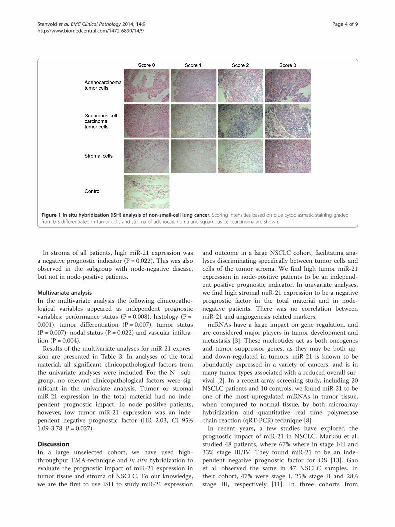
Figure 1 In situ hybridization (ISH) analysis of non-small-cell lung cancer. Scoring intensities based on blue cytoplasmatic staining gradedfrom 0-3 differentiated in tumor cells and stroma of adenocarcinoma and squamous cell carcinoma are shown.
Stenvold et al. BMC Clinical Pathology 2014, 14:9 Page 4 of 9http://www.biomedcentral.com/1472-6890/14/9
In stroma of all patients, high miR-21 expression wasa negative prognostic indicator (P = 0.022). This was alsoobserved in the subgroup with node-negative disease,but not in node-positive patients.
Multivariate analysisIn the multivariate analysis the following clinicopatho-logical variables appeared as independent prognosticvariables: performance status (P = 0.008), histology (P =0.001), tumor differentiation (P = 0.007), tumor status(P = 0.007), nodal status (P = 0.022) and vascular infiltra-tion (P = 0.004).Results of the multivariate analyses for miR-21 expres-
sion are presented in Table 3. In analyses of the totalmaterial, all significant clinicopathological factors fromthe univariate analyses were included. For the N + sub-group, no relevant clinicopathological factors were sig-nificant in the univariate analysis. Tumor or stromalmiR-21 expression in the total material had no inde-pendent prognostic impact. In node positive patients,however, low tumor miR-21 expression was an inde-pendent negative prognostic factor (HR 2.03, CI 95%1.09-3.78, P = 0.027).
DiscussionIn a large unselected cohort, we have used high-throughput TMA-technique and in situ hybridization toevaluate the prognostic impact of miR-21 expression intumor tissue and stroma of NSCLC. To our knowledge,we are the first to use ISH to study miR-21 expression
and outcome in a large NSCLC cohort, facilitating ana-lyses discriminating specifically between tumor cells andcells of the tumor stroma. We find high tumor miR-21expression in node-positive patients to be an independ-ent positive prognostic indicator. In univariate analyses,we find high stromal miR-21 expression to be a negativeprognostic factor in the total material and in node-negative patients. There was no correlation betweenmiR-21 and angiogenesis-related markers.miRNAs have a large impact on gene regulation, and
are considered major players in tumor development andmetastasis [3]. These nucleotides act as both oncogenesand tumor suppressor genes, as they may be both up-and down-regulated in tumors. miR-21 is known to beabundantly expressed in a variety of cancers, and is inmany tumor types associated with a reduced overall sur-vival [2]. In a recent array screening study, including 20NSCLC patients and 10 controls, we found miR-21 to beone of the most upregulated miRNAs in tumor tissue,when compared to normal tissue, by both microarrayhybridization and quantitative real time polymerasechain reaction (qRT-PCR) technique [8].In recent years, a few studies have explored the
prognostic impact of miR-21 in NSCLC. Markou et al.studied 48 patients, where 67% where in stage I/II and33% stage III/IV. They found miR-21 to be an inde-pendent negative prognostic factor for OS [13]. Gaoet al. observed the same in 47 NSCLC samples. Intheir cohort, 47% were stage I, 25% stage II and 28%stage III, respectively [11]. In three cohorts from

Table 1 Patient characteristics and their variables aspredictors for disease-specific survival in 335 NSCLCpatients (univariate analyses; log-rank test)
CharacteristicsPatients Median
survivalmonth
5-yearsurvival % P
n (91)
Age
≤65years 156 (47) 98 550.42
>65 years 179 (53) NR 60
Sex
Female 82 (24) 190 640.22
Male 253 (76) 98 56
Smoking
Never 15 (5) 19 43
0.26Current 215 (64) NR 60
Former 105 (31) 84 55
Performance status
PS 0 197 (59) NR 63
0.016PS 1 120 (36) 64 52
PS 2 18 (5) 25 33
Weight loss
<10% 303 (90) 190 580.76
>10% 32 (10) 98 57
Histology
SCC 191 (57) NR 66
0.028Adenocarcinoma 113 (34) 54 46
LCC 31 (9) 98 56
Differentiation
Poor 138 (41) 47 47
<0.001Moderate 144 (43) 190 65
Well 53 (16) NR 68
Surgical procedure
Lobectomy + Wedge* 243 (73) 190 620.007
Pitunionectomy 92 (27) 37 47
Pathological stage
I 157 (47) NR 61
<0.001II 136 (40) 62 51
IIIa 42 (13) 17 23
Tumor status
1 85 (25) 190 75
<0.0012 188 (56) 84 57
3 62 (19) 25 36
Nodal status
0 232 (69) NR 67
<0.0011 76 (23) 35 43
2 27 (8) 18 18
Table 1 Patient characteristics and their variables aspredictors for disease-specific survival in 335 NSCLCpatients (univariate analyses; log-rank test) (Continued)
Surgical margins
Free 3)7 (92) 190 590.37
Not free 28 (8) 47 48
Vascular infiltration
No 284 (85) 190 620.001
Yes 51 (15) 27 33
*Wedge, n= 10.Abbreviations: NR not reached, PS performance status, SCC squanos cellcarcinoma, LCC large-cell carcinoma.Statistically significant results in bold font.
Stenvold et al. BMC Clinical Pathology 2014, 14:9 Page 5 of 9http://www.biomedcentral.com/1472-6890/14/9
Maryland, US (64% stage I, 25% stage II, 11% stageIII), Norway (57% stage I, 14% stage II, 29% stage III),and Japan (74% stage I, 26% stage II), Saito and col-leagues showed miR-21 to be an independent negativeprognostic factor. In the Norwegian and American ma-terial, overall survival was the endpoint, while in theJapanese cohort relapse free survival was [14]. Landiand colleagues used an oligo array with 440 humanmiRNAs to evaluate differences in miRNA expressiondepending on histology and clinical outcome in 290NSCLC tissues, constituted by 40% stage I, 29% stageII, 26% stage III and 4% stage IV cancers. They foundmiR-21 to differentiate between adenocarcinoma andsquamous cell carcinoma, but there was no differencein survival according to miR-21 expression rate [12].In a large study on 639 patients (35% stage I, 23%stage II and 42% stage III), Voortman et al. observedno prognostic impact of miR-21 on NSCLC survival.There was a tendency towards a better prognosis forhigh miR-21 expression, but this finding was not sig-nificant (P = 0.06) [15].There were some differences in methodology between
these studies, as fresh frozen tissue was used in three[11,13,14] and paraffin-embedded material in two stud-ies [12,15]. For quantification of miRNA, all except theLandi study used qRT-PCR. In the studies where miR-21was associated with a worse prognosis, subgroup ana-lyses were not performed. The Landi and Gao studies[11,12] were numerously too small and the Saito studyconsisting of three cohorts (89, 37 and 189 patients re-spectively) [14], was not suited for subgroup analyses.Yang et al. performed a meta-analysis based on the
studies mentioned above. They also included two otherstudies analyzing miR-21 in serum. The conclusion wasthat high miR-21 expression was significantly associatedwith poor survival [23].When using the summarized score of tumor cell and
stromal expression in the whole cohort, miR-21 waswithout any significant prognostic impact. In the stromal
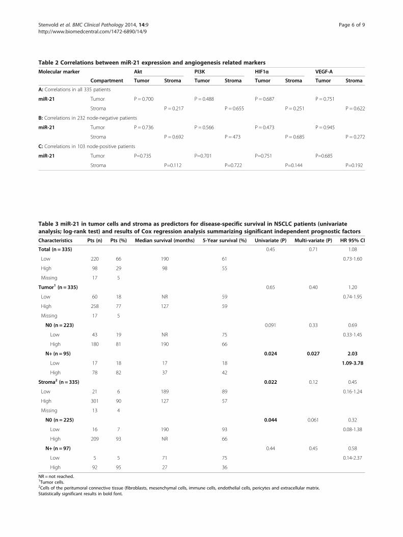
Table 2 Correlations between miR-21 expression and angiogenesis related markers
Molecular marker Akt PI3K HIF1α VEGF-A
Compartment Tumor Stroma Tumor Stroma Tumor Stroma Tumor Stroma
A: Correlations in all 335 patients
miR-21 Tumor P = 0.700 P = 0.488 P = 0.687 P = 0.751
Stroma P = 0.217 P = 0.655 P = 0.251 P = 0.622
B: Correlations in 232 node-negative patients
miR-21 Tumor P = 0.736 P = 0.566 P = 0.473 P = 0.945
Stroma P = 0.692 P = 473 P = 0.685 P = 0.272
C: Correlations in 103 node-positive patients
miR-21 Tumor P=0.735 P=0.701 P=0.751 P=0.685
Stroma P=0.112 P=0.722 P=0.144 P=0.192
Table 3 miR-21 in tumor cells and stroma as predictors for disease-specific survival in NSCLC patients (univariateanalysis; log-rank test) and results of Cox regression analysis summarizing significant independent prognostic factors
Characteristics Pts (n) Pts (%) Median survival (months) 5-Year survival (%) Univariate (P) Multi-variate (P) HR 95% CI
Total (n = 335) 0.45 0.71 1.08
Low 220 66 190 61 0.73-1.60
High 98 29 98 55
Missing 17 5
Tumor1 (n = 335) 0.65 0.40 1.20
Low 60 18 NR 59 0.74-1.95
High 258 77 127 59
Missing 17 5
N0 (n = 223) 0.091 0.33 0.69
Low 43 19 NR 75 0.33-1.45
High 180 81 190 66
N+ (n = 95) 0.024 0.027 2.03
Low 17 18 17 18 1.09-3.78
High 78 82 37 42
Stroma2 (n = 335) 0.022 0.12 0.45
Low 21 6 189 89 0.16-1.24
High 301 90 127 57
Missing 13 4
N0 (n = 225) 0.044 0.061 0.32
Low 16 7 190 93 0.08-1.38
High 209 93 NR 66
N+ (n = 97) 0.44 0.45 0.58
Low 5 5 71 75 0.14-2.37
High 92 95 27 36
NR = not reached.1Tumor cells.2Cells of the peritumoral connective tissue (fibroblasts, mesenchymal cells, immune cells, endothelial cells, pericytes and extracellular matrix.Statistically significant results in bold font.
Stenvold et al. BMC Clinical Pathology 2014, 14:9 Page 6 of 9http://www.biomedcentral.com/1472-6890/14/9
Figure 2 Disease-specific survival curves according to expression of A) miR-21 in stroma, B) miR-21 in tumor, C) miR-21 in tumor innode negative patients and D) miR-21 in tumor in node positive patients.
Stenvold et al. BMC Clinical Pathology 2014, 14:9 Page 7 of 9http://www.biomedcentral.com/1472-6890/14/9
compartment, miR-21 expression was a negative prog-nosticator in univariate, but not in the multivariate ana-lysis. In the studies mentioned above [11,13,14], exceptfor the studies by Voortman [15] and Landi [12], miR-21appears to be a negative prognostic factor in NSCLC,corroborating our stromal results. In contrast, we foundmiR-21 expression in tumor cells to be an independentpositive prognostic factor in node positive lung cancerpatients. One may speculate if the differences we ob-serve between our results and some of the other studiesare caused by the different methodologies used. As weuse ISH-technique, miR-21 expression can be assessedseparately in tumor and stromal cells. When using theqRT-PCR method without prior microdissection, itcan not be differentiated between the tumor cells andthe stromal compartment. Gregg and colleagues per-formed microdissection on a prostate cancer material toseparate tumor and stromal cells, and showed a largedifference regarding gene expression between the two
compartments [24]. Our findings show that there is aprognostic difference in expression between the twocompartments. Using qRT-PCR the contribution of miR-21 from the stromal compartment may override the con-tribution from tumor cells, especially at significant dif-ferences in expression. Consequently, the data willreflect the situation in stroma, and a divergent situationin the tumor cells will not be detected. The conclusionin the Yang meta-analysis reflects the findings in thesestudies, and does not take into account possible expres-sion differences between the compartments.The mechanistic functions of miR-21 are still being ex-
plored, but some functions have recently been suggested.In human umbilical vein endothelial cells (HUVECs),Sabatel and colleagues found miR-21 to be a potentialinhibitor of angiogenesis via inhibition of RhoB, resultingin a reduction in endothelial proliferation, migrationand vessel formation [10]. On the other hand, Liu et al.demonstrated miR-21 to induce angiogenesis in human

Stenvold et al. BMC Clinical Pathology 2014, 14:9 Page 8 of 9http://www.biomedcentral.com/1472-6890/14/9
prostate cancer cells through up-regulating HIF-1α andVEGF and through activating the Akt and ERK pathways[9]. Hence, miR-21 may have both pro- and anti-angiogenic functions. Angiogenesis is an important traitof cancer progression [25], and inhibiting angiogenesismay contribute to slow down cancer growth. In the lightof these opposing findings and our data, we may specu-late if miR-21 has opposite impacts in different stages ofthe disease. Could high miR-21 in early stages (node-negative) act pro-angiogenic and contribute to a fasterprogression, while in a node positive stage, it acts anti-angiogenic and protects against further progression?We did not find any correlations between miR-21 and
angiogenic markers of pathways earlier described formiR-21 and angiogenesis [9,10]. These pathways are pos-sibly tissue and cell type specific. Studies exploring miR-21 as a modulator of angiogenesis have in some casesused endothelial cells in their models [10]. In our mater-ial, we have not specifically studied endothelial cells,so the connection between miR-21 and angiogeneticmarkers seen in endothelial cells will not necessarily bemirrored by our tissue samples as we assessed the sumoff all stromal cell types.
ConclusionWe found tumor cell miR-21 expression to be an inde-pendent positive prognostic factor in node-positiveNSCLC. In the total material, stromal miR-21 expressionwas a negative prognostic factor in univariate analysis.We identified diverging impacts of miR-21 related to cellcompartment and nodal status. These findings should befurther explored, and may have implications for the fu-ture use of miR-21 in diagnostics and therapy.
Competing interestsThe authors declare that they have no competing interests.
Authors’ contributionsHS participated in the design of the study, contributed to the clinical anddemographic database, did the statistical analysis and drafted themanuscript. TD, SA and SAS contributed to the clinical and demographicdatabase and SAS and MIP in making the TMAs. TD and SA contributedto the statistical analysis. SAS and AV scored the cores. MIP carried out theISH. RB and LTB supervised and participated in the study design, resultinterpretation and writing. All authors read and approved the finalmanuscript.
AcknowledgementsThe study was solely funded by the Northern Norway Regional HealthAuthority (Helse Nord RHF) which is responsible for the public hospitals innorthern Norway. The funders had no role in study design, data collectionand analysis, decision to publish, or preparation of the manuscript.
Author details1Institute of Clinical Medicine, University of Tromso, Tromso, Norway.2Department of Oncology, University Hospital of North Norway, Tromso9038, Norway. 3Institute of Medical Biology, University of Tromso, Tromso,Norway. 4Dept of Clinical Pathology, University Hospital of North Norway,Tromso, Norway.
Received: 16 May 2013 Accepted: 27 January 2014Published: 13 February 2014
References1. Jemal A, Siegel R, Xu J, Ward E: Cancer statistics, 2010. CA Cancer J Clin
2010, 60:277–300.2. Fu X, Han Y, Wu Y, Zhu X, Lu X, Mao F, et al: Prognostic role of microRNA-
21 in various carcinomas: a systematic review and Meta-analysis. Eur JClin Invest 2011, 41:1245–53.
3. Iorio MV, Croce CM: MicroRNAs in cancer: small molecules with a hugeimpact. J Clin Oncol 2009, 27:5848–5856.
4. Kasinski AL, Slack FJ: Epigenetics and genetics. MicroRNAs en route to theclinic: progress in validating and targeting microRNAs for cancertherapy. Nat Rev Cancer 2011, 11:849–864.
5. Krichevsky AM, Gabriely G: miR-21: a small multi-faceted RNA. J Cell MolMed 2009, 13:39–53.
6. Olson P, Lu J, Zhang H, Shai A, Chun MG, Wang Y, et al: MicroRNAdynamics in the stages of tumorigenesis correlate with hallmarkcapabilities of cancer. Genes Dev 2009, 23:2152–2165.
7. Suarez Y, Sessa WC: MicroRNAs as novel regulators of angiogenesis.Circ Res 2009, 104:442–454.
8. Donnem T, Fenton CG, Lonvik K, Berg T, Eklo K, Andersen S, et al: MicroRNASignatures in Tumor Tissue Related to Angiogenesis in Non-Small CellLung Cancer. PLoS One 2012, 7:e29671.
9. Liu LZ, Li C, Chen Q, Jing Y, Carpenter R, Jiang Y, et al: MiR-21 inducedangiogenesis through AKT and ERK activation and HIF-1alphaexpression. PLoS One 2011, 6:e19139.
10. Sabatel C, Malvaux L, Bovy N, Deroanne C, Lambert V, Gonzalez ML, et al:MicroRNA-21 exhibits antiangiogenic function by targeting RhoBexpression in endothelial cells. PLoS One 2011, 6:e16979.
11. Gao W, Yu Y, Cao H, Shen H, Li X, Pan S, et al: Deregulated expression ofmiR-21, miR-143 and miR-181a in non small cell lung cancer is related toclinicopathologic characteristics or patient prognosis. BiomedPharmacother 2010, 64:399–408.
12. Landi MT, Zhao Y, Rotunno M, Koshiol J, Liu H, Bergen AW, et al: MicroRNAexpression differentiates histology and predicts survival of lung cancer.Clin Cancer Res 2010, 16:430–441.
13. Markou A, Tsaroucha EG, Kaklamanis L, Fotinou M, Georgoulias V,Lianidou ES: Prognostic value of mature microRNA-21 and microRNA-205 overexpression in non-small cell lung cancer by quantitativereal-time RT-PCR. Clin Chem 2008, 54:1696–1704.
14. Saito M, Schetter AJ, Mollerup S, Kohno T, Skaug V, Bowman ED, et al:The association of microRNA expression with prognosis andprogression in early-stage, non-small cell lung adenocarcinoma:a retrospective analysis of three cohorts. Clin Cancer Res 2011,17:1875–1882.
15. Voortman J, Goto A, Mendiboure J, Sohn JJ, Schetter AJ, Saito M, et al:MicroRNA expression and clinical outcomes in patients treated withadjuvant chemotherapy after complete resection of non-small cell lungcarcinoma. Cancer Res 2010, 70:8288–8298.
16. Dillhoff M, Liu J, Frankel W, Croce C, Bloomston M: MicroRNA-21 isoverexpressed in pancreatic cancer and a potential predictor of survival.J Gastrointest Surg 2008, 12:2171–2176.
17. Nielsen BS, Jorgensen S, Fog JU, Sokilde R, Christensen IJ, Hansen U, et al:High levels of microRNA-21 in the stroma of colorectal cancers predictshort disease-free survival in stage II colon cancer patients. Clin ExpMetastasis 2011, 28:27–38.
18. Andersen S, Eilertsen M, Donnem T, Al-Shibli K, Al-Saad S, Busund LT, et al:Diverging prognostic impacts of hypoxic markers according to NSCLChistology. Lung Cancer 2011, 72:294–302.
19. Al-Saad S, Donnem T, Al-Shibli K, Persson M, Bremnes RM, Busund LT:Diverse prognostic roles of Akt isoforms, PTEN and PI3K in tumorepithelial cells and stromal compartment in non-small cell lung cancer.Anticancer Res 2009, 29:4175–4183.
20. Donnem T, Al-Saad S, Al-Shibli K, Delghandi MP, Persson M, Nilsen MN, et al:Inverse prognostic impact of angiogenic marker expression in tumorcells versus stromal cells in non small cell lung cancer. Clin Cancer Res2007, 13:6649–6657.
21. Rami-Porta R, Chansky K, Goldstraw P: Updated lung cancer stagingsystem. Future Oncol 2009, 5:1545–1553.
Stenvold et al. BMC Clinical Pathology 2014, 14:9 Page 9 of 9http://www.biomedcentral.com/1472-6890/14/9
22. Jorgensen S, Baker A, Moller S, Nielsen BS: Robust one-day in situhybridization protocol for detection of microRNAs in paraffin samplesusing LNA probes. Methods 2010, 52:375–381.
23. Yang M, Shen H, Qiu C, Ni Y, Wang L, Dong W, et al: High expression ofmiR-21 and miR-155 predicts recurrence and unfavourable survival innon-small cell lung cancer. Eur J Cancer 2012, 49:604–15.
24. Gregg JL, Brown KE, Mintz EM, Piontkivska H, Fraizer GC: Analysis of geneexpression in prostate cancer epithelial and interstitial stromal cellsusing laser capture microdissection. BMC Cancer 2010, 10:165.
25. Hanahan D, Weinberg RA: Hallmarks of cancer: the next generation.Cell 2011, 144:646–674.
doi:10.1186/1472-6890-14-9Cite this article as: Stenvold et al.: High tumor cell expression ofmicroRNA-21 in node positive non-small cell lung cancer predicts afavorable clinical outcome. BMC Clinical Pathology 2014 14:9.
Submit your next manuscript to BioMed Centraland take full advantage of:
• Convenient online submission
• Thorough peer review
• No space constraints or color figure charges
• Immediate publication on acceptance
• Inclusion in PubMed, CAS, Scopus and Google Scholar
• Research which is freely available for redistribution
Submit your manuscript at www.biomedcentral.com/submit