High expression of HLA-E in colorectal carcinoma is associated with a favorable prognosis
10
RESEARCH Open Access High expression of HLA-E in colorectal carcinoma is associated with a favorable prognosis Maria Benevolo 1 , Marcella Mottolese 1 , Elisa Tremante 2 , Francesca Rollo 1 , Maria Grazia Diodoro 1 , Cristiana Ercolani 1 , Isabella Sperduti 3 , Elisa Lo Monaco 2 , Maurizio Cosimelli 4 and Patrizio Giacomini 2* Abstract Background: Human Leukocyte Antigen (HLA)-E is a non-classical class I HLA molecule that can be stabilized by ligands donated by other classical (HLA-A, -B, -C) and non-classical (HLA-G) family members. HLA-E engages a variety of immune receptors expressed by cytotoxic T lymphocytes (CTLs), Natural killer (NK) cells and NK-CTLs. In view of the opposing outcomes (activation or inhibition) of the different HLA-E receptors, the preferred role (if any) of HLA-E expressed in vivo on tumor cells remains to be established. Methods: Taking advantage of MEM-E/02, a recently characterized antibody to denatured HLA-E molecules, HLA-E expression was assessed by immunohistochemistry on an archival collection (formalin-fixed paraffin-embedded) of 149 colorectal primary carcinoma lesions paired with their morphologically normal mucosae. Lymphoid infiltrates were assessed for the expression of the HLA-E-specific, inhibitory, non-rearranging receptor NKG2A. Results: High HLA-E expression did not significantly correlate with the expression of classical HLA-B and HLA-C molecules, but it did correlate with high expression of its preferential ligand donor HLA-A. In addition, it correlated with lymphoid cell infiltrates expressing the inhibitory NKG2A receptor, and was an independent predictor of good prognosis, particularly in a subset of patients whose tumors express HLA-A levels resembling those of their paired normal counterparts (HLA-A). Thus, combination phenotypes (HLA-E lo-int /HLA-AE and HLA-E hi /HLA-AE) of classical and non-classical class I HLA molecules mark two graded levels of good prognosis. Conclusions: These results suggest that HLA-E favors activating immune responses to colorectal carcinoma. They also provide evidence in humans that tumor cells entertain extensive negotiation with the immune system until a compromise between recognition and escape is reached. It is implied that this process occurs stepwise, as predicted by the widely accepted ‘immunoediting’ model. Background Human Leukocyte Antigen (HLA)-E is a cell surface, non-classical Major Histocompatibility class I molecule recognized by immune receptors expressed by cytotoxic T lymphocytes (CTLs), Natural Killer (NK) cells, and the more recently described subset of NK-CTLs. These receptors are either inhibitory or activating [1-3]. Inhibition, on the one hand, results from the engage- ment of the NKG2A receptor with HLA-E heavy chains that have been stabilized upon heterotrimeric assembly with their light chain subunit, called b 2 -microglobulin ( b 2 m), and peptide ligands derived from the signal sequences of ‘permissive’ class I heavy chains, both classi- cal (HLA-A, -B, -C) and non-classical (HLA-G). Activa- tion, on the other hand, results from: (a) the competitive relief of NKG2A-mediated inhibition upon HLA-E assembly with peptides from donor proteins other than HLA class I; (b) the direct engagement of the activating NKG2C receptor isoform; and (c) antigen-specific recog- nition through the T cell receptor (TcR) expressed by NK-CTLs [1-4]. Balancing and integration of opposing signals (often dubbed activation-inhibition) is not unique of HLA-E, but is indeed widely adopted to control cyto- toxic responses and regulate complex immune networks. Thus, HLA-E may provide key information to understand how virus-infected [5] and tumor cells walk the thin line between immune surveillance and immune evasion. * Correspondence: [email protected] 2 Laboratory of Immunology, Regina Elena National Cancer Institute, Via delle Messi d’Oro 156, 00158 Rome, Italy Full list of author information is available at the end of the article Benevolo et al. Journal of Translational Medicine 2011, 9:184 http://www.translational-medicine.com/content/9/1/184 © 2011 Benevolo et al; licensee BioMed Central Ltd. This is an Open Access article distributed under the terms of the Creative Commons Attribution License (http://creativecommons.org/licenses/by/2.0), which permits unrestricted use, distribution, and reproduction in any medium, provided the original work is properly cited.
-
Upload
independent -
Category
Documents
-
view
1 -
download
0
Transcript of High expression of HLA-E in colorectal carcinoma is associated with a favorable prognosis

RESEARCH Open Access
High expression of HLA-E in colorectal carcinomais associated with a favorable prognosisMaria Benevolo1, Marcella Mottolese1, Elisa Tremante2, Francesca Rollo1, Maria Grazia Diodoro1, Cristiana Ercolani1,Isabella Sperduti3, Elisa Lo Monaco2, Maurizio Cosimelli4 and Patrizio Giacomini2*
Abstract
Background: Human Leukocyte Antigen (HLA)-E is a non-classical class I HLA molecule that can be stabilized byligands donated by other classical (HLA-A, -B, -C) and non-classical (HLA-G) family members. HLA-E engages avariety of immune receptors expressed by cytotoxic T lymphocytes (CTLs), Natural killer (NK) cells and NK-CTLs. Inview of the opposing outcomes (activation or inhibition) of the different HLA-E receptors, the preferred role (if any)of HLA-E expressed in vivo on tumor cells remains to be established.
Methods: Taking advantage of MEM-E/02, a recently characterized antibody to denatured HLA-E molecules, HLA-Eexpression was assessed by immunohistochemistry on an archival collection (formalin-fixed paraffin-embedded) of149 colorectal primary carcinoma lesions paired with their morphologically normal mucosae. Lymphoid infiltrateswere assessed for the expression of the HLA-E-specific, inhibitory, non-rearranging receptor NKG2A.
Results: High HLA-E expression did not significantly correlate with the expression of classical HLA-B and HLA-Cmolecules, but it did correlate with high expression of its preferential ligand donor HLA-A. In addition, it correlatedwith lymphoid cell infiltrates expressing the inhibitory NKG2A receptor, and was an independent predictor of goodprognosis, particularly in a subset of patients whose tumors express HLA-A levels resembling those of their pairednormal counterparts (HLA-A). Thus, combination phenotypes (HLA-Elo-int/HLA-AE and HLA-Ehi/HLA-AE) of classicaland non-classical class I HLA molecules mark two graded levels of good prognosis.
Conclusions: These results suggest that HLA-E favors activating immune responses to colorectal carcinoma. Theyalso provide evidence in humans that tumor cells entertain extensive negotiation with the immune system until acompromise between recognition and escape is reached. It is implied that this process occurs stepwise, aspredicted by the widely accepted ‘immunoediting’ model.
BackgroundHuman Leukocyte Antigen (HLA)-E is a cell surface,non-classical Major Histocompatibility class I moleculerecognized by immune receptors expressed by cytotoxicT lymphocytes (CTLs), Natural Killer (NK) cells, and themore recently described subset of NK-CTLs. Thesereceptors are either inhibitory or activating [1-3].Inhibition, on the one hand, results from the engage-
ment of the NKG2A receptor with HLA-E heavy chainsthat have been stabilized upon heterotrimeric assemblywith their light chain subunit, called b2-microglobulin(b2m), and peptide ligands derived from the signal
sequences of ‘permissive’ class I heavy chains, both classi-cal (HLA-A, -B, -C) and non-classical (HLA-G). Activa-tion, on the other hand, results from: (a) the competitiverelief of NKG2A-mediated inhibition upon HLA-Eassembly with peptides from donor proteins other thanHLA class I; (b) the direct engagement of the activatingNKG2C receptor isoform; and (c) antigen-specific recog-nition through the T cell receptor (TcR) expressed byNK-CTLs [1-4]. Balancing and integration of opposingsignals (often dubbed activation-inhibition) is not uniqueof HLA-E, but is indeed widely adopted to control cyto-toxic responses and regulate complex immune networks.Thus, HLA-E may provide key information to understandhow virus-infected [5] and tumor cells walk the thin linebetween immune surveillance and immune evasion.
* Correspondence: [email protected] of Immunology, Regina Elena National Cancer Institute, Via delleMessi d’Oro 156, 00158 Rome, ItalyFull list of author information is available at the end of the article
Benevolo et al. Journal of Translational Medicine 2011, 9:184http://www.translational-medicine.com/content/9/1/184
© 2011 Benevolo et al; licensee BioMed Central Ltd. This is an Open Access article distributed under the terms of the CreativeCommons Attribution License (http://creativecommons.org/licenses/by/2.0), which permits unrestricted use, distribution, andreproduction in any medium, provided the original work is properly cited.
HLA-A, -B, -C down-regulation has been viewed as amajor subterfuge to deceive T cells [6] for some timenow, but it is unlikely to provide a comprehensive expla-nation of immune evasion, since it impairs ligand dona-tion to HLA-E and the direct engagement of inhibitoryNK receptors [1,7-9]. Accordingly, several immunohisto-chemical studies failed to confirm an association betweenHLA-A, -B, - C loss and poor prognosis [10-12], and ourown studies were consistent with activation-inhibitionmodels [13-16]. We showed that early-passage mela-noma, breast carcinoma, and lung carcinoma cells, likevirus-infected cells, avoid both extremes of overly low orhigh HLA class I expression, which would expose themto lysis by NK and CTLs, respectively. Similar ‘low pro-file’ HLA phenotypes were also observed in vivo, in color-ectal carcinoma lesions, and were associated with afavorable prognosis, whereas extreme down-and up-regu-lation with respect to the normal autologous mucosawere rare and associated with a poor prognosis, particu-larly when involving the HLA-A locus [16]. Possibly,these altered HLA phenotypes mark tumor cells refrac-tory to immune elimination.In an effort to characterize monoclonal antibodies
(mAbs) to HLA-E, we found that MEM-E/02 binds a lin-ear epitope highly restricted to the HLA-E polypeptideand fully available upon denaturation [17]. Using MEM-E/02, we recently observed that HLA-E is constitutivelyco-expressed with HLA-A, -B, -C molecules in a fractionof neoplastic tissues and on the surface of most culturedtumor cell lines. In these conditions, HLA-E is functional[18]. In the present report, we describe the use of MEM-E/02 on archival collections of formalin-fixed/paraffin-embedded colorectal adenocarcinoma tissues and theirpaired, morphologically normal mucosae. We found thatoverall HLA-E expression is a significant prognosticator,and in combination with HLA-A expression and the pre-sence of NKG2A infiltrating lymphoid cells provides aclue to understand tumor immune surveillance in vivo.
MethodsPatients and histological specimensPatients (149 cases, 71 men and 78 women, median age64, range 34-90 years) were radically resected for primarycolorectal adenocarcinoma between 1988 and 2000.Pathological staging [19], performed according to theAmerican Joint Committee on Cancer (AJCC)/Interna-tional Union Against Cancer (UICC), identified 98 and51 tumors at stages II and III, respectively. The latterwere treated by adjuvant chemotherapy. According to theWHO classification, well (G1), moderately (G2), andpoorly (G3) differentiated tumors were 10, 114, and 25,respectively. T2, T3 and T4 tumors were 26, 63, and 60,respectively. The present archival tissue collection largelyoverlaps (n = 126) with that previously described [16].
Uninvolved mucosal tissues were obtained from the sametissue block as the tumor lesion in 108 cases, and from adistinct tissue block in 41 cases. No patients were lost toclinical follow-up (continuously updated), and the med-ian follow-up was 71.0 (range 3.0 - 175.1) months, with45 relapses and 37 deaths due to cancer-related causes.This study was approved by the Ethical Committee.
Antibodies and ImmunohistochemistryMEM-E/02 is the only available mAb to HLA-E with anextensive biochemical characterization. It binds a linearepitope present on HLA-E but not HLA-A, -B, -C, -F or-G heavy chains [17]. HCA2 binds all the denaturedHLA-A alleles except A24, and crossreacts with the rareHLA-B73 allele, HLA-G, HLA-E and HLA-F [20,21].However, HLA-E and HLA-F bind weakly and almostexclusively under native conditions [21], and HLA-Gaccounts for less than 5% of the total HCA2 reactivity ina large cell panel [15]. HC10 binds HLA-B and weaklycrossreacts with HLA-C, whereas L31 binds HLA-C anda few crossreacting HLA-B alleles [20,22,23]. CD8 andCD56 were identified by mAbs 1A5 and 1B6 (Novocastra,UK), respectively. The goat polyclonal antibody N-19 toNKG2A was from Santa Cruz (CA, USA). Sections (5-μmthick) from routinely formalin-fixed (18-24 h) paraffin-embedded tissue blocks were stained by Immunohisto-chemistry (IHC). Antigen retrieval for HCA2, HC10, L31,CD8 and CD56 staining was carried out as described[23]. For MEM-E/02 and NKG2A staining, slides weredewaxed, rehydrated and heated in a water bath at 96°Cfor 40 minutes in EDTA (pH 8.0), followed by cooling atroom temperature. Staining was revealed by a supersensi-tive streptavidin-biotin immunoperoxidase system (Bio-genex, Menarini, Italy) for all antibodies. In addition,NKG2A staining was also revealed by a Goat AP-PolymerKit (Biocare Medical, CA, USA). The chromogenic sub-strate was 3,3’-diaminobenzidine for all stainings. Inaddition, NKG2A staining was also revealed by WarpRed (Biocare Medical). Preliminary specificity assess-ments were performed on a panel of HLA-E-positive andHLA-E-negative cell lines [17] fixed and embedded asabove. Different staining sessions included repeatedstaining of an internal normalization control (a sectionfrom a known specimen) to ensure day-to-day consis-tency. Because weak staining was invariably associatedwith the presence of negative areas, whereas strong stain-ing tended to be homogeneous (this applies to both nor-mal and tumor tissues), staining heterogeneity andstaining intensity were factored into a single scale(termed ’absolute’ intensity score hereafter). The absoluteintensity score may acquire one of four possible values: 0(undetectable or faint in <20% of the cells); 1 (faint toweak in 20% but ≤70% of the cells); 2 (weak to moderatein >70% of the cells); 3 (intense in>70% of the cells).
Benevolo et al. Journal of Translational Medicine 2011, 9:184http://www.translational-medicine.com/content/9/1/184
Page 2 of 10

In analogy with our previously cited study [16], we calcu-lated an additional intensity scale (termed ’relative’ here-after) by subtracting the absolute score of the normalmucosa from that of its paired colon carcinoma speci-men. Subtraction of two series of scores, each graded ona four-digit scale, resulted in the assignment of one of 7possible algebraic values (-3, -2, -1, 0, +1, +2, +3) to eachnormal-neoplastic pair. Intermediate (-1, 0, and +1) andextreme (-3, -2, +2, and +3) relative scores identify pairsin which the tumor is similar to and deviates from,respectively, its normal paired mucosa. CD3, CD56 andNKG2A infiltrates were assessed in six randomly chosenhigh power fields (HPF, ×400), and expressed as eitherthe percentage of positive cells in the total infiltrate, orthe average number of positive cells in each HPF.
Statistical analysisDescriptive statistics were used to summarize pertinentstudy information. The Spearman rho correlation wasused to determine relationships between parameters. TheHazard risk and the confidence limits were estimated foreach variable using the Cox univariate model. Signifi-cance was defined at the p < 0.05 level. A multivariateCox proportional hazard model was also developed usingstepwise regression (forward selection) by selecting thosepredictive variables that were significant upon univariateanalysis. Enter limit and remove limit were p = 0.10 andp = 0.15 respectively. Survival was calculated by theKaplan-Meier product-limit method from the date of sur-gery until the time of death (overall survival; OS), relapse(disease-free survival; DFS), or last visit (OS and DFS),whichever applicable. The log-rank test was used toassess differences between subgroups. Significance wasdefined at the p < 0.05 level. Statistical analysis was car-ried out by the SPSS v. 13.0 (SPSS, Milan, Italy) software.Optimal cut-offs of NKG2A immunoreactivity definingtwo groups of patients with significantly different MEM-E/02 intensity scores were found using the ReceiverOperating Characteristic (ROC) curve analysis.
HLA-A, -B, -C typingGenomic DNAs were extracted from the available frozentumor tissue samples (n = 29), and typed for HLA-A, -B,and -C using sequence-specific primers (Olerup-SSP,Genovision, Vienna, Austria).
ResultsAnalysis of MEM-E/02 immunoreactivity in colorectalcarcinomas and paired mucosa specimensColorectal carcinomas from stage II/III patients (n = 149),each paired with morphologically normal colonic mucosafrom the same patient, were stained by immunohisto-chemistry with the HLA-E-specific MEM-E/02 antibody.Representative examples of staining grades (recorded as
absolute scores, see Materials and Methods) are providedin Figure 1A-C. HLA-E was detected in nearly all the spe-cimens, approximately half of which displayed the highestabsolute score of 3. High absolute scores were most fre-quent also with HCA2 (to HLA-A), HC10 (to HLA-B) andL31 (to HLA-C), as shown in Additional file 1, Fig. S1A.Similar results were obtained by plotting the stainingintensities of the normal mucosae (not shown). Thus,HLA-E is expressed in essentially all normal and neoplas-tic colonic tissues, often at high levels.Next, we calculated the seven-digit relative intensity
scores (see Materials and Methods) of paired immunohis-tochemical determinations. Representative results (Addi-tional file 1, Fig. S1B) demonstrate that tumors resemblingtheir normal paired mucosa (relative scores: +1, 0, and -1)were far more numerous than tumors undergoing consis-tent up-regulation or down-regulation (relative scores: -2,-3 and +2, +3), a finding that applies to all the tested HLAclass I molecules: HLA-E, HLA-A, HLA-B and HLA-C.Finally, flow cytometry of 3 representative colorectal car-
cinoma cell lines provided evidence that at least some ofthe denatured HLA-A and HLA-E molecules detected byHCA2 and MEM-E/02 on archival tissues derive fromnative molecules that are co-expressed on the surface oflive cells (Additional file 1, Fig. S2).
Co-expression of HLA-E and permissive class I allelesIt has been shown that signal sequence-derived peptidesfrom permissive alleles (most HLA-A and some HLA-C)carry methionine at P2, whereas peptides from non-per-missive alleles (most HLA-B) carry threonine [24,25]. Onthis basis, absolute HLA-E levels are expected to correlatewith (a) the levels of co-expressed products from at leastsome HLA-A, -B, and -C loci, and (b) the number of per-missive alleles carried at the DNA level. To verify predic-tion (a), the staining intensities of MEM-E/02 (to HLA-E)were correlated with the staining intensities of HCA2,HC10, and L31 (preferentially reactive with HLA-A, HLA-B and HLA-C, respectively) in the 149 tumor specimens.A significant (Table 1, p = 0.01) correlation was detectedbetween HLA-E and HLA-A, but not HLA-B or HLA-C.To verify prediction (b), 29 high-quality genomic DNAs(required for reliable allele-specific PCR) from colon carci-noma lesions were typed for HLA-A, -B, -C. From 3 to 6permissive alleles (alleles and signal sequences listed at<http://hla.alleles.org/data/index.html>) were identified ineach DNA. As in the complete case collection, HLA-E andHLA-A staining intensities did correlate also in this smal-ler subset (Table 1, p = 0.02; also see Additional file 1, Fig.S3A). In addition, there was a weak non-significant trendfor correlation between the number of the putative HLA-A/-B/-C donor alleles and HLA-E staining intensity (Addi-tional file 1, Fig. S3B). We conclude that HLA-E and atleast its preferred ligand donors (from the HLA-A locus)
Benevolo et al. Journal of Translational Medicine 2011, 9:184http://www.translational-medicine.com/content/9/1/184
Page 3 of 10

are coordinately expressed in colorectal carcinoma lesionsin vivo.
Association between HLA-E expression and survivalNext, univariate analysis and multivariate analysis of DFSand OS (Cox model) were performed on the entire seriesof 149 stage II/III colorectal cancer patients. In agree-ment with our previous study [16], advanced tumor stageand extreme up-/down-regulation of HCA2-reactive
(HLA-A) molecules (relative scores of +3, +2, -3, and -2)were significant and independent predictors of both DFSand OS (Table 2, top two lines in both sections), whereasthe absolute reactivity scores of HCA2, HC10 and L31(Table 2, last three lines in both sections) as well as therelative reactivity scores of HC10 and L31 (not shown)were not. Contrary to HCA2, and quite surprisingly, therelative MEM-E/02 scale did not identify any significantprognostic category (not shown).However, a high MEM-E/02 absolute staining intensity
score of 3 did display a significant correlation withfavorable prognosis upon both univariate analysis andmultivariate analysis (Table 2, third line from top inboth sections). Accordingly, Kaplan-Meier analysis (Fig-ure 2A and 2B) was consistent with significantly longerDFS and OS in patients bearing tumors with high levelsof absolute MEM-E/02 reactivity (HLA-Ehi).Next, we investigated whether a combined evaluation
of relative HCA2 reactivity and absolute MEM-E/02
Figure 1 Immunohistochemical staining of formalin-fixed, paraffin-embedded colorectal carcinoma tissues. A-C: Representative reactivityscores of MEM-E/02 to HLA-E (see Materials and Methods): A = 1, B = 2, C = 3. D: staining of lymphoid infiltrates with an antibody to the HLA-E-specific receptor NKG2A. Magnification: 10×, 40×, 20×, and 40× respectively. Scale bar = 100 μm.
Table 1 Correlations among antibody reactivity scores
HCA2(HLA-A)
HC10(HLA-B)
L31(HLA-C)
r p r p r p
MEM-E/02
All specimens(n = 149)
0.20 0.01 0.13 0.11 0.07 0.28
HLA-typed specimens (n= 29)
0.42 0.02 0.08 0.69 0.13 0.51
Significant correlations boldface.
Benevolo et al. Journal of Translational Medicine 2011, 9:184http://www.translational-medicine.com/content/9/1/184
Page 4 of 10

reactivity might improve the accuracy of outcome pre-diction. In the patients with unfavorable HCA2 pheno-types (extreme HLA-A up-/down-regulation), absoluteHLA-E expression did not significantly associate with 5-year DFS and OS (not shown), suggesting that ‘abnor-mal’ HCA2 reactivity is a dominant prognostic factor,and the influence of HLA-E in this tumor subset is neg-ligible. In contrast (Figure 2C and 2D), among patientsbearing tumors with HLA-A levels similar to that of thenormal autologous mucosa (that are by themselves asso-ciated with favorable prognosis), high and low/inter-mediate HLA-E expression levels further discriminatedtwo numerically equivalent populations (n = 49 vs 45)significantly differing in their chances of relapse anddeath within 5 years.
Correlation between HLA-E expression in tumors andNKG2A expression in lymphoid infiltratesSixteen lesions expressing the highest HLA-E levels(MEM-E/02 score = 3), and 12 lesions expressing the low-est levels (score = 1) were assessed for NKG2A (Figure1D), CD8 and CD56 infiltrates (not shown). Up to 5% and20% of colorectal carcinoma infiltrates were reactive withNKG2A and CD8, respectively. In contrast, only scatteredCD56 cells (no more than 2 per microscopic field) were
detected. As shown by a representative stain of serial sec-tions (Figure 3), the distribution of NKG2A-positive andCD8-positive cells partly overlaps.Upon ROC analysis there was no significant correla-
tion between CD8 infiltrate (either the extent or num-ber of positive cells) and HLA-E expression. Incontrast, high and low levels of the HLA-E ligand intumor tissues correlated with high and low numbers,respectively, of infiltrating lymphoid cells expressing itsspecific receptor NKG2A (Additional file 1, Table S1;p = 0.04).
DiscussionAlthough intracellular transport of HLA-E is believed tobe quite inefficient [26], colorectal carcinoma cells (likemost neoplastic cell lines) co-express on the surfaceHLA-E and classical HLA class I molecules, includingHLA-A ([18] and Additional file 1, Fig. S2).The present immunohistochemical study of colorectal
carcinoma lesions with the MEM-E/02 antibody [17]reveals that high expression of HLA-E significantly corre-lates with high expression of HLA-A, but not HLA-B orHLA-C. Since HLA-E ligands donated by most HLA-Band at least some HLA-C alleles are poorer NKG2A inhi-bitors than those donated by HLA-A alleles [27], in vitro
Table 2 Univariate and multivariate analyses of prognostic factors for disease-free and overall survival of stage II andIII colon carcinoma patients
DISEASE-FREE SURVIVAL
Univariate Analysis Multivariate Analysis
Prognostic factors HR1 (95% CI) 2 p value HR (95% CI) p value
Stage III vs II 2.55 (1.41-4.60) 0.002 2.77 (1.45-5.28) 0.002
HCA2 relative score (+3/+2/-3/-2 vs others) 2.50 (0.18-5.31) 0.02 2.94 (1.36-6.34) 0.006
MEM-E/02 absolute score (0/1/2 vs 3) 1.90 (1.05-3.45) 0.03 1.82 (0.96-3.45) 0.06
T (3/4 vs 1/2) 1.26 (0.56-2.82) 0.58
Grading (2/3 vs 1) 1.95 (0.47-8.04) 0.36
HCA2 absolute score (0/1 vs 2/3) 1.47 (0.77-2.79) 0.25
HC10 absolute score (2/3 vs 0/1) 2.40 (0.74-7.74) 0.14
L31 absolute score (0/1 vs 2/3) 1.36 (0.73-2. 57) 0.33
OVERALL SURVIVAL
Univariate Analysis Multivariate Analysis
Prognostic factors HR1 (95% CI) 2 p value HR (95% CI) p value
Stage III vs II 2.78 (1.45-5.34) 0.002 2.49 (1.23-5.04) 0.01
HCA2 relative score (+3/+2/-3/-2 vs others) 3.09 (1.43-6.69) 0.004 3.55 (1.61-7.87) 0.002
MEM-E/02 absolute score (0/1/2 vs 3) 1.96 (1.01-3.81) 0.05 1.81 (0.90-3.63) 0.09
T (3/4 vs 1/2) 1.41 (0.55-3.61) 0.48
Grading (2/3 vs 1) 1.57 (0.38-6.52) 0.54
HCA2 absolute score (0/1 vs 2/3) 1.66 (0.83-3.31) 0.15
HC10 absolute score (2/3 vs 0/1) 1.91 (0.59-6.22) 0.28
L31 absolute score (0/1 vs 2/3) 1.21 (0.60-2.44) 0.611 HR: hazard risk2 CI: 95% confidence interval
Significant correlations boldface
Benevolo et al. Journal of Translational Medicine 2011, 9:184http://www.translational-medicine.com/content/9/1/184
Page 5 of 10

mechanisms of ligand donation and HLA-E stabilizationappear to be recapitulated in vivo.Ligand donation by permissive HLA class I alleles is a
well-known strategy to signal integrity of the so-calledantigen processing machinery, and prevent inappropriateimmune lysis upon HLA-E:NKG2A inhibition [1,2].Consistent with HLA-E enabling active scrutiny by theimmune system (rather than immunoevasive responses),we show herein that HLA-Ehi phenotypes are associatedwith a lymphoid infiltrate (mainly CD8) enriched in theHLA-E-specific inhibitory receptor NKG2A, and a favor-able prognosis.
Similar to previous studies [28,29], in which the extentof the colorectal carcinoma lymphoid infiltrate was cor-related with a favorable prognosis, only scattered NKcells were detected herein. However, this does notobviously rule out a role of infiltrating NK cells in vivo.Interestingly, we found that high HLA-E expression is
associated per se with a favorable outcome, but it is thecombination between high HLA-E (on an absolutescale) and similar HLA-A levels in the tumor comparedto the normal mucosa (on a relative expression scale)that results in the precise identification of graded levelsof prognosis.
Figure 2 Kaplan-Meier survival curves of patients with colorectal carcinoma. Five-year disease-free (A) and overall (B) survival rates of 149stage II-III colorectal cancer patients according to the MEM-E/02 (HLA-E) absolute immunohistochemical score (MEM-E/02hi= 3; MEM-E/02lo/int: 0,1 and 2). Five-year disease-free (C) and overall (D) survival of stage II-III colon carcinoma patients according to the HCA2 (HLA-A) relativeimmunohistochemical score (0, -1, +1 vs +2, +3, -2, -3). Patients (n = 94) with an HCA2 reactivity mimicking that seen in the normal pairedmucosa (relative scores 0, -1 and +1) were sorted into MEM-E/02hi (absolute reactivity score = 3) and MEM-E/02lo/int (0, 1, and 2). The percentsurvival, shown in ordinate, is analytically displayed for the different groups at the end of the observation period, along with the statisticalsignificance (p) of the observations.
Benevolo et al. Journal of Translational Medicine 2011, 9:184http://www.translational-medicine.com/content/9/1/184
Page 6 of 10
Also of note, HLA-A clearly dominates the prognosticlandscape both in quantitative and qualitative terms.Quantitatively, HLA-A identifies a wider prognostic gapbetween favorable and poor outcomes. Qualitatively,HLA-A was shown previously to be an early-stage prog-nosticator [16], whereas HLA-E is shown herein to influ-ence prognosis particularly within a cohort of patientspre-selected for a favorable outcome through HLA-Aphenotyping.The easiest interpretation of these results involves the
assignment of the observed HLA-A and HLA-E phenoty-pic variants to the three classical phases (elimination, equi-librium and escape; boxes n. 1, 2 and 3 in Figure 4) of thewidely accepted ‘immunoediting’ theory. This theory wasoriginally proposed by Dunn et al.[30] to interpret murinemodels of tumor escape. We are not aware of previousattempts to classify specific class I HLA phenotypes in thecontext of stepwise immunoediting. We propose that inthe first immunoediting phase (box 1, elimination), HLA-Ahi and HLA-Alo phenotypes are suppressed, for instanceby anti-tumor CTL and NK cells equipped with function-ally dominant activating and inhibitory receptors, respec-tively. As described by us several years ago [13,14], this
results in the selection of ‘low profile’ (HLA-A phenotypes,e.g. colorectal carcinoma cells expressing HLA-A levelssimilar to those of their normal counterparts. Next, twophenotypes arise characterized by different HLA-E levels(box 2, immunoediting): HLA-A/HLA-Elo/int and HLA-A/HLA-Ehi. Low/intermediate and high HLA-E levels arehypothesized to result: (a) the former from limited liganddonation and HLA-E stabilization, as a result of HLA-Ahi
suppression during the previous elimination phase; (b) thelatter from HLA-A-independent events of ligand donation.Why both might incite anti-tumor immune responses, attwo different levels, could be explained by relief of lyticinhibition and/or triggering immune responses. There areprecedents for both. Relief of inhibition by competitivebinding of non-HLA-derived ligands is known to favor NKlysis [4]. Direct triggering of cytotoxic CD8 lymphocyteshas been described to result from the presentation of anon-conventional tumor antigen in the context of anundefined non-classical class I molecule [31].Finally (box 3, escape), extreme HLA-A phenotypes
(HLA-A↑↓)1, very different from those seen in the nor-mal mucosa, and initially suppressed during the elimina-tion phase, may (re)appear. Having forced all the
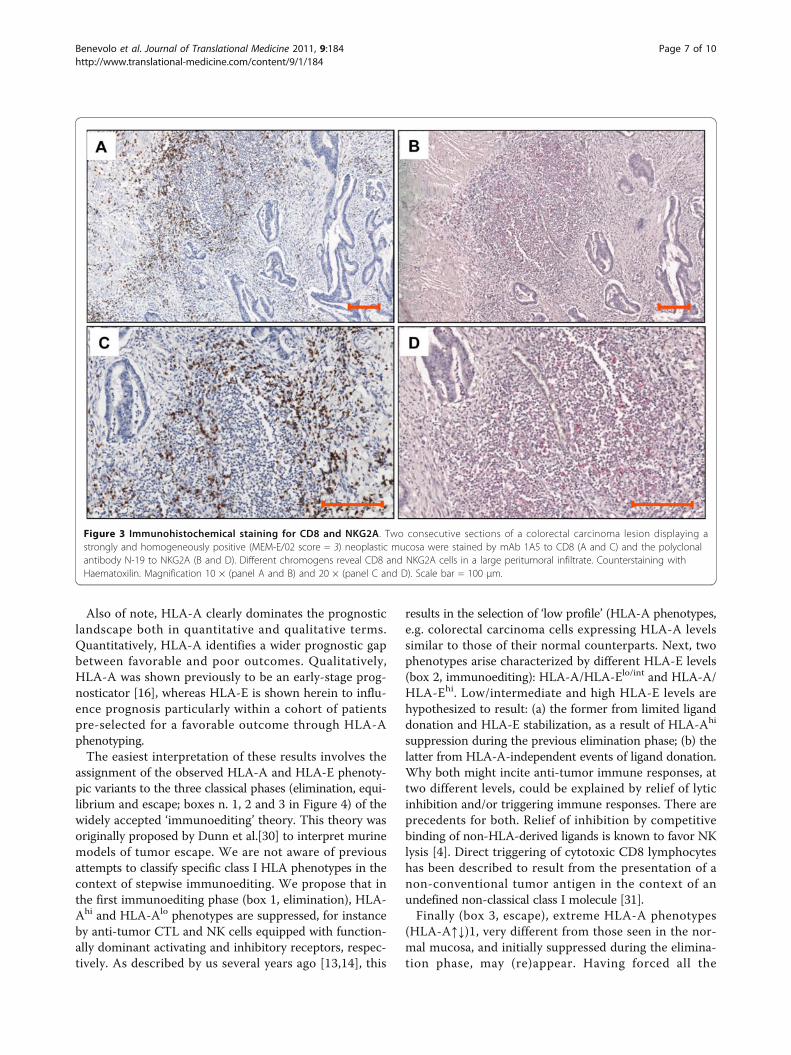
Figure 3 Immunohistochemical staining for CD8 and NKG2A. Two consecutive sections of a colorectal carcinoma lesion displaying astrongly and homogeneously positive (MEM-E/02 score = 3) neoplastic mucosa were stained by mAb 1A5 to CD8 (A and C) and the polyclonalantibody N-19 to NKG2A (B and D). Different chromogens reveal CD8 and NKG2A cells in a large peritumoral infiltrate. Counterstaining withHaematoxilin. Magnification 10 × (panel A and B) and 20 × (panel C and D). Scale bar = 100 μm.
Benevolo et al. Journal of Translational Medicine 2011, 9:184http://www.translational-medicine.com/content/9/1/184
Page 7 of 10

immune checkpoints, these phenotypes are not surpris-ingly associated with the poorest outcomes regardless ofHLA-E expression, found to be irrelevant in this group.Thus, we propose a dual outcome model whereby
HLA-E expression, far from being a manifestation ofimmune inhibition and escape, represents instead a pre-requisite for active immunological scrutiny of editedHLA tumor variants on the one hand, and triggeringimmune responses on the other.Recently, Levy et al. reported poor MEM-E/02 reactivity
in most colorectal carcinomas, and an association betweenhigh HLA-E expression, detected in just 8 lesions, andpoor prognosis [32]. Poor reactivity is difficult to reconcilewith the high levels of HLA-E detected by us with thesame MEM-E/02 antibody in most lesions, and the prog-nostic associations proposed by Levy et al. are based on alimited number of cases. Another example of HLA-E asso-ciation with poor prognosis is a recent study on early-stage breast carcinoma [33].Our results better agree with two cDNA profiling stu-
dies of advanced melanoma in which high levels ofHLA-E transcripts contributed molecular signatures of
favourable prognosis [34,35], and a recent study on glio-blastoma [36]. Possibly, the balance between activatingand inhibitory functions of HLA-E may differ in differ-ent tumor histotypes. Additional work is required toinvestigate these issues in the context of activation-inhi-bition models of tumor immune surveillance.
ConclusionsIn summary, this is one of the few reports [32,33,37]addressing the prognostic significance of non-classicalclass I molecules in colorectal carcinoma. Additionally, weprovide evidence (albeit indirect) for a functional interplaybetween classical and non-classical HLA molecules invivo. This is also the first report to suggest in situ engage-ment of tumor cells and T lymphocytes expressing adefined ligand:non-rearranging immune receptor pair.
Note added in proofAt the time of proof-editing a paper [38] appeared inPubMed describing a poor prognosis associated withHLA-E expression in colorectal carcinoma and NKG2Aexpression in the lymphoid infiltrate.
Figure 4 Class I HLA expression, immunoediting, and survival through colorectal carcinoma stages . Stepwise selection of HLAphenotypes results in distinct tumor variants populating the three classical steps (elimination, equilibrium and escape; shaded boxes) ofimmunoediting. Equilibrium is defined herein quasi-equilibrium. Unlike ‘true’ equilibrium, that corresponds to occult cancer and dormancy [30],and was elusive even in experimental models of mouse tumorigenesis [39], quasi-equilibrium refers to edited tumor cells embedded in aclinically evident tumor mass, sufficiently large to be immunophenotyped in a human pathology facility.
Benevolo et al. Journal of Translational Medicine 2011, 9:184http://www.translational-medicine.com/content/9/1/184
Page 8 of 10

Additional material
Additional file 1: Supplemental materials. Additional Tables, Figures,and Legends [7,17,20,40,41].
AcknowledgementsSupported by funds from the Italian Ministry of Health agreement 2007 (toMB and PG) and by Associazione Italiana Ricerca sul Cancro (AIRC IG to PG).Dr. Hidde Ploegh (Whitehead Institute for Biomedical Research, Cambridge,MA) and Dr. Antonio Siccardi (S. Raffaele Institute, Milan, Italy) are gratefullyacknowledged for generously providing HCA2, HC-10, and L31. Rocco Fraioli,Maria Vincenza Sarcone, and Paula Franke are gratefully acknowledged forskillful technical assistance, secretarial support, and linguistic revision,respectively.
Author details1Department of Pathology, Regina Elena National Cancer Institute, Via E.Chianesi 53, 00144 Rome, Italy. 2Laboratory of Immunology, Regina ElenaNational Cancer Institute, Via delle Messi d’Oro 156, 00158 Rome, Italy.3Biostatistics Unit, Istituti Fisioterapici Ospitalieri, Via E. Chianesi 53, 00144Rome, Italy. 4Hepato-Biliary-Pancreatic Surgery, Regina Elena National CancerInstitute, Via E. Chianesi 53, 00144 Rome, Italy.
Authors’ contributionsMC provided surgical specimens and supervised patient follow-up. MM andMC organized the tissue bank and the clinical pathological database. MGDreviewed and staged pathological specimens. MB, MM, CE and FRperformed and evaluated immunohistochemistry. ET and ELM prepared theantibodies and performed flow cytometry and molecular HLA typing. ISparticipated in the design of the study and performed statistical analysis. MB,MM, MC and PG conceived, designed, and coordinated the study. MB andPG wrote the manuscript. All authors read and approved the finalmanuscript.
Competing interestsThe authors declare that they have no competing interests.
Received: 10 June 2011 Accepted: 27 October 2011Published: 27 October 2011
References1. Braud VM, Allan DSJ, O’Callaghan CA, Söderström K, D’Andrea A, Ogg GS,
Lazetic S, Young NT, Bell JI, Phillips JH, Lanier LL, McMichael AJ: HLA-Ebinds to natural killer cell receptors CD94/NKG2A, B and C. Nature 1998,391:795-799.
2. Rodgers J, Cook R: MHC class Ib molecules bridge innate and acquiredimmunity. Nat Rev Immunol 2005, 5:459-471.
3. Moretta L, Romagnani C, Pietra G, Moretta A, Mingari MC: NK-CTLs, a novelHLA-E-restricted T-cell subset. Trends Immunol 2003, 24:136-143.
4. Michaelsson J, Teixeira de Matos C, Achour A, Lanier LL, Kärre K,Soderstrom K: A signal peptide derived from hsp60 binds HLA-E andinterferes with CD94/NKG2A recognition. J Exp Med 2005, 196:1403-1414.
5. Cohen GB, Gandhi RT, Davis DM, Mandelboim O, Chen BK, Strominger JL,Baltimore D: The selective downregulation of class I majorhistocompatibility complex proteins by HIV-1 protects HIV-infected cellsfrom NK cells. Immunity 1999, 10:661-671.
6. Smith MEF, Bodmer WF, Bodmer JG: Selective loss of HLA-A, B, C locusproducts in colorectal adenocarcinoma. Lancet 1988, I:823-824.
7. Lee N, Goodlett DR, Ishitani A, Marquardt H, Geraghty DE: HLA-E surfaceexpression depends on binding of TAP-dependent peptides derivedfrom certain HLA class I signal sequences. J Immunol 1998,160:4951-4960.
8. Tomasec P, Braud VM, Rickards C, Powell MB, McSharry BP, Gadola S,Cerundolo V, Borysiewicz LK, McMichael AJ, Wilkinson GWG: Surfaceexpression of HLA-E, an inhibitor of natural killer cells, enhanced byhuman cytomegalovirus gpUL40. Science 2000, 287:1031-1033.
9. Ulbrecht M, Martinozzi S, Grzeschik M, Hengel H, Ellwart JW, Pla M,Weiss EH: The human cytomegalovirus UL40 gene product contains a
ligand for HLA-E and prevents NK cell-mediated lysis. J Immunol 2000,164:5019-5022.
10. Gudmundsdóttir I, Jónasson JG, Sigurdsson H, Olafsdóttir K, Tryggvadóttir L,Ogmundsdóttir HM: Altered expression of HLA class I antigens in breastcancer: Association with prognosis. Int J Cancer 2000, 89:500-505.
11. Menon AG, Morreau H, Tollenaar RA, Alphenaar E, Van Puijenbroek M,Putter H, Janssen-Van Rhijn CM, Van De Velde CJ, Fleuren GJ, Kuppen PJ:Down-regulation of HLA-A expression correlates with a better prognosisin colorectal cancer patients. Lab Invest 2002, 82:1725-1733.
12. Watson NF, Ramage JM, Madjd Z, Spendlove I, Ellis IO, Scholefield JH,Durrant LG: Immunosurveillance is active in colorectal cancer asdownregulation but not complete loss of MHC class I expressioncorrelates with a poor prognosis. Int J Cancer 2005, 118:6-10.
13. Giacomini P, Giorda E, Fraioli R, Nicotra MR, Vitale N, Setini A, Delfino L,Morabito A, Benevolo M, Venturo I, Mottolese M, Ferrara GB, Natali PG: Lowprevalence of selective human leukocyte antigen (HLA)-A and HLA-Bepitope losses in early-passage tumor cell lines. Cancer Res 1999,59:2657-2667.
14. Giorda E, Sibilio L, Martayan A, Moretti S, Venturo I, Mottolese M, Ferrara GB,Cappellacci S, Eibenschutz L, Catricalà C, Grammatico P, Giacomini P: Theantigen processing machinery of Human Leukocyte Antigens: linkedpatterns of gene expression in neoplastic cells. Cancer Res 2003,63:4119-4127.
15. Fruci D, Ferracuti S, Limongi MZ, Cunsolo V, Giorda E, Fraioli R, Sibilio L,Carroll O, Hattori A, van Endert PM, Giacomini P: Expression ofendoplasmic reticulum aminopeptidases in EBV-B cell lines from healthydonors and in leukemia/lymphoma, carcinoma, and melanoma cell lines.J Immunol 2006, 176:4869-4879.
16. Benevolo M, Mottolese M, Piperno G, Sperduti I, Cione A, Sibilio L,Martayan A, Donnorso RP, Cosimelli M, Giacomini P: HLA-A, -B, -Cexpression in colon carcinoma mimics that of the normal colonicmucosa and is prognostically relevant. Am J Surg Pathol 2007, 31:76-84.
17. Lo Monaco E, Sibilio L, Melucci E, Tremante E, Suchànek M, Horejsi V,Martayan A, Giacomini P: HLA-E: strong association with β2m and surfaceexpression in the absence of HLA class I signal sequence-derivedpeptides. J Immunol 2008, 181:5442-5450.
18. Lo Monaco E, Tremante E, Cerboni C, Melucci E, Sibilio L, Zingoni A,Nicotra MR, Natali PG, Giacomini P: Human Leukocyte Antigen Econtributes to protect tumor cells from lysis by natural killer cells.Neoplasia 2011, 13:822-830.
19. TNM classification of malignant tumors. 6 edition. Hoboken, NJ: John Wiley& Sons; 2002.
20. Stam NJ, Vroom TM, Peters PJ, Pastoors EB, Ploegh HL: HLA-A and HLA-B-specific monoclonal antibodies reactive with free heavy chains inWestern blots, in formalin-fixed, paraffin-embedded tissue sections andin cryoimmuno-electron microscopy. Int Immunol 1990, 2:113-125.
21. Seitz C, Uchanska-Ziegler B, Zank A, Ziegler A: The monoclonal antibodyHCA2 recognises a broadly shared epitope on selected classical as wellas several non-classical HLA class I molecules. Mol Immunol 1998,35:819-827.
22. Setini A, Beretta A, De Santis C, Meneveri R, Martayan A, Mazzilli MC,Appella E, Siccardi AG, Natali PG, Giacomini P: Distinctive features of theα1 domain α helix of HLA-C heavy chains free of β2-microglobulin. HumImmunol 1996, 46:69-81.
23. Giacomini P, Beretta A, Nicotra MR, Ciccarelli G, Martayan A, Cerboni C,Lopalco L, Bini D, Delfino L, Ferrara GB, Siccardi AG, Natali PG: HLA-C heavychains free of β2m: distribution in normal tissues and neoplastic lesionsof non-lymphoid origin and IFN-γ responsiveness. Tissue Antigens 1997,50:555-566.
24. Braud VM, Jones EY, McMichael A: The human major histocompatibilitycomplex class Ib molecule HLA-E binds signal sequence-derivedpeptides with primary anchor residues at positions 2 and 9. Eur JImmunol 1997, 27:1164-1169.
25. Borrego F, Ulbrecht M, Weiss EH, Coligan JE, Brooks AG: Recognition ofhuman histocompatibility leukocyte antigen (HLA)-E complexed withHLA class I signal sequence-derived peptides by CD94/NKG2 confersprotection from natural killer cell-mediated lysis. J Exp Med 1998,187:813-818.
26. Ulbrecht M, Kellermann J, Johnson JP, Weiss EH: Impaired intracellulartransport and cell surface expression of nonpolymorphic HLA-E:evidence for inefficient peptide binding. J Exp Med 1992, 176:1083-1090.
Benevolo et al. Journal of Translational Medicine 2011, 9:184http://www.translational-medicine.com/content/9/1/184
Page 9 of 10

27. Brooks AG, Borrego F, Posch PE, Patamawenu A, Scorzelli CJ, Ulbrecht M,Weiss EH, Coligan JE: Specific recognition of HLA-E, but not classical, HLAclass I molecules by soluble CD94/NKG2A and NK cells. J Immunol 1999,162:305-313.
28. Galon J, Costes A, Sanchez-Cabo F, Kirilovsky A, Mlecnik B, Lagorce-Pages C,Tosolini M, Camus M, Berger A, Wind P, Zinzindohoue F, Bruneval P,Cugnenc PH, Trajanoski Z, Fridman WH, Pages F: Type, density, andlocation of immune cells within human colorectal tumors predict clinicaloutcome. Science 2006, 313:1960-1964.
29. Ohtani H: Focus on TILs: prognostic significance of tumor infiltratinglymphocytes in human colorectal cancer. Cancer Immun 2007, 7:4-12.
30. Dunn GP, Bruce AT, Ikeda H, Old LJ, Schreiber RD: Cancer immunoediting:from immunosurveillance to tumor escape. Nat Immunol 2002, 3:991-998.
31. Housseau F, Bright RK, Simonis T, Nishimura MI, Topalian SL: Recognition ofa shared human prostate cancer-associated antigen by nonclassicalMHC-restricted CD8+ T cells. J Immunol 1999, 163:6330-6337.
32. Levy EM, Bianchini M, Von Euw EM, Barrio MM, Bravo AI, Furman D,Domenichini E, Macagno C, Pinsky V, Zucchini C, Valvassori L, Mordoh J:Human leukocyte antigen-E protein is overexpressed in primary humancolorectal cancer. Int J Oncol 2008, 32:633-641.
33. de Kruijf EM, Sajet A, van Nes JG, Natanov R, Putter H, Smit VT, Liefers GJ,van den Elsen PJ, van de Velde CJ, Kuppen PJ: HLA-E and HLA-GExpression in Classical HLA Class I-Negative Tumors Is of PrognosticValue for Clinical Outcome of Early Breast Cancer Patients. J Immunol2010, 185:7452-7459.
34. John T, Black MA, Toro TT, Leader D, Gedye CA, Davis ID, Guilford PJ,Cebon JS: Predicting clinical outcome through molecular profiling instage III melanoma. Clin Cancer Res 2008, 14:5173-5180.
35. Mandruzzato S, Callegaro A, Turcatel G, Francescato S, Montesco MC,Chiarion-Sileni V, Mocellin S, Rossi CR, Bicciato S, Wang E, Marincola FM,Zanovello P: A gene expression signature associated with survival inmetastatic melanoma. J Transl Med 2006, 4:50.
36. Kren L, Slaby O, Muckova K, Lzicarova E, Sova M, Vybihal V, Svoboda T,Fadrus P, Lakomy R, Vanhara P, Krenova Z, Sterba J, Smrcka M, Michalek J:Expression of immune-modulatory molecules HLA-G and HLA-E bytumor cells in glioblastomas: An unexpected prognostic significance?Neuropathology 2011, 31:129-134.
37. Ye SR, Yang H, Li K, Dong DD, Lin XM, Yie SM: Human leukocyte antigenG expression: as a significant prognostic indicator for patients withcolorectal cancer. Mod Pathol 2007, 20:375-383.
38. Bossard C, Bezieau S, Matysiak-Budnik T, Volteau C, Laboisse CL, Jotereau F,Mosnier JF: HLA-E/beta2 microglobulin over-expression in colorectalcancer is associated with recruitment of inhibitory immune cells andtumor progression. Int J Cancer 2011.
39. Koebel CM, Vermi W, Swann JB, Zerafa N, Rodig SJ, Old LJ, Smyth MJ,Schreiber RD: Adaptive immunity maintains occult cancer in anequilibrium state. Nature 2007, 450:903-907.
40. Cifaldi L, Lo Monaco E, Forloni M, Giorda E, Lorenzi S, Petrini S, Tremante E,Pende D, Locatelli F, Giacomini P, Fruci D: NK cells efficiently rejectlymphoma silenced for the endoplasmic reticulum aminopeptidaseassociated with antigen processing. Cancer Res 2011.
41. Bicknell DC, Rowan A, Bodmer WF: β2-microglobulin mutations: a studyof established colorectal cell lines and fresh tumors. ProcNatlAcadSci1994, 91:4751-4755.
doi:10.1186/1479-5876-9-184Cite this article as: Benevolo et al.: High expression of HLA-E incolorectal carcinoma is associated with a favorable prognosis. Journal ofTranslational Medicine 2011 9:184. Submit your next manuscript to BioMed Central
and take full advantage of:
• Convenient online submission
• Thorough peer review
• No space constraints or color figure charges
• Immediate publication on acceptance
• Inclusion in PubMed, CAS, Scopus and Google Scholar
• Research which is freely available for redistribution
Submit your manuscript at www.biomedcentral.com/submit
Benevolo et al. Journal of Translational Medicine 2011, 9:184http://www.translational-medicine.com/content/9/1/184
Page 10 of 10