Mycoplasma salivarium as a Dominant Coloniser of Fanconi Anaemia Associated Oral Carcinoma
Upload
independentCategory
view
3download
0
BioMed CentralBMC Microbiology
ss
Open AcceResearch articleβ-D-Glucoside utilization by Mycoplasma mycoides subsp. mycoides SC: possible involvement in the control of cytotoxicity towards bovine lung cellsEdy M Vilei*1, Ivone Correia2, M Helena Ferronha2, Daniela F Bischof1 and Joachim Frey1Address: 1Institute of Veterinary Bacteriology, University of Bern, Länggass-Strasse 122, Postfach, CH-3001 Bern, Switzerland and 2Laboratório Nacional de Investigação Veterinária, Departamento de Biologia Celular, Estrada de Benfica 701, P-1549-011 Lisbon, Portugal
Email: Edy M Vilei* - [email protected]; Ivone Correia - [email protected]; M Helena Ferronha - [email protected]; Daniela F Bischof - [email protected]; Joachim Frey - [email protected]
* Corresponding author
AbstractBackground: Contagious bovine pleuropneumonia (CBPP) caused by Mycoplasma mycoides subsp.mycoides small-colony type (SC) is among the most serious threats for livestock producers in Africa.Glycerol metabolism-associated H2O2 production seems to play a crucial role in virulence of thismycoplasma. A wide number of attenuated strains of M. mycoides subsp. mycoides SC are currentlyused in Africa as live vaccines. Glycerol metabolism is not affected in these vaccine strains andtherefore it does not seem to be the determinant of their attenuation. A non-synonymous singlenucleotide polymorphism (SNP) in the bgl gene coding for the 6-phospho-β-glucosidase (Bgl) hasbeen described recently. The SNP differentiates virulent African strains isolated from outbreakswith severe CBPP, which express the Bgl isoform Val204, from strains to be considered less virulentisolated from CBPP outbreaks with low mortality and vaccine strains, which express the Bglisoform Ala204.
Results: Strains of M. mycoides subsp. mycoides SC considered virulent and possessing the Bglisoform Val204, but not strains with the Bgl isoform Ala204, do trigger elevated levels of damage toembryonic bovine lung (EBL) cells upon incubation with the disaccharides (i.e., β-D-glucosides)sucrose and lactose. However, strains expressing the Bgl isoform Val204 show a lower hydrolysingactivity on the chromogenic substrate p-nitrophenyl-β-D-glucopyranoside (pNPbG) whencompared to strains that possess the Bgl isoform Ala204. Defective activity of Bgl in M. mycoidessubsp. mycoides SC does not lead to H2O2 production. Rather, the viability during addition of β-D-glucosides in medium-free buffers is higher for strains harbouring the Bgl isoform Val204 than forthose with the isoform Ala204.
Conclusion: Our results indicate that the studied SNP in the bgl gene is one possible cause of thedifference in bacterial virulence among strains of M. mycoides subsp. mycoides SC. Bgl does not actas a direct virulence factor, but strains possessing the Bgl isoform Val204 with low hydrolysingactivity are more prone to survive in environments that contain high levels of β-D-glucosides, thuscontributing in some extent to mycoplasmaemia.
Published: 17 April 2007
BMC Microbiology 2007, 7:31 doi:10.1186/1471-2180-7-31
Received: 22 September 2006Accepted: 17 April 2007
This article is available from: http://www.biomedcentral.com/1471-2180/7/31
© 2007 Vilei et al; licensee BioMed Central Ltd. This is an Open Access article distributed under the terms of the Creative Commons Attribution License (http://creativecommons.org/licenses/by/2.0), which permits unrestricted use, distribution, and reproduction in any medium, provided the original work is properly cited.
Page 1 of 15(page number not for citation purposes)
BMC Microbiology 2007, 7:31 http://www.biomedcentral.com/1471-2180/7/31
BackgroundMycoplasma species are known to represent the smallestself replicating organisms on earth, possessing the small-est genomes that contain basically fundamental functionsto ensure autonomous life. Potential virulence genes arefound in many mycoplasmas [1-9]. However, the genomeof Mycoplasma mycoides subsp. mycoides small-colony type(SC), a Mycoplasma species causing one of most severeinfectious animal diseases as defined by the World Organ-ization for Animal Health and included in the list of dis-eases notifiable to the Office International des Epizooties(OIE), does not show classical virulence genes such astoxin, cytolysin and invasin genes [10]. M. mycoides subsp.mycoides SC causes contagious bovine pleuropneumonia(CBPP), the most serious cattle disease in Africa since thesuccessful control of rinderpest [11]. In Europe, CBPP re-emerged at the end of the last century after being com-pletely eradicated between 1967 and the 1980s [12], thusshowing that CBPP is still a threat also to continents thathave been able to eradicate the disease.
Virulence of M. mycoides subsp. mycoides SC seems to bedetermined by intrinsic functions such as: i) capsularpolysaccharide (CPS) that seems to be involved in serum-resistance [13-17]; ii) lipoproteins that are expected toplay a role as triggers in mechanisms of pathogenicity andin the release of pro-inflammatory cytokines [18-22]; iii)yet unknown but necessary adhesion factors that may playa central role in the intimate interactions of pathogenicmycoplasmas with mammalian cells for long periods thustriggering a cascade of signals which are transduced to thehost cell and induce inflammation [23]; iv) repeating ele-ments in variable membrane proteins of mycoplasmasthat are suggested to increase the pathogen's ability toadhere to host cells and to evade the host immuneresponse [24-26]; v) immunomodulating factors that cancause apoptosis of the mononuclear cells triggered by liveM. mycoides subsp. mycoides SC or by a substance releasedby these mycoplasmas [27]; and vi) toxic metabolic sideproducts such as H2O2 [28-31], which is translocated effi-ciently to the host cells where it can cause cell death [32].
Since no genetic tools for targeted mutagenesis are availa-ble for M. mycoides subsp. mycoides SC, analysis of viru-lence factors requires the use of various strains, includingfield strains from outbreaks of CBPP with various degreesof severity, strains from geographically different areas andlive vaccine strains. Detailed genetic studies comparing M.mycoides subsp. mycoides SC isolates has provided evidencethat deletion of gtsC and part of gtsB (two of three genesinvolved in glycerol transport) in the moderately virulentEuropean strains correlated with a reduced ability to pro-duce H2O2 compared to the highly virulent strains of theAfrican/Australian cluster [31]. Vaccine strains of M.mycoides subsp. mycoides SC such as T1/44, T1/Sr50 and
KH3J possess however an intact glycerol uptake systemand metabolism [33,34], which do not distinguish themfrom virulent field strains by means of H2O2 production.Hence, other functions that determine virulence areexpected in M. mycoides subsp. mycoides SC. For instance,these vaccine strains seem to produce lower amounts ofCPS compared to field strains [16]. CPS seems to play arole in the capacity of persistence and dissemination of M.mycoides subsp. mycoides SC in the infected host [17].However, the genetic locus of the CPS regulation stillremains unknown.
We have recently identified by PCR amplification andrestriction enzyme analysis (PCR-REA) a genetic differ-ence in a structural gene which divides M. mycoides subsp.mycoides SC strains in two groups, one including virulentAfrican field strains and the other comprising the T1-derived vaccine strains T1/44 and T1/Sr50, as well as Euro-pean and Australian strains and the type strain PG1 [35].This genetic difference was found in the bgl gene thatcodes for 6-phospho-β-glucosidase (Bgl). Bgl is anenzyme that is associated with the phosphoenolpyruvate-dependent sugar:phosphotransferase system (PEP-PTS), amulticomponent system involved in the simultaneoustranslocation and phosphorylation of sugars by bacteriafrom both gram-positive and gram-negative genera [36].The PEP-PTS and Bgl are involved in incorporation andsubsequent catabolism of β-D-glucosides (e.g., disaccha-rides).
The present work focuses on the characterization of Bgland related disaccharide catabolism, and the possiblecontribution of sugar metabolism to control cytotoxicityof M. mycoides subsp. mycoides SC.
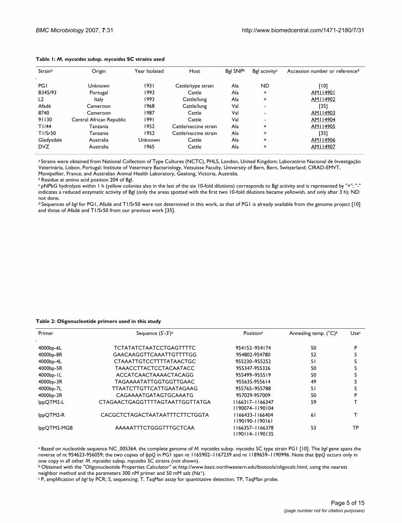
ResultsGenetic variability of the bgl gene in M. mycoides subsp. mycoides SCThe 9.8 kb genomic region containing the genes involvedin disaccharide uptake and metabolism in M. mycoidessubsp . mycoides SC type strain PG1, whose genomesequence was published by Westberg et al. [10], was con-sidered in this study. Figure 1 shows the expected roles ofEIIBC, sugar hydrolase, EIIA, Bgl and Suk in the disaccha-ride transport system and sugar metabolism. The bgl genefrom ten selected strains of M. mycoides subsp . mycoidesSC (Table 1) was sequenced with primers listed in Table 2.The sequences from the African field strains Afadé, 8740and 91130 were identical, although these strains were iso-lated in geographically distant locations in an interval ofas much as 23 years. The bgl sequences obtained from thevaccine strains T1/44 and T1/Sr50, as well as from typestrain PG1, two European and two Australian strains pre-sented a single nucleotide polymorphism (SNP) whencompared to those of the African field strains. The non-
Page 2 of 15(page number not for citation purposes)

BMC Microbiology 2007, 7:31 http://www.biomedcentral.com/1471-2180/7/31
synonymous SNP involves an amino acid change at posi-tion 204 of the Bgl protein sequence reported recently[35]. The group of the three African field strains possessesthe Bgl isoform Val204, while the vaccine strains, the Euro-pean and Australian outbreak strains and PG1 possess theBgl isoform Ala204.
Carbohydrate utilization of M. mycoides subsp. mycoides SCThe biochemical reactions of all strains tested indicatedthat M. mycoides subsp. mycoides SC can use the monosac-charides glucose, fructose and galactose, and the disaccha-rides sucrose and lactose. In fact, after incubation ofmycoplasma suspensions in serum-free media with highamounts of either substrate in the presence of the indica-tor bromothymol blue for 6 days, all reactions turnedlight green to bright yellow (depending on the substrate)in colour, indicating production of CO2 and therefore car-bohydrate uptake and utilization. Daily examination ofthe reactions revealed that there were no significant differ-ences in the substrate utilization rates among the 5 strainstested (not shown).
M. mycoides subsp. mycoides SC induction of cell damage in the presence of sugarsThe effect of M. mycoides subsp. mycoides SC on embryonicbovine lung (EBL) cells in the presence and absence of dis-accharides was studied. Figure 2 shows the cell damagemediated by strain 8740, chosen as representative of M.mycoides subsp. mycoides SC strains with the Bgl isoformVal204, and of the vaccine strain T1/44, as representativefor strains with the Bgl isoform Ala204. Cell damage wasexpressed by morphological changes mainly characterizedby cell retraction, which was evident in monolayers of EBLcells inoculated with strain 8740 in the presence ofsucrose or lactose but not in EBL cells inoculated with vac-cine strain T1/44, with or without these disaccharides. Therelative cell damage as a measure for cytotoxicityincreased with increasing concentrations of the disaccha-rides (not shown). Morphological changes were alreadynoticeable 3 h post-inoculation with strain 8740 in thepresence of low concentrations of sucrose and lactose. At4 h, those changes became easily observable (Figure 2,panels A and B) and were more evident at 8 h post-inocu-lation (not shown). Mycoplasmas incubated in the pres-ence of monosaccharides for 4 h had no cytotoxic effectstowards EBL cells (Figure 2, panel C).
After 24 h, cell monolayers infected with strain 8740 inthe presence of sucrose and lactose had almost completelylost confluence and numerous detached rounded cellswere observed (Figure 2, panels D and E). Morphologicalcell changes after 24 h were also seen with strain 8740when incubated with glucose, fructose or galactose, butthe degree of cell damage mediated by these monosaccha-
rides was significantly lower when compared to that of thedisaccharides (Figure 2, panel F). Strain T1/44 did notinduce cell damage, either in the presence or absence ofthe five tested sugars (Figure 2, panels G, H and I). Thesame was observed with the European strains L2 andB345/93 (not shown).
The cytotoxic effect observed upon incubation with strain8740 in the presence of the disaccharides could not beblocked when the cell monolayers were pre-treated withcatalase at a concentration of 160 U/ml. Under these con-ditions, the EBL cells showed dramatic morphologicalchanges 24 h post infection as in the absence of catalase(Figure 2, panel J). Strain T1/44 in the presence of disac-charides and catalase (Figure 2, panel K) and catalasealone (Figure 2, panel L) did not induce cell damage.Moreover, no morphological changes were detected whenEBL cell monolayers were incubated either with sugarsalone, with strain 8740 alone or with axenic medium (Fig-ure 2, panels M, N and O).
pNPbG hydrolysis of M. mycoides subsp. mycoides SC grown on solid mediumHydrolysis of p-nitrophenyl-β-D-glucopyranoside(pNPbG; colourless) to give p-nitrophenol (yellow) wasdetermined qualitatively to assess 6-phospho-β-glucosi-dase activity by flooding cultures of M. mycoides subsp .mycoides SC on nutrient agar plates with pNPbG solution.After 1 h, no colour change was seen with the African M.mycoides subsp . mycoides SC field strains tested (Afadé,8740 and 91130), indicating no detectable hydrolyticactivity of Bgl isoform Val204. In contrast, individual colo-nies of the other strains tested, i.e., the vaccine strains T1/44 and T1/Sr50, as well as the European strains B345/93and L2 and the Australian strains Gladysdale and DVZ,which express the Bgl isoform Ala204, were coloured yel-low, indicating hydrolysis of pNPbG (Table 1). It has to benoted that after prolonged incubation, the three Africanfield strains also showed some hydrolysis of pNPbG, asrevealed by a light yellow coloration in areas of confluentgrowth.
Production of H2O2 by M. mycoides subsp. mycoides SCProduction of H2O2 after the addition of mono- and dis-accharides to suspensions of M. mycoides subsp . mycoidesSC strain 8740 and the vaccine strain T1/44, chosen asrepresentative strains in this study, was measured. Addi-tion of physiological concentrations of the different sug-ars to the two mycoplasmas tested resulted in a weakrelease of H2O2 into the incubation buffer after 1 h. Thereleased H2O2 levels measured in the suspensions were atconcentrations below or equal to 0.5 µg/ml, the detectionlevel of the assay. In contrast, the addition of 100 µM glyc-erol to the mycoplasmas instantly resulted in a strongrelease of H2O2 into the incubation buffer, reaching 8.75
Page 3 of 15(page number not for citation purposes)

BMC Microbiology 2007, 7:31 http://www.biomedcentral.com/1471-2180/7/31
Page 4 of 15(page number not for citation purposes)
Identification of the genes involved in oligosaccharide uptake and Bgl-dependent utilization in M. mycoides subsp. mycoides SCFigure 1Identification of the genes involved in oligosaccharide uptake and Bgl-dependent utilization in M. mycoides subsp. mycoides SC. (A) Genetic map of the locus involved in the metabolism of β-D-glucosides. The open box indicates the IS element IS1296 and the large horizontal arrows indicate open reading frames found in the 9.8 kb DNA portion. (B) Model for the Bgl-dependent metabolism of β-D-glucosides in M. mycoides subsp. mycoides SC. Oligosaccharides are incorporated into the mycoplasma through the protein EIIBC. Once in the cytoplasm, sugar hydrolase may split complex β-D-glucosides into less complex β-D-glucosides (e.g., monosaccharides and disaccharides). Phosphorylation of these intracellular sugar molecules is preceded by the transfer of a phosphoryl group (P) from phosphoenolpyruvate (PEP) to EIIA in a pathway also involving enzyme I (EI) and the phosphoryl carrier protein (HPr). Then, Bgl hydrolyzes β-glycosidic linkages in the phospho-β-D-gluco-sides and residual monosaccharides are phosphorylated by the sugar kinase (Suk) before entering glycolysis.
EIIBC sugar hydrolase EIIA Suk Bgl outer surfaceprotein
IS1296 frvA suk bgl
0 1 2 3 4 5 6 7 8 9 10 kb
A
B
MEMBRANEEIIBC
oligosaccharides ( -D-glucosides)
sugarhydrolase
low complex -D-glucosides
phospho- -D-glucosides
P EIIA HPr EI PEP
P
PP
Bgl
phospho-monosaccharide
Suk
monosaccharide phospho-monosaccharide
CYTOPLASM
GLYCOLYSIS
P
frvB
BMC Microbiology 2007, 7:31 http://www.biomedcentral.com/1471-2180/7/31
Page 5 of 15(page number not for citation purposes)
Table 1: M. mycoides subsp. mycoides SC strains used
Straina Origin Year Isolated Host Bgl SNPb Bgl activityc Accession number or referenced
PG1 Unknown 1931 Cattle/type strain Ala ND [10]B345/93 Portugal 1993 Cattle Ala + AM114901L2 Italy 1993 Cattle/lung Ala + AM114902Afadé Cameroon 1968 Cattle/lung Val - [35]8740 Cameroon 1987 Cattle Val - AM11490391130 Central African Republic 1991 Cattle Val - AM114904T1/44 Tanzania 1952 Cattle/vaccine strain Ala + AM114905T1/Sr50 Tanzania 1952 Cattle/vaccine strain Ala + [35]Gladysdale Australia Unknown Cattle Ala + AM114906DVZ Australia 1965 Cattle Ala + AM114907
a Strains were obtained from National Collection of Type Cultures (NCTC), PHLS, London, United Kingdom; Laboratório Nacional de Investigação Veterinária, Lisbon, Portugal; Institute of Veterinary Bacteriology, Vetsuisse Faculty, University of Bern, Bern, Switzerland; CIRAD-EMVT, Montpellier, France; and Australian Animal Health Laboratory, Geelong, Victoria, Australia.b Residue at amino acid position 204 of Bgl.c pNPbG hydrolysis within 1 h (yellow colonies also in the last of the six 10-fold dilutions) corresponds to Bgl activity and is represented by "+"; "-" indicates a reduced enzymatic activity of Bgl (only the areas spotted with the first two 10-fold dilutions became yellowish, and only after 3 h); ND: not done.d Sequences of bgl for PG1, Afadé and T1/Sr50 were not determined in this work, as that of PG1 is already available from the genome project [10] and those of Afadé and T1/Sr50 from our previous work [35].
Table 2: Oligonucleotide primers used in this study
Primer Sequence (5'-3')a Positiona Annealing temp. (°C)b Usec
4000bp-6L TCTATATCTAATCCTGAGTTTTC 954152–954174 50 P4000bp-8R GAACAAGGTTCAAATTGTTTTGG 954802-954780 52 S4000bp-4L CTAAATTGTCCTTTTATAACTGC 955230–955252 51 S4000bp-5R TAAACCTTACTCCTACAATACC 955347-955326 50 S4000bp-1L ACCATCAACTAAAACTACAGG 955499–955519 50 S4000bp-3R TAGAAAATATTGGTGGTTGAAC 955635-955614 49 S4000bp-7L TTAATCTTGTTCATTGAATAGAAG 955765–955788 51 S4000bp-2R CAGAAAATGATAGTGCAAATG 957029-957009 50 PlppQTM2-L CTAGAACTGAGGTTTTAGTAATTGGTTATGA 1166317–1166347
1190074–119010459 T
lppQTM2-R CACGCTCTAGACTAATAATTTCTTCTGGTA 1166433-11664041190190-1190161
61 T
lppQTM2-MGB AAAAATTTCTGGGTTTGCTCAA 1166357–11663781190114–1190135
53 TP
a Based on nucleotide sequence NC_005364, the complete genome of M. mycoides subsp. mycoides SC type strain PG1 [10]. The bgl gene spans the reverse of nt 954623-956059; the two copies of lppQ in PG1 span nt 1165902–1167239 and nt 1189659–1190996. Note that lppQ occurs only in one copy in all other M. mycoides subsp. mycoides SC strains (not shown).b Obtained with the "Oligonucleotide Properties Calculator" at http://www.basic.northwestern.edu/biotools/oligocalc.html, using the nearest neighbor method and the parameters 300 nM primer and 50 mM salt (Na+).c P, amplification of bgl by PCR; S, sequencing; T, TaqMan assay for quantitative detection; TP, TaqMan probe.

BMC Microbiology 2007, 7:31 http://www.biomedcentral.com/1471-2180/7/31
Page 6 of 15(page number not for citation purposes)
Effect of sugars on cytotoxicity of M. mycoides subsp. mycoides SC on bovine lung cells in vitroFigure 2Effect of sugars on cytotoxicity of M. mycoides subsp. mycoides SC on bovine lung cells in vitro. Representative photomicrographs (320 ×) of EBL cell morphology upon mycoplasma infection in the presence of sugars. Pictures were taken after a 4 h incubation with the African field strain 8740 in the presence of sucrose (2 µM, panel A), lactose (200 µM, panel B) or the monosaccharide glucose (10 mM, panel C) and again after 24 h (panels D-F). After 24 h, pictures of EBL cell monolayers incubated with the less virulent vaccine strain T1/44 in the presence of these three sugars were also taken (panels G-I). Cata-lase (160 U/ml) was added to the EBL cells, which were then incubated for 24 h with strain 8740 and sucrose (panel J), or with strain T1/44 and sucrose (panel K). Controls performed to exclude eventual toxic effects of catalase alone (panel L), sugars alone (e.g., sucrose, panel M) or of strain 8740 without sugars (panel N) are also shown. Panel O shows EBL cells that were not treated at all.
sucrose 2 µM lactose 200 µM glucose 10 mM
A
8740
B
8740
C
8740
4 h
D
8740
E
8740
F
8740
24 h
G
T1/44
H
T1/44
I
T1/44
24 h
sucrose sucrose -
J
8740
K
T1/44
L
no mycoplasmas
24 h
catalase catalase catalase
sucrose - -
M
no mycoplasmas
N
8740
O
no mycoplasmas
24 h
BMC Microbiology 2007, 7:31 http://www.biomedcentral.com/1471-2180/7/31
µg/ml by strain 8740, and 7.5–7.75 µg/ml by vaccinestrain T1/44 after 1 h. All H2O2 concentrations measuredwere retained up to 2 h and showed no significant differ-ences between the two strains.
Viability of M. mycoides subsp. mycoides SC upon incubation with different sugars and growth analysis of surviving cellsStarvation in incubation buffer alone, or in incubationbuffer supplemented with 5 mM glucose or 5 µM sucroseaffected the viability of M. mycoides subsp. mycoides SCstrains 8740 and T1/44. Over the first 4 h of starvation,there was an overall loss of 7–39% of viable mycoplasmacells. After 18 h of starvation, the number of CFU forstrain T1/44 declined sharply to a rate of < 1% of the orig-inal in the three assays (loss of > 99%), while that forstrain 8740 was reduced to < 1% in incubation bufferalone and in the presence of glucose, and to approxi-mately 12% in the presence of sucrose (Table 3).
The estimated quantities of mycoplasma cells of strains8740 and T1/44 grown for 2 days after a precedingimpregnation phase of 18 h with sucrose or glucose weredetermined in the TaqMan real-time PCR with primersand probe listed in Table 2. The Ct values shown in Table4 are mean values of two or more replicate experiments (P< 0.0001; ANOVA: single factor analysis). Results showedthat pre-treatment with incubation buffer in the absenceof sugars for 18 h completely blocked the growth of bothstrains. Also pre-incubation of the two strains of M.mycoides subsp. mycoides SC with 5 mM glucose in incuba-tion buffer affected dramatically their growth rate and theamounts of mycoplasmas grown in the 2-day culture werebelow 4% when compared to those of non-treated myco-plasmas (Table 4). Pre-incubation with 5 µM sucrose
blocked the subsequent growth of strain T1/44 (≈1%),while strain 8740 retained a growth of approximately15% of the rate without treatment (Table 4).
DiscussionA single genetic difference between African field strains ofM. mycoides subsp. mycoides SC and T1-derived vaccinestrains was reported recently in the genomic region har-bouring the genes frvA, suk and bgl [35]. This genetic dif-ference is an SNP in the bgl gene coding for the 6-phospho-β-glucosidase (Bgl). Here we report the correla-tion of the two isoforms of Bgl to the cytotoxic potentialof M. mycoides subsp. mycoides SC.
In cytotoxicity studies where we examined the effect of M.mycoides subsp. mycoides SC grown in the presence of cer-tain mono- and disaccharides towards cultivated embry-onic bovine lung (EBL) cells, the group of strainsexpressing the Bgl isoform Val204 showed high cytotoxicityin the presence of β-D-glucosides. This group containedprincipally highly virulent African field strains such asstrains Afadé and 8740 [37-39]. In contrast, strains withthe Bgl isoform Ala204 showed virtually no cytotoxicity inthe presence of the two disaccharides sucrose or lactose.Most of the strains in this group are considered to be lessvirulent, in particular the vaccine strains T1/44 and T1/Sr50 used in this study [12,40]. Other strains in this groupare the type strain PG1 that is not considered to be viru-lent (probably due to a high number of in vitro passages[41]), the European strains from outbreaks of 1980–2000,which primarily caused epidemics with low mortality [42]and of which a characteristic strain was shown experimen-tally to be of low virulence [37,38], and the Australianstrains whose virulence in relation to the former strainshas not been described. Other disaccharides such as the α-
Table 3: Viability of M. mycoides subsp. mycoides SC cell suspensions upon starvation at 37°C in medium-free buffers supplemented with 5 µM sucrose or 5 mM glucose
incubation time (h) CFUa
HEPES glucose sucrose
strain 8740
0 ≈ 107 ≈ 107 ≈ 107
4 61 × 105 (61%) 82 × 105 (82%) 93 × 105 (93%)18 165 × 102 (0.16%) 21 × 103 (0.21%) 123 × 104 (12.3%)
strain T1/44
0 ≈ 107 ≈ 107 ≈ 107
4 77 × 105 (77%) 87 × 105 (87%) 83 × 105 (83%)18 131 × 102 (0.13%) 35 × 103 (0.35%) 104 × 102 (0.10%)
a For all assays, the original CFU (at time zero) was considered as being ≈ 107 (10 µl of a suspension containing ≈ 109 CFU/ml). CFUs are reported as number of colonies formed in a specific sector of the plate X dilution factor. Numbers in parentheses represent the percentages in relation to the initial 107 CFU.
Page 7 of 15(page number not for citation purposes)

BMC Microbiology 2007, 7:31 http://www.biomedcentral.com/1471-2180/7/31
D-glucosides maltose and trehalose were not tested, as ithas been shown that M. mycoides subsp. mycoides SC areunable to metabolize them [43-45]. Qualitative determi-nation of pNPbG hydrolysis by M. mycoides subsp.mycoides SC grown on solid medium indicated that strainsconsidered virulent and expressing the Bgl isoform Val204have a reduced 6-phospho-β-glucosidase activity. It isworthwhile noting at this point, that the pattern and ratesof disaccharide utilization were similar among the M.mycoides subsp. mycoides SC strains tested and thus therewere no differences in substrate uptake that could accountfor different rates of pNPbG hydrolysis, which could thusbe appropriately referred to as Bgl activity. However, it hasto be considered that the SNP was found in the bgl gene,while no genetic differences were found in the genes frvAand suk, but it is possible that differences in frvB or in thesugar hydrolase gene are also present and could accountfor different pNPbG hydrolysis patterns as well. Othersugar metabolism genes are likely to be involved in theflow of β-D-glucosides into glycolysis, thus contributingto the similar pattern of disaccharide utilization amongstrains. In this regard, there are a number of other PTSenzymes or sugar metabolism proteins that are annotatedin the genome of M. mycoides subsp . mycoides SC [10] andwhich may also be involved in β-D-glucoside metabolism.
Glucosides like disaccharides are among the most impor-tant and most common carbon and energy sources forbacteria. Disaccharides are accumulated as phosphor-ylated derivatives by the translocation-phosphorylationmechanism PEP-PTS and are cleaved by intracellular sub-
strate-specific phospho-glycosylhydrolases such as Bgl[36]. Specifically, Bgl hydrolyzes the β-glycosidic linkagebetween the anomeric carbon and glycosidic oxygen ofintracellular phospho-β-D-glucosides. The residual mon-osaccharides obtained by this enzymatic reaction are thenphosphorylated before being metabolized during glycoly-sis or undergo other metabolic reactions such as sugar oxi-dation. In mycoplasmas, oxidation of sugars together withoxidation of organic acids or glycerol lead to productionof H2O2 [46]. Glycerol-dependent H2O2 production wasshown to be a virulence determinant in M. mycoides subsp.mycoides SC and attenuated strains were found to have adefect in glycerol-related H2O2 generation [31,32]. In con-trast, the amount of H2O2 produced by mycoplasmasgrown in the presence of sugars is approximately 20 timesless if compared to that produced in the presence of glyc-erol [47]. The low production of H2O2 by M. mycoidessubsp. mycoides SC upon incubation with sugars was con-firmed in our work and, moreover, in our cytotoxicitystudies we could demonstrate that sugar-mediated H2O2production by this mycoplasma does not contribute toinduction of cell damage in the host, as addition of cata-lase, which behaves as a catalyst for the conversion ofH2O2 into water and oxygen, could not block the cytotoxiceffect.
Our study indicated that cytotoxicity of M. mycoides subsp.mycoides SC is related to defective Bgl activity. We thusassumed that β-D-glucosides could mediate in someextent repression of certain virulence factors of M.mycoides subsp. mycoides SC, as already reported for other
Table 4: Estimated growth of M. mycoides subsp. mycoides SC after treatment with 5 µM sucrose or 5 mM glucose
Pre-treatment of 18 ha Ct Estimated quantity (geq)b Culture titre (mycoplasmas/ml)c Growth efficacy (%)d
strain 8740
none 18.26 6.94 × 10⁶ 2.77 × 10⁹ 100.00HEPES 25.50 6.12 × 10⁴ 2.45 × 10⁷ -0.20glucose 22.85 3.46 × 10⁵ 1.38 × 10⁸ 3.94sucrose 21.07 1.11 × 10⁶ 4.42 × 10⁸ 15.02
strain T1/44
none 18.13 7.55 × 10⁶ 3.02 × 10⁹ 100.00HEPES 24.85 9.35 × 10⁴ 3.74 × 10⁷ 0.25glucose 23.57 2.16 × 10⁵ 8.63 × 10⁷ 1.88sucrose 24.20 1.43 × 10⁵ 5.72 × 10⁷ 0.91
a After pre-treatment with the indicated compound, mycoplasmas were grown for 2 days at 37°C in mycoplasma medium. Compounds: none, mycoplasmas were not subjected to any pre-treatment but were immediately diluted with mycoplasma medium for cultivation of 2 days at 37°C; sucrose/glucose, mycoplasmas were pre-treated for 18 h at 37°C with sucrose or glucose in incubation buffer prior to cultivation for 2 days in mycoplasma medium; HEPES, mycoplasmas were pre-treated with incubation buffer alone.b As determined by the formula geq = 1.05 × 10¹² × e-0.65 × Ct generated by the TaqMan standard curve.c The culture titre for each test was obtained by multiplying the corresponding geq (amount of total mycoplasmas in 2.5 µl of lysate) by a factor of 400.d Calculated by first subtracting the 3 × 10⁷ CFU of departure from each culture titre and considering then as 100% the resulting amount of non-treated mycoplasmas grown in 2-day cultures.
Page 8 of 15(page number not for citation purposes)
BMC Microbiology 2007, 7:31 http://www.biomedcentral.com/1471-2180/7/31
pathogens [48,49]. This mechanism is known as carboncatabolite repression (CCR). In most bacteria, theenzymes involved in sugar transport and phosphorylationare known to play an essential role in signal generationleading through different transduction mechanisms toCCR that may mediate downregulation of virulence genes[50]. Viability tests and growth analysis of mycoplasmacells that have come through preceding impregnationwith sugars in medium-free buffers indicated howeverthat Bgl activity was rather involved in the control ofgrowth of M. mycoides subsp. mycoides SC. In fact, the Afri-can field strains that possess the defective Bgl isoformVal204 were found to be more able than vaccine strains,which express the Bgl isoform Ala204, to preserve viablemycoplasma cells in the presence of β-D-glucosides suchas sucrose or lactose, i.e., strains that seem to be particu-larly virulent are capable to subsist in some degree in envi-ronments that contain β-D-glucosides, while strains thathave an attenuated virulence are not prone to β-D-gluco-sides and may succumb. Bacterial growth inhibition byelevated sugar metabolism was already described forEscherichia coli [51]. Two different mechanisms for thiscessation of growth and substantial loss of viability weredistinguished. One mechanism involved cell killing andthe other mechanism resulted in growth inhibition with-out cell death. Cell killing was related to increased fluxthrough glycolysis and consequent overproduction of thetoxic metabolite methylglyoxal. Inhibition of growthresulted from excessive accumulation of organophos-phates in the cell or depletion of inorganic phosphatepools [51]. It is possible that one or both of these "self-inflicted injury" mechanisms for growth inhibition alsooccurs in strains of M. mycoides subsp. mycoides SC consid-ered of low virulence and possessing the active Bgl iso-form Ala204 upon elevated utilization of β-D-glucosides.On the other hand, strains possessing the defective Bglisoform Val204 do not metabolize β-D-glucosides in thesame "self-inflicting" way and may rather use the latter ina different molecular mechanism of pathogenicity thatenables them to exert cytotoxicity on host cells.
Almost no EBL cell damage was observed upon incuba-tion with mycoplasmas in the presence of monosaccha-rides. Minor cell damage was observed only with strains8740 or Afadé after a prolonged incubation of 24 h withglucose, fructose or galactose, while T1/44 was completelynon-cytotoxic upon monosaccharide incubation. Thisindicated the presence of additional determinant(s) ofcytotoxicity other than defective Bgl activity and glycerolmetabolism in M. mycoides subsp. mycoides SC. A possibleexplanation why significant cytotoxicity in our experi-ments was only seen in the presence of some strains withsucrose and lactose, and not with monosaccharides, canreside in the fact that M. mycoides subsp. mycoides SC maybe sensitive to high levels of monosaccharides. Note that
the determined saturation constants (Km) for glucose andfructose in M. mycoides subsp. mycoides SC are < 5 µM[44,52], while we have applied calculated physiologicalconcentrations for monosaccharides which were signifi-cantly (up to > 3 log) higher than the Km values. It is pos-sible that such high carbohydrate concentrations maydetermine a high sugar metabolism rate and, conse-quently, a high production of toxic metabolites. The lat-ter, in turn, may induce cessation of growth of all M.mycoides subsp. mycoides SC isolates, similarly to whatobserved upon starvation of less cytotoxic strains with β-D-glucosides.
As gene inactivation by homologous recombination [53]is currently impossible to achieve in M. mycoides subsp.mycoides SC (C. Janis & A. Blanchard, personal communi-cation), no bgl inactivation could be assessed in the vac-cine strain T1/44 in order to directly verify if the differencein the bgl gene is related to modulation of cytotoxicity ofM. mycoides subsp. mycoides SC or not. However, there isevidence that the detected difference in the Bgl isoformsAla204 and Val204 is close to (hence can influence) the cat-alytic site of the 6-phospho-β-glucosidase, as evidenced inan in silico three-dimensional (3D) localization of aminoacid 204 of Bgl (see additional files 1 and 2). As no 3Dstructures are available for Bgl of M. mycoides subsp.mycoides SC, comparison of the Bgl sequence from strainAfadé with available sequences from the NCBI site wasperformed with the Conserved Domain Database (CDD).Alignment with sequences belonging to the cluster oforthologous groups of proteins COG2723 (BglB: β-glu-cosidase/6-phospho-β-glucosidase/β-galactosidase) andto the protein domain family pfam00232(glyco_hydro_1: glycosyl hydrolase family 1) wasobtained (see additional file 1). Comparison of Bgl withrelated proteins that have known 3D structures revealedhomologies with the chain A sequences of the β-glycosi-dase from Sulfolobus solfataricus (1GOW_A) [54], the β-glucosidase from Zea mays (1HXJ_A) [55], and the 6-phospho-β-galactosidase from Lactococcus lactis (1PBG_A)[56]. As 1PBG_A presented the highest homology with Bglfrom M. mycoides subsp. mycoides SC among the threecharacterized proteins, the software Cn3D 4.1 from NCBIwas used to visualize the 3D structure of 1PBG_A with theaim to envisage the structure of Bgl (see additional file 2).Protein 1PBG_A showed three Glu residues in the activesite and a Val residue in its vicinity. These four aminoacids are equivalent to the catalytic triad and the SNP-related Val residue (Val204) in the Bgl protein of Africanfield strains of M. mycoides subsp. mycoides SC. Protein1PBG_A is an active enzyme in L. lactis [57,58] but we donot have any information as to if its Val185 is essential ornot for enzymatic activity. It has to be noted at this point,that a transition between Ala and Val not involving thecatalytic site has been already reported to be implicated in
Page 9 of 15(page number not for citation purposes)

BMC Microbiology 2007, 7:31 http://www.biomedcentral.com/1471-2180/7/31
modulation of enzyme activities [59-61]. Such amino acidsubstitutions may modify the steric properties that areassociated with the enzymatic activities by promptingconformational changes near the active site. These confor-mational changes may arise from the fact that Ala and Valhave a different side-chain volume and therefore a transi-tion Ala to Val can confer a different secondary structureto the enzyme [62,63].
ConclusionTo cause disease or to be virulent, M.mycoides subsp.mycoides SC possesses multiple molecular mechanisms ofpathogenicity [64]. These mechanisms permit myco-plasma to adhere to specific host tissue, to evade the host'simmune defence, to enable persistence and disseminationin the infected animal, to exert a cytotoxic effect, and tocause inflammation and disease symptoms. The loss ofany of these mechanisms can lead to attenuation or lossof virulence. Our observations of significant differences incytotoxicity among Bgl variants of M. mycoides subsp.mycoides SC grown in the presence of different sugars indi-cate that the β-D-glucoside metabolism is involved in con-trol of pathogenicity of M. mycoides subsp. mycoides SC.This mechanism is independent of H2O2 production, incontrast to glycerol metabolism [31,32]. Rather, cytotox-icity observed in the presence of β-D-glucosides seems tobe due to the effect of defective 6-phospho-β-glucosidaseactivity on the containment of cell death. At this point, itis important to know that lungs have an effective innatedefence system to counteract the airborne bacteria that areconstantly inhaled. These organisms land in the thin layerof the liquid that overlays the cells lining the inside of thelungs. The lung cells produce natural antibiotic sub-stances, such as polypeptides and glycoproteins, andsecrete them into the airway surface liquid where they killthe invading bacteria [65-67]. Also, degradation of thesaccharide moiety of glycoproteins generates sugars [68],which, acting synergistically with other agents, may con-tribute to provide a defence barrier to colonization andinfection in the host's lungs. As sugars are able to affectviability of those bacteria that are able to metabolize themin large amounts [51], a defective 6-phospho-β-glucosi-dase activity in strains harbouring the Bgl isoform Val204can contribute indirectly to their higher virulence, due tothe fact that they possess a sort of "resistance" to elevatedlevels of certain sugars and may use them as a supplementfor cytotoxicity on host cells.
MethodsMycoplasma strains, growth conditions and DNA extractionStrains of M. mycoides subsp. mycoides SC used in thisstudy are listed in Table 1. Mycoplasmas were grown in astandard mycoplasma medium (Axcell Biotechnologies,St. Genis l'Argentière, France) for 3 days at 37°C to a den-
sity of 108-109 cells/ml or on solid mycoplasma agarmedium (Axcell Biotechnologies). Growth and handlingof live M. mycoides subsp. mycoides SC were performed ina biological safety laboratory fulfilling the BSL-3 contain-ment safety standards. Lysis of mycoplasmas with GESbuffer (5 M guanidium thiocyanate, 100 mM EDTA,0.5%N-lauroylsarcosine) and extraction of genomic DNAwere performed as previously described [69].
Sequencing strategiesTo sequence the bgl gene of the strains listed in Table 1,polymerase chain reaction (PCR) was first performed witha DNA thermal cycler Gene Amp 9600 (Applied Biosys-tems, Foster City, CA, USA) in a 50-µl reaction mixture [50mM Tris-HCl, pH 9.2, 1.75 mM MgCl2, 16 mM(NH4)2SO4, 350 µM of each dNTP] that containedapproximately 50 ng of genomic template DNA, 300 nMof oligonucleotide primers 4000bp_6L and 4000bp_2R(Table 2), and 1.75 U of a mixture of Taq and Pwo DNApolymerases (Expand Long Template PCR System kit,Roche Diagnostics, Rotkreuz, Switzerland). The mixtureswere subjected to 2-min denaturation at 94°C followedby 35 cycles of amplification with the parameters: 30s at94°C, 30s at 48°C, and 3 min extension at 68°C. Ampli-cons were purified with the High pure PCR product puri-fication kit (Roche Diagnostics).
DNA sequencing of the purified amplicons was per-formed with a DNA Sequenator AB 3100 genetic analyzerand the Taq dye deoxy terminator cycle sequencing kit(Applied Biosystems) by primer walking using internal bglprimers (Table 2). Assembling of DNA sequences andalignments of sequenced segments were done using theprogram Sequencher 4.6 (GeneCodes, Ann Arbor, MI,USA). Comparisons of DNA sequences and their deducedamino acid sequences with the EMBL/GenBank databasewere performed using the BLAST programs blastn, blastxand blastp [70].
Mycoplasma preparation for the assaysMycoplasma cultures were grown to the exponentialphase, determined by counting the number of colonyforming units (CFU) on solid medium and by evaluating3-(4,5-dimethyltiazol-2-yl)-2,5-diphenyl-tetrazoliumbromide reduction rate as previously described [71]. Aftercentrifuging the cultures at 36,000 × g for 30 min, pelletswere resuspended in fresh medium to 50% of the initialvolume. Aliquots of 5 ml were stored at -80°C. The daybefore the assays, these cultures were thawed and incu-bated overnight at 37°C. They were then centrifuged asstated above and the pellets were washed once and resus-pended with the aid of a 27-gauge needle in the appropri-ate buffer or medium (depending on the assay). Thenumber of CFU was counted and adjusted to the desireddensities (see below).
Page 10 of 15(page number not for citation purposes)
BMC Microbiology 2007, 7:31 http://www.biomedcentral.com/1471-2180/7/31
Carbohydrate utilization testsConventional biochemical tests were performed to meas-ure the utilization of glucose, fructose, galactose, sucroseand lactose in a serum-free medium by M. mycoides subsp.mycoides SC strains L2, Afadé, 8740, T1/44 and Gladysdale(Table 1). Sera or heat-inactivated sera were omitted toavoid possible saccharolytic activity by enzymes that cangive fallacious results in sugar fermentation tests. The ster-ile laboratory-prepared basal medium containing bovineserum albumin, lipids, glycerol and penicillin G was pre-pared as described [43]. The basal medium was supple-mented with bromothymol blue as indicator, whichdetects the presence of CO2 in the media. Either filter-ster-ilized carbohydrate solutions were added to the sterilebasal medium (pH 7.6) to give final concentrations of0.1%. The biochemical tests were inoculated with approx-imately 108 CFU/ml M. mycoides subsp. mycoides SC fromthe pellet preparations described above, incubated at37°C in ambient air for 6 days and examined daily forreactions. Media turning yellow in colour indicated a pos-itive carbohydrate utilization test.
Bovine lung cell cultivationEmbryonic bovine lung (EBL) cells (ACC192; DSMZ,Braunschweig, Germany) were cultivated in Eagle's Mini-mum Essential Medium (MEM; Gibco Life Technologies,Gaithersburg, MD, USA) containing 2 mM L-glutaminesupplemented with 4% foetal bovine serum (Gibco LifeTechnologies) and 50 µg/ml gentamicin. This mediumwas previously filtered by 0.1 µm filters (PALL GelmanLaboratory, Ann Arbor, MI, USA). To obtain a confluentmonolayer, 5 × 10⁴ freshly trypsinized cells were inocu-lated into each cavity of 24-well tissue culture plates(NUNC Brand Products, Rochester, NY, USA) and incu-bated for approximately 48 h at 37°C in an atmosphere of5% CO2. The MycoFluor KIT (Molecular Probes, Eugene,OR, USA) was used as described by the manufacturer toensure that cell cultures were free of mycoplasma contam-ination.
Effect of M. mycoides subsp. mycoides SC on EBL cells in the presence of different sugarsTo study the cellular effect of mycoplasma metabolism inthe presence of different sugars, pellets of M. mycoidessubsp. mycoides SC strains Afadé, 8740, T1/44, B345/93and L2 (Table 1) were resuspended in MEM (without sup-plements and gentamicin) at a density of approximately 5× 10⁸ CFU/ml and then used to infect EBL cells.
Different final concentrations of the disaccharides sucrose(2, 5 and 10 µM) and lactose (50, 100 and 200 µM), andof the monosaccharides glucose (2, 5 and 10 mM), fruc-tose (50, 100 and 200 µM) and galactose (50, 100 and200 µM) were used. The sugar concentrations corre-sponded to the physiological levels in bovine serum and
were computed as follows: the lactose concentration wasderived from previous work [72,73] and ranges from 40–60 µM up to 230 µM in lactating cows; the concentrationapplied for glucose was derived from physiological levelsmeasured in adult cattle (2.5–4.2 mM, up to 8.9 mM inveal calves; J. Blum, personal communication). As no datawere available for concentrations of the other three sugarsin cattle, we have considered as references their standardvalues in human blood (approximately 2 µM sucrose, 30–50 µM fructose and 50–100 µM galactose). Prior to infec-tion of EBL cells, equal volumes (250 µl) of mycoplasmasuspensions (≈1.25 × 10⁸ CFU) and sugars (2 × final con-centration) were incubated for 20 min at 37°C. Cell mon-olayers (≈2 × 10⁵ cells) were washed twice with Hank'sBalanced Salt Solutions (Gibco Life Technologies) andthen incubated with the inocula (500 µl) for 24 h. TheEBL cells were thus infected at a multiplicity of infection(MOI) of 550–700 mycoplasmas per cell. In controlexperiments, 160 U/ml catalase (Sigma-Aldrich ChemieGmbH, Buchs, Switzerland) was added to the cell monol-ayers prior to addition of the 500-µl inocula in order toblock the effect of H2O2 produced by mycoplasmas. Othercontrols consisted of cell monolayers inoculated with oneof the following: i) MEM; ii) mycoplasmas; iii) sugars; andiv) catalase. EBL cell morphology was monitored by phasecontrast every hour for 8 hours and at 24 h using a LeicaDMIL microscope. Each assay was performed in triplicate.
Bgl activity assay6-Phospho-β-glucosidase activity was determined qualita-tively by measuring the hydrolysis of the chromogenicsubstrate p-nitrophenyl-β-D-glucopyranoside (pNPbG,Merck KGaA, Darmstadt, Germany) by intact mycoplas-mas as already described for a similar substrate [45]. Thisis actually a coupled assay because degradation of β-glu-cosides by M. mycoides subsp. mycoides SC occurs by phos-phorylation of the sugar during transport followed byhydrolysis of the glucosidic bond by the cytoplasmic Bglto release p-nitrophenol [48].
Mycoplasma pellets from preparations described abovewere resuspended at a density of approximately 108 CFU/ml in fresh standard mycoplasma medium and aliquots of5 µl of 10-fold dilutions (6 log units, from non-diluted to10-5) were spotted on solid mycoplasma agar medium. M.mycoides subsp. mycoides SC were grown for 3 days at 37°Cand then colonies were flooded with 600 µl of 2 mMpNPbG dissolved in incubation buffer (67.6 mM HEPES,pH7.3, 140 mM NaCl, 7 mM MgCl2). Incubation was con-tinued for another 3 h at 37°C. The reaction was blockedby adding 600 µl of 500 mM Na2CO3; this stopped thehydrolysis reaction and enhanced the yellow coloration ofcolonies. Colony coloration was examined using aNIKON SMZ 1500 with 40 × and 100 × final magnifica-tions.
Page 11 of 15(page number not for citation purposes)
BMC Microbiology 2007, 7:31 http://www.biomedcentral.com/1471-2180/7/31
Quantification of H2O2 productionTo measure H2O2 production, pellets of strains 8740 andT1/44 of M. mycoides subsp. mycoides SC (Table 1) resus-pended at a density of approximately 109 CFU/ml in pre-warmed incubation buffer were incubated at 37°C for 1 h.To induce H2O2 production, final concentrations of thedisaccharides sucrose (5 µM) and lactose (100 µM) and ofthe monosaccharides glucose (5 mM), fructose (50 µM)and galactose (100 µM) were added to the mycoplasmasuspensions. The production of H2O2 was measured withthe peroxide test (Merck KGaA) as previously described[31,32] at time zero and after 1- and 2-h incubation at37°C in the presence of sugars. Each assay was performedin duplicate. The samples were then subjected to the myc-oplasma viability and growth assay (see below). As a con-trol for induction of H2O2 production, the twomycoplasmas were incubated with 100 µM final concen-tration of glycerol.
Viability measurement and growth analysis of surviving cellsViability measurement of strains 8740 and T1/44 upon 2-h treatment with incubation buffer alone, with 5 µMsucrose or with 5 mM glucose was performed by countingthe CFU formed on solid medium after another 2 h(totally 4 h incubation) and another 16 h (totally 18 h)incubations at 37°C of the above H2O2 quantificationassays. Aliquots of 10 µl of 10-fold serial dilutions werestreaked with sterile loops on the solid medium in specificsectors of the plates, which were labelled with the corre-sponding dilution factors.
For measuring mycoplasma growth after pre-treatmentwith 5 µM sucrose or 5 mM glucose, we took 30-µl aliq-uots from the above H2O2 quantification assay – origi-nally containing approximately 3 × 10⁷ CFU of strains8740 and T1/44 incubated with the corresponding sugars– and kept them at 37°C for another 16 h (totally 18 hincubation). As controls, 30 µl of cells in incubationbuffer for the two mycoplasmas (≈3 × 10⁷ CFU) were keptat 37°C for 18 h in the absence of sugars. Cells were resus-pended in 1 ml of fresh standard mycoplasma mediumand incubation at 37°C was continued for another 2 days.Other controls consisted of 60 µl from 3-day-old cultures(≈3 × 10⁷ CFU) that were not pre-warmed with incubationbuffer but were immediately diluted with 1 ml of freshmycoplasma medium and then grown for 2 days. Myco-plasmas were then harvested by centrifugation at 8,000 ×g for 10 min, washed and resuspended in 1 ml sterile puri-fied water, and boiled for 10 min. These lysates werefinally used to quantitatively evaluate the amount of myc-oplasmas by TaqMan real-time PCR, where a time-dependent signal increase was taken as a measure forgrowth efficacy of the mycoplasmas, revealing their viabil-
ity. Reactions were performed using 2.5 µl of each lysate,a 900 nM concentration of each TaqMan primer (Table 2)and 300 nM TaqMan probe (Table 2) in a 25-µl volume.Primers and probe specific to the lipoprotein gene lppQ ofM. mycoides subsp. mycoides SC were designed using thePrimer Express software (Applied Biosystems). 6FAMreporter dye and MGB quencher were affixed on the 5' and3' ends of the probe, respectively. PCR reactions were runon an ABI 7500 instrument (Applied Biosystems) usingthe following cycling parameters: after one step at 50°Cfor 2 min and at 95°C for 10 min, 40 cycles of denatura-tion at 95°C for 15s and extension at 60°C for 1 min wereperformed. Real-time fluorescence measurements weretaken and the cycle number at which the fluorescent sig-nal crossed the threshold for each sample was recorded(Ct value). Each assay was repeated at least twice.
A standard curve was produced by analyzing 10-fold dilu-tions of the lppQ-containing plasmid pJFFLP4811 [74]containing 0.7 to 7 × 10¹⁰ genome equivalents (geq) inthe TaqMan assay three times. It was linear over a range of8 log units, with amounts ranging from 7 to 7 × 10⁷ geq in2.5 µl of analyzed samples, showing a correlation coeffi-cient of 0.9987 (r2 = 0.9975). The estimated quantities ofmycoplasma cells were determined using the formula geq= 1.05 × 10¹² × e-0.65 × Ct, generated by the linear regressionof standard curve [Ct values vs. log10(geq)].
Nucleotide sequence accession numbersThe EMBL/GenBank accession numbers for the nucleotidesequences of the bgl genes from seven of the ten M.mycoides subsp. mycoides SC strains used in this study(Table 1) have been deposited under accession numbersAM114901–AM114907.
Authors' contributionsEMV, MHF and JF participated in the discussions on thestudy design. EMV, IC and MHF participated in analysisand interpretation of the data. EMV and DFB performedsequence analysis, quantification of H2O2 production andviability measurement by TaqMan. IC and MHF carriedout the experiments with EBL cells. EMV, MHF and JF par-ticipated in the writing of the manuscript. EMV was theprincipal author of the manuscript. All authors read andapproved the final manuscript.
Page 12 of 15(page number not for citation purposes)
BMC Microbiology 2007, 7:31 http://www.biomedcentral.com/1471-2180/7/31
Additional material
AcknowledgementsWe are grateful to A. Blanchard and C. Janis (INRA, Université Victor Seg-alen Bordeaux 2, Villenave d'Ornon, France), and to J. Blum (Institute of Animal Genetics, Nutrition and Housing, University of Bern, Bern, Switzer-land) for helpful contribution. This study was initially financed by the Swiss Ministry of Education and Science grant No. BBW99.0849 as part of the EU 5th Framework programme INCO, contract No. ICA4-CT-2000-30015, and is currently supported by grant no. 075804 "A genomics approach to understanding the immunopathology of contagious bovine pleuropneumo-nia (CBPP): improvement of current live vaccines and the development of next generation vaccines" of the Wellcome Trust, London, UK.
References1. Su CJ, Tryon VV, Baseman JB: Cloning and sequence analysis of
cytadhesin P1 gene from Mycoplasma pneumoniae. InfectImmun 1987, 55:3023-3029.
2. Dallo SF, Chavoya A, Baseman JB: Characterization of the genefor a 30-kilodalton adhesion-related protein of Mycoplasmapneumoniae. Infect Immun 1990, 58:4163-4165.
3. Dallo SF, Lazzell AL, Chavoya A, Reddy SP, Baseman JB: Biofunc-tional domains of the Mycoplasma pneumoniae P30 adhesin.Infect Immun 1996, 64:2595-2601.
4. Washburn LR, Weaver KE, Weaver EJ, Donelan W, Al-Sheboul S:Molecular characterization of Mycoplasma arthritidis varia-ble surface protein MAA2. Infect Immun 1998, 66:2576-2586.
5. Chambaud I, Heilig R, Ferris S, Barbe V, Samson D, Galisson F, MoszerI, Dybvig K, Wróblewski H, Viari A, Rocha EPC, Blanchard A: Thecomplete genome sequence of the murine respiratory path-
ogen Mycoplasma pulmonis. Nucleic Acids Res 2001,29:2145-2153.
6. Papazisi L, Gorton TS, Kutish G, Markham PF, Browning GF, NguyenDK, Swartzell S, Madan A, Mahairas G, Geary SJ: The completegenome sequence of the avian pathogen Mycoplasma galli-septicum strain R(low). Microbiology 2003, 149:2307-2316.
7. Minion FC, Lefkowitz EJ, Madsen ML, Cleary BJ, Swartzell SM,Mahairas GG: The genome sequence of Mycoplasma hyopneu-moniae strain 232, the agent of swine mycoplasmosis. J Bac-teriol 2004, 186:7123-7133.
8. Hudson P, Gorton TS, Papazisi L, Cecchini K, Jr FS, Geary SJ: Identi-fication of a virulence-associated determinant, dihydrolipoa-mide dehydrogenase (lpd), in Mycoplasma gallisepticumthrough in vivo screening of transposon mutants. Infect Immun2006, 74:931-939.
9. Kannan TR, Baseman JB: ADP-ribosylating and vacuolating cyto-toxin of Mycoplasma pneumoniae represents unique viru-lence determinant among bacterial pathogens. Proc Natl AcadSci USA 2006, 103:6724-6729.
10. Westberg J, Persson A, Holmberg A, Goesmann A, Lundeberg J,Johansson KE, Pettersson B, Uhlen M: The genome sequence ofMycoplasma mycoides subsp. mycoides SC type strainPG1T, the causative agent of contagious bovine pleuropneu-monia (CBPP). Genome Res 2004, 14:221-227.
11. Windsor RS: The eradication of contagious bovine pleurop-neumonia from south western Africa. A plan for action. AnnN Y Acad Sci 2000, 916:326-332.
12. Egwu GO, Nicholas RAJ, Ameh JA, Bashiruddin JB: Contagiousbovine pleuropneumonia: an update. Vet Bull 1996, 66:875-888.
13. Plackett P, Buttery SH: A galactofuranose disaccharide from thegalactan of Mycoplasma mycoides. Biochem J 1964, 90:201-205.
14. Hudson JR, Buttery S, Cottew GS: Investigations into the influ-ence of the galactan of Mycoplasma mycoides on experimen-tal infection with that organism. J Pathol Bact 1967, 94:257-273.
15. Buttery SH, Lloyd LC, Titchen DA: Acute respiratory, circulatoryand pathological changes in the calf after intravenous injec-tions of the galactan from Mycoplasma mycoides subsp.mycoides. J Med Microbiol 1976, 9:379-391.
16. March JB, Jones GE, Hitchen P, Morris HR, Dell A: Analysis of thecapsular polysaccharide of Mycoplasma mycoides subsp.mycoides SC, the causal agent of CBPP: purification, compo-sition and its role in infection and immunity. In Mycoplasmas ofruminants: pathogenicity, diagnostics, epidemiology and molecular genetics- Vol. 3 Edited by: Stipkovits L, Rosengarten R and Frey J. Brussels,European Communities; 1999:69-72.
17. March JB, Brodlie M: Comparison of the virulence of Europeanand African isolates of Mycoplasma mycoides subspeciesmycoides small colony type. Vet Rec 2000, 147:20-21.
18. Herbelin A, Ruuth E, Delorme D, Michel-Herbelin C, Praz F: Myco-plasmaarginini TUH-14 membrane lipoproteins induce pro-duction of interleukin-1, interleukin-6, and tumor necrosisfactor alpha by human monocytes. Infect Immun 1994,62:4690-4694.
19. Mühlradt PF, Frisch M: Purification and partial biochemicalcharacterization of a Mycoplasmafermentans-derived sub-stance that activates macrophages to release nitric oxide,tumor necrosis factor and interleukin-6. Infect Immun 1994,62:3801-3807.
20. Brenner C, Wroblewski H, Le Henaff M, Montagnier L, Blanchard A:Spiralin, a mycoplasmal membrane lipoprotein, induces T-cell-independent B-cell blastogenesis and secretion of proin-flammatory cytokines. Infect Immun 1997, 65:4322-4329.
21. Calcutt MJ, Kim MF, Karpas AB, P.F. M, Wise KS: Differential post-translational processing confers intraspecies variation of amajor surface lipoprotein and a macrophage-activatinglipopeptide of Mycoplasma fermentans. Infect Immun 1999,67:760-771.
22. Marie C, Roman-Roman S, Rawadi G: Involvment of mitogen-activated protein kinase pathways in interleukin-8 produc-tion by human monocytes and polymorphonuclear cellsstimulated with lipopolysaccharide or Mycoplasma fermen-tans membrane lipoproteins. Infect Immun 1999, 67:688-693.
23. Razin S, Yogev D, Naot Y: Molecular biology and pathogenicityof mycoplasmas. Microbiol Mol Biol Rev 1998, 62:1094-1156.
24. Peterson SN, Bailey CC, Jensen JS, Borre MB, King ES, Bott KF,Hutchison CA: Characterization of repetitive DNA in the Myc-
Additional file 1Alignment of the Bgl amino acid sequence with those of related pro-teins. The Bgl sequence from strain Afadé was aligned with the protein sequences COG2723, pfam00232, 1PBG_A, 1HXJ_A and 1GOW_A using the program DIALIGN 2, and the alignment was elaborated with Boxshade 3.21. The sequences for COG2723 (from Enterococcus fae-cium) and pfam00232 (from Streptococcus pneumoniae) were obtained with blastp and showed very high similarities with the Bgl sequence from M. mycoides subsp. mycoides SC. Black backgrounds indicate identical amino acids and grey backgrounds indicate similar res-idues. Asterisks (*) indicate the three Glu residues of the catalytic site and the circle (o) indicates the amino acid 204 in the Bgl sequence from M. mycoides subsp. mycoides SC.Click here for file[http://www.biomedcentral.com/content/supplementary/1471-2180-7-31-S1.pdf]
Additional file 2Three-dimensional model showing the secondary structures in the vicinity of the active site of 1PBG_A. The "worm" model was obtained with the program Cn3D 4.1. The residues Glu160, Glu320 and Glu375 that comprise the catalytic site are shown in yellow. The Val185 (corresponding to Val204 in the Bgl of M. mycoides subsp. mycoides SC strain Afadé) is shown in green. All other residues are shown in pink. Note that Glu320
was not displayed by Cn3D 4.1, as Bgl amino acids 315–324 do not have a well-defined secondary structure, therefore its approximate location was introduced manually.Click here for file[http://www.biomedcentral.com/content/supplementary/1471-2180-7-31-S2.pdf]
Page 13 of 15(page number not for citation purposes)
BMC Microbiology 2007, 7:31 http://www.biomedcentral.com/1471-2180/7/31
oplasma genitalium genome: possible role in the generationof antigenic variation. Proc Natl Acad Sci USA 1995,92:11829-11833.
25. Sachse K, Helbig JH, Lysnyansky I, Grajetzki C, Muller W, Jacobs E,Yogev D: Epitope mapping of immunogenic and adhesivestructures in repetitive domains of Mycoplasma bovis varia-ble surface lipoproteins. Infect Immun 2000, 68:680-687.
26. Persson A, Jacobsson K, Frykberg L, Johansson KE, Poumarat F: Var-iable surface protein Vmm of Mycoplasma mycoides subsp.mycoides small colony type. J Bacteriol 2002, 184:3712-3722.
27. Dedieu L, Chapey E, Balcer-Rodrigues V: Mycoplasma mycoidesssp. mycoides biotype small colony-secreted componentsinduce apoptotic cell death in bovine leucocytes. Scand JImmunol 2005, 62:528-538.
28. Wadher BJ, Henderson CL, Miles RJ, Varsani H: A mutant of Myc-oplasma mycoides subsp. mycoides lacking the H2O2-pro-ducing enzyme L-a-glycerophosphate oxidase. FEMS MicrobiolLett 1990, 72:127-130.
29. Houshaymi BM, Miles RJ, Nicholas RA: Oxidation of glycerol dif-ferentiates African from European isolates of Mycoplasmamycoides subspecies mycoides SC (small colony). Vet Rec1997, 140:182-183.
30. Rice P, Houshaymi BM, Abu-Groun EA, Nicholas RA, Miles RJ: Rapidscreening of H2O2 production by Mycoplasma mycoides anddifferentiation of European subsp. mycoides SC (small col-ony) isolates. Vet Microbiol 2001, 78:343-351.
31. Vilei EM, Frey J: Genetic and biochemical characterization ofglycerol uptake in Mycoplasma mycoides subsp. mycoidesSC: its impact on H2O2 production and virulence. Clin DiagnLab Immunol 2001, 8:85-92.
32. Pilo P, Vilei EM, Peterhans E, Bonvin-Klotz L, Stoffel MH, DobbelaereD, Frey J: A metabolic enzyme as a primary virulence factorof Mycoplasma mycoides subsp. mycoides Small Colony. JBacteriol 2005, 187:6824-6831.
33. Vilei EM, Abdo EM, Nicolet J, Botelho A, Gonçalves R, Frey J:Genomic and antigenic differences between the Europeanand African/Australian clusters of Mycoplasma mycoidessubsp. mycoides SC. Microbiology 2000, 146:477-486.
34. Djordjevic SP, Vilei EM, Frey J: Characterization of a chromo-somal region of Mycoplasma sp. bovine group 7 strain PG50encoding a glycerol transport locus (gtsABC). Microbiology2003, 149:195-204.
35. Vilei EM, Frey J: Differential clustering of Mycoplasmamycoides subsp. mycoides SC strains by PCR-REA of the bgllocus. Vet Microbiol 2004, 100:283-288.
36. Thompson J, Robrish SA, Bouma CL, Freedberg DI, Folk JE: Phos-pho-b-glucosidase from Fusobacterium mortiferum: purifi-cation, cloning, and inactivation by 6-phosphoglucono-delta-lactone. J Bacteriol 1997, 179:1636-1645.
37. Pilloud T: Etude sur la reproduction expérimentale de lapéripneumonie contagieuse bovine. In PhD thesis University ofBern, Institute of Veterinary Bacteriology; 1996.
38. Abdo EM, Nicolet J, Miserez R, Gonçalves R, Regalla J, Griot C, Ben-saide A, Krampe M, Frey J: Humoral and bronchial immuneresponses in cattle experimentally infected with Mycoplas-mamycoides subsp. mycoides small colony type. Vet Microbiol1998, 59:109-122.
39. Lorenzon S, Arzul I, Peyraud A, Hendrikx P, Thiaucourt F: Molecularepidemiology of contagious bovine pleuropneumonia bymultilocus sequence analysis of Mycoplasma mycoides sub-species mycoides biotype SC strains. Vet Microbiol 2003,93:319-333.
40. Thiaucourt F, Lorenzon S, David A, Tulasne JJ, Domenech J: Vacci-nation against contagious bovine pleuropneumonia and theuse of molecular tools in epidemiology. Ann N Y Acad Sci 1998,849:146-151.
41. Edward DG, Freundt EA: Type strains of species of the orderMycoplasmatales, including designation of neotypes for Myc-oplasma mycoides subsp. mycoides, Mycoplasma agalactiaesubsp. agalactiae and Mycoplasma arthritidis. Int J Syst Bacteriol1973, 23:55-61.
42. Nicholas RAJ, Santini FG, Clark KM, Palmer NMA, DeSantis P,Bashiruddin JB: A comparison of serological tests and grosslung pathology for detecting contagious bovine pleuropneu-monia in two groups of Italian cattle. Vet Rec 1996, 139:89-93.
43. Miles RJ, Lee DH: Carbohydrate utilisation by Mycoplasmamycoides var mycoides strain T1 in a medium withoutserum. Microbios Lett 1983, 24:133.
44. Abu-Groun EA, Taylor RR, Varsani H, Wadher BJ, Leach RH, Miles R:Biochemical diversity within the 'Mycoplasma mycoides'cluster. Microbiology 1994, 140:2033-2042.
45. Rice P, Houshaymi BM, Nicholas RAJ, Miles RJ: A rapid biochemicaltest to aid identification of Mycoplasma mycoides subsp.mycoides small colony (SC) strains. Lett Appl Microbiol 2000,30:70-74.
46. Miles RJ: Catabolism in mollicutes. J Gen Microbiol 1992,138:1773-1783.
47. Miles RJ, Taylor RR, Varsani H: Oxygen uptake and H2O2 pro-duction by fermentative Mycoplasma spp. J Med Microbiol 1991,34:219-223.
48. Kerppola TK, Serwold-Davis T, Gross DC, Kahn ML: Effect ofincreased b-glucosidase activity on virulence of Erwinia amy-lovora. Appl Environ Microbiol 1987, 53:677-682.
49. Brehm K, Ripio MT, Kreft J, Vazquez-Boland JA: The bvr locus ofListeria monocytogenes mediates virulence gene repressionby b-glucosides. J Bacteriol 1999, 181:5024-5032.
50. Stulke J, Hillen W: Carbon catabolite repression in bacteria.Curr Opin Microbiol 1999, 2:195-201.
51. Kadner RJ, Murphy GP, Stephens CM: Two mechanisms forgrowth inhibition by elevated transport of sugar phosphatesin Escherichia coli. J Gen Microbiol 1992, 138:2007-2014.
52. Miles RJ, Beezer AE, Lee DH: Kinetics of utilization of organicsubstrates by Mycoplasma mycoides subsp. mycoides in asalts solution: a flow-microcalorimetric study. J Gen Microbiol1985, 131:1845-1852.
53. Janis C, Lartigue C, Frey J, Wroblewski H, Thiaucourt F, Blanchard A,Sirand-Pugnet P: Versatile use of oriC plasmids for functionalgenomics of Mycoplasma capricolum subsp. capricolum.Appl Environ Microbiol 2005, 71:2888-2893.
54. Pearl LH, Hemmings AM, Nucci R, Rossi M: Crystallization andpreliminary X-ray analysis of the b-galactosidase from theextreme thermophilic archaebacterium Sulfolobus solfatari-cus. J Mol Biol 1993, 229:561-563.
55. Vevodova J, Marek J, Zouhar J, Brzobohaty B, Su XD: Purification,crystallization and preliminary X-ray analysis of a maizecytokinin glucoside specific b-glucosidase. Acta Crystallogr D BiolCrystallogr 2001, 57:140-142.
56. Wiesmann C, Beste G, Hengstenberg W, Schulz GE: The three-dimensional structure of 6-phospho-b-galactosidase fromLactococcus lactis. Structure 1995, 3:961-968.
57. Wiesmann C, Hengstenberg W, Schulz GE: Crystal structures andmechanism of 6-phospho-b-galactosidase from Lactococcuslactis. J Mol Biol 1997, 269:851-860.
58. Schulte D, Hengstenberg W: Engineering the active center ofthe 6-phospho-b-galactosidase from Lactococcus lactis. Pro-tein Eng 2000, 13:515-518.
59. Goswitz VC, Brooker RJ: Isolation of lactose permease mutantswhich recognize arabinose. Membr Biochem 1993, 10:61-70.
60. Kirchberger J, Edelmann A, Kopperschlager G, Heinisch JJ: A singlepoint mutation leads to an instability of the hetero-octa-meric structure of yeast phosphofructokinase. Biochem J 1999,341:15-23.
61. Zhang Y, Wang PG, Brew K: Specificity and mechanism of metalion activation in UDP-galactose:b-galactoside-a-1,3-galacto-syltransferase. J Biol Chem 2001, 276:11567-11574.
62. Deber CM, Khan AR, Li Z, Joensson C, Glibowicka M, Wang J: Val-->Ala mutations selectively alter helix-helix packing in thetransmembrane segment of phage M13 coat protein. ProcNatl Acad Sci USA 1993, 90:11648-11652.
63. Soto C, Castaño EM, Frangione B, Inestrosa NC: The a-helical tob-strand transition in the amino-terminal fragment of theamyloid b-peptide modulates amyloid formation. J Biol Chem1995, 270:3063-3067.
64. Pilo P, Frey J, Vilei EM: Molecular mechanisms of pathogenicityof Mycoplasma mycoides subsp. mycoides SC. Vet J 2006,doi:10.1016/j.tvjl.2006.10.016:.
65. Bals R, Weiner DJ, Wilson JM: The innate immune system incystic fibrosis lung disease. J Clin Invest 1999, 103:303-307.
66. Zabner J, Seiler MP, Launspach JL, Karp PH, Kearney WR, Look DC,Smith JJ, Welsh MJ: The osmolyte xylitol reduces the salt con-
Page 14 of 15(page number not for citation purposes)

BMC Microbiology 2007, 7:31 http://www.biomedcentral.com/1471-2180/7/31
Publish with BioMed Central and every scientist can read your work free of charge
"BioMed Central will be the most significant development for disseminating the results of biomedical research in our lifetime."
Sir Paul Nurse, Cancer Research UK
Your research papers will be:
available free of charge to the entire biomedical community
peer reviewed and published immediately upon acceptance
cited in PubMed and archived on PubMed Central
yours — you keep the copyright
Submit your manuscript here:http://www.biomedcentral.com/info/publishing_adv.asp
BioMedcentral
centration of airway surface liquid and may enhance bacte-rial killing. Proc Natl Acad Sci USA 2000, 97:11614-11619.
67. Ganz T: Antimicrobial polypeptides. J Leukoc Biol 2004, 75:34-38.68. Mager S, Sloan J: Possible role of amino acids, peptides, and
sugar transporter in protein removal and innate lungdefense. Eur J Pharmacol 2003, 479:263-267.
69. Cheng X, Nicolet J, Poumarat F, Regalla J, Thiaucourt F, Frey J: Inser-tion element IS1296 in Mycoplasma mycoides subsp.mycoides small colony identifies a European clonal line dis-tinct from African and Australian strains. Microbiology 1995,141:3221-3228.
70. Altschul SF, Gish W, Miller W, Myers EW, Lipman DJ: Basic localalignment search tool. J Mol Biol 1990, 215:403-410.
71. Lin YC, Correia I, Ferronha MH, Shahram M, Miles R: Monitoringmycoplasma growth: A review with recent applications oftetrazolium reduction methods. In Mycoplasmas of ruminants:pathogenicity, diagnostics, epidemiology and molecular genetics - Vol. 5Edited by: Poveda JB, Fernandez A, Frey J and Johansson KE. Brussels,European Communities; 2001:56-59.
72. Stelwagen K, Davis SR, Farr VC, Eichler SJ, Politis I: Effect of oncedaily milking and concurrent somatotropin on mammarytight junction permeability and yield of cows. J Dairy Sci 1994,77:2994-3001.
73. Lacy-Hulbert SJ, Woolford MW, Nicholas GD, Prosser CG, Stelwa-gen K: Effect of milking frequency and pasture intake on milkyield and composition of late lactation cows. J Dairy Sci 1999,82:1232-1239.
74. Abdo EM, Nicolet J, Frey J: Antigenic and genetic characteriza-tion of lipoprotein LppQ from Mycoplasma mycoides subsp.mycoides SC. Clin Diagn Lab Immunol 2000, 7:588-595.
Page 15 of 15(page number not for citation purposes)
Copyright © 2022 FDOKUMEN