Cytochrome c Release and Mitochondria Involvement in Programmed Cell Death Induced by Acetic Acid in...
36
Cytochrome c release and mitochondria involvement in programmed cell death induced by acetic acid in Saccharomyces cerevisiae Paula Ludovico 1,3,4 , Fernando Rodrigues 2 , Agostinho Almeida 2 , Manuel T. Silva 3 , Antoni Barrientos 4 and Manuela Côrte-Real 1 1 Centro de Ciências do Ambiente — Departamento de Biologia, Universidade do Minho, 4710- 057 Braga, Portugal 2 Escola de Ciências da Saúde, Universidade do Minho, 4710-057 Braga, Portugal 3 Imunobiologia, Instituto de Biologia Molecular e Celular (IBMC), 4150-171 Porto, Portugal 4 Department of Biological Sciences, Columbia University, New York, USA Running title: Cytochrome c release in yeast apoptosis Key words: Yeast apoptosis, Cytochrome c release, Mitochondria and apoptosis Address for correspondence: Manuela Côrte-Real Centro de Ciências do Ambiente-Departamento de Biologia Universidade do Minho Campus de Gualtar 4710-057 Braga Portugal Phone: 00351-253-604317 Fax: 00351-253-678980 E-mail: [email protected] MBC in Press, published on June 6, 2002 as 10.1091/mbc.E01-12-0161
Transcript of Cytochrome c Release and Mitochondria Involvement in Programmed Cell Death Induced by Acetic Acid in...

Cytochrome c release and mitochondria involvement in programmed cell death
induced by acetic acid in Saccharomyces cerevisiae
Paula Ludovico1,3,4
, Fernando Rodrigues2
, Agostinho Almeida2
, Manuel T. Silva3
, Antoni
Barrientos4and Manuela Côrte-Real
1
1Centro de Ciências do Ambiente — Departamento de Biologia, Universidade do Minho, 4710-
057 Braga, Portugal
2Escola de Ciências da Saúde, Universidade do Minho, 4710-057 Braga, Portugal
3Imunobiologia, Instituto de Biologia Molecular e Celular (IBMC), 4150-171 Porto, Portugal
4Department of Biological Sciences, Columbia University, New York, USA
Running title: Cytochrome c release in yeast apoptosis
Key words: Yeast apoptosis, Cytochrome c release, Mitochondria and apoptosis
Address for correspondence:
Manuela Côrte-Real
Centro de Ciências do Ambiente-Departamento de Biologia
Universidade do Minho
Campus de Gualtar
4710-057 Braga
Portugal
Phone: 00351-253-604317
Fax: 00351-253-678980
E-mail: [email protected]
MBC in Press, published on June 6, 2002 as 10.1091/mbc.E01-12-0161
2
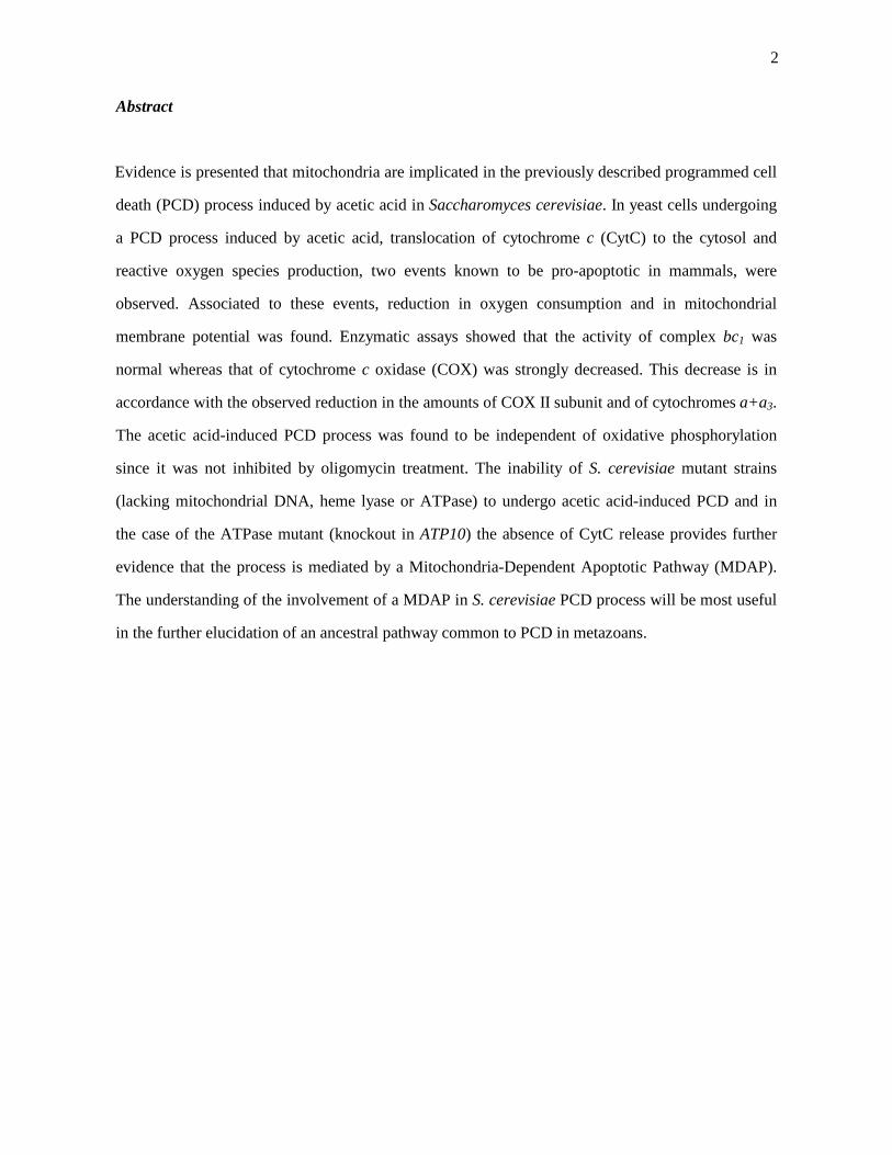
Abstract
Evidence is presented that mitochondria are implicated in the previously described programmed cell
death (PCD) process induced by acetic acid in Saccharomyces cerevisiae. In yeast cells undergoing
a PCD process induced by acetic acid, translocation of cytochrome c (CytC) to the cytosol and
reactive oxygen species production, two events known to be pro-apoptotic in mammals, were
observed. Associated to these events, reduction in oxygen consumption and in mitochondrial
membrane potential was found. Enzymatic assays showed that the activity of complex bc1 was
normal whereas that of cytochrome c oxidase (COX) was strongly decreased. This decrease is in
accordance with the observed reduction in the amounts of COX II subunit and of cytochromes a+a3.
The acetic acid-induced PCD process was found to be independent of oxidative phosphorylation
since it was not inhibited by oligomycin treatment. The inability of S. cerevisiae mutant strains
(lacking mitochondrial DNA, heme lyase or ATPase) to undergo acetic acid-induced PCD and in
the case of the ATPase mutant (knockout in ATP10) the absence of CytC release provides further
evidence that the process is mediated by a Mitochondria-Dependent Apoptotic Pathway (MDAP).
The understanding of the involvement of a MDAP in S. cerevisiae PCD process will be most useful
in the further elucidation of an ancestral pathway common to PCD in metazoans.

3
Introduction
Programmed cell death (PCD), of which apoptosis is the most common morphological expression,
is described as an orchestrated collapse of the cell. This process plays an important role in the
normal development and homeostasis mechanisms of multicellular organisms. At least two major
apoptotic pathways have been described in mammalian cells. One requiring the participation of
mitochondria, called “intrinsic pathway”, and another one in which mitochondria are bypassed and
caspases are activated directly, called “extrinsic pathway” (Matsuyama et al., 2000; Hengartner,
2000). Regarding the mitochondrial pathway, two main events have been proposed as integral
control elements in the cell’s decision to dye, namely, the release of apoptogenic factors such as
cytochrome c (CytC) and the production of reactive oxygen species (Liu et al., 1996; Kluck et al.,
1997; Pham et al., 2000). Release of CytC to the cytosol drives the assembly of a high-molecular
weight complex, the mitochondrial apoptosome that activates caspases (Adrain and Martin, 2001).
Translocation of CytC to the cytosol is, therefore, a pivotal event in apoptosis. CytC is a soluble
protein loosely bound to the outer face of the inner mitochondrial membrane, and its release is
associated with an interruption of the normal electron flow at the complex III site of the respiratory
chain, that could divert electron transfer to the generation of superoxide (Cai and Jones, 1998). The
mechanism by which CytC is released from mitochondria during apoptosis remains unknown.
However, two competing models have been proposed, a volume-dependent mechanism, involving
mitochondrial swelling and the rupture of the outer membrane, and a volume-independent
mechanism, in which the permeability of the outer membrane is selectively altered (Matsuyama et
al., 2000). Pavlov et al. (2001) reported, very recently, a new high conductance channel named
mitochondrial apoptosis-induced channel (MAC) linked to apoptosis in mammalian cells and Bax
expression in yeast, which is in agreement with the latter model. The authors propose this channel as
a candidate for the outer mitochondrial membrane pore through which CytC and possible other
factors exit mitochondria during apoptosis.
4
Recently, we have shown that acetic acid (20-80 mM) induces death in exponential cells of
Saccharomyces cerevisiae which displays the most common PCD hallmarks such as chromatin
condensation along the nuclear envelope; exposure of phosphatidylserine on the outer surface of the
cytoplasmic membrane and occurrence of DNA fragmentation (Ludovico et al., 2001a). It was also
shown that similar to hydrogen peroxide (Madeo et al., 1999), acetic acid at high doses (above 120
mM) induces cell morphological changes typical of necrosis without apoptotic markers such as
TUNEL-positive phenotype. Acetic acid is a normal end-product of fermentation carried out by S.
cerevisiae and may be produced by contaminating acetic acid bacteria being therefore quite familiar
to the yeast environment. In this context the occurrence of acetic acid-induced PCD points to a
physiological role for such a cell death in yeast.
In the last few years the occurrence of a PCD process independent of Bax-expression and with an
apoptotic phenotype in S. cerevisiae has been reported (Madeo et al., 1997; Madeo et al., 1999;
Laun et al., 2001; Ludovico et al., 2001a; Yamaki et al., 2001; Carratore et al., 2002). However,
only Yamaki and coworkers (2001) studied the involvement of mitochondria in such a cell death
pathway. The authors observed that cell death mediated by deletion of histone chaperone
ASF1/CIA1 is associated to a decrease of mitochondrial membrane potential, dysfunction of
mitochondrial ATPase and release of CytC to the cytoplasm, a phenotype that largely resembles
mammalian apoptosis. Although controversial, there is a interpretation that mitochondria seem to be
involved in Bax-induced cell death in yeast. Matusyama and coworkers (1998) reported that ATP4,
which is a nuclear gene encoding subunit 4 of the yeast mitochondrial ATPase complex, is required
for yeast death induced by Bax-expression. Additionally, as in mammalian cells, Bax-expression
leads to CytC release from yeast mitochondria (Manon et al., 1997; Kluck et al., 2000). Manon and
coworkers (1997) described that Bax-expression in yeast cells stimulates the release of CytC to the
cytosol and causes a concomitant decrease in the amount of COX complex.

5
In the present paper a large body of evidence supporting the involvement of mitochondria in the S.
cerevisiae PCD process triggered by acetic acid is presented, indicating that, like in mammalian
cells, the PCD in yeast can be mediated by a Mitochondria-Dependent Apoptotic Pathway (MDAP).

6
Materials and Methods
Microorganisms and growth conditions
The yeast Saccharomyces cerevisiae W303-1A (MAT a ade2 leu2 his3 trp1 ura3), the respective
knockout in ATP10 and CYC3 genes and the isogenic derivative ρ0 strain (lacking mitochondrial
DNA) were used. These organisms were maintained on YEPD agar slants containing glucose (2 %,
w/v), yeast extract (1 %, w/v), peptone (2 %, w/v) and agar (2 %, w/v). In experiments, the yeast
cells were sub-cultured in liquid YEPD medium. The growth experiments were performed in 250
ml flasks containing a ratio 2:1 of air to liquid phase, and incubated on a mechanical shaker (150
rpm) at 26 °C.
Treatments with acetic acid and inhibition of protein synthesis or oxidative phosphorylation
Stationary phase cells were harvested and suspended (107cells/ml) in treatment medium (YEPD, pH
3.0, set with HCl) containing 0, 120, 140, 180, 200 and 240 mM acetic acid. The treatments were
carried out for 200 minutes at 26 °C with magnetic stirring (150 rpm). Inhibition of protein
synthesis or oxidative phophorylation was performed by adding 50 µg/ml cycloheximide (Merck) or
10 µM oligomycin (Matsuyama et al., 1998), respectively, at the same time as the different acetic
acid concentrations tested. At these concentrations cycloheximide and oligomycin were not
cytotoxic after 200 minutes incubation, as assessed by counting colony forming units (cfu). Viability
was determined by cfu counts after 2 days of incubation at 26 ºC on YEPD agar plates. No further
colonies appeared after that incubation period. In all the above experiments, the extracellular pH did
not change during the incubations.
7
Terminal deoxynucleotidyl transferase-mediated dUTP Nick End Labelling (TUNEL) and
propidium iodide staining
DNA strand breaks were demonstrated by Terminal Deoxynucleotidyl Transferase-mediated dUTP
Nick End Labelling (TUNEL) with the “In Situ Cell Death Detection Kit, Fluorescein”, from
Boehringer Mannheim (Indianapolis, IN) as described before (Ludovico et al., 2001a).
Isolation of yeast mitochondria and mitochondrial respiratory chain activity measurements
Mitochondria were isolated from S. cerevisiae (W303-1A and ATP10 mutant) cells grown to
stationary phase, harvested and resuspended in YEPD, pH 3.0 in the absence or presence of 140
mM acetic acid for 200 min. Mitochondria were prepared essentially as described by Faye et al.
(1974) except that Zymolyase 20,000 instead of Glusulase was used to digest the cell wall. The
post-mitochondrial supernatant (supernatant after the first 12,000 rpm centrifugation) was kept for
the detection of cytochrome c (CytC). Mitochondria were washed twice with 0.5 M sorbitol at
12,000 rpm for 15 min and suspended in 0.5 M sorbitol. Protein concentration was ascertained by
the method published by Lowry et al. (1951).
Oxygen utilization in isolated mitochondria was measured polarographically in 1 ml of standard
medium (0.5 M sorbitol, 20 mM K2PO4 pH 7.4, 1 mM EDTA) with a Clark oxygen electrode
(model 5300: Yellow Springs Instruments Co., Yellow Springs Ohio), in a water-jacketed cell,
magnetically stirred at room temperature. Twenty µg of mitochondrial protein were used to assay
for NADH oxidase activity. Oxygen consumption was monitored using NADH (0.8 mM) as a
substrate. In a set of reactions, the effect of exogenous CytC on the oxidative rate was checked out
by adding 10 µM horse CytC (Sigma, St Louis, MO) to the chamber. Respiration was inhibited by
addition of 700 µM potassium cyanide (KCN).

8
Measurement of respiratory chain enzymatic activities in isolated mitochondria was performed
essentially as described by Tzagoloff et al. (1975). Cytochrome c oxidase (COX) activity was
measured at room temperature in 20 mM K2HPO4, pH 7.5; containing 65 µM reduced CytC. COX
activity was assayed in mitochondria (10 µg protein) permeabilized with potassium deoxycholate
(KDOC) to maximize the access of the substrate to the enzyme, by measuring oxidation of
ferrocytochrome c at 550 nm. NADH-Cytochrome c reductase activity was measured in KDOC
permeabilized mitochondria at room temperature in 10 mM K2HPO4, pH 7.5, containing 100 µM
KCN by following the reduction of CytC (65 µM) at 550 nm in the presence of 1 M NADH.
Cytochromes spectra
Optical absorption spectra of mitochondrial cytochromes were obtained on an Aminco DW-2A dual
wavelength scanning spectrophotometer. The spectrophotometer was operated in the split beam
mode with 1 nm band pass. Mitochondria were extracted at a protein concentration of 5 mg/ml with
potassium deoxycholate under conditions that quantitatively solubilize all the cytochromes
(Tzagoloff et al., 1975). Difference spectra of the reduced (sodium dithionite) versus oxidized
(potassium ferricyanide) extracts were recorded at room temperature. The α absorption bands
corresponding to cytochromes a and a3 have maxima at 603 nm. The maxima for cytochrome b and
for cytochrome c and c1 are 560 nm and 550 nm, respectively.
Cytochrome c distribution
The release of CytC from mitochondria to cytoplasm when S. cerevisiae (W303-1A and ATP10
mutant) cells were grown in the presence or absence of acetic acid was checked out by Western blot
detection of CytC in the subcellular compartments. Total mitochondrial proteins (5, 10 or 20 µg)
and the post-mitochondrial supernatant (PMS) were electrophoretically separated on 12.0 % SDS-
9
polyacrylamide gels (Laemmli, 1970) and transferred to 0.2 µm nitrocellulose membranes. The
membranes were blocked with 5 % (w/v) milk powder for at least 1 hour followed by the incubation
for another hour with anti-yeast CytC polyclonal antibody (kind gift from Dr. R. Lill). The blots
were then incubated in goat anti-rabbit IgG conjugated to horseradish peroxidase (Sigma). The
SuperSignal chemiluminescent substrate kit (Pierce, Il, USA) was used for the final detection.
Steady-state levels of bc1, complex COX and ATPase subunits
The detection of cytochrome b, COX subunit II and V, and ATPase subunit 6 were performed by
Western blotting as described above for CytC detection, and using primary polyclonal antibodies
against the yeast forms of the mentioned subunits. All the antibodies used were a kind gift of Dr.
Alexander Tzagoloff (Columbia University, New York).
Assessment of mitochondrial membrane potential (∆Ψ∆Ψ∆Ψ∆Ψm) and of Reactive Oxygen Species (ROS)
production
Assessment of ∆Ψm in whole cells was performed by flow cytometry and epifluorescence
microscopy as described by Ludovico et al. (2001b). The control suspensions of killed cells used
both in the assessment of ∆Ψm and of ROS production were prepared by boiling for 10 min the
cell suspensions as referred by Ludovico et al. (2001b).
ROS production by mitochondria, was monitored by using the MitoTracker® Red CM-H2XRos
staining. The reduced version of MitoTracker Red CMXRos does not fluoresce until entering an
actively respiring cell, where it is oxidized by ROS to a red fluorescent compound, which is
sequestered in the mitochondria. Cells were harvested and then suspended in the treatment
medium (107cells/ml). Before the addition of acetic acid, cells were pre-loaded with 50 µg/ml of

10
the dye for 20 minutes at 37 ºC. After the pre-loading the different acetic acid concentrations were
added and the fluorescence was detected by flow cytometry and epifluorescence microscopy.
Flow cytometric analysis was performed in a basic FACS Calibur (Beckton Dickinson) flow
cytometer equipped with an argon-ion laser emitting a 488 nm beam at 15 mW. Red fluorescence
was collected through a 560 nm short-pass dichroic, a 640 nm long-pass and another 670 nm
long-pass. Twenty thousand cells per sample were analysed at low flow rate. An acquisition
protocol was defined to measure forward scatter (FS Log), side scatter (SS Log) and red
fluorescence (FL3 Log) on a four decades logarithmic scale. Data were acquired and analysed
with CELLQuest PRO 3.3 (Beckton Dickinson).

11
Results
Wild-type S. cerevisiae stationary cells undergo a PCD process induced by acetic acid
The study was focused on the role of mitochondria in the previously reported acetic acid induced
programmed cell death (PCD) process in Saccharomyces cerevisiae W303-1A (Ludovico et al.,
2001a). Stationary cells rather than exponential cells were used for that purpose. Cells from
stationary growth phase are more advantageous since they possess fully active mitochondria and
display a higher mitochondrial mass. When exposed to different acetic acid concentrations at pH
3.0, and in the presence of glucose, S. cerevisiae stationary cells also committed to a PCD process.
This conclusion is based on the detection of cell death accompanied by DNA strand breaks
evaluated by the TUNEL assay (data not shown), an evident apoptotic marker, and by the fact that
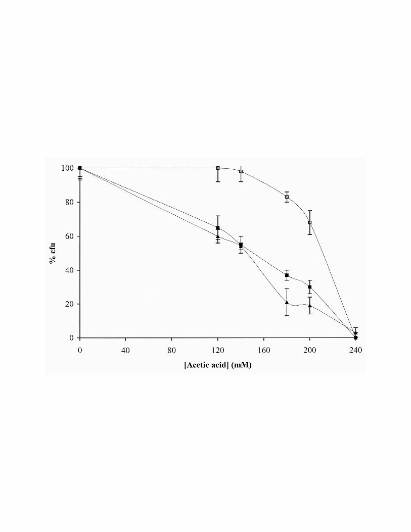
cycloheximide inhibited cell death (Fig. 1). Consistently with the recognized higher resistance of
stationary cells to different stress agents, the effect was only observed for higher acetic acid
concentrations. Actually, while acetic acid concentrations above 120 mM are necrotic for
exponential cells, they induce a PCD process in stationary cells. In fact, 140 mM acetic acid
concentration induces, after 200 min, about 50 % of loss of cell viability evaluated by cfu and about
30 % of TUNEL-positive cells. This concentration (140 mM) was selected to perform mitochondrial
function analysis. Incubation with acetic acid concentrations above to 200 mM resulted in no
detectable TUNEL staining (data not shown).
In order to evaluate whether the acetic acid-induced PCD in S. cerevisiae is affected by the
inhibition of oxidative phosphorylation, the treatment with acetic acid was carried out in the
presence of oligomycin. Figure 1 shows that cell death was not affected by the drug.

12
Cytochrome c is translocated from mitochondria to the cytosol during the acetic-acid induced
PCD process
To check if acetic acid-induced PCD process was accompanied by a release of CytC from
mitochondria to cytosol, the levels of CytC in mitochondria and in the post-mitochondrial
supernatant (PMS, containing soluble cytosolic proteins), from S. cerevisiae W303-1A cells
undergoing a acid-induced PCD, were detected by Western blot analysis. The amount of CytC
present in mitochondria of cells treated with 140 mM acetic acid was decreased by a 2-3 fold
compared with the mitochondria from untreated cells (Fig. 2). This portion of "lost" CytC in
mitochondria from acetic acid treated cells was detected in its PMS, while no CytC was detected in
the PMS of untreated control cells (Fig. 2). Levels of other mitochondrial proteins, such as COX
subunits II and V were not detected in PMS (data not shown), although COX II was found to be
reduced in mitochondria (described hereafter). Decrease of the CytC amount in mitochondria was
confirmed by cytochrome spectra analysis of mitochondria from treated and untreated cells. As
shown in Figure 3A, there was some decrease in the amount of cytochromes c+c1 extracted from the
mitochondrial membranes.
The levels of CytC were also evaluated in mitochondria and in the PMS from S. cerevisiae ATP10
mutant cells which do not undergo a PCD process induced by acetic acid as shown hereafter. CytC
was not detected in PMS and it was found to be at a normal level in mitochondria, comparatively to
untreated cells (data not shown).
These results indicate that CytC is specifically translocated from mitochondria to the cytosol during
the acid-induced PCD.
13
Reactive Oxygen Species (ROS) are produced in mitochondria during the acetic acid-induced
PCD process
S. cerevisiae W303-1A stationary cells stained with MitoTracker® Red CM-H2Xros, and treated
with 140 mM acetic acid, were analysed by flow cytometry. An increase in the red fluorescence
indicative of cells with an increased mitochondrial ROS production could only be detected after 100
min of treatment (data not shown). The results obtained after 200 min of treatment showed a
heterogeneous population and a second sub-population (about 45 %) with a higher mean
fluorescence intensity (Fig. 4). The epifluorescence analysis showed that this sub-population display
a bright red fluorescence localized in mitochondria (data not shown). Moreover, killed cells
displayed a highest red fluorescence corresponding to an unspecific cell staining (data not shown).
Acetic acid-induced PCD is accompanied by mitochondrial alterations
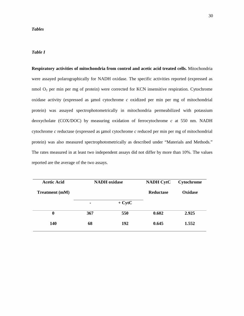
The respiratory capacity of mitochondria isolated from control and acetic acid-treated cells was
assayed polarographically by measuring oxygen uptake using NADH as substrate. The effect on
respiration of exogenously added CytC was evaluated in a set of experiments. Results presented in
Table I show that mitochondria isolated from cells treated with 140 mM acetic acid have a
dramatically reduced oxygen consumption, with a decrease of nearly 75 % in NADH oxidase
activity. Although an increase of about 2.8 fold of the oxygen consumption was observed after
addition of CytC, the inability to fully restore the respiration rate with this addition suggested that
an enzymatic portion of the respiratory chain could be intrinsically affected.
To establish the biochemical basis for the decrease in the respiratory activity of mitochondria, the
NADH-CytC reductase and COX activities of isolated mitochondria from untreated and acid-treated
cells, were measured. As shown in Table I, the treatment with 140 mM acetic acid resulted in a
decrease of 50 % of COX activity in mitochondrial membranes, while NADH-CytC reductase
14
activity was essentially identical to that obtained with mitochondria from untreated cells. These
results confirm that COX complex was affected whereas complex bc1 was unaffected by the
treatment. Cytochromes spectra were recorded in isolated mitochondria (Fig. 3A) to clarify the
observed respiratory chain alterations (Table I). In agreement with the observed lower COX activity,
a decrease in the amount of cytochromes a+a3 in mitochondrial membranes of cells treated with
acetic acid was observed while the levels of cytochrome b were not affected (Fig. 3A).
When the COX subunits II and V were analysed by immunodetection of mitochondrial proteins
obtained from cells treated with 140 mM acetic acid, the amount of COX II protein but not COX V
was found to be lower than in the control (Fig. 3B). Additionally, the amount of cytochrome b from
bc1 complex and of subunit 6 of ATPase remained identical to control (Fig. 3B).
Some studies on mammalian apoptosis reported an increase in mitochondrial membrane potential
(∆Ψm) after a lethal stimulus, with ∆Ψm decreasing later in the death process (Vander Heiden et
al., 1997; Vander Heiden et al., 1999). Consistently with such observations, in our study, acetic acid
treatment induced a transient slight hyperpolarization followed by a depolarisation (data not shown).
However, although with a lower ∆Ψm, cells maintained the specific mitochondria staining
indicating that mitochondria membrane integrity is still preserved (data not shown).
Mitochondrial respiration is essential for S. cerevisiae to undergo a PCD process induced by
acetic acid
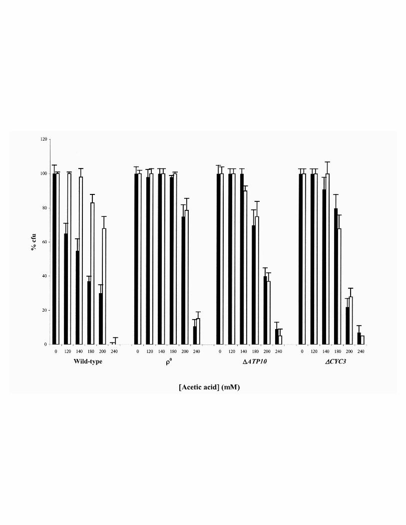
The requirement of mitochondria function in the PCD process induced by acetic acid was analysed
by the study of three S. cerevisiae W303-1A mutant strains, namely the ρ0, lacking mitochondrial
DNA, the null ATP10 mutant, deleted in an assembly factor of mitochondrial ATPase, and the null
CYC3 mutant, deleted in the gene encoding a heme lyase, essential for the covalent binding of the
heme group to isoform 1 and 2 of apocytochrome c (Pearce and Sherman, 1995). It was observed

15
that these mutant strains were more resistant to death induced by acetic acid, comparatively to the
wild type strain. In addition, cycloheximide had no effect on survival (Fig. 5) and no TUNEL-
positive cells were found, for any of the acetic acid concentrations tested, even for concentrations
inducing a percentage of dead cells identical to that obtained for the wild-type (data not shown).
16
Discussion
Mitochondria have been implicated in mammalian PCD processes (Skulachev, 1996, 1998, 2000,
2001; Green and Reed, 1998; Yang et al., 1997; Kluck et al., 1997) being called the “cell death
organelle”. Data reported in this paper allowed us to conclude that mitochondria are, as well,
implicated in the active cell death of Saccharomyces cerevisiae W303-1A induced by acetic acid.
The strategy followed to analyse the possible role of mitochondria in that PCD process involved the
use of stationary cells of S. cerevisiae which have a high mass of fully active mitochondria, and of
selected S. cerevisiae strains with mutations affecting mitochondria. Regarding the first strategy, we
found that S. cerevisiae W303-1A stationary cells, like exponential cells (Ludovico et al., 2001a),
can organise its own death in response to acetic acid. Using those cells, the possibility was addressed
that CytC could be in this unicellular eukaryont, as in mammalian cells, a direct or indirect
apoptogenic factor. The analysis of CytC localization by immunodetection revealed that acetic acid
triggers CytC release from mitochondria to the cytosol in S. cerevisiae W303-1A under PCD. CytC
extrusion from mitochondria was also concluded from the results of cytochromes spectra analysis.
Recently, Yamaki et al. (2001) observed a release of CytC to the cytoplasm associated to a
phenotype that largely resembles mammalian apoptosis in S. cerevisiae cells lacking the histone
chaperone ASF1/CIA1. The human homologue of this chaperone appears to be involved in the
regulation of apoptosis. Manon and coworkers (1997), using heterologous expression of the pro-
apoptotic protein Bax, had previously described CytC release in S. cerevisiae. In this context, since
CytC release was found in the apoptosis-like cell death induced by Bax-expression, deletion of the
histone chaperone ASF1/CIA1 and acetic acid treatment (Ligr et al., 1998; Ludovico et al., 2001a;
Yamaki et al., 2001), we have an explanation why the apoptotic phenotype is identical in those three
situations. It is conceivable that, when in the cytosol after its translocation from mitochondria, yeast
CytC could work as an apoptogenic factor, as reported for mammalian cells. In these cells the pro-
apoptotic activity of cytoplasmic CytC is due to the activation of a caspase cascade. Very recently,
17
Madeo and coworkers (2002) reported a caspase-related protease that regulates yeast apoptosis,
named Yeast Caspase-1 (YCA-1). These authors showed that the disruption of YCA-1 gene resulted
in a higher survival to acetic acid treatment comparatively to the wild-type, whereas its
overexpression led to the enhancement of cell death. In this context, when in cytosol, CytC may also
work as an apoptogenic factor in S. cerevisiae through the activation of YCA-1, but further research
is needed to get a complete frame of the interaction between these two proteins.
On the other hand, the release of CytC from mitochondria to the cytosol, being accompanied by the
loss of the molecule at the mitochondria, leads to the occurrence of another pro-apoptotic event,
namely the production of reactive oxygen species (ROS). Indeed, the observed increase in ROS
production in S. cerevisiae W303-1A cells exposed to acetic acid may well be the consequence of
the reduction in COX activity due to the loss of CytC from mitochondria. Whether loss of COX
activity is a consequence of CytC release or a primary effect of acetic acid on the assembly of the
enzyme remains to be studied although we favour the first possibility. The observed deficiency in
COX activity could be responsible for an inhibition of the electron transfer with the accumulation of
reducing equivalents in the middle portion of the electron transfer chain, and thus directing one-
electron transfer to O2, resulting in the production of superoxide (Cai and Jones, 1998). ROS
production appears as an event common to all the instances of S. cerevisiae apoptotic phenotypes
reported so far (Madeo et al., 1997; Madeo et al., 1999; Fröhlich and Madeo 2000; Laun et al.,
2001; Levine et al., 2001; Narasimhan et al., 2001; present results).
In mitochondria, CytC is an essential component of the respiratory chain acting as an electron
carrier between complex bc1 and COX complex. Therefore, the decrease in the amount of CytC in
mitochondria of S. cerevisiae exposed to acetic acid must have other functional implications. The
biochemical basis for the observed decrease in oxygen consumption and in ∆Ψm is the important
reduction in COX activity. Such a reduction could be due to an inefficient COX assembly in the
absence of CytC. The decrease of steady-state levels of COX II protein observed in mitochondria
18
from acetic acid treated cells that have lost CytC and the impossibility to detect it in PMS indicate
that, probably a degradation of the subunit occurred, as has been reported in mutants lacking CytC
(Pearce and Sherman, 1995). In fact, COX II participates in CytC binding (Bisson et al., 1977) and
the absence of CytC appears to destabilise COX II, making it susceptible to degradation (Pearce and
Sherman, 1995). Manon et al., (2001) reported very recently that Bax-induced CytC release is
directly involved in the decrease of COX activity by activating the mitochondrial AAA-type
protease Yme1p, which leads to COX II degradation.
A pattern of mitochondrial dysfunction identical to that above discussed was observed in S.
cerevisiae cells expressing Bax (Manon et al., 1997). With this new insight, a link between major
alterations in the respiratory chain, namely decrease in the amount of CytC and reduction of the
COX activity, and the PCD process in yeast, can be envisaged, being the first time that the apoptotic
mitochondrial pathway was found to be activated in yeast wild type cells independent of the
heterologous expression of Bax.
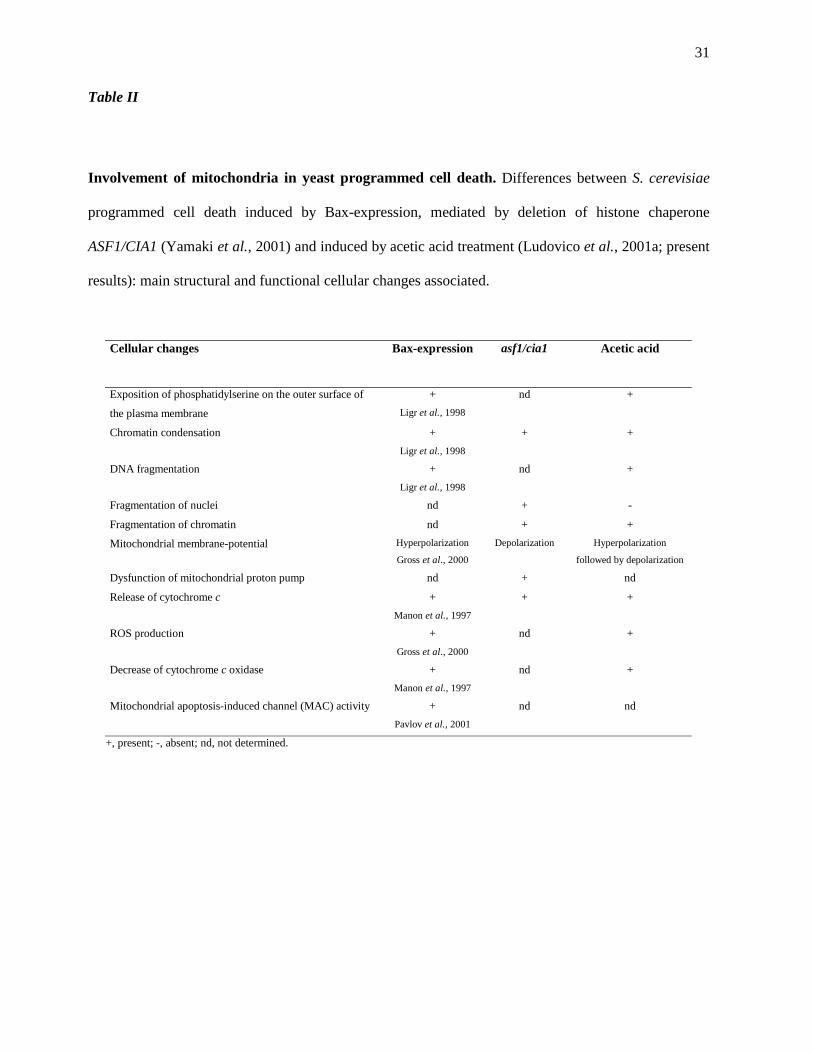
The comparison between main structural and functional cellular changes, including mitochocondrial
changes, associated to S. cerevisiae programmed cell death induced by Bax-expression, by deletion
of histone chaperone ASF1/CIA1 or by acetic acid treatment is outlined in Table II.
The above discussed results, obtained with experiments using stationary cells of S. cerevisiae
W303-1A, indicated that mitochondria would be involved in the PCD process induced by acetic
acid through the release of CytC and ROS production. The use of three S. cerevisiae strains with
mutations affecting mitochondrial respiratory chain function led to results that further indicate that
mitochondria are required for that process. Although the literature concerning the mitochondria
involvement in Bax-induced cell death is controversial (Greenhalf et al., 1996; Kissova et al.,
2000), our results clearly demonstrate that a yeast cell depleted of mitochondrial DNA does not
undergo a PCD process in response to acetic acid.
Supporting the interpretation that CytC exhibits apoptogenic properties in S. cerevisiae, the mutant
in CYC3 gene, which encodes a heme lyase essential for the heme binding to apocytochrome c

19
(isoform 1 and 2) (Pearce and Sherman, 1995), did not display PCD when exposed to acetic acid. It
is interesting to notice that in ρo strains, the CYC1 gene (coding for iso-1-CytC) is 4-6 fold
repressed, probably due to the lack of mitochondrial DNA and of mitochondrial respiratory activity
(Epstein et al., 2001), perhaps explaining why those strains do not undergo a PCD induced by acetic
acid.
Matsuyama et al. (1998) related cell death induced by Bax-expression with the mitochondrial
ATPase complex, namely with ATP subunit 4. Our results regarding the inability of ATP10 mutant
[ATP10 codes a mitochondrial protein encoded by nuclear DNA that acts as an ATPase assembly
factor (Paul et al., 2000)] to undergo PCD by exposure to acetic acid indicate that a fully assembled
F0F1-ATPase is required for the PCD process. Moreover, and because it has been proposed that
ATPase complex could be involved in the mechanism of CytC release in apoptotic mammalian cells
(Matsuyama et al., 2000), our results with ATP10 null mutant showing that treatment with 140 mM
acetic acid does not result in PCD nor CytC release suggest that the ATPase complex would also be
involved in the mechanism of CytC release from mitochondria during PCD induced by acetic acid
in S. cerevisiae. Because of the absence of oxidative phosphorylation in the ρ0 and ATP10 mutant
strains, it could be argued that the inability of those strains to develop the acid-induced PCD process
could be due to the failure in mitochondrial ATP generation. Nevertheless, this can not be the
explanation since cells of wild-type strain are able to induce PCD even when mitochondrial ATP
synthesis is inhibited by oligomycin.
In summary, the experimental evidence herein presented strongly supports that the PCD process
induced by acetic acid in S. cerevisiae is mediated by a Mitochondria-Dependent Apoptotic
Pathway (MDAP). Therefore, in yeast the pivotal role of mitochondria is related not only to the
exogeneous cell death induced by Bax-expression but also, and more relevantly, to the endogenous
PCD induced with acetic acid. These advances surpass the contemporary knowledge on PCD in
unicellular eukaryotes and raise new questions regarding PCD in yeast, such as: i) is CytC an

20
activating factor of YCA-1; ii) is CytC release an event common to all endogenous PCD processes?
The answers to these questions will be most useful in the further elucidation of ancestral pathway(s)
common to PCD in metazoans and will validate the yeast cell as the tool of choice in apoptosis
research.

21
Acknowledgements
The stay of Paula Ludovico in Department of Biology at Columbia University, New York, was
supported in part by “Fundação Luso-Americana para o Desenvolvimento”. Paula Ludovico has a
fellowship from PRAXIS XXI (Fundação da Ciência e Tecnologia, Portugal). Antoni Barrientos is a
recipient of a grant MDACU01991001 from the Muscular Dystrophy Association. The authors
would like to thank Prof. Vladimir Skulachev and Dr. Frank Madeo for their critical reading of the
manuscript and all the helpful comments and suggestions.
22
Abbreviations List
COX, Cytochrome c oxidase; CytC, cytochrome c; MDAP, mitochondria-dependent apoptotic
pathway; ∆Ψm, mitochondria membrane potential; PCD, programmed cell death; PMS, post-
mitochondrial supernatant; ROS, reactive oxygen species; TUNEL, TdT-mediated dUTP nick end
labelling.
23
References
Adrian, C. and Martin, S.J. (2001). The mitochondrial apoptosome: a killer unleashed by the
cytochrome seas. Trends Biochem. Sci. 26:390-397.
Bisson, R., Gutweniger, H., Montecucco, C., Colonna, R., Zanotti, A. and Azzi, A. (1977).
Covalent binding of arylazido derivatives of cytochrome c to cytochrome oxidase. FEBS Lett.
81:147-150.
Cai, J. and Jones, D.P. (1998). Superoxide in apoptosis. Mitochondrial generation triggered by
cytochrome c loss. J. Biol. Chem. 273:11401-11404.
Carratore, M.R., Croce, C., Simili, M., Taccini, E., Scavuzzo, M. and Sbrana, S. (2002). Cell
cycle and morphological alterations as indicative of apoptosis promoted by UV irradiation in S.
cerevisiae. Mutat Res. 513: 183-191.
Epstein, C., Waddle, J.A., Hale IV, W., Davé, V., Thornton, J., Macatee, T.L., Garner, H.R.
and Butow, R. (2001). Genome-wide responses to mitochondrial dysfunction. Mol. Biol. Cell
12:297-308.
Faye, G., Kujawa, C. and Fukuhara, H. (1974). Physical and genetic organization of petite and
grande yeast mitochondrial DNA. IV. In vivo transcription products of mitochondrial DNA and
localization of 23 S ribosomal RNA in petite mutants of Saccharomyces cerevisiae. J. Mol. Biol.
88:185-203.
Fröhlich, K.U. and Madeo, F. (2000). Apoptosis in yeast – a monocellular organism exhibits
altruistic behaviour. FEBS Lett. 473:6-9.
Green, D.R. and Reed, J.C. (1998). Mitochondria and apoptosis. Science 281:1309-1312.
Greenhalf, W., Stephan, C. and Chaudhuri, B. (1996). Role of mitochondria and C-terminal
membrane anchor of Bcl-2 in Bax induced growth arrest and mortality in Saccharomyces cerevisiae.
FEBS Lett. 380:169-175.
24
Gross, A., Pilcher, K., Blachly-Dyson, E., Basso, E., Jockel, J., Bassik, M., Korsmeyer, S. and
Forte, M. (2000). Biochemical and genetic analysis of the mitochondrial response of yeast to BAX
and BCL-XL. Mol. Cell Biol., 20 (9):3125-3136.
Hengartner, M.O. (2000). The biochemistry of apoptosis. Nature 407:770-776.
Kissova, I., Polcic, P., Kempna, P., Zeman, I., Sabova, L. and Kolarov, J. (2000). The cytotoxic
action of Bax on yeast cells does not require mitochondrial ADP/ATP carrier but may be related to
its import to the mitochondria. FEBS Lett. 471:113-118.
Kluck, R., Bossy-Wetzel, E., Green, D.R. and Newmeyer, D.D. (1997). The release of
cytochrome c from mitochondria: a primary site for Bcl-2 regulation of apoptosis. Science
275:1132-1136.
Kluck, R., Ellerby, L.M., Ellerby, H.M., Naiem, S., Yaffe, M., Margoliash, E., Bredesen, D.,
Mauk, A.G., Sherman, F. and Newmeyer, D. (2000). Determinants of cytochrome c pro-apoptotic
activity – The role of lysine 72 trimethylation. J. Biol. Chem. 275:16127-16133.
Laemmli, U.K. (1970). Cleavage of structural proteins during the assembly of the head of
bacteriophage T4. Nature 227:680-685.
Laun, P., Pichova, A., Madeo, F., Fuchs, J., Ellinger, A., Kohlwein, S., Dawes, I., Frohlich,
K.U. and Breitenbach, M. (2001). Aged mother cells of Saccharomyces cerevisiae show markers
of oxidative stress and apoptosis. Mol. Microbiol. 39:1166-1173.
Levine, A., Belenghi, B., Damari-Weisler, H. and Granot, D. (2001). Vesicle-associated
Membrane Protein of Arabidopsis Suppresses Bax-induced Apoptosis in Yeast Downstream of
Oxidative Burst. J. Biol. Chem. 276:46284-46289.
Ligr, M., Madeo, F., Frohlich, E., Hilt, W., Frohlich, K.U. and Wolf, D.H. (1998). Mammalian
Bax triggers apoptotic changes in yeast. FEBS Lett. 438:61-65.
Liu, X., Kim, C.N., Yang, J., Jemmerson, R. and Wang, X. (1996). Induction of apoptotic
program in cell-free extracts: requirement for dATP and cytochrome c. Cell 86:147-157.
25
Lowry, O.H., Rosebrough, N.J., Farr, A.L. and Randall R.J. (1951). Protein measurement with
Folin phenol reagent. J. Biol. Chem. 193:265-275.
Ludovico, P., Sousa, M.J., Silva, M.T., Leão, C. and Côrte-Real, M. (2001a). Saccharomyces
cerevisiae commits to a programmed cell death process in response to acetic acid. Microbiology
147:2409-2415.
Ludovico, P., Sansonetty, F. and Côrte-Real, M. (2001b). Assessment of mitochondrial
membrane potential in yeast cell populations by flow cytometry. Microbiology 147:3335-3343.
Madeo, F., Fröhlich, E. and Fröhlich, K.U. (1997). A yeast mutant showing diagnostic markers of
early and late apoptosis. J. Cell Biol. 139:729-734.
Madeo F., Fröhlich E., Ligr M., Grey M., Sigrist S. J., Wolf D.H., and Fröhlich K.U. (1999).
Oxygen stress: a regulator of apoptosis in yeast. J. Cell Biol. 145:757-767.
Madeo, F., Herker, E., Maldener, C., Wissing, S., Lächelt, S., Herlan, M., Fehr, M., Lauber,
K., Sigrist, S., Wesselborg, S. and Fröhlich, K.-U. (2002). A caspase-related protease regulates
apoptosis in yeast. Mol. Cell 9:1-20.
Manon, S., Chaudhuri, B. and Guérin, M. (1997). Release of cytochrome c and decrease of
cytochrome c oxidase in Bax-expressing cells, and prevention of these effects by coexpression of
Bcl-xL. FEBS Lett. 415:29-32.
Manon, S., Priault, M. and Camougrand, N. (2001). Mitochondrial AAA-Type protease Yme1p
is involved in Bax effects on Cytochrome c Oxidase. Biochem. Biophys. Res. Commun. 289:1314-
1319.
Matsuyama, S., Xu, Q., Velours, J. and Reed, J.C. (1998). The mitochondrial F0F1-ATPase
proton pump is required for function of the proapoptotic protein Bax in yeast and mammalian cells.
Mol. Cell 1:327-336.

26
Matsuyama, S., Llopis, J., Deveraux, Q.L., Tsien, R. and Reed, J.C. (2000). Changes in
mitochondrial and cytosolic pH: early events that modulate caspase activation during apoptosis.
Nature Cell Biology 2:318-325.
Narasimhan, M.L., Damsz, B., Coca, M.A., Ibeas, J.I., Yun, D.J., Pardo, J.M., Hasegawa, P.M.
and Bressan, R.A. (2001). A plant defense response effector induces microbial apoptosis. Mol. Cell
8:921-30.
Paul, M.F., Barrientos, A. and Tzagoloff, A. (2000). A single amino acid change in subunit 6 of
the yeast mitochondrial ATPase suppresses a null mutation in ATP10. J. Biol. Chem. 275:29238-
29243.
Pavlov, E., Priault, M., Pietkiewicz, D., Cheng, E., Antonsson, B., Manon, S., Korsmeyer, S.,
Mannella, C. and Kinnally, K. (2001). A novel, high conductance channel of mitochondria linked
to apoptosis in mammalian cells and Bax expression in yeast. J. Cell Biol. 155:725-731.
Pearce, D. and Sherman, F. (1995). Degradation of cytochrome oxidase subnunits in mutants of
yeast lacking cytochrome c and suppression of the degradation by mutation of yme1. J. Biol. Chem.
270:20879-20882.
Pham, N., Robison, B. and Hedley, D. (2000). Simultaneous detection of mitochondrial respiration
chain activity and reactive oxygen in digitonin-permeabilized cells using flow cytometry. Cytometry
41:245-251.
Skulachev, V. P. (1996). Why are mitochondria involved in apoptosis? Permeability transition
pores and apoptosis as selective mechanisms to eliminate superoxide-producing mitochondria and
cell. FEBS Lett. 397:7-10 17.
Skulachev, V. P. (1998). Cytochrome c in the apoptotic and antioxidant cascades. FEBS Lett.
423:275-280.
Skulachev, V. P. (2000). Mitochondria in the programmed death phenomena; A principle of
biology: "It is better to die than to be wrong". IUBMB Life 49, 365-373.
27
Skulachev, V. P. (2001). The programmed death phenomena, aging, and the Samurai law of
biology. Exp. Gerontol. 36:995-1024.
Tzagoloff, A., Akai, A. and Needleman, R.B. (1975). Assembly of the mitochondrial membrane
system. Characterization of nuclear mutants of Saccharomyces cerevisiae with defects in
mitochondrial ATPase and respiratory enzymes. J. Biol. Chem. 250:8228-8235.
Vander Heiden, M.G., Chandel, N.S., Williamson, E.K., Schumacker, P.T. and Thompson,
C.B. (1997). Bcl-xL regulates the membrane potential and volume homeostasis of mitochondria.
Cell 91:627-637.
Vander Heiden, M.G., Chandel, N.S., Schumacker, P.T. and Thompson, C.B. (1999). Bcl-xL
prevents cell death following growth factor withdrawal by facilitating mitochondrial ATP/ADP
exchange. Mol. Cell 3:159-167.
Yamaki, M., Umehara, T., Chimura, T. and Horikoshi, M. (2001). Cell death with predominant
apoptotic features in Saccharomyces cerevisiae mediated by deletion of the histone chaperone
ASF1/CIA1. Genes to Cells 6: 1043-1054.
Yang, J., Liu, X., Bhalla, K., Kim, N., Ibrado, A. M., Cai, J., Peng, T., Jones, D. and Wang, X.
(1997). Prevention of apoptosis by Bcl-2: release of cytochrome c from mitochondria blocked.
Science 275: 1129-1132.
28
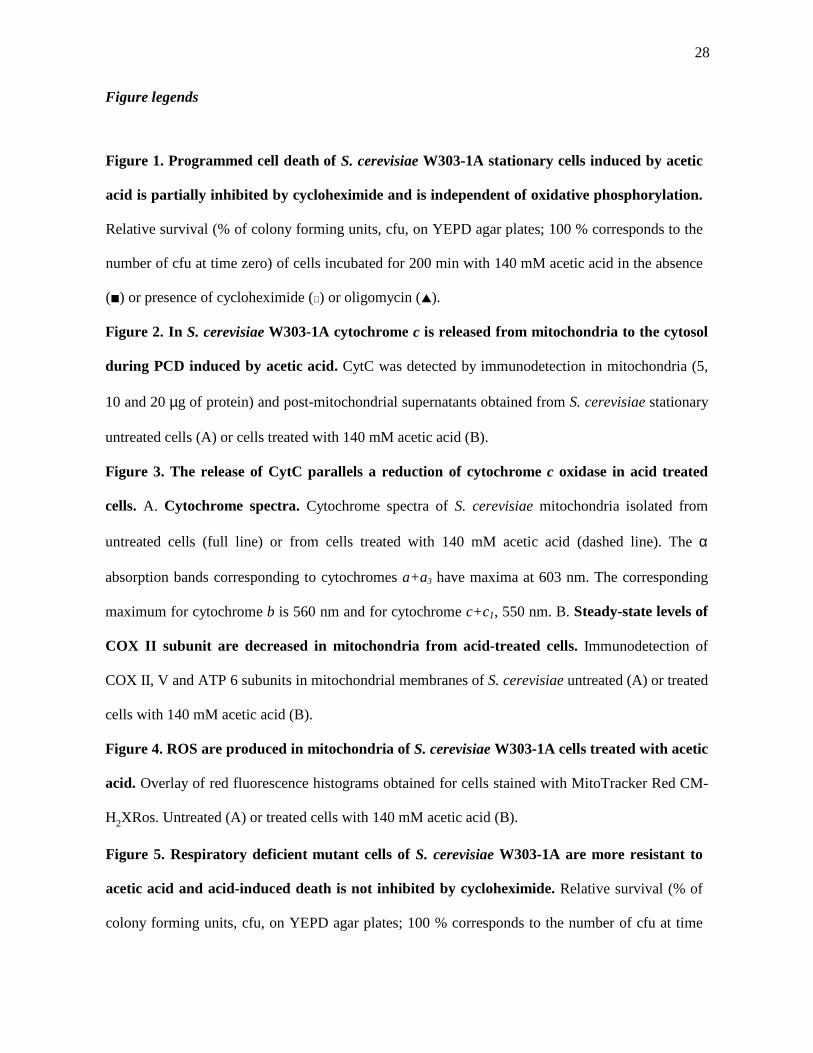
Figure legends
Figure 1. Programmed cell death of S. cerevisiae W303-1A stationary cells induced by acetic
acid is partially inhibited by cycloheximide and is independent of oxidative phosphorylation.
Relative survival (% of colony forming units, cfu, on YEPD agar plates; 100 % corresponds to the
number of cfu at time zero) of cells incubated for 200 min with 140 mM acetic acid in the absence
(!) or presence of cycloheximide (∀ ) or oligomycin (!).
Figure 2. In S. cerevisiae W303-1A cytochrome c is released from mitochondria to the cytosol
during PCD induced by acetic acid. CytC was detected by immunodetection in mitochondria (5,
10 and 20 µg of protein) and post-mitochondrial supernatants obtained from S. cerevisiae stationary
untreated cells (A) or cells treated with 140 mM acetic acid (B).
Figure 3. The release of CytC parallels a reduction of cytochrome c oxidase in acid treated
cells. A. Cytochrome spectra. Cytochrome spectra of S. cerevisiae mitochondria isolated from
untreated cells (full line) or from cells treated with 140 mM acetic acid (dashed line). The α
absorption bands corresponding to cytochromes a+a3 have maxima at 603 nm. The corresponding
maximum for cytochrome b is 560 nm and for cytochrome c+c1, 550 nm. B. Steady-state levels of
COX II subunit are decreased in mitochondria from acid-treated cells. Immunodetection of
COX II, V and ATP 6 subunits in mitochondrial membranes of S. cerevisiae untreated (A) or treated
cells with 140 mM acetic acid (B).
Figure 4. ROS are produced in mitochondria of S. cerevisiae W303-1A cells treated with acetic
acid. Overlay of red fluorescence histograms obtained for cells stained with MitoTracker Red CM-
H2XRos. Untreated (A) or treated cells with 140 mM acetic acid (B).
Figure 5. Respiratory deficient mutant cells of S. cerevisiae W303-1A are more resistant to
acetic acid and acid-induced death is not inhibited by cycloheximide. Relative survival (% of
colony forming units, cfu, on YEPD agar plates; 100 % corresponds to the number of cfu at time

29
zero) of cells of wild-type, ρ0 (lacking mitochondrial DNA), ∆ATP10 (depleted of ATPase) and of
∆CYC3 (depleted of CytC) cells of S. cerevisiae incubated for 200 min with acetic acid in the
absence (black bars) or presence (white bars) of cycloheximide.
30
Tables
Table I
Respiratory activities of mitochondria from control and acetic acid treated cells. Mitochondria
were assayed polarographically for NADH oxidase. The specific activities reported (expressed as
nmol O2 per min per mg of protein) were corrected for KCN insensitive respiration. Cytochrome
oxidase activity (expressed as µmol cytochrome c oxidized per min per mg of mitochondrial
protein) was assayed spectrophotometrically in mitochondria permeabilized with potassium
deoxycholate (COX/DOC) by measuring oxidation of ferrocytochrome c at 550 nm. NADH
cytochrome c reductase (expressed as µmol cytochrome c reduced per min per mg of mitochondrial
protein) was also measured spectrophotometrically as described under “Materials and Methods.”
The rates measured in at least two independent assays did not differ by more than 10%. The values
reported are the average of the two assays.
Acetic Acid
Treatment (mM)
NADH oxidase
- + CytC
NADH CytC
Reductase
Cytochrome
Oxidase
0 367 550 0.602 2.925
140 68 192 0.645 1.552
31
Table II
Involvement of mitochondria in yeast programmed cell death. Differences between S. cerevisiae
programmed cell death induced by Bax-expression, mediated by deletion of histone chaperone
ASF1/CIA1 (Yamaki et al., 2001) and induced by acetic acid treatment (Ludovico et al., 2001a; present
results): main structural and functional cellular changes associated.
Cellular changes Bax-expression asf1/cia1 Acetic acid
Exposition of phosphatidylserine on the outer surface of
the plasma membrane
+
Ligr et al., 1998
nd +
Chromatin condensation +
Ligr et al., 1998
+ +
DNA fragmentation +
Ligr et al., 1998
nd +
Fragmentation of nuclei nd + -
Fragmentation of chromatin nd + +
Mitochondrial membrane-potential Hyperpolarization
Gross et al., 2000
Depolarization Hyperpolarization
followed by depolarization
Dysfunction of mitochondrial proton pump nd + nd
Release of cytochrome c +
Manon et al., 1997
+ +
ROS production +
Gross et al., 2000
nd +
Decrease of cytochrome c oxidase +
Manon et al., 1997
nd +
Mitochondrial apoptosis-induced channel (MAC) activity +
Pavlov et al., 2001
nd nd
+, present; -, absent; nd, not determined.