Cryo-EM Reveals Promoter DNA Binding and Conformational Flexibility of the General Transcription...
11
Structure Article Cryo-EM Reveals Promoter DNA Binding and Conformational Flexibility of the General Transcription Factor TFIID Hans Elmlund, 1,2, * Vera Baraznenok, 3 Tomas Linder, 3 Zsolt Szilagyi, 2,3 Reza Rofougaran, 4 Anders Hofer, 4 Hans Hebert, 5 Martin Lindahl, 5,6 and Claes M. Gustafsson 2,3 1 Department of Structural Biology, Fairchild Building, Stanford University School of Medicine, Stanford, CA 94305, USA 2 Department of Medical Biochemistry and Cell Biology, Go ¨ teborg University, SE-405 30 Go ¨ teborg, Sweden 3 Division of Metabolic Diseases, Karolinska Institutet, Novum, SE-141 86 Huddinge, Sweden 4 Department of Medical Biochemistry and Biophysics, Umea ˚ University, SE-901 87 Umea ˚ , Sweden 5 Department of Biosciences and Nutrition, Karolinska Institutet and School of Technology and Health, Royal Institute of Technology, Novum, SE-141 87 Huddinge, Sweden 6 Department of Molecular Biophysics, Lund University, SE-221 00 Lund, Sweden *Correspondence: [email protected] DOI 10.1016/j.str.2009.09.007 SUMMARY The general transcription factor IID (TFIID) is required for initiation of RNA polymerase II-dependent tran- scription at many eukaryotic promoters. TFIID com- prises the TATA-binding protein (TBP) and several conserved TBP-associated factors (TAFs). Recogni- tion of the core promoter by TFIID assists assembly of the preinitiation complex. Using cryo-electron microscopy in combination with methods for ab initio single-particle reconstruction and hetero- geneity analysis, we have produced density maps of two conformational states of Schizosaccharomyces pombe TFIID, containing and lacking TBP. We report that TBP-binding is coupled to a massive histone- fold domain rearrangement. Moreover, docking of the TBP-TAF1 N-terminus atomic structure to the TFIID map and reconstruction of a TAF-promoter DNA complex helps to account for TAF-dependent regula- tion of promoter-TBP and promoter-TAF interac- tions. INTRODUCTION Transcription factor IID (TFIID) is composed of the TATA-binding protein (TBP) and several evolutionary conserved TBP-associ- ated factors (TAFs) (Burley and Roeder, 1996), of which 13 are essential for cell viability in yeast (Yatherajam et al., 2003). Many TAFs also exists as components of other multiprotein complexes involved in the regulation of transcription, such as the SAGA complex (Grant et al., 1998) and the mammalian PCAF complex (Ogryzko et al., 1998). Most genes in yeast are dependent on TFIID (Lee et al., 2000) for the assembly of a prei- nitiation complex (PIC) (Hahn et al., 1989; Horikoshi et al., 1989; Peterson et al., 1990), and there is mounting evidence pointing to the importance of TAFs (Chalkley and Verrijzer, 1999; Oelgeschl- a ¨ ger et al., 1996; Sawadogo and Roeder, 1985; Shao et al., 2005) and TBP (Kim et al., 1993; Nikolov et al., 1995) in promoter recog- nition. Direct TAF interactions to TBP have been reported for only TAF7 (Yatherajam et al., 2003) and the N terminus of TAF1 (Liu et al., 1998). TBP does not seem to play a crucial role in the integ- rity of the TFIID complex. Accordingly, TBP dynamically associ- ates with the Saccharomyces cerevisiae holo-TAF complex (Sanders et al., 2002), and human TAF complexes lacking TBP have been identified (Wieczorek et al., 1998). Atomic resolution structural information is only available for parts of the TFIID complex (Bhattacharya et al., 2007; Liu et al., 1998; Romier et al., 2007; Werten et al., 2002; Xie et al., 1996). The human histone-fold domain (HFD) subunits TAF4 and TAF12 form crystals in a (TAF4-TAF12) 2 arrangement (Werten et al., 2002). and the TBP-TAF1 11-77 NMR-structure (Liu et al., 1998) shows that the TAF1 N terminus is a protein mimic of the bent TATA box, which inhibits TBP-promoter inter- actions. Structural analysis of the complete TFIID assembly has so far been limited to low-resolution electron microscopy studies (20–30 A ˚ ) of yeast and human TFIID (Andel et al., 1999; Brand et al., 1999; Grob et al., 2006; Leurent et al., 2002, 2004; Papai et al., 2009) reporting a horseshoe-shaped structure with two approximately equally sized quaternary structure domains protruding from a central core, forming a pore through the struc- ture. Immunolabeling of yeast TFIID (Leurent et al., 2002; Leurent et al., 2004) has localized all dimerical HFD TAFs in lobes sepa- rated at distances approaching hundreds of A ˚ ngstro ¨ ms, which argues against the existence of a coherent HFD arrangement within TFIID. However, because of the limited resolution of previous electron microscopic data, it has not been possible to recognize and dock the HFD crystal structures (Werten et al., 2002; Xie et al., 1996) or the TBP-TAF1 11-77 NMR-structure (Liu et al., 1998). On the basis of immunolabeling of TBP in a recon- struction of human TFIID (Andel et al., 1999), promoter DNA was suggested to bind in the central pore, but structural evidence for this interaction has not been provided. Elucidation of the structure and dynamics of TFIID is neces- sary for understanding transcriptional regulation. We have taken steps in this direction and have determined single-particle cryo-EM reconstructions of two conformational states of 1442 Structure 17, 1442–1452, November 11, 2009 ª2009 Elsevier Ltd All rights reserved
Transcript of Cryo-EM Reveals Promoter DNA Binding and Conformational Flexibility of the General Transcription...

Structure
Article
Cryo-EM Reveals Promoter DNA Binding andConformational Flexibility of the GeneralTranscription Factor TFIIDHans Elmlund,1,2,* Vera Baraznenok,3 Tomas Linder,3 Zsolt Szilagyi,2,3 Reza Rofougaran,4 Anders Hofer,4 Hans Hebert,5
Martin Lindahl,5,6 and Claes M. Gustafsson2,3
1Department of Structural Biology, Fairchild Building, Stanford University School of Medicine, Stanford, CA 94305, USA2Department of Medical Biochemistry and Cell Biology, Goteborg University, SE-405 30 Goteborg, Sweden3Division of Metabolic Diseases, Karolinska Institutet, Novum, SE-141 86 Huddinge, Sweden4Department of Medical Biochemistry and Biophysics, Umea University, SE-901 87 Umea, Sweden5Department of Biosciences and Nutrition, Karolinska Institutet and School of Technology and Health, Royal Institute of Technology, Novum,
SE-141 87 Huddinge, Sweden6Department of Molecular Biophysics, Lund University, SE-221 00 Lund, Sweden
*Correspondence: [email protected]
DOI 10.1016/j.str.2009.09.007
SUMMARY
The general transcription factor IID (TFIID) is requiredfor initiation of RNA polymerase II-dependent tran-scription at many eukaryotic promoters. TFIID com-prises the TATA-binding protein (TBP) and severalconserved TBP-associated factors (TAFs). Recogni-tion of the core promoter by TFIID assists assemblyof the preinitiation complex. Using cryo-electronmicroscopy in combination with methods for abinitio single-particle reconstruction and hetero-geneity analysis, we have produced density maps oftwo conformational states of Schizosaccharomycespombe TFIID, containing and lacking TBP. We reportthat TBP-binding is coupled to a massive histone-fold domain rearrangement. Moreover, docking ofthe TBP-TAF1N-terminus atomic structure to the TFIIDmap and reconstruction of a TAF-promoter DNAcomplex helps to account for TAF-dependent regula-tion of promoter-TBP and promoter-TAF interac-tions.
INTRODUCTION
Transcription factor IID (TFIID) is composed of the TATA-binding
protein (TBP) and several evolutionary conserved TBP-associ-
ated factors (TAFs) (Burley and Roeder, 1996), of which 13 are
essential for cell viability in yeast (Yatherajam et al., 2003).
Many TAFs also exists as components of other multiprotein
complexes involved in the regulation of transcription, such as
the SAGA complex (Grant et al., 1998) and the mammalian
PCAF complex (Ogryzko et al., 1998). Most genes in yeast are
dependent on TFIID (Lee et al., 2000) for the assembly of a prei-
nitiation complex (PIC) (Hahn et al., 1989; Horikoshi et al., 1989;
Peterson et al., 1990), and there is mounting evidence pointing to
the importance of TAFs (Chalkley and Verrijzer, 1999; Oelgeschl-
ager et al., 1996; Sawadogo and Roeder, 1985; Shao et al., 2005)
1442 Structure 17, 1442–1452, November 11, 2009 ª2009 Elsevier L
and TBP (Kim et al., 1993; Nikolov et al., 1995) in promoter recog-
nition. Direct TAF interactions to TBP have been reported for only
TAF7 (Yatherajam et al., 2003) and the N terminus of TAF1 (Liu
et al., 1998). TBP does not seem to play a crucial role in the integ-
rity of the TFIID complex. Accordingly, TBP dynamically associ-
ates with the Saccharomyces cerevisiae holo-TAF complex
(Sanders et al., 2002), and human TAF complexes lacking TBP
have been identified (Wieczorek et al., 1998).
Atomic resolution structural information is only available for
parts of the TFIID complex (Bhattacharya et al., 2007; Liu
et al., 1998; Romier et al., 2007; Werten et al., 2002; Xie et al.,
1996). The human histone-fold domain (HFD) subunits TAF4
and TAF12 form crystals in a (TAF4-TAF12)2 arrangement
(Werten et al., 2002). and the TBP-TAF111-77 NMR-structure
(Liu et al., 1998) shows that the TAF1 N terminus is a protein
mimic of the bent TATA box, which inhibits TBP-promoter inter-
actions. Structural analysis of the complete TFIID assembly has
so far been limited to low-resolution electron microscopy studies
(20–30 A) of yeast and human TFIID (Andel et al., 1999; Brand
et al., 1999; Grob et al., 2006; Leurent et al., 2002, 2004; Papai
et al., 2009) reporting a horseshoe-shaped structure with two
approximately equally sized quaternary structure domains
protruding from a central core, forming a pore through the struc-
ture. Immunolabeling of yeast TFIID (Leurent et al., 2002; Leurent
et al., 2004) has localized all dimerical HFD TAFs in lobes sepa-
rated at distances approaching hundreds of Angstroms, which
argues against the existence of a coherent HFD arrangement
within TFIID. However, because of the limited resolution of
previous electron microscopic data, it has not been possible to
recognize and dock the HFD crystal structures (Werten et al.,
2002; Xie et al., 1996) or the TBP-TAF111-77 NMR-structure (Liu
et al., 1998). On the basis of immunolabeling of TBP in a recon-
struction of human TFIID (Andel et al., 1999), promoter DNA was
suggested to bind in the central pore, but structural evidence for
this interaction has not been provided.
Elucidation of the structure and dynamics of TFIID is neces-
sary for understanding transcriptional regulation. We have
taken steps in this direction and have determined single-particle
cryo-EM reconstructions of two conformational states of
td All rights reserved

Structure
Promoter DNA Binding and Flexibility of TFIID
Schizosaccharomyces pombe TFIID, containing (TFIID) and
lacking TBP (TFIIDDTBP). We have also reconstructed
TFIIDDTBP in complex with promoter DNA. We report that TBP
binding to and release from the TAFs is coupled to a massive
rearrangement of the quaternary structure region comprising
TAF4. Moreover, we describe the structural dynamics underlying
formation of a TBP-binding site within TFIID, which indicates that
promoter-TBP and promoter-TAF interactions are inhibited
when TBP is bound to the TAFs.
RESULTS
Purification and GEMMA Analysis of Fission Yeast TFIIDTo isolate TFIID from S. pombe, we used tandem affinity puri-
fication (TAP) with a TAP-tag on TAF4. After purification over
IgG-Sepharose and MiniS columns, 12 distinct TAF subunits
(TAF1–TAF12) were identified using SDS-PAGE (see Figures
S1A and S1B available online) and MALDI-TOF mass finger-
printing of excised bands. Among the identified proteins were
two closely related TAF5 paralogs, TAF5a and TAF5b, which
are encoded for by two separate genes. An additional protein
band of about 10 kDa could not be identified with mass finger-
printing, but the size agrees with what would be expected
for TAF13. Immunoblotting showed that purified TFIID con-
tained TBP (Figure S1C). We used Gas-phase-Electropho-
retic-Mobility Macromolecule Analysis (GEMMA) to estimate
the molecular weight of TFIID (Figure S1D) to 874 ± 52 kDa.
GEMMA is a relatively new method for separating protein mole-
cules in diluted protein samples according to their diameter by
analyzing their different mobility in gas phase induced by
a charged reduced electrospray process (Bacher et al., 2001;
Kaufman et al., 1996). The method runs at atmospheric pres-
sure and allows for determination of the mass of protein mole-
cules with an error of ±5.6% (Bacher et al., 2001). Determina-
tion of the molecular weight of S. pombe TFIID to 874 ± 52
kDa agrees with the predicted molecular weight of 876 kDa.
The prediction accounts for the existence of two copies each
of the TAF4, TAF6, TAF9, and TAF12 subunits (Selleck et al.,
2001).
3D Reconstruction of TFIID and TFIIDDTBPWe calculated the TFIID reconstructions from 62,300 particle
images, collected from 22 micrographs of a single TFIID spec-
imen, preserved in vitrified ice, and imaged under low-dose
conditions (10–15 e�/A2) using a transmission electron micro-
scope (EM) equipped with a field-emission gun. We used an
ab initio reconstruction method based on reference-free com-
mon lines (Elmlund et al., 2008) to generate an initial TFIID 3D
reconstruction. Refinement of the entire TFIID cryo-data set
was performed without using sorting procedures to exclude
images. The refinement halted initially at a resolution of �25 A,
which was significantly lower than that expected for the given
size of the data set. We interpreted this finding as a sign of the
data set being heterogeneous and used multivariate statistical
analysis to investigate the number of conformational states
present in the population (see Figure S2). Two predominant
conformational states were identified. We developed a method
to divide the data set into homogeneous groups (see Supple-
mental Data). The two resulting reconstructions were identified
Structure 17, 1442–14
as containing (TFIID) and lacking TBP (TFIIDDTBP) (see below),
which is in agreement with the known ability of TBP to dynami-
cally associate with TAFs (Sanders et al., 2002). We compared
projections in five evenly distributed orientations of each of the
two states with corresponding projection averages. Projection
averages showed an excellent agreement with projections
(Figure 1B). The resolution of the two final density maps was
assessed by Fourier shell correlation (FSC) to 8–10 A according
to the FSC = 0.5 criterion (Figure 1D), and the maps were low-
pass filtered to 8 A. Local symmetry analysis detected the two-
fold symmetry and defined the direction of the symmetry axis
for a module present in the back end of the core, the TBP-TAF
region, and for two segments of the back, of which one corre-
sponded to the (TAF4–TAF12)2 assembly (see Supplemental
Data and Figure S6).
Determination of the Absolute HandThe absolute hand of a reconstruction cannot be resolved for
independent projections alone. One of the human TFIID recon-
structions (Grob et al., 2006) agreed well with our TFIIDDTBP
reconstruction at low resolution. The fact that the human TFIID
reconstructions originate from a random conical tilt reconstruc-
tion therefore led us to attempt to determine the absolute hand
by using the human TFIID reconstruction. We projected the
human and S. pombe TFIID reconstructions in 116 even direc-
tions and aligned the S. pombe TFIID images in 3D by using
the human TFIID projections as a reference set. We calculated
a reconstruction from the aligned set of S. pombe TFIID projec-
tions and compared it to both hands of our reconstruction, which
revealed a significant difference in correlation (Figure S5b;
0.6523 for hand1 vs. 0.4389 for hand2). Visual inspection of the
results from the correlation search confirmed the assignment
of absolute hand.
Docking of TBP into the TFIID MapTo simplify the interpretation of our TFIID and TFIIDDTBP maps,
we divided the reconstructions into five regions (Figure 2), which
we called the core-, arm-, back, deco1-, and deco2-regions
(‘‘deco’’ stands for ‘‘decorative protein module’’). In one of the
two states (TFIID), we detected a sixth module that corre-
sponded in size and shape to what would be expected for TBP
in complex with the N-terminal stirrup of TAF1 (Figure 2A: right
panel, gray). Docking of the most probable conformer of the
Drosophila TBP-TAF111-77 NMR-ensemble (PDBid: 1TBA) into
the TFIID map was aided by local symmetry analysis (see
Figure S6) and the wealth of distinct fine structural details
present in the TFIID reconstruction. Positioning of the TAF111-77
NMR-structure was determined by automatic real space rigid
body docking in Chimera (Pettersen et al., 2004). To validate
the docking, we performed common line correlation search
using projections of the density map, calculated from the most
probable conformer of the NMR-ensemble and the TFIID recon-
struction as references and targets, respectively. The search
was performed over the entire projection direction space and
resulted in three correlation peaks. The first peak represented
the presented docking (highest correlation peak), and the other
two correlation maxima were explained by the pseudo-two-
fold symmetry of the TBP-TAF complex, equivalent to the
behavior of the local symmetry search (see Figure S6). We
52, November 11, 2009 ª2009 Elsevier Ltd All rights reserved 1443

Structure
Promoter DNA Binding and Flexibility of TFIID
Figure 1. Validation Data for the Single-Particle 3D Reconstructions
(A) Refinement cycle for the S. pombe TFIIDDTBP and TFIID reconstructions. RAD (Reference-free Alignment in a Discrete angular space) is the ab initio recon-
struction (Elmlund et al., 2008). Numbering denotes round of model-based refinement. Heterogeneity analysis and supervised classification were performed as
described above and in the Supplemental Data.
(B) Projections of the reconstructions (right column) and corresponding projection averages (left column) for the two states, selected according to five evenly
distributed projection directions.
(C) Histogram of defocus values for the 22 micrographs of the data set.
(D) Fourier shell correlation plots (upper panel) and diagrams over the distribution of the two nonazimuthal Euler angles (lower panel). The resolution of the two
TFIID reconstructions was determined to 8–10 A according to the FSC = 0.5 criterion.
(E) Gallery of windowed particles from a micrograph acquired at 3.8 mm defocus, band-pass filtered using a [12,160] A resolution window.
observed density features that matched well with the skewed
b sheet and the C-terminal helix of TBP (Figures 3B–3D).
Although our fit of TBP-TAF111-77 was excellent, density for the
N-terminal TBP helix was hard to interpret, which may be
explained by pronounced TAF-TBP interactions. The peripheral
1444 Structure 17, 1442–1452, November 11, 2009 ª2009 Elsevier
localization of TBP explains why it does not play a crucial role
for the integrity of the TFIID complex and is in agreement with
findings that direct TAF interactions to TBP occur only for
TAF1 (Liu et al., 1998) and TAF7 (Yatherajam et al., 2003). The
central position of TBP, just above the central pore, agrees
Ltd All rights reserved

Structure
Promoter DNA Binding and Flexibility of TFIID
Figure 2. Quaternary Structure Arrangement of
TFIID
Rendering volume was selected according to the molec-
ular weight of TFIID.
(A) Structurally distinct and coherent quaternary regions
were masked out in the TBP-lacking state (left panel) and
the TBP-containing state (right panel). We describe
our TFIID reconstructions as divided into five common
quaternary structure regions: the core-region (brown),
arm-region (yellow), back-region (blue), deco1-region
(red), and deco2-region (green) (‘‘deco’’ stands for
‘‘decorative protein module’’), sorted in order of
descending molecular weight. The tbp-taf region (gray)
was present exclusively in one of the states (right panel).
Viewing directions are defined as follows: I, front; II,
back; III, bottom; IV, arm-side; and V, core-side. Scale
bar is 150 A.
(B) Formation of a TBP-binding site within TFIID, with the
deco2 segment (green) within TFIIDDTBP (left) and the fit
of the segment to the TBP-containing reconstruction
(right).
Structure 17, 1442–1452, November 11, 2009 ª2009 Elsevier Ltd All rights reserved 1445

Structure
Promoter DNA Binding and Flexibility of TFIID
with the earlier immunolabeling of TBP in human TFIID (Andel
et al., 1999). Moreover, the high degree of structural similarity
between the TBP-TAF111-77 NMR data and the corresponding
region of our TFIID reconstruction supports the previously
reported inhibitory role of the TAFs (Liu et al., 1998; Mal et al.,
2004). We could not identify the TBP-TAF11-77 NMR structure
within the other conformer (TFIIDDTBP), which evidently lacked
the TBP density. In agreement with Sanders et al. (2002), we
therefore identify the two states as containing and lacking TBP.
The formation of a TBP-binding site involved one of the small
decorative protein modules (Figure 2; deco2). To characterize
the rearrangement of deco2 in the TFIIDDTBP to TFIID transi-
tion, we fitted the segment of the TBP-lacking reconstruction
into the TBP-containing map. Figure 2B shows the deco2
segment within TFIIDDTBP (left) and the fit of the segment to
the TBP-containing reconstruction (right). To confirm the ability
of TBP to dynamically associate with the TAFs, we added puri-
fied TBP in seven-fold molar excess to the original heteroge-
neous preparation and collected 14,991 single-particle cryo-
EM images. To investigate any preference of the population
to any of the two resolved TFIID states, we applied the principle
of supervised classification (Gao et al., 2004), using our final
reconstructions as templates for the common line correlation-
based matching. Addition of TBP significantly affected the
Figure 3. Docking of the TBP-TAF111-77
NMR-Structure
Rendering volume was selected to maximize the
helical densities.
(A) Cut through the TFIID density and definition of
the viewing direction in (b).
(B) View orthogonal to the cut demonstrates that
TAFs occupy the concave DNA-binding surface
of TBP. Viewing directions in (c) and (d) are indi-
cated.
(C) Fit of the C-terminal TBP helix in stereo.
(D) View facing the DNA-binding pocket of TBP
shows the fit of the C-terminal end of the skewed
TBP b sheet.
(E) Validation of the fit of the TBP-TAF111-77 NMR
structure by common line correlation-based
search over the entire projection direction space.
The analysis revealed three correlation peaks. The
first (leftmost) correlation peak represents the
presented docking (highest correlation peak),
and the second peak is an artificial peak induced
by the pseudo-two-fold symmetry axis present
perpendicular to the real two-fold symmetry axis
of TBP. All two-fold symmetrical objects have
a pseudo-two-fold symmetry axis perpendicular
to the real axis. First in the D2 point-group this
pseudo-axis becomes a real two-fold axis. The
third peak represents the symmetry-induced
correlation peak.
equilibrium between the two states.
Although the original preparation showed
an almost equal distribution between the
states (TFIIDDTBP, 31,974 particles;
TFIID, 30,326 particles), the preparation
with added TBP showed a preference
for the TBP-containing conformation (TFIIDDTBP, 5510 parti-
cles; TFIID, 9481 particles).
Docking of (hTAF4-hTAF12)2 into the TFIID MapThe crystal structure of human TAF4 and in complex with human
TAF12 has been solved. The two hTAF4-hTAF12 heterodimers
interact to form a heterotetramer (hTAF4-hTAF12)2 (Werten
et al., 2002). Multiple sequence alignment between the hTAF4
and hTAF12 crystal structure sequences (PDBid: 1H3O) and
the corresponding complete human, Drosophila, S. pombe,
and S. cerevisiae TAF sequences, using ClustalW (Thompson
et al., 1994), did not reveal any inserts or deletions within the
structurally characterized regions (data not shown). The
(hTAF4-hTAF12)2 crystal structure should therefore be a good
template for modeling the S. pombe (TAF4-TAF12)2 complex.
Local symmetry analysis identified a two-fold symmetrical
module in the back region of the TFIID map (see Figure S6C),
which fitted the size and shape of (hTAF4-hTAF12)2. Docking
was performed as described above. As expected, the correlation
search used for validation resulted in three peaks. The first peak
represented the presented docking (highest correlation peak),
and the other two maxima were explained by the two-fold
symmetry of the (TAF4-TAF12)2 assembly (Figure 4). In the
fitting of (TAF4-TAF12)2, rodlike density features in the TFIID
1446 Structure 17, 1442–1452, November 11, 2009 ª2009 Elsevier Ltd All rights reserved

Structure
Promoter DNA Binding and Flexibility of TFIID
reconstruction coincided with the C-terminal hTAF4 helices. Our
finding that the (TAF4-TAF12)2 crystallographic unit also corre-
sponded to a biological unit was puzzling, because previous
immunolabeling of yeast TFIID identified TAF4 in lobes sepa-
rated at distances approaching hundreds of Angstroms (Leurent
et al., 2002, 2004). We therefore used an antibody recognizing
the calmodulin-binding peptide of the C-terminal TAP-tag on
TAF4 to specifically label the TAF4 C terminus. Low-resolution
reconstructions were calculated from 22,000 cryo-EM images,
with 11,201 and 10,799 particles assigned to the TFIIDDTBP
and TFIID states, respectively. No labeling was observed in the
TFIIDDTBP state, whereas binding to two closely associated
regions was observed in the TBP-containing state, located in
the center of the back (Figure 4D). It was obvious that the labeled
protein module was present in the TFIIDDTBP state, and the
absence of structured antibody regions in the TBP-lacking
reconstruction indicated that the conformational rearrangement
had masked the epitope-binding surfaces. It appeared as if the
Figure 4. Identification, Docking, and
Structural Dynamics Analysis of the (TAF4-
TAF12)2 Histone-Fold Tetramer Within TFIID
(A) The (TAF4-TAF12)2 segment (yellow) within the
TBP-containing state (right) and the fit of the
segment (blue) to the TBP-lacking state (left).
(B) Views parallel and perpendicular to the two-
fold symmetry axis of the (TAF4-TAF12)2 segment
within the TBP-lacking (blue) and TBP-containing
(yellow) state, with the relative rotation and tilt
around the two-fold symmetry axis indicated.
(C) Validation of the fit of the (hTAF4-hTAF12)2crystal structure by common line correlation-
based search over the entire projection direction
space. The analysis revealed three correlation
peaks. The first (leftmost) correlation peak repre-
sents the presented docking (highest correlation
peak), and the second peak is an artificial peak
induced by the pseudo-two-fold symmetry axis
present perpendicular to the real two-fold sym-
metry axis of (TAF4-TAF12)2. All two-fold symmet-
rical objects have a pseudo-two-fold symmetry
axis perpendicular to the real axis. First in the D2
point-group this pseudo-axis becomes a real
two-fold axis. The third peak represents the
symmetry-induced correlation peak.
(D) Immunolabeling of the C terminus of TAF4 with
the density of the ordered antibody regions in red
according to the difference calculated between
the labeled and the native structure. I, Stereo
view along the two-fold symmetry axis of the
docked (hTAF4-hTAF12)2 crystal structure. II,
View perpendicular to the two-fold symmetry
axis of the docked (hTAF4-hTAF12)2 crystal
structure.
(TAF4-TAF12)2 units in the TFIIDDTBP
and TFIID reconstructions were related
by a composite rotation. To further inves-
tigate this hypothesis, we masked out the
region corresponding to (TAF4-TAF12)2 in
the TBP-containing state. Positioning of
the (TAF4-TAF12)2 density within the
TFIIDDTBP reconstruction revealed a coordinate transformation
involving a �30� tilt of and a �90� rotation around the two-fold
symmetry axis of the docked (TAF4-TAF12)2 structure (Figures
4A and 4B). We concluded that TBP binding to and release
from the TAFs was coupled to a massive conformational rear-
rangement of the back region, likely involving a composite rota-
tion of the (TAF4-TAF12)2 tetramer.
3D Reconstruction of a TFIIDDTBP-Promoter DNAComplexFinally, we wanted to monitor interactions between TFIID and
promoter DNA. A DNA fragment composed of 101 base pairs
of the S. pombe NMT1 promoter (50 and 51 base pairs upstream
and downstream of the transcription start site, respectively) was
added in seven-fold molar excess to the original TFIID prepara-
tion, and low-resolution reconstructions were calculated from
14,300 cryo-EM images, with 7280 and 7020 particles assigned
to the TFIIDDTBP and TFIID states, respectively. In previous
Structure 17, 1442–1452, November 11, 2009 ª2009 Elsevier Ltd All rights reserved 1447
Structure
Promoter DNA Binding and Flexibility of TFIID
gel-shift experiments, we had observed direct binding between
this DNA fragment and the TFIID complex (data not shown).
The bent DNA structure appeared in the reconstruction calcu-
lated from particles mapped to the TFIIDDTBP state in the posi-
tion exactly expected from docking of TBP (Figure 5). No DNA
binding was observed to the TBP-containing state. Our inability
to visualize DNA binding to the TBP-containing complex does
not imply that DNA associates only with TFIIDDTBP. Rather, if
promoter DNA binds with less specificity or exhibits a higher
degree of flexibility when associated to the TBP-containing
complex, this may prevent us from visualizing it as a result of
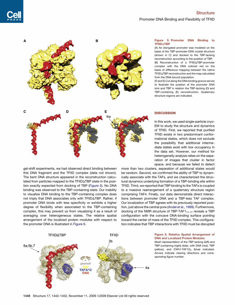
averaging over heterogeneous states. The relative spatial
arrangement of the localized protein modules with respect to
the promoter DNA is illustrated in Figure 6.
Figure 5. Promoter DNA Binding to
TFIIDDTBP
(A) An elongated promoter was modeled on the
basis of the TBP-promoter-DNA crystal structure
(shown in C) and docked to the TBP-lacking
reconstruction according to the position of TBP.
(B) Reconstruction of a TFIIDDTBP-promoter
complex with the DNA colored red on the
basis of difference mapping between the native
TFIIDDTBP reconstruction and the map calculated
from the DNA-bound population.
(D and E) Cut along the DNA binding groove serves
to illustrate the position of the promoter DNA
kink and TBP in relation the TBP-lacking (D) and
TBP-containing (E) reconstruction. Quaternary
structure regions are indicated.
DISCUSSION
In this work, we used single-particle cryo-
EM to study the structure and dynamics
of TFIID. First, we reported that purified
TFIID exists in two predominant confor-
mational states, which does not exclude
the possibility that additional interme-
diate states exist with low occupancy in
the data set. However, our method for
heterogeneity analysis relies on the sepa-
ration of images that cluster in factor
space, and because we failed to detect
more than two clusters, separation of additional states would
be random. Second, we confirmed the ability of TBP to dynam-
ically associate with the TAFs, and we characterized the struc-
tural dynamics underlying formation of a TBP-binding site within
TFIID. Third, we reported that TBP binding to the TAFs is coupled
to a massive rearrangement of a quaternary structure region
comprising TAF4. Finally, our data demonstrate direct interac-
tions between promoter DNA and a TBP-less TAF complex.
Our localization of TBP agrees with its previously reported posi-
tion, just above the central pore (Andel et al., 1999). Furthermore,
docking of the NMR structure of TBP-TAF111-77 reveals a TBP
configuration with the concave DNA-binding surface pointing
toward the center of mass of the TFIID complex. This configura-
tion indicates that TBP interactions with TFIID must be disrupted
Figure 6. Relative Spatial Arrangement of
DNA and Localized Protein Modules
Mesh representation of the TBP-lacking (left) and
TBP-containing (right) state, with DNA (red), TBP
(yellow), and (TAF4-TAF12)2 (blue) indicated.
Arrows indicate viewing directions and corre-
sponding figure number.
1448 Structure 17, 1442–1452, November 11, 2009 ª2009 Elsevier Ltd All rights reserved

Structure
Promoter DNA Binding and Flexibility of TFIID
before a complex between TBP and promoter DNA can be
formed (Kim et al., 1993).
Structural Architecture of TFIIDThe HFD TAF4 structure is present in two closely associated
copies located in the center of the back. Our localization of
TAF4 is based on vitrified specimens and new methods for 3D
reconstruction, and it receives strong support from the (hTAF4-
hTAF12)2 crystal structure (Werten et al., 2002) in which two
copies of each of the TAF4 and TAF12 HFD structures form
a closely entangled two-fold symmetric tetramer. The two-fold
symmetry of the (TAF4-TAF12)2 structure is confirmed by local
symmetry analysis of the back region. TAF4 appears to be
present in two copies, and our positioning of the C-terminal
TAF4 helix within the EM-density corresponds exactly with the
extra density attributed to the structured regions of the anti-
TAF4 antibodies. Our data therefore suggest that the (TAF4-
TAF12)2 crystallographic unit also corresponds to a biological
unit. Certain regions of the crystal structure are not well defined
in the TFIID map. Residues 17–26 of hTAF4 and 47–57 of hTAF12
in the crystallized sequence segments are highly conserved loop
and helix end regions that are fully accessible to the solvent. The
potentially high flexibility of these regions explains why they have
poor occupancy in the TFIID reconstruction. The conformational
transition coupled to binding of TBP leads to a composite rota-
tion of (TAF4-TAF12)2 such that exactly these conserved and
potentially flexible regions become involved in interactions with
the arm segment. Docking of (TAF4-TAF12)2 places the terminal
regions where the sequence continues either toward the arm
segment or toward the additional domains of the back. A likely
explanation is that the remaining TAF4 and TAF12 sequence
parts entangle with the arm segment or the additional domains
of the back, which moves in concert with the HFD tetramer
and appears to be the ‘‘glue’’ that holds the arm and core
segments together in the conformational transition of the TFIID
structure. This observation agrees with the finding that TAF4,
rather than TBP or TAF1, plays the most critical role in maintain-
ing the stability of TFIID (Wright et al., 2006). Similar to what has
been described for human TFIID (Grob et al., 2006), a conforma-
tional breathing of the molecule occurs, with the two main
quaternary domains changing their relative position. The HFD re-
arrangement is directly related to TBP binding to and releasing
from the TAFs. How this rearrangement may relate to other
aspects of TFIID biology remains to be established.
DNA-TBP-, DNA-TAF, and TAF-TBP-Interactionsand Regulation of TranscriptionLoading of TBP onto the promoter is a crucial step in gene acti-
vation. Transcriptional activity in yeast strongly correlates with
promoter occupancy by general factors such as TBP, TFIIA,
and TFIIB, but not with occupancy of TAFs (Kuras et al., 2000).
Our docking of the TBP-TAF111-77 NMR-structure into the TFIID
map and reconstruction of TFIIDDTBP bound to promoter DNA
give a number of important structural insights, which may help
to explain why we observe promoter DNA binding to TFIID only
in the absence of TBP. We observe DNA binding to a two-fold
symmetric groove that protrudes from the TBP-binding site
and along the surface of the core. TBP thus appears to form
a molecular lid over the groove, with its DNA-binding surface
Structure 17, 1442–14
pointing toward the center of mass of the TFIID structure. This
configuration of TBP within TFIID may prevent access of
promoter DNA to the concave DNA-binding pocket of TBP by
steric hindrance. It is tempting to speculate that TBP-TAF inter-
actions must be disrupted before promoter-DNA interactions
can take place with either TBP or TAFs. The TFIIDDTBP to TFIID
transition resolved here may describe the structural dynamics
underlying this process, involving a reversible conformational
change that may affect binding of TBP to the TAFs and allow
for DNA entrance. It should be noted that this model does not
exclude the existence of a TBP-containing and promoter-bound
TAF complex, which may represent the last intermediate in
the TFIID-dependent catalytic cycle of loading TBP onto the
promoter.
In stark contrast to binding of TBP, promoter DNA binding to
the TAFs does not introduce any conformational rearrangements
of note. The DNA configuration within the promoter-bound TAF
complex would result in major steric clashes between TAFs
and pol II according to the current model for the positioning of
pol II with respect to promoter DNA in the PIC (see Bushnell
et al., 2004 and Chen et al., 2007 and references therein). This
observation could indicate that the TAFs need to either partly
dissociate from the promoter or change their positioning before
entry of the polymerase into the PIC. In support of this notion,
immobilized template assays have demonstrated that TFIID
and TFIIA can assemble at promoters and block further PIC
assembly (Ranish et al., 1999). How this block is relieved is not
well understood, but it could perhaps be a regulatory target for
transcriptional activators and repressors.
The TATA consensus does not appear to be a major determi-
nant of TBP binding in yeast (Kim and Iyer, 2004). Only approx-
imately 20% of the genes in yeast contain a TATA box, and these
genes are associated with responses to stress, they are highly
regulated, and they preferentially utilize SAGA before TFIID to
load TBP (Basehoar et al., 2004). One possibility is therefore
that TFIIDDTBP plays an especially important role in DNA
bending and TBP loading at promoters lacking a TATA con-
sensus (illustrated in Figure 7). Another possibility is that of tran-
scription without TBP, which would require stabilization of the
bent promoter structure by general factors other than TBP to
accomplish initiation. The latter possibility agrees with what
has been observed for the TBP-lacking human TAF complex,
TFTC (Wieczorek et al., 1998). We see no reason to exclude
any of the two pathways. The TAFs are involved in both activa-
tion and repression of transcription. They inhibit TBP-mediated
basal transcription in the absence of activator and they stimulate
activated transcription in synergy with the Mediator complex
(Burley and Roeder, 1998; Guermah et al., 1998, 2001). The
steric hindrance of promoter-TAF and promoter-TBP interac-
tions exerted by TBP when bound to the TAFs may provide
one part of the structural basis for the dual role of TAFs in tran-
scription regulation.
EXPERIMENTAL PROCEDURES
Purification of TBP and TFIID
Recombinant spTBP with 6xHis tag in the PET21b(+) vector was expressed in
Escherichia coli (Cod+) cells for 15 hr at room temperature and purified over Ni
column followed by heparin column. For purification of TAP-tagged TFIID, 15 l
52, November 11, 2009 ª2009 Elsevier Ltd All rights reserved 1449

Structure
Promoter DNA Binding and Flexibility of TFIID
of S. pombe cells was grown to OD600 3.0–4.5 in YES medium supplemented
with 0.2 g/l adenine. Cells were collected by centrifugation (JA-10, Beckman
Coulter, at 2,500 rpm, for 7 min at +4�C), washed once with ice-cold water,
and frozen in liquid nitrogen. Cells were broken in a Freezer/Mill 6850 (SPEX
CertiPrep, NJ) using the following program: 10 min precooling, 5 cycles with
2 min beating, and 2 min rest at stringency 14. Broken cells were suspended
in 0.5 ml of 3*TAP buffer (200 mM KOH-HEPES [pH 7.8], 15 mM KCl,
1.5 mM MgCl2, 0.5 mM EDTA, and 15% glycerol) per gram of cell pellet.
DTT (0.5 mM) and protease inhibitors were added last, and the amount was
adjusted to fit the total volume. After clearing of the supernatant by centrifuga-
tion (JA-10, at 9000 rpm, for 15 min, at +4�C), 1/9 volume of 2 M KCl was added
followed by stirring for 15 min. After ultracentrifugation (Ti45, Beckman
Coulter, at 42,000 rpm, for 30 min, at +4�C), the supernatant was frozen.
IgG beads (300 ml; slurry; Amersham Biosciences) was added per each of
30–45 ml of extract and was incubated for 1 hr at +4�C. IgG beads were
collected by centrifugation (JA-17, Beckman Coulter, at 1,000 rpm, for
2 min, at +4�C) and were washed with IgG buffer (10 mM Tris-HCl and
150 mM KOAc [pH 8.0]) and then by tobacco etch virus (TEV) protease
cleavage buffer (25 mM HEPES-KOH [pH 7.6], 150 mM KOAc, 1 mM DTT,
0.5 mM EDTA, and 0.05% NP-40). TFIID was eluted by incubation for 1.5–2
hr at +16�C with 200 units of TEV protease in 1.5–2 ml of TEV protease
cleavage buffer. Eluted TFIID was loaded onto a MiniS column (PE 4.6/50)
and was subjected to 10 column volumes of gradient 0.15–1.5 M KOAc in
the buffer (25 mM HEPES-KOH [pH 7.6], 10% glycerol, 0,05% NP-40, 1 mM
DTT, protease inhibitors, and 2 mM MgOAc). TFIID eluted around 0.5 M
KOAc. Fractions containing 0.03–0.08 g/l of TFIID were used for the following
experiments. TFIID was concentrated by TCA precipitation, and TAF subunits
were resolved on 12% SDS PAGE. Protein bands were excised, were in-gel
digested by sequencing grade-modified trypsin from Promega, and were
analyzed by MALDI-TOF MS on an Ultraflex TOF/TOF instrument (Bruker).
TBP was detected by Western blot, developed with anti-S. cerevisiae TBP
(Santa Cruz TBP (y-240): sc-33736).
GEMMA
TFIID was exchanged into a buffer consisting of 150 mM ammonium acetate
(pH 7.8) using Sephadex-25 chromatography and was diluted into a protein
concentration of 0.005 mg/ml in ammonium acetate (final concentration,
Figure 7. A Model for TFIID Function Based
on Our Current Structural Knowledge about
TFIID
TFIID (brown) exists in a dynamic equilibrium
between TBP-containing and TBP-lacking states.
Promoter DNA (red) binding occurs primarily to
the TBP-lacking state. TFIIDDTBP introduces the
bent promoter structure required for initiation
and in TBP-dependent transcription it may func-
tion by presenting a protein-DNA surface for TBP
to interact with. At promoters that are not depen-
dent on TBP, general factors other than TBP
must stabilize the promoter kink.
95 mM). GEMMA analysis was performed as
described elsewhere (Rofougaran et al., 2006)
using a pressure drop of 1.8 psi to direct the
flow-rate. The results from the GEMMA analysis
are shown in Figure S1.
Generation of Complex TFIID with
TAP-TAF4 Antibody
Affinity-purified rabbit polyclonal anti-TAP anti-
body, recognizing C terminus of the TAP construct
after TEV cleavage, came from OpenBiosystems
(CAB1001). To prepare complexes, approximately
five times molar excess of antibodies were added
to the TFIID preparation. After mixing, TFIID-anti-
body complexes were dialyzed against binding buffer (10 mM Tris-HCl
[pH 8.0], 150 mM NaCl, 0.05% NP-40, and 1 mM DTT).
Cryo-EM and Single-Particle Processing
Vitrified specimen was prepared, and image acquisition and digitization were
performed as described elsewhere (Elmlund et al., 2008), resulting in a sampling
size of 2.33 A/pixel at the specimen level. Defocus of the micrographs varied
from 1.0 to 5.5 mm to avoid systematic loss of information due to the contrast
transfer function (CTF). Selection of particles, CTF-correction, 2D alignment,
image classification, and initial model generation were performed as described
elsewhere (Elmlund et al., 2008). We used the method described in Supple-
mental Data to do heterogeneity analysis. Refinement and supervised classifi-
cation was performed in Strul (Lindahl, 2001). UCSF chimera (Pettersen et al.,
2004) was used for segmentation, docking and visualization.
ACCESSION NUMBERS
Maps have been deposited in the EMDataBank with accession codes EMD-
5134 and EMD 5135.
SUPPLEMENTAL DATA
Supplemental data include six figures and may be found with this article online
at http://www.cell.com/structure/supplemental/S0969-2126(09)00375-X.
ACKNOWLEDGMENTS
We thank Roger D. Kornberg, Anders Liljas, David Bushnell, Stefan Bjorklund,
Per Elias, and Philip Koeck for critical reading of the manuscript and helpful
suggestions; the LUNARC centre for distributed computing; and Pasi Purho-
nen for microscope assistance. This work was supported by grants from the
Swedish Research Council to H.E, C.M.G., and H.H, from the Swedish Cancer
Society to C.M.G., and from the Swedish Foundation for Strategic Research to
C.M.G. The remaining authors have no financial interest related to this work.
H.E. and C.M.G. designed research; H.E., V.B., T.L., and R.R. performed
research; H.E. developed the method for heterogeneity analysis; and H.E.
and C.M.G. analyzed data. All authors contributed to writing of the article.
1450 Structure 17, 1442–1452, November 11, 2009 ª2009 Elsevier Ltd All rights reserved

Structure
Promoter DNA Binding and Flexibility of TFIID
Received: March 27, 2009
Revised: September 7, 2009
Accepted: September 12, 2009
Published: November 10, 2009
REFERENCES
Andel, F., 3rd, Ladurner, A.G., Inouye, C., Tjian, R., and Nogales, E. (1999).
Three-dimensional structure of the human TFIID-IIA-IIB complex. Science
286, 2153–2156.
Bacher, G., Szymanski, W.W., Kaufman, S.L., Zollner, P., Blaas, D., and Allma-
ier, G. (2001). Charge-reduced nano electrospray ionization combined with
differential mobility analysis of peptides, proteins, glycoproteins, noncovalent
protein complexes and viruses. J. Mass Spectrom. 36, 1038–1052.
Basehoar, A.D., Zanton, S.J., and Pugh, B.F. (2004). Identification and distinct
regulation of yeast TATA box-containing genes. Cell 116, 699–709.
Bhattacharya, S., Takada, S., and Jacobson, R.H. (2007). Structural analysis
and dimerization potential of the human TAF5 subunit of TFIID. Proc. Natl.
Acad. Sci. USA 104, 1189–1194.
Brand, M., Leurent, C., Mallouh, V., Tora, L., and Schultz, P. (1999). Three-
dimensional structures of the TAF(II)-containing complexes TFIID and TFTC.
Science 286, 2151–2153.
Burley, S.K., and Roeder, R.G. (1996). Biochemistry and structural biology of
transcription factor IID (TFIID). Annu. Rev. Biochem. 65, 769–799.
Burley, S.K., and Roeder, R.G. (1998). TATA box mimicry by TFIID: Autoinhibi-
tion of pol II transcription. Cell 94, 551–553.
Bushnell, D.A., Westover, K.D., Davis, R.E., and Kornberg, R.D. (2004). Struc-
tural basis of transcription: An RNA polymerase II-TFIIB cocrystal at 4.5
angstroms. Science 303, 983–988.
Chalkley, G.E., and Verrijzer, C.P. (1999). DNA binding site selection by RNA
polymerase II TAFs: a TAF(II)250-TAF(II)150 complex recognizes the Initiator.
EMBO J. 18, 4835–4845.
Chen, H.T., Warfield, L., and Hahn, S. (2007). The positions of TFIIF and TFIIE in
the RNA polymerase II transcription preinitiation complex. Nat. Struct. Mol.
Biol. 14, 696–703.
Elmlund, H., Lundqvist, J., Al-Karadaghi, S., Hansson, M., Hebert, H., and Lin-
dahl, M. (2008). A new cryo-EM single-particle ab initio reconstruction method
visualizes secondary structure elements in an ATP-fuelled AAA+ motor. J. Mol.
Biol. 375, 934–947.
Gao, H.X., Valle, M., Ehrenberg, M., and Frank, J. (2004). Dynamics of EF-G
interaction with the ribosome explored by classification of a heterogeneous
cryo-EM dataset. J. Struct. Biol. 147, 283–290.
Grant, P.A., Schieltz, D., Pray-Grant, M.G., Steger, D.J., Reese, J.C., Yates,
J.R., and Workman, J.L. (1998). A subset of TAF(II)s are integral components
of the SAGA complex required for nucleosome acetylation and transcriptional
stimulation. Cell 94, 45–53.
Grob, P., Cruse, M.J., Inouye, C., Peris, M., Penczek, P.A., Tjian, R., and
Nogales, E. (2006). Cryo-electron microscopy studies of human TFIID: Confor-
mational breathing in the integration of gene regulatory cues. Structure 14,
511–520.
Guermah, M., Malik, S., and Roeder, R.G. (1998). Involvement of TFIID and
USA components in transcriptional activation of the human immunodeficiency
virus promoter by NF-kappa B and Sp1. Mol. Cell. Biol. 18, 3234–3244.
Guermah, M., Tao, Y., and Roeder, R.G. (2001). Positive and negative TAF(II)
functions that suggest a dynamic TFIID structure and elicit synergy with TRAPs
in activator-induced transcription. Mol. Cell. Biol. 21, 6882–6894.
Hahn, S., Buratowski, S., Sharp, P.A., and Guarente, L. (1989). Isolation of the
gene encoding the yeast Tata binding-protein TFIID: a gene identical to the
Spt15 suppressor of Ty element insertions. Cell 58, 1173–1181.
Horikoshi, M., Wang, C.K., Fujii, H., Cromlish, J.A., Weil, P.A., and Roeder,
R.G. (1989). Cloning and structure of a yeast gene encoding a general tran-
scription initiation-factor TFIID that binds to the Tata box. Nature 341,
299–303.
Structure 17, 1442–14
Kaufman, S.L., Skogen, J.W., Dorman, F.D., Zarrin, F., and Lewis, K.C. (1996).
Macromolecule analysis based on electrophoretic mobility in air: Globular
proteins. Anal. Chem. 68, 1895–1904.
Kim, J., and Iyer, V.R. (2004). Global role of TATA box-binding protein recruit-
ment to promoters in mediating gene expression profiles. Mol. Cell. Biol. 24,
8104–8112.
Kim, J.L., Nikolov, D.B., and Burley, S.K. (1993). Co-crystal structure of Tbp
recognizing the minor-groove of a Tata element. Nature 365, 520–527.
Kuras, L., Kosa, P., Mencia, M., and Struhl, K. (2000). TAF-containing and
TAF-independent forms of transcriptionally active TBP in vivo. Science 288,
1244–1248.
Lee, T.I., Causton, H.C., Holstege, F.C.P., Shen, W.C., Hannett, N., Jennings,
E.G., Winston, F., Green, N.R., and Young, R.A. (2000). Redundant roles for the
TFIID and SAGA complexes in global transcription. Nature 405, 701–704.
Leurent, C., Sanders, S., Ruhlmann, C., Mallouh, V., Weil, P.A., Kirschner,
D.B., Tora, L., and Schultz, P. (2002). Mapping histone fold TAFs within yeast
TFIID. EMBO J. 21, 3424–3433.
Leurent, C., Sanders, S.L., Demeny, M.A., Garbett, K.A., Ruhlmann, C., Weil,
P.A., Tora, L., and Schultz, P. (2004). Mapping key functional sites within yeast
TFIID. EMBO J. 23, 719–727.
Lindahl, M. (2001). Strul: a method for 3D alignment of single-particle projec-
tions based on common line correlation in Fourier space. Ultramicroscopy
87, 165–175.
Liu, D., Ishima, R., Tong, K.I., Bagby, S., Kokubo, T., Muhandiram, D.R., Kay,
L.E., Nakatani, Y., and Ikura, M. (1998). Solution structure of a TBP-TAF(II)230
complex: Protein mimicry of the minor groove surface of the TATA box
unwound by TBP. Cell 94, 573–583.
Mal, T.K., Masutomi, Y., Zheng, L., Nakata, Y., Ohta, H., Nakatani, Y., Kokubo,
T., and Ikura, M. (2004). Structural and functional characterization on the inter-
action of yeast TFIID subunit TAF1 with TATA-binding protein. J. Mol. Biol. 339,
681–693.
Nikolov, D.B., Chen, H., Halay, E.D., Usheva, A.A., Hisatake, K., Lee, D.K.,
Roeder, R.G., and Burley, S.K. (1995). Crystal-structure of a Tfiib-Tbp-Tata-
element ternary complex. Nature 377, 119–128.
Oelgeschlager, T., Chiang, C.M., and Roeder, R.G. (1996). Topology and reor-
ganization of a human TFIID-promoter complex. Nature 382, 735–738.
Ogryzko, V.V., Kotani, T., Zhang, X.L., Schiltz, R.L., Howard, T., Yang, X.J.,
Howard, B.H., Qin, J., and Nakatani, Y. (1998). Histone-like TAFs within the
PCAF histone acetylase complex. Cell 94, 35–44.
Papai, G., Tripathi, M.K., Ruhlmann, C., Werten, S., Crucifix, C., Weil, P.A., and
Schultz, P. (2009). Mapping the initiator binding Taf2 subunit in the structure of
hydrated yeast TFIID. Structure 17, 363–373.
Peterson, M.G., Tanese, N., Pugh, B.F., and Tjian, R. (1990). Functional
domains and upstream activation properties of cloned human TATA binding
protein. Science 248, 1625–1630.
Pettersen, E.F., Goddard, T.D., Huang, C.C., Couch, G.S., Greenblatt, D.M.,
Meng, E.C., and Ferrin, T.E. (2004). UCSF chimera: A visualization system
for exploratory research and analysis. J. Comput. Chem. 25, 1605–1612.
Ranish, J.A., Yudkovsky, N., and Hahn, S. (1999). Intermediates in formation
and activity of the RNA polymerase II preinitiation complex: holoenzyme
recruitment and a postrecruitment role for the TATA box and TFIIB. Genes
Dev. 13, 49–63.
Rofougaran, R., Vodnala, M., and Hofer, A. (2006). Enzymatically active
mammalian ribonucleotide reductase exists primarily as an alpha(6)beta(2)
octamer. J. Biol. Chem. 281, 27705–27711.
Romier, C., James, N., Birck, C., Cavarelli, J., Vivares, C., Collart, M.A., and
Moras, D. (2007). Crystal structure, biochemical and genetic characterization
of yeast and E-cuniculi TAF(II)5 N-terminal domain: Implications for TRID
assembly. J. Mol. Biol. 368, 1292–1306.
Sanders, S.L., Garbett, K.A., and Weil, P.A. (2002). Molecular characterization
of Saccharomyces cerevisiae TFIID. Mol. Cell. Biol. 22, 6000–6013.
Sawadogo, M., and Roeder, R.G. (1985). Interaction of a gene-specific tran-
scription factor with the adenovirus major late promoter upstream of the
Tata box region. Cell 43, 165–175.
52, November 11, 2009 ª2009 Elsevier Ltd All rights reserved 1451

Structure
Promoter DNA Binding and Flexibility of TFIID
Selleck, W., Howley, R., Fang, Q.J., Podolny, V., Fried, M.G., Buratowski, S.,
and Tan, S. (2001). A histone fold TAF octamer within the yeast TFIID transcrip-
tional coactivator. Nat. Struct. Biol. 8, 695–700.
Shao, H., Revach, M., Moshonov, S., Tzuman, Y., Gazit, K., Albeck, S., Unger,
T., and Dikstein, R. (2005). Core promoter binding by histone-like TAF
complexes. Mol. Cell. Biol. 25, 206–219.
Thompson, J.D., Higgins, D.G., and Gibson, T.J. (1994). Clustal-W: Improving
the sensitivity of progressive multiple sequence alignment through sequence
weighting, position-specific gap penalties and weight matrix choice. Nucleic
Acids Res. 22, 4673–4680.
Werten, S., Mitschler, A., Romier, C., Gangloff, Y.G., Thuault, S., Davidson, I.,
and Moras, D. (2002). Crystal structure of a subcomplex of human transcrip-
tion factor TFIID formed by TATA binding protein-associated factors hTAF4
(hTAF(II)135) and hTAF12 (hTAF(II)20). J. Biol. Chem. 277, 45502–45509.
1452 Structure 17, 1442–1452, November 11, 2009 ª2009 Elsevier L
Wieczorek, E., Brand, M., Jacq, X., and Tora, L. (1998). Function of TAF(II)-con-
taining complex without TBP in transcription by RNA polymerase II. Nature
393, 187–191.
Wright, K.J., Ii, M.T.M., and Tjian, R. (2006). TAF4 nucleates a core subcom-
plex of TFIID and mediates activated transcription from a TATA-less promoter.
Proc. Natl. Acad. Sci. USA 103, 12347–12352.
Xie, X., Kokubo, T., Cohen, S.L., Mirza, U.A., Hoffmann, A., Chait, B.T., Roeder,
R.G., Nakatani, Y., and Burley, S.K. (1996). Structural similarity between
TAFs and the heterotetrameric core of the histone octamer. Nature 380,
316–322.
Yatherajam, G., Zhang, L., Kraemer, S.M., and Stargell, L.A. (2003). Protein-
protein interaction map for yeast TFIID. Nucleic Acids Res. 31, 1252–1260.
td All rights reserved