Comparative evaluation of community detection algorithms: a topological approach
Upload
khangminh22Category
view
3download
0
European Journal of Molecular & Clinical Medicine
ISSN 2515-8260 Volume 7, Issue 07, 2020
1958
COMPARATIVE EVALUATION OF
CHEEK FLEXIBILITY USING CHEEK
RETRACTOR AND MOUTH BLOWING
METHOD AND CORRELATION WITH
PINDBORG’S GRADING FOR ORAL
FIBROSIS Authors:
Bhavika M Khated1, Rahul M Jain
2, Shravani G Deolia
3, R Zothan Puii
4,
Wendy M Ralte5, Gargi Nimbulkar
6
1Bhavika M Khated (BDS) Department of Public Health Dentistry, Sharad Pawar Dental
College and Hospital, Datta Meghe Institute of Medical Sciences, Wardha, India.
2Rahul M Jain (BDS) Department of Public Health Dentistry, Sharad Pawar Dental College
and Hospital, Datta Meghe Institute of Medical Sciences, Wardha, India.
3Shravani G Deolia, MDS, Associate Professor and Head, Department of Public Health
Dentistry, Sharad Pawar Dental College and Hospital, Datta Meghe Institute of Medical
Sciences, Wardha, India. [email protected]
4R Zothan Puii (BDS) Department of Public Health Dentistry, Sharad Pawar Dental College
and Hospital, Datta Meghe Institute of Medical Sciences, Wardha, India.
5Wendy M Ralte (BDS) Department of Public Health Dentistry, Sharad Pawar Dental
College and Hospital, Datta Meghe Institute of Medical Sciences, Wardha, India.
6Gargi Nimbulkar, MDS, Assistant Professor, Department of Public Health Dentistry, Sharad
Pawar Dental College and Hospital, Datta Meghe Institute of Medical Sciences, Wardha,
India. [email protected]
Address for correspondence
Dr. Shravani G Deolia, Associate Professor, Department of Public Health Dentistry, Sharad
Pawar Dental College and Hospital, Sawangi (M), Wardha, Maharashtra, India, Phone no-
9503523656; Email id- [email protected]

European Journal of Molecular & Clinical Medicine
ISSN 2515-8260 Volume 7, Issue 07, 2020
1959
Abstract
Background: OSMF is a chronic debilitating but preventable and curable oral disease with
potential of malignant transformation. It affects oral, oropharyngeal, and sometimes the
oesophageal mucosa. The treatment of OSMF varies according to the stages and early
detection is the key for appropriate management and prevention of progression of disease.
Aim: Comparative evaluation of cheek flexibility using cheek retractor method and mouth
blowing method and its correlation with Pindborg’s grading system for oral submucous
fibrosis patient.
Material and method:The study included 118 patients who had been diagnosed with OSMF
attending to the Department of Oral Medicine and Radiology. Patient who had diagnose
with grade 4 OSMF are left out from the study. A detailed history and examination of the
patients was performed with special emphasis on measuring cheek flexibility and mouth
blowing method. Statistical analysis was done using Chi-square test and p<0.05 was
considered to be statistically significant.
Results: Values obtained by cheek retraction and blowing method are not in association
with Pindborg et al grading system.
Conclusion: It is essential to diagnose and treat OSMF as per the stage of the disease.
None of the methods fulfills all the criteria necessitating the need to further do the
research in this direction.
Clinical significance: This method can be used under resource constraint setting to grade
OSMF.
KEY WORDS: Cheek flexibility, Mouth blowing, Oral Submucous fibrosis, Pindborg’s
grading.
INTRODUCTION
Most invasive oral carcinomas are preceded by clinical premalignant conditions and
lesionslike oral lichen planus, leukoplakia, oral submucous fibrosis (OSMF) and
erythroplakia.These remain in pre-invasive stage for years and the cancerous alterations
remain indolent and not readily recognizable on clinical and histopathologic examination.(1)
Among these OSMFis an insidious progressive and chronic oral mucosal disorder that mainly
affects the oral cavity and is characterized by juxta epithelial inflammatory reaction. This is
succeeded by progressive fibrosis of the lamina propria and the underlying submucosal layer,
associated with epithelial atrophy.(2)
Its typical features are anincrease in loss of tissue
mobility, blanching and rigidityof the oral mucosa which leads to restricted mouth opening
and prejudice to spicy and hot food.(3-4)
There are numerous causative factors for OSMF but pathogenesis of disease is still
unknown.(5)
The predisposing factors are areca nut and chilies,(6-7)
pan (betel leaf with tobacco
powder and other ingredients) and alcohol.Various study has revealed the association of
routine practice of areca nut chewing with precancerous conditions, either alone or as
anelement of betel quid.(2)
Alcohol and tobacco smoking have been predictedas major risk
factors.(8-9)
Various forms of tobacco usage are predominant in India, and several of them are
definite to certain areas. The practice of placing tobacco mixed with lime; commonlyseen in
the canine–premolar region of the mandibular sulcusis widespread in the rural population of
Central Maharashtra, India.(9)
It is equally essential to detect and control the premalignant
conditions.(10)

European Journal of Molecular & Clinical Medicine
ISSN 2515-8260 Volume 7, Issue 07, 2020
1960
Transition to oral cancer is high risk in OSMF. In an epidemiologic study in India, over a
period of 17 years the conversion of OSMF into malignant form was 7.6% to 12% was
noted.(2,11)
Inflammation is the initial presentation in OSMF and it is followed by
hypovascularity and fibrosis which is visible as blanching of the oral mucosa.(12)
OSMF is a chronic debilitating but preventable and curable oral disease.(13)
Majorly affecting
oral, oropharyngeal, and sometimes the oesophageal mucosa.(1,14)
The most frequently
involved sites are buccal mucosa, retromolar region, faucial pillars and pharynx followed by
palate.(4)
The periods of exacerbation manifested as vesiculation, ulceration, pigmentation
changes, dryness of mouth, depapillation of tongue. Gingiva become depigmented, fibrotic
with loss of stippling.(15)
As the blood supplydecreased, muscle activity increased and
simultaneouslychanges in connective tissue owing to extensive oral sub mucous fibrosis
which leads to fibrosis and degeneration of muscle.(16)
Itsdistinguishing features areinability
to open the mouth due to loss of elasticity, development of vertical fibrous bands in labial and
buccal tissue and sunkening of cheeks.(17)
When soft palate is involved, it appears as a heavy
curtain hanging from the hard palate and the uvula becomes shrunken which appears as bud
shape.(15)
In advance stageof OSMF, fibrosis leads to difficulty in mouth opening, blowing out
a candle and to whistle and also sometimes difficulty in deglutition.(18)
Various classifications stated based on histopathological and clinical aspects, have been put
forward by various researchers based on different stages of OSMF.(19)
Lal (1953) classified
OSMF based on the severity of clinical features.(20)
Its staging system lacks specificity of
criteria and also lacks symptoms such as burning sensation and mouth opening Criteria of the
staging werevery subjective, causing variability in staging the disease, thus making it difficult
to compare with the histological grades of the disease. Pindborg et al,(21)
gave clinical staging
according to clinical features such as blanching, fibrosis, and precancerous lesions. However,
it was subjective and not specifically related to the clinical features. It did not consider the
alterations in the mouth opening (interincisal distance) of the patients, which is one of the
essential features of OSMF. Pindborg JJ,(22)
reviewed the first clinical classification of OSMF
based on the physical findingsof the disease. But this classification did not include the mouth
opening of the patients. Patil S, Maheshwari S classified OSMF based on Cheek flexibility.
Normally observed cheek flexibility in male was 35-45 mm and 30-40 mm in female.(23)
But it
is lacking with clinical features and mouth opening.
The advantages and disadvantages of these classifications supersede each other thus leading
to confusion and difficulty in diagnosis.(19)
Yet a big lacunae is present in this scenario which
correlates the clinical findings among each other. Pindborg’s suggested the classification
based on the clinical findings.(24)
There are few researches done to grade OSMF on the basis
of cheek flexibility using mouth blowing technique(13)
and Patil S, Maheshwari S proposed
new grading of oral submucous fibrosis on the basis of cheek flexibility by using cheek
retractor method.(23)
But there is no grading to assess mouth blowing method.
Till date no research is done on Comparative evaluation of cheek flexibility using cheek
retractor method and mouth blowing method and its correlation with Pindborg’s grading
system for oral submucous fibrosis patient.
MATERIALS AND METHOD
Ethical approval was given by Institutional Ethical Committee (DMIMS (DU)/ IEC/2018-
19/7572).This study was conducted on the patients visiting private Dental College of Central
India who were clinically screened and diagnosed with OSMF.

European Journal of Molecular & Clinical Medicine
ISSN 2515-8260 Volume 7, Issue 07, 2020
1961
The inclusion criteria were clinically diagnosed cases of OSMF by Pindborg’s grading
system.The exclusion criteria were patients with any other systemic disorder, Patients with
reduced mouth opening other than OSMF, Patient with grade-IV OSMF is not taken in the
study due to severe pain and difficulty in performing both the method and also they are less
co-operative for doing those method.
The study purpose was explained with a written consent taken from the patients who were
willing to participate in the study.Brief detail of patient was recorded with interviewer’s
method and cheek flexibility was examined in patient using mouth blowing and cheek
retractor method in a validated proforma by single investigator within 10 minutes.
Cheek retractor method was performed by placing cheek retractor in the patient’s mouth and
the distance was measured from maxillary incisal midline to the cheek retractor in both sides
using a thread and that measurement is recorded in millimeters on a scale. OSMF can be
graded as follows based on cheek flexibility by Patil S, Maheshwari S -(23)
Where Grade 1- is an early stage which is cheek flexibility of 30 mm and above. Grade 2- is a
mild stage which contains cheek flexibility between 20-30 mm. Grade 3- is a moderate stage
which contains cheek flexibility less than 20 mm. Grade 4- is a severe stage which contains
any of the above condition without concurrent presence of potential malignant lesions. Grade
5- isa advanced stage which contains any of the above condition with concurrent presence of
oral carcinoma.
In mouth blowing method, we requested the patients to blow their mouth in order to check
the presence or absence of mouth blowing ability.And we also measured the distance from
the corner of mouth to the tragus of the ear on both the side by requesting the patient to blow
his mouth fully and by using a thread, that distance was measuredand that measurement of
the thread is taken by the scale and written in millimeters.
Pindborg’s grading of OSMF was used to decide the severity of the patients.After all the
procedure a feedback was obtained from the patient using interviewer’s method regarding
convenience of both the procedures.Then mouth blowing method and cheek retractor method
was compared and the best choice was correlatedwith Pindborg’s classification through the
feedback obtained.The collection and arrangement of data was done in Excel 2016 program
with a statistical analysis done on the SPSS version 21.
RESULTS
The level of statistical significance for multivariate model was set at p-value ≤0.005using
Chi-square test and p<0.05 was considered to be statistically significant.
Table 1: Demographics of patient details
Variables Number Percentage
Age (in years)
17-25 20 16.9%
26-35 44 37.2%
36-45 32 27.1%
>45 23 19.4%
Gender Male 92 77.9%
European Journal of Molecular & Clinical Medicine
ISSN 2515-8260 Volume 7, Issue 07, 2020
1962
Female 26 22.1%
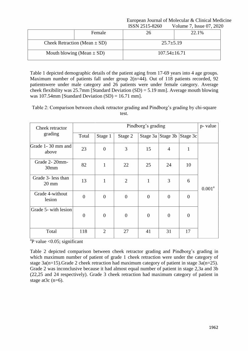
Cheek Retraction (Mean ± SD) 25.7±5.19
Mouth blowing (Mean ± SD) 107.54±16.71
Table 1 depicted demographic details of the patient aging from 17-69 years into 4 age groups.
Maximum number of patients fall under group 2(n=44). Out of 118 patients recorded, 92
patientswere under male category and 26 patients were under female category. Average
cheek flexibility was 25.7mm [Standard Deviation (SD) = 5.19 mm]. Average mouth blowing
was 107.54mm [Standard Deviation (SD) = 16.71 mm].
Table 2: Comparison between cheek retractor grading and Pindborg’s grading by chi-square
test.
aP value <0.05; significant
Table 2 depicted comparison between cheek retractor grading and Pindborg’s grading in
which maximum number of patient of grade 1 cheek retraction were under the category of
stage 3a(n=15).Grade 2 cheek retraction had maximum category of patient in stage 3a(n=25).
Grade 2 was inconclusive because it had almost equal number of patient in stage 2,3a and 3b
(22,25 and 24 respectively). Grade 3 cheek retraction had maximum category of patient in
stage at3c (n=6).
Cheek retractor
grading
Pindborg’s grading p- value
Total Stage 1 Stage 2 Stage 3a Stage 3b Stage 3c
Grade 1- 30 mm and
above 23 0 3 15 4 1
0.001a
Grade 2- 20mm-
30mm 82 1 22 25 24 10
Grade 3- less than
20 mm 13 1 2 1 3 6
Grade 4-without
lesion 0 0 0 0 0 0
Grade 5- with lesion
0 0 0 0 0 0
Total 118 2 27 41 31 17

European Journal of Molecular & Clinical Medicine
ISSN 2515-8260 Volume 7, Issue 07, 2020
1963
Table 3: Comparison between mouth blowing grading and Pindborg’s grading by chi-square
test
Mouth
blowing
grade
Total
Pindborg’s grading
p- value Stage 1 Stage 2 Stage 3a Stage 3b Stage 3c
70-80 5 0 3 0 0 2
0.001a
81-90 21 2 5 4 4 6
91-100 21 0 7 6 7 1
101-110 17 0 4 3 9 1
111-120 27 0 4 8 9 6
121-130 18 0 3 12 2 1
131-140 9 0 1 8 0 0
TOTAL 118 2 27 41 31 17
aP value <0.05; significant
Table 3 depicted comparison between mouth blowing and Pindborg’s grading in which
patient having mouth blowing of range 70-80mm had maximum number of patients falling
under stage 2 of Pindborg’s classification (n=3). Patient who had mouth blowing of range 81-
90mm had maximum number of patients in stage 3c category (n=6).Patient who had mouth
blowing of range 91-100 had maximum of patients falling under stage 2 and 3b(n=7). Patient
who had mouth blowing of range 101-110mm and 111-120mm had maximum number of
patients falling in stage 3b category (n=9).Patient who had mouth blowing of range 121-130
and 131-140 had maximum number of patients in stage 3a category
ofPindborg’sclassification(n=12 and 8 respectively).
Table 4: Comparison between mouth blowing grading and cheek retractor grade by chi-
square test
Mb range TOTAL
Cheek retractor grade
p- value Grade 1- 30
mm and
above
Grade 2-
20mm-
30mm
Grade 3-
less than 20
mm
Grade 4-
without
lesion
Grade 5-
with
lesion
70-80 5 0 4 1 0 0
0.001a
81-90 21 0 15 6 0 0
91-100 21 2 16 3 0 0

European Journal of Molecular & Clinical Medicine
ISSN 2515-8260 Volume 7, Issue 07, 2020
1964
aP value <0.05; significant
Table 4 depicted comparison of mouth blowing and cheek retractor grading in which patient
who had mouth blowing of range 70-80, 81-90, 91-100,101-110 and 111-120 maximum of
them fall under the category of grade 2 cheek retraction (n=4,15,16,16 and 20
respectively).While patient who are having mouthblowing of range 121-130 and 131-140 had
maximum numbers of patient in grade 1 category of cheek retraction (n=10 and 6
respectively).
Table 5: Comparison between cheek retractor grading and mouth blowing grade of right side
by chi-square test
Cheek
retractor
grading
right side
Mouth blowing grade right side p-
value TOTAL 70-80 81-90 91-100
101-
110 111-120 121-130 131-140
Grade 1-
30 mm
and above
25 0 0 4 1 6 7 7
0.001a
Grade 2-
20mm-
30mm
83 11 8 24 11 20 4 5
Grade 3-
less than
20 mm
10 5 0 4 1 0 0 0
Grade 4-
without
lesion
0 0 0 0 0 0 0 0
Grade 5-
with lesion
0 0 0 0 0 0 0 0
Total 118 16 8 32 13 26 11 12
aP value <0.05; significant
101-110 17 1 16 0 0 0
111-120 27 4 20 3 0 0
121-130 18 10 8 0 0 0
131-140 9 6 3 0 0 0
Total 118 23 82 13 0 0
European Journal of Molecular & Clinical Medicine
ISSN 2515-8260 Volume 7, Issue 07, 2020
1965
Table 5 depicted comparison of mouth blowing and cheek retractor grading of right side in
which grade 1 cheek retraction had maximum number of patients falling under the range 121-
130 and 131-140 of mouth blowing (n=7). Grade 2 cheek retraction had maximum number of
patients falling under the range 91-100 of mouth blowing (n=24). While in grade 3 cheek
retraction had maximum number of patients falling under the range 70-80 and 91-100 of
mouth blowing (n=5 and 4 respectively).
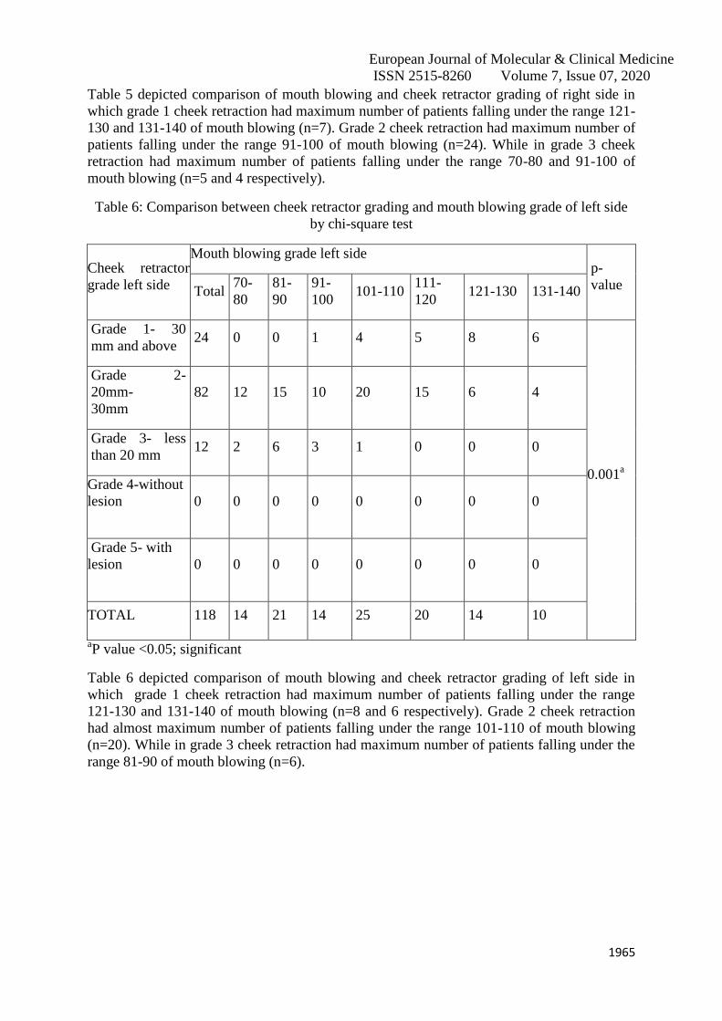
Table 6: Comparison between cheek retractor grading and mouth blowing grade of left side
by chi-square test
Cheek retractor
grade left side
Mouth blowing grade left side p-
value Total 70-
80
81-
90
91-
100 101-110
111-
120 121-130 131-140
Grade 1- 30
mm and above 24 0 0 1 4 5 8 6
0.001a
Grade 2-
20mm-
30mm
82 12 15 10 20 15 6 4
Grade 3- less
than 20 mm 12 2 6 3 1 0 0 0
Grade 4-without
lesion
0 0 0 0 0 0 0 0
Grade 5- with
lesion
0 0 0 0 0 0 0 0
TOTAL 118 14 21 14 25 20 14 10
aP value <0.05; significant
Table 6 depicted comparison of mouth blowing and cheek retractor grading of left side in
which grade 1 cheek retraction had maximum number of patients falling under the range
121-130 and 131-140 of mouth blowing (n=8 and 6 respectively). Grade 2 cheek retraction
had almost maximum number of patients falling under the range 101-110 of mouth blowing
(n=20). While in grade 3 cheek retraction had maximum number of patients falling under the
range 81-90 of mouth blowing (n=6).

European Journal of Molecular & Clinical Medicine
ISSN 2515-8260 Volume 7, Issue 07, 2020
1966
Table 7: Frequency of patient feedback
Items Cheek retractor n[%] Mouth blowing n[%]
More comfortable 10[8.5] 108[91.5]
Makes you more aware about the condition 42[35.6] 76[64.4]
Table 7depicted feedback of patients in which 91.5% felt more comfortable with mouth
blowing technique and 64.4% patient get awared about their condition from mouth blowing
method.
DISCUSSION
Oral submucous fibrosis, a potentially malignant condition associated with tobacco and areca
nut chewing is primarily seen in the Southeast Asian countries and Indian subcontinent.[14]
It
is universally considered as an Indian disease. The overall prevalence rate 0.2% to 0.5% is
believed to be in India.(20)
OSMF, if diagnosed by a method which is less time consuming and
easy to perform in clinical practice will decrease the chair-side time taken which also
subsequently will decrease the time taken for further investigation and the management of the
condition.
Presently, this study is done to analyse the role of different variables which play a vital role in
the clinical diagnosing of OSMF. This study was done to find out comparative evaluation of
cheek flexibility using cheek retractor method and mouth blowing method and its correlation
with Pindborg’s grading system for oral submucous fibrosis patient.
In this study of 118 patients, the age ranging starts from 17- 69 years, where majority of
OSMF cases appear in the range 26-35 years of age. It is similar to another hospital based
study by Ahmed(18)
where it was reported that majority of the OSMF cases belonged to the
age group of 21-40 years. Similarly, Sirsat[25]
reported OSMF cases from 20 to 40 years of
age. A similarity was seen between the study conducted by Borle RM and this present study
with the occurrence of OSMF in particular age group(26)
Vanaja Reddy.(13)
In contrast to
Ranganathan et al, half of the study population appear in the age group of third
decade.(27)
This shows that middle age group of the population are more affected with OSMF
in the Vidarbha region which is considered as the working population in India.
In the study of118 OSMF patients with 92 patients of male and 26 patients of female, a male
predominance was shown. Similarly male predominance was reported by Ranganathan
(2004),(27)
Kumar et al. (2007),(28)
and Pandya et al. (2009)(29)
Ceena et al. (2009)(30)
.However
other investigations like Pindborg (1970),(31)
Caniff (1986)(32)
and Johnson (2000) shows
female predominance.(33)
In our study male predominance can be due to the eccentric lifestyle
of the youngsters in our society which brought the males to be more accessible into using of
arecanut and its product more frequently than females.
The average cheek flexibility in this study was 25.7±5.19mm. Similar method was used by
Hassan Shahid(34)
for measuring the cheek flexibility. While in contrast to that Syeda Arshiya
Ara et al.(35)
showed average cheek flexibility as 7.18 mm. Ranganathan et al. (2001)(36)
showed that the mean values of cheek flexibility in males as 9.7 mm, and in females as 9.0
mm. This difference in their mean value may be due to difference in the method of
measurement where Ranganathan et al. and Syeda Arshiya Ara et al. used a method in which
a line is joined in between the tragus of the ear and angle of the mouth. An imaginary
perpendicular line from the outer canthus of the ipsilateral eye was extended downwards to
intersect the angle-tragus line. The point of intersection was marked as a reference point. This

European Journal of Molecular & Clinical Medicine
ISSN 2515-8260 Volume 7, Issue 07, 2020
1967
was done on the right and left sides for measuring cheek flexibility. The average mouth
blowing in our study was 107.54±16.71mm.
On co-relating cheek flexibility and Pindborg’s grading,though the inter-relation between
them is not reliable, the statistical analysis with Chi square test shows a high significant with
p<0.001. Cheek flexibility method is also used in Hassan Shahid et al.(34)
to estimate oral
health impact profile (OHIP) in patients who are suffering from oral submucosal fibrosis
(OSMF) and its co-relation with Pindborg's clinical grading. Sadiya Khan(37)
also mentioned
the classification of cheek flexibility in their study for classifying the OSMF. Advantages of
cheek retractor method was its easy to assess which side is more affected by OSMF on the
other hand advantage of mouth blowing method are easy to perform, less time consuming and
patient co-operation is more. Disadvantages of cheek retractor method are time consuming,
painful for the patient, need certain materials to perform and for mouth blowing method
disadvantages are some time its really difficult to diagnose by the investigator whether it is
normal or diseased, and even mouth breather and asthmatic patient cannot blow for longer
time. Limitation of this study a larger sample size may be needed to establish the significance
and magnitude of this association. To diagnose OSMF by cheek retraction and mouth
blowing we required certain materials like cheek retractor, scale and thread. Even this study
does not include patients who had severe stage of OSMF.
CONCLUSION
Oral Submucous fibrosis is considered as a potentially malignant disorder, therefore it is
essential to diagnose and treat as per the stage of the disease. The method or criteria choosing
for diagnosing the condition should be easy to perform with minimum armamentarium as
well as sensitive and less time consuming. None of the methods fulfills all the criteria
necessitating the need to further do the research in this direction.
CLINICAL SIGNIFICANCE
This method can be used under resource constraint setting for preliminary grading of the
OSMF.
REFRENCES
[1] Alka HH, Prajakta ZR, Minal CS, Madhuri GN, Swati P, Aakruti A.
Immunohistochemical analysis of tumor-associated stroma in oral squamous cell
carcinoma with and without preexisting oral submucous fibrosis. J Datta Meghe Inst
Med SciUniv 2017;12:170-6
[2] Chaudhary M, Bajaj S, Bohra S, Swastika N,Hande A. The domino effect: Role of
hypoxia in malignant transformation of oral submucous fibrosis. J Oral
MaxillofacPathol 2015;19:122-7.
[3] Borle RM, Nimonkar PV, &Rajan R, Extended nasolabial flaps in the management of
oral submucous fibrosis. Br J Oral Maxillofac Surg. 2009 Jul;47(5):382-5.
[4] Chole RH, Gondivkar SM, Gadbail AR, Balsaraf S, Chaudhary S, Dhore SV, Parikh
RV. Review of drug treatment of oral submucous fibrosis. Oral Oncol. 2012
May;48(5):393-8.
[5] Cox SC, Walker DM. Oral submucous fibrosis. A review. Aust Dent J. 1996;41:294-9
[6] Tilakaratne WM, Klinikowski MF, Takashi S, Peters TJ, Warnakulasuriya S. Oral
submucous fibrosis: review on etiology and pathogenesis Oral Oncol 2006 42:561-68.

European Journal of Molecular & Clinical Medicine
ISSN 2515-8260 Volume 7, Issue 07, 2020
1968
[7] Tekade SA, Chaudhary MS, Tekade SS, Sarode SC, Wanjari SP, Gadbail AR. Early
stage oral submucous fibrosis is characterized by increased vascularity as opposed to
advanced stages. J ClinDiagn Res. 2017 May;11(5):ZC92-ZC96.
[8] Lewin F, Norell SE, Johansson H, Gustavsson P, Wennerberg J, Biörklund A, Rutqvist
LE. Smoking tobacco, oral snuff, and alcohol in the etiology of squamous cell
carcinoma of the head and neck: a population based case-referent study in Sweden.
Cancer. 1998 Apr 1;82(7):1367-75.
[9] Hande AH, Chaudhary MS.Cytomorphometric analysis of buccal mucosa of tobacco
chewers. Rom J MorpholEmbryol. 2010;51(3):527-32.
[10] Lohe VK, Degwekar SS, Bhowate RR, Kadu RP, Dangore SB. Evaluation of
correlation of serum lipid profile in patients with oral cancer and precancer and its
association with tobacco abuse. J Oral Pathol Med (2010) 39:141–148.
[11] Tilakaratne WM, Iqbal Z, Teh MT, Ariyawardana A, Pitiyage G, Cruchley A, et al.
Upregulation of HIF‑ 1alpha in malignant transformation of oral submucous fibrosis. J
Oral Pathol Med 2008;37:372‑ 7.
[12] Haider SM, Merchant AT, Fikree FF, Rahbar MH. Clinical and functional staging of
oral submucous fibrosis. Br J Oral Maxillofacial Surg. 2000;38(1):12–15.
[13] ReddyV,Wanjari PV, Banda NR, Reddy P. Oral Submucous Fibrosis: Correlation of
Clinical Grading to various habit factors. International journal of dental clinics
2011:3(1): 21-24.
[14] Afroz N, Hasan SA, Naseem S. Oral submucous fibrosis a distressing disease with
malignant potential. Indian J Community Med 2006;31:270-1.
[15] Gupta M, Mishra P, Shrivastava K, Singh N. Oral Submucous Fibrosis- Current
Concepts of Aetiology & its Management J App. Dent. Med. Sci. 2015; 1(1):28-39.
[16] Kant P, Bhowate RR, ShardaN.Assessment of cross-sectional thickness and activity of
masseter, anterior temporalis and orbicularis oris muscles in oral submucous fibrosis
patients and healthy controls: An ultrasonography and electromyography study.
Dentomaxillofac Radiol. 2014;43(3).
[17] Tupakri JV, Bhavathankar JD, Mandale MS. Oral submucous fibrosis. A study of 100
cases. J Indian Acad Oral Med Radiol 2007;19:311-8.
[18] Ahmad MS, Ali SA, Ali AS, Chaubey KK. Epidemiological and etiological study of
oral submucous fibrosis among gutkha chewers of Patna, Bihar, India. J Indian
SocPedodPrev Dent 2006;24:84‑ 9.
[19] CB, Gupta S, Joshi J, Varma SN. Classification system for OSMF. J Indian Aca Oral
Med Radiol 2012;24(1):24-29.
[20] Mani NJ, Singh B. Studies on oral submucous fibrosis. Oral Surg Oral Med Oral Pathol
1976;41:203‑ 14.
[21] Pindborg JJ, Odont, Sirsat M. Oral submucous fibrosis. Oral Surg Oral Med Oral Pathol
Oral Radiol 1966;22:764‑ 79.
[22] Luqaman M, Vidya V. The role of serum copper and iron in oral submucous fibrosis. J
Indian Acad Oral Med Radiol 2004;16:30‑ 2.
[23] Patil S, Maheshwari S. Proposed new grading of oral submucous fibrosis based on
cheek flexibility. J ClinExp Dent. 2014;6(3):e255-8.
[24] Ranganathan k, Mishra G. An overview of classification schemes of osmf. Journal of
oral and maxillofacial pathology. 2006 jul-dec;10 (2);55-58.
[25] Sirsat S, Khanolkar V. The effect of arecoline on the palatal and buccal mucosa of the
Wistar rat. An optical and electron microscope study.Indian J Med Sci 1962;16:198-
202.
[26] Borle R, Borle S. Management of oral submucous fibrosis: a conservative approach.
J Oral Maxillofac Surg. 1991 Aug;49(8):788-91
European Journal of Molecular & Clinical Medicine
ISSN 2515-8260 Volume 7, Issue 07, 2020
1969
[27] Ranganathan K, Devi MU, Joshua E, Kirankumar K, Saraswathi T. Oral submucous
fibrosis: a case control study in Chennai, South India. J Oral Pathol Med. 2004
May;33(5):274-7.
[28] Kiran Kumar K, Saraswathi T R, Ranganathan K, Uma Devi M, Elizabeth J. Oral
submucous fibrosis: A clinico-histopathological study in Chennai. Indian J Dent Res
2007;18:106-11
[29] Pandya S, Chaudhary AK, Singh M, et al . Correlation of histopathological diagnosis
with habits and clinical findings in oral submucous fibrosis. Head Neck Oncol, 1, 10.
[30] Ceena DE, Bastian TS, Ashok L, Rajeshwari G Annigeri. Comparative study of
clinicofunctional staging of oral submucous fibrosis with qualitative analysis of
collagen fibers under polarizing microscopy. Indian J Dent Res.2009;20, 271-6.
[31] Pindborg JJ, Mehta FS, Daftary DK. Occurrence of epithelial atypia in 51 Indian
villagers with oral submucous fibrosis. Bri J Cancer.1970;24, 253-7.
[32] Canniff JP, Harvey W, Harris M. Oral submucous fibrosis: its pathogenesis and
management. Br Dent J.1986;21, 429-34.
[33] Johnson NW, Maher R, Trivedy C, WarnekulasureiyaS.The clinical condition and
pathology of oral submucous fibrosis. JADA. 2000;3, 278-9.
[34] Shahid H, Qadri M, Hassan S. Oral submucous fibrosis; Oral health impact profile of
patients and its correlation with its clinical grading. Professional Med J
2017;24(11):1719-1726.
[35] Ara SA, Arora V, Zakaullah S, Raheel SA, Rampure P, Ashraf S.Correlation of Habits
and Clinical Findings with Histopathological Diagnosis in Oral Submucosal Fibrosis
Patients Asian Pac J Cancer Prev. 2013;14(12):7075-80.
[36] K. Ranganathan, U. Devi, J Elizabeth, B Arun, T Rooban, R. R. Viswanathan.
Mouth Opening, Cheek Flexibility And Tongue Protrusion Parameters Of 800 Normal
Patients In Chennai, South India A Baseline Study To Enable Assessment Of
Alterations In Oral Submucous Fibrosis.Jida Vol. 72 March 2001
[37] Khan S, Sinha A, Kumar S, Iqbal H. Oral submucous fibrosis: Current concepts on
aetiology and management – A Review. J Indian Acad Oral Med Radiol. 2018;30:407-
11.
Copyright © 2022 FDOKUMEN